understanding morphological variation in the extant koala as a … · 2013-06-06 · boundaries in...
TRANSCRIPT
This article was downloaded by: [UQ Library]On: 14 May 2013, At: 16:35Publisher: Taylor & FrancisInforma Ltd Registered in England and Wales Registered Number: 1072954 Registered office: Mortimer House,37-41 Mortimer Street, London W1T 3JH, UK
Journal of Systematic PalaeontologyPublication details, including instructions for authors and subscription information:http://www.tandfonline.com/loi/tjsp20
Understanding morphological variation in the extantkoala as a framework for identification of speciesboundaries in extinct koalas (Phascolarctidae;Marsupialia)Karen H. Black a , Julien Louys b c & Gilbert J. Price ca School of Biological, Earth and Environmental Sciences , University of New South Wales ,Sydney , New South Wales , Australiab Geosciences, Queensland Museum , South Brisbane BC , Queensland , Australiac School of Earth Sciences , The University of Queensland , St. Lucia , Queensland ,AustraliaPublished online: 14 May 2013.
To cite this article: Karen H. Black , Julien Louys & Gilbert J. Price (2013): Understanding morphological variation in theextant koala as a framework for identification of species boundaries in extinct koalas (Phascolarctidae; Marsupialia), Journalof Systematic Palaeontology, DOI:10.1080/14772019.2013.768304
To link to this article: http://dx.doi.org/10.1080/14772019.2013.768304
PLEASE SCROLL DOWN FOR ARTICLE
Full terms and conditions of use: http://www.tandfonline.com/page/terms-and-conditions
This article may be used for research, teaching, and private study purposes. Any substantial or systematicreproduction, redistribution, reselling, loan, sub-licensing, systematic supply, or distribution in any form toanyone is expressly forbidden.
The publisher does not give any warranty express or implied or make any representation that the contentswill be complete or accurate or up to date. The accuracy of any instructions, formulae, and drug doses shouldbe independently verified with primary sources. The publisher shall not be liable for any loss, actions, claims,proceedings, demand, or costs or damages whatsoever or howsoever caused arising directly or indirectly inconnection with or arising out of the use of this material.
Journal of Systematic Palaeontology, 2013http://dx.doi.org/10.1080/14772019.2013.768304
Understanding morphological variation in the extant koala as a frameworkfor identification of species boundaries in extinct koalas (Phascolarctidae;
Marsupialia)Karen H. Blacka∗, Julien Louysb,c and Gilbert J. Pricec
aSchool of Biological, Earth and Environmental Sciences, University of New South Wales, Sydney, New South Wales, Australia;bGeosciences, Queensland Museum, South Brisbane BC, Queensland, Australia; cSchool of Earth Sciences,
The University of Queensland, St. Lucia, Queensland, Australia
(Received 27 September 2011; accepted 15 April 2012)
We document morphological variation (both geographical and sexual) in the dentition of the extant koala, Phascolarctoscinereus, in order to facilitate discrimination of species boundaries in extinct phascolarctids. Considerable variation is evidentin dental structures previously used to diagnose several phascolarctid fossil species. Consistent patterns of morphologicalvariation are not evident between sexes or geographic regions, with variation as great between samples as within them. Metricvariation is evident between the sexes in upper molar dimensions with Victorian (southern) males significantly larger thanVictorian females, although this is not reflected in lower molar dimensions or in the Queensland (northern) sample. Malekoalas from southern populations generally display significantly larger molars than their northern counterparts; however thistrend is not evident in female upper molar dimensions. In both males and females, some, but not all, lower molar dimensionsare larger in southern populations than northern. In light of these results, a systematic revision of species of Litokoala suggestsL. ‘dicktedfordi’ is a junior synonym of L. kutjamarpensis, and the poorly known L. thurmerae is regarded to be a nomendubium. Further, we describe a partial cranium of a new species of koala from Early Miocene sediments in the RiversleighWorld Heritage Area, northern Australia. Litokoala dicksmithi sp. nov. is the fifth koala species recorded from the diverserainforest assemblages of Riversleigh and the third species referred to the Oligo-Miocene genus Litokoala. Aspects of cranialmorphology, including a shortened robust rostrum and broad, irregular nasal aperture, confirm placement of Litokoala assister taxon to the modern genus Phascolarctos. Relatively large orbits and small body size suggest the possibility that L.dicksmithi was nocturnal, had enhanced visual acuity, and was a more agile arboreal species than the relatively sedentaryextant koala.
http://zoobank.org/urn:lsid:zoobank.org:pub:EE5D13C1-47BB-4432-B281-16398A1781E1
Keywords: intraspecific variation; morphometric; Phascolarctomorphia; rainforest; Miocene; Riversleigh
Introduction
Accurately assessing the number of species in the fossilrecord is fundamental to understanding evolutionary histo-ries, past biodiversity, and responses of species andpalaeocommunities to environmental change. However,determining species boundaries in fossil taxa may be achallenge for palaeontologists who are often confronted bylimited fossil samples, poor preservation and the absence ofmodern analogues. Conspicuous morphological variabilitywithin a species may result from geographical effects (withconcomitant environmental or climatic influences), biolog-ical factors (e.g. sexual dimorphism and ontogeny) or acombination of both (Albrecht et al. 2003). Understand-ing the nature of such variation within modern analoguesis important to assessing the validity of morphologicalfeatures used in species determinations of extinct groups.Surprisingly, despite the need for such data, there are few
∗Corresponding author. Email: [email protected]
comprehensive published accounts of intraspecific variationin either extant or extinct marsupials.
Among extant marsupials, analyses of both qualitativeand quantitative dental variation have been investigated inspecies of Perameles (Peramelidae; e.g. Freedman 1967;Freedman & Joffe 1967a, b), Macropus (Macropodidae; e.g.Bartholomai 1971; Easton 2006), and the Patagonian opos-sum Lestodelphys halli (Didelphidae; e.g. Martin 2005).Studies of variation in fossil marsupials are few owing tothe relative paucity of fossil samples from single localities.Archer & Dawson (1982) analysed a moderate sample (n =55) of marsupial lion cranial and dental remains referable tothe genus Thylacoleo, from Pleistocene deposits of Welling-ton Caves (NSW). Prideaux (2004) undertook an analysisof craniodental morphological variation among sthenurinesas part of a taxonomic review of the macropodid subfam-ily. Recently, several benchmark studies (e.g. Murray et al.2000a, b; Price 2008a; Black & Hand 2010; Price & Sobbe
C© 2013 Natural History Museum
Dow
nloa
ded
by [
UQ
Lib
rary
] at
16:
35 1
4 M
ay 2
013
2 K. H. Black et al.
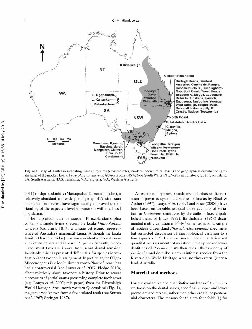
Figure 1. Map of Australia indicating main study sites (closed circles, modern; open circles, fossil) and geographical distribution (greyshading) of the modern koala, Phascolarctos cinereus. Abbreviations: NSW, New South Wales; NT, Northern Territory; QLD, Queensland;SA, South Australia; TAS, Tasmania; VIC, Victoria; WA, Western Australia.
2011) of diprotodontids (Marsupialia: Diprotodontidae), arelatively abundant and widespread group of Australasianmarsupial herbivores, have significantly improved under-standing of the expected level of variation within a fossilpopulation.
The diprotodontian infraorder Phascolarctomorphiacontains a single living species, the koala Phascolarctoscinereus (Goldfuss, 1817), a unique yet iconic represen-tative of Australia’s marsupial fauna. Although the koalafamily (Phascolarctidae) was once evidently more diversewith seven genera and at least 17 species currently recog-nized, most taxa are known from scant dental remains.Inevitably, this has presented difficulties for species identi-fication and taxonomic assignment. In particular, the Oligo-Miocene genus Litokoala, sister taxon to Phascolarctos, hashad a controversial (see Louys et al. 2007; Pledge 2010),albeit relatively short, taxonomic history. Prior to recentdiscoveries of partial crania preserving complete tooth rows(e.g. Louys et al. 2007; this paper) from the RiversleighWorld Heritage Area, north-western Queensland (Fig. 1),the genus was known from a few isolated teeth (see Stirtonet al. 1967; Springer 1987).
Assessment of species boundaries and intraspecific vari-ation in previous systematic studies of koalas by Black &Archer (1997), Louys et al. (2007) and Price (2008b) havebeen based on unpublished qualitative accounts of varia-tion in P. cinereus dentitions by the authors (e.g. unpub-lished thesis of Black 1992). Bartholomai (1968) docu-mented metric variation in P3–M2 dimensions for a sampleof modern Queensland Phascolarctos cinereus specimensbut restricted discussion of morphological variation to afew aspects of P3. Here we present both qualitative andquantitative assessments of variation in the upper and lowerdentitions of P. cinereus. We then revisit the taxonomy ofLitokoala, and describe a new rainforest species from theRiversleigh World Heritage Area, north-western Queens-land, Australia.
Material and methods
For our qualitative and quantitative analyses of P. cinereuswe focus on the dental series, specifically upper and lowerpremolars and molars, rather than other cranial or postcra-nial characters. The reasons for this are four-fold: (1) for
Dow
nloa
ded
by [
UQ
Lib
rary
] at
16:
35 1
4 M
ay 2
013
Identification of species boundaries in extinct koalas 3
consistency across other taxonomic studies that focus oncheek teeth; (2) teeth are relatively common elements inthe fossil record with most fossil marsupials having beendescribed on the basis of dentitions; (3) teeth are system-atically and taxonomically important elements; and (4)all fossil koala species that have been described to datehave been based largely on dental characteristics with someknown only from isolated teeth.
Modern koala specimens examined were derivedfrom collections of the Queensland Museum (Brisbane),Australian Museum (Sydney), University of New SouthWales (Sydney) and Museum Victoria (Melbourne). Fossilmaterial described here is registered in the fossil collectionof the Queensland Museum. Reference to Litokoala kutja-marpensis throughout the text is sensu Louys et al. (2007)unless stated otherwise. Higher-level systematic nomen-clature follows Aplin & Archer (1987). Molar morphol-ogy follows Archer (1978) with revisions by Tedford &Woodburne (1987; such that the metaconule is consideredhomologous to the older term ‘hypocone’, and the cuspbetween the metacone and ‘true’ metaconule is deemed the‘neometaconule’). Cheek tooth homology follows Luckett(1993). Biostratigraphic nomenclature follows Travouillonet al. (2006), Woodburne et al. (1993) and Creaser (1997).
Qualitative analysisA sample of 109 skulls from 55 localities in New SouthWales, Victoria and Queensland (Fig. 1) was used to inves-tigate qualitative morphological variation in the dentition ofmodern Phascolarctos cinereus (see Online SupplementaryMaterial). Ten of these skulls were from unknown localitiesbut were included in the analysis because of their clean,relatively unworn dentitions. Only cheek teeth were exam-ined and both sexes were represented in the sample. It isrecognized that P. cinereus exhibits marked sexual dimor-phism with males being significantly larger than females(Martin et al. 2008). However, it is unclear whether sexuallyrelated differences are exhibited within dental morphology.Thus, an initial assessment of morphological variation ineach sex was made to determine whether any morphologiesor patterns might be gender specific. Similarly, in order toassess whether morphologies or patterns of morphologicalvariation were evident within and/or between geographi-cal regions, variation in specimens from New South Wales,Victoria and Queensland populations was assessed inde-pendently. An example of Phascolarctos cinereus upper andlower dentitions and the dental nomenclature used in thisanalysis is provided in Figure 2. A representative sampleof morphologies for each tooth position is illustrated inFigures 3–12. Figured specimens were selected on the basisof least wear.
Quantitative analysesPhascolarctos cinereus also exhibits distinctly differentbody sizes throughout its modern geographical range. For
instance, adult individuals within southern, higher latitudepopulations (e.g. Victoria) typically range in body sizefrom 8.5 to 12 kg. In contrast, individuals from northern,lower latitudes (e.g. Queensland) are significantly smaller,weighing on average from 5.1 to 6.5 kg (Martin et al.2008). Thus, P. cinereus body size appears to representa latitudinal morphocline reflecting Bergmann’s rule (e.g.Meiri & Dayan 2003). However, it has never been demon-strated that such latitudinal differences are also reflectedin dental morphometrics. Thus, using teeth as a surro-gate for body size (following Gould 1975; Myers 2001),we test the following hypotheses: (1) that no signifi-cant difference in premolar and molar dimensions existbetween koalas from southern (Victorian) and northern(Queensland) populations; and (2) that no significant differ-ence in premolar and molar dimensions exist betweenmale and female individuals from the same geographi-cal region. We test these hypotheses independently forupper and lower dentitions. Understanding such size vari-ation in the modern koala is critical for establishing thesignificance of morphometric differences between fossilspecies.
In order to test the hypotheses, we took a series ofdental measurements from museum specimens originallysourced from Queensland and Victorian populations. Linearmeasurements were made for premolars and molars of adultindividuals using Mitutoyo digital callipers and included:maximum length, anterior width (maximum width acrossanterior root) and posterior width (maximum width acrossposterior root) for premolars, and maximum length, ante-rior width (maximum width across trigon/trigonid) andposterior width (maximum width across talon/talonid)for molars. This approach is consistent with numerousother morphometric dental studies of not only koalas (e.g.Bartholomai 1968; Price 2008b; Price et al. 2009; Pledge2010), but marsupials in general (e.g. Freedman 1967;Freedman & Joffe 1967a, b; Bartholomai 1971; Price 2002,2005, 2008a; Easton 2006; Black & Hand 2010), andallows for direct comparison of results between respec-tive investigations. Both upper (n = 49) and lower (n =70) dentitions were measured. Analyses were performedusing PAST (version 1.51; Hammer et al. 2006) computersoftware. Univariate statistics for dental measurements areprovided in Online Supplementary Material Appendix 1.Multivariate analysis of variance (MANOVA) was used toassess any differences indicated by whole tooth rows, aswell as individual teeth. This method enables simultaneouscomparisons of all three measurements and, in the case oftooth rows, more than one tooth position at a time. Differ-ences were considered to be significant at the 95% confi-dence interval. Where significant differences were foundin the morphometrics of a particular tooth position, themeasurements of that tooth were compared between groupsusing t-tests. Only significant differences are reported(Tables 1–3).
Dow
nloa
ded
by [
UQ
Lib
rary
] at
16:
35 1
4 M
ay 2
013
4 K. H. Black et al.
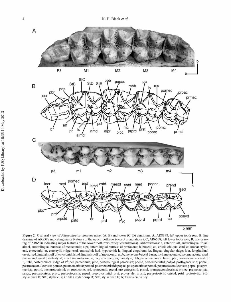
Figure 2. Occlusal view of Phascolarctos cinereus upper (A, B) and lower (C, D) dentitions. A, AR8398, left upper tooth row; B, linedrawing of AR8398 indicating major features of the upper tooth row (except crenulations); C, AR6508, left lower tooth row; D, line draw-ing of AR6508 indicating major features of the lower tooth row (except crenulations). Abbreviations: a, anterior; alf, anterolingual fossa;almcl, anterolingual buttress of metaconule; alpr, anterolingual buttress of protocone; b, buccal; co, cristid obliqua; cstd, columnar stylid;end, entoconid; er, entostylid ridge; estd, entostylid; hyd, hypoconid; lc, lingual cingulum; lcr, lingual cingular ridge; locr, longitudinalcrest; lsed, lingual shelf of entoconid; lsmd, lingual shelf of metaconid; mbb, metacone buccal basin; mcl, metaconule; me, metacone; med,metaconid; mestd, metastylid; nmcl, neometaconule; pa, paracone; pas, parastyle; pbb, paracone buccal basin; pbc, posterobuccal crest ofP3; pbr, posterobuccal ridge of P3; pcl, paraconule; plpc, posterolingual paracrista; poend, postentocristid; pohyd, posthypocristid; pomcl,postmetaconulecrista; pomec, postmetacrista; pomed, postmetacristid; popac, postparacrista; pomcl, postmetaconulecrista; poprc, postpro-tocrista; poprd, postprotocristid; pr, protocone; prd, protoconid; prend, pre-entocristid; prmcl, premetaconulecrista; prmec, premetacrista;prpac, preparacrista; prprc, preprotocrista; prprd, preprotocristid; prst, protostyle; prpstd, preprotostylid cristid; pstd, protostylid; StB,stylar cusp B; StC, stylar cusp C; StD, stylar cusp D; StE, stylar cusp E; tv, transverse valley.
Dow
nloa
ded
by [
UQ
Lib
rary
] at
16:
35 1
4 M
ay 2
013
Identification of species boundaries in extinct koalas 5
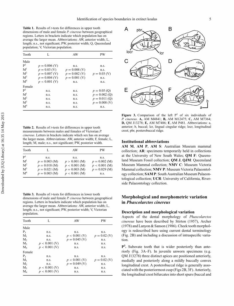
Table 1. Results of t-tests for differences in upper toothdimensions of male and female P. cinereus between geographicalregions. Letters in brackets indicate which population has onaverage the larger mean. Abbreviations: AW, anterior width; L,length; n.s., not significant; PW, posterior width; Q, Queenslandpopulation; V, Victorian population.
Tooth L AW PW
MaleP3 p = 0.006 (V) n.s. n.s.M1 p = 0.03 (V) p = 0.008 (V) n.s.M2 p = 0.007 (V) p = 0.002 (V) p = 0.03 (V)M3 p = 0.004 (V) p = 0.001 (V) n.s.M4 p < 0.001 (V) n.s. n.s.
FemaleP3 n.s. n.s. p = 0.05 (Q)M1 n.s. n.s. p = 0.002 (Q)M2 n.s. n.s. p = 0.011 (Q)M3 n.s. n.s. p = 0.008 (V)M4 n.s. n.s. n.s.
Table 2. Results of t-tests for differences in upper toothmeasurements between males and females of Victorian P.cinereus. Letters in brackets indicate which sex has on averagethe larger mean. Abbreviations: AW, anterior width; F, female; L,length; M, male; n.s., not significant; PW, posterior width.
Tooth L AW PW
P3 n.s. n.s. n.s.M1 p = 0.003 (M) p > 0.001 (M) p = 0.002 (M)M2 p = 0.018 (M) p < 0.001 (M) p < 0.001 (M)M3 p = 0.021 (M) p = 0.003 (M) p = 0.029 (M)M4 p = 0.003 (M) p < 0.001 (M) n.s.
Table 3. Results of t-tests for differences in lower toothdimensions of male and female P. cinereus between geographicalregions. Letters in brackets indicate which population has onaverage the larger mean. Abbreviations: AW, anterior width; L,length; n.s., not significant; PW, posterior width; V, Victorianpopulation.
Tooth L AW PW
MaleP3 n.s. n.s. n.s.M1 n.s. p < 0.001 (V) p = 0.02 (V)M2 n.s. p = 0.045 (V) n.s.M3 p < 0.001 (V) n.s. n.s.M4 p < 0.001 (V) n.s. n.s.
FemaleP3 n.s. n.s. n.s.M1 n.s. p < 0.001 (V) p = 0.02 (V)M2 n.s. p = 0.049 (V) n.s.M3 p < 0.001 (V) n.s. n.s.M4 p < 0.001 (V) n.s. n.s.
Figure 3. Comparison of the left P3 of six individuals ofP. cinereus. A, AM M4841; B, AM M12475; C, AM M7364;D, QM J13278; E, AM M7486; F, AM P481. Abbreviations: a,anterior; b, buccal; lcr, lingual cingular ridge; locr, longitudinalcrest; pbr, posterobuccal ridge.
Institutional abbreviationsAM M, AM P, AM S: Australian Museum mammalcollection; AR: specimens temporarily held in collectionsat the University of New South Wales; QM F: Queens-land Museum Fossil collection; QM J, QJM: QueenslandMuseum Mammal collection; NMV C: Museum VictoriaMammal collection; NMV P: Museum Victoria Palaeontol-ogy collection; SAM P: South Australian Museum Palaeon-tological collection; UCR: University of California, River-side Palaeontology collection.
Morphological and morphometric variationin Phascolarctos cinereus
Description and morphological variationAspects of the dental morphology of Phascolarctoscinereus have been described by Stirton (1957), Archer(1978) and Lanyon & Sanson (1986). Cheek tooth morphol-ogy is redescribed here using current dental terminology(Fig. 2B) and including a discussion of intraspecific varia-tion.
P3. Subovate tooth that is wider posteriorly than ante-riorly (Fig. 3A–F). In juvenile unworn specimens (e.g.QM J13278) three distinct apices are positioned anteriorly,medially and posteriorly along a mildly buccally convexlongitudinal crest. A posterobuccal ridge is generally asso-ciated with the posteriormost cusp (Figs 2B, 3F). Anteriorly,the longitudinal crest bifurcates into short spurs (buccal and
Dow
nloa
ded
by [
UQ
Lib
rary
] at
16:
35 1
4 M
ay 2
013
6 K. H. Black et al.
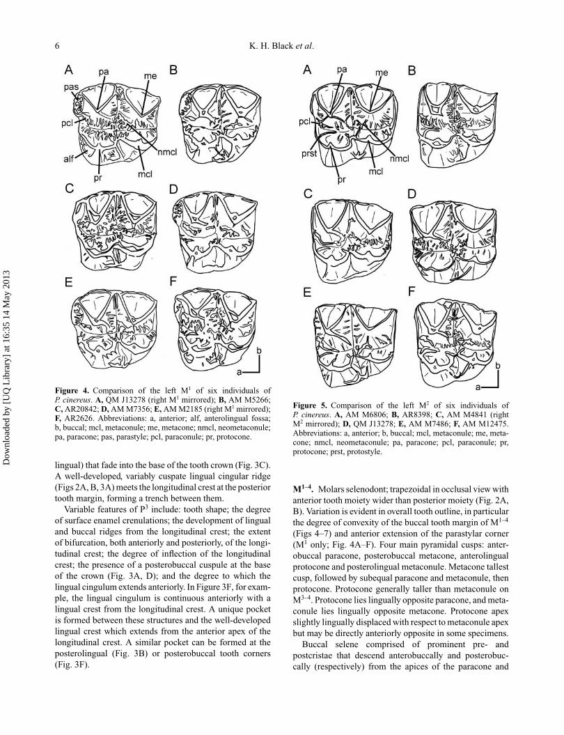
Figure 4. Comparison of the left M1 of six individuals ofP. cinereus. A, QM J13278 (right M1 mirrored); B, AM M5266;C, AR20842; D, AM M7356; E, AM M2185 (right M1 mirrored);F, AR2626. Abbreviations: a, anterior; alf, anterolingual fossa;b, buccal; mcl, metaconule; me, metacone; nmcl, neometaconule;pa, paracone; pas, parastyle; pcl, paraconule; pr, protocone.
lingual) that fade into the base of the tooth crown (Fig. 3C).A well-developed, variably cuspate lingual cingular ridge(Figs 2A, B, 3A) meets the longitudinal crest at the posteriortooth margin, forming a trench between them.
Variable features of P3 include: tooth shape; the degreeof surface enamel crenulations; the development of lingualand buccal ridges from the longitudinal crest; the extentof bifurcation, both anteriorly and posteriorly, of the longi-tudinal crest; the degree of inflection of the longitudinalcrest; the presence of a posterobuccal cuspule at the baseof the crown (Fig. 3A, D); and the degree to which thelingual cingulum extends anteriorly. In Figure 3F, for exam-ple, the lingual cingulum is continuous anteriorly with alingual crest from the longitudinal crest. A unique pocketis formed between these structures and the well-developedlingual crest which extends from the anterior apex of thelongitudinal crest. A similar pocket can be formed at theposterolingual (Fig. 3B) or posterobuccal tooth corners(Fig. 3F).
Figure 5. Comparison of the left M2 of six individuals ofP. cinereus. A, AM M6806; B, AR8398; C, AM M4841 (rightM2 mirrored); D, QM J13278; E, AM M7486; F, AM M12475.Abbreviations: a, anterior; b, buccal; mcl, metaconule; me, meta-cone; nmcl, neometaconule; pa, paracone; pcl, paraconule; pr,protocone; prst, protostyle.
M1–4. Molars selenodont; trapezoidal in occlusal view withanterior tooth moiety wider than posterior moiety (Fig. 2A,B). Variation is evident in overall tooth outline, in particularthe degree of convexity of the buccal tooth margin of M1–4
(Figs 4–7) and anterior extension of the parastylar corner(M1 only; Fig. 4A–F). Four main pyramidal cusps: anter-obuccal paracone, posterobuccal metacone, anterolingualprotocone and posterolingual metaconule. Metacone tallestcusp, followed by subequal paracone and metaconule, thenprotocone. Protocone generally taller than metaconule onM3–4. Protocone lies lingually opposite paracone, and meta-conule lies lingually opposite metacone. Protocone apexslightly lingually displaced with respect to metaconule apexbut may be directly anteriorly opposite in some specimens.
Buccal selene comprised of prominent pre- andpostcristae that descend anterobuccally and posterobuc-cally (respectively) from the apices of the paracone and
Dow
nloa
ded
by [
UQ
Lib
rary
] at
16:
35 1
4 M
ay 2
013
Identification of species boundaries in extinct koalas 7
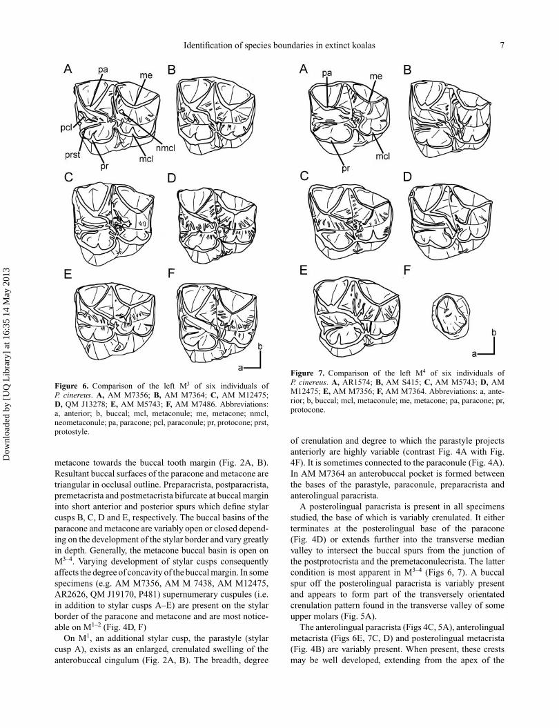
Figure 6. Comparison of the left M3 of six individuals ofP. cinereus. A, AM M7356; B, AM M7364; C, AM M12475;D, QM J13278; E, AM M5743; F, AM M7486. Abbreviations:a, anterior; b, buccal; mcl, metaconule; me, metacone; nmcl,neometaconule; pa, paracone; pcl, paraconule; pr, protocone; prst,protostyle.
metacone towards the buccal tooth margin (Fig. 2A, B).Resultant buccal surfaces of the paracone and metacone aretriangular in occlusal outline. Preparacrista, postparacrista,premetacrista and postmetacrista bifurcate at buccal margininto short anterior and posterior spurs which define stylarcusps B, C, D and E, respectively. The buccal basins of theparacone and metacone are variably open or closed depend-ing on the development of the stylar border and vary greatlyin depth. Generally, the metacone buccal basin is open onM3–4. Varying development of stylar cusps consequentlyaffects the degree of concavity of the buccal margin. In somespecimens (e.g. AM M7356, AM M 7438, AM M12475,AR2626, QM J19170, P481) supernumerary cuspules (i.e.in addition to stylar cusps A–E) are present on the stylarborder of the paracone and metacone and are most notice-able on M1–2 (Fig. 4D, F)
On M1, an additional stylar cusp, the parastyle (stylarcusp A), exists as an enlarged, crenulated swelling of theanterobuccal cingulum (Fig. 2A, B). The breadth, degree
Figure 7. Comparison of the left M4 of six individuals ofP. cinereus. A, AR1574; B, AM S415; C, AM M5743; D, AMM12475; E, AM M7356; F, AM M7364. Abbreviations: a, ante-rior; b, buccal; mcl, metaconule; me, metacone; pa, paracone; pr,protocone.
of crenulation and degree to which the parastyle projectsanteriorly are highly variable (contrast Fig. 4A with Fig.4F). It is sometimes connected to the paraconule (Fig. 4A).In AM M7364 an anterobuccal pocket is formed betweenthe bases of the parastyle, paraconule, preparacrista andanterolingual paracrista.
A posterolingual paracrista is present in all specimensstudied, the base of which is variably crenulated. It eitherterminates at the posterolingual base of the paracone(Fig. 4D) or extends further into the transverse medianvalley to intersect the buccal spurs from the junction ofthe postprotocrista and the premetaconulecrista. The lattercondition is most apparent in M3–4 (Figs 6, 7). A buccalspur off the posterolingual paracrista is variably presentand appears to form part of the transversely orientatedcrenulation pattern found in the transverse valley of someupper molars (Fig. 5A).
The anterolingual paracrista (Figs 4C, 5A), anterolingualmetacrista (Figs 6E, 7C, D) and posterolingual metacrista(Fig. 4B) are variably present. When present, these crestsmay be well developed, extending from the apex of the
Dow
nloa
ded
by [
UQ
Lib
rary
] at
16:
35 1
4 M
ay 2
013
8 K. H. Black et al.
paracone and metacone, or less defined, extending basal tothe apices of these cusps.
The lingual selene is comprised of prominent pre- andpostcristae that descend anterobuccally and posterobuccally(respectively) from the apices of the protocone and meta-conule towards the longitudinal tooth valley (Fig. 2A, B).The preprotocrista and postmetaconulecrista are continu-ous with the anterior and posterior cingula, respectively.The postprotocrista and premetaconulecrista meet lingualof the longitudinal valley and radiate at their juncture intoa series of enamel crenulations (Fig. 4A, C). The post-protocrista often bifurcates before its junction with thepremetaconulecrista into a well-developed posterolingualarm that extends into the valley between the protocone andmetaconule (Fig. 4B, C). A well-developed anterolingualbuttress of the metaconule (Fig. 4B, F) is variably devel-oped, as are buccal ribs from the apices of the protocone(Figs 4D, 5E) and metaconule (Figs 5F, 6A, 7C).
A variably cuspate lingual cingulum, which blocks thelingual exit of the transverse valley, is present on M1–3 (Figs4E, F, 5E) and sometimes M4 (Fig. 7A). It is continuouswith the anterolingual buttress of the metaconule and theposterolingual arm of the postprotocrista (generally on M1)in individuals in which these ridges are present (Fig. 4B, C)and may be highly crenulated (Fig. 4B). In AM M2185 (Fig.4E), the lingual cingulum is cuspate and projects linguallywell beyond the bases of the protocone and metaconule.In AM M5266 (Fig. 4B) it is similarly developed to theneomorphic cuspules characteristic of L. kutjamarpensisand L. garyjohnstoni. An anterolingual buttress of the proto-cone (Fig. 4B, D) is variably present, as is the developmentof an anterolingual fossette. The latter structure appears asa well-developed, often crenulated pocket at the anterolin-gual base of the protocone, bounded by the anterolingualcingulum, the anterolingual buttress of the protocone andthe preprotocrista (Fig. 2A, B). It is well developed on M1
of QM J13278 (Fig. 4A) and AR2626 (Fig. 4F) but gener-ally absent on M2–4; except in QM J13278 (Fig. 5D) whereit is developed, but to a lesser extent, on M2 also.
Molars are generally crenulated, although the degree ofexpression and pattern of surface enamel crenulations arenot constant between individuals. Most notably, in the trans-verse valley of M1–4 enamel crenulations vary from a highlyreticulate pattern in some individuals (Fig. 4A) to well-developed transverse parallel ridges which extend linguallyfrom the postparacrista and the premetacrista (Fig. 4D, E) inothers. This feature also varies along the tooth row within anindividual specimen. The M1 may exhibit a reticular crenu-lation pattern whereas M4 in the same individual exhibitswell-developed transverse ridges, or vice versa.
As a result of variation in enamel crenulations, thestructures of the parastyle, paraconule, neometaconuleand protostyle exhibit considerable intraspecific variation.These structures vary not only in size and shape, but also intheir orientation and connection to surrounding structures.
The paraconule is situated at the anterolingual base ofthe paracone with its long axis variably running parallel tothe preparacrista (Figs 4D, 5C) or more anteriorly directedin some individuals (Fig. 4B, C). It is either isolated at thispoint (Fig. 4D, E) or connected to the lingual base of theparacone and/or parastyle (M1 only; Fig. 4C) and/or anteriorcingulum (Fig. 5B, D). The paraconule varies from being abulbous cuspate structure (Figs 5C, 7B, D) to a relativelylinear structure or, depending on the degree of crenula-tion, in some individuals it is represented by a number ofbifurcate arms that are relatively indistinguishable from theenamel crenulations on the rest of the crown (Fig. 5B, D).
The neometaconule is situated at the anterolingual baseof the metacone (Fig. 2A, B) and may be isolated at thispoint (Fig. 4D), or connected to the base of the metacone(Fig. 4A) or anterolingual metacrista (Fig. 6E). It maybe arcuate or relatively linear and is generally highlycrenulated. It is variably developed in individuals and, inthe more posterior molars, may be poorly distinguishable.The degree to which the neometaconule extends posteri-orly along the longitudinal valley between the metaconeand metaconule varies between individuals, as does itsanterobuccal extension along the transverse valley. Theneometaconule variably connects to the buccal spursthat extend from the junction of the postprotocrista andpremetaconulecrista (Fig. 5A, E).
The protostyle is a short ridge that originates from thepreprotocrista at a point just lingual to the longitudinalvalley (Fig. 2A, B). It is generally linear (although bifurcatein some individuals) and varies in its extension posteriorly.In general it extends further posteriorly in more posteriormolars. In some specimens (e.g. QM J13278) it is indistin-guishable from the molar crenulation pattern (Figs 4A, 5D).
A small pocket is formed between the anterior cingu-lum and an anterior crest from the preparacrista at theanterobuccal corner of M4 of one individual studied (AMM12475, Fig. 7D). The anterior crest of the preparacrista isnot evident in any other tooth studied.
Similarly, the left and right M4 of one individual studied(AM M7364, Fig. 7F) are unique in that these teeth arereduced (or malformed) to such an extent that they existas small rounded structures consisting of a rounded basinbordered by a continuous cingulum on which only the apexof the presumed metacone is evident. Similar abnormalvariations have been noted in kangaroos (Archer 1975).
P3. Subovate tooth, wider posteriorly, tapering anteriorly(Fig. 2C, D). Tooth shape varies from elongate and narrowin some individuals (Fig. 8F), to shorter and bulbousin others (Fig. 8E). The buccal and lingual demarcationbetween the anterior and posterior moieties also variesbetween individuals, as does the lingual curvature of thetooth with respect to the molar row. There are three maincusps positioned anteriorly, medially and posteriorly alonga longitudinal crest which varies in its degree of lingual
Dow
nloa
ded
by [
UQ
Lib
rary
] at
16:
35 1
4 M
ay 2
013

Identification of species boundaries in extinct koalas 9
Figure 8. Comparison of the left P3 of six individuals ofP. cinereus. A, AM M7356; B, AM M7364; C, QM J13278; D,AM M2185; E, AM M7486; F, AM M12475. Abbreviations: a,anterior; l, lingual; locr, longitudinal crest; pbc, posterobuccalcrest.
deflection. The extent to which the longitudinal crest curvesaround the lingual tooth margin, both anteriorly and poste-riorly, is variable (Fig. 8C, E). Buccal ribs from the apicesof the cusps on the longitudinal crest (Fig. 8F) are variablypresent. A well-developed, variably cuspate, crescentic crestoccupies the posterobuccal corner of the tooth, the natureof which is not constant between individuals. In general,it extends posterobuccally from the medial cusp apex (Fig.8B), but it can exist as an isolated crest at the posterobuccaltooth corner (Fig. 8C). In some individuals it terminatesprior to meeting the posterior tooth margin, resulting in aposteriorly open crescentic trench basal to the longitudinalcrest (Fig. 8B). In other individuals the posterobuccal crestis highly crescentic and curves towards (and may meet)the posterior apex of the longitudinal crest, resulting in aposterobuccal pocket (Fig. 8A, D, E). An additional cuspuleoccupies this pocket in Figure 8F. The height of the poster-obuccal crest varies also from sitting relatively high onthe crown in some individuals (e.g. AR1574), to relativelylower in others (e.g. AM M7486, Fig. 8E).
M1. Morphology of M1 differs to that of M2–4 in itsconstruction of the trigonid which is comprised of threemajor cuspids: metaconid, protoconid and protostylid (Fig.2C, D). The protoconid occupies a more lingual positionthan on M2–4 with its apex just buccal to that of the anterolin-gually positioned metaconid to which it is connected by ashort transverse crest. The main anterobuccal cuspid is alarge protostylid. An arcuate preprotostylid cristid extendsanterolingually to meet a linear anteriorly directed prepro-tocristid at the anterior tooth margin. A slight swelling
Figure 9. Comparison of the left M1 of six individuals ofP. cinereus. A, AM M7486; B, AR1574; C, QM J13278; D, AMM12475; E, AM M7356; F, AM M7364. Abbreviations: a, ante-rior; end, entoconid; er, entostylid ridge; hyd, hypoconid; l, lingual;med, metaconid; prd, protoconid; pstd, protostylid.
at this point may represent a weak paraconid. The linearpostprotostylid cristid descends posteriorly into the mediantransverse valley, becoming crenulate at its posterior base(Fig. 9D). Generally it bifurcates into lingual and posteriorarms, but the extent to which these arms extend linguallyand posteriorly, respectively, varies. In Figure 9B, E andF the lingual arm terminates basal to the postprotocristid,whereas it is only weakly developed in Figures 9A andD. The posterior arm either terminates in the transversevalley between the opposing bases of the protostylid andhypoconid (Fig. 9E), or transgresses this valley and meetsthe anterior base of the hypoconid, consequently blockingthe buccal exit of the transverse valley (Fig. 9A). Addition-ally, the posterior arm of the postprotostylid cristid meetsa (variably present) anterobuccal spur from the cristid obli-qua, blocking the buccal exit of the transverse valley slightlylingual to the buccal margin (Fig. 9C, D).
The postprotocristid of M1 extends posteriorly (some-times posterolingually) into the transverse median valleywhere it generally meets an elongate anterolinguallydirected prehypocristid (cristid obliqua) (Fig. 9A, C–F).In some specimens (e.g. AM M7438, AR1574) the post-protocristid terminates before meeting the prehypocristid,the latter extending further lingually and connecting to the
Dow
nloa
ded
by [
UQ
Lib
rary
] at
16:
35 1
4 M
ay 2
013
10 K. H. Black et al.
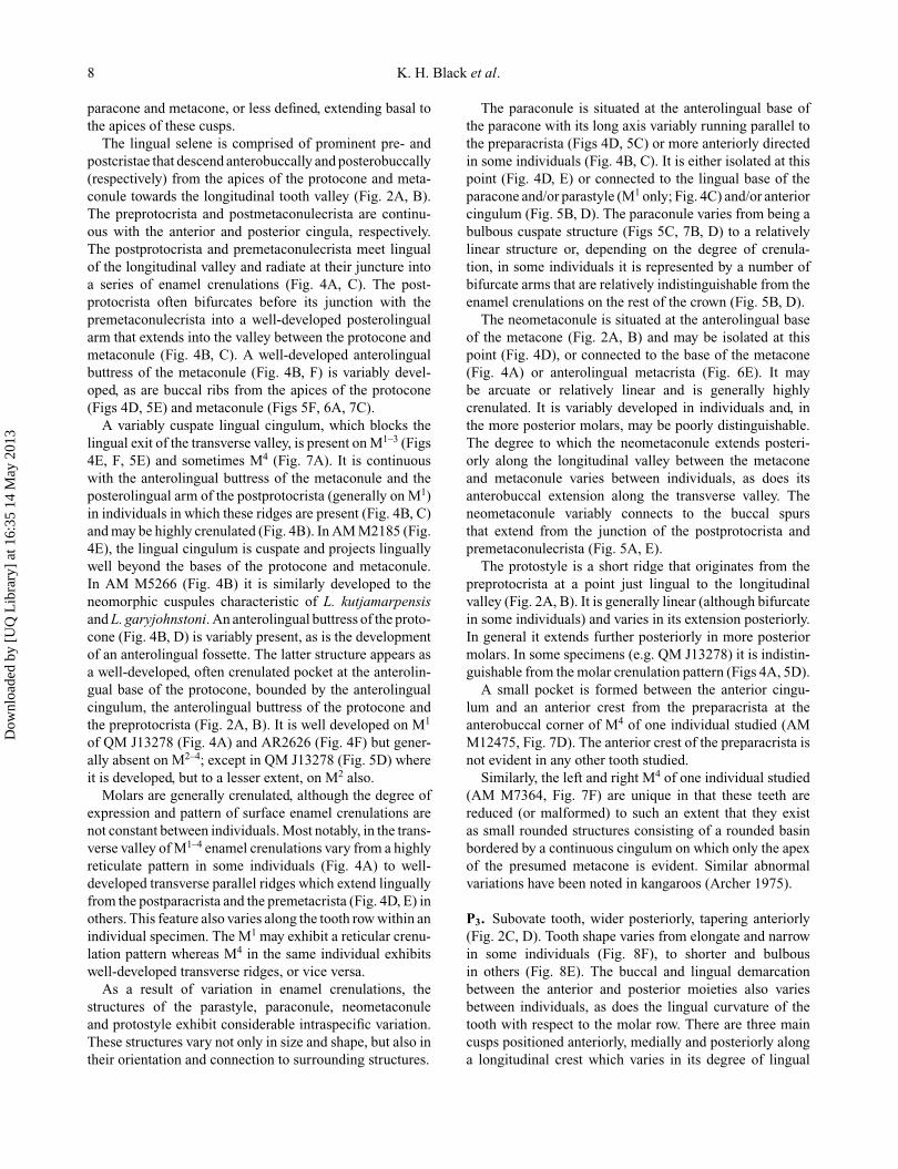
Figure 10. Comparison of the left M2 of six individuals ofP. cinereus. A, AM M5266; B, AM M5743; C, AM M12475;D, AM M6582; E, QM J13278; F, AM M7356. Abbreviations: a,anterior; cstd, columnar stylid; end, entoconid; er, entostylid ridge;hyd, hypoconid; l, lingual; lsed, lingual shelf of entoconid; lsmd,lingual shelf of metaconid; med, metaconid; prd, protoconid.
pre-entocristid (Fig. 9B) or terminating basal to the post-metacristid (e.g. AM M7438).
There does not appear to be a premetacristid on M1,although in some specimens a small columnar stylid at thelingual base of the metaconid possesses a short anteriorspur (Fig. 9C, E). It is possible that the transverse crestlinking the apices of the metaconid and protoconid on M1
may represent a premetacristid. A well-developed columnarstylid is present on the lingual face of the entoconid, givingthis cusp the appearance of having a twinned apex (Fig.9E, F). On M1–4 the postmetacristid extends posterolin-gually from the metaconid apex and may bifurcate justprior to the lingual tooth margin into short anterolingualand posterolingual spurs (Fig. 10D, F). These spurs (thepremetastylid cristid and postmetastylid cristid) define theapex of the metastylid (Fig. 2C, D). The pre-entocristidvaries from a linear (Fig. 9A) to highly arcuate crest (Fig.9D), and is generally continuous with the postmetastylidcristid at the median lingual tooth margin. This featureis often referred to as the metastylid fold (see Black &Archer 1997). In some specimens (e.g. AR1574), however,the pre-entocristid connects to the posterior base of the post-metacristid (Fig. 9B). A similar bifurcation of the posten-tocristid at the posterobuccal tooth corner into a weak
Figure 11. Comparison of the left M3 of six individuals ofP. cinereus. A, AM M7356; B, AM S415; C, AM M2185; D,AM M5743; E, AM M12475; F, AM M7486. Abbreviations: a,anterior; cstd, columnar stylid; end, entoconid; er, entostylid ridge;hyd, hypoconid; l, lingual; lsed, lingual shelf of entoconid; lsmd,lingual shelf of metaconid; med, metaconid; prd, protoconid.
buccal preentostylid cristid and a distinct postentostylidcristid defines the apex of the entostylid (Fig. 2C, D). Thepostentostylid cristid is continuous with the posterior cingu-lum which is in turn continuous with the posthypocristid.An entostylid ridge extends anterobuccally from the junc-tion of the postentostylid cristid and posterior cinguluminto the talonid basin. The entostylid ridge on M1–3 (gener-ally absent in M4s examined except AM M5266) may existas a well-developed cuspate structure (Fig. 9D) or be rela-tively indistinguishable existing as a series of discontinu-ous crenulate ridges at the posterobuccal base of the ento-conid (Fig. 10E, F). In some specimens it is linear andmirrored by a parallel crest that extends anteriorly fromthe posthypocristid (Figs 10C, 11A). In some individu-als the entostylid ridge extends anterobuccally from thepostentostylid cristid and variably connects to the posteriorcingulum (AM M7486, AR1574) and a variably presentanteriorly directed ridge off the posthypocristid (Fig. 9A,B). It is isolated at the posterobuccal base of the entoconidin others. The above-mentioned variations for the entostylidridge vary significantly along the tooth row in individ-ual specimens. In some specimens a variably defined crestor lingual rib descends from the hypoconid apex into the
Dow
nloa
ded
by [
UQ
Lib
rary
] at
16:
35 1
4 M
ay 2
013
Identification of species boundaries in extinct koalas 11
longitudinal valley between the hypoconid and entoconid(Fig. 9C, D).
M2–4. Subrectangular in occlusal view, longer than wideand much narrower than their corresponding upper molars(Fig. 2C, D). Comprised of four main cusps: an anterolin-gual metaconid; posterolingual entoconid; anterobuccalprotoconid; and posterobuccal hypoconid. The lingualselene is composed of prominent pre- and postcristids thatdescend anterolingually and posterolingually (respectively)from the apices of the metaconid and entoconid towards thelingual tooth margin. The premetacristid is continuous withthe anterior cingulum. A slight swelling at their junctionhas been referred to by some authors (e.g. Pledge 1987) asa parastylid. The construction of the talonid is similar tothat described for M1. Again, the pre-entocristid, althoughgenerally continuous with the postmetastylid cristid, mayterminate at the posterior base of the postmetacristid. Thisappears to be variable within a single individual. In AR1574for example, the latter condition is evident on the rightM1 and M3 and the left M1–3, whereas the pre-entocristidconnects to the postmetastylid cristid on the remainingmolars.
The buccal selene is composed of prominent pre- andpostcristids that descend anterolingually and posterolin-gually (respectively) from the apices of the protoconidand hypoconid towards the median longitudinal valley (Fig.2C, D). The preprotocristid terminates at the anterior toothmargin and generally abuts the posterior cingulum of thepreceding tooth. It may be highly crenulated at its anteriorextent (Figs 10F, 11E). On M2–3 of all specimens stud-ied the postprotocristid and cristid obliqua (prehypocristid)join at the longitudinal valley and, in some individuals, aseries of variably developed spurs spread lingually from thisjunction (Fig 10C, 11E). On M4 the cristid obliqua gener-ally meets the postprotocristid (Fig. 12C) and/or the pre-entocristid (Fig. 12A, D). A relatively deep buccal valleyis created between the posterior and anterior bases of theprotoconid and hypoconid, respectively, and the junction ofthe postprotocristid and cristid obliqua. A buccal cingulumis generally present on M2–4, but it is variably developed,may be crenulated (Fig. 10C), and in some individuals mayascend the anterobuccal face of the hypoconid (Fig. 10A). Asmall stylar cusp medially positioned on the buccal cingu-lum at the buccal tip of the valley separating the trigonidand talonid of M2–4 is variably developed.
Columnar stylids are present on both the metaconid andentoconid of M2–4, giving these cuspids a ‘twinned’ appear-ance (Fig. 2C, D). Their development varies from strongly(Fig. 10D) to poorly developed between individuals andalong the tooth row becoming reduced in the more poste-rior molars (Fig. 12B–D). In some individuals the colum-nar stylid of the metaconid of M2 is anteroposteriorlybroad with well-developed anterior, posterior and trans-verse accessory crests (Fig. 10F). In others (e.g. AR6071)
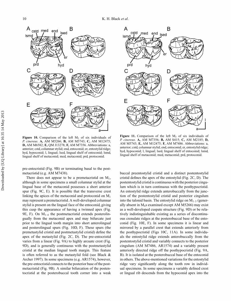
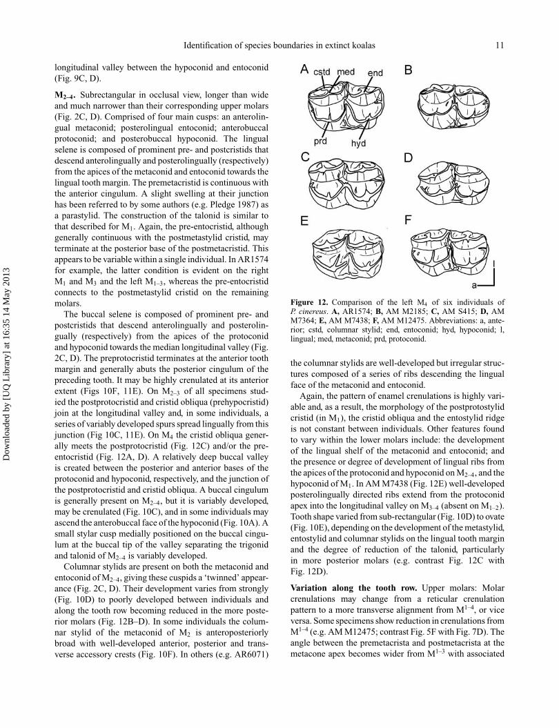
Figure 12. Comparison of the left M4 of six individuals ofP. cinereus. A, AR1574; B, AM M2185; C, AM S415; D, AMM7364; E, AM M7438; F, AM M12475. Abbreviations: a, ante-rior; cstd, columnar stylid; end, entoconid; hyd, hypoconid; l,lingual; med, metaconid; prd, protoconid.
the columnar stylids are well-developed but irregular struc-tures composed of a series of ribs descending the lingualface of the metaconid and entoconid.
Again, the pattern of enamel crenulations is highly vari-able and, as a result, the morphology of the postprotostylidcristid (in M1), the cristid obliqua and the entostylid ridgeis not constant between individuals. Other features foundto vary within the lower molars include: the developmentof the lingual shelf of the metaconid and entoconid; andthe presence or degree of development of lingual ribs fromthe apices of the protoconid and hypoconid on M2–4, and thehypoconid of M1. In AM M7438 (Fig. 12E) well-developedposterolingually directed ribs extend from the protoconidapex into the longitudinal valley on M3–4 (absent on M1–2).Tooth shape varied from sub-rectangular (Fig. 10D) to ovate(Fig. 10E), depending on the development of the metastylid,entostylid and columnar stylids on the lingual tooth marginand the degree of reduction of the talonid, particularlyin more posterior molars (e.g. contrast Fig. 12C withFig. 12D).
Variation along the tooth row. Upper molars: Molarcrenulations may change from a reticular crenulationpattern to a more transverse alignment from M1–4, or viceversa. Some specimens show reduction in crenulations fromM1–4 (e.g. AM M12475; contrast Fig. 5F with Fig. 7D). Theangle between the premetacrista and postmetacrista at themetacone apex becomes wider from M1–3 with associated
Dow
nloa
ded
by [
UQ
Lib
rary
] at
16:
35 1
4 M
ay 2
013
12 K. H. Black et al.
increased length of the metacone buccal margin (Fig. 2A,B). On M3–4 the postmetacrista shortens both absolutelyand with respect to the length of the premetacrista (Fig.2A, B). As a consequence, the buccal margin of the meta-cone becomes obliquely oriented with respect to the antero-posterior plane of the tooth. The buccal basins/surfacesof the paracone and metacone become progressively shal-lower from M1–4. This feature is most noticeable on themetacone wherein the stylar shelf is progressively reducedand often absent in M3–4. Generally the paraconule andneometaconule become weaker through M1–4 (Fig. 2A, B),becoming indistinguishable from the enamel crenulationpattern in more posterior molars (Fig. 6D), although somespecimens (e.g. AR2626) have a paraconule on M2 that is aswell developed as that of M1. The posterior moiety becomesprogressively reduced (buccolingually) with respect to theanterior moiety. This is most pronounced in M4 with oftenextreme reduction of both the metacone and metaconule(Fig. 7A). The protostyle generally becomes more antero-posteriorly elongate in M2–3 (and sometimes M4) but maybe reduced in M4 (although in some specimens the proto-style is progressively reduced from M2–4; e.g. AR2626).The posterolingual paracrista may become more promi-nent from M1–4. The anterolingual fossette (and associatedanterolingual buttress from the protocone apex) if presentin M1 is generally absent in M2–4 (although it is present inM1–3 of AR8398). M2–4 lack the well-developed parastylarregion of M1 and in most M4 a well-developed anterolingualmetacrista is developed (Fig. 7A, C–E) (yet absent in M1–3).The lingual pocket between the posterior base of the proto-cone, the anterior base of the metaconule and the lingualcingulum is progressively reduced from M1–4 and a lingualcingulum may be absent entirely on M4 (Fig. 2A, B).
Lower molars: The columnar stylids of the metaconidand entoconid become progressively reduced from M2–4
as do the metastylid and entostylid (Fig. 2C, D). Toothshape generally changes from subrectangular in M2 to ovatein M4 with more arcuate anterior, lingual and posteriortooth margins. The molars become progressively narrowerthrough M1 to M4. The entostylid ridge is reduced fromM1–3 and is absent in all M4s (and some M3s). Thebuccal valley between the protoconid and hypoconid isreduced in area and depth from M2–M4 (Fig. 2C, D),and the buccal cingulum (including associated crenula-tions/cuspids, if developed) is also reduced in more poste-rior molars.
Sexual dimorphism and populational morphologicalvariation. Conspicuous patterns of qualitative morpholog-ical variation within or between sexes and within or betweenregions were not evident in the Phascolarctos cinereussample studied here.
Summary of dental characters useful for distinguishingspecies. Because significant variation occurs along themolar row with many features becoming progressively
reduced, attenuated or absent in the more posterior molarsof P. cinereus, morphological features that may be usefulin distinguishing phascolarctid species are generallyrestricted to the premolar and first molar of the upper andlower tooth rows.
Features consistently developed on P3 of P. cinereusinclude: the presence of a midline longitudinal crest; thenumber of major cusps (three) developed along this crestand the presence of a lingual cingulum. Consistent featuresdeveloped on M1 include: the relative height of major cusps;and the presence of a parastyle, protostyle, paraconule,neometconule, posterolingual paracrista and lingual cingu-lum. All P. cinereus M1s possess enamel crenulations (albeitthey vary in degree and pattern of expression). Consistentfeatures developed on P3 include: a midline longitudinalcrest with three cuspids (anterior, medial and posterior) andthe presence of a posterobuccal crest. Consistent features ofM1 include: the relative height of major cuspids; the devel-opment and position of the protostylid; the presence of ametastylid, entostylid and entostylid ridge; the presence ofa columnar stylid on the entoconid; and the presence of atransverse crest connecting the apices of the metaconid andprotoconid. As in M1, all lower first molars possess enamelcrenulations.
In general, the shape, extent and degree of developmentof many structures on P. cinereus teeth were found to behighly variable. Consequently, as a general rule, the pres-ence or absence of a structure (as opposed to its shape orrelative development) appears more useful in discriminat-ing species boundaries in phascolarctids. Nevertheless, therelative development of a structure may fall outside theexpected range of variation for a species and, as such, maystill be a useful diagnostic feature. Nimiokoala greystanesiM1s for example exhibit consistently large, pyramidialparastyles and large, bicuspid neometaconules that are diag-nostic for the species (Black & Archer 1997).
Morphometric analysisUpper dentition. MANOVA of upper tooth dimensionsof male P. cinereus showed significant differences betweennorthern (Queensland) and southern (Victorian) popula-tions (F = 3.225, df = 15, 11, p = 0.028). Likewisesignificant differences in upper tooth dimensions betweenfemales from Queensland and Victoria were observed(F = 7.451, df = 15, 9, p = 0.002). For both male andfemale koalas, significant differences between geographi-cal regions existed for each tooth position. (Male: P3: F =5.064, df = 3, 23, p = 0.008; M1: F = 3.661, df = 3, 23, p =0.027; M2: F = 4.817, df = 3, 23, p = 0.01; M3: F = 4.732,df = 3, 23, p = 0.01; M4: F = 5.376, df = 3, 23, p = 0.006.Female: P3: F = 3.973, df = 3, 21, p = 0.022; M1: F = 5.404,df = 3, 21, p = 0.006; M2: F = 3.742, df = 3, 21, p = 0.027;M3: F = 7.113, df = 3, 21, p = 0.002; M4: F = 3.219, df = 3,21, p = 0.044.) Analysis of individual tooth measurementssuggests that the Victorian male population has on average
Dow
nloa
ded
by [
UQ
Lib
rary
] at
16:
35 1
4 M
ay 2
013
Identification of species boundaries in extinct koalas 13
greater lengths of both premolars and molars, and largeranterior widths of molars (M1–M3) than Queensland males.Only the M2 showed significantly different posterior widths(Table 1). For females, very few of the individual toothmeasurements were significantly different between regions,the notable exception being the posterior widths of P3, M1
and M2 which were larger for Queensland koalas (Table 1).MANOVA of upper tooth dimensions between male
and female koalas from Queensland showed no significantdifferences between the sexes (F = 1.267, df = 15, 13, p =0.338), yet significant differences were evident in the Victo-rian sample (F = 5.405, df = 15, 7, p = 0.0157). Male andfemale premolars did not differ significantly; only molarsshowed significant differences between the sexes (M1: F =7.309, df = 3, 19, p = 0.002; M2: F = 10.64, df = 3, 19,p < 0.001; M3: F = 4.48, df = 3, 19, p = 0.0154; M4:F = 9.752, df = 3, 19, p < 0.001). Analysis of individualtooth measurements showed that Victorian male koalas havesignificantly bigger molars in all dimensions with respectto females, except for the posterior width of M4 (Table 2).
Lower dentition. MANOVA of lower tooth dimensions ofmale P. cinereus showed significant differences betweennorthern (Queensland) and southern (Victorian) popula-tions (F = 13.5, df = 15, 14, p < 0.001). With respect toindividual teeth, both premolars and molars of males fromnorthern and southern populations differed significantly indimensions (P3: F = 5.661, df = 3, 26, p = 0.004; M1: F =10.57, df = 3, 26, p < 0.001; M2: F = 7.956, df = 3, 26,p < 0.001; M3: F = 7.121, df = 3, 26, p = 0.001; M4: F =6.973, df = 3, 26, p = 0.001). MANOVA of lower toothdimensions of female P. cinereus showed significant differ-ences between northern (Queensland) and southern (Victo-rian) populations (F = 5.621, df = 15, 24, p < 0.001). Withrespect to individual teeth positions, premolars did not showany significant differences in dimensions measured (F =0.6496, df = 3, 36, p = 0.6); however, molars from north-ern and southern populations differed significantly (M1:F = 7.568, df = 3, 36, p = 0.0004; M2: F = 4.01, df =3, 36, p = 0.015; M3: F = 5.356, df = 3, 36, p = 0.004;M4: F = 4.746, df = 3, 36, p = 0.007). Analysis of theindividual tooth measurements showed that for both maleand female koalas, Victorian specimens were significantlylarger than Queensland specimens in the following dimen-sions: M3 and M4 length, M1 and M2 anterior width, and M1
posterior width (Table 3). MANOVA of lower tooth dimen-sions between male and female koalas from Victoria andbetween male and female koalas from Queensland showedno differences existing along the tooth row (Queensland:F = 1.41, df = 15, 31, p = 0.2035; Victoria: F = 2.359, df= 15, 7, p = 0.1275).
Systematic palaeontology
Class Marsupialia Illiger, 1811Order Diprotodontia Owen, 1866
Suborder Vombatiformes Woodburne, 1984Infraorder Phascolarctomorphia Aplin & Archer, 1987
Family Phascolarctidae Owen, 1839Genus Litokoala Stirton et al., 1967
Type species. Litokoala kutjamarpensis Stirton et al.,1967.
Additional species. L. garyjohnstoni Louys et al., 2007;Litokoala dicksmithi sp. nov.
Diagnosis. In addition to the dental (e.g. Black & Archer1997; Louys et al. 2007) and cranial features (Louyset al. 2009) described elsewhere, species of Litokoaladiffer from all other phascolarctids in having a twinnedor secondary infraorbital foramen, a prominent massetericprocess composed entirely of maxilla, an almost verticallyoriented premaxillomaxilla suture on the lateral face ofthe rostrum, and a large, anteriorly extensive lacrimal.Litokoala species differ from Phascolarctos species inhaving proportionately larger orbits, a less constrictedrostrum anteriorly at the level of the incisor arcade, ashallow maxillolabial fossa, and a zygomatic arch thatprojects posterolaterally (as opposed to laterally) from theface. Litokoala species differ from Nimiokoala speciesin having a relatively shorter, broader, deeper rostrum,a broader maxillary palate, and a larger, broader narialaperture with an irregular border (as in Phascolarctos).Louys et al. (2007, p. 100) differentiated Litokoala speciesfrom other phascolarctids by possession of “. . . a well-developed neomorphic cuspule at the anterolingual baseof the metaconule of M1 (with the exception of speciesof Phascolarctos de Blainville, 1816 where it is variablypresent)”. Because this feature is variably present in Phas-colarctos, absent in Litokoala dicksmithi and unknown forL. thurmerae (known only from M3), we do not regard it tobe diagnostic for the genus.
Litokoala kutjamarpensis Stirton et al., 1967
1987 Litokoala kanunkaensis Springer; 320, figs 1, 2.2010 Litokoala dicktedfordi Pledge; 81, fig. 5.
Holotype. SAM P13845, right M1.
Material. From Kanunka North Site: SAM P32397, a rightM2; UCR21945, a right M4; UCR21980, a metacone of aright M3; UCR21979, a metacone of a left M1. From River-sleigh: QM F30500, right P3, QM F13079, right dentaryfragment with posterior half of P3, M1–2, QM F30502, rightM3, Henks Hollow Local Fauna; QM F30501, right M1,Gag Site; QM F30503, Mx fragment containing paraconeand buccal half of protocone, Gotham Site; QM F51382,a partial skull with left and right P3, M1–4, Jim’s CarouselSite; QM F20809, right M3, JC9 Site.
Occurrence. The holotype is from the Kutjamarpu LocalFauna, Leaf Locality, Lake Ngapakaldi, Wipajiri Forma-tion, South Australia, which is interpreted to be EarlyMiocene in age (Woodburne et al. 1993). Kanunka North
Dow
nloa
ded
by [
UQ
Lib
rary
] at
16:
35 1
4 M
ay 2
013
14 K. H. Black et al.
Site (Zone E of the Etadunna Formation) is located on thewest side of Lake Kanunka, South Australia. This depositis Late Oligocene in age (Woodburne et al. 1993). TheHenk’s Hollow, Gag, Gotham, Jim’s Carousel and JC9Sites from the Riversleigh World Heritage Area, Lawn HillNational Park, north-western Queensland, are Faunal ZoneC deposits which are interpreted to be Middle Miocene inage (Creaser 1997; Arena 2005; Travouillon et al. 2006).
Remarks. Dental descriptions of L. kutjamarpensis areprovided in Stirton et al. (1967), Black & Archer (1997; asL. kanunkaensis) and Louys et al. (2007), and a descriptionof the cranium is given by Louys et al. (2009). Argumentsfor subsuming L. kanunkaensis into L. kutjamarpensis canbe found in Louys et al. (2007) and are recounted in thediscussion below. Arguments for synonymizing L. ‘dickt-edfordi’ with L. kutjamarpensis are also given below.
Litokoala garyjohnstoni Louys et al., 2007
Holotype. QM F51405, left partial maxilla with P3, M1–3.
Material. Paratype, QM F51406, left M4.
Occurrence. Outasite, Godthelp Hill, Riversleigh WorldHeritage Area, Lawn Hill National Park, north-westernQueensland. Outasite is a Faunal Zone B deposit and isinterpreted to be Early Miocene in age (Creaser 1997;Travouillon et al. 2006).
Remarks. Litokoala garyjohnstoni is described in Louyset al. (2007).
Litokoala dicksmithi sp. nov(Figs 13, 14)
Diagnosis. Rostrum short with twinned infraorbital fora-men; narial aperture broad with irregular border. Differsfrom other Litokoala spp. in: its deeper transverse andlongitudinal valleys in the upper molars; its larger, broaderprotocone in the upper molars; lacking a neomorphiccuspule at the anterolingual base of the metaconule on M1
(unknown for L. thurmerae); and P3 lacking a lingual cingu-lum with narrower, shallow lingual valley (unknown for L.thurmerae). Differs from L. garyjohnstoni in: its longer,more prominent masseteric process; P3 with reduced medialand posterior lingual cusps and reduced anterobuccal crestfrom medial midline cusp; reduced molar crenulations; andposteriorly narrow upper molars across the metacone andmetaconule. Differs from L. kutjamarpensis in: its lesslinear arrangement of midline cusps and crests on P3; andlacking an anterolingual metacrista on upper molars.
Derivation of name. The specific name honours DickSmith, Australian aviator, adventurer and philanthropistfor his long-term financial support of Australian scientificendeavour and in particular fossil research at Riversleigh.
Holotype. QM F54567, partial anterior skull with left I1,P3, M1, partial M2; right P3, M1–4.
Occurrence. Ross Scott Orr (RSO) Site, Faunal Zone B(Travouillon et al. 2006), Riversleigh World Heritage Area,north-western Queensland; Early Miocene.
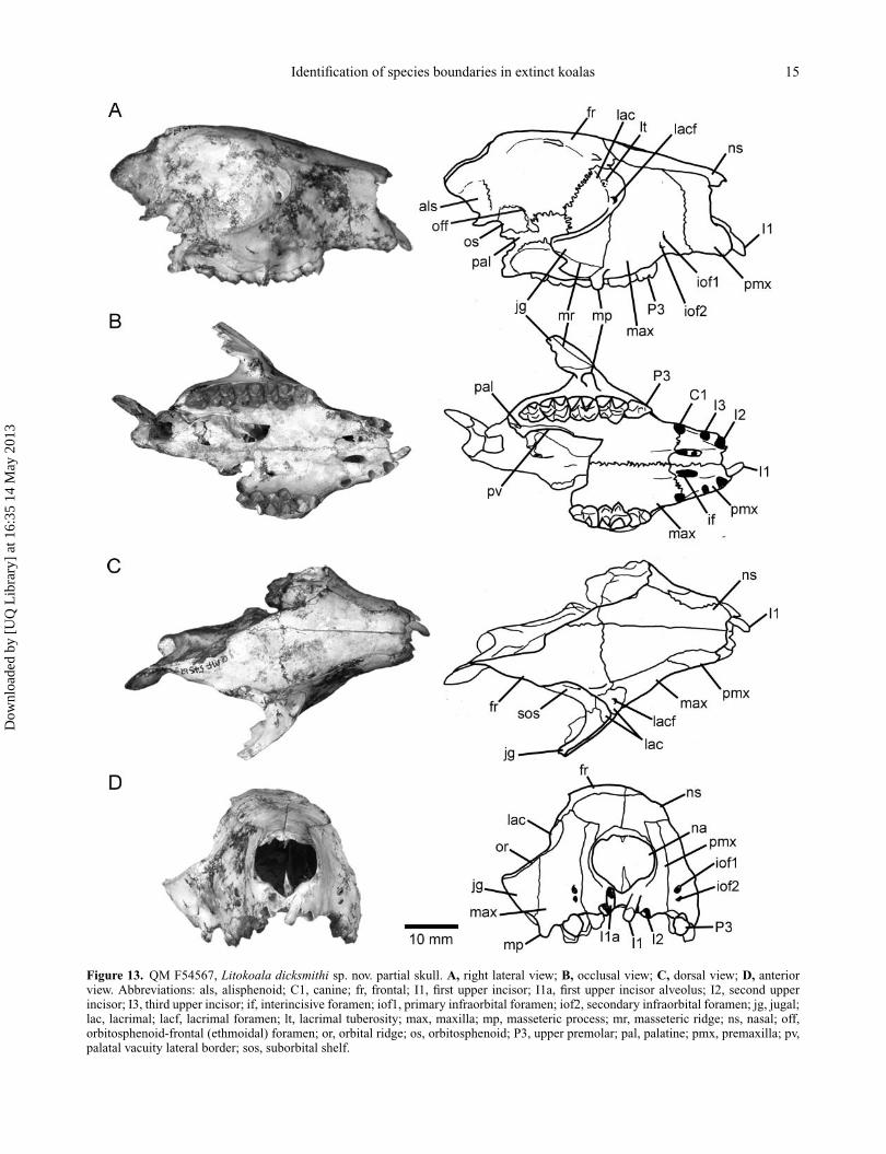
DescriptionAdult skull with right P3–M4, LI1, LP3–M2. Approxi-mately the same size as Litokoala kutjamarpensis skull(QM F51382) judging by width of the palate and length oftooth row, and slightly shorter than Nimiokoala. We esti-mate the total length of the skull (based on overlap withQM F51382) to have been approximately 76 mm, whichis suggestive of a relatively short, broad skull. Right sidepreserves nasal, premaxilla, maxilla, lacrimal, part of thefrontal, orbitosphenoid, endocranial portion of the alisphe-noid, anterior jugal and partial palatine (Fig. 13A). Left sideis largely missing, preserving only the premaxilla, nasal andpartial maxilla. Cranial and dental measurements are givenin Tables 4 and 5, respectively. The description is restrictedto the right hand side. Comparisons are made throughoutwith Phascolarctos cinereus (hereafter Phascolarctos) andNimiokoala greystanesi (hereafter Nimiokoala), the onlytwo koala species with which there is considerable overlapwith the skull portion preserved.
Facial region. Rostrum short and relatively deep (17.9 mmat C1 alveoli), tapers slightly anteriorly. Shorter and deeperthan Nimiokoala and similarly proportioned to Phasco-larctos. Slight inflation of premaxilla portion of rostrumdorsolaterally, but not to the extent as seen in Phasco-larctos in which the rostrum in anterior view is distinctlykeyhole-shaped (i.e. broad dorsally, narrow ventrally). In L.dicksmithi and Nimiokoala, the rostrum is slightly broaderventrally than dorsally and not constricted as in Phascolarc-tos. Nasals are wide posteriorly, taper anteriorly and flareslightly at their anterior extremity (Fig. 13C). Althoughthe nasals are broken anteriorly it appears that their ante-rior border was quite irregular, as in Phascolarctos whichhas numerous short anterior projections. This is unlikethe condition found in most other diprotodontians (possi-bly including Nimiokoala) in which the anterior borderof the nasals is well defined and medially tapering. Thenasals are dorsally flattened as in Nimiokoala and unlike inPhascolarctos where they are mildly convex and inflated.Nasofrontal suture is broad (11.1 mm) and transverselyorientated (Fig. 13C). The nasals are slightly inflated later-ally at this suture and the frontals more inflated dorsallythan in Nimiokoala, indicating a slightly deeper skull for L.dicksmithi. Nasal aperture (Fig. 13D) broader (12.91 mm)than high (10.15 mm), larger than Nimiokoala (8.47 mmwide, 7.42 mm high), and similarly proportioned to Phas-colarctos. Premaxilla short, premaxillomaxilla suture verti-cally orientated in lateral view terminating dorsally at itsjunction with the nasals at a point vertically in line withthe infraorbital foramina (Fig. 13A). This is unlike thecondition in Nimiokoala and Phascolarctos where it runs
Dow
nloa
ded
by [
UQ
Lib
rary
] at
16:
35 1
4 M
ay 2
013
Identification of species boundaries in extinct koalas 15
Figure 13. QM F54567, Litokoala dicksmithi sp. nov. partial skull. A, right lateral view; B, occlusal view; C, dorsal view; D, anteriorview. Abbreviations: als, alisphenoid; C1, canine; fr, frontal; I1, first upper incisor; I1a, first upper incisor alveolus; I2, second upperincisor; I3, third upper incisor; if, interincisive foramen; iof1, primary infraorbital foramen; iof2, secondary infraorbital foramen; jg, jugal;lac, lacrimal; lacf, lacrimal foramen; lt, lacrimal tuberosity; max, maxilla; mp, masseteric process; mr, masseteric ridge; ns, nasal; off,orbitosphenoid-frontal (ethmoidal) foramen; or, orbital ridge; os, orbitosphenoid; P3, upper premolar; pal, palatine; pmx, premaxilla; pv,palatal vacuity lateral border; sos, suborbital shelf.
Dow
nloa
ded
by [
UQ
Lib
rary
] at
16:
35 1
4 M
ay 2
013
16 K. H. Black et al.
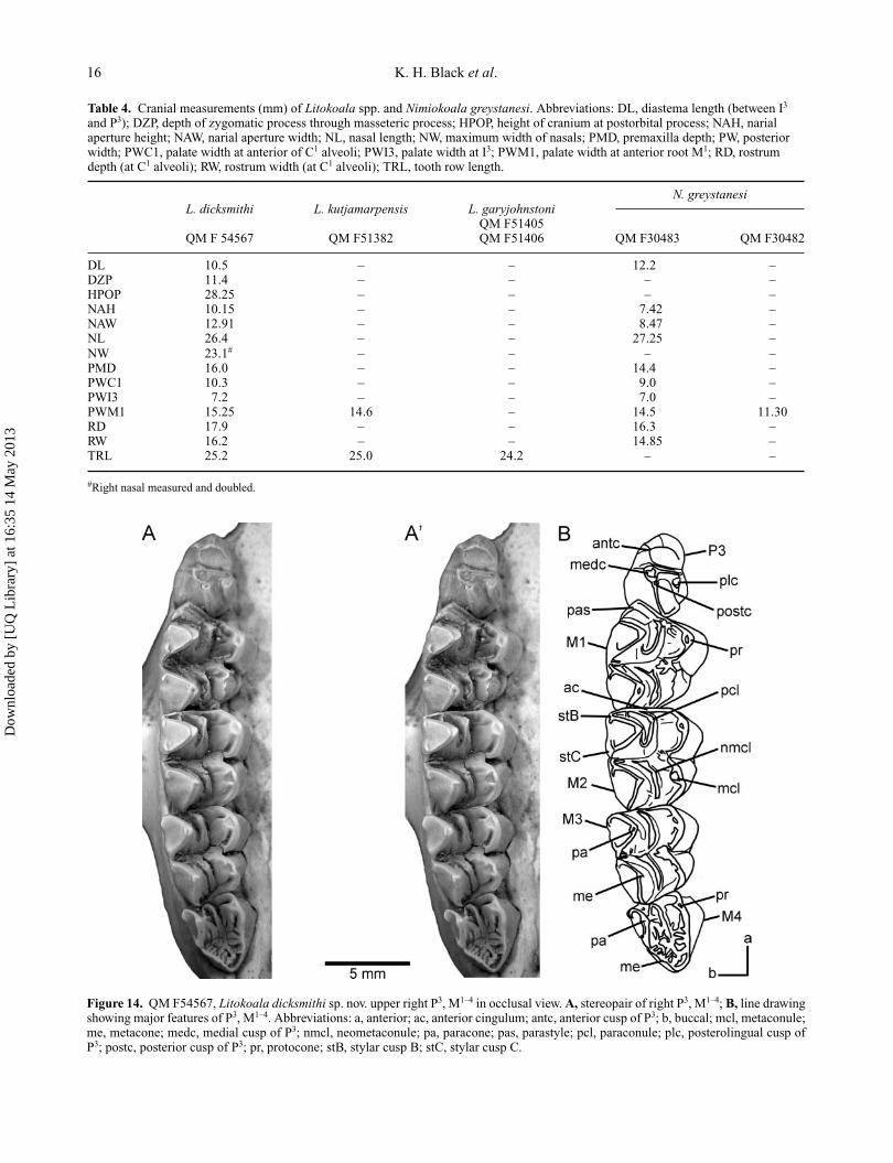
Table 4. Cranial measurements (mm) of Litokoala spp. and Nimiokoala greystanesi. Abbreviations: DL, diastema length (between I3
and P3); DZP, depth of zygomatic process through masseteric process; HPOP, height of cranium at postorbital process; NAH, narialaperture height; NAW, narial aperture width; NL, nasal length; NW, maximum width of nasals; PMD, premaxilla depth; PW, posteriorwidth; PWC1, palate width at anterior of C1 alveoli; PWI3, palate width at I3; PWM1, palate width at anterior root M1; RD, rostrumdepth (at C1 alveoli); RW, rostrum width (at C1 alveoli); TRL, tooth row length.
N. greystanesiL. dicksmithi L. kutjamarpensis L. garyjohnstoni
QM F51405QM F 54567 QM F51382 QM F51406 QM F30483 QM F30482
DL 10.5 – – 12.2 –DZP 11.4 – – – –HPOP 28.25 – – – –NAH 10.15 – – 7.42 –NAW 12.91 – – 8.47 –NL 26.4 – – 27.25 –NW 23.1# – – – –PMD 16.0 – – 14.4 –PWC1 10.3 – – 9.0 –PWI3 7.2 – – 7.0 –PWM1 15.25 14.6 – 14.5 11.30RD 17.9 – – 16.3 –RW 16.2 – – 14.85 –TRL 25.2 25.0 24.2 – –
#Right nasal measured and doubled.
Figure 14. QM F54567, Litokoala dicksmithi sp. nov. upper right P3, M1–4 in occlusal view. A, stereopair of right P3, M1–4; B, line drawingshowing major features of P3, M1–4. Abbreviations: a, anterior; ac, anterior cingulum; antc, anterior cusp of P3; b, buccal; mcl, metaconule;me, metacone; medc, medial cusp of P3; nmcl, neometaconule; pa, paracone; pas, parastyle; pcl, paraconule; plc, posterolingual cusp ofP3; postc, posterior cusp of P3; pr, protocone; stB, stylar cusp B; stC, stylar cusp C.
Dow
nloa
ded
by [
UQ
Lib
rary
] at
16:
35 1
4 M
ay 2
013
Identification of species boundaries in extinct koalas 17
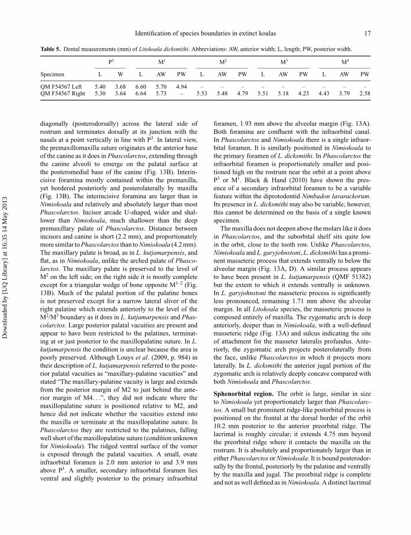
Table 5. Dental measurements (mm) of Litokoala dicksmithi. Abbreviations: AW, anterior width; L, length; PW, posterior width.
P3 M1 M2 M3 M4
Specimen L W L AW PW L AW PW L AW PW L AW PW
QM F54567 Left 5.40 3.68 6.60 5.70 4.94 – – – – – – – – –QM F54567 Right 5.30 3.64 6.64 5.73 – 5.53 5.48 4.79 5.51 5.18 4.23 4.43 3.79 2.58
diagonally (posterodorsally) across the lateral side ofrostrum and terminates dorsally at its junction with thenasals at a point vertically in line with P3. In lateral view,the premaxillomaxilla suture originates at the anterior baseof the canine as it does in Phascolarctos, extending throughthe canine alveoli to emerge on the palatal surface atthe posteromedial base of the canine (Fig. 13B). Interin-cisive foramina mostly contained within the premaxilla,yet bordered posteriorly and posterolaterally by maxilla(Fig. 13B). The interincisive foramina are larger than inNimiokoala and relatively and absolutely larger than mostPhascolarctos. Incisor arcade U-shaped, wider and shal-lower than Nimiokoala, much shallower than the deeppremaxillary palate of Phascolarctos. Distance betweenincisors and canine is short (2.2 mm), and proportionatelymore similar to Phascolarctos than to Nimiokoala (4.2 mm).The maxillary palate is broad, as in L. kutjamarpensis, andflat, as in Nimiokoala, unlike the arched palate of Phasco-larctos. The maxillary palate is preserved to the level ofM2 on the left side; on the right side it is mostly completeexcept for a triangular wedge of bone opposite M1–2 (Fig.13B). Much of the palatal portion of the palatine bonesis not preserved except for a narrow lateral sliver of theright palatine which extends anteriorly to the level of theM2/M3 boundary as it does in L. kutjamarpensis and Phas-colarctos. Large posterior palatal vacuities are present andappear to have been restricted to the palatines, terminat-ing at or just posterior to the maxillopalatine suture. In L.kutjamarpensis the condition is unclear because the area ispoorly preserved. Although Louys et al. (2009, p. 984) intheir description of L. kutjamarpensis referred to the poste-rior palatal vacuities as “maxillary-palatine vacuities” andstated “The maxillary-palatine vacuity is large and extendsfrom the posterior margin of M2 to just behind the ante-rior margin of M4. . .”, they did not indicate where themaxillopalatine suture is positioned relative to M2, andhence did not indicate whether the vacuities extend intothe maxilla or terminate at the maxillopalatine suture. InPhascolarctos they are restricted to the palatines, fallingwell short of the maxillopalatine suture (condition unknownfor Nimiokoala). The ridged ventral surface of the vomeris exposed through the palatal vacuities. A small, ovateinfraorbital foramen is 2.0 mm anterior to and 3.9 mmabove P3. A smaller, secondary infraorbital foramen liesventral and slightly posterior to the primary infraorbital
foramen, 1.93 mm above the alveolar margin (Fig. 13A).Both foramina are confluent with the infraorbital canal.In Phascolarctos and Nimiokoala there is a single infraor-bital foramen. It is similarly positioned in Nimiokoala tothe primary foramen of L. dicksmithi. In Phascolarctos theinfraorbital foramen is proportionately smaller and posi-tioned high on the rostrum near the orbit at a point aboveP3 or M1. Black & Hand (2010) have shown the pres-ence of a secondary infraorbital foramen to be a variablefeature within the diprotodontid Nimbadon lavarackorum.Its presence in L. dicksmithi may also be variable; however,this cannot be determined on the basis of a single knownspecimen.
The maxilla does not deepen above the molars like it doesin Phascolarctos, and the suborbital shelf sits quite lowin the orbit, close to the tooth row. Unlike Phascolarctos,Nimiokoala and L. garyjohnstoni, L. dicksmithi has a promi-nent masseteric process that extends ventrally to below thealveolar margin (Fig. 13A, D). A similar process appearsto have been present in L. kutjamarpensis (QMF 51382)but the extent to which it extends ventrally is unknown.In L. garyjohnstoni the masseteric process is significantlyless pronounced, remaining 1.71 mm above the alveolarmargin. In all Litokoala species, the masseteric process iscomposed entirely of maxilla. The zygomatic arch is deepanteriorly, deeper than in Nimiokoala, with a well-definedmasseteric ridge (Fig. 13A) and sulcus indicating the siteof attachment for the masseter lateralis profundus. Ante-riorly, the zygomatic arch projects posterolaterally fromthe face, unlike Phascolarctos in which it projects morelaterally. In L. dicksmithi the anterior jugal portion of thezygomatic arch is relatively deeply concave compared withboth Nimiokoala and Phascolarctos.
Sphenorbital region. The orbit is large, similar in sizeto Nimiokoala yet proportionately larger than Phascolarc-tos. A small but prominent ridge-like postorbital process ispositioned on the frontal at the dorsal border of the orbit10.2 mm posterior to the anterior preorbital ridge. Thelacrimal is roughly circular; it extends 4.75 mm beyondthe preorbital ridge where it contacts the maxilla on therostrum. It is absolutely and proportionately larger than ineither Phascolarctos or Nimiokoala. It is bound posterodor-sally by the frontal, posteriorly by the palatine and ventrallyby the maxilla and jugal. The preorbital ridge is completeand not as well defined as in Nimiokoala. A distinct lacrimal
Dow
nloa
ded
by [
UQ
Lib
rary
] at
16:
35 1
4 M
ay 2
013
18 K. H. Black et al.
foramen is situated just anterior and outside the orbit(Fig. 13A). A smaller foramen is positioned 3.65 mm dorso-posteriorly to the lacrimal foramen and perforates the preor-bital ridge just dorsal to a weak lacrimal tuberosity. Threesmall foramina pierce the lacrimal close to its ventral suturewith the maxilla. Below these foramina on the lacrimojugalsuture is a small unperforated yet deep ovate depression.A well-defined infraorbital canal perforates the maxillajust below this suture line; like Nimiokoala it is situatedat the anterior end of a well-developed sulcus. Posteriorto the infraorbital canal and within the infraorbital sulcuslies the sphenopalatine foramen. Again as in Nimiokoalait lies on the maxillopalatine suture. The dorsal margin ofthe palatine is preserved; it interdigitates with the maxillaventrally and anteriorly, makes contact with the lacrimalanterodorsally, interdigitates with the frontal dorsally andmakes contact with the orbitosphenoid at its posterior-most preserved point (Fig. 13A). The orbital portion of themaxilla is roughly ovate, its anteriormost point being coin-cident with the lacrimojugal suture, and is entirely bounddorsally by the palatine. The orbitosphenoid is present asa roughly circular bone at the posteroventral corner of thespecimen preserved; it contacts the palatine anteroventrallyand the frontal anteriorly and dorsally (Fig. 13A). At thesuture between the orbitosphenoid and the frontal lies theethmoidal foramen; this foramen is also present in Phasco-larctos. Posteriorly it is bound by the alisphenoid, but thesuture between these two bones is difficult to discern. At theposteroventral margin of the orbitosphenoid, the anterodor-sal rim of the sphenorbital fissure can be seen. The alisphe-noid is almost only preserved as an endocranial lamina,its suture line with the frontal is however discernable. Thefrontal makes a large ventral contribution to the sphenor-bital region; on the cranial roof it is preserved only as atriangular wedge (Fig. 13C).
Dentition. (Fig. 14A, B). QM F54567 preserves the LI1,LP3-M2 and RP3-M4. The adult upper tooth formula wouldbe I1–3, C1, P3, M1–4. The alveolus for I2 is larger thanI3, the latter being similar in size to the alveolus of C1
(Fig. 13B). In basic dimensions the alveoli for I2–3 and C1
are comparable with Nimiokoala. As in other species ofLitokoala, the premolar is positioned more in line with themolar row (Fig. 14A, B) and less divergent anteromediallyas in Nimiokoala.
I1. Left I1 preserved. Short, gracile tooth with enamel onall surfaces and a small ovate wear facet on the poste-rior surface of its tip. Relatively blunt tip but this maybe partially attributed to abrasion. Projects anteroventrallyand would have converged medially on tip of right I1 (Fig.13D). Less robust, more protracted and less ventrally exten-sive than I1 of Nimiokoala or Phascolarctos (the only otherphascolarctids for which a first upper incisor is known) andin this regard is more reminiscent of an I3 of the latter taxathan an I1.
P3. Bulbous P3 with four main cusps, three of which arepositioned anteriorly, medially, and posteriorly along thelongitudinal tooth midline (Fig. 14A, B). The fourth andsmallest cusp is positioned at the posterolingual toothmargin. The P3 is widest posteriorly at the level of theposterolingual cusp and tapers in width to the anterior toothmargin. There is significant wear on the posterolingual faceof the anterior cusp, the apices of the medial and posterolin-gual cusps and the posterior blade of the posterior cusp. Asmall ovate wear facet that probably represented a smalllingual cuspule is positioned at the anterior base of theposterolingual cusp along the lingual tooth margin in thesame position as the small lingual cusp in L. garyjohnstoniand the small cuspule in L. kutjamarpensis (QM F51382).The relative heights of the major cusps are difficult todiscern as a result of wear. The anterior cusp is situatedapproximately 0.95 mm from the anterior margin. Ante-rior, buccal and lingual crests extend from its apex andfade towards the base of the crown. The buccal crest isnot as extensive, nor prominent as in L. garyjohnstoni inwhich it meets a well-developed anterobuccally directedcrest from the apex of the medial cusp. In L. dicksmithithe buccal crest of the medial cusp is weak and fades outmidway to the base of the crown. A better-developed yetworn lingual crest extends from the apex of the medialcusp and terminates at the base of the crown at the smallovate wear facet (mentioned above) resembling the condi-tion found in L. garyjohnstoni. In L. kutjamarpensis (QMF51382) the lingual crest of the medial cusp terminatesabruptly before meeting the small lingual cuspule.
The apex of the medial cusp is positioned 2.39 mmposterior to the anterior tooth margin. Short anterior andposterior crests link the apices of the medial and anteriorcusps (respectively) in a shallow valley which defines theanterior and posterior tooth moieties (Fig. 14A, B). A shortposterolingually directed crest links the apices of the medialand posterior cusps. The apex of the posterior cusp is posi-tioned 3.04 mm posterior to the anterior tooth margin. Theposterolingual placement of the posterior cusp with respectto the medial cusp is also found in L. garyjohnstoni and lessso in L. kutjamarpensis. Both L. dicksmithi and L. garyjohn-stoni lack the lingual crest of the posterior cusp found inL. kutjamarpensis. A prominent posteriorly directed crestconnects the posterior cusp apex to the anterior cingulumof M1. The posterolingual cusp is small and similarly devel-oped to that of L. kutjamarpensis but weaker with respect toL. garyjohnstoni. Short weak anterior and posterior crestsextend from its apex. The anterior crest is heavily wornbut appears to have been connected to the small lingualcuspule represented by an ovate wear facet. In L. dicksmithithe well-developed crest found in both L. garyjohnstoni andL. kutjamarpensis that links the small lingual cuspule ante-riorly with the lingual crest of the anterior cusp is absent.The resultant effect is a less trenchant, more bulbous look-ing premolar overall.
Dow
nloa
ded
by [
UQ
Lib
rary
] at
16:
35 1
4 M
ay 2
013
Identification of species boundaries in extinct koalas 19
M1. Description based on LM1 because enamel is missingfrom the lingual face of metaconule on RM1. The enamelis moderately crenulated although less apparent due towear. Crenulations are evident in the transverse valley, onthe anterior bases of the paracone, metacone, paraconuleand neometaconule, the posterior bases of the parastyle,paracone and metacone, the buccal base of the protocone,and the anterior cingulum. M1 is longer (across the buccalmargin) than it is wide. The buccal margin is convex andthe anterior margin slopes posterolingually. The tooth ismarkedly wider anteriorly (i.e. across the paracone andprotocone) than posteriorly (across the metacone and meta-conule) and in this regard resembles the M1 of Nimiokoalamore so than either L. garyjohnstoni or L. kutjamarpen-sis. This is in part due to the absence of the ‘neomorphiccuspule’ (sensu Stirton et al. 1967) located at the anterolin-gual base of the metaconule in other Litokoala species.The apices of the paracone and metacone slightly over-hang their lingual bases as they do in L. kutjamarpensis andcontra L. garyjohnstoni. The buccal surface of the paraconeis reduced relative to the metacone as it is in L. garyjohn-stoni. The respective heights of the major cusps are difficultto discern due to moderate wear with dentine exposed onall apices; however, the metacone appears to have beentaller than the paracone. The metaconule is the most wornof the major cusps. Dentine is also exposed on the apexof the parastyle, the neometaconule, the buccal end of thepremetaconule crista and the lingual end of the posterolin-gual paracrista. The parastyle is a large (albeit very worn),pyramidal cusp which occupies the anterobuccal corner ofthe tooth and is similarly developed as in L. garyjohnstoniand slightly larger than in L. kutjamarpensis. The degreeand pattern of wear is similar to that found in the holotype ofL. kutjamarpensis (SAM P13845), the worn posterior baseof the parastyle appearing as a buccal ridge connectingthe paraconule and buccal cingulum. The tooth is slightlyhigher crowned and the transverse and longitudinal valleysare slightly deeper than in other Litokoala spp. Stylar cuspsare weakly developed, similar to L. kutjamarpensis, andreduced relative to L. garyjohnstoni. Unlike other Litokoalaspp., stylar cusps B and C do not form a continuous ridgethat effectively closes off the paracone buccal basin alongthe buccal tooth margin. The preparacrista is short, heav-ily worn and bifurcates at the buccal tooth margin into ashort anterior spur that terminates just prior to meetingthe posterobuccal base of the parastyle, and a short poste-rior ridge that represents a poorly developed stylar cuspB. The postparacrista is linear, longer and less worn thanthe preparacrista and similarly bifurcates at the buccal toothmargin into a short anterior ridge representing stylar cusp C,and a short posterobuccal arm that meets the premetacristaat the buccal margin, effectively closing off the buccalend of the transverse valley. Both the postparacrista andpremetacrista meet a series of short, transversely orientedridges (that form part of the molar crenulation pattern)
at this point. The premetacrista is moderately worn, elon-gate, anterobuccally directed (running in a parallel planeto the preparacrista) and branches before its junction withthe postparacrista into a short posterior ridge that repre-sents stylar cusp D. The postmetacrista is more heavilyworn than the premetacrista (having dentine exposed atthe metacone apex) and is directed slightly more poste-riorly than the postparacrista, terminating ventral to theanterior cingulum of M2 at the anterobuccal tooth corner.A slight swelling at this point represents stylar cusp E. Alarge crescentic paraconule is situated at the anterolingualbase of the paracone. A short, linear crest extends anteri-orly from the paraconule apex and terminates at the lingualbase of the parastyle. A more elongate, slightly crescen-tic posterior crest curves around the base of the paraconeto meet a well-developed and moderately worn posterolin-gual paracrista as it does in other species of Litokoala.A third posterolingually directed crest extends from theparaconule apex and terminates at the buccal base of theprotocone. In the RM1, this crest is anterolingually directedand meets a short posterior spur from the anterior cingu-lum, the latter representing a weakly developed protostyle(Fig. 14A, B). A large crescentic neometaconule occupiesthe longitudinal valley between the metacone and meta-conule, its apex positioned at the anterolingual base of themetacone. A well-developed anterior neometaconule crestextends anterobuccally into the transverse valley, becom-ing part of the crenulation pattern. A posterior neometa-conule crest extends posteriorly, curving slightly buccallyat the posterolingual base of the metacone where it meetsa weakly developed posterolingual metacrista. A linear andslightly worn preprotocrista is continuous with the ante-rior cingulum anteriorly. A crescentic postprotocrista meetsa bifurcate premetaconule crista in the transverse valleywell buccal to the lingual margin. A weak anterolinguallydirected crest extends down the lingual face of the proto-cone, becoming more prominent towards the base of thecrown. It is more prominent in RM1 and becomes progres-sively better developed in M2–4 and originates from theprotocone apex (Fig. 14A, B). This crest is well developedin M1–4 of L. kutjamarpensis but weakly expressed in L.garyjohnstoni. The lingual face of the protocone slopes lesssteeply towards its base and consequently projects furtherlingually than that of the metaconule. A crescentic post-metaconule crista extends posteriorly from the metaconuleapex becoming continuous with the posterior cingulum, atwhich point it abuts and extends ventral to the anteriorcingulum of M2. The valley between the posterior baseof the protocone and the anterior base of the metaconuleis narrow in comparison with other Litokoala spp. and isan artefact of the absence of a neomorphic cuspule at theanterolingual base of the metaconule.
M2. Similar in most respects to M1 except for the follow-ing features: a large pyramidal parastyle is absent and
Dow
nloa
ded
by [
UQ
Lib
rary
] at
16:
35 1
4 M
ay 2
013
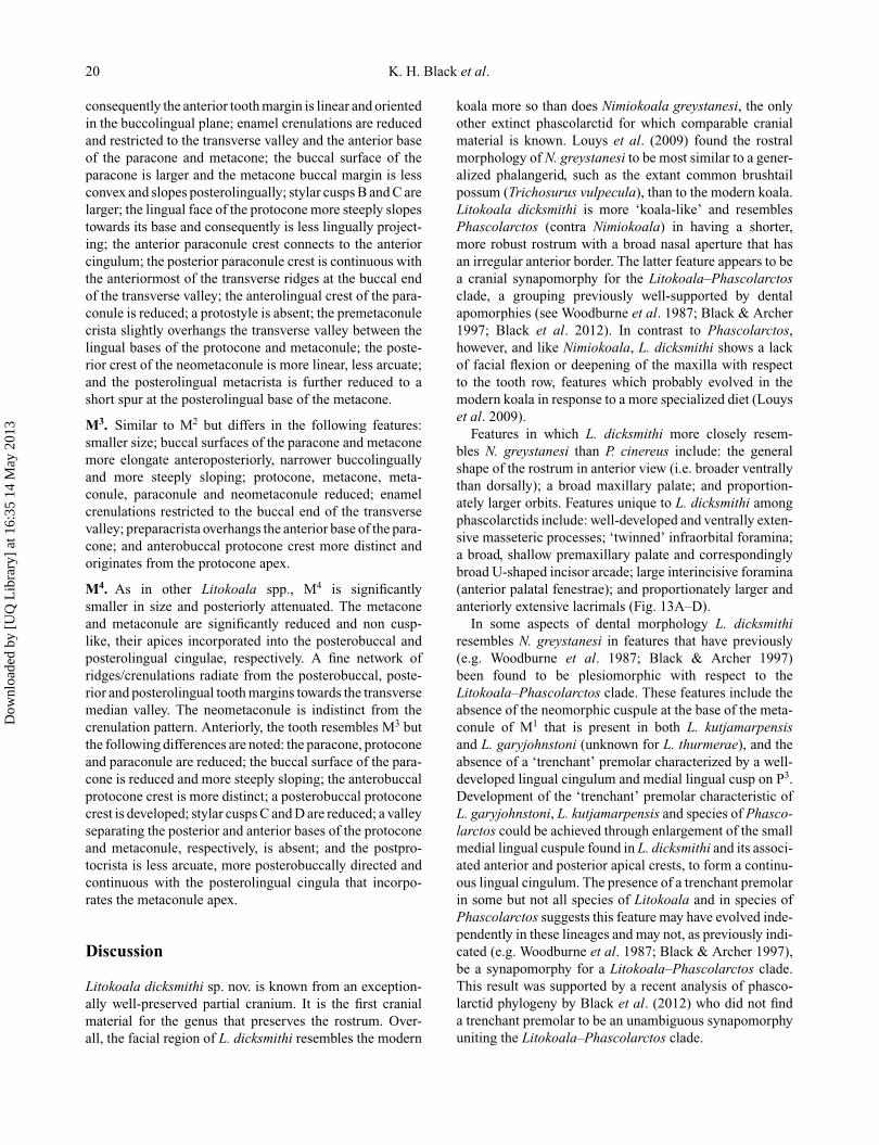
20 K. H. Black et al.
consequently the anterior tooth margin is linear and orientedin the buccolingual plane; enamel crenulations are reducedand restricted to the transverse valley and the anterior baseof the paracone and metacone; the buccal surface of theparacone is larger and the metacone buccal margin is lessconvex and slopes posterolingually; stylar cusps B and C arelarger; the lingual face of the protocone more steeply slopestowards its base and consequently is less lingually project-ing; the anterior paraconule crest connects to the anteriorcingulum; the posterior paraconule crest is continuous withthe anteriormost of the transverse ridges at the buccal endof the transverse valley; the anterolingual crest of the para-conule is reduced; a protostyle is absent; the premetaconulecrista slightly overhangs the transverse valley between thelingual bases of the protocone and metaconule; the poste-rior crest of the neometaconule is more linear, less arcuate;and the posterolingual metacrista is further reduced to ashort spur at the posterolingual base of the metacone.
M3. Similar to M2 but differs in the following features:smaller size; buccal surfaces of the paracone and metaconemore elongate anteroposteriorly, narrower buccolinguallyand more steeply sloping; protocone, metacone, meta-conule, paraconule and neometaconule reduced; enamelcrenulations restricted to the buccal end of the transversevalley; preparacrista overhangs the anterior base of the para-cone; and anterobuccal protocone crest more distinct andoriginates from the protocone apex.
M4. As in other Litokoala spp., M4 is significantlysmaller in size and posteriorly attenuated. The metaconeand metaconule are significantly reduced and non cusp-like, their apices incorporated into the posterobuccal andposterolingual cingulae, respectively. A fine network ofridges/crenulations radiate from the posterobuccal, poste-rior and posterolingual tooth margins towards the transversemedian valley. The neometaconule is indistinct from thecrenulation pattern. Anteriorly, the tooth resembles M3 butthe following differences are noted: the paracone, protoconeand paraconule are reduced; the buccal surface of the para-cone is reduced and more steeply sloping; the anterobuccalprotocone crest is more distinct; a posterobuccal protoconecrest is developed; stylar cusps C and D are reduced; a valleyseparating the posterior and anterior bases of the protoconeand metaconule, respectively, is absent; and the postpro-tocrista is less arcuate, more posterobuccally directed andcontinuous with the posterolingual cingula that incorpo-rates the metaconule apex.
Discussion
Litokoala dicksmithi sp. nov. is known from an exception-ally well-preserved partial cranium. It is the first cranialmaterial for the genus that preserves the rostrum. Over-all, the facial region of L. dicksmithi resembles the modern
koala more so than does Nimiokoala greystanesi, the onlyother extinct phascolarctid for which comparable cranialmaterial is known. Louys et al. (2009) found the rostralmorphology of N. greystanesi to be most similar to a gener-alized phalangerid, such as the extant common brushtailpossum (Trichosurus vulpecula), than to the modern koala.Litokoala dicksmithi is more ‘koala-like’ and resemblesPhascolarctos (contra Nimiokoala) in having a shorter,more robust rostrum with a broad nasal aperture that hasan irregular anterior border. The latter feature appears to bea cranial synapomorphy for the Litokoala–Phascolarctosclade, a grouping previously well-supported by dentalapomorphies (see Woodburne et al. 1987; Black & Archer1997; Black et al. 2012). In contrast to Phascolarctos,however, and like Nimiokoala, L. dicksmithi shows a lackof facial flexion or deepening of the maxilla with respectto the tooth row, features which probably evolved in themodern koala in response to a more specialized diet (Louyset al. 2009).
Features in which L. dicksmithi more closely resem-bles N. greystanesi than P. cinereus include: the generalshape of the rostrum in anterior view (i.e. broader ventrallythan dorsally); a broad maxillary palate; and proportion-ately larger orbits. Features unique to L. dicksmithi amongphascolarctids include: well-developed and ventrally exten-sive masseteric processes; ‘twinned’ infraorbital foramina;a broad, shallow premaxillary palate and correspondinglybroad U-shaped incisor arcade; large interincisive foramina(anterior palatal fenestrae); and proportionately larger andanteriorly extensive lacrimals (Fig. 13A–D).
In some aspects of dental morphology L. dicksmithiresembles N. greystanesi in features that have previously(e.g. Woodburne et al. 1987; Black & Archer 1997)been found to be plesiomorphic with respect to theLitokoala–Phascolarctos clade. These features include theabsence of the neomorphic cuspule at the base of the meta-conule of M1 that is present in both L. kutjamarpensisand L. garyjohnstoni (unknown for L. thurmerae), and theabsence of a ‘trenchant’ premolar characterized by a well-developed lingual cingulum and medial lingual cusp on P3.Development of the ‘trenchant’ premolar characteristic ofL. garyjohnstoni, L. kutjamarpensis and species of Phasco-larctos could be achieved through enlargement of the smallmedial lingual cuspule found in L. dicksmithi and its associ-ated anterior and posterior apical crests, to form a continu-ous lingual cingulum. The presence of a trenchant premolarin some but not all species of Litokoala and in species ofPhascolarctos suggests this feature may have evolved inde-pendently in these lineages and may not, as previously indi-cated (e.g. Woodburne et al. 1987; Black & Archer 1997),be a synapomorphy for a Litokoala–Phascolarctos clade.This result was supported by a recent analysis of phasco-larctid phylogeny by Black et al. (2012) who did not finda trenchant premolar to be an unambiguous synapomorphyuniting the Litokoala–Phascolarctos clade.
Dow
nloa
ded
by [
UQ
Lib
rary
] at
16:
35 1
4 M
ay 2
013
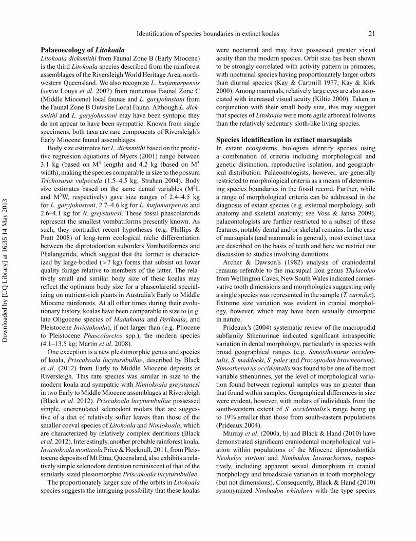
Identification of species boundaries in extinct koalas 21
Palaeoecology of LitokoalaLitokoala dicksmithi from Faunal Zone B (Early Miocene)is the third Litokoala species described from the rainforestassemblages of the Riversleigh World Heritage Area, north-western Queensland. We also recognize L. kutjamarpensis(sensu Louys et al. 2007) from numerous Faunal Zone C(Middle Miocene) local faunas and L. garyjohnstoni fromthe Faunal Zone B Outasite Local Fauna. Although L. dick-smithi and L. garyjohnstoni may have been syntopic theydo not appear to have been sympatric. Known from singlespecimens, both taxa are rare components of Riversleigh’sEarly Miocene faunal assemblages.
Body size estimates for L. dicksmithi based on the predic-tive regression equations of Myers (2001) range between3.1 kg (based on M3 length) and 4.2 kg (based on M3
width), making the species comparable in size to the possumTrichosurus vulpecula (1.5–4.5 kg; Strahan 2004). Bodysize estimates based on the same dental variables (M3Land M3W, respectively) gave size ranges of 2.4–4.5 kgfor L. garyjohnstoni, 2.7–4.6 kg for L. kutjamarpensis and2.6–4.1 kg for N. greystanesi. These fossil phascolarctidsrepresent the smallest vombatiforms presently known. Assuch, they contradict recent hypotheses (e.g. Phillips &Pratt 2008) of long-term ecological niche differentiationbetween the diprotodontian suborders Vombatiformes andPhalangerida, which suggest that the former is character-ized by large-bodied (>7 kg) forms that subsist on lowerquality forage relative to members of the latter. The rela-tively small and similar body size of these koalas mayreflect the optimum body size for a phascolarctid special-izing on nutrient-rich plants in Australia’s Early to MiddleMiocene rainforests. At all other times during their evolu-tionary history, koalas have been comparable in size to (e.g.late Oligocene species of Madakoala and Perikoala, andPleistocene Invictokoala), if not larger than (e.g. Plioceneto Pleistocene Phascolarctos spp.), the modern species(4.1–13.5 kg; Martin et al. 2008).
One exception is a new plesiomorphic genus and speciesof koala, Priscakoala lucyturnbullae, described by Blacket al. (2012) from Early to Middle Miocene deposits atRiversleigh. This rare species was similar in size to themodern koala and sympatric with Nimiokoala greystanesiin two Early to Middle Miocene assemblages at Riversleigh(Black et al. 2012). Priscakoala lucyturnbullae possessedsimple, uncrenulated selenodont molars that are sugges-tive of a diet of relatively softer leaves than those of thesmaller coeval species of Litokoala and Nimiokoala, whichare characterized by relatively complex dentitions (Blacket al. 2012). Interestingly, another probable rainforest koala,Invictokoala monticola Price & Hocknull, 2011, from Pleis-tocene deposits of Mt Etna, Queensland, also exhibits a rela-tively simple selenodont dentition reminiscent of that of thesimilarly sized plesiomorphic Priscakoala lucyturnbullae.
The proportionately larger size of the orbits in Litokoalaspecies suggests the intriguing possibility that these koalas
were nocturnal and may have possessed greater visualacuity than the modern species. Orbit size has been shownto be strongly correlated with activity pattern in primates,with nocturnal species having proportionately larger orbitsthan diurnal species (Kay & Cartmill 1977; Kay & Kirk2000). Among mammals, relatively large eyes are also asso-ciated with increased visual acuity (Kiltie 2000). Taken inconjunction with their small body size, this may suggestthat species of Litokoala were more agile arboreal folivoresthan the relatively sedentary sloth-like living species.
Species identification in extinct marsupialsIn extant ecosystems, biologists identify species usinga combination of criteria including morphological andgenetic distinction, reproductive isolation, and geograph-ical distribution. Palaeontologists, however, are generallyrestricted to morphological criteria as a means of determin-ing species boundaries in the fossil record. Further, whilea range of morphological criteria can be addressed in thediagnosis of extant species (e.g. external morphology, softanatomy and skeletal anatomy; see Voss & Jansa 2009),palaeontologists are further restricted to a subset of thesefeatures, notably dental and/or skeletal remains. In the caseof marsupials (and mammals in general), most extinct taxaare described on the basis of teeth and here we restrict ourdiscussion to studies involving dentitions.
Archer & Dawson’s (1982) analysis of craniodentalremains referable to the marsupial lion genus Thylacoleofrom Wellington Caves, New South Wales indicated conser-vative tooth dimensions and morphologies suggesting onlya single species was represented in the sample (T. carnifex).Extreme size variation was evident in cranial morphol-ogy, however, which may have been sexually dimorphicin nature.
Prideaux’s (2004) systematic review of the macropodidsubfamily Sthenurinae indicated significant intraspecificvariation in dental morphology, particularly in species withbroad geographical ranges (e.g. Simosthenurus occiden-talis, S. maddocki, S. pales and Procoptodon browneorum).Simosthenurus occidentalis was found to be one of the mostvariable sthenurines, yet the level of morphological varia-tion found between regional samples was no greater thanthat found within samples. Geographical differences in sizewere evident, however, with molars of individuals from thesouth-western extent of S. occidentalis’s range being upto 19% smaller than those from south-eastern populations(Prideaux 2004).
Murray et al. (2000a, b) and Black & Hand (2010) havedemonstrated significant craniodental morphological vari-ation within populations of the Miocene diprotodontidsNeohelos stirtoni and Nimbadon lavarackorum, respec-tively, including apparent sexual dimorphism in cranialmorphology and broadscale variation in tooth morphology(but not dimensions). Consequently, Black & Hand (2010)synonymized Nimbadon whitelawi with the type species
Dow
nloa
ded
by [
UQ
Lib
rary
] at
16:
35 1
4 M
ay 2
013
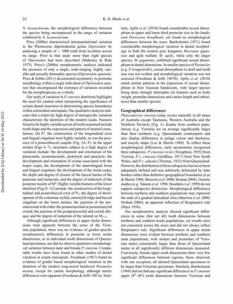
22 K. H. Black et al.
N. lavarackorum, the morphological differences betweenthe species being encompassed in the range of variationexhibited by N. lavarackorum.
Price (2008a) characterized interpopulational variationin the Pleistocene diprotodontid genus Diprotodon byanalysing a sample of > 1000 teeth from localities acrossits range. Prior to that study more than eight speciesof Diprotodon had been described (Mahoney & Ride1975). Price’s (2008a) morphometric analysis indicatedthe presence of only a single, wide-ranging, highly vari-able and sexually dimorphic species (Diprotodon optatum).Price & Sobbe (2011) documented asymmetry in premolarmorphology within a single individual of Diprotodon opta-tum that encompassed the extremes of variation recordedfor the morphospecies as a whole.
Our study of variation in P. cinereus dentitions highlightsthe need for caution when interpreting the significance ofcertain dental structures in determining species boundarieswithin selenodont marsupials. Our qualitative analysis indi-cates that a relatively high degree of intraspecific variationcharacterizes the dentition of the modern koala. Featuresfound to be highly variable across all teeth examined includetooth shape and the expression and pattern of enamel crenu-lations. On P3, the construction of the longitudinal crestand lingual cingulum were highly variable, as was the pres-ence of a posterobuccal cuspule (Fig. 3A–F). In the uppermolars (Figs 4–7), structures subject to a high degree ofvariation include: the development and orientation of theparaconule, neometaconule, protostyle and parastyle; thedevelopment and orientation of cristae associated with themajor cusps; the development of the anterolingual fossaand lingual cingulum; the development of the stylar cusps;the depth and degree of closure of the buccal basins of theparacone and metacone; and the degree of reduction of theposterior moiety of M4. Highly variable features of the lowerdentition (Figs 8–12) include: the construction of the longi-tudinal and posterobuccal crest of P3; the degree of devel-opment of the columnar stylids, entostylid ridge and buccalcingulum on the lower molars; the junction of the pre-entocristid with either the postmetacristid or postmetastylidcristid; the junction of the postprotocristid and cristid obli-qua; and the degree of reduction of the talonid on M3–4.
Although significant differences in upper molar dimen-sions were apparent between the sexes of the Victo-rian population, there was no evidence of gender-specificmorphometric differences in premolar or lower molardimensions, or in individual tooth dimensions of Queens-land specimens, nor did we observe qualitative morphologi-cal variation between male and female P. cinereus. Compa-rable results have been found in other studies of dentalvariation in extant marsupials. Freedman (1967) found noevidence of gender based morphological variation in thedentition of the modern long-nosed bandicoot Peramelesnasuta, except for canine morphology, although metricdifferences were apparent (Freedman & Joffe 1967a). Simi-
larly, Aplin et al. (2010) found considerable sexual dimor-phism in upper and lower third premolar size in the bandi-coot Peroryctes broadbenti, yet found no morphologicaldifferences between the sexes. Bartholomai (1971) foundconsiderable morphological variation in dental morphol-ogy in both the eastern grey kangaroo Macropus gigan-teus and agile wallaby M. agilis, while only the largerspecies, M. giganteus, exhibited significant sexual dimor-phism in dental dimensions. In smaller species of Perameles(e.g. P. bougainville), sexual dimorphism in skull and toothsize was not evident and morphological variation was notassessed (Freedman & Joffe 1967b). Aplin et al. (2010)noted similar patterns in the expression of sexual dimor-phism in New Guinean bandicoots, with larger speciesbeing more strongly dimorphic (in features such as bodyweight, premolar dimensions and canine length and robust-ness) than smaller species.
Geographical differencesPhascolarctos cinereus today occurs naturally in all statesof Australia except Tasmania, Western Australia and theNorthern Territory (Fig. 1). Koalas from southern popu-lations (e.g. Victoria) are on average significantly largerthan their northern (e.g. Queensland) counterparts andalso display differences in pelage colour and thickness,and muzzle shape (Lee & Martin 1988). To reflect thesemorphological differences, early taxonomists recognizedthree subspecies: P. cinereus victor (Troughton, 1935) fromVictoria; P. c. cinereus (Goldfuss, 1817) from New SouthWales; and P. c. adustus (Thomas, 1923) from Queensland.However, the distribution of these subspecies has never beenadequately defined and was arbitrarily delineated by stateborders rather than definitive geographical boundaries (Lee& Martin 1988; Sherwin et al. 2000; Strahan 2004). Geneticstudies (e.g. Takami et al. 1998; Houlden et al. 1999) do notsupport subspecies distinction. Morphological differencesbetween northern and southern populations may representthe ends of a gradual latitudinal cline (Sherwin et al. 2000;Strahan 2004); an apparent reflection of Bergmann’s rule(Mayr 1956).
Our morphometric analysis showed significant differ-ences in some (but not all) tooth dimensions betweennorthern and southern koala populations, yet results werenot consistent across the sexes and did not always reflectBergmann’s rule. Significant differences in upper molardimensions were evident between northern and southernmale populations, with molars and premolars of Victo-rian males consistently larger than those of Queenslandmales in all significantly different dimensions measured.Conversely, female upper tooth dimensions show very fewsignificant differences between regions; those observed,with one exception, all showed Queensland specimens tobe larger than Victorian specimens. Similarly, Bartholomai(1968) did not find any significant differences in P. cinereusupper (P3–M2) tooth dimensions between Victorian and
Dow
nloa
ded
by [
UQ
Lib
rary
] at
16:
35 1
4 M
ay 2
013
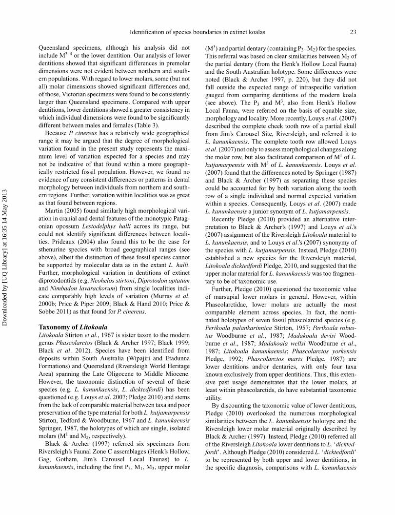
Identification of species boundaries in extinct koalas 23
Queensland specimens, although his analysis did notinclude M3–4 or the lower dentition. Our analysis of lowerdentitions showed that significant differences in premolardimensions were not evident between northern and south-ern populations. With regard to lower molars, some (but notall) molar dimensions showed significant differences and,of those, Victorian specimens were found to be consistentlylarger than Queensland specimens. Compared with upperdentitions, lower dentitions showed a greater consistency inwhich individual dimensions were found to be significantlydifferent between males and females (Table 3).
Because P. cinereus has a relatively wide geographicalrange it may be argued that the degree of morphologicalvariation found in the present study represents the maxi-mum level of variation expected for a species and maynot be indicative of that found within a more geograph-ically restricted fossil population. However, we found noevidence of any consistent differences or patterns in dentalmorphology between individuals from northern and south-ern regions. Further, variation within localities was as greatas that found between regions.
Martin (2005) found similarly high morphological vari-ation in cranial and dental features of the monotypic Patag-onian opossum Lestodelphys halli across its range, butcould not identify significant differences between locali-ties. Prideaux (2004) also found this to be the case forsthenurine species with broad geographical ranges (seeabove), albeit the distinction of these fossil species cannotbe supported by molecular data as in the extant L. halli.Further, morphological variation in dentitions of extinctdiprotodontids (e.g. Neohelos stirtoni, Diprotodon optatumand Nimbadon lavarackorum) from single localities indi-cate comparably high levels of variation (Murray et al.2000b; Price & Piper 2009; Black & Hand 2010; Price &Sobbe 2011) as that found for P. cinereus.
Taxonomy of LitokoalaLitokoala Stirton et al., 1967 is sister taxon to the moderngenus Phascolarctos (Black & Archer 1997; Black 1999;Black et al. 2012). Species have been identified fromdeposits within South Australia (Wipajiri and EtadunnaFormations) and Queensland (Riversleigh World HeritageArea) spanning the Late Oligocene to Middle Miocene.However, the taxonomic distinction of several of thesespecies (e.g. L. kanunkaensis, L. dicktedfordi) has beenquestioned (e.g. Louys et al. 2007; Pledge 2010) and stemsfrom the lack of comparable material between taxa and poorpreservation of the type material for both L. kutjamarpensisStirton, Tedford & Woodburne, 1967 and L. kanunkaensisSpringer, 1987, the holotypes of which are single, isolatedmolars (M1 and M2, respectively).
Black & Archer (1997) referred six specimens fromRiversleigh’s Faunal Zone C assemblages (Henk’s Hollow,Gag, Gotham, Jim’s Carousel Local Faunas) to L.kanunkaensis, including the first P3, M1, M3, upper molar
(M3) and partial dentary (containing P3–M2) for the species.This referral was based on clear similarities between M2 ofthe partial dentary (from the Henk’s Hollow Local Fauna)and the South Australian holotype. Some differences werenoted (Black & Archer 1997, p. 220), but they did notfall outside the expected range of intraspecific variationgauged from comparing dentitions of the modern koala(see above). The P3 and M3, also from Henk’s HollowLocal Fauna, were referred on the basis of equable size,morphology and locality. More recently, Louys et al. (2007)described the complete cheek tooth row of a partial skullfrom Jim’s Carousel Site, Riversleigh, and referred it toL. kanunkaensis. The complete tooth row allowed Louyset al. (2007) not only to assess morphological changes alongthe molar row, but also facilitated comparison of M1 of L.kutjamarpensis with M3 of L. kanunkaensis. Louys et al.(2007) found that the differences noted by Springer (1987)and Black & Archer (1997) as separating these speciescould be accounted for by both variation along the toothrow of a single individual and normal expected variationwithin a species. Consequently, Louys et al. (2007) madeL. kanunkaensis a junior synonym of L. kutjamarpensis.
Recently Pledge (2010) provided an alternative inter-pretation to Black & Archer’s (1997) and Louys et al.’s(2007) assignment of the Riversleigh Litokoala material toL. kanunkaensis, and to Louys et al.’s (2007) synonymy ofthe species with L. kutjamarpensis. Instead, Pledge (2010)established a new species for the Riversleigh material,Litokoala dicktedfordi Pledge, 2010, and suggested that theupper molar material for L. kanunkaensis was too fragmen-tary to be of taxonomic use.
Further, Pledge (2010) questioned the taxonomic valueof marsupial lower molars in general. However, withinPhascolarctidae, lower molars are actually the mostcomparable element across species. In fact, the nomi-nated holotypes of seven fossil phascolarctid species (e.g.Perikoala palankarinnica Stirton, 1957; Perikoala robus-tus Woodburne et al., 1987; Madakoala devisi Wood-burne et al., 1987; Madakoala wellsi Woodburne et al.,1987; Litokoala kanunkaensis; Phascolarctos yorkensisPledge, 1992; Phascolarctos maris Pledge, 1987) arelower dentitions and/or dentaries, with only four taxaknown exclusively from upper dentitions. Thus, this exten-sive past usage demonstrates that the lower molars, atleast within phascolarctids, do have substantial taxonomicutility.
By discounting the taxonomic value of lower dentitions,Pledge (2010) overlooked the numerous morphologicalsimilarities between the L. kanunkaensis holotype and theRiversleigh lower molar material originally described byBlack & Archer (1997). Instead, Pledge (2010) referred allof the Riversleigh Litokoala lower dentitions to L. ‘dickted-fordi’. Although Pledge (2010) considered L. ‘dicktedfordi’to be represented by both upper and lower dentitions, inthe specific diagnosis, comparisons with L. kanunkaensis
Dow
nloa
ded
by [
UQ
Lib
rary
] at
16:
35 1
4 M
ay 2
013
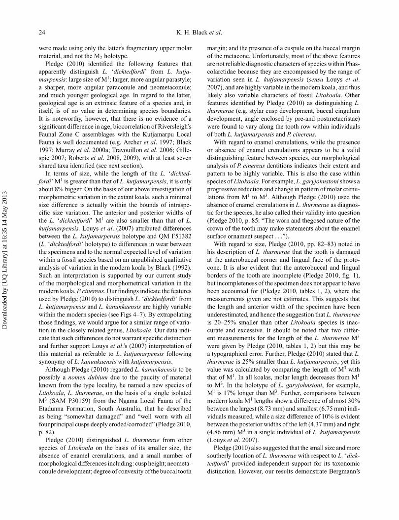
24 K. H. Black et al.
were made using only the latter’s fragmentary upper molarmaterial, and not the M2 holotype.
Pledge (2010) identified the following features thatapparently distinguish L. ‘dicktedfordi’ from L. kutja-marpensis: large size of M1; larger, more angular parastyle;a sharper, more angular paraconule and neometaconule;and much younger geological age. In regard to the latter,geological age is an extrinsic feature of a species and, initself, is of no value in determining species boundaries.It is noteworthy, however, that there is no evidence of asignificant difference in age; biocorrelation of Riversleigh’sFaunal Zone C assemblages with the Kutjamarpu LocalFauna is well documented (e.g. Archer et al. 1997; Black1997; Murray et al. 2000a; Travouillon et al. 2006; Gille-spie 2007; Roberts et al. 2008, 2009), with at least sevenshared taxa identified (see next section).
In terms of size, while the length of the L. ‘dickted-fordi’ M1 is greater than that of L. kutjamarpensis, it is onlyabout 8% bigger. On the basis of our above investigation ofmorphometric variation in the extant koala, such a minimalsize difference is actually within the bounds of intraspe-cific size variation. The anterior and posterior widths ofthe L. ‘dicktedfordi’ M1 are also smaller than that of L.kutjamarpensis. Louys et al. (2007) attributed differencesbetween the L. kutjamarpensis holotype and QM F51382(L. ‘dicktedfordi’ holotype) to differences in wear betweenthe specimens and to the normal expected level of variationwithin a fossil species based on an unpublished qualitativeanalysis of variation in the modern koala by Black (1992).Such an interpretation is supported by our current studyof the morphological and morphometrical variation in themodern koala, P. cinereus. Our findings indicate the featuresused by Pledge (2010) to distinguish L. ‘dicktedfordi’ fromL. kutjamarpensis and L. kanunkaensis are highly variablewithin the modern species (see Figs 4–7). By extrapolatingthose findings, we would argue for a similar range of varia-tion in the closely related genus, Litokoala. Our data indi-cate that such differences do not warrant specific distinctionand further support Louys et al.’s (2007) interpretation ofthis material as referable to L. kutjamarpensis followingsynonymy of L. kanunkaensis with kutjamarpensis.
Although Pledge (2010) regarded L. kanunkaensis to bepossibly a nomen dubium due to the paucity of materialknown from the type locality, he named a new species ofLitokoala, L. thurmerae, on the basis of a single isolatedM3 (SAM P30159) from the Ngama Local Fauna of theEtadunna Formation, South Australia, that he describedas being “somewhat damaged” and “well worn with allfour principal cusps deeply eroded/corroded” (Pledge 2010,p. 82).
Pledge (2010) distinguished L. thurmerae from otherspecies of Litokoala on the basis of its smaller size, theabsence of enamel crenulations, and a small number ofmorphological differences including: cusp height; neometa-conule development; degree of convexity of the buccal tooth
margin; and the presence of a cuspule on the buccal marginof the metacone. Unfortunately, most of the above featuresare not reliable diagnostic characters of species within Phas-colarctidae because they are encompassed by the range ofvariation seen in L. kutjamarpensis (sensu Louys et al.2007), and are highly variable in the modern koala, and thuslikely also variable characters of fossil Litokoala. Otherfeatures identified by Pledge (2010) as distinguishing L.thurmerae (e.g. stylar cusp development, buccal cingulumdevelopment, angle enclosed by pre-and postmetacristae)were found to vary along the tooth row within individualsof both L. kutjamarpensis and P. cinereus.
With regard to enamel crenulations, while the presenceor absence of enamel crenulations appears to be a validdistinguishing feature between species, our morphologicalanalysis of P. cinereus dentitions indicates their extent andpattern to be highly variable. This is also the case withinspecies of Litokoala. For example, L. garyjohnstoni shows aprogressive reduction and change in pattern of molar crenu-lations from M1 to M3. Although Pledge (2010) used theabsence of enamel crenulations in L. thurmerae as diagnos-tic for the species, he also called their validity into question(Pledge 2010, p. 85: “The worn and thegosed nature of thecrown of the tooth may make statements about the enamelsurface ornament suspect . . .”).
With regard to size, Pledge (2010, pp. 82–83) noted inhis description of L. thurmerae that the tooth is damagedat the anterobuccal corner and lingual face of the proto-cone. It is also evident that the anterobuccal and lingualborders of the tooth are incomplete (Pledge 2010, fig. 1),but incompleteness of the specimen does not appear to havebeen accounted for (Pledge 2010, tables 1, 2), where themeasurements given are not estimates. This suggests thatthe length and anterior width of the specimen have beenunderestimated, and hence the suggestion that L. thurmeraeis 20–25% smaller than other Litokoala species is inac-curate and excessive. It should be noted that two differ-ent measurements for the length of the L. thurmerae M3
were given by Pledge (2010, tables 1, 2) but this may bea typographical error. Further, Pledge (2010) stated that L.thurmerae is 25% smaller than L. kutjamarpensis, yet thisvalue was calculated by comparing the length of M3 withthat of M1. In all koalas, molar length decreases from M1
to M3. In the holotype of L. garyjohnstoni, for example,M1 is 17% longer than M3. Further, comparisons betweenmodern koala M1 lengths show a difference of almost 30%between the largest (8.73 mm) and smallest (6.75 mm) indi-viduals measured, while a size difference of 10% is evidentbetween the posterior widths of the left (4.37 mm) and right(4.86 mm) M3 in a single individual of L. kutjamarpensis(Louys et al. 2007).
Pledge (2010) also suggested that the small size and moresoutherly location of L. thurmerae with respect to L. ‘dick-tedfordi’ provided independent support for its taxonomicdistinction. However, our results demonstrate Bergmann’s
Dow
nloa
ded
by [
UQ
Lib
rary
] at
16:
35 1
4 M
ay 2
013
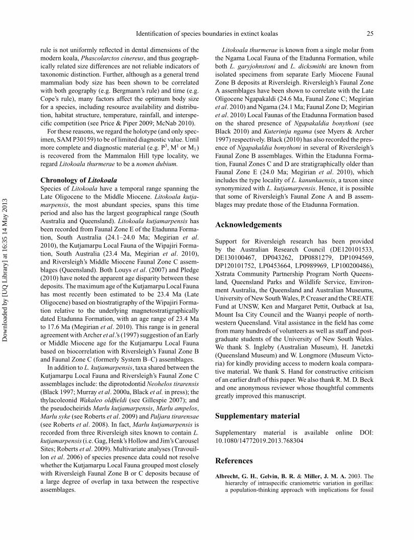
Identification of species boundaries in extinct koalas 25
rule is not uniformly reflected in dental dimensions of themodern koala, Phascolarctos cinereus, and thus geograph-ically related size differences are not reliable indicators oftaxonomic distinction. Further, although as a general trendmammalian body size has been shown to be correlatedwith both geography (e.g. Bergmann’s rule) and time (e.g.Cope’s rule), many factors affect the optimum body sizefor a species, including resource availability and distribu-tion, habitat structure, temperature, rainfall, and interspe-cific competition (see Price & Piper 2009; McNab 2010).
For these reasons, we regard the holotype (and only spec-imen, SAM P30159) to be of limited diagnostic value. Untilmore complete and diagnostic material (e.g. P3, M1 or M1)is recovered from the Mammalon Hill type locality, weregard Litokoala thurmerae to be a nomen dubium.
Chronology of LitokoalaSpecies of Litokoala have a temporal range spanning theLate Oligocene to the Middle Miocene. Litokoala kutja-marpensis, the most abundant species, spans this timeperiod and also has the largest geographical range (SouthAustralia and Queensland). Litokoala kutjamarpensis hasbeen recorded from Faunal Zone E of the Etadunna Forma-tion, South Australia (24.1–24.0 Ma; Megirian et al.2010), the Kutjamarpu Local Fauna of the Wipajiri Forma-tion, South Australia (23.4 Ma, Megirian et al. 2010),and Riversleigh’s Middle Miocene Faunal Zone C assem-blages (Queensland). Both Louys et al. (2007) and Pledge(2010) have noted the apparent age disparity between thesedeposits. The maximum age of the Kutjamarpu Local Faunahas most recently been estimated to be 23.4 Ma (LateOligocene) based on biostratigraphy of the Wipajiri Forma-tion relative to the underlying magnetostratigraphicallydated Etadunna Formation, with an age range of 23.4 Mato 17.6 Ma (Megirian et al. 2010). This range is in generalagreement with Archer et al.’s (1997) suggestion of an Earlyor Middle Miocene age for the Kutjamarpu Local Faunabased on biocorrelation with Riversleigh’s Faunal Zone Band Faunal Zone C (formerly System B–C) assemblages.
In addition to L. kutjamarpensis, taxa shared between theKutjamarpu Local Fauna and Riversleigh’s Faunal Zone Cassemblages include: the diprotodontid Neohelos tirarensis(Black 1997; Murray et al. 2000a, Black et al. in press); thethylacoleonid Wakaleo oldfieldi (see Gillespie 2007); andthe pseudocheirids Marlu kutjamarpensis, Marlu ampelos,Marlu syke (see Roberts et al. 2009) and Paljara tirarensae(see Roberts et al. 2008). In fact, Marlu kutjamarpensis isrecorded from three Riversleigh sites known to contain L.kutjamarpensis (i.e. Gag, Henk’s Hollow and Jim’s CarouselSites; Roberts et al. 2009). Multivariate analyses (Travouil-lon et al. 2006) of species presence data could not resolvewhether the Kutjamarpu Local Fauna grouped most closelywith Riversleigh Faunal Zone B or C deposits because ofa large degree of overlap in taxa between the respectiveassemblages.
Litokoala thurmerae is known from a single molar fromthe Ngama Local Fauna of the Etadunna Formation, whileboth L. garyjohnstoni and L. dicksmithi are known fromisolated specimens from separate Early Miocene FaunalZone B deposits at Riversleigh. Riversleigh’s Faunal ZoneA assemblages have been shown to correlate with the LateOligocene Ngapakaldi (24.6 Ma, Faunal Zone C; Megirianet al. 2010) and Ngama (24.1 Ma; Faunal Zone D; Megirianet al. 2010) Local Faunas of the Etadunna Formation basedon the shared presence of Ngapakaldia bonythoni (seeBlack 2010) and Kuterintja ngama (see Myers & Archer1997) respectively. Black (2010) has also recorded the pres-ence of Ngapakaldia bonythoni in several of Riversleigh’sFaunal Zone B assemblages. Within the Etadunna Forma-tion, Faunal Zones C and D are stratigraphically older thanFaunal Zone E (24.0 Ma; Megirian et al. 2010), whichincludes the type locality of L. kanunkaensis, a taxon sincesynonymized with L. kutjamarpensis. Hence, it is possiblethat some of Riversleigh’s Faunal Zone A and B assem-blages may predate those of the Etadunna Formation.
Acknowledgements
Support for Riversleigh research has been providedby the Australian Research Council (DE120101533,DE130100467, DP043262, DP0881279, DP1094569,DP120101752, LP0453664, LP0989969, LP100200486),Xstrata Community Partnership Program North Queens-land, Queensland Parks and Wildlife Service, Environ-ment Australia, the Queensland and Australian Museums,University of New South Wales, P. Creaser and the CREATEFund at UNSW, Ken and Margaret Pettit, Outback at Isa,Mount Isa City Council and the Waanyi people of north-western Queensland. Vital assistance in the field has comefrom many hundreds of volunteers as well as staff and post-graduate students of the University of New South Wales.We thank S. Ingleby (Australian Museum), H. Janetzki(Queensland Museum) and W. Longmore (Museum Victo-ria) for kindly providing access to modern koala compara-tive material. We thank S. Hand for constructive criticismof an earlier draft of this paper. We also thank R. M. D. Beckand one anonymous reviewer whose thoughtful commentsgreatly improved this manuscript.
Supplementary material
Supplementary material is available online DOI:10.1080/14772019.2013.768304
References
Albrecht, G. H., Gelvin, B. R. & Miller, J. M. A. 2003. Thehierarchy of intraspecific craniometric variation in gorillas:a population-thinking approach with implications for fossil
Dow
nloa
ded
by [
UQ
Lib
rary
] at
16:
35 1
4 M
ay 2
013
26 K. H. Black et al.
species recognition studies. Pp. 62–103 in A. B. Taylor &M. L. Goldsmith (eds) Gorilla biology: a multidisciplinaryperspective. Cambridge University Press, Cambridge.
Aplin, K. P. & Archer, M. 1987. Recent advances in marsupialsystematics with a new syncretic classification. Pp. xv–lxxiiin M. Archer (ed.) Possums and Opossums: Studies in Evolu-tion. Surrey Beatty & Sons, Chipping Norton, New SouthWales.
Aplin, K. P., Helgen, K. M. & Lunde, D. P. 2010. A reviewof Peroryctes broadbenti, the giant bandicoot of Papua NewGuinea. American Museum Novitates, 3696, 1–41.
Archer, M. 1975. Abnormal dental development and its signif-icance in dasyurids and other marsupials. Memoirs of theQueensland Museum, 17, 251–265.
Archer, M. 1978. Koalas (phascolarctids) and their significance inmarsupial evolution. Pp. 20–28 in T. Bergin (ed.) The Koala.Zoological Parks Board of New South Wales, Sydney.
Archer, M. & Dawson, L. 1982. Revision of marsupial lionsof the genus Thylacoleo Gervais (Thylacoleonidae: Marsupi-alia) and thylacoleonid evolution in the late Cainozoic. Pp.477–494 in M. Archer (ed.) Carnivorous Marsupials. RoyalZoological Society of New South Wales, Sydney.
Archer, M., Hand, S. J., Godthelp, H. & Creaser, P. 1997.Correlation of the Cainozoic sediments of the RiversleighWorld Heritage Fossil Property, Queensland, Australia. Pp.131–152 in J.-P. Aguilar, S. Legendre & J. Michaux (eds) Actesdu Congres BiocroM’97. Memoirs et Traveaux Ecole Pratiquedes Hautes Etudes, Institut de Montpellier, Montpellier.
Arena, D. A. 2005. The geological history and development ofthe terrain at the Riversleigh World Heritage Area during themiddle Tertiary. Unpublished PhD thesis, The University ofNew South Wales. 275 pp.
Bartholomai, A. 1968. A new fossil koala from Queensland anda reassessment of the taxonomic position of the problematicalspecies Koalemus ingens De Vis. Memoirs of the QueenslandMuseum, 15, 65–73.
Bartholomai, A. 1971. Morphology and variation of the cheekteeth in Macropus giganteus Shaw and Macropus agilis(Gould). Memoirs of the Queensland Museum, 16, 1–18.
Black, K. 1992. Systematics and palaeobiology of fossil phasco-larctids from Riversleigh. Unpublished Honours thesis, TheUniversity of New South Wales, 63 pp.
Black, K. 1997. Diversity and biostratigraphy of the Diprotodon-toidea of Riversleigh, northwestern Queensland. Memoirs ofthe Queensland Museum, 41, 187–192.
Black, K. 1999. Diversity and relationships of living and extinctkoalas (Phascolarctidae, Marsupialia). Australian Mammal-ogy, 21, 16–17, 34–45.
Black, K. H. 2010. Ngapakaldia bonythoni (Marsupialia,Diprotodontidae): new material from Riversleigh, northwest-ern Queensland, and a reassessment of the genus Bemath-erium. Alcheringa, 34, 471–492.
Black, K. & Archer, M. 1997. Nimiokoala gen. nov. (Marsupialia,Phascolarctidae) from Riversleigh, northwestern Queensland,with a revision of Litokoala. Memoirs of the QueenslandMuseum, 41, 209–228.
Black, K. H. & Hand, S. J. 2010. First crania and assessment ofspecies boundaries in Nimbadon (Marsupialia: Diprotodon-tidae) from the Middle Miocene of Australia. AmericanMuseum Novitates, 3678, 1–60.
Black, K. H., Archer, M. & Hand, S. J. 2012. New Tertiary koala(Marsupialia, Phascolarctidae) from Riversleigh, Australiawith a revision of phascolarctid phylogenetics, paleoecologyand paleobiodiversity. Journal of Vertebrate Paleontology, 32,125–138.
Black, K. H., Archer, M., Hand, S. J., Godthelp, H. Inpress. Revision in the marsupial diprotodontid genus Neoh-elos: systematics and biostratigraphy. Acta PalaeontologicaPolonica.
Blainville, H. M. D. de 1816. Prodrome d’une nouvelle distribu-tion systematique du regne animal. Bulletin des sciences parla Societe philomathique de Paris, 1816, 113–124.
Creaser, P. 1997. Oligocene-Miocene sediments of Riversleigh:the potential significance of topography. Memoirs of theQueensland Museum, 41, 303–314.
Easton, L. C. 2006. Pleistocene grey kangaroos from the fossilchamber of Victoria Fossil Cave, Naracoorte, South Australia.Transactions of the Royal Society of South Australia, 130,17–28.
Freedman, L. 1967. Skull and tooth variation in the genusPerameles. Part I: anatomical features. Records of theAustralian Museum, 27, 147–165.
Freedman, L. & Joffe, A. D. 1967a. Skull and tooth variation inthe genus Perameles. Part 2: metrical features of P. nasuta.Records of the Australian Museum, 27, 183–195.
Freedman, L. & Joffe, A. D. 1967b. Skull and tooth variationin the genus Perameles. Part 3: metrical features of P. gunniiand P. bougainville. Records of the Australian Museum, 27,197–212.
Gillespie, A. K. 2007. Diversity and systematics of marsupiallions from the Riversleigh World Heritage Area and the evolu-tion of the Thylacoleonidae. Unpublished PhD thesis, TheUniversity of New South Wales, 394 pp.
Goldfuss, G. A. 1817. In J. C. D. von Schreber (ed.) DieSaugethiere Abbildungen nach der Natur, mit Beschreibun-gen. Fortgesetzt von A. Goldfuss. 65e cahier. Walther, Erlan-gen, Germany.
Gould, S. J. 1975. On the scaling of tooth size in mammals.American Zoologist, 15, 353–362.
Hammer, Ø., Harper, D. A. T. & Ryan, P. D. 2006. PAST: Pale-ontological Statistics Software Package for Education andData Analysis. Version 1.51.
Houlden, B. A., Costello, B. H., Sharkey, D., Fowler, E. V.,Melzer, A., Ellis, W., Carrick, F., Baverstock, P. R. &Elphinstone, M. S. 1999. Phylogeographic differentiationin the mitochondrial control region in the koala, Phasco-larctos cinereus (Goldfuss 1817). Molecular Ecology, 8,999–1011.
Illiger, C. 1811. Prodromus systematis mammalium et aviumadditis terminis zoographicis utriudque classis. C. Salfeld,Berlin, xviii+301 pp.
Kay, R. F. & Cartmill, M., 1977. Cranial morphology and adap-tation of Palaechthon nacimienti and other Paraomomyidae(Plesiadapoidea? Primates), with a description of a new genusand species. Journal of Human Evolution, 6, 19–35.
Kay, R. F. & Kirk, E. C. 2000. Osteological evidence for theevolution of activity pattern and visual acuity in primates.American Journal of Physical Anthropology, 113, 235–262.
Kiltie, R. A. 2000. Scaling of visual acuity with body size inmammals and birds. Functional Ecology, 14, 226–234.
Lanyon, J. M. & Sanson, G. D. 1986. Koala (Phascolarctoscinereus) dentition and nutrition. I. Morphology and occlusionof cheekteeth. Journal of Zoology, 209, 155–168.
Lee, A. & Martin, R. 1988. The koala, a natural history. NewSouth Wales University Press, Kensington, New South Wales.
Louys, J., Black, K., Archer, M., Hand, S. J. & Godthelp,H. 2007. Descriptions of koala fossils from the Mioceneof Riversleigh, northwestern Queensland and implicationsfor Litokoala (Marsupialia, Phascolarctidae). Alcheringa, 31,99–110.
Dow
nloa
ded
by [
UQ
Lib
rary
] at
16:
35 1
4 M
ay 2
013
Identification of species boundaries in extinct koalas 27
Louys, J., Aplin, K., Beck, R. M. D. & Archer, M. 2009.Cranial anatomy of Oligo-Miocene koalas (Diprotodontia:Phascolarctidae): stages in the evolution of an extreme leaf-eating specialization. Journal of Vertebrate Paleontology, 29,981–992.
Luckett, W. P. 1993. An ontogenetic assessment of dentalhomologies in therian mammals. Pp. 182–204 in F. S. Szalay,M. J. Novacek & M. C. McKenna (eds) Mammal Phylogeny:Mesozoic differentiation, multituberculates, monotremes,early therians and marsupials. Springer-Verlag, New York.
Mahoney, J. A. & Ride, W. D. 1975. Index to the genera andspecies of fossil Mammalia described from Australia andNew Guinea between 1838 and 1968, including citations oftype species and primary type specimens. Western AustralianMuseum Special Publication, 6, 1–250.
Martin, G. M. 2005. Intraspecific variation in Lestodelphys halli(Marsupialia: Didelphimorphia). Journal of Mammalogy, 86,793–802.
Martin, R. W., Handasyde, K. A. & Krockenberger, A. 2008.Koala. Pp. 198–201 in S. van Dyke & R. Strahan (eds) TheMammals of Australia. Reed New Holland, Sydney, NewSouth Wales.
Mayr, E. 1956. Geographical character gradients and climaticadaptation. Evolution, 10, 105–108.
McNab, B. 2010. Geographic and temporal correlations ofmammalian size reconsidered: a resource rule. Oecologia,164, 13–23.
Megirian, D., Prideaux, G. J., Murray, P. F. & Smit, N. 2010.An Australian land mammal age biochronological scheme.Paleobiology, 36, 658–671.
Meiri, S. & Dayan, T. 2003. On the validity of Bergmann’s rule.Journal of Biogeography, 30, 331–351.
Murray, P., Megirian, D., Rich, T., Plane, M., Black, K.,Archer, M., Hand, S. & Vickers-Rich, P. 2000a. Morphol-ogy, systematics, and evolution of the marsupial genus Neohe-los Stirton (Diprotodontidae, Zygomaturinae). Museums andArt Galleries of the Northern Territory Research Report No.6, 1–141.
Murray, P., Megirian, D., Rich, T., Plane, M. & Vickers-Rich,P. 2000b. Neohelos stirtoni, a new species of Zygomaturi-nae (Diprotodontidae: Marsupialia) from the mid-Tertiary ofnorthern Australia. Memoirs of the Queen Victoria Museum,105, 1–47.
Myers, T. J. 2001. Prediction of marsupial body mass. AustralianJournal of Zoology, 49, 99–118.
Myers, T. & Archer, M. 1997. Kuterintja ngama (Marsupialia,Ilariidae): a revised systematic analysis based on materialfrom the late Oligocene of Riversleigh, northwestern Queens-land. Memoirs of the Queensland Museum, 41, 379–392.
Owen, R. 1839. Outlines of a classification of the Marsupialia.Proceedings of the Zoological Society of London, 7, 5–19.
Owen, R. 1866. On the Anatomy of Vertebrates. Longmans Green,London.
Phillips, M. J. & Pratt, R. C. 2008. Family-level relationshipsamong the Australasian marsupial “herbivores” (Diprotodon-tia: Koala, wombats, kangaroos and possums). MolecularPhylogenetics and Evolution, 46, 594–605.
Pledge, N. S. 1987. Phascolarctos maris, a new species of koala(Marsupialia: Phascolarctidae) from the early Pliocene ofSouth Australia. Pp. 327–330 in M. Archer (ed.) Possumsand Opossums: Studies in Evolution. Surrey Beatty & Sons,Chipping Norton, New South Wales.
Pledge, N. S. 1992. The Curramulka Local Fauna: a newlate Tertiary fossil assemblage from Yorke Peninsula, SouthAustralia. The Beagle, 9, 115–142.
Pledge, N. S. 2010. A new koala (Marsupialia: Phascolarcti-dae) from the late Oligocene Etadunna Formation, Lake EyreBasin, South Australia. Australian Mammalogy, 32, 79–86.
Price, G. J. 2002. Perameles sobbei sp. nov. (Marsupialia,Peramelidae), a Pleistocene bandicoot from the DarlingDowns, south-eastern Queensland. Memoirs of the Queens-land Museum, 48, 193–197.
Price, G. J. 2005. Fossil bandicoots (Marsupialia, Perameli-dae) and environmental change during the Pleistocene on theDarling Downs, southeastern Queensland, Australia. Journalof Systematic Palaeontology, 2, 347–356.
Price, G. J. 2008a. Taxonomy and palaeobiology of the largest-ever marsupial, Diprotodon Owen, 1838 (Diprotodontidae,Marsupialia). Zoological Journal of the Linnean Society, 153,369–397.
Price, G. J. 2008b. Is the modern koala (Phascolarctos cinereus)a derived dwarf of a Pleistocene giant? Implications for test-ing megafauna extinction hypotheses. Quaternary ScienceReviews, 27, 2516–2521.
Price, G. J. & Hocknull, S. A. 2011. Invictokoala monticolagen. et sp. nov. (Phascolarctidae, Marsupialia), a Pleistoceneplesiomorphic koala holdover from Oligocene ancestors.Journal of Systematic Palaeontology, 9, 327–335.
Price, G. J. & Piper, K. J. 2009. Gigantism of the AustralianDiprotodon Owen 1838 (Marsupialia, Diprotodontoidea)through the Pleistocene. Journal of Quaternary Science, 24,1029–1038.
Price, G. J. & Sobbe, I. H. 2011. Morphological variation withinan individual Pleistocene Diprotodon optatum Owen, 1838(Diprotodontinae; Marsupialia): implications for taxonomywithin diprotodontoids. Alcheringa, 35, 21–29.
Price, G. J., Zhao, J.-x., Feng, Y.-x. & Hocknull, S. A. 2009. Newrecords of Plio-Pleistocene koalas from Australia: palaeoeco-logical and taxonomic implications. Records of the AustralianMuseum, 61, 39–4
Prideaux, G. J. 2004. Systematics and evolution of the sthenurinekangaroos. University of California Publications in Geologi-cal Sciences, 146, 1–622.
Roberts, K. K., Bassarova, M. & Archer, M. 2008. Oligo-Miocene ringtail possums of the genus Paljara (Pseudocheiri-dae: Marsupialia) from Queensland, Australia. Geobios, 41,833–844.
Roberts, K. K., Archer, M., Hand, S. J. & Godthelp, H.2009. New Australian Oligocene to Miocene ringtail possums(Pseudocheiridae) and revision of the genus Marlu. Palaeon-tology, 52, 441–456.
Sherwin, W. B., Timms, P., Wilcken, J. & Houlden, B. 2000.Analysis and conservation implications of koala genetics.Conservation Biology, 14, 639–649.
Springer, M. S. 1987. Lower molars of Litokoala (Marsupialia:Phascolarctidae) and their bearing on phascolarctid evolu-tion. Pp. 319–325 in M. Archer (ed.) Possums and Opossums:Studies in Evolution. Surrey Beatty & Sons, Chipping Norton,New South Wales.
Stirton, R. A. 1957. A new koala from the Pliocene Palankarinnafauna of South Australia. Records of the South AustralianMuseum, 13, 71–81.
Stirton, R. A., Tedford, R. H. & Woodburne, M. O. 1967.A new Tertiary formation and fauna from the Tirari Desert,South Australia. Records of the South Australian Museum, 15,427–462.
Strahan, R. 2004. The Mammals of Australia. New HollandPublishers, Sydney, 756 pp.
Takami, K., Yoshida, M., Yamamoto, Y., Harada, M. &Furuyama, J. 1998. Genetic variation of mitochondrial
Dow
nloa
ded
by [
UQ
Lib
rary
] at
16:
35 1
4 M
ay 2
013
28 K. H. Black et al.
cytochrome b genes among the subspecies of koala, Phas-colarctos cinereus. Journal of Veterinary Medical Science,60, 1161–1163.
Tedford, R. H. & Woodburne, M. O. 1987. The Ilariidae, a newfamily of vombatiform marsupials from Miocene strata ofSouth Australia and an evaluation of the homology of molarcusps in the Diprotodontia. Pp. 401–418 in M. Archer (ed.)Possums and Opossums: Studies in Evolution. Surrey Beatty& Sons, Chipping Norton, New South Wales.
Thomas, O. 1923. On some Queensland Phalangeridae. Annalsand Magazine of Natural History, 9, 246–250.
Travouillon, K. J., Archer, M., Hand, S. J. & Godthelp, H.2006. Multivariate analyses of Cenozoic mammalian faunasfrom Riversleigh, northwestern Queensland. Alcheringa, 30(2supp 1), 323–349.
Troughton, E. & Le, G. 1935. Phascolarctos cinereus Victor.Australian Naturalist, 9, 139.
Voss, R. S. & Jansa, S. A. 2009. Phylogenetic relationships andclassification of didelphid marsupials, an extant radiation of
New World metatherian mammals. Bulletin of the AmericanMuseum of Natural History, 322, 1–177.
Woodburne, M. O. 1984. Families of marsupials: relationships,evolution and biogeography. Pp. 48–71 in T. W. Broadhead(ed.) Mammals: Notes for a Short Course. University ofTennessee Department of Geological Sciences, Studies inGeology, Tennessee.
Woodburne, M. O., Tedford, R. H., Archer, M. & Pledge,N. S. 1987. Madakoala, a new genus and two species ofMiocene koalas (Marsupialia: Phascolarctidae) from SouthAustralia and a new species of Perikoala. Pp. 293–317 inM. Archer (ed.) Possums and Opossums: Studies in Evolu-tion. Surrey Beatty & Sons, Chipping Norton, New SouthWales.
Woodburne, M. O., MacFadden, B. J., Case, J. A., Springer,M. S. & Springer, K. B. 1993. Land Mammal Biostratig-raphy and Magnetostratigraphy of the Etadunna Formation(Late Oligocene) of South Australia. Journal of VertebratePaleontology, 13, 483–515.
Dow
nloa
ded
by [
UQ
Lib
rary
] at
16:
35 1
4 M
ay 2
013