universidade federal de goiÁs faculdade de medicina ...€¦ · biodentine® e bio-c repair® e...
TRANSCRIPT
UNIVERSIDADE FEDERAL DE GOIÁS FACULDADE DE MEDICINA
PROGRAMA DE PÓS-GRADUAÇÃO EM CIÊNCIAS DA SAÚDE
SARA RODRIGUES RENOVATO
Adaptação marginal de cimentos à base de silicato de cálcio à
parede dentinária de cavidades retrógadas
Goiânia
2018

SARA RODRIGUES RENOVATO
Adaptação marginal de cimentos à base de silicato de cálcio
à parede dentinária de cavidades retrógadas
Tese de Doutorado apresentada ao Programa de Pós-Graduação em Ciências da Saúde da Universidade Federal de Goiás para obtenção do título de Doutor em Ciências da Saúde
Orientador: Prof. Dr. Carlos Estrela Co-orientador: Prof. Dr. Julio Almeida Silva

Goiâ


Programa de Pós-Graduação em Ciências da Saúde
da Universidade Federal de Goiás
BANCA EXAMINADORA DA TESE DE DOUTORADO
Aluno(a): Sara Rodrigues Renovato
Orientador(a): Prof. Dr. Carlos Estrela
Membros:
1. Prof. Dr. Carlos Estrela (Presidente)
2. Profa. Dra. Ana Helena Gonçalves de Alencar
3. Prof. Dr. Daniel de Almeida Decurcio
4. Prof. Dr. Jesus Djalma Pécora
5. Profa. Dra. Patrícia Correia de Siqueira
Suplentes:
1. Prof. Dr. Julio Almeida Silva
2. Prof. Dr. João Batista de Souza
Data: 05/07/18
DEDICATÓRIA
Dedico este trabalho a minha pequena Luísa,
que ainda no ventre tem transformado meu
mundo e me ensinado sobre amor incondicional.
A ela, todo amor e dedicação.
E ao meu esposo, Rafael, que me impulsiona e
me ampara. Meu companheiro em todos os
momentos.

AGRADECIMENTOS
Meus eternos agradecimentos.
Primeiramente a Deus, por me permitir chegar até aqui. Por ouvir,
abençoar, guardar e cuidar. Por estar sempre ao meu lado. “ Em seu coração
o homem planeja seu caminho, mas o Senhor determina os seus passos”
(Provérbio 16:9)
Aos meus pais, Regina e José, meus exemplos e fonte de coragem.
Meus grandes mestres, que me ensinaram os maiores valores. Obrigada por
me formarem na escola mais importante da vida e serem meu alicerce em
todos os momentos. Vocês são meu orgulho e eu espero um dia retribuir tudo
o que fizeram por mim. Eu sonho em ser para Luísa exatamente o que vocês
são para mim. Obrigada por serem os melhores.
Ao meu esposo, Rafael, que me ampara em todos os momentos, que
vive comigo as lutas e desafios diários. Juntos construímos um lar e hoje
esperamos ansiosamente pelo nosso maior presente: nossa filha. Sua
dedicação me inspira e me move. Obrigada por ter paciência nas turbulências
e por comemorar comigo cada degrau que conquistamos. De mãos dadas a
caminhada é leve.
À minha família, minha base, minha fonte de amor inesgotável.
Obrigada pelo constante incentivo e por serem o meu refúgio. Amo cada um
de vocês.
Ao professor Carlos Estrela, exemplo de ser humano e educador.
Obrigada por todas as oportunidades e orientações onde pude aprender não
somente ciência, mas ensinamentos para a vida, que nos incentivam,
desafiam e, acima de tudo, nos tornam pessoas melhores, É uma honra e
privilégio poder conviver com o senhor. Obrigada por me ajudar a ser a
pessoa/profissional que sou hoje.
À professora Ana Helena Gonçalves de Alencar, meu muitíssimo
obrigada. Palavras nunca serão suficientes para descrever minha admiração,
carinho e gratidão. Exímia educadora e fonte de inspiração. Obrigada por
estar sempre disponível para ajudar e ensinar. O convívio com a senhora me
oportunizou momentos de ensinamentos valiosos e aprendizados únicos.
Aos professores Julio Almeida Silva e Daniel de Almeida Decurcio,
docentes que muito admiro e que tanto me auxiliaram desde a graduação.
Obrigada pela convivência, ensinamentos, apoio e incentivo.
À professora, amiga e quase irmã, Patrícia Correia de Siqueira,
companheira de caminhada. Compartilhamos tantos momentos pessoais e
profissionais que neste não poderia ser diferente. Tê-la na banca de defesa
enche meu coração de alegria. Obrigada pela amizade.
Ao professor Jesus Djalma Pécora, é uma honra tê-lo na banca de
defesa. Obrigada pelo aceite do convite e por contribuir com sua experiência
e sabedoria.
Ao professor João Batista de Souza, obrigada por tantos ensinamentos
e pelas palavras de apoio que fizeram diferença.
Ao professor Fernando Barletta que nos disponibilizou o Biodentine,
obrigada pela contribuição com o trabalho
A todos os colegas de pós-graduação, Alexandre, Alessandra,
Alessandro, Caixeta, Denise, Iury, Juliano, Giulliano, Mamede, Gabriela,
Olavo, Gustavo, Lorena, Luiz Eduardo, Luma, Marcela, Marco Antônio,
Marcus Vinícius, Matheus, Paulo Otávio pela parceria nessa jornada. Um
agradecimento especial à Keila e Mônica, amigas que Deus colocou em meu
caminho, que não medem esforços para sempre ajudar. Tenho uma
admiração enorme por vocês.
Ao CRTI, na pessoa do Sérgio Paulo, pela gentileza e pelo auxílio na
execução da etapa experimental da pesquisa.
À Faculdade de Odontologia da UFG, que me acolheu e vem sendo
minha segunda casa desde a graduação. Agradeço a todos os professores
pela contribuição na minha formação, aos colegas TAES pela colaboração
constante, e todos os demais funcionários pelo cuidado, preocupação e
auxílio sempre.
Aos amigos Gabriella, Leuçon, Elisângela e Karitta que contribuíram
diretamente na execução experimental do trabalho. Obrigada pelo apoio e
pelo convívio diário. É um prazer trabalhar com vocês.
Ao Programa de Pós-Graduação em Ciências da Saúde, junto à
Faculdade de Medicina da UFG, pela oportunidade e apoio necessários para
que esta etapa fosse alcançada.
À Capes, pelos recursos financeiros concedidos na forma de bolsa de
estudo de Doutorado.
Aos amigos que estão sempre ao meu lado.
“Talvez não tenha conseguido fazer o melhor, mas lutei para que o melhor fosse feito. Não sou o que deveria ser, mas Graças a Deus, não sou o que era antes”
Marthin Luther King
RESUMO
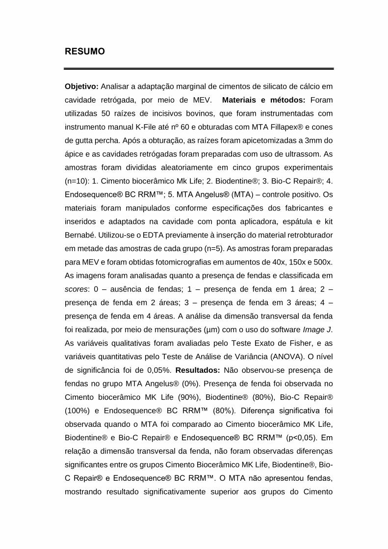
Objetivo: Analisar a adaptação marginal de cimentos de silicato de cálcio em
cavidade retrógada, por meio de MEV. Materiais e métodos: Foram
utilizadas 50 raízes de incisivos bovinos, que foram instrumentadas com
instrumento manual K-File até nº 60 e obturadas com MTA Fillapex® e cones
de gutta percha. Após a obturação, as raízes foram apicetomizadas a 3mm do
ápice e as cavidades retrógadas foram preparadas com uso de ultrassom. As
amostras foram divididas aleatoriamente em cinco grupos experimentais
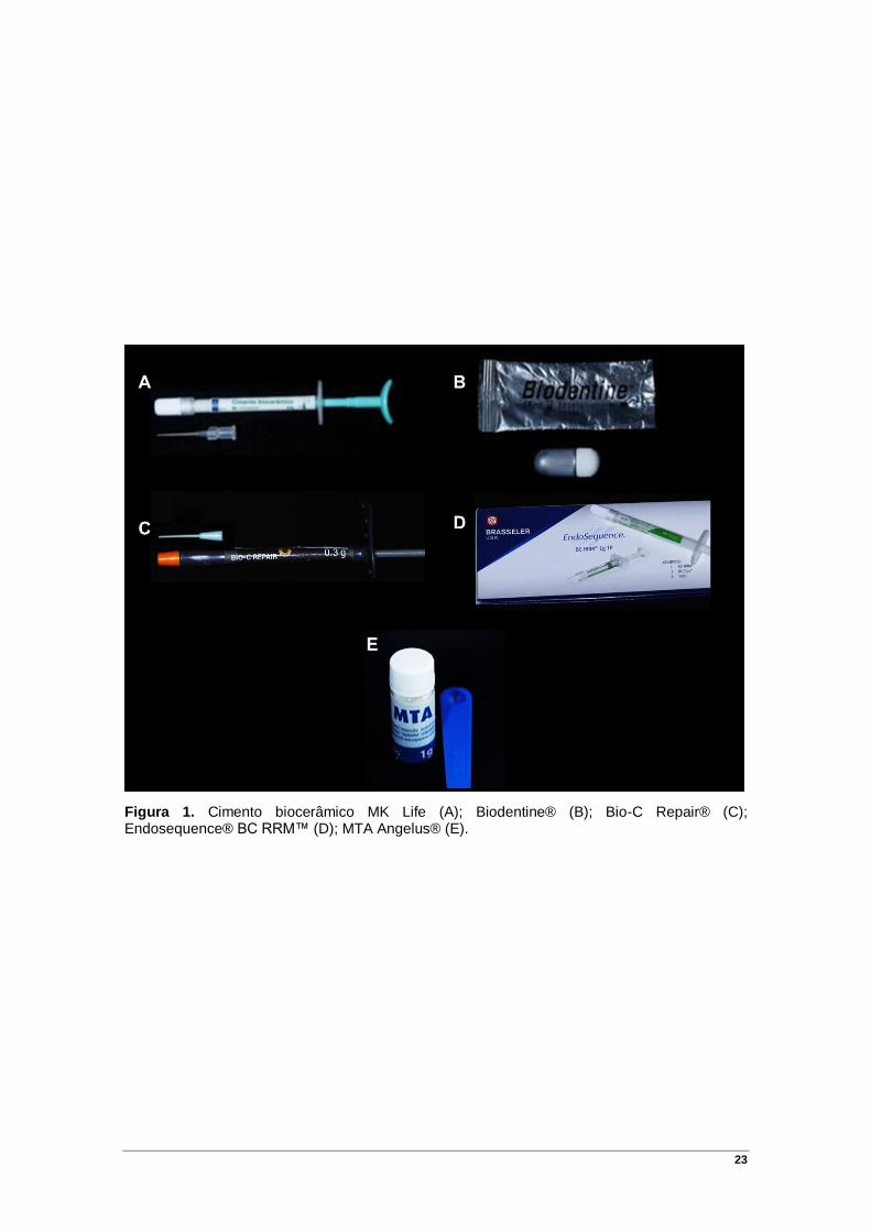
(n=10): 1. Cimento biocerâmico Mk Life; 2. Biodentine®; 3. Bio-C Repair®; 4.
Endosequence® BC RRM™; 5. MTA Angelus® (MTA) – controle positivo. Os
materiais foram manipulados conforme especificações dos fabricantes e
inseridos e adaptados na cavidade com ponta aplicadora, espátula e kit
Bernabé. Utilizou-se o EDTA previamente à inserção do material retrobturador
em metade das amostras de cada grupo (n=5). As amostras foram preparadas
para MEV e foram obtidas fotomicrografias em aumentos de 40x, 150x e 500x.
As imagens foram analisadas quanto a presença de fendas e classificada em
scores: 0 – ausência de fendas; 1 – presença de fenda em 1 área; 2 –
presença de fenda em 2 áreas; 3 – presença de fenda em 3 áreas; 4 –
presença de fenda em 4 áreas. A análise da dimensão transversal da fenda
foi realizada, por meio de mensurações (µm) com o uso do software Image J.
As variáveis qualitativas foram avaliadas pelo Teste Exato de Fisher, e as
variáveis quantitativas pelo Teste de Análise de Variância (ANOVA). O nível
de significância foi de 0,05%. Resultados: Não observou-se presença de
fendas no grupo MTA Angelus® (0%). Presença de fenda foi observada no
Cimento biocerâmico MK Life (90%), Biodentine® (80%), Bio-C Repair®
(100%) e Endosequence® BC RRM™ (80%). Diferença significativa foi
observada quando o MTA foi comparado ao Cimento biocerâmico MK Life,
Biodentine® e Bio-C Repair® e Endosequence® BC RRM™ (p<0,05). Em
relação a dimensão transversal da fenda, não foram observadas diferenças
significantes entre os grupos Cimento Biocerâmico MK Life, Biodentine®, Bio-
C Repair® e Endosequence® BC RRM™. O MTA não apresentou fendas,
mostrando resultado significativamente superior aos grupos do Cimento
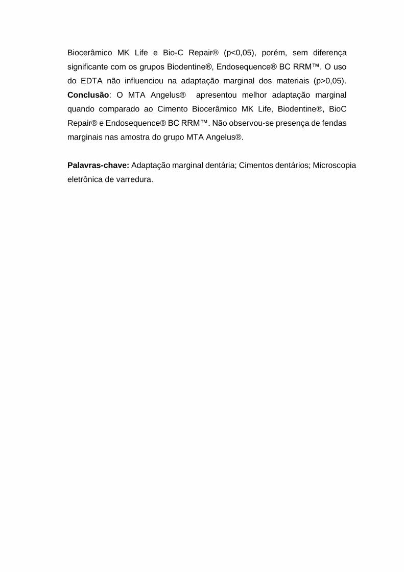
Biocerâmico MK Life e Bio-C Repair® (p<0,05), porém, sem diferença
significante com os grupos Biodentine®, Endosequence® BC RRM™. O uso
do EDTA não influenciou na adaptação marginal dos materiais (p>0,05).
Conclusão: O MTA Angelus® apresentou melhor adaptação marginal
quando comparado ao Cimento Biocerâmico MK Life, Biodentine®, BioC
Repair® e Endosequence® BC RRM™. Não observou-se presença de fendas
marginais nas amostra do grupo MTA Angelus®.
Palavras-chave: Adaptação marginal dentária; Cimentos dentários; Microscopia
eletrônica de varredura.

ABSTRACT
Objective: To analyse the marginal adaptation of calcium silicate-based cements
in root-end cavity by SEM. Material and Methods: Fifty bovine roots were
prepared with K-File #60 and filled with gutta-percha and MTA Fillapex. Roots
were apicetomized and a 3-mm-deep root-end cavity was prepared using
ultrasonic tips. Samples were randomly divided into five groups (n = 10): 1. Mk
Life bioceramic cement; 2. Biodentine®; 3. Bio-C Repair®; 4. Endosequence®
BC RRM™; 5. MTA Angelus® (MTA) - positive control. Root-end cavities were
filled with the materials prepared according to the manufacturers’ instructions.
EDTA was used prior the retro-filling material insertion in half of the samples from
each group (n = 5). Samples were prepared for SEM and photomicrographs were
taked in x40, x150 and x500. The images of root-end fillings were divided into
four áreas and distributed into five scores: 0 - absence of gaps; 1 - presence of
gap in 1 area; 2 - presence of gap in 2 areas; 3 - presence of gap in 3 areas; 4 -
presence of gaps in 4 areas. The analysis of transverse dimension of the gap
(μm) was performed using Image J software. Qualitative variables were
evaluated by Fisher Exact Test and quantitative variables by Analysis of Variance
(ANOVA) . The level of significance was 0.05%. Results: No gaps were
observed in MTA Angelus® group (0%). Presence of gaps were observed in Mk
Life bioceramic cement (90%), Biodentine® (80%), Bio-C Repair® (100%) and
Endosequence® BC RRM™ (80%). Significant difference was observed when
MTA were compared to Mk Life bioceramic cement, Biodentine®, Bio-C Repair®
and Endosequence® BC RRM™ (p <0.05). No significant difference were
observed in the transverse dimension of the gap between Mk Life bioceramic
cement, Biodentine®, Bio-C Repair® and Endosequence® BC RRM™ groups.
MTA show significantly better result than Mk Life bioceramic cement and Bio-C
Repair® groups (p <0.05), but without significant difference with Biodentine® and
Endosequence® BC RRM™ Conclusion: MTA Angelus® showed better
marginal adaptation compared to MK Life bioceramic cement, Biodentine® , BioC
Repair® and Endosequence® BC RRM™. Marginal gaps were not observed in
samples of MTA Angelus® group.

Keywords: Dental marginal adaptation; Dental cements; Scanning electron
microscopy.

QUADROS, FIGURAS E TABELAS
Quadro 1. Distribuição dos grupos experimentais de acordo com o material retrobturador e composição química dos cimentos de acordo com o fabricante ..................................................................................................
21
Quadro 2. Classificação das amostras em scores de acordo com a presença de fendas entre o material retrobturador e a parede dentinária da cavidade retrógada .................................................................................
27
Figura 1. Cimento biocerâmico MK Life (A); Biodentine® (B); Bio-C Repair® (C); Endosequence® BC RRM™ (D); MTA Angelus® (E) .............
23
Figura 2. Seccionamento da raiz (A); Instrumentação (B); Obturação (C); Apicectomia (D); Retropreparo (E); Cavidade retrógada (F); Inserção do material retrobturador com seringa (G); Inserção do material retrobturador com espátula (H); Condensação do material retrobturador (I); Retrobturação (J); Amostras preparadas para análise em MEV (K); MEV (JSM - IT300) (L) ..........................................................................................
25
Figura 3. Representação esquemática da divisão da retrobturação em áreas (área A, B, C e D) e da classificação utilizada na avaliação da presença ou ausência de fendas entre o material retrobturador e a parede dentinária da cavidade retrógada ................................................................
26
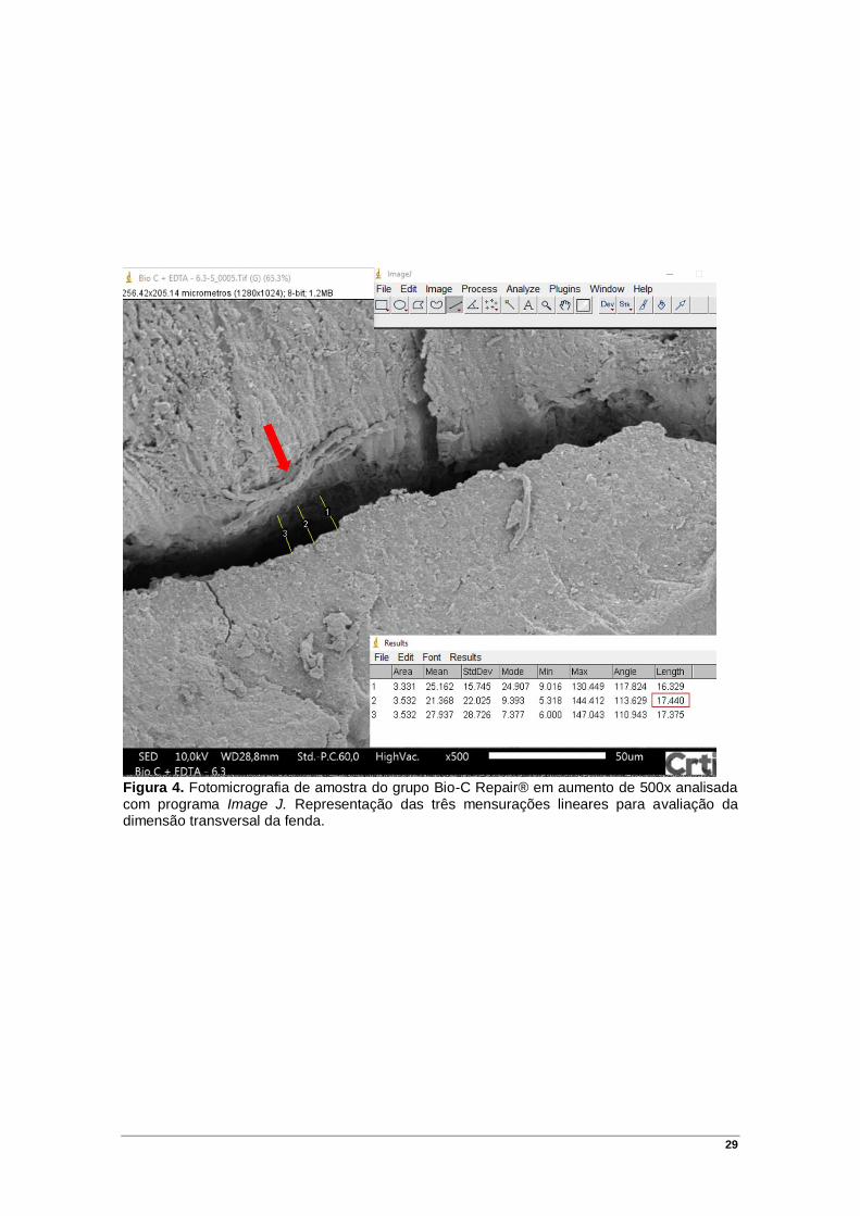
Figura 4. Fotomicrografia de amostra do grupo Bio-C Repair® em aumento de 500x analisada com programa Image J. Representação das três mensurações lineares para avaliação da dimensão transversal da fenda ...........................................................................................................
29
Figura 5. Fotomicrografias de MEV da adaptação marginal com Cimento biocerâmico MK Life (I.1 – I.3), Biodentine® (II.1 – II.3), Bio-C Repair® (III.1 – III.3), Endosequence® BC RRM™ (IV.1 – IV.3) e MTA Angelus® (V.1 – V.3) em magnificações de 40x (1), 150x (2) e 500x (3). Em destaque (amarelo) região selecionada para realizar fotomicrografia em aumento de 500x ............................................................................................................
33
Tabela 1. Frequência dos scores nos diferentes grupos ........................... 31
Tabela 2. Comparação da dimensão transversal da fenda (µm) em relação aos cimentos retrobturadores, por meio de MEV (500x) ..............................
32
Tabela 3. Comparação da dimensão transversal da fenda (µm) em relação aos cimentos retrobturadores sem e com o uso de EDTA ............................
32
SÍMBOLOS, ABREVIATURAS E SIGLAS
�̅� Média
# Number
% Porcentagem
< Menor que
® Marca registrada
ANOVA Análise de Variância
CRTI Centro Regional para o Desenvolvimento Tecnológico e
Inovação
DP Desvio padrão
EDTA Ácido etilenodiamino tetra-acético
et al. e colaboradores
mµ Micrômetro
MEV Microscopia eletrônica de varredura
mL Mililitro
mm Milímetro
MTA Mineral Trioxide Aggregate
n Número de amostras
n. Número
º Graus
p Nível de significância
RRM Root Repair Material
™ Trademark (marca registrada)
SUMÁRIO
1 INTRODUÇÃO ................................................................................... 15
2 OBJETIVOS ....................................................................................... 18
2.1 OBJETIVO GERAL .............................................................................. 18
2.2 OBJETIVO ESPECÍFICO ....................................................................... 18
3 MATERIAIS E MÉTODOS ................................................................. 19
3.1 SELEÇÃO E PREPARO DAS AMOSTRAS ................................................. 19
3.2 PREPARO DOS ESPÉCIMES PARA MEV ................................................. 24
3.3 AVALIAÇÃO DAS IMAGENS OBTIDAS NO MEV .......................................... 24
3.4 ANÁLISE ESTATÍSTICA ....................................................................... 30
4 RESULTADOS ................................................................................... 31
5 DISCUSSÃO ...................................................................................... 34
6 CONCLUSÃO .................................................................................... 39
REFERÊNCIAS ...................................................................................... 40
APÊNDICE ............................................................................................. 46
ANEXO ................................................................................................... 66
15
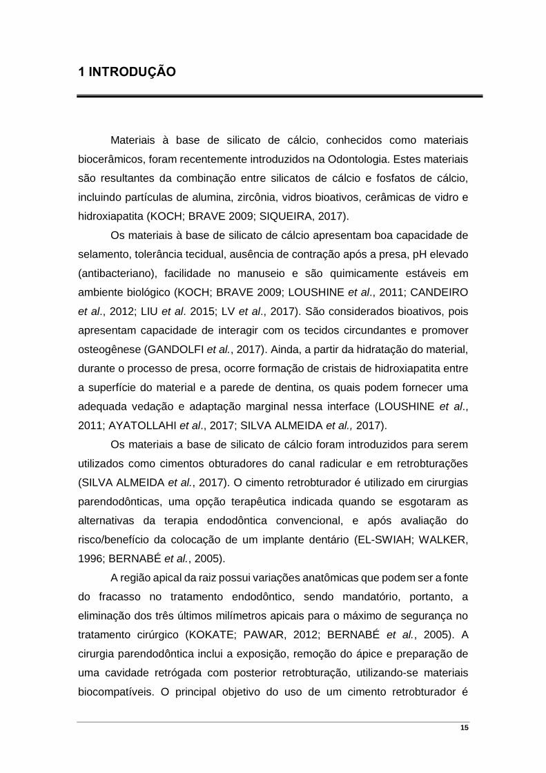
1 INTRODUÇÃO
Materiais à base de silicato de cálcio, conhecidos como materiais
biocerâmicos, foram recentemente introduzidos na Odontologia. Estes materiais
são resultantes da combinação entre silicatos de cálcio e fosfatos de cálcio,
incluindo partículas de alumina, zircônia, vidros bioativos, cerâmicas de vidro e
hidroxiapatita (KOCH; BRAVE 2009; SIQUEIRA, 2017).
Os materiais à base de silicato de cálcio apresentam boa capacidade de
selamento, tolerância tecidual, ausência de contração após a presa, pH elevado
(antibacteriano), facilidade no manuseio e são quimicamente estáveis em
ambiente biológico (KOCH; BRAVE 2009; LOUSHINE et al., 2011; CANDEIRO
et al., 2012; LIU et al. 2015; LV et al., 2017). São considerados bioativos, pois
apresentam capacidade de interagir com os tecidos circundantes e promover
osteogênese (GANDOLFI et al., 2017). Ainda, a partir da hidratação do material,
durante o processo de presa, ocorre formação de cristais de hidroxiapatita entre
a superfície do material e a parede de dentina, os quais podem fornecer uma
adequada vedação e adaptação marginal nessa interface (LOUSHINE et al.,
2011; AYATOLLAHI et al., 2017; SILVA ALMEIDA et al., 2017).
Os materiais a base de silicato de cálcio foram introduzidos para serem
utilizados como cimentos obturadores do canal radicular e em retrobturações
(SILVA ALMEIDA et al., 2017). O cimento retrobturador é utilizado em cirurgias
parendodônticas, uma opção terapêutica indicada quando se esgotaram as
alternativas da terapia endodôntica convencional, e após avaliação do
risco/benefício da colocação de um implante dentário (EL-SWIAH; WALKER,
1996; BERNABÉ et al., 2005).
A região apical da raiz possui variações anatômicas que podem ser a fonte
do fracasso no tratamento endodôntico, sendo mandatório, portanto, a
eliminação dos três últimos milímetros apicais para o máximo de segurança no
tratamento cirúrgico (KOKATE; PAWAR, 2012; BERNABÉ et al., 2005). A
cirurgia parendodôntica inclui a exposição, remoção do ápice e preparação de
uma cavidade retrógada com posterior retrobturação, utilizando-se materiais
biocompatíveis. O principal objetivo do uso de um cimento retrobturador é
16
promover um selamento apical para prevenir a infiltração de bactérias e seus
subprodutos para os tecidos periapicais (BERNABÉ et al., 2007).
Enfatiza-se que o material retrobturador ideal deve promover o selamento
tridimensional do canal radicular, impedir a infiltração bacteriana, ser
biologicamente tolerado pelos tecidos periapicais, não reabsorvível, de fácil
manipulação, dimensionalmente estável, radiopaco e possibilitar o reparo
tecidual (TORABINEJAD; WATSON; PITT FORD, 1993; KOKATE; PAWAR,
2012).
Vários materiais foram propostos para o selamento de cavidades
retrógadas, incluindo gutta-percha, amálgama, cimento de óxido de zinco e
eugenol, Cavit, resinas compostas, cimentos de ionômero de vidro, Super-EBA,
cimentos de hidróxido de cálcio, cimento Portland, agregado de trióxido mineral
(MTA), além de alguns cimentos endodônticos (BERNABÉ; HOLLAND 2009).
O amálgama, durante muito tempo, foi utilizado como principal material
retrobturador. O MTA, nas duas últimas décadas, tornou-se o material de
referência para este fim. O MTA foi desenvolvido em 1993 (LEE et al., 1993;
(TORABINEJAD; WATSON; PITT FORD, 1993), consiste em um cimento com
partículas hidrofílicas à base de silicato tricálcio, silicato dicálcio, aluminato
tricálcio e óxido de cálcio (CAMILLERI et al., 2005), e tem sido amplamente
aceito devido sua biocompatibilidade e suas propriedades físico-químicas
(OLIVEIRA et al., 2013; KÜÇÜKKAYA EREN; PARASHOS, 2018). No entanto,
trata-se de um material que apresenta dificuldade de colocação nas cavidades
retrógadas e tempo de presa elevado (OROSCO et al. 2010; KÜÇÜKKAYA
EREN; PARASHOS, 2018).
A busca por um material retrobturador ideal tem direcionado as pesquisas
aos novos cimentos a base de silicato de cálcio, que apresentam constituintes
semelhantes ao MTA (RAVICHANDRA et al., 2014; SHOKOUHINEJAD et al.,
2014; SOUNDAPPAN et al., 2014). Diferentes apresentações comerciais de
cimentos de silicato de cálcio estão disponíveis no mercado, tais como:
Biodentine®, Endosequence® BC, iRoot SP®, Cimento biocerâmico MK Life,
entre outros. A Angelus® recentemente desenvolveu um cimento reparador de
silicato de cálcio, o Bio-C Repair®, apresentado de forma experimental
(SIQUEIRA, 2017).

17
A qualidade do selamento apical obtida pelo material retrobturador
influencia diretamente no sucesso da cirurgia parendodôntica. A capacidade de
selamento dos materiais retrobturadores tem sido avaliada por diversas
metodologias, como: penetração de corante, bacteriana e de radioisótopos,
método eletroquímico e transporte de fluidos (SHEETAL et al., 2015). A análise
da adaptação marginal é uma metodologia que mensura, indiretamente, a
capacidade de selamento do material retrobturador (STABHOLZ et al., 1985;
AYATOLLAHI et al., 2017) e a microscopia eletrônica de varredura tem sido o
método mais utilizado no estudo da interface do material com a parede dentinária
(PETERS; PETERS 2002; XAVIER et al., 2005).
A retrobturação é um procedimento importante que visa impedir a
microinfiltração de irritantes aos tecidos periapicais, sendo que, uma
retrobturação insuficiente é considerada a principal causa de insucesso no
procedimento cirúrgico (BOLHARI et al., 2015). Portanto, a seleção de um
material biocompatível e que apresenta boa adaptação marginal torna-se
importante, sendo a análise da adaptação marginal capaz de fornecer
informações valiosas sobre a capacidade de selamento do material com vistas à
aplicação e benefícios na clínica endodôntica (STABHOLZ et al., 1985).
Considerando a importância dos materiais retrobturadores no contexto do
selamento e do sucesso do tratamento endodôntico cirúrgico, aliada à recente
introdução dos cimentos de silicato de cálcio no mercado odontolológico, tornou-
se oportuno e justificável o presente estudo.

18
2 OBJETIVOS
2.1 Objetivo geral
Analisar a adaptação marginal de cimentos de silicato de cálcio em
cavidades retrógadas.
2.2 Objetivo específico
• Comparar a adaptação marginal do Cimento biocerâmico MK Life,
Biodentine®, Bio-C Repair® e Endosequence® BC RRM™ com o MTA
Angelus® em cavidade retrógada, por meio de microscopia eletrônica de
varredura.
19
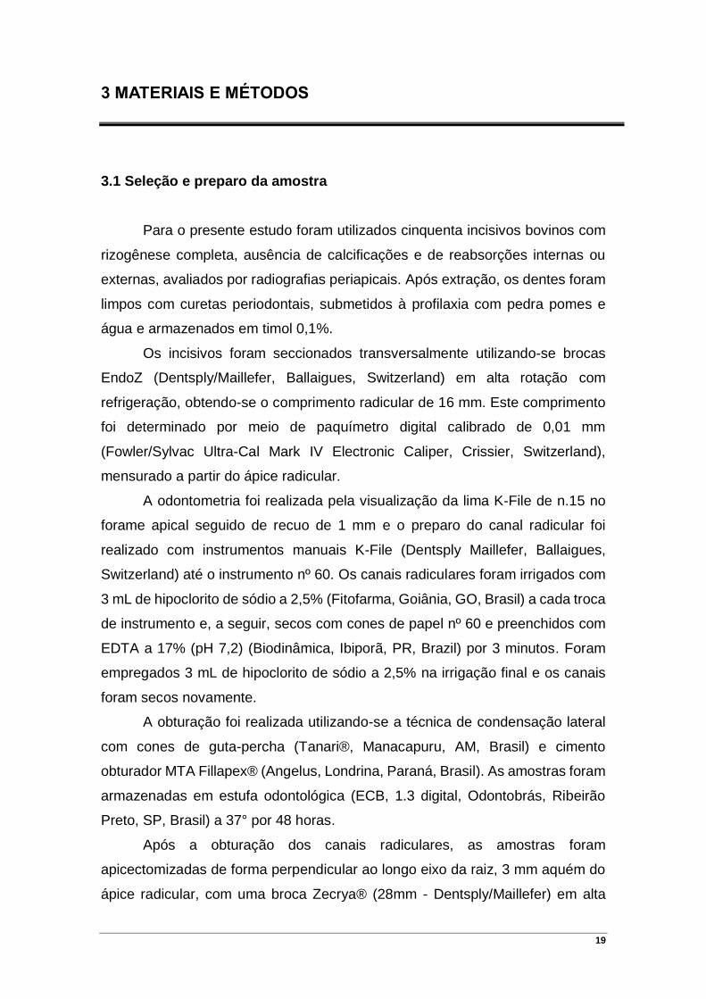
3 MATERIAIS E MÉTODOS
3.1 Seleção e preparo da amostra
Para o presente estudo foram utilizados cinquenta incisivos bovinos com
rizogênese completa, ausência de calcificações e de reabsorções internas ou
externas, avaliados por radiografias periapicais. Após extração, os dentes foram
limpos com curetas periodontais, submetidos à profilaxia com pedra pomes e
água e armazenados em timol 0,1%.
Os incisivos foram seccionados transversalmente utilizando-se brocas
EndoZ (Dentsply/Maillefer, Ballaigues, Switzerland) em alta rotação com
refrigeração, obtendo-se o comprimento radicular de 16 mm. Este comprimento
foi determinado por meio de paquímetro digital calibrado de 0,01 mm
(Fowler/Sylvac Ultra-Cal Mark IV Electronic Caliper, Crissier, Switzerland),
mensurado a partir do ápice radicular.
A odontometria foi realizada pela visualização da lima K-File de n.15 no
forame apical seguido de recuo de 1 mm e o preparo do canal radicular foi
realizado com instrumentos manuais K-File (Dentsply Maillefer, Ballaigues,
Switzerland) até o instrumento nº 60. Os canais radiculares foram irrigados com
3 mL de hipoclorito de sódio a 2,5% (Fitofarma, Goiânia, GO, Brasil) a cada troca
de instrumento e, a seguir, secos com cones de papel nº 60 e preenchidos com
EDTA a 17% (pH 7,2) (Biodinâmica, Ibiporã, PR, Brazil) por 3 minutos. Foram
empregados 3 mL de hipoclorito de sódio a 2,5% na irrigação final e os canais
foram secos novamente.
A obturação foi realizada utilizando-se a técnica de condensação lateral
com cones de guta-percha (Tanari®, Manacapuru, AM, Brasil) e cimento
obturador MTA Fillapex® (Angelus, Londrina, Paraná, Brasil). As amostras foram
armazenadas em estufa odontológica (ECB, 1.3 digital, Odontobrás, Ribeirão
Preto, SP, Brasil) a 37° por 48 horas.
Após a obturação dos canais radiculares, as amostras foram
apicectomizadas de forma perpendicular ao longo eixo da raiz, 3 mm aquém do
ápice radicular, com uma broca Zecrya® (28mm - Dentsply/Maillefer) em alta
20
velocidade, sob refrigeração. O preparo das cavidades retrógradas foi realizado
com auxílio de lupa de 2,5x de aumento (Bioart, São Carlos, SP, Brasil) a 3 mm
de profundidade, empregando-se ponta ultrassônica P1M (Helse, São Paulo,
Brasil) acoplada ao aparelho de ultrassom EMS Piezon Master 400 (Electro
Medical Systems, Vallée de Joux, Suíça), com constante irrigação de solução
fisiológica. Posteriormente, os espécimes foram divididos aleatoriamente em
cinco grupos experimentais de acordo com o material retrobturador (Quadro 1).
O grupo do MTA Angelus® foi utilizado como controle.
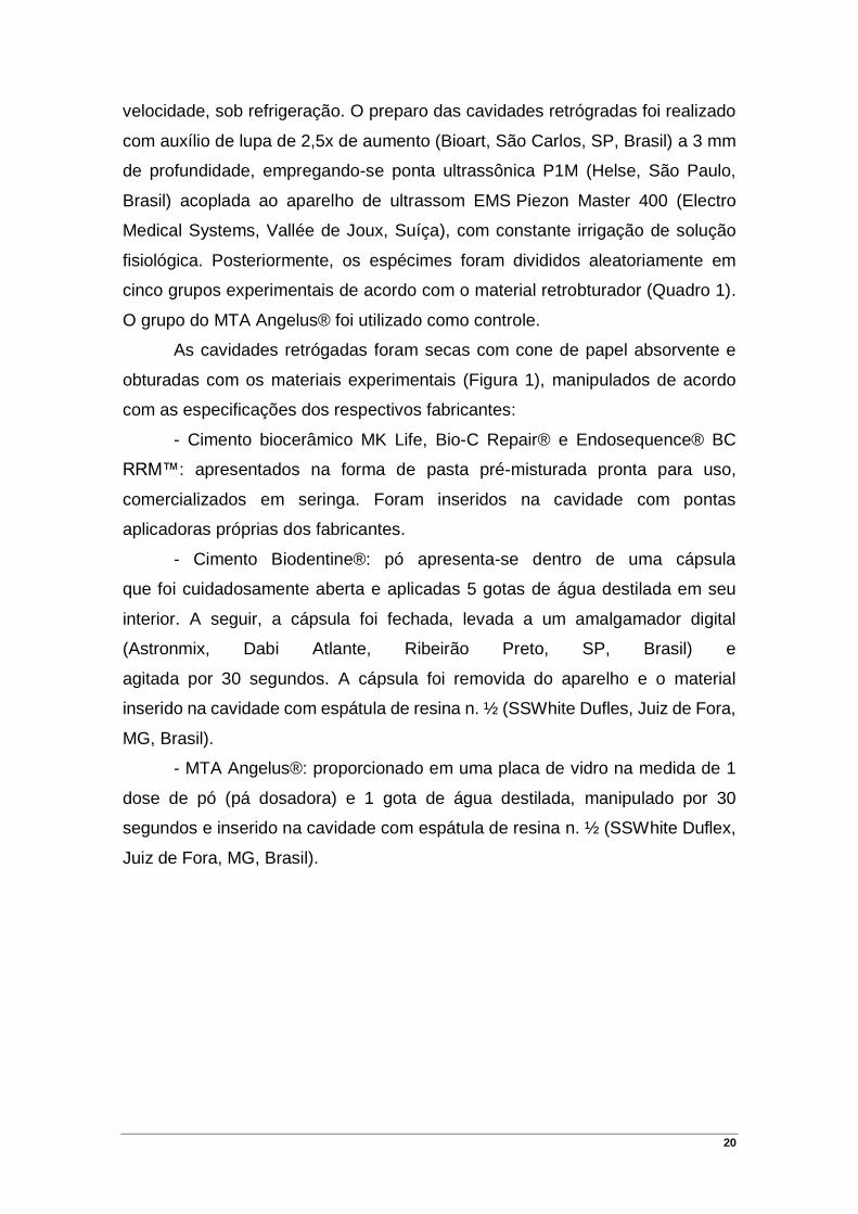
As cavidades retrógadas foram secas com cone de papel absorvente e
obturadas com os materiais experimentais (Figura 1), manipulados de acordo
com as especificações dos respectivos fabricantes:
- Cimento biocerâmico MK Life, Bio-C Repair® e Endosequence® BC
RRM™: apresentados na forma de pasta pré-misturada pronta para uso,
comercializados em seringa. Foram inseridos na cavidade com pontas
aplicadoras próprias dos fabricantes.
- Cimento Biodentine®: pó apresenta-se dentro de uma cápsula
que foi cuidadosamente aberta e aplicadas 5 gotas de água destilada em seu
interior. A seguir, a cápsula foi fechada, levada a um amalgamador digital
(Astronmix, Dabi Atlante, Ribeirão Preto, SP, Brasil) e
agitada por 30 segundos. A cápsula foi removida do aparelho e o material
inserido na cavidade com espátula de resina n. ½ (SSWhite Dufles, Juiz de Fora,
MG, Brasil).
- MTA Angelus®: proporcionado em uma placa de vidro na medida de 1
dose de pó (pá dosadora) e 1 gota de água destilada, manipulado por 30
segundos e inserido na cavidade com espátula de resina n. ½ (SSWhite Duflex,
Juiz de Fora, MG, Brasil).
21
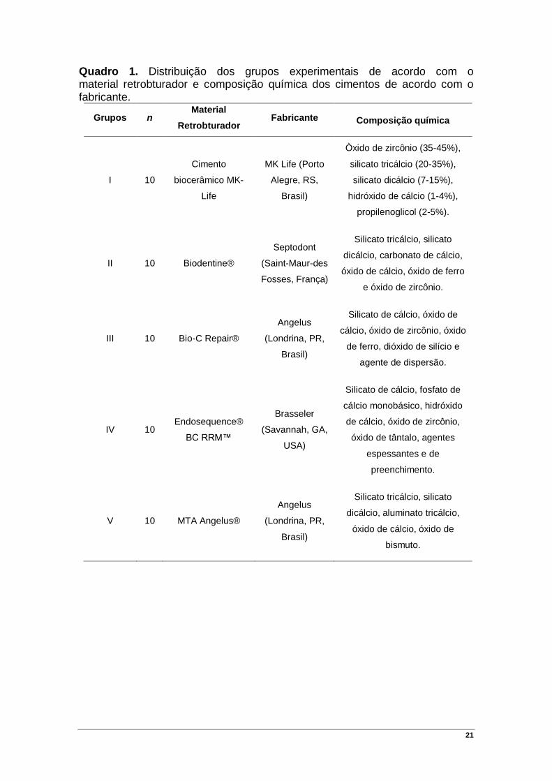
Quadro 1. Distribuição dos grupos experimentais de acordo com o material retrobturador e composição química dos cimentos de acordo com o fabricante.
Grupos n Material
Retrobturador Fabricante Composição química
I 10
Cimento
biocerâmico MK-
Life
MK Life (Porto
Alegre, RS,
Brasil)
Òxido de zircônio (35-45%),
silicato tricálcio (20-35%),
silicato dicálcio (7-15%),
hidróxido de cálcio (1-4%),
propilenoglicol (2-5%).
II 10 Biodentine®
Septodont
(Saint-Maur-des
Fosses, França)
Silicato tricálcio, silicato
dicálcio, carbonato de cálcio,
óxido de cálcio, óxido de ferro
e óxido de zircônio.
III 10 Bio-C Repair®
Angelus
(Londrina, PR,
Brasil)
Silicato de cálcio, óxido de
cálcio, óxido de zircônio, óxido
de ferro, dióxido de silício e
agente de dispersão.
IV 10 Endosequence®
BC RRM™
Brasseler
(Savannah, GA,
USA)
Silicato de cálcio, fosfato de
cálcio monobásico, hidróxido
de cálcio, óxido de zircônio,
óxido de tântalo, agentes
espessantes e de
preenchimento.
V 10 MTA Angelus®
Angelus
(Londrina, PR,
Brasil)
Silicato tricálcio, silicato
dicálcio, aluminato tricálcio,
óxido de cálcio, óxido de
bismuto.
22
Após a inserção dos materiais nas cavidades retrógadas, os cimentos e
pasta foram adaptados com kit de calcadores Bernabé (Thimon, São Paulo, SP,
Brasil) e cones de papel.
Em todos os grupos, metade das amostras (n=5) tiveram a cavidade
inundada com EDTA 17% por 3 minutos, previamente à colocação do material
retrobturador seguida de irrigação com solução fisiológica.
Os espécimes foram armazenados em tubos eppendorf por 72 horas e
100% de umidade em estufa odontológica (ECB, 1.3 digital, Odontobrás,
Ribeirão Preto, SP, Brasil). Todos os procedimentos foram realizados por um
único operador previamente calibrado e com mais de 5 anos de experiência em
Endodontia.
23
Figura 1. Cimento biocerâmico MK Life (A); Biodentine® (B); Bio-C Repair® (C); Endosequence® BC RRM™ (D); MTA Angelus® (E).

24
3.2 Preparo dos espécimes para MEV
Para avaliação em MEV, os 3 mm apicais foram seccionados com uma
broca Zecrya® (28mm - Dentsply/Maillefer) em alta velocidade, sob refrigeração
e, posteriormente, desidratados em álcool 70º, álcool 96º e álcool absoluto, em
imersões de 30 minutos em cada, em ordem crescente de concentração, sendo
que a solução foi renovada a cada 10 minutos.
As amostras foram analisadas no Centro Regional para o
Desenvolvimento Tecnológico e Inovação (CRTI) da Universidade Federal de
Goiás, onde permaneceram em estufa e baixo vácuo para completa secagem.
As amostras foram codificadas e fixadas em fita adesiva de carbono sobre porta
amostras de alumínio e recobertas com ouro. As fotomicrografias foram obtidas
no microscópio eletrônico de varredura - MEV (JSM-IT300, JEOL Ltd, Fukuoka,
Japão) com aumento de 40x, 150x e 500x para as análises (Figura 2).
3.3 Avaliação da adaptação marginal nas imagens obtidas por MEV
Avaliação da presença de fendas entre o cimento retrobturador e a parede
dentinária da cavidade retrógada
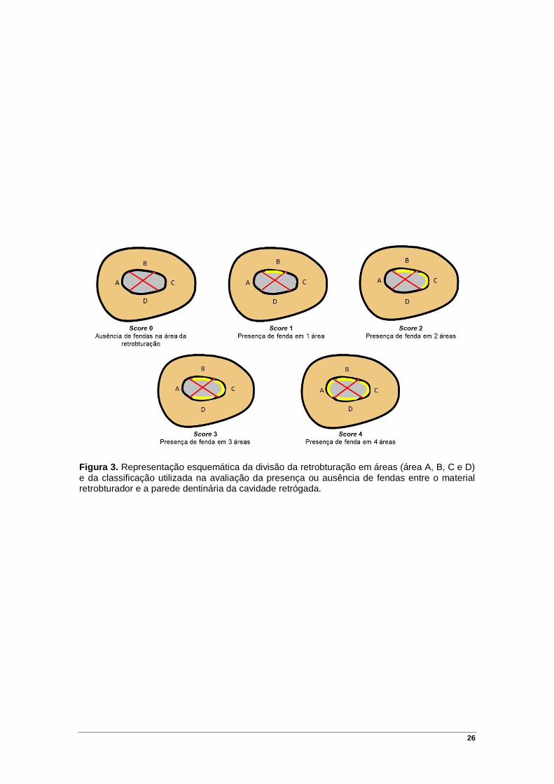
Empregando-se a imagem de 40x de aumento, a área total do material
retrobturador de cada espécime foi dividida em quatro (Figura 3). As áreas A, B,
C e D foram analisadas com aumento de 150x com relação a presença de fendas
entre o cimento retrobturador e a parede dentinária da cavidade retrógada.
Cada espécime foi classificado em escores conforme metodologia (Figura
3) empregada por Oliveira et al. (2013), constituindo cinco diferentes categorias
(Quadro 2).
25
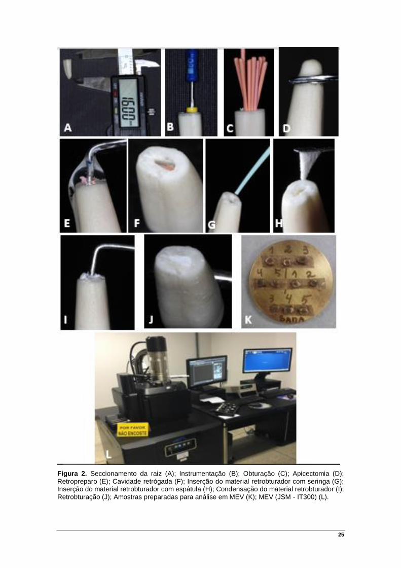
Figura 2. Seccionamento da raiz (A); Instrumentação (B); Obturação (C); Apicectomia (D); Retropreparo (E); Cavidade retrógada (F); Inserção do material retrobturador com seringa (G); Inserção do material retrobturador com espátula (H); Condensação do material retrobturador (I); Retrobturação (J); Amostras preparadas para análise em MEV (K); MEV (JSM - IT300) (L).
26
Figura 3. Representação esquemática da divisão da retrobturação em áreas (área A, B, C e D) e da classificação utilizada na avaliação da presença ou ausência de fendas entre o material retrobturador e a parede dentinária da cavidade retrógada.
27
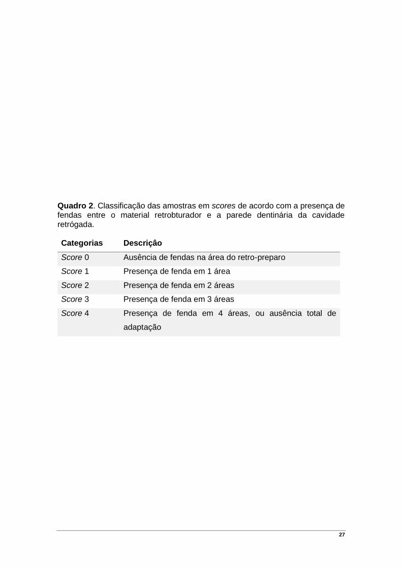
Quadro 2. Classificação das amostras em scores de acordo com a presença de fendas entre o material retrobturador e a parede dentinária da cavidade retrógada. Categorias Descriçâo
Score 0 Ausência de fendas na área do retro-preparo
Score 1 Presença de fenda em 1 área
Score 2 Presença de fenda em 2 áreas
Score 3 Presença de fenda em 3 áreas
Score 4 Presença de fenda em 4 áreas, ou ausência total de
adaptação

28
Avaliação da dimensão transversal da fenda entre o cimento retrobturador e a
parede dentinária da cavidade retrógada
Empregando-se a imagem de 40x de aumento, a área total do material
retrobturador de cada espécime foi dividida em quatro (área A, B, C e D), como
descrito anteriormente. Nas fotomicrografias com aumento de 150x selecionou-
se a área de maior dimensão transversal da fenda entre o cimento retrobturador
e a parede dentinária da cavidade retrógada e obteve-se imagens com aumento
de 500x para efetuação das mensurações. Com o uso do software Image J (NIH,
Bethesda, Maryland, EUA), foram realizadas três mensurações lineares (em
micrômetros) da maior dimensão transversal da fenda visível. Das três
mensurações, utilizou-se a de maior valor de cada área para calcular a média do
espécime (Figura 4). As análises foram realizadas por um único avaliador, com
experiência em MEV.
29
Figura 4. Fotomicrografia de amostra do grupo Bio-C Repair® em aumento de 500x analisada com programa Image J. Representação das três mensurações lineares para avaliação da dimensão transversal da fenda.

30
3.4 Análise estatística
A frequência e porcentagem das variáveis qualitativas e a média e o
desvio padrão das variáveis quantitativas foram obtidas. As variáveis qualitativas
foram avaliadas pelo Teste Exato de Fisher. A normalidade das variáveis
quantitativas foi avaliada pelo teste de Kolmogorov-Smirnov. As variáveis
quantitativas foram avaliadas pelo Teste de Análise de Variância (ANOVA) post
hoc Bonferroni. O nível de significância foi de 0,05%.
31
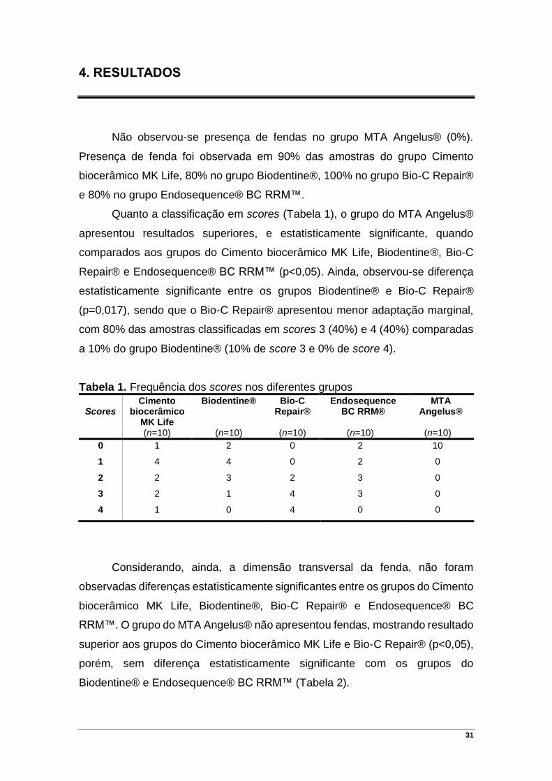
4. RESULTADOS
Não observou-se presença de fendas no grupo MTA Angelus® (0%).
Presença de fenda foi observada em 90% das amostras do grupo Cimento
biocerâmico MK Life, 80% no grupo Biodentine®, 100% no grupo Bio-C Repair®
e 80% no grupo Endosequence® BC RRM™.
Quanto a classificação em scores (Tabela 1), o grupo do MTA Angelus®
apresentou resultados superiores, e estatisticamente significante, quando
comparados aos grupos do Cimento biocerâmico MK Life, Biodentine®, Bio-C
Repair® e Endosequence® BC RRM™ (p<0,05). Ainda, observou-se diferença
estatisticamente significante entre os grupos Biodentine® e Bio-C Repair®
(p=0,017), sendo que o Bio-C Repair® apresentou menor adaptação marginal,
com 80% das amostras classificadas em scores 3 (40%) e 4 (40%) comparadas
a 10% do grupo Biodentine® (10% de score 3 e 0% de score 4).
Tabela 1. Frequência dos scores nos diferentes grupos
Scores
Cimento biocerâmico
MK Life
Biodentine®
Bio-C Repair®
Endosequence BC RRM®
MTA Angelus®
(n=10) (n=10) (n=10) (n=10) (n=10) 0 1 2 0 2 10
1 4 4 0 2 0
2 2 3 2 3 0
3 2 1 4 3 0
4 1 0 4 0 0
Considerando, ainda, a dimensão transversal da fenda, não foram
observadas diferenças estatisticamente significantes entre os grupos do Cimento
biocerâmico MK Life, Biodentine®, Bio-C Repair® e Endosequence® BC
RRM™. O grupo do MTA Angelus® não apresentou fendas, mostrando resultado
superior aos grupos do Cimento biocerâmico MK Life e Bio-C Repair® (p<0,05),
porém, sem diferença estatisticamente significante com os grupos do
Biodentine® e Endosequence® BC RRM™ (Tabela 2).

32
Tabela 2. Comparação da dimensão transversal da fenda (µm) em relação aos cimentos retrobturadores, por meio de MEV (500x).
Grupos n �̅� ± 𝑫𝑷 p*
Cimento biocerâmico MK Life 10 5,58 ± 4,50a 0,000
Biodentine® 10 4,13 ± 3,28a,b
Bio-C Repair® 10 8,43 ± 3,90a
Endosequence® BC RRM™ 10 4,28 ± 4,14a,b
MTA Angelus® 10 0,00b
*ANOVA post hoc Bonferroni. Letras iguais indicam que não há diferença significativa entre os grupos (p > 0,05). Letras diferentes indicam diferença significativa entre os grupos (p < 0,05).
Em relação à influência do uso do EDTA na dimensão transversal da
fenda entre o material retrobturador e a parede da cavidade retrógrada,
observou-se que não houve diferença significativa (p>0,05) quando o EDTA foi
utilizado previamente à inserção de nenhum dos cimentos retrobturadores
utilizados (Tabela 3).
Tabela 3. Comparação da dimensão transversal da fenda (µm) em relação aos cimentos retrobturadores sem e com o uso de EDTA.
Grupos Sem EDTA Com EDTA
p*
n �̅� ± 𝑫𝑷 n �̅� ± 𝑫𝑷
Cimento biocerâmico MK Life
5 6,91 ± 4,65 5 4,25 ± 4,41 0,380
Biodentine® 5 4,39 ± 3,60 5 3,87 ± 3,34 0,820
Bio-C Repair® 5 7,12 ± 4,14 5 9,73 ± 3,59 0,319
Endosequence® BC RRM™
5 4,54 ± 3,74 5 4,02 ± 4,94 0,857
MTA Angelus® 5 0,00 ± 0,00 5 0,00 ± 0,00 -
*Teste-t para amostras independentes.

33
Figura 5. Fotomicrografias de MEV da adaptação marginal com Cimento biocerâmico MK Life (I.1 – I.3), Biodentine® (II.1 – II.3), Bio-C Repair® (III.1 – III.3), Endosequence® BC RRM™ (IV.1 – IV.3) e MTA Angelus® (V.1 – V.3) em magnificações de 40x (1), 150x (2) e 500x (3). Em destaque (amarelo) região selecionada para realizar fotomicrografia em aumento de 500x.
Cimento biocerâmico MK Life Cimento biocerâmico MK Life Cimento biocerâmico MK Life
Biodentine® Biodentine® Biodentine®
Bio-C Repair® Bio-C Repair® Bio-C Repair®
Endosequence® BC RRM™ Endosequence® BC RRM™ Endosequence® BC RRM™
MTA Angelus® MTA Angelus® MTA Angelus®

34
5. DISCUSSÃO
O MTA Angelus® apresentou maior adaptação marginal quando
comparado aos demais cimentos de silicato de cálcio avaliados, sendo que não
foram observadas fendas em nenhuma amostra deste grupo. Estes resultados
são concordantes com os de estudos que demonstraram que o MTA apresentou
capacidade de adaptação marginal superior comparado a outros materiais
retrobturadores (PETERS; PETERS 2002; GONDIM et al., 2003; XAVIER et al.,
2005; GOMES et al., 2009; MUNHOZ et al., 2011; ROSALES-LEAL et al., 2011;
SHAHI et al., 2011; ROSA et al., 2014; SHOKOUHINEJAD et al., 2014;
SOUNDAPPAN et al., 2014; BOLHARI et al., 2015).
Entende-se por selamento, o vedamento da cavidade com finalidade de
prevenir microinfiltração de bactérias, fluidos, moléculas ou íons entre a margem
da cavidade e o material (KIDD, 1976; MOREIRA et al., 2010). A adaptação
marginal pode ser definida como o grau de aproximação ou ajuste do material
de preenchimento à superfície dentária (DeCS, 2018). Portanto, a avaliação da
adaptação marginal representa uma metodologia que analisa indiretamente a
capacidade de selamento do material retrobturador (STABHOLZ et al., 1985;
AYATOLLAHI et al., 2017).
A existência de correlação entre adaptação marginal e capacidade de
selamento dos materiais obturadores radiculares tem sido discutida na literatura
(BOLHARI et al., 2015). Diversos estudos têm mostrado uma relação positiva
entre estas duas propriedades (SHANI et al., 1984; STABHOLZ et al., 1985;
TORABINEJAD et al., 1995; TEWARI; TEWARI, 1999; COSTA et al., 2009;
AYATOLLAHI et al., 2017; KÜÇÜKKAYA EREN; GORGUYSUS; SAHIN, 2017).
Mesmo em estudos empregando metodologias diferentes (STABHOLZ et al.,
1985; AYATOLLAHI et al., 2017; KÜÇÜKKAYA EREN; GORGUYSUS; SAHIN,
2017), como microscopia eletrônica de varredura, penetração de radioisótopos
e infiltração por transporte de fluidos, esta correlação tem sido evidenciada
O emprego do microscópio eletrônico de varredura tem sido o método
mais utilizado para avaliação da adaptação marginal (PETERS; PETERS 2002;
XAVIER et al., 2005). A principal vantagem da microscopia eletrônica de

35
varredura é a capacidade de fornecer elevada magnificação e resolução.
Entretanto, este método também apresenta limitações, visto que, a preparação
convencional de amostras biológicas e a evaporação em vácuo pode estar
associada à introdução de artefatos, como trincas em tecidos duros (BADR,
2010). Estudos anteriores analisaram a adaptação marginal do material
retrobturador utilizando réplicas de resina para evitar artefatos nas amostras
(PETERS; PETERS 2002; GONDIM et al., 2003; ROSALES-LEAL et al., 2011;
SHOKOUHINEJAD et al., 2014; BOLHARI et al., 2015). Orosco et al. (2010),
relataram que para avaliação da adaptação marginal as amostras podem ser
vistas diretamente sob microscopia eletrônica de varredura após recobrimento
com ouro, não necessitando de criação de réplicas de resina. Em concordância,
no presente estudo, não foram realizadas réplicas das amostras, que foram
analisadas diretamente no microscópio eletrônico de varredura sem que
houvesse prejuízo ou perdas por formação de artefatos.
Para avaliação da adaptação marginal dos materiais testados, foram
realizadas duas análises no estudo: avaliação da presença de fenda com
classificação em scores (análise qualitativa) e a mensuração da dimensão
transversal da fenda (análise quantitativa), com o objetivo de obter-se resultados
que se complementassem, ampliando as possibilidades de discussão sobre o
assunto. Em uma revisão sistemática que avaliou trabalhos que compararam a
adaptação marginal do MTA com a de outros cimentos retrobturadores, foram
apresentados estudos que avaliaram a presença de fendas por análises
qualitativas, quantitativas ou utilizando ambas. As análises qualitativas foram
realizadas por meio da interpretação das imagens em termos de presença ou
ausência de fendas ou utilizando scores para indicar a distribuição das fendas
em relação a quadrantes da área da retrobturação. As análises quantitativas
foram realizadas com mensurações do comprimento, largura, área ou volume da
fenda. As duas análises, por score e por mensuração da dimensão da fenda,
também foram realizadas, assim como no presente trabalho (KÜÇÜKKAYA
EREN; PARASHOS, 2018).
Apesar das diferenças estruturais entre dentes bovinos e humanos,
estudos têm considerados dentes bovinos como possíveis substitutos para
dentes humanos em pesquisa odontológica, visto que, a dentina bovina e
36
humana possuem características similares, como, por exemplo, número e
diâmetro de túbulos dentinários (Soares et al., 2010).
No presente estudo avaliou-se a adaptação marginal dos cimentos de
silicato de cálcio (Cimento biocerâmico MK Life, Biodentine®, Bio-C Repair® e
Endosequence® BC RRM™), comparados ao MTA Angelus® (controle positivo)
e a pasta de hidróxido de cálcio (controle negativo).
O Biodentine® foi introduzido em 2009 pela Septodont® e apresenta
indicações endodônticas semelhantes às do MTA, promovendo o reparo
dentinário (CAMILLERI; SORRENTINO; DAMIDOT 2013; BOLHARI et al.,
2015).
No presente estudo, o MTA Angelus® mostrou maior adaptação marginal
quando comparado ao Biodentine® . No grupo do MTA Angelus® não foi
verificado fenda em nenhuma amostra analisada. O Biodentine® apresentou
média de 4,13 ± 3,28 µm de dimensão transversal de fenda, resultados
semelhantes com os de Soundappan et al. (2014). Este estudo comparou a
adaptação marginal do Biodentine e o MTA em MEV. Os resultados, quando o
MTA foi utilizado, mostraram-se superiores. Contraditoriamente, Ravichandra et
al. (2014) observaram melhor adaptação marginal do Biodentine® comparado
ao MTA em análise de microscopia confocal de varredura a laser. Outros estudos
(BOLHARI et al., 2015; KÜÇÜKKAYA EREN; GORGUYSUS; SAHIN, 2017)
demonstraram não haver diferença significativa entre os dois materiais.
Quanto à presença de fenda, o Biodentine® apresentou maior adaptação
marginal do que o BioC Repair®, e sem diferença significante quando
comparado ao Cimento biocerâmico MK Life ou ao Endosequence® BC RRM™.
O cimento Endosequence e o MTA foram comparados, por microscopia
eletrônica de varredura, quanto a adaptação marginal quando utilizados como
cimentos retrobturadores (SHOKOUHINEJAD et al., 2014; NAGESH et al.,
2016). Shokouhinejad et al. (2014) observaram que os dois cimentos
apresentaram resultados semelhantes. No entanto, Nagesh et al. (2016),
demonstraram que o Endosequence® apresentou significativamente menor
quantidade de fendas marginais quando comparado ao MTA. Os autores
relacionaram o resultado às desvantagens do MTA, como a dificuldade de
manipulação e o tempo de presa elevado, que podem contribuir para que ocorra
infiltração, desintegração da superfície levando à perda de adaptação marginal
37
e de continuidade do material. No presente estudo o MTA apresentou maior
adaptação marginal quando comparado ao Endosequence® BC RRM™, que por
sua vez, não apresentou diferença significante quando comparado ao Cimento
biocerâmico MK Life, Biodentine® ou BioC Repair®.
Há uma variedade muito grande de técnicas e metodologias para avaliar
a capacidade de selamento, o que dificulta a sua padronização e comparações
entre resultados (SHEETAL et al., 2015). Para Costa et al. (2009) e Bolhari et
al. (2015) os materiais apresentaram uma composição semelhante, tendo o
silicato de cálcio como constituinte principal, fato que possivelmente explica os
resultados similares de adaptação marginal. Estes cimentos são hidrofílicos,
absorvem líquido durante a presa e sofrem uma pequena expansão, além de
formarem cristais de hidroxiapatita entre a superfície do material e a parede de
dentina, os quais podem fornecer uma apropriada adaptação marginal
(AYATOLLAHI et al., 2017).
Apesar dos resultados promissores dos trabalhos que avaliaram os novos
cimentos de silicato de cálcio, no presente estudo, o MTA Angelus® apresentou
maior adaptação marginal quando comparado aos demais materiais testados. O
que nos conduz a questionar os motivos que levaram o MTA a apresentar
adaptação marginal consideravelmente superior. As justificativas de uma boa
adaptação marginal do MTA se enquadram a todos os cimentos a base de
silicato de cálcio, o que nos leva a admitir se a presença de compostos que
fornecem maior plasticidade aos novos materiais e, consequentemente, facilitam
o seu manuseio, poderiam resultar em uma diminuição das propriedades fisico-
químicas do material.
Além das características físico-químicas dos materiais, um dos fatores
que pode influenciar na qualidade da adaptação marginal em obturações
retrógadas é a presença de smear layer, porém, esta influência permanece uma
questão controversa na literatura (DI LENARDA; CADENARO; SHAIZERO,
2000). Vários agentes como hipoclorito de sódio, EDTA, mistura de detergente
ácido de tetraciclina (MTAD) e ácidos orgânicos foram introduzidos para
remoção de smear layer (DECHICHI; CHRISTIAN, 2006).
Estudos avaliaram a influência do uso do EDTA na adaptação marginal e
na capacidade de selamento em retrobturações com MTA. Por meio de
microtomografia computadorizada, Al Fouzan et al. (2015) observaram uma

38
melhora significativa na adaptação do material à dentina quando o EDTA foi
utilizado. No entanto, estudos mostraram que a capacidade de selamento apical,
avaliada por transporte de fluidos (YILDIRIM; ORUCOGLU; COBANKARA,
2008) e infiltração microbiana (YILDIRIM et al., 2010; ESTRELA et al., 2011)
diminuiu significativamente quando a smear layer foi removida. No presente
estudo, o uso do EDTA previamente a inserção dos materiais retrobturadores
testados não influenciou na adaptação marginal dos mesmos.
Os cimentos de silicato de cálcio possuem várias aplicações na
Odontologia. O conhecimento atualizado desses novos materiais é essencial
para garantir a seleção do mais adequado em diferentes situações clínicas (AL-
HADDAD; CHE AB AZIZ, 2016; JITARU et al, 2016; RAGHAVENDRA et al,
2017). A maioria dos estudos que avaliaram adaptação marginal são
laboratoriais, sendo este um aspecto que deve-se ter cuidado. Por se tratar de
um modelo in vitro, não há presença de umidade e secreções próprias do
ambiente cirúrgico, o que interfere na presa e comportamento do material.
Considerando condições clínicas complexas, a questão de quanto de dimensão
da fenda levaria a uma redução da taxa de sucesso do tratamento precisa ser
respondida em futuros estudos clínicos.

39
6. CONCLUSÃO
O MTA Angelus® apresentou melhor adaptação marginal quando
comparado ao Cimento Biocerâmico MK Life, Biodentine®, BioC Repair® e
Endosequence® BC RRM™. Não observou-se presença de fendas marginais
em nenhuma amostra do grupo MTA Angelus®.

40
REFERÊNCIAS
1. AL FOUZAN, K.; AWADH, M.; BADWELAN, M.; GAMAL, A.; GEEVARGHESE, A.; BABHAIR, S.; AL-REJAIE, M.; AL HEZAIMI, K.; ROTSTEIN, I. Marginal adaptation of mineral trioxide aggregate (MTA) to root dentin surface with orthograde/retrograde application techniques: A microcomputed tomographic analysis. J Conserv Dent, v. 18, p. 109-113, 2015.
2. AL-HADDAD, A.; CHE AB AZIZ, Z. A. Bioceramic-Based Root Canal Sealers: A Review. Int J Biomater, 2016
3. AYATOLLAHI, F.; HAZERI BAQDAD ABAD, M.; RAZAVI, S. H.;
TABRIZIZADEH, M.; AYATOLLAHI, R.; ZAREBIDOKI, F. Evaluating the Accuracy of Two Microleakage Assessment Methods for Mineral Trioxide Aggregate and Calcium-enriched Mixture Cement. Iran Endod J, v. 12, n. 4, p. 497-501, 2017.
4. BADR, A. E. Marginal adaptation and cytotoxicity of bone cement
compared with amalgam and mineral trioxide aggregate as root-end filling materials. J Endod, v. 36, p. 1056–1060, 2010.
5. BERNABE, P. F. E.; GOMES-FILHO, J. E.; ROCHA, W. C.; NERY, M. J.; OTOBONI-FILHO, J. A.; DEZAN-JUNIOR, E. Histological evaluation of MTA as a root-end filling material. International Endodontic Journal, v. 40, p. 758–765, 2007.
6. BERNABÉ, P. F. E.; HOLLAND, R. Endodontic Surgery. In: Estrela C.
Endodontic Science. São Paulo: Ed. Artes Médicas, 2009. p.1079- 1173.
7. BERNABE, P. F.; HOLLAND, R.; MORANDI, R.; DE SOUZA, V.; NERY, M. J.;
OTOBONI FILHO, J. A.; DEZAN JUNIOR, E.; GOMES-FILHO, J. E. Comparative study of MTA and other materials in retrofilling of pulpless dogs' teeth. Braz Dent J, v. 16, n. 2, p. 149-55, 2005.
8. BOLHARI, B.; ASHOFTEH YAZDI, K.; SHARIFI, F.; PIRMOAZEN, S.
Comparative scanning electron microscopic study of the marginal adaptation of four root-end filling materials in presence and absence of blood. J Dent (Tehran), v. 12, p. 226–34, 2015.
9. CAMILLERI, J.; MONTESIN, F.E.; BRADY, K.; SWEENEY, R.; CURTIS, R.V.; PITT FORD, T.R. The constitution of mineral trioxide aggregate. Dent Mat, v.21, p.297–303, 2005.
10. CAMILLERI, J.; SORRENTINO, F.; DAMIDOT, D. Investigation of the
hydration and bioactivity of radiopacified tricalcium silicate cement, Biodentine and MTA Angelus. Dent Mat, v. 29, p. 580-593, 2013.
41
11. CANDEIRO, G. T.; CORREIA, F. C.; DUARTE, M. A.; et al. Evaluation of radiopacity, pH, release of calcium ions, and flow of a bioceramic root canal sealer. J Endod, v.38, p.842–5, 2012.
12. COSTA, A. T.; KONRATH, F.; DEDAVID, B.; WEBER, J. B.; DE OLIVEIRA, M. G. Marginal adaptation of root-end filling materials: an in vitro study with teeth and replicas. J Contemp Dent Pract, v. 10, n. 2, p. 75-82, 2009.
13. DECHICHI, P.; CHRISTIAN, C. Smear layer: A brief review of general concepts. Part II. The most common agents to remove endodontic smear layer. RFO UPF, v. 11, p. 100-4, 2006.
14. Descritores em Ciências da Saúde: DECS. 2018. ed. rev. e ampl. São Paulo: BIREME / OPAS / OMS, 2018. Disponível em http://decs.bvsalud.org. Acesso em 22 de jun. 2018.
15. DI LENARDA, R.; CADENARO, M.; SBAIZERO, O. Effectiveness of 1 mol L-1 citric acid and 15% EDTA irrigation on smear layer removal. Int Endod J, v. 33, p. 46-52, 2000.
16. EL-SWIAH, J. M.; WALKER, R. T. Reasons for apicectomies. A retrospective study. Endod Dent Traumatol, v. 12, p. 185–91, 1996.
17. ESTRELA, C.; ESTRADA-BERNABÉ, P. F.; DE ALMEIDA-DECURCIO, D.; ALMEIDA-SILVA, J.; RODRIGUES-ARAÚJO-ESTRELA, C.; POLI-FIGUEIREDO, J. A. Microbial leakage of MTA, Portland cement, Sealapex and zinc oxide-eugenol as root-end flling materials. Med Oral Patol Oral Cir Bucal, v. 16, n. 3, p.418-24, 2011.
18. GANDOLFI MG, IEZZI G, PIATTELLI A, PRATI C, SCARANO A. Osteoinductive potential and bone-bonding ability of ProRoot MTA, MTA Plus and Biodentine in rabbit intramedullary model: Microchemical characterization and histological analysis. Dent Mater, v. 33, n. 5, p. 221-238, 2017.
19. GOMES, C. C.; ACCETTA, R. F.; GOMES CAMOES, I. C.; FREITAS, F. L.; PINTO, S. S. Marginal adaptation of root-end filling materials. Pesqui Bras Odontopediatr Clin Integr, v. 9, p. 31–5, 2009.
20. GONDIM, E.; ZAIA, A. A.; GOMES, B. P.; FERRAZ, C. C.; TEIXEIRA, F. B.; SOUZA-FILHO, F. J. Investigation of the marginal adaptation of root-end filling materials in root-end cavities prepared with ultrasonic tips. Int Endod J, v. 36, p. 491–9, 2003.
21. JITARU, S.; HODISAN, I.; TIMIS, L.; LUCIAN, A.; BUD, M. The use of bioceramics in endodontics - literature review. Clujul Med, v. 89, n. 4, p. 470-473, 2016.
42
22. KIDD, E. A. Microleakage: A review. J Dent, v. 4, p. 199–206, 1976.
23. KOCH, K.; BRAVE, D. Bioceramic technology - the game changer in endodontics. Endodontic Practice, v. 2, p.17-21, 2009.
24. KOKATE, S. R.; PAWAR, A. M. An in vitro comparative stereomicroscopic evaluation of marginal seal between MTA, glass ionomer cement and biodentine as root end filling materials using 1% methylene blue as tracer. Endodontology, v. 24, p. 36-42, 2012.
25. KÜÇÜKKAYA EREN, S.; GORDUYSUS, M. O.; SAHIN, C. Sealing ability and adaptation of root-end filling materials in cavities prepared with different techniques. Microsc Res Tech, v. 80, p. 756–62, 2017.
26. KÜÇÜKKAYA EREN, S.; PARASHOS, P. Adaptation of mineral trioxide aggregate to dentine walls compared with other root-end filling materials: A systematic review. Aust Endod J, v. 16, 2018.
27. LEE, S. J.; MONSEF, M.; TORABINEJAD, M. Sealing ability of a mineral trioxide aggregate for repair of lateral root perforations. J Endod, v. 19, p. 541-4, 1993.
28. LIU, S.; WANG, S.; DONG, Y. Evaluation of a bioceramic as a pulpar capping agent in vitro and in vivo. J Endod, v. 41, n.5, p. 652-7, 2015.
29. LOUSHINE, B. A.; BRYAN, T. E.; LOONEY, S. W.; et al. Setting properties and cytotoxicity evaluation of a premixed bioceramic root canal sealer. J Endod, v. 37, p. 673–7, 2011.
30. LV, F.; ZHU, L.; ZHANG, J.; YU, J.; CHENG, X.; PENG, B. Evaluation of the in vitro biocompatibility of a new fast-setting ready-to-use root filling and repair material. Int Endod J, v.50, p.540–548, 2017.
31. MOREIRA, A. L. B.; D’ASSUNÇÃO, F. L. C.; SALAZAR-SILVA, J. R.; PEREIRA, J. B. Systematic review about capacity of Resilon/Epiphany® and gutta-percha/cement to seal the apex of the root canal. Rev Odontol UNESP, v. 39, n. 2, p. 123-129, 2010.
32. MUNHOZ, M. F.; MARCHESAN, M. A.; CARDOSO, D. R.; SILVA, S. R.; SILVA-SOUSA, Y. T.; SOUSA-NETO, M. D. Quantitative 3D profilometry and SEM analysis of the adaptation of root-end filling materials placed under an optical microscope. Int Endod J, v. 44, p. 560–6, 2011.
33. NAGESH, B.; JEEVANI, E.; SUJANA, V.; DAMARAJU, B.; SREEHA, K.; RAMESH, P. Scanning electron microscopy (SEM) evaluation of sealing ability of MTA and EndoSequence as rootend filling materials with chitosan and carboxymethyl chitosan (CMC) as retrograde smear layer removing agents. J Conserv Dent, v. 19, p. 143–6, 2016.

43
34. OLIVEIRA, H. F.; GONÇALVES ALENCAR, A. H.; JOSÉ FIGUEIREDO, A. P.; GUEDES, O. A.; DE ALMEIDA DECURCIO, D.; ESTRELA, C. Marginal adaptation evaluation of root-end filling materials using scanning electron microscopy. Iran Endod J, v. 8, n. 4, p. 183-7, 2013.
35. OROSCO, F. A.; BRAMANTE, C. M.; GARCIA, R. B.; BERNARDINELI, N.; DE MORAES, I. G. Sealing ability, marginal adaptation and their correlation using three root-end filling materials as apical plugs. J Appl Oral Sci, v. 18, p.127–34, 2010.
36. PETERS, C. I.; PETERS, O. A. Occlusal loading of EBA and MTA root-end fillings in a computer-controlled masticator: a scanning electron microscopic study. Int Endod J, v. 35, p. 22–9, 2002.
37. RAGHAVENDRA, S. S.; JADHAV, G. R.; GATHANI, K. M.; KOTADIA, P. Bioceramics in endodontics – a revıew. J Istanb Univ Fac Dent, v. 51, n. 3, p. 128-137, 2017.
38. RAVICHANDRA, P. V.; HARIKUMAR, V.; DEEPTHI, K. et al. Comparative evaluation of marginal adaptation of Biodentine(TM) and other commonly used root end filling materials-na invitro study. J Clin Diagn Res, v. 8, p. 243–5, 2014.
39. ROSA, R. A.; SANTINI, M. F.; HEIDEN, K.; SÓ, B. B.; KUGA, M. C.; PEREIRA, J. R.; SÓ, M. V. SEM evaluation of the interface between filling and root-end filling materials. Scanning, v. 36, n. 2, p. 252–7, 2014.
40. ROSALES-LEAL, J. I.; OLMEDO-GAYA, V.; VALLECILLO-CAPILLA, M.; LUNA-DEL CASTILLO, J. D. Influence of cavity preparation technique (rotary vs. ultrasonic) on microleakage and marginal fit of six end-root filling materials. Med Oral Patol Oral Cir Bucal, v. 16, p. 185–9, 2011.
41. SHAHI, S.; YAVARI, H. R.; ESKANDARINEZHAD, M.; KASHANI, A.; RAHIMI, S.; SADRHAGHIGHI, H. Comparative investigation of marginal adaptation of mineral trioxide aggregate (MTA) and PC as root-end filling materials: a scanning eléctron microscopy (SEM) study. Afr J Biotechnol, v. 10, p. 16084–8, 2011.
42. SHANI, J.; FRIEDMAN, S.; STABHOLZ, A.; ABED, J. A radionuclide model for evaluating sealability of retrograde filling materials. Int J Nucl Med Biol, v. 11, n. 1, p. 46-52, 1984.
43. SHEETAL, M.; AMIT, P.; DHAIRAYSHEEL, E.; AMIT, C.; ABHIJIT, P.; RAHUL, S. R. Methodologies for assessing root canal sealing: a review. IJOCR, v. 13, n. 3, p. 55-8, 2015.
44. SHOKOUHINEJAD, N.; NEKOOFAR, M. H.; ASHOFTEHYAZDI, K.; ZAHRAEE, S.; KHOSHKHOUNEJAD, M. Marginal adaptation of new
44
bioceramic materials and mineral trioxide aggregate: a scanning electron microscopy study. Iran Endod J, v. 9, p. 144–8, 2014.
45. SILVA ALMEIDA, L.H.; MORAES, R.R.; MORGENTAL, R.D.; PAPPEN, F.G. Are Premixed Calcium Silicate–based Endodontic Sealers Comparable to Conventional Materials? A Systematic Review of In Vitro Studies. J Endod, v. 43, n. 4, 2017.
46. SIQUEIRA, P. C. Caracterização de elementos químicos de cimentos biocerâmicos. 2017. 77 f. Tese (Doutorado em Ciências da Saúde) - Universidade Federal de Goiás, Goiânia, 2017.
47. SOARES, C. J.; BARBOSA, L. M.; SANTANA, F. R.; SOARES, P. B. F.; MOTA, A. S.; SILVA, G. R. Fracture strength of composite fixed partial denture using bovine teeth as a substitute for human teeth with or without fiber-reinforcement. Braz Dent J. v. 21, n. 3, p. 235-240, 2010.
48. SOUNDAPPAN, S.; SUNDARAMURTHY, J. L.; RAGHU, S.; NATANASABAPATHY, V. Biodentine versus mineral trioxide aggregate versus intermediate restorative material for retrograde root end filling: an invitro study. J Dent (Tehran), v. 11, p. 143–9, 2014.
49. STABHOLZ, A.; FRIEDMAN, S.; ABED, J. Marginal adaptation of retrograde fillings and its correlation with sealability. J Endod, v. 11, p. 218–23, 1985.
50. TEWARI, S.; TEWARI, S. Marginal adaptation and sealability of orthograde and retrograde amalgam obturation. An in vitro study. Indian J Dent Res, v. 10, n. 3, p. 96-103, 1999.
51. TORABINEJAD, M.; SMITH, P. W.; KETTERING, J. D.; PITT FORD, T. R. Comparative investigation of marginal adaptation of mineral trioxide aggregate and other commonly used root-end filling materials. J Endod, v. 21, p. 295–9, 1995.
52. TORABINEJAD, M.; WATSON, T. F.; PITT FORD, T. R. Sealing ability of a mineral trioxide aggregate when used as a root end filling material. J Endod, v. 19, p. 591–5,1993.
53. XAVIER, C. B.; WEISMANN, R.; DE OLIVEIRA, M. G.; DEMARCO, F. F.; POZZA, D. H. Root-end filling materials: apical microleakage and marginal adaptation. J Endod, v. 31, p. 539–42, 2005.
54. YILDIRIM, T.; ER, K.; TASDEMIR, T.; TAHAN, E.; BURUK, K.; SERPER, A. Effect of smear layer and root-end cavity thickness on apical sealing ability of MTA as a root-end filling material: A bacterial leakage study. Oral Surg Oral Med Oral Pathol Oral Radiol Endod, v. 109, p. 67–72, 2010.

45
55. YILDIRIM, T.; ORUCOGLU, H.; COBANKARA, F. K. Long-term evaluation of the influence of smear layer on the apical sealing ability of MTA. J Endod, v. 34, p. 1537–1540, 2008.

46
APÊNDICE
ARTIGO
Title: Marginal adaptation of calcium silicate-based materials to dentine wall in root-end cavities
Renovato SR, Alencar AHG, Estrela C
Sara Rodrigues Renovato, MSc
Department of Stomatologic Sciences, Federal University of Goiás, Goiânia, GO,
Brazil
Ana Helena Gonçalves de Alencar, PhD
Department of Stomatologic Sciences, Federal University of Goiás, Goiânia, GO,
Brazil
Carlos Estrela, PhD
Department of Stomatologic Sciences, Federal University of Goiás, Goiânia, GO,
Brazil
Running title: Calcium silicate-based cements
Keywords: Dental cements; Dental marginal adaptation; Scanning electron
microscopy
Corresponding author:
Sara Rodrigues Renovato
Federal University of Goiás, Department of Stomatologic Sciences, Praça
Universitária s/n, Setor Universitário.
CEP 74605-220, Goiânia, GO, Brazil.
Phone number: 55 62 982388504
E-mail address: [email protected]
47
Abstract
Aim: To analyse the marginal adaptation of calcium silicate-based cements in root-
end cavity by SEM.
Methodology: Fifty bovine roots were prepared with K-File #60 and filled with gutta-
percha and MTA Fillapex. Roots were apicetomized and a 3-mm-deep root-end
cavity was prepared using ultrasonic tips. Samples were randomly divided into five
groups (n = 10): 1. Mk Life bioceramic cement (MKL); 2. Biodentine® (BD); 3. Bio-
C Repair® (BCR); 4. Endosequence® BC RRM™ (ERRM); 5. MTA Angelus®
(MTA) - positive control. Root-end cavities were filled with materials prepared
according to the manufacturers’ instructions. EDTA was used prior to the retro-filling
material insertion in half of samples from each group (n = 5). Photomicrographs in
SEM were taked in x40, x150 and x500. Images of root-end fillings were divided into
four áreas and distributed into five scores: 0 - absence of gaps; 1 - presence of gap
in 1 area; 2 - presence of gap in 2 areas; 3 - presence of gap in 3 areas; 4 - presence
of gaps in 4 areas. Transverse dimension of the gap (μm) was performed using
Image J software. Qualitative variables were evaluated by Fisher Exact Test and
quantitative variables by Analysis of Variance (ANOVA) . The level of significance
was 0.05%.
Results: No gaps were observed in MTA Angelus® group (0%). Presence of gaps
were observed in MKL (90%), BD (80%), BCR (100%) and ERRM (80%). Significant
difference was observed between MTA to MKL, BD, BCR and ERRM (p <0.05). No
significant difference were observed in transverse dimension of gap between MKL,
BD, BCR and ERRM groups. MTA show significantly better result than MKL and
BCR groups (p <0.05), but without significant difference with BD and ERRM.
Conclusions: MTA Angelus® showed better marginal adaptation compared to MK
Life bioceramic cement, Biodentine® , BioC Repair® and Endosequence® BC
RRM™. Marginal gaps were not observed in samples of MTA Angelus® group.

48
Introduction
Calcium silicate-based materials, known as bioceramic materials, were
recently introduced in dentistry. These materials are resulting from combination
of calcium silicates and calcium phosphates, and include alumina, zirconia,
bioactive glass, glass ceramics and hydroxyapatite (Koch & Brave 2009,
Siqueira, 2017).
These materials exhibit good sealing ability, biocompatibility, high pH
(antibacterial), good handling, do not shrink and are chemically stable in
biological environment (Koch & Brave 2009, Loushine et al. 2011, Candeiro et al.
2012, Liu et al. 2015, Lv et al. 2017). They are considered bioactive material, as
they have ability to interact with the surrounding tissues and promote
osteogenesis (Gandolfi et al. 2017). From the hydration of the material during
setting process occurs hydroxyapatite crystals formation between material
surface and dentin wall, which can provide adequate sealing and marginal
adaptation in interface (Loushine et al. 2011, Ayatollahi et al. 2017, Silva Almeida
et al. 2017).
Calcium silicate-based materials were introduced to be used as root canal
sealers and root-end filling material (Silva Almeida et al. 2017). Root-end filling
materials are used in endodontic surgeries, a therapeutic option indicated when
the alternatives of conventional endodontic therapy have been exhausted, and
after assessment of the risk / benefit of dental implant placement (El-Swiah &
Walker 1996, Bernabe et al. 2005 ).
Apical region of the root has anatomical variations that can be the source
of failure in endodontic treatment, so it is mandatory the last three apical
millimeters elimination for maximum safety in surgical treatment (Kokate & Pawar
2012, Bernabé et al. 2005). Endodontic surgery includes exposure, apex
removal, and preparation of a retrograde cavity with subsequent retrofilling, using
biocompatible materials. The main objective of a root-end filling material use is
promote an apical sealing to prevent bacteria and their by-products infiltration into
periapical tissues (Bernabé et al. 2007).
An ideal root-end filling material should promote tridimensional filling of
root canal, prevent leakage of microrganisms, be biologically tolerated by
periapical tissue, non-absorbable, easy to handle, dimensionally stable,
49
radiopaque and allow tissue repair (Torabinejad et al. 1993, Kokate & Pawar
2012).
Several materials were proposed for root-end cavity sealing, including
gutta-percha, amalgam, zinc oxide and eugenol cement, Cavit, composite resins,
glass ionomers, super-EBA, calcium hydroxide cements, Portland cement,
mineral trioxide aggregate (MTA), and some endodontic sealers (Bernabé &
Holland 2009).
Amalgam, for a long time, was the main root-end filling material used.
MTA, in the last two decades, has become the reference material for this purpose.
MTA was developed in 1993 (Lee et al. 1993, Torabinejad et al., 1993), consisting
of a cement with hydrophilic particles composed by tricalcium silicate, dicalcium
silicate, tricalcium aluminate and calcium oxide (Camilleri et al. 2005), and has
been widely accepted because of its biocompatibility and its physico-chemical
properties (Oliveira et al. 2013, Küçükkaya Eren & Parashos 2018). However,
has difficult handling characteristics and long setting time (Orosco et al. 2010,
Küçükkaya Eren & Parashos 2018).
The search for an ideal root-end filling material has directed researchs to
the new calcium silicate-based cements, which have constituents similar to MTA
(Ravichandra et al. 2014, Shokouhinejad et al. 2014, Soundappan et al. 2014).
Differents commercial presentations of calcium silicate-based cements are
available in the market, such as: Biodentine®, Endosequence® BC, iRoot SP®,
MK Life Bioceramic Cement, among others. Angelus® recently developed a
calcium silicate repair cement, Bio-C Repair®, experimentally presented
(Siqueira, 2017).
The apical sealing quality obtained by root-end filling material directly
influences at endodontic surgery success. The sealing ability of root-end filling
materials has been evaluated by several methodologies, such as: dye, bacterial
and radioisotope penetration, electrochemical method and fluid transport
(Sheetal et al. 2015). The marginal adaptation analysis is a methodology that
indirectly measures the sealing ability of the root-end filling material (Stabholz et
al. 1985, Ayatollahi et al. 2017) and scanning electron microscopy has been the
most used method in the study of the interface between material and dentin wall
(Peters & Peters 2002, Xavier et al. 2005).
50
Retrofilling is an important procedure that aims to prevent microleakage of
irritants to the periapical tissues. Insufficient retrofilling is considered the main
cause of failure in surgical procedure (Bolhari et al. 2015). Therefore, a
biocompatible material selection with good marginal adaptation becomes
important, and is capable of providing valuable information about material sealing
ability for application and benefits in the endodontic clinic (Stabholz et al. 1985).
Considering the importance of root-end filling materials in sealing ability context
and in surgical endodontic treatment succes, and with the recent introduction of
calcium silicate-based cements in the dental market, the present study was timely
and justifiable.
The aim of the study was too analyze the marginal adaptation of calcium
silicate- based cements in root-end cavities, by SEM.
Materials and Methods
Fifty bovine incisors with fully formed apex, absence of calcified root
canals, internal or external resorption, evaluated by periapical radiographs, were
used. After extraction, teeth were cleaned with periodontal curettes, submitted to
prophylaxis with pumice and water and stored on 0.1% thymol solution.
Teeth were cross-sectioned using EndoZ drills (Dentsply / Maillefer,
Ballaigues, Switzerland) at high speed to prepare standardized 16-mm tooth
length from the root Apex measured by calibrated digital caliper gauge of 0.01
mm (Fowler / Sylvac Electronic caliper Ultra-Cal Mark IV, Crissier, Switzerland).
Odontometry was performed by K-File #15 visualization on apical foramen
and canals were prepared using K-Files (Dentsply Maillefer, Ballaigues,
Switzerland) up to size #60, 1 mm short of the apical foramen. During
instrumentation, the root canals were irrigated with 3 mL of 2,5% sodium
hypochlorite (Fitofarma, Goiânia, GO, Brazil) at each change of files. Root canals
were dried and filled with 17% EDTA (pH 7.2) (Biodinâmica, Ibiporã, PR, Brazil)
for 3 min. After that, 3 mL of 2.5% sodium hypochlorite were used as final
irrigation and dried again with paper points (Dentsply Maillefer, Ballaigues,
Switzerland).
The root canals were obturated with guttapercha points (Tanari®,
Manacapuru, AM, Brazil) and MTA Fillapex® sealer (Angelus, Londrina, Paraná,

51
Brazil) using the conventional lateral compaction technique. After that, samples
were placed in an incubator (ECB, 1.3 digital, Odontobrás, Ribeirão Preto, SP,
Brazil) at 37ºC for 48 hours.
The apical 3 mm of roots was sectioned perpendicular to the long axis of
the tooth with a high-speed Zecrya drill (Dentsply Maillefer, Ballaigues,
Switzerland) under continuous air/water spray. Then, a 3-mm-deep root-end
cavity was prepared using P1M ultrasonic tips (Helse, São Paulo, Brazil) powered
by a EMS Piezon Master 400 (Electro Medical Systems, Vallée de Joux,
Switzerland), ultrassonic unit under continuous irrigation with saline solution.
Subsequently, samples were randomly assigned to five groups according to root-
end filling material tested (Table 1). MTA Angelus® group was used as positive
control.
Root-end cavities were dried with paper points and filled with experimental
materials that were prepared according to the manufacturers’ instruction. MK Life
bioceramic cement, Bio-C Repair® and Endosequence® BC RRM™ are
marketed in syringes and were inserted into the cavity with applicator tips specific
to the manufacturers. Biodentine® and MTA Angelus® were manipulated and
inserted into the cavity with resin spatula n. ½ (SSWhite Dufles, Juiz de Fora,
MG, Brasil).
Cements and paste were adapted with Bernabé kit (Thimon, São Paulo,
SP, Brazil) and paper points. EDTA was used prior to the retro-filling material
insertion in half of the samples from each group (n = 5) for 3 minutes, followed by
irrigation with saline solution.
Samples were stored in eppendorf tubes for 72 hours and 100% humidity,
placed in na incubator (ECB, 1.3 digital, Odontobrás, Ribeirão Preto, SP, Brazil).
All procedures were performed by a single operator previously calibrated and with
more than 5 years experience in Endodontics.
The apical 3-mm specimens were sectioned with a high-speed Zecrya drill
(Dentsply Maillefer, Ballaigues, Switzerland) under continuous air/water spray,
then, specimens were dehydrated in increasing concentrations of alcohol
(70º, 96º and 99º), in immersions of 30 minutes in each. Samples were analyzed
in the Regional Center for Technological Development and Innovation (CRTI) of
Federal University of Goiás, where they remained in incubator and under vacuum
for complete drying. Photomicrographs were obtained in scanning electron

52
microscope (SEM) (JSM-IT300, JEOL Ltd, Fukuoka, Japan) with an increase of
40x, 150x and 500x for analyzes.
Evaluation of the presence of marginal gaps
Using the 40x magnification image, the root end area of each specimen was
divided into four. The areas A, B, C and D were analyzed in a 150x magnification
image about the presence of gaps between root-end filling cement and dentin
wall of root-end cavity. Each specimen was classified in scores according to
methodology (Figure 1) employed by Oliveira et al. (2013), constituting five
different scores: score 0 - no marginal gap; score 1- marginal gap in one área;
score 2 – marginal gap in two áreas; score 3 – marginal gap in three areas and
score 4 – marginal gap in four áreas or in entire área.
Evaluation of the transverse dimension of the marginal gap
Using the 40x magnification image, the root end area of each specimen was
divided into four (areas A, B, C and D) as previously described. In 150x
magnification photomicrographs, greater transverse dimension of the gap was
selected and 500x magnification images were obtained for measurements. Using
Image J software (NIH, Bethesda, Maryland, USA), three linear measurements
(in micrometers) of the largest visible transverse dimension of the gap were
performed. The highest value of the three measurements of each area was used
to calculate the mean of transverse dimension of the specimen. The analyzes
were performed by a single evaluator, with experience in SEM.
Statistical analysis
The frequency and percentage of the qualitative variables and the mean and
standard deviation of the quantitative variables were obtained. Qualitative
variables were analyzed by Fisher's Exact Test. The normality of the quantitative
variables was evaluated by Kolmogorov-Smirnov test. The quantitative variables
were compared by Variance Analysis (ANOVA) post hoc Bonferroni. The level of
significance was 0.05%.

53
Results
No gaps were observed in MTA Angelus® group (0%). The presence of
gaps were observed in 90% of samples in MK Life bioceramic cement group,
80% in Biodentine® group, 100% in Bio-C Repair® group and 80% in
Endosequence® BC RRM™ group.
The MTA Angelus® group presented higher and statistically significant
results when compared to the MK Life bioceramic cement, Biodentine®, Bio-C
Repair® and Endosequence® BC RRM™ groups (p <0.05). There was also a
statistically significant difference between Biodentine® and Bio-C Repair®
groups (p=0.017). Bio-C Repair® presented the lowest marginal adaptation, with
80% of the samples classified as scores 3 (40%) and 4 (40%) compared to 10%
of the Biodentine® group (10% score 3 and 0% score 4).
Considering the transverse dimension of the marginal gap, no statistically
significant differences were observed between MK Life bioceramic cement,
Biodentine®, Bio-C Repair® and Endosequence® BC RRM™ groups. MTA
Angelus® group did not show marginal gaps, showing a superior result to MK Life
bioceramic cement and Bio-C Repair® groups (p <0.05), but without significant
difference with Biodentine® and Endosequence® BC RRM™ groups (Table 2).
Regarding the influence of the use of EDTA on transverse dimension of
the gap between the root-end filling material and the dentine wall of root-end
cavity, it was observed that there was no significant difference (p> 0.05) when
EDTA was used prior the insertion of any material used (Table 3).
Discussion
MTA Angelus® showed better marginal adaptation when compared to the
others calcium silicate cements evaluated. In the present study, no gaps were
observed in samples of MTA Angelus® group. In agrément, studies have shown
that MTA presented greater marginal adaptation compared to other root-end
filling materials (Peters & Peters 2002, Gondim et al. 2003, Xavier et al. 2005,
Gomes et al. 2009, Munhoz et al. 2011, Rosales-Leal et al. 2011, Shahi et al.

54
2011, Rosa et al. 2014, Shokouhinejad et al. 2014, Soundappan et al. 2014,
Bolhari et al. 2015).
The analysis of the marginal adaptation represents a methodology that
indirectly analyzes the sealing ability of root-end filling material (Stabholz et al.
1985, Ayatollahi et al. 2017). It is understood that sealing abilitty is the sealing of
the cavity in order to prevent microinfiltration of bacteria, fluids, molecules or ions
between the cavity margin and material (KIDD, 1976; MOREIRA et al., 2010).
Marginal adaptation can be defined as the degree of approximation or adjustment
of the filling material to dental surface (DeCS, 2018).
The correlation between marginal adaptation and sealing ability of root-
end filling materials has been discussed in literature (Bolhari et al. 2015). Studies
have shown a positive relation between these two properties (Shani et al. 1984,
Stabholz et al. 1985, Torabinejad et al. 1995, Tewari & tewari 1999, Costa et al.
2009, Ayatollahi et al. 2017, Küçükkaya Eren et al. 2017). Based on diverse
methodologies, using scanning electron microscopy, radioisotope penetration
and fluid transport filtration, Stabholz et al. (1985), Ayatollahi et al. (2017) and
Küçükkaya Eren et al. (2017) verified that exist correlation between marginal
adaptation and sealing capacity.
Marginal adaptation can be determined by different techniques. The use
of scanning electron microscope has been the most used (Peters & Peters 2002,
Xavier et al. 2005). The main advantage of scanning electron microscopy is the
ability to provide high magnification and resolution. This method also presents
limitations, since conventional preparation of biological samples and evaporation
in vacuum may be associated with introduction of artifacts, such as cracks in hard
tissues (Badr 2010). Previous studies have analyzed marginal adaptation of root-
end filling material using resin replicates to avoid artifacts in the samples (Peters
& Peters 2002, Gondim et al. 2003, Rosales-Leal et al. 2011, Shokouhinejad et
al. 2014, Bolhari et al. 2015). Orosco et al. (2010), reported that for evaluation of
the marginal adaptation the samples can be seen directly under scanning
electron microscopy after coating with gold, not requiring the creation of resin
replicas. In agreement, in the present study, no replicas of samples were taken,
which were analyzed directly in the scanning electron microscope without any
damage or losses due to artifact formation.
55
In order to evaluate marginal adaptation of tested materials, two analyzes
were performed in the study: evaluation of the presence of gap with classification
in scores (qualitative analysis) and measurement of the transversal dimension of
the gap (quantitative analysis), with objective to obtaining results that
complement each other, expanding the possibilities of discussion on the subject.
In a systematic review that evaluated studies that compared marginal adaptation
of MTA with others root-end filling materials, show that they evaluated the
presence of gaps by qualitative, quantitative or using both analysis. Two studies
of this systematic review, the qualitative analysis was performed by interpretation
of the images in terms of presence or absence of gaps, while in four studies
scores were used to indicate the distribution of the gaps in relation to quadrants
of the root-end cavity area. In 15 studies of this systematic review, quantitative
analysis was performed by measurements of the length, width, area or volume of
gap, and only two studies performed both the score classification and
measurements of the gap analysis as in the present study (Küçükkaya Eren &
Parashos 2018).
Despite some structural differences between bovine and human teeth,
studies have considered bovine teeth as possible substitutes for human teeth in
dental research, since bovine and human dentine have similar characteristics,
such as number and diameter of dentinal tubules (Soares et al. 2010).
In the present study, marginal adaptation of calcium silicate cements (MK
Life bioceramic cement, Biodentine®, Bio-C Repair® and Endosequence® BC
RRM™) was evaluated, compared to MTA Angelus® (positive control) and
calcium hydroxide paste (negative control).
Biodentine® was introduced by Septodont® in 2009 and presents
endodontic indications similar to those of the MTA. It performs an application in
dental repair (Camilleri et al. 2013, Bolhari et al. 2015).
In present study, MTA Angelus® showed better marginal adaptation when
compared to Biodentine®. In MTA Angelus® group, no gap was observed in any
sample analyzed. Biodentine® presented a mean of 4.13 ± 3.28 μm of transverse
gap size, similar results with those of Soundappan et al. (2014). This study
compared marginal adaptation of Biodentine and MTA by MEV. The results were
superior when MTA was used. Ravichandra et al. (2014) observed a better
marginal adaptation of Biodentine® compared to MTA in confocal laser scanning

56
microscopy. Other studies (Bolhari et al. 2015, Küçükkaya Eren et al. 2017)
showed no significant difference between these two materials.
Regarding the presence of gap, Biodentine® presented better marginal
adaptation than BioC Repair®, but without significant difference when compared
to MK Life bioceramic cement or Endosequence® BC RRM™.
Endosequence and MTA were compared about marginal adaptation by
scanning electron microscopy when used as root-end filling materials
(Shokouhinejad et al. 2014; Nagesh et al. 2016). Shokouhinejad et al. (2014)
observed that these two cements presented similar results. However, Nagesh et
al. (2016), demonstrated that Endosequence® presented significantly less
marginal gaps when compared to MTA. The authors related the results to MTA
disadvantages, such as the difficulty of handling and the high setting time, which
can contribute to infiltration, surface disintegration leading to loss marginal
adaptation and loss continuity of material. In the present study, MTA presented
greater marginal adaptation when compared to Endosequence® BC RRM™,
which did not present significant difference when compared to MK Life bioceramic
cement, Biodentine® or BioC Repair®.
There is a very wide variety of techniques and methodologies to evaluate
sealing ability, which makes difficult to standardize and compare the results
(Sheetal et al. 2015). For Costa et al. (2009) and Bolhari et al. (2015) the
materials presented a similar composition, with calcium silicate as main
constituent, a fact that possibly explains the similar results of marginal adaptation.
These cements are hydrophilic, absorb liquid during setting and suffer a small
expansion, in addition they form hydroxyapatite crystals between the surface of
material and dentin wall, which can provide an appropriate marginal adaptation
(Ayatollahi et al. 2017).
In spite of the promising results of the studies that evaluated new calcium
silicate cements, in the present study, MTA Angelus® presented better marginal
adaptation when compared to other materials tested. This result leads to
questioning the reasons that MTA presents considerably higher marginal
adaptation. Results (Peters & Peters 2002, Gondim et al. 2003, Xavier et al. 2005,
Gomes et al. 2009, Munhoz et al. 2011, Rosales-Leal et al. 2011, Shahi et al.
2011, Rosa et al. 2014, Shokouhinejad et al. 2014, Soundappan et al. 2014,
Bolhari et al. 2015) that justifie good marginal adaptation of MTA, justifie all
57
calcium silicate-based cements bahavior, which leads us to admit that the
presence of compounds that provide greater plasticity to new materials and
consequently facilitate their handling could result in a decrease of
physicochemical properties of the material.
In addition to materials physicochemical characteristics, one of the factors
that may influence the quality of marginal adaptation in root-end fillings is the
presence of smear layer, however, this influence remains a controversial issue in
literature (Di Lenarda et al. 2000) . Several agentes, such as sodium hypochlorite,
EDTA, blend of tetracycline acid detergent (MTAD) and organic acids were
introduced to remove smear layer (Dechichi & Christian 2006).
Studies have evaluated the influence of EDTA on marginal adaptation and
sealing ability on root-end filling with MTA. By computerized microtomography, Al
Fouzan et al. (2015) observed a significant improvement in the material
adaptation to dentin when EDTA was used. However, some studies have shown
that apical sealing ability, assessed by fluid transport (Yildirim et al. 2008) and
bacterial leakage (Yildirim et al. 2010; Estrela et al. 2011) decreased significantly
when the smear layer was removed. The use of EDTA prior the insertion of root-
end filling materials tested did not influence their marginal adaptation.
Calcium silicate-based materials have gained several applications in
dentistry. The updated knowledge of these new materials is essential to ensure
the selection of the most appropriate in different clinical situations (Al-Haddad &
Che Ab Aziz, 2016, Jitaru et al, 2016, Raghavendra et al, 2017). Most of studies
that evaluated marginal adaptation are laboratorial, and this should be taken care
of. Because it is an in vitro model, there is no presence of moisture and secretions
of surgical environment, which interferes with setting and behavior of material.
Considering complex clinical conditions, the question of how much dimensiono f
the gap would lead to a reduction in treatment success needs to be answered in
future clinical studies.
Conclusion
MTA Angelus® presented better marginal adaptation when compared to
MK Life bioceramic cement, Biodentine®, BioC Repair® and Endosequence®

58
BC RRM™. No marginal gaps were observed in all samples tested in MTA
Angelus® gropup.
References
Al Fouzan K, Awadh M, Badwelan M, et al (2015) Marginal adaptation of mineral trioxide aggregate (MTA) to root dentin surface with orthograde/retrograde application techniques: A microcomputed tomographic analysis. J Conserv Dent 18, 109-113.
Al-Haddad A, Che Ab Aziz ZA (2016) Bioceramic-Based Root Canal Sealers: A Review. Int J Biomater 4, 1-10. Ayatollahi F, Hazeri Baqdad Abad M, Razavi SH, Tabrizizadeh M, Ayatollahi R, Zarebidoki F (2017). Evaluating the Accuracy of Two Microleakage Assessment Methods for Mineral Trioxide Aggregate and Calcium-enriched Mixture Cement. Iran Endod J 12, 497-501. Badr AE (2010) Marginal adaptation and cytotoxicity of bone cement compared with amalgam and mineral trioxide aggregate as root-end filling materials. J Endod 36, 1056–1060.
Bernabe PFE, Gomes-Filho JE, Rocha WC, Nery MJ, Otoboni-Filho JA, Dezan-Junior E (2007) Histological evaluation of MTA as a root-end filling material. International Endodontic Journal 40, 758–765. Bernabé PFE, Holland R (2009) Endodontic Surgery. In: Estrela C (2009) Endodontic Science São Paulo, SP, Brasil: Ed. Artes Médicas, 1079-1173. Bernabe PFE, Holland R, Morandi R, et al (2005) Comparative study of MTA and other materials in retrofilling of pulpless dogs' teeth. Braz Dent J 16, 149-55. Bolhari B, Ashofteh Yazdi K, Sharifi F, Pirmoazen S (2015) Comparative scanning electron microscopic study of the marginal adaptation of four root-end filling materials in presence and absence of blood. J Dent (Tehran) 12, 226–34. Camilleri J, Montesin FE, Brady K, Sweeney R, Curtis RV, Pitt Ford TR (2005) The constitution of mineral trioxide aggregate. Dent Mat 21, 297–303. Camilleri J, Sorrentino F, Damidot D (2013) Investigation of the hydration and bioactivity of radiopacified tricalcium silicate cement, Biodentine and MTA Angelus. Dent Mat 29, 580-593.
Candeiro GT, Correia FC, Duarte MA, et al (2012) Evaluation of radiopacity, pH, release of calcium ions, and flow of a bioceramic root canal sealer. J Endod 38, 842–5.
59
Costa AT, Konrath F, Dedavid B, Weber JB, de Oliveira MG (2009) Marginal adaptation of root-end filling materials: an in vitro study with teeth and replicas. J Contemp Dent Pract 10, 75-82.
Dechichi P, Christian C (2006) Smear layer: A brief review of general concepts. Part II. The most common agents to remove endodontic smear layer. RFO UPF 11, 100-4.
Di Lenarda R, Cadenaro M, Sbaizero O (2000) Effectiveness of 1 mol L-1 citric acid and 15% EDTA irrigation on smear layer removal. Int Endod J 33, 46-52.
El-Swiah JM, Walker RT (1996) Reasons for apicectomies. A retrospective study. Endod Dent Traumatol 12, 185–91.
Estrela C, Bernabé PFE, Decurcio DA, Silva JA, Estrela CRA, Figueiredo JAP (2011) Microbial leakage of MTA, Portland cement, Sealapex and zinc oxide-eugenol as root-end flling materials. Med Oral Patol Oral Cir Bucal 16, 418-24.
Gandolfi MG, Iezzi G, Piattelli A, Prati C, Scarano A (2017) Osteoinductive potential and bone-bonding ability of ProRoot MTA, MTA Plus and Biodentine in rabbit intramedullary model: Microchemical characterization and histological analysis. Dent Mater 33, 221-238.
Gomes CC, Accetta RF, Gomes Camoes IC, Freitas FL, Pinto SS (2009) Marginal adaptation of root-end filling materials. Pesqui Bras Odontopediatr Clin Integr 9, 31–5.
Gondim E, Zaia AA, Gomes BP, Ferraz CC, Teixeira FB, Souza-Filho FJ (2003) Investigation of the marginal adaptation of root-end filling materials in root-end cavities prepared with ultrasonic tips. Int Endod J 36, 491–9. Health Sciences Descriptors: DECS. 2018. rev. and enl. ed. São Paulo: BIREME / PAHO / WHO, 2018. Available from: http://decs.bvsalud.org. Acess on 22 june 2018.
Jitaru S, Hodisan I, Timis L, Lucian A, Bud M (2016) The use of bioceramics in endodontics - literature review. Clujul Med 89, 470-473.
Kidd EA (1976) Microleakage: A review. J Dent 4, 199–206.
Koch K, Brave D (2009) Bioceramic technology - the game changer in endodontics. Endodontic Practice 2, 17-21.
Kokate SR, Pawar AM (2012) An in vitro comparative stereomicroscopic evaluation of marginal seal between MTA, glass ionomer cement and biodentine as root end filling materials using 1% methylene blue as tracer. Endodontology 24, 36-42.
60
Küçükkaya Eren S, Gorduysus MO, Sahin C (2017) Sealing ability and adaptation of root-end filling materials in cavities prepared with different techniques. Microsc Res Tech 80, 756–62.
Küçükkaya Eren S, Parashos P (2018) Adaptation of mineral trioxide aggregate to dentine walls compared with other root-end filling materials: A systematic review. Aust Endod J 16, 1-11.
Lee SJ, Monsef M, Torabinejad M (1993) Sealing ability of a mineral trioxide aggregate for repair of lateral root perforations. J Endod 19, 541-4.
Liu S, Wang S, Dong Y (2015) Evaluation of a bioceramic as a pulpar capping agent in vitro and in vivo. J Endod 41, 652-7.
Loushine BA, Bryan TE, Looney SW, et al (2011) Setting properties and cytotoxicity evaluation of a premixed bioceramic root canal sealer. J Endod 37, 673–7.
Lv F, Zhu L, Zhang J, Yu J, Cheng X, Peng B (2017) Evaluation of the in vitro biocompatibility of a new fast-setting ready-to-use root filling and repair material. Int Endod J 50, 540–548.
Moreira ALB, D’Assunção FLC, Salazar-Silva JR, Pereira JB (2010) Systematic review about capacity of Resilon/Epiphany® and gutta-percha/cement to seal the apex of the root canal. Rev Odontol UNESP 39, 123-129.
Munhoz MF, Marchesan MA, Cardoso DR, Silva SR, Silva-Sousa YT, Sousa-Neto MD (2011) Quantitative 3D profilometry and SEM analysis of the adaptation of root-end filling materials placed under an optical microscope. Int Endod J 44, 560–6. Nagesh B, Jeevani E, Sujana V, Damaraju B, Sreeha K, Ramesh P (2016) Scanning electron microscopy (SEM) evaluation of sealing ability of MTA and EndoSequence as rootend filling materials with chitosan and carboxymethyl chitosan (CMC) as retrograde smear layer removing agents. J Conserv Dent, 19, 143–6.
Oliveira HF, Alencar AHG, Figueiredo APJ, Guedes OA, Decurcio DA, Estrela C (2013) Marginal adaptation evaluation of root-end filling materials using scanning electron microscopy. Iran Endod J 8, 183-7.
Orosco FA, Bramante CM, Garcia RB, Bernardineli N, de Moraes IG (2010) Sealing ability, marginal adaptation and their correlation using three root-end filling materials as apical plugs. J Appl Oral Sci 18, 127–34.
Peters CI, Peters OA (2002) Occlusal loading of EBA and MTA root-end fillings in a computer-controlled masticator: a scanning electron microscopic study. Int Endod J 35, 22–9.

61
Raghavendra SS, Jadhav GR, Gathani KM, Kotadia P (2017) Bioceramics in endodontics – a revıew. J Istanb Univ Fac Dent 51, 128-137.
Ravichandra PV, Harikumar V, Deepthi K, et al (2014) Comparative evaluation of marginal adaptation of Biodentine(TM) and other commonly used root end filling materials-na invitro study. J Clin Diagn Res 8, 243–5.
Rosa RA, Santini MF, Heiden K, et al (2014) SEM evaluation of the interface between filling and root-end filling materials. Scanning 36, 252–7.
Rosales-leal JI, Olmedo-Gaya V, Vallecillo-Capilla M, Luna-del Castillo JD (2011) Influence of cavity preparation technique (rotary vs. ultrasonic) on microleakage and marginal fit of six end-root filling materials. Med Oral Patol Oral Cir Bucal 16, 185–9.
Shahi S, Yavari HR, Eskandarinezhad M, Kashani A, Rahimi S, Sadrhaghighi H (2011) Comparative investigation of marginal adaptation of mineral trioxide aggregate (MTA) and PC as root-end filling materials: a scanning eléctron microscopy (SEM) study. Afr J Biotechnol 10, 16084–8.
Shani J, Friedman S, Stabholz A, Abed J (1984) A radionuclide model for evaluating sealability of retrograde filling materials. Int J Nucl Med Biol 11, 46-52.
Sheetal M, Amit P, Dhairaysheel E, Amit C, Abhijit P, Rahul SR (2015) Methodologies for assessing root canal sealing: a review. IJOCR 13, 55-8.
Shokouhinejad N, Nekoofar MH, Ashoftehyazdi K, Zahraee S, Khoshkhounejad M (2014) Marginal adaptation of new bioceramic materials and mineral trioxide aggregate: a scanning electron microscopy study. Iran Endod J 9, 144–8.
Silva Almeida LH, Moraes RR, Morgental RD, Pappen FG (2017) Are Premixed Calcium Silicate–based Endodontic Sealers Comparable to Conventional Materials? A Systematic Review of In Vitro Studies. J Endod 43, 1-9.
Siqueira PC (2017) Caracterização de elementos químicos de cimentos biocerâmicos. 2017. 77 f. Tese (Doutorado em Ciências da Saúde) - Universidade Federal de Goiás, Goiânia.
Soares CJ, Barbosa LM, Santana FR, Soares PBF, Mota AS, Silva GR (2010) Fracture strength of composite fixed partial denture using bovine teeth as a substitute for human teeth with or without fiber-reinforcement. Braz Dent J 21,235-240.
Soundappan S, Sundaramurthy JL, Raghu S, Natanasabapathy V (2014) Biodentine versus mineral trioxide aggregate versus intermediate restorative material for retrograde root end filling: an invitro study. J Dent (Tehran), 11, 143–9.
62
Stabholz A, Friedman S, Abed J (1985) Marginal adaptation of retrograde fillings and its correlation with sealability. J Endod 11, 218–23.
Tewari S, Tewari S (1999) Marginal adaptation and sealability of orthograde and retrograde amalgam obturation. An in vitro study. Indian J Dent Res 10, 96-103.
Torabinejad M, Smith PW, Kettering JD, Pitt Ford TR (1995) Comparative investigation of marginal adaptation of mineral trioxide aggregate and other commonly used root-end filling materials. J Endod 21, 295–9.
Torabinejad M, Watson TF, Pitt Ford TR (1993) Sealing ability of a mineral trioxide aggregate when used as a root end filling material. J Endod 19, 591–5.
Xavier CB, Weismann R, de Oliveira MG, Demarco FF, Pozza DH (2005) Root-end filling materials: apical microleakage and marginal adaptation. J Endod 31, 539–42.
Yildirim T, Er K, Tasdemir T, Tahan E, Buruk K, Serper A (2010) Effect of smear layer and root-end cavity thickness on apical sealing ability of MTA as a root-end filling material: A bacterial leakage study. Oral Surg Oral Med Oral Pathol Oral Radiol Endod 109,67–72.
Yildirim T, Orucoglu H, Cobankara FK (2008) Long-term evaluation of the influence of smear layer on the apical sealing ability of MTA. J Endod 34, 1537–1540.
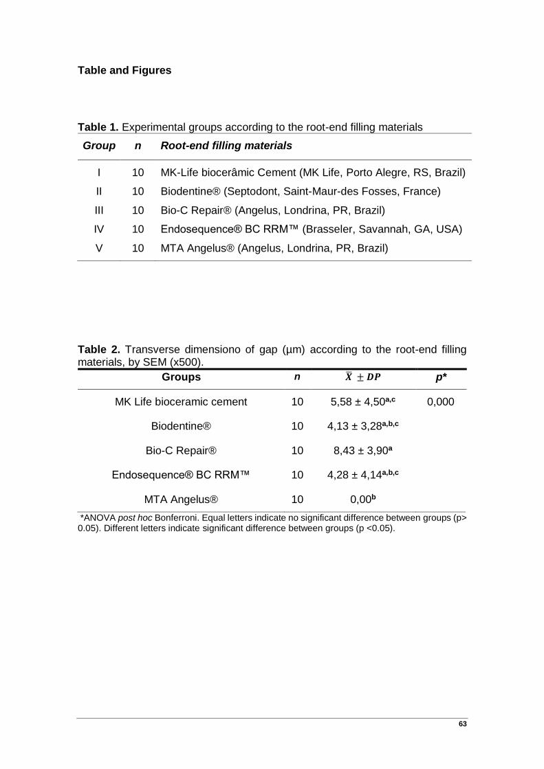
63
Table and Figures
Table 1. Experimental groups according to the root-end filling materials
Group n Root-end filling materials
I 10 MK-Life biocerâmic Cement (MK Life, Porto Alegre, RS, Brazil)
II 10 Biodentine® (Septodont, Saint-Maur-des Fosses, France)
III 10 Bio-C Repair® (Angelus, Londrina, PR, Brazil)
IV 10 Endosequence® BC RRM™ (Brasseler, Savannah, GA, USA)
V 10 MTA Angelus® (Angelus, Londrina, PR, Brazil)
Table 2. Transverse dimensiono of gap (µm) according to the root-end filling materials, by SEM (x500).
Groups n �̅� ± 𝑫𝑷 p*
MK Life bioceramic cement 10 5,58 ± 4,50a,c 0,000
Biodentine® 10 4,13 ± 3,28a,b,c
Bio-C Repair® 10 8,43 ± 3,90a
Endosequence® BC RRM™ 10 4,28 ± 4,14a,b,c
MTA Angelus® 10 0,00b
*ANOVA post hoc Bonferroni. Equal letters indicate no significant difference between groups (p> 0.05). Different letters indicate significant difference between groups (p <0.05).

64
Table 3. Conparison of transverse dimension of gap (µm) in relation to root-end filling cement with and without the use of EDTA.
Groups Without EDTA With EDTA
p*
n �̅� ± 𝑫𝑷 n �̅� ± 𝑫𝑷
MK Life bioceramic cement
5 6,91 ± 4,65 5 4,25 ± 4,41 0,380
Biodentine® 5 4,39 ± 3,60 5 3,87 ± 3,34 0,820
Bio-C Repair® 5 7,12 ± 4,14 5 9,73 ± 3,59 0,319
Endosequence® BC RRM™
5 4,54 ± 3,74 5 4,02 ± 4,94 0,857
MTA Angelus® 5 0,00 ± 0,00 5 0,00 ± 0,00 -
*Teste-t for independent samples.
Figure 1. Schematic representation of root-end filling division in areas (A, B, C and D) and classification used to evaluation of the presence or absence of gaps between root-end filling material and root-end cavity dentine.
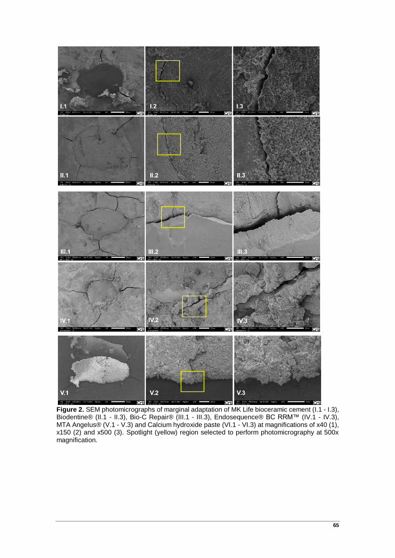
65
Figure 2. SEM photomicrographs of marginal adaptation of MK Life bioceramic cement (I.1 - I.3), Biodentine® (II.1 - II.3), Bio-C Repair® (III.1 - III.3), Endosequence® BC RRM™ (IV.1 - IV.3), MTA Angelus® (V.1 - V.3) and Calcium hydroxide paste (VI.1 - VI.3) at magnifications of x40 (1), x150 (2) and x500 (3). Spotlight (yellow) region selected to perform photomicrography at 500x magnification.

66
ANEXO
Normas para publicação
Author Guidelines
Content of Author Guidelines:
1. General, 2. Ethical Guidelines, 3. Manuscript Submission Procedure, 4. Manuscript Types Accepted, 5. Manuscript Format and Structure, 6. After Acceptance Useful Websites:
Submission Site, Articles published in International Endodontic Journal, Author Services, Wiley's Ethical Guidelines, Guidelines for Figures
The journal to which you are submitting your manuscript employs a plagiarism detection system. By submitting your manuscript to this journal you accept that your manuscript may be screened for plagiarism against previously published works.
1. GENERAL
International Endodontic Journal publishes original scientific articles, reviews, clinical articles and case reports in the field of Endodontology; the branch of dental sciences dealing with health, injuries to and diseases of the pulp and periradicular region, and their relationship with systemic well-being and health. Original scientific articles are published in the areas of biomedical science, applied materials science, bioengineering, epidemiology and social science relevant to endodontic disease and its management, and to the restoration of root-treated teeth. In addition, review articles, reports of clinical cases, book reviews, summaries and abstracts of scientific meetings and news items are accepted.
Please read the instructions below carefully for details on the submission of manuscripts, the journal's requirements and standards as well as information concerning the procedure after a manuscript has been accepted for publication in International Endodontic Journal. Authors are encouraged to visit Wiley Author Services for further information on the preparation and submission of articles and figures.

67
2. ETHICAL GUIDELINES
International Endodontic Journal adheres to the below ethical guidelines for publication and research.
2.1. Authorship and Acknowledgements
Authors submitting a paper do so on the understanding that the manuscript has been read and approved by all authors and that all authors agree to the submission of the manuscript to the Journal.
International Endodontic Journal adheres to the definition of authorship set up by The International Committee of Medical Journal Editors (ICMJE). According to the ICMJE, authorship criteria should be based on 1) substantial contributions to conception and design of, or acquisiation of data or analysis and interpretation of data, 2) drafting the article or revising it critically for important intellectual content and 3) final approval of the version to be published. Authors should meet conditions 1, 2 and 3.
Acknowledgements: Under acknowledgements please specify contributors to the article other than the authors accredited. Please also include specifications of the source of funding for the study and any potential conflict of interests if appropriate. Please find more information on the conflict of interest form in section 2.6.
2.2. Ethical Approvals
Experimentation involving human subjects will only be published if such research has been conducted in full accordance with ethical principles, including the World Medical AssociationDeclaration of Helsinki (version 2008) and the additional requirements, if any, of the country where the research has been carried out. Manuscripts must be accompanied by a statement that the experiments were undertaken with the understanding and written consent of each subject and according to the above mentioned principles. A statement regarding the fact that the study has been independently reviewed and approved by an ethical board should also be included. Editors reserve the right to reject papers if there are doubts as to whether appropriate procedures have been used.
When experimental animals are used the methods section must clearly indicate that adequate measures were taken to minimize pain or discomfort. Experiments should be carried out in accordance with the Guidelines laid down by the National Institute of Health (NIH) in the USA regarding the care and use of animals for experimental procedures or with the European Communities Council Directive of 24 November 1986 (86/609/EEC) and in accordance with local laws and regulations.
All studies using human or animal subjects should include an explicit statement in the Material and Methods section identifying the review and ethics committee approval for each study. The authors MUST upload a copy of the ethical approval letter when submitting their manuscript. Editors reserve the right to reject papers if there is doubt as to whether appropriate procedures have been used.
2.3 Clinical Trials
The International Endodontic Journal asks that authors submitting manuscripts reporting from a clinical trial to register the trials in any of the following public
68
clinical trials registries: www.clinicaltrials.gov, https://www.clinicaltrialsregister.eu/, http://isrctn.org/. Other primary registries if named in the WHO network will also be considered acceptable. The clinical trial registration number and name of the trial register should be included in the Acknowledgements at the submission stage.
2.3.1 Randomised control clinical trials
Randomised control clinical trials should be reported using the guidelines available at www.consort-statement.org. A CONSORT checklist and flow diagram (as a Figure) should also be included in the submission material.
2.3.2 Epidemiological observational trials
Submitting authors of epidemiological human observations studies are required to review and submit a 'strengthening the reporting of observational studies in Epidemiology' (STROBE) checklist and statement. Compliance with this should be detailed in the materials and methods section. (www.strobe-statement.org)
2.4 Systematic Reviews
Systematic reviews should be reported using the PRISMA guidelines available at http://prisma-statement.org/. A PRISMA checklist and flow diagram (as a Figure) should also be included in the submission material.
2.5 DNA Sequences and Crystallographic Structure Determinations
Papers reporting protein or DNA sequences and crystallographic structure determinations will not be accepted without a Genbank or Brookhaven accession number, respectively. Other supporting data sets must be made available on the publication date from the authors directly.
2.6 Conflict of Interest and Source of Funding
International Endodontic Journal requires that all authors (both the corresponding author and co-authors) disclose any potential sources of conflict of interest. Any interest or relationship, financial or otherwise that might be perceived as influencing an author’s objectivity is considered a potential source of conflict of interest. These must be disclosed when directly relevant or indirectly related to the work that the authors describe in their manuscript. Potential sources of conflict of interest include but are not limited to patent or stock ownership, membership of a company board of directors, membership of an advisory board or committee for a company, and consultancy for or receipt of speaker's fees from a company. If authors are unsure whether a past or present affiliation or relationship should be disclosed in the manuscript, please contact the editorial office at [email protected]. The existence of a conflict of interest does not preclude publication in this journal.
The above policies are in accordance with the Uniform Requirements for Manuscripts Submitted to Biomedical Journals produced by the International Committee of Medical Journal Editors (http://www.icmje.org/).
It is the responsibility of the corresponding author to have all authors of a manuscript fill out a conflict of interest disclosure form, and to upload all forms individually (do not combine the forms into one file) together with the manuscript on submission. The disclosure statement should be included under Acknowledgements. Please find the form below: Conflict of Interest Disclosure Form

69
2.7 Appeal of Decision
The decision on a paper is final and cannot be appealed.
2.8 Permissions
If all or parts of previously published illustrations are used, permission must be obtained from the copyright holder concerned. It is the author's responsibility to obtain these in writing and provide copies to the Publishers.
2.8 Copyright Assignment
If your paper is accepted, the author identified as the formal corresponding author for the paper will receive an email prompting them to login into Author Services; where via the Wiley Author Licensing Service (WALS) they will be able to complete the license agreement on behalf of all authors on the paper. Your article cannot be published until this has been done.
For authors choosing OnlineOpen
If the OnlineOpen option is selected the corresponding author will have a choice of the following Creative Commons License Open Access Agreements (OAA):
Creative Commons Attribution License OAA
Creative Commons Attribution Non-Commercial License OAA
Creative Commons Attribution Non-Commercial - No Derivs License OAA
To preview the terms and conditions of these open access agreements please visit the Copyright FAQs hosted on Wiley Author Services http://exchanges.wiley.com/authors/faqs---copyright-_301.html and visithttp://www.wileyopenaccess.com/details/content/12f25db4c87/Copyright--License.html.
If you select the OnlineOpen option and your research is funded by certain funders [e.g. The Wellcome Trust and members of the Research Councils UK (RCUK) or the Austrian Science Fund (FWF)] you will be given the opportunity to publish your article under a CC-BY license supporting you in complying with Wellcome Trust and Research Councils UK requirements. For more information on this policy and the Journal’s compliant self-archiving policy please visit: http://www.wiley.com/go/funderstatement.
3. OnlineOpen
OnlineOpen is available to authors of primary research articles who wish to make their article available to non-subscribers on publication, or whose funding agency requires grantees to archive the final version of their article. With OnlineOpen, the author, the author's funding agency, or the author's institution pays a fee to ensure that the article is made available to non-subscribers upon publication via Wiley Online Library, as well as deposited in the funding agency's preferred archive. For the full list of terms and conditions, see http://wileyonlinelibrary.com/onlineopen#OnlineOpen_Terms
Any authors wishing to send their paper OnlineOpen will be required to complete the payment form available from our website at: https://authorservices.wiley.com/bauthor/onlineopen_order.asp
Prior to acceptance there is no requirement to inform an Editorial Office that you intend to publish your paper OnlineOpen if you do not wish to. All OnlineOpen articles are treated in the same way as any other article. They go through the

70
journal's standard peer-review process and will be accepted or rejected based on their own merit.
3.1 MANUSCRIPT SUBMISSION PROCEDURE
Manuscripts should be submitted electronically via the online submission site http://mc.manuscriptcentral.com/iej. The use of an online submission and peer review site enables immediate distribution of manuscripts and consequentially speeds up the review process. It also allows authors to track the status of their own manuscripts. Complete instructions for submitting a paper is available online and below. Further assistance can be obtained from [email protected].
3.2. Getting Started
• Launch your web browser (supported browsers include Internet Explorer 5.5 or higher, Safari 1.2.4, or Firefox 1.0.4 or higher) and go to the journal's online Submission Site: http://mc.manuscriptcentral.com/iej
• Log-in, or if you are a new user, click on 'register here'.
• If you are registering as a new user.
- After clicking on 'register here', enter your name and e-mail information and click 'Next'. Your e-mail information is very important.
- Enter your institution and address information as appropriate, and then click 'Next.'
- Enter a user ID and password of your choice (we recommend using your e-mail address as your user ID), and then select your areas of expertise. Click 'Finish'.
• If you are registered, but have forgotten your log in details, please enter your e-mail address under 'Password Help'. The system will send you an automatic user ID and a new temporary password.
• Log-in and select 'Author Centre '
3.3. Submitting Your Manuscript
• After you have logged into your 'Author Centre', submit your manuscript by clicking on the submission link under 'Author Resources'.
• Enter data and answer questions as appropriate. You may copy and pastedirectly from your manuscript and you may upload your pre-prepared covering letter.
• Click the 'Next' button on each screen to save your work and advance to the next screen.
• You are required to upload your files.
- Click on the 'Browse' button and locate the file on your computer.
- Select the designation of each file in the drop down next to the Browse button.
- When you have selected all files you wish to upload, click the 'Upload Files' button.
• Review your submission (in HTML and PDF format) before completing your submission by sending it to the Journal. Click the 'Submit' button when you are finished reviewing.
3.4. Manuscript Files Accepted
71
Manuscripts should be uploaded as Word (.doc) or Rich Text Format (.rft) files (not write-protected) plus separate figure files. GIF, JPEG, PICT or Bitmap files are acceptable for submission, but only high-resolution TIF or EPS files are suitable for printing. The files will be automatically converted to HTML and PDF on upload and will be used for the review process. The text file must contain the abstract, main text, references, tables, and figure legends, but no embedded figures or Title page. The Title page should be uploaded as a separate file. In the main text, please reference figures as for instance 'Figure 1', 'Figure 2' etc to match the tag name you choose for the individual figure files uploaded. Manuscripts should be formatted as described in the Author Guidelines below.
3.5. Blinded Review
Manuscript that do not conform to the general aims and scope of the journal will be returned immediately without review. All other manuscripts will be reviewed by experts in the field (generally two referees). International Endodontic Journal aims to forward referees´ comments and to inform the corresponding author of the result of the review process. Manuscripts will be considered for fast-track publication under special circumstances after consultation with the Editor.
International Endodontic Journal uses double blinded review. The names of the reviewers will thus not be disclosed to the author submitting a paper and the name(s) of the author(s) will not be disclosed to the reviewers. To allow double blinded review, please submit (upload) your main manuscript and title page as separate files.
Please upload:
• Your manuscript without title page under the file designation 'main document'
• Figure files under the file designation 'figures'
• The title page and Acknowledgements where applicable, should be uploaded under the file designation 'title page'
All documents uploaded under the file designation 'title page' will not be viewable in the html and pdf format you are asked to review in the end of the submission process. The files viewable in the html and pdf format are the files available to the reviewer in the review process.
3.6. Suspension of Submission Mid-way in the Submission Process
You may suspend a submission at any phase before clicking the 'Submit' button and save it to submit later. The manuscript can then be located under 'Unsubmitted Manuscripts' and you can click on 'Continue Submission' to continue your submission when you choose to.
3.7. E-mail Confirmation of Submission
After submission you will receive an e-mail to confirm receipt of your manuscript. If you do not receive the confirmation e-mail after 24 hours, please check your e-mail address carefully in the system. If the e-mail address is correct please contact your IT department. The error may be caused by some sort of spam filtering on your e-mail server. Also, the e-mails should be received if the IT department adds our e-mail server (uranus.scholarone.com) to their whitelist.
3.8. Manuscript Status
72
You can access ScholarOne Manuscripts any time to check your 'Author Centre' for the status of your manuscript. The Journal will inform you by e-mail once a decision has been made.
3.9. Submission of Revised Manuscripts
To submit a revised manuscript, locate your manuscript under 'Manuscripts with Decisions' and click on 'Submit a Revision'. Please remember to delete any old files uploaded when you upload your revised manuscript.
4. MANUSCRIPT TYPES ACCEPTED
Original Scientific Articles: must describe significant and original experimental observations and provide sufficient detail so that the observations can be critically evaluated and, if necessary, repeated. Original Scientific Articles must conform to the highest international standards in the field.
Review Articles: are accepted for their broad general interest; all are refereed by experts in the field who are asked to comment on issues such as timeliness, general interest and balanced treatment of controversies, as well as on scientific accuracy. Reviews should generally include a clearly defined search strategy and take a broad view of the field rather than merely summarizing the authors´ own previous work. Extensive or unbalanced citation of the authors´ own publications is discouraged.
Mini Review Articles: are accepted to address current evidence on well-defined clinical, research or methodological topics. All are refereed by experts in the field who are asked to comment on timeliness, general interest, balanced treatment of controversies, and scientific rigor. A clear research question, search strategy and balanced synthesis of the evidence is expected. Manuscripts are limited in terms of word-length and number of figures.
Clinical Articles: are suited to describe significant improvements in clinical practice such as the report of a novel technique, a breakthrough in technology or practical approaches to recognised clinical challenges. They should conform to the highest scientific and clinical practice standards.
Case Reports: illustrating unusual and clinically relevant observations are acceptable but they must be of sufficiently high quality to be considered worthy of publication in the Journal. On rare occasions, completed cases displaying non-obvious solutions to significant clinical challenges will be considered. Illustrative material must be of the highest quality and healing outcomes, if appropriate, should be demonstrated.
Supporting Information: International Endodontic Journal encourages submission of adjuncts to printed papers via the supporting information website (see submission of supporting information below). It is encouraged that authors wishing to describe novel procedures or illustrate cases more fully with figures and/or video may wish to utilise this facility.
Letters to the Editor: are also acceptable.
Meeting Reports: are also acceptable.
5. MANUSCRIPT FORMAT AND STRUCTURE
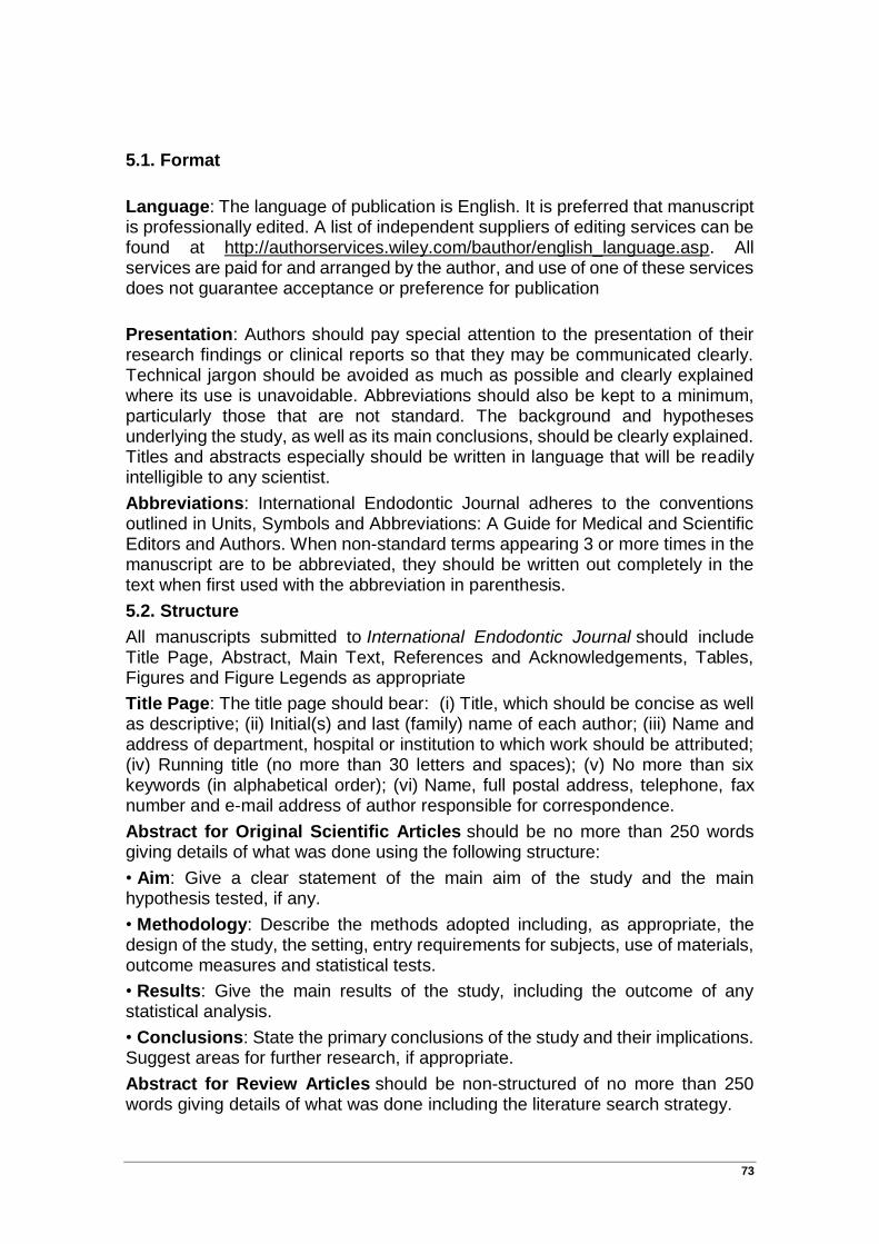
73
5.1. Format
Language: The language of publication is English. It is preferred that manuscript is professionally edited. A list of independent suppliers of editing services can be found at http://authorservices.wiley.com/bauthor/english_language.asp. All services are paid for and arranged by the author, and use of one of these services does not guarantee acceptance or preference for publication
Presentation: Authors should pay special attention to the presentation of their research findings or clinical reports so that they may be communicated clearly. Technical jargon should be avoided as much as possible and clearly explained where its use is unavoidable. Abbreviations should also be kept to a minimum, particularly those that are not standard. The background and hypotheses underlying the study, as well as its main conclusions, should be clearly explained. Titles and abstracts especially should be written in language that will be readily intelligible to any scientist.
Abbreviations: International Endodontic Journal adheres to the conventions outlined in Units, Symbols and Abbreviations: A Guide for Medical and Scientific Editors and Authors. When non-standard terms appearing 3 or more times in the manuscript are to be abbreviated, they should be written out completely in the text when first used with the abbreviation in parenthesis.
5.2. Structure
All manuscripts submitted to International Endodontic Journal should include Title Page, Abstract, Main Text, References and Acknowledgements, Tables, Figures and Figure Legends as appropriate
Title Page: The title page should bear: (i) Title, which should be concise as well as descriptive; (ii) Initial(s) and last (family) name of each author; (iii) Name and address of department, hospital or institution to which work should be attributed; (iv) Running title (no more than 30 letters and spaces); (v) No more than six keywords (in alphabetical order); (vi) Name, full postal address, telephone, fax number and e-mail address of author responsible for correspondence.
Abstract for Original Scientific Articles should be no more than 250 words giving details of what was done using the following structure:
• Aim: Give a clear statement of the main aim of the study and the main hypothesis tested, if any.
• Methodology: Describe the methods adopted including, as appropriate, the design of the study, the setting, entry requirements for subjects, use of materials, outcome measures and statistical tests.
• Results: Give the main results of the study, including the outcome of any statistical analysis.
• Conclusions: State the primary conclusions of the study and their implications. Suggest areas for further research, if appropriate.
Abstract for Review Articles should be non-structured of no more than 250 words giving details of what was done including the literature search strategy.
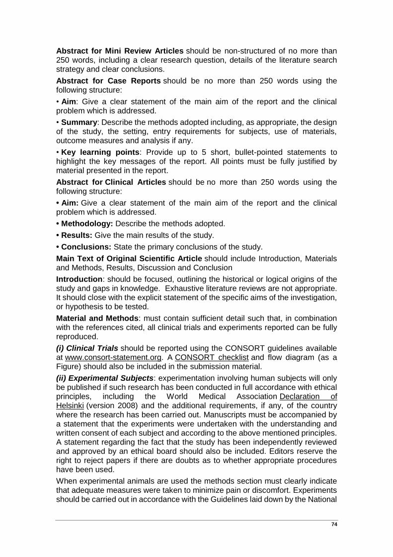
74
Abstract for Mini Review Articles should be non-structured of no more than 250 words, including a clear research question, details of the literature search strategy and clear conclusions.
Abstract for Case Reports should be no more than 250 words using the following structure:
• Aim: Give a clear statement of the main aim of the report and the clinical problem which is addressed.
• Summary: Describe the methods adopted including, as appropriate, the design of the study, the setting, entry requirements for subjects, use of materials, outcome measures and analysis if any.
• Key learning points: Provide up to 5 short, bullet-pointed statements to highlight the key messages of the report. All points must be fully justified by material presented in the report.
Abstract for Clinical Articles should be no more than 250 words using the following structure:
• Aim: Give a clear statement of the main aim of the report and the clinical problem which is addressed.
• Methodology: Describe the methods adopted.
• Results: Give the main results of the study.
• Conclusions: State the primary conclusions of the study.
Main Text of Original Scientific Article should include Introduction, Materials and Methods, Results, Discussion and Conclusion
Introduction: should be focused, outlining the historical or logical origins of the study and gaps in knowledge. Exhaustive literature reviews are not appropriate. It should close with the explicit statement of the specific aims of the investigation, or hypothesis to be tested.
Material and Methods: must contain sufficient detail such that, in combination with the references cited, all clinical trials and experiments reported can be fully reproduced.
(i) Clinical Trials should be reported using the CONSORT guidelines available at www.consort-statement.org. A CONSORT checklist and flow diagram (as a Figure) should also be included in the submission material.
(ii) Experimental Subjects: experimentation involving human subjects will only be published if such research has been conducted in full accordance with ethical principles, including the World Medical Association Declaration of Helsinki (version 2008) and the additional requirements, if any, of the country where the research has been carried out. Manuscripts must be accompanied by a statement that the experiments were undertaken with the understanding and written consent of each subject and according to the above mentioned principles. A statement regarding the fact that the study has been independently reviewed and approved by an ethical board should also be included. Editors reserve the right to reject papers if there are doubts as to whether appropriate procedures have been used.
When experimental animals are used the methods section must clearly indicate that adequate measures were taken to minimize pain or discomfort. Experiments should be carried out in accordance with the Guidelines laid down by the National
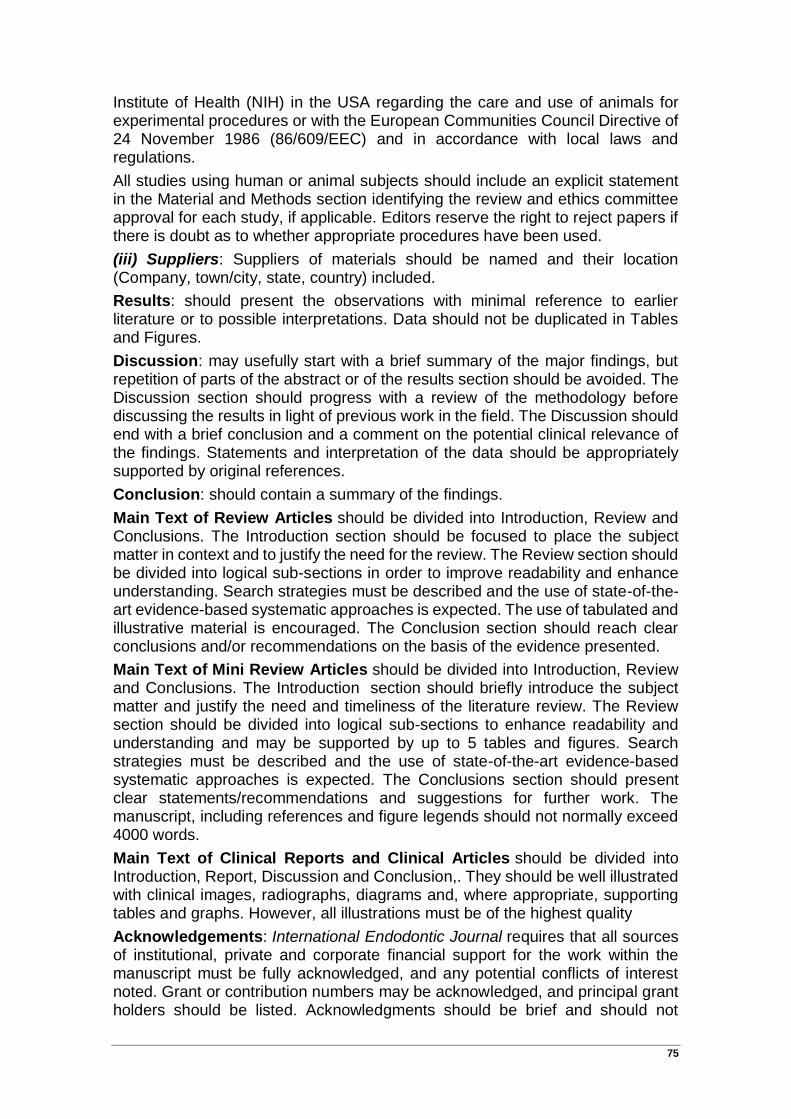
75
Institute of Health (NIH) in the USA regarding the care and use of animals for experimental procedures or with the European Communities Council Directive of 24 November 1986 (86/609/EEC) and in accordance with local laws and regulations.
All studies using human or animal subjects should include an explicit statement in the Material and Methods section identifying the review and ethics committee approval for each study, if applicable. Editors reserve the right to reject papers if there is doubt as to whether appropriate procedures have been used.
(iii) Suppliers: Suppliers of materials should be named and their location (Company, town/city, state, country) included.
Results: should present the observations with minimal reference to earlier literature or to possible interpretations. Data should not be duplicated in Tables and Figures.
Discussion: may usefully start with a brief summary of the major findings, but repetition of parts of the abstract or of the results section should be avoided. The Discussion section should progress with a review of the methodology before discussing the results in light of previous work in the field. The Discussion should end with a brief conclusion and a comment on the potential clinical relevance of the findings. Statements and interpretation of the data should be appropriately supported by original references.
Conclusion: should contain a summary of the findings.
Main Text of Review Articles should be divided into Introduction, Review and Conclusions. The Introduction section should be focused to place the subject matter in context and to justify the need for the review. The Review section should be divided into logical sub-sections in order to improve readability and enhance understanding. Search strategies must be described and the use of state-of-the-art evidence-based systematic approaches is expected. The use of tabulated and illustrative material is encouraged. The Conclusion section should reach clear conclusions and/or recommendations on the basis of the evidence presented.
Main Text of Mini Review Articles should be divided into Introduction, Review and Conclusions. The Introduction section should briefly introduce the subject matter and justify the need and timeliness of the literature review. The Review section should be divided into logical sub-sections to enhance readability and understanding and may be supported by up to 5 tables and figures. Search strategies must be described and the use of state-of-the-art evidence-based systematic approaches is expected. The Conclusions section should present clear statements/recommendations and suggestions for further work. The manuscript, including references and figure legends should not normally exceed 4000 words.
Main Text of Clinical Reports and Clinical Articles should be divided into Introduction, Report, Discussion and Conclusion,. They should be well illustrated with clinical images, radiographs, diagrams and, where appropriate, supporting tables and graphs. However, all illustrations must be of the highest quality
Acknowledgements: International Endodontic Journal requires that all sources of institutional, private and corporate financial support for the work within the manuscript must be fully acknowledged, and any potential conflicts of interest noted. Grant or contribution numbers may be acknowledged, and principal grant holders should be listed. Acknowledgments should be brief and should not
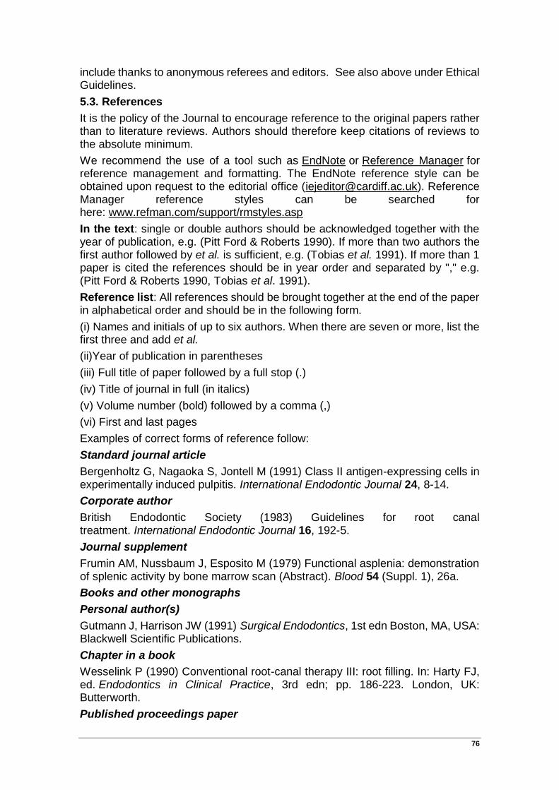
76
include thanks to anonymous referees and editors. See also above under Ethical Guidelines.
5.3. References
It is the policy of the Journal to encourage reference to the original papers rather than to literature reviews. Authors should therefore keep citations of reviews to the absolute minimum.
We recommend the use of a tool such as EndNote or Reference Manager for reference management and formatting. The EndNote reference style can be obtained upon request to the editorial office ([email protected]). Reference Manager reference styles can be searched for here: www.refman.com/support/rmstyles.asp
In the text: single or double authors should be acknowledged together with the year of publication, e.g. (Pitt Ford & Roberts 1990). If more than two authors the first author followed by et al. is sufficient, e.g. (Tobias et al. 1991). If more than 1 paper is cited the references should be in year order and separated by "," e.g. (Pitt Ford & Roberts 1990, Tobias et al. 1991).
Reference list: All references should be brought together at the end of the paper in alphabetical order and should be in the following form.
(i) Names and initials of up to six authors. When there are seven or more, list the first three and add et al.
(ii)Year of publication in parentheses
(iii) Full title of paper followed by a full stop (.)
(iv) Title of journal in full (in italics)
(v) Volume number (bold) followed by a comma (,)
(vi) First and last pages
Examples of correct forms of reference follow:
Standard journal article
Bergenholtz G, Nagaoka S, Jontell M (1991) Class II antigen-expressing cells in experimentally induced pulpitis. International Endodontic Journal 24, 8-14.
Corporate author
British Endodontic Society (1983) Guidelines for root canal treatment. International Endodontic Journal 16, 192-5.
Journal supplement
Frumin AM, Nussbaum J, Esposito M (1979) Functional asplenia: demonstration of splenic activity by bone marrow scan (Abstract). Blood 54 (Suppl. 1), 26a.
Books and other monographs
Personal author(s)
Gutmann J, Harrison JW (1991) Surgical Endodontics, 1st edn Boston, MA, USA: Blackwell Scientific Publications.
Chapter in a book
Wesselink P (1990) Conventional root-canal therapy III: root filling. In: Harty FJ, ed. Endodontics in Clinical Practice, 3rd edn; pp. 186-223. London, UK: Butterworth.
Published proceedings paper
77
DuPont B (1974) Bone marrow transplantation in severe combined immunodeficiency with an unrelated MLC compatible donor. In: White HJ, Smith R, eds. Proceedings of the Third Annual Meeting of the International Society for Experimental Rematology; pp. 44-46. Houston, TX, USA: International Society for Experimental Hematology.
Agency publication
Ranofsky AL (1978) Surgical Operations in Short-Stay Hospitals: United States-1975. DHEW publication no. (PHS) 78-1785 (Vital and Health Statistics; Series 13; no. 34.) Hyattsville, MD, USA: National Centre for Health Statistics.8
Dissertation or thesis
Saunders EM (1988) In vitro and in vivo investigations into root-canal obturation using thermally softened gutta-percha techniques (PhD Thesis). Dundee, UK: University of Dundee.
URLs Full reference details must be given along with the URL, i.e. authorship, year, title of document/report and URL. If this information is not available, the reference should be removed and only the web address cited in the text. Smith A (1999) Select committee report into social care in the community [WWW document]. URL http://www.dhss.gov.uk/reports/report015285.html [accessed on 7 November 2003]
5.4. Tables, Figures and Figure Legends
Tables: Tables should be double-spaced with no vertical rulings, with a single bold ruling beneath the column titles. Units of measurements must be included in the column title.
Figures: All figures should be planned to fit within either 1 column width (8.0 cm), 1.5 column widths (13.0 cm) or 2 column widths (17.0 cm), and must be suitable for photocopy reproduction from the printed version of the manuscript. Lettering on figures should be in a clear, sans serif typeface (e.g. Helvetica); if possible, the same typeface should be used for all figures in a paper. After reduction for publication, upper-case text and numbers should be at least 1.5-2.0 mm high (10 point Helvetica). After reduction, symbols should be at least 2.0-3.0 mm high (10 point). All half-tone photographs should be submitted at final reproduction size. In general, multi-part figures should be arranged as they would appear in the final version. Reduction to the scale that will be used on the page is not necessary, but any special requirements (such as the separation distance of stereo pairs) should be clearly specified.
Unnecessary figures and parts (panels) of figures should be avoided: data presented in small tables or histograms, for instance, can generally be stated briefly in the text instead. Figures should not contain more than one panel unless the parts are logically connected; each panel of a multipart figure should be sized so that the whole figure can be reduced by the same amount and reproduced on the printed page at the smallest size at which essential details are visible.
Figures should be on a white background, and should avoid excessive boxing, unnecessary colour, shading and/or decorative effects (e.g. 3-dimensional skyscraper histograms) and highly pixelated computer drawings. The vertical axis of histograms should not be truncated to exaggerate small differences. The line
78
spacing should be wide enough to remain clear on reduction to the minimum acceptable printed size.
Figures divided into parts should be labelled with a lower-case, boldface, roman letter, a, b, and so on, in the same typesize as used elsewhere in the figure. Lettering in figures should be in lower-case type, with the first letter capitalized. Units should have a single space between the number and the unit, and follow SI nomenclature or the nomenclature common to a particular field. Thousands should be separated by a thin space (1 000). Unusual units or abbreviations should be spelled out in full or defined in the legend. Scale bars should be used rather than magnification factors, with the length of the bar defined in the legend rather than on the bar itself. In general, visual cues (on the figures themselves) are preferred to verbal explanations in the legend (e.g. broken line, open red triangles etc.)
Figure legends: Figure legends should begin with a brief title for the whole figure and continue with a short description of each panel and the symbols used; they should not contain any details of methods.
Permissions: If all or part of previously published illustrations are to be used, permission must be obtained from the copyright holder concerned. This is the responsibilty of the authors before submission.
Preparation of Electronic Figures for Publication: Although low quality images are adequate for review purposes, print publication requires high quality images to prevent the final product being blurred or fuzzy. Submit EPS (lineart) or TIFF (halftone/photographs) files only. MS PowerPoint and Word Graphics are unsuitable for printed pictures. Do not use pixel-oriented programmes. Scans (TIFF only) should have a resolution of 300 dpi (halftone) or 600 to 1200 dpi (line drawings) in relation to the reproduction size (see below). EPS files should be saved with fonts embedded (and with a TIFF preview if possible). For scanned images, the scanning resolution (at final image size) should be as follows to ensure good reproduction: lineart: >600 dpi; half-tones (including gel photographs): >300 dpi; figures containing both halftone and line images: >600 dpi.
Further information can be obtained at Wiley Blackwell’s guidelines for figures: http:/authorservices.wiley.com/bauthor/illustration.asp. Check your electronic artwork before submitting it: http://authorservices.wiley.com/bauthor/eachecklist.asp.
5.5. Supporting Information
Publication in electronic formats has created opportunities for adding details or whole sections in the electronic version only. Authors need to work closely with the editors in developing or using such new publication formats.
Supporting information, such as data sets or additional figures or tables, that will not be published in the print edition of the journal, but which will be viewable via the online edition, can be submitted. It should be clearly stated at the time of submission that the supporting information is intended to be made available through the online edition. If the size or format of the supporting information is such that it cannot be accommodated on the journal's website, the author agrees to make the supporting information available free of charge on a permanent Web

79
site, to which links will be set up from the journal's website. The author must advise Wiley Blackwell if the URL of the website where the supporting information is located changes. The content of the supporting information must not be altered after the paper has been accepted for publication.
The availability of supporting information should be indicated in the main manuscript by a paragraph, to appear after the References, headed 'Supporting Information' and providing titles of figures, tables, etc. In order to protect reviewer anonymity, material posted on the authors Web site cannot be reviewed. The supporting information is an integral part of the article and will be reviewed accordingly.
Preparation of Supporting Information: Although provision of content through the web in any format is straightforward, supporting information is best provided either in web-ready form or in a form that can be conveniently converted into one of the standard web publishing formats:
• Simple word-processing files (.doc or .rtf) for text.
• PDF for more complex, layout-dependent text or page-based material. Acrobat files can be distilled from Postscript by the Publisher, if necessary.
• GIF or JPEG for still graphics. Graphics supplied as EPS or TIFF are also acceptable.
• MPEG or AVI for moving graphics.
Subsequent requests for changes are generally unacceptable, as for printed papers. A charge may be levied for this service.
Video Imaging: For the on-line version of the Journal the submission of illustrative video is encouraged. Authors proposing the use such media should consult with the Editor during manuscript preparation.
6. AFTER ACCEPTANCE
Upon acceptance of a paper for publication, the manuscript will be forwarded to the Production Editor who is responsible for the production of the journal.
6.1. Figures
Hard copies of all figures and tables are required when the manuscript is ready for publication. These will be requested by the Editor when required. Each Figure copy should be marked on the reverse with the figure number and the corresponding author’s name.
6.2 Proof Corrections
The corresponding author will receive an email alert containing a link to a web site. A working email address must therefore be provided for the corresponding author. The proof can be downloaded as a PDF (portable document format) file from this site. Acrobat Reader will be required in order to read this file. This software can be downloaded (free of charge) from the following Web site: www.adobe.com/products/acrobat/readstep2.html. This will enable the file to be opened, read on screen, and printed out in order for any corrections to be added. Further instructions will be sent with the proof. Hard copy proofs will be posted if no e-mail address is available; in your absence, please arrange for a

80
colleague to access your e-mail to retrieve the proofs. Proofs must be returned to the Production Editor within three days of receipt. As changes to proofs are costly, we ask that you only correct typesetting errors. Excessive changes made by the author in the proofs, excluding typesetting errors, will be charged separately. Other than in exceptional circumstances, all illustrations are retained by the publisher. Please note that the author is responsible for all statements made in his work, including changes made by the copy editor.
6.3 Early Online Publication Prior to Print
International Endodontic Journal is covered by Wiley Blackwell's Early View service. Early View articles are complete full-text articles published online in advance of their publication in a printed issue. Early View articles are complete and final. They have been fully reviewed, revised and edited for publication, and the authors' final corrections have been incorporated. Because they are in final form, no changes can be made after online publication. The nature of Early View articles means that they do not yet have volume, issue or page numbers, so Early View articles cannot be cited in the traditional way. They are therefore given a Digital Object Identifier (DOI), which allows the article to be cited and tracked before it is allocated to an issue. After print publication, the DOI remains valid and can continue to be used to cite and access the article.
6.4 Online Production Tracking
Online production tracking is available for your article through Blackwell's Author Services. Author Services enables authors to track their article - once it has been accepted - through the production process to publication online and in print. Authors can check the status of their articles online and choose to receive automated e-mails at key stages of production. The author will receive an e-mail with a unique link that enables them to register and have their article automatically added to the system. Please ensure that a complete e-mail address is provided when submitting the manuscript. Visit http://authorservices.wiley.com/bauthor/ for more details on online production tracking and for a wealth of resources including FAQs and tips on article preparation, submission and more.
6.5 Author Material Archive Policy
Please note that unless specifically requested, Wiley Blackwell will dispose of all hardcopy or electronic material submitted two months after publication. If you require the return of any material submitted, please inform the editorial office or production editor as soon as possible.
6.6 Offprints
Free access to the final PDF offprint of your article will be available via Author Services only. Please therefore sign up for Author Services if you would like to access your article PDF offprint and enjoy the many other benefits the service offers.
Additional paper offprints may be ordered online. Please click on the following link, fill in the necessary details and ensure that you type information in all of the required fields: Offprint Cosprinters. If you have queries about offprints please email [email protected]
The corresponding author will be sent complimentary copies of the issue in which the paper is published (one copy per author).
81
6.7 Author Services
For more substantial information on the services provided for authors, please see Wiley Blackwell Author Services
6.8 Note to NIH Grantees: Pursuant to NIH mandate, Wiley Blackwell will post the accepted version of contributions authored by NIH grant-holders to PubMed Central upon acceptance. This accepted version will be made publicly available 12 months after publication. For further information, see www.wiley.com/go/nihmandate
7 Guidelines for reporting of DNA microarray data
The International Endodontic Journal gives authors notice that, with effect from 1st January 2011, submission to the International Endodontic Journal requires the reporting of microarray data to conform to the MIAME guidelines. After this date, submissions will be assessed according to MIAME standards. The complete current guidelines are available athttp://www.mged.org/Workgroups/MIAME/miame_2.0.html. Also, manuscripts will be published only after the complete data has been submitted into the public repositories, such as GEO (http://www.ncbi.nlm.nih.gov/geo/) or ArrayExpress (http://www.ebi.ac.uk/microarray/submissions_overview.html), in MIAME compliant format, with the data accession number (the identification number of the data set in the database) quoted in the manuscript. Both databases are committed to keeping the data private until the associated manuscript is published, if requested.
Prospective authors are also encouraged to search for previously published microarray data with relevance to their own data, and to report whether such data exists. Furthermore, they are encouraged to use the previously published data for qualitative and/or quantitative comparison with their own data, whenever suitable. To fully acknowledge the original work, an appropriate reference should be given not only to the database in question, but also to the original article in which the data was first published. This open approach will increase the availability and use of these large-scale data sets and improve the reporting and interpretation of the findings, and in increasing the comprehensive understanding of the physiology and pathology of endodontically related tissues and diseases, result eventually in better patient care.