universidade nova de lisboa faculdade de ciências médicas marta td 2013... · department of...
TRANSCRIPT

Universidade Nova de Lisboa
Faculdade de Ciências Médicas
DNA DAMAGE INDUCED BY ACRYLAMIDE: ROLE OF
GENETIC POLYMORPHISMS IN DNA DAMAGE
LEVELS
Marta Sofia Pereira Pingarilho Fazendeiro
Doutoramento em Ciências da Vida
(Especialidade Genética)
2013
Universidade Nova de Lisboa
Faculdade de Ciências Médicas
DNA DAMAGE INDUCED BY ACRYLAMIDE: ROLE OF
GENETIC POLYMORPHISMS IN DNA DAMAGE
LEVELS
Marta Sofia Pereira Pingarilho Fazendeiro
Doutoramento em Ciências da Vida
(Especialidade Genética)
This thesis was supervised by:
Supervisor: Professor Doutor Jorge Francisco Gaspar
Co-supervisor: Professor Doutor José Rueff
2013


Acknowledgments
I want to acknowledgment tightly to my supervisor Professor Jorge Gaspar, who
invited and convinced me to join to this scientific journey. Thank you for giving me the
opportunity to play a quality scientific work, for your mentorship, availability and
scientific rigor and especially for your friendship and support.
I want to express my deepest gratitude to Professor José Rueff director of
department of Genetics of Faculdade de Ciências Médicas da Universidade Nova de
Lisboa for being my co-supervisor and for received me in this research group with so
kindness and enormous friendship.
My deepest gratitude to Professor Nuno Oliveira for all comments, opinions and
constant help those were so important throughout this work. Thank you above all for
your friendship.
I want to express my acknowledgments to all colleagues and friends of the
Genetic department of Faculdade de Ciências Médicas da Universidade Nova de Lisboa.
Each one collaborated with important knowledge that made me grow up in scientific
and also in personal terms. Everyone was very important, but I must emphasize the help
and friendship of Professor Sebastião Rodrigues, Célia Martins, Bruno Gomes, Susana
Silva and João Pereira de Lima. Thank you very much.
A very special thanks to my family, specially to my fathers, because they always
believed it would be possible. Thank you for your constant support and encouragement.
A special thanks to João Pedro and my daughters Carminho and Leonor that had to
share my attention and time with this thesis
Finally, I want to acknowledge Fundação Calouste Gulbenkian that financially
supported the work presented in this thesis, through the Grant 76438. I also thank you
Fundação para a Ciência e Tecnologia that funded this work through the Project
PTDC/SAU-OSM/105572/208 and through my PhD fellowship
(SFRH/BD/22612/2005).


i
Abstract
Acrylamide (AA) has been classified as a probable human carcinogen by IARC.
Besides being used in numerous industrial applications, AA is also present in a variety
of starchy cooked foods. This AA exposure scenario raised concerns about risk in
human health and suggests that the oral consumption of AA is an additional risk factor
for cancer. A considerable number of findings strongly suggest that the reactive
metabolite glycidamide (GA), an epoxide generated presumably by cytochrome P450
2E1, plays a central role in AA carcinogenesis.
Until now there are a scarcity of results concerning the mechanisms of
genotoxicity of AA and GA in mammalian cells. In view of that, the study described in
this thesis aims to unveil the genetic consequences of AA and GA exposure using
mammalian cells as a model system.
With this aim we evaluated the cytotoxicity of AA and GA using the MTT assay
and subsequently performed two cytogenetic end-points: chromosomal aberrations
(CAs) and sister chromatid exchanges (SCEs), in order to evaluate DNA damage
induced by these compounds in V79 Chinese hamster cell line. The results showed that
GA was more cytotoxic and clastogenic than AA.
Within the scope of this thesis the quantification of specific DNA adducts were
also performed, namely N7-(2-carbamoyl-2-hydroxyethyl)guanine (N7-GA-Gua) and
N3-(2-carbamoyl-2-hydroxyethyl)adenine (N3-GA-Ade). Interestingly, the GA
concentration and the levels of N7-GA-Gua presented a linear dose-response
relationship. Further, a very good correlation between the levels of N7-GA-Gua and the
extent of SCEs were observed.
In order to understand the mechanisms of AA-induced toxicity, the modulation
of reduced glutathione (GSH)-dependent mechanisms were studied, namely the
evaluation of the effect of buthionine sulfoximine (BSO), an effective inhibitor of GSH
synthesis, of GSH-monoethyl ester (GSH-EE), a cell permeable compound that is
intracellularly hydrolysed to GSH and also of GSH endogenously added to culture
medium,z in V79 cell line. The overall results reinforced the role of GSH in the
modulation of the cytotoxic and clastogenic effects induced by AA.

ii
Complementary to the studies performed in V79 cells, SCEs, specific DNA-
adducts and alkaline comet assay in lymphocytes from healthy donors exposed to AA
and GA were also evaluated. Both, the frequency of SCE and the quantification of
specific GA DNA adducts, produced comparable results with those obtained in V79 cell
line, reinforcing the idea that GA is far more genotoxic than AA. Further, the DNA
damaging potential of AA and GA in whole blood leukocytes evaluated by the alkaline
comet assay, showed that GA, but not AA, increases DNA damage.
Additionally, this study aimed to identify associations between DNA damage
and biomarkers of susceptibility, concerning individual genetic polymorphisms
involved in detoxification and DNA repair pathways (BER, NER, HRR and NHEJ) on
the GA-induced genotoxicity assessed by the SCE assay and by the alkaline comet
assay. The extent of DNA damage determined by the levels of SCEs induced by GA
seems to be modulated by GSTP1 (Ile105Val) and GSTA2 (Glu210Ala) genotypes.
Moreover, the results obtained from the comet assay suggested associations between
DNA damage and polymorphisms of BER (MUTYH Gln335His and XRCC1
Gln399Arg) and NER (XPC Ala499Val and Lys939Gln) genes, either alone or in
combination.
The overall results from this study contribute to a better understanding of the
genotoxicity and carcinogenicity of AA and GA in mammalian cells, as well as the
knowledge about the variability in individual susceptibility involved in detoxification
and repair of DNA damage due to these dietary xenobiotics.
Keywords: acrylamide; glycidamide; sister chromatid exchange; chromosomal
aberrations; DNA-adducts; comet assay; SNPs; genotoxicity; carcinogenicity.

iii
Resumo
Em 1994 a acrilamida (AA) foi classificada pela IARC como um provável
cancerígeno para o homem. Para além da utilização de AA em numerosas aplicações
industriais, a AA está também presente numa grande variedade de alimentos ricos em
amido e processados a temperaturas elevadas. Esta exposição através da ingestão de
produtos alimentares despoletou elevadas preocupações ao nível do risco para a saúde
pública e poderá implicar um risco adicional para o aparecimento de cancro. A
glicidamida (GA), o metabolito epóxido formado a partir da oxidação da AA
provavelmente através do citocromo P450 2E1, é considerada por vários estudos, o
principal responsável pela carcinogenicidade da AA.
Actualmente existe uma escassez de resultados relativamente aos mecanismos de
genotoxicidade da AA e GA em células de mamífero. Por este motivo, o objectivo deste
estudo centra-se na avaliação das consequências genéticas da exposição à AA e GA,
recorrendo-se para tal ao uso de células de mamífero como modelo.
Tendo como base este objectivo avaliou-se a citotoxicidade da AA e GA, através
do ensaio do MTT, e realizaram-se dois testes citogenéticos, o teste das aberrações
cromossómicas (CAs) e o teste da troca de cromátides irmãs (SCEs), de modo a avaliar
as lesões de DNA induzidas por estes compostos em células de hamster Chinês V79. Os
resultados globalmente mostraram que a GA é mais citotóxica e clastogénica do que a
AA.
No âmbito deste trabalho, foi também efectuada a quantificação de aductos
específicos de DNA, nomeadamente N7-(2-carbamoil-2-hidroxietil)guanina (N7-GA-
Gua) e N3-(2-carbamoil-2-hidroxietil)adenina (N3-GA-Ade). Os resultados obtidos
permitem afirmar que os níveis de N7-GA-Gua e a concentração de GA apresentam
uma relação linear dose-resposta. Foi também identificada uma óptima correlação entre
os níveis de N7-GA-Gua e a frequência de troca de cromátides irmãs.
Adicionalmente, e de forma a compreender os mecanismos de toxicidade da AA,
estudaram-se os mecanismos dependentes da modulação do glutationo reduzido (GSH),
nomeadamente da butionina sulfoximina (BSO), um inibidor da síntese de GSH, do
GSH-monoetil estér (GSH-EE), um composto permeável nas células e que é intra-
celularmente hidrolisado a GSH e ainda do GSH adicionado exogenamente ao meio de

iv
cultura, em células V79. Os resultados obtidos reforçaram o papel da modulação do
GSH nos efeitos de citotoxicidade e clastogenicidade da AA.
Para além dos estudos efetuados com células V79, procedeu-se também à
determinação da frequência de SCEs, à quantificação de aductos específicos de DNA,
bem como ao ensaio do cometa alcalino em amostras de dadores saudáveis expostos à
AA e GA. Tanto os resultados obtidos através do ensaio das SCE, como pela
quantificação de aductos específicos de DNA, ambos efectuados em linfócitos
estimulados, originaram resultados comparáveis aos obtidos anteriormente para as
células V79, reforçando a ideia de que a GA é bastante mais genotóxica do que a AA.
Por outro lado, os resultados obtidos pelo ensaio do cometa para exposição à AA e GA
mostraram que apenas esta última aumenta o nível das lesões de DNA.
Outro objectivo deste trabalho, foi a identificação de possíveis associações
existentes entre as lesões de DNA, quantificadas através do ensaio das SCEs e do
cometa, e biomarcadores de susceptibilidade, tendo em conta os polimorfismos
genéticos individuais envolvidos na destoxificação e nas vias de reparação do DNA
(BER, NER, HRR e NHEJ) em linfócitos expostos à GA. Tal permitiu identificar
associações entre os níveis de lesão de DNA determinados através do ensaio das SCEs,
e os polimorfismos genéticos estudados, apontando para uma possível associação entre
o GSTP1 (Ile105Val) e GSTA2 (Glu210Ala) e a frequência de SCEs. Por outro lado, os
resultados obtidos através do ensaio do cometa sugerem uma associação entre as lesões
de DNA e polimorfismos da via BER (MUTYH Gln335His e XRCC1 Gln39Arg) e da
via NER (XPC Ala499val e Lys939Gln), considerando os genes isoladamente ou
combinados.
Estes estudos contribuem para um melhor entendimento da genotoxicidade e
carcinogenicidade da AA e GA em células de mamífero, bem como da variabilidade da
susceptibilidade individual na destoxificação e reparação de lesões de DNA provocadas
pela exposição a estes xenobióticos alimentares.
Palavras chave: acrilamida; glicidamida; troca de cromátides irmãs; aberrações
cromossómicas, aductos de DNA; ensaio do cometa; SNPs; genotoxicidade;
carcinogenicidade.

v
List of publications and communications
From the results presented in this thesis, the following papers were published in
international refereed journals:
1. Induction of Sister Chromatid Exchange by acrylamide and glycidamide in human
lymphocytes: Role of polymorphisms in detoxification and DNA-repair genes in the
genotoxicity of glycidamide
Pingarilho M, Oliveira NG, Martins C, Gomes BC, Fernandes AS, Martins V, Labilloy
A, Lima JP, Rueff J and Gaspar JF.
Mutation Research (2013), 752, 1-7.
2. Genetic polymorphisms in detoxification and DNA repair genes and susceptibility to
glycidamide-induced DNA damage
Pingarilho M, Oliveira NG, Martins C, Fernandes AS, Lima JP, Rueff J and Gaspar JF.
Journal of Toxicology and Environmental Health, Part A (2012) 75:13-15, 920-933.
3. Cytotoxicity and chromosomal aberrations induced by acrylamide in V79 cells: role
of glutathione modulators.
Oliveira NG, Pingarilho M, Martins C, Fernandes AS, Vaz S, Martins V, Rueff J,
Gaspar JF.
Mutation Research (2009) 676, 87-92.
4. Cytogenetic damage induced by acrylamide and glycidamide in mammalian cells:
correlation with specific glycidamide-DNA adducts.
Martins C, Oliveira NG, Pingarilho M, Gamboa da Costa G, Martins V, Marques MM,
Beland FA, Churchwell MI, Doerge DR, Rueff J, Gaspar JF.
Toxicology Science (2007) 95, 383-390.

vi
This thesis also contains methods published in the following papers:
1. Genotoxic effects of occupational exposure to lead and influence of polymorphisms
in genes involved in lead toxicokinetics and in DNA repair
García-Lestón J, Roma-Torres J, Vilares M, Pinto R, Prista J, Teixeira JP, Mayan O,
Conde J, Pingarilho M, Gaspar JF, Pásaro E, Méndez J, Laffon B.
Environment International (2012), 43: 29-36.
2. Genotoxic effects of doxorubicin in cultured human lymphocytes with different
glutathione S-transferase genotypes
Ramos DL. Gaspar JF, Pingarilho M, Gil OM, Fernandes AS, Rueff J, Oliveira NG
Mutation Research (2011), 724: 28-34.
The results related with this study were also presented in scientific meetings in the
following poster communications:
1. Gene expression induced by Acrylamide and Glycidamide in mammalian cells.
Pingarilho M, Lima JP, Martins C, Rueff J, Gaspar JF
Post-GWAS Horizons in Molecular Epidemiology: Digging Deeper into the
Environment, Genome Wide Association Studies (GWAS), November 2012,
Hollywood, California, USA. (The abstract relative to this poster presentation will be
reprinting as a supplement associated with the online journal of Cancer Epidemiology,
Biomarkers & Prevention.)
2. Genetic variation in the in vitro genotoxic response to glycidamide and gene
expression of DNA repair genes.
Pingarilho M, Oliveira NG, Martins C, Gomes BC, Fernandes AS, Martins V, Silva
AR, Rueff J and Gaspar JF
American Association for Cancer research, 2011, Orlando, Flórida, USA.

vii
3. Glycidamide-induced cytotoxic and genotoxic effects in human mammary MCF10A
cells.”
Bandarra S, Gaspar J, Pingarilho M, Gil OM, Fernandes AS, Miranda J, Castro M,
Rueff J, Oliveira NG.
Society for Free Radical Research - Europe Meeting, September 2010, Oslo, Norway.
4. Interindividual Variability in the In Vitro Genotoxic Response to Glycidamide: Role
of BER and NER Genetic Polymorphisms.
Pingarilho M., Oliveira NG, Gomes BC,
Martins C, Rueff J and Gaspar JF,
Environmental Mutagen Society, 2009, St. Louis, Missouri, USA.
5. Evaluation and modulation of the cytotoxic potential of acrylamide and glycidamide
Oliveira N G, Martins C, Vaz S, Martins V, Pingarilho M, Rueff J, Gaspar JF
Gordons Research Conference, 2008, Ventura, California, USA.
6. Glycidamide-DNA. adducts and sister chromatid exchanges in human lymphocytes
exposed to acrylamide and glycidamide.
Pingarilho M, Martins C, Oliveira NG, Vaz S, Gamboa da Costa G, Martins V,
Marques MM,. Beland FA, Churchwell MI, Doerge DR, Rueff J and Gaspar JF.
98th
Annual Meeting of the American Association for Cancer Research, 2007, Los
Angeles, California, USA.
7. Role of Gluthatione Status on the Cytotoxity induced by Acrilamide and Glycidamide
in Mammalian Cells.
Oliveira NG, Martins C, Vaz S, Martins V, Pingarilho M, Ruef J and Gaspar J.
13th
Annual Meeting of Free Radical Biology & Medicine, November 2006, Denver,
Colorado, USA.
8. Correlation between DNA adduct formation and cytogenetic damage induction in
mammalian cells exposed to acrylamide and glycidamide.
Costa GG, Oliveira NG, Martins C, Pingarilho M, Rueff J, Gaspar JF, Martins V,
Marques MM,. Beland FA, Churchwell MI and Doerge DR
36th
Annual Meeting of the European Environmental Mutagen Society, July 2006,
Prague.
viii
9. Cytogenetic Damage Induced by Acrylamide and Glycidamide in Mammalian Cells:
Correlation with specific Glycidamide DNA-adducts”.
Oliveira NG, Martins C, Pingarilho M, Costa GG, Martins V, Marques MM,. Beland
FA, Churchwell MI, Doerge DR, Rueff J and Gaspar JF.
97th
Annual Meeting of the American Association for Cancer Research, April 2006,
Washington, DC, USA.
Within the scope of this thesis, the following oral communication was presented in a
scientific meeting:
In vitro genotoxic response to glycidamide: Role of individual genetic polymorphisms
of biotransformation and DNA repair genes.
Gaspar J.F, Pingarilho M, Oliveira NG, Martins C, Gomes BC, Fernandes AS, Martins
V, Silva AR, Rueff J.
International Conference on Occupational and Environmental Health. Porto, Portugal,
Outubro de 2011.

ix
Table of contents
Abstract .............................................................................................................................. i
Resumo ............................................................................................................................ iii
List of publications and communications ......................................................................... v
Table of contents ............................................................................................................. ix
List of Figures ................................................................................................................. xv
List of Tables ................................................................................................................ xvii
Abbreviations ................................................................................................................ xix
Chapter 1 – General Introduction ................................................................................. 1
1.1. Causes of Cancer ................................................................................................... 3
1.1.1. Principles ............................................................................................................ 3
1.1.2. Food contaminants .............................................................................................. 5
1.2. Biomarkers of genetic DNA damage......................................................................... 8
1.2.1. Principles ............................................................................................................ 8
1.2.2. Biomarker of exposure ........................................................................................ 9
1.2.2.1. Biomarkers of internal dose ....................................................................... 10
1.2.2.2. Biomarkers of effective dose...................................................................... 10
1.2.3. Biomarkers of effects ........................................................................................ 12
1.2.3.1. Micronucleus (MN) .................................................................................... 13
1.2.3.2. Chromosomal aberrations (CAs) ................................................................ 13
1.2.3.3. Sister Chromatid Exchange (SCE) ............................................................. 14
1.2.3.4. Comet assay................................................................................................ 15
1.2.4. Biomarker of susceptibility ............................................................................... 16

x
1.2.4.1. Main Metabolism/Detoxification polymorphisms ..................................... 17
1.2.4.2. DNA repair enzymes polymorphisms ........................................................ 20
1.3. DNA Damage .......................................................................................................... 21
1.3.1. Principles .......................................................................................................... 21
1.3.2. DNA Damage Repair System ........................................................................... 22
1.3.2.1. Direct Repair (DR) ..................................................................................... 22
1.3.2.2. Base Excision Repair (BER) ...................................................................... 23
1.3.2.4. Double Strand Breaks................................................................................. 27
1.3.2.5. Mismatch Repair (MMR) ........................................................................... 29
1.4. Reactive Oxygen Species ........................................................................................ 32
1.4.1. Principles .......................................................................................................... 32
1.4.2. Antioxidant defenses ......................................................................................... 34
1.5. Acrylamide .............................................................................................................. 35
1.5.1. Acrylamide: production, uses and sources of exposure .................................... 35
1.5.2. Acrylamide dietary exposure ............................................................................ 37
1.5.3. Acrylamide Toxicokinetics ............................................................................... 40
1.5.4. Adducts of AA and GA..................................................................................... 44
1.5.4.1. Hemoglobin adducts ................................................................................... 44
1.5.4.2. DNA adducts .............................................................................................. 45
1.5.5. Genotoxicity of acrylamide and glycidamide ................................................... 46
1.5.5.1. Genotoxicity in Prokaryotes ....................................................................... 47
1.5.5.2. Genotoxicity in mammalian cells ............................................................... 47
1.5.6. Animal carcinogenicity ..................................................................................... 49
1.5.7. Epidemiologic studies ....................................................................................... 50
1.6. References: .............................................................................................................. 55

xi
Chapter 2 – Problem and Method ............................................................................... 75
2.1. Problem and Method ............................................................................................... 77
2.2. References ............................................................................................................... 80
Chapter 3 – Cytogenetic damage induced by acrylamide and glycidamide in
mammalian cells: correlation with specific DNA adducts ........................................ 83
Abstract ........................................................................................................................... 84
3.1. Introduction ............................................................................................................. 85
3.2. Materials and methods ............................................................................................. 87
3.2.1. Chemicals .......................................................................................................... 87
3.2.2. V79 Cells culture .............................................................................................. 87
3.2.3. MTT cytotoxicity assay .................................................................................... 88
3.2.4. Chromosomal aberration assay ......................................................................... 88
3.2.5. Sister Chromatid assay ...................................................................................... 89
3.2.6. DNA adducts ..................................................................................................... 90
3.2.6.1. Chemical exposure and DNA extraction .................................................... 90
3.2.6.2. DNA quantification .................................................................................... 90
3.2.6.3. Quantification of DNA adducts.................................................................. 91
3.2.7. Statistical analyses ............................................................................................ 91
3.3. Results ..................................................................................................................... 91
3.4. Discussion ................................................................................................................ 97
3.5. References ............................................................................................................. 102
Chapter 4 – Cytotoxicity and chromosomal aberrations induced by acrylamide in
V79 cells: role of glutathione modulators ................................................................. 107
Abstract ......................................................................................................................... 108
4.1. Introduction ........................................................................................................... 109
4.2. Materials and methods ........................................................................................... 110

xii
4.2.1. Chemicals ........................................................................................................ 110
4.2.2. MTT reduction assay ...................................................................................... 111
4.2.3. Chromosomal aberrations assay ..................................................................... 111
4.2.4. GSH conjugation assay ................................................................................... 112
4.2.5. Statistical analysis ........................................................................................... 113
4.3. Results ................................................................................................................... 113
4.3.1. MTT reduction assay ...................................................................................... 113
4.3.2. Chromosome aberration assay ........................................................................ 116
4.3.3. GSH conjugation assay ................................................................................... 118
4.4. Discussion .............................................................................................................. 120
4.5. References ............................................................................................................. 124
Chapter 5 – Induction of sister chromatid exchanges by acrylamide and
glycidamide in human lymphocytes: Role of polymorphisms in detoxification and
DNA-repair genes in the genotoxicity of glycidamide ............................................. 129
Abstract ......................................................................................................................... 130
5.1. Introduction ........................................................................................................... 131
5.2. Materials and Methods .......................................................................................... 133
5.2.1. Chemicals ........................................................................................................ 133
5.2.2. Blood samples collection ................................................................................ 133
5.2.3. Lymphocytes culture ....................................................................................... 134
5.2.4. Sister Chromatid Exchange Assay .................................................................. 134
5.2.5. Mitotic Index ................................................................................................... 135
5.2.6. DNA adducts ................................................................................................... 135
5.2.6.1. Chemical exposure and DNA extraction. ................................................. 135
5.2.7. DNA quantification. ........................................................................................ 135
5.2.8. Quantification of DNA adducts. ..................................................................... 136
5.2.9. Genotyping ...................................................................................................... 136

xiii
5.2.9.1. Detoxification pathways ........................................................................... 136
5.2.9.2. DNA repair Pathways .............................................................................. 137
5.2.10. Statistical Analysis ........................................................................................ 138
5.3. Results ................................................................................................................... 141
5.3.1. GA markedly increases the formation of SCE in human lymphocytes .......... 141
5.3.2. GA-induced SCE showed inter-individual variability .................................... 142
5.3.3. DNA-adducts levels induced by AA and GA ................................................. 143
5.3.3. Role of genetic polymorphisms in induction of SCE by GA ......................... 145
5.4. Discussion .............................................................................................................. 151
5.5. References ............................................................................................................. 156
Chapter 6 – Genetic polymorphisms in detoxification and DNA repair genes and
susceptibility to glycidamide-induced DNA damage ............................................... 161
Abstract ......................................................................................................................... 162
6.1. Introduction ........................................................................................................... 163
6.2.Materials and Methods ........................................................................................... 164
6.2.1.Chemicals ......................................................................................................... 164
6.2.2. Blood Sample Collection ................................................................................ 165
6.2.3. Comet Assay ................................................................................................... 165
6.2.5. Genotyping ...................................................................................................... 166
6.2.5.1. Detoxification pathways ........................................................................... 167
6.2.5.2. DNA repair pathways ............................................................................... 167
6.2.6. Statistical Analysis .......................................................................................... 168
6.3. Results: .................................................................................................................. 168
6.4. Discussion: ............................................................................................................ 178

xiv
Chapter 7 – Concludind remarks and futures prospects ........................................ 187
7.1. Concluding remarks and future prospects ............................................................. 189
7.2. References ............................................................................................................. 194
Supplement .................................................................................................................. 195

xv
List of Figures
Page
Fig. 1.1 Relation of events and biomarker classification
9
Fig. 1.2 DNA damage, repair mechanisms and consequences
21
Fig. 1.3 Base excision repair mechanisms in mammalian cells
25
Fig. 1.4 Mechanism of nucleotide excision repair (NER). A- global genome repair
and B- transcription-coupled repair
27
Fig. 1.5 Double strand break/recombinational repair. A- Homologous recombination
and B- Nonhomologous end-joining
29
Fig. 1.6 Mismatch repair pathway.
31
Fig. 1.7 The sources and cellular responses to reactive oxygen species
33
Fig. 1.8 Proposed pathway for the formation of AA after thermal processing
36
Fig. 1.9 CYP2E1 mediated biotransformation of AA to GA
41
Fig. 1.10 Proposed mammalian metabolic pathway for AA.
42
Fig. 1.11 Chemical structures of major DNA adducts of AA and GA
45
Fig. 3.1 CYP2E1 mediated biotransformation of acrylamide to glycidamide and
chemical structures of the depurinating adducts
86
Fig. 3.2 Cytotoxicity of acrylamide and glycidamide in V79 cells (MTT assay).
92
Fig. 4.1 Effect of glutathione modulators on cell viability presented by V79
fibroblasts exposed to acrylamide (% MTT reduction). (A) Effect of BSO
(0.1 mM) pre-treatment (14 h). (B) Effect of GSH-EE (1.0 and 2.5 mM) pre-
treatment (14 h). (C) Effect of GSH (1.0 mM) co-incubation (24 h).
115
Fig. 4.2 Effect of glutathione modulators on the induction of chromosomal
aberrations by acrylamide (2.0 mM) in V79 cells. (A) Effect of BSO (0.1
mM) or GSH-EE (1.0 mM) pre-treatment (14 h). (B) Effect of GSH (1.0 and
3.0 mM) co-incubation (24 h).
117

xvi
Fig. 4.3 In vitro non-enzymatic GSH conjugation studies with acrylamide (AA) using
the fluorimetric monochlorobimane (MCB) assay. (A) Dose-effect curves of
GSH-MCB fluorescence intensity presented by GSH alone or GSH
incubated 1 h with AA (0.5 mM). (B) Dose-effect curve of free GSH (%)
observed upon incubation of GSH (0.5 mM) during 1 hour with different
concentrations of AA. (C) Dose-effect curve of free GSH (%) observed upon
incubation of GSH (1.0 mM) during 24 h with different concentrations of
AA.
119-120
Fig. 5.1 Chemical structures of N7-(2-carbamoyl-2-hydroxyethyl)guanine (N7-GA-
Gua) and N3-(2-carbamoyl-2-hydroxyethyl)adenine (N3-GA-Ade)
132
Fig. 5.2 Sister chromatid exchanges (SCE/metaphase) induced by acrylamide (AA)
and glycidamide (GA) in cultured lymphocytes from two donors
141
Fig. 5.3 Sister chromatid exchanges (SCE/metaphase) induced by glycidamide (GA)
in cultured lymphocytes from 13 donors. (A) Individual frequencies of
SCE/metaphase (B) Collective average values of SCE/metaphase
143
Fig. 5.4 Correlation between N7-GA-Gua adduct levels and SCE in lymphocyte
exposed to glycidamide (100–750 µM) for two independent donors
145
Fig. 6.1 Percent DNA in Tail (%Tail DNA) induced by AA and GA in whole blood
leucocytes from a healthy donor
169
Fig. 6.2 Percent DNA in Tail (% Tail DNA) induced by GA in whole blood
leukocytes for 25 healthy donors. (A) Individual values of % Tail DNA. (B)
Collective values of % Tail DNA
170
xvii
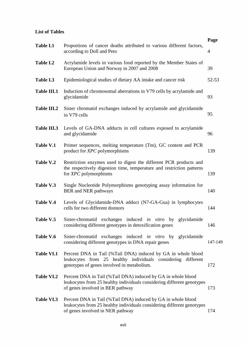
List of Tables
Page
Table I.1 Proportions of cancer deaths attributed to various different factors,
according to Doll and Peto
4
Table I.2 Acrylamide levels in various food reported by the Member States of
European Union and Norway in 2007 and 2008
39
Table I.3 Epidemiological studies of dietary AA intake and cancer risk
52-53
Table III.1 Induction of chromosomal aberrations in V79 cells by acrylamide and
glycidamide
93
Table III.2 Sister chromatid exchanges induced by acrylamide and glycidamide
in V79 cells
95
Table III.3 Levels of GA-DNA adducts in cell cultures exposed to acrylamide
and glycidamide
96
Table V.1 Primer sequences, melting temperature (Tm), GC content and PCR
product for XPC polymorphisms
139
Table V.2 Restriction enzymes used to digest the different PCR products and
the respectively digestion time, temperature and restriction patterns
for XPC polymorphisms
139
Table V.3 Single Nucleotide Polymorphisms genotyping assay information for
BER and NER pathways
140
Table V.4 Levels of Glycidamide-DNA adduct (N7-GA-Gua) in lymphocytes
cells for two different donnors
144
Table V.5 Sister-chromatid exchanges induced in vitro by glycidamide
considering different genotypes in detoxification genes
146
Table V.6 Sister-chromatid exchanges induced in vitro by glycidamide
considering different genotypes in DNA repair genes
147-149
Table VI.1 Percent DNA in Tail (%Tail DNA) induced by GA in whole blood
leukocytes from 25 healthy individuals considering different
genotypes of genes involved in metabolism.
172
Table VI.2 Percent DNA in Tail (%Tail DNA) induced by GA in whole blood
leukocytes from 25 healthy individuals considering different genotypes
of genes involved in BER pathway
173
Table VI.3 Percent DNA in Tail (%Tail DNA) induced by GA in whole blood
leukocytes from 25 healthy individuals considering different genotypes
of genes involved in NER pathway
174
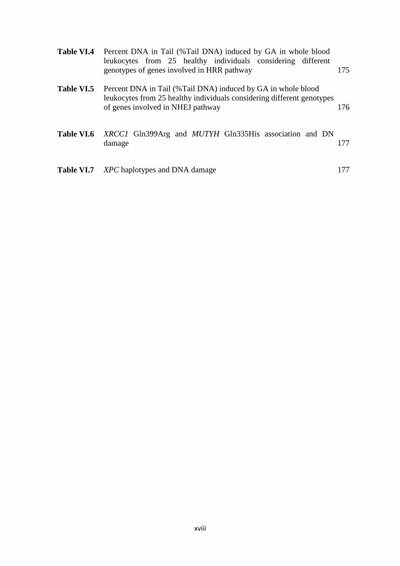
xviii
Table VI.4 Percent DNA in Tail (%Tail DNA) induced by GA in whole blood
leukocytes from 25 healthy individuals considering different
genotypes of genes involved in HRR pathway
175
Table VI.5 Percent DNA in Tail (%Tail DNA) induced by GA in whole blood
leukocytes from 25 healthy individuals considering different genotypes
of genes involved in NHEJ pathway
176
Table VI.6 XRCC1 Gln399Arg and MUTYH Gln335His association and DNA
damage
177
Table VI.7 XPC haplotypes and DNA damage
177

xix
Abbreviations
8-OHdG 8-hydroxy-2’-deoxyguanosine
AA Acrylamide
ACEG Aberrant cells excluding gaps
ACIG Aberrant cells including gaps
AFB1 Aflatoxin B1
AP Apurinic site
BER Base excision repair
BrDU 5-Bromo-2’deoxyuridine
BSO buthionine sulfoximine
CAs Chromosomal aberrations
CAT Catalase
CBMN Cytokinesis block micronucleus
CYP 2E1 Cytochrome P450 2E1
DMSO Dimethylsulphoxide
DNA Deoxyribonucleic acid
DR Direct repair
EDTA Ethylynediaminetetractic acid
EPHX Epoxide hydrolase
FCS Fetal calf serum
FFQ Food frequency questionnaire
FPG Fluorescence-plus-Giemsa
GA Glycidamide
GC-MS Gaseous chromatography coupled with mass spectrometry
GG-NER Global genome nucleotide excision repair
GP Genetic olymorphism
GPX Glutathione peroxidase
GSH L-Glutathione reduced
GSH-EE GSH-monoethyl ester
GST Glutathione S-transferase
Hb Hemoglobin
HCA Heterocyclic amine
MGHT Methyltranferase

xx
HPLC-MS/MS High pressure liquid chromatography coupled with double mass
spectrometry
HRR Homologous recombination repair
HuGE Human genome epidemiology
IARC International agency for research on cancer
LMP Low melting point
MCB Monochlorobimane
MI Mitotic index
MMR Mismatch repair
MN Micronucleus
mRNA Messenger ribonucleic acid
MTT 3-(4,5-dimethylthiazol-2-yl)-2,5-diphenyl-2H-tetrazolium
bromide
N1-GA-dA N1-(2-carboxy-2-hydroxyethyl)-2’-deoxyadenosine
N3-GA-Ade N3-(2-carbamoyl-2-hydroxyethyl)adenine
N7-GA-Gua N7-(2-carbamoyl-2-hydroxyethyl)guanine
NAT N-acetyltransferase
NER Nucleotide Excision repair
NHEJ Non homologous end-joining
NMP Normal melting point
NOCs N-nitroso compounds
O6MeGua O
6-methylguanine
PAHs Polycyclic aromatic hdrocarbons
PAs Pyrrolizidine alkaloids
PBL Peripheral blood lymphocytes
PBS phosphate buffered saline
PCR Polymerase chain reaction
PHA Phytohemagglutinin
RFLP Restriction fragment length polymorphism
RFU Relative fluorescence units
RNS Reactive nitrogen specie
ROS Reactive oxygen species
RS Reactive species
SCE Sister Chromatid exchange

xxi
SH Sulfydryl
SOD Superoxide dismutase
SSB Single strand breaks
SULT Sulfotransferase
TCR Transcriptional-coupled repair
TTD Trichothiodystrophy
UGT UDP-glucuronosyltranferase
UV Ultraviolet
UVSS UV-sensitive syndrome


Chapter 1
General introduction


3
1.1. Causes of Cancer
1.1.1. Principles
Cancer is a leading cause of death all over the world and is characterized by
uncontrolled cellular growth as a result of changes in the genetic and epigenetic
information of cells. Each year, tens of millions of people are diagnosed with cancer and
more than half of the patients eventually die from it. Moreover, cancer rates could
further increase by 50% to 15 million new cases in the year 2020, according to the
World Cancer Report [1, 2].
It is known that about 5-10% of all cancers are caused by genetic defects, while
90-95% are caused by environmental factors and lifestyle, including diet (30-35%),
tobacco smoking (25-30%) and alcohol (4-6%) [1]. Cancer related with genetic defects
can result directly from inherited mutated genes. However, the majority involves
alterations or damage accumulation over time of the genetic material within cells [3].
This damage can be caused by both endogenous (internal) and exogenous
(environmental) factors, known as important for cancer development [1]. The
endogenous causes can be inherited germ line mutation, oxidative stress, generated
through normal oxidative metabolism and pathophysiologic states, such as
inflammation [1, 3]. There are also several known exogenous factors including tobacco
smoking, infectious agents (e.g. viruses, bacteria and parasites), drug intake, radiation,
that can damage DNA, both directly by causing breaks in DNA strands and indirectly
by interacting with water molecules and generating reactive oxygen species (ROS),
industrial chemicals and carcinogenic agents in food and drink that are established as
carcinogenic by IARC [3].
Already in 1981 Doll and Peto [4] identified and attempted to quantify the
causes of cancer. The factors related with cancer and their relation with the proportions
of cancer deaths are listed in next table.

4
Table I.1-Proportions of cancer deaths attributed to various different factors, according
to Doll and Peto (from [4]).
Factor or class of factors Percent of all cancer deaths
Best estimate Range of acceptable estimates
Tobacco 30 25-40
Alcohol 3 2-4
Diet 35 10-70
Food additives <1 -5-2 a
Reproductive and sexual
behavior
7 1-13
Occupation 4 2-8
Pollution 2 <1-5
Industrial products <1 <1-2
Medicines and medical
procedures
1 0.5-3
Geophysical factors b
3 2-4
Infection 10? 1-?
Unknown ? ? a Allowing for a possibly protective effect of antioxidants and other preservatives.
b Only about 1% could reasonably be described as “avoidable”. Geophysical factors also
cause a much greater proportion of nonfatal cancers (up to 30% of all cancers, depending on
ethnic mix and latitude) because of the importance of UV light in causing the relatively nonfatal
basal cell and squamous cell carcinomas of sunlight-exposed skin.
According to the previous table, the cause of 97% of all human cancers is
explainable and it was estimated that 35% of cancer deaths might be avoidable through
changes in diet [4-6]. Diet has long been recognized as potentially important modifiers
of cancer risk, beyond that, human beings are often being exposed to carcinogenic
factors during their life, some of which are nutritional factors [1]. It is important to note
that during their life a human being ingests about 15 tons of dry matter in the form of
food [7]. Although many foodstuffs contain genotoxic compounds, the majority of these
only occur at low levels, however, multiple genotoxic substances in the same food may
result in cumulative or synergistic actions leading to neoplasia in humans [8]. These
findings moved attention away from environmental factors such as pollution or viruses
or occupational factors, and turned the focus instead onto dietary factors as a major
contributor to disease risk [9]. In the same way, through the 1970s and 1980s, many

5
chemicals from various sources (e.g. environment, occupation and diet) were tested for
mutagenic effects with the Ames test and concluded that natural chemicals, present in
human diet as complex mixtures may be a more important source of human mutation
than environmental or occupational exposure [9].
1.1.2. Food contaminants
Food contaminants can be classified as genotoxic and non-genotoxic mutagens
according to the mechanistic view of carcinogenesis. Genotoxic agents begin their
action at the DNA level, causing DNA damage (gene point mutations, deletions and
insertions, recombination, rearrangements and amplifications, as well as chromosomal
aberrations). Non-genotoxic agents presumably affect indirectly the cell through tumor
promoters, however their modes of action are less defined. These non-genotoxic agents
are generally macro-components, e.g. high fat [1].
Genotoxic mutagens are frequently natural products that can be avoided. For
instance, through fungal contamination, mycotoxins (e.g. Aflatoxin B1); or
anthropogenic chemicals produced through cooking or preserving methods, (e.g.
heterocyclic amines (HCAs), polycyclic aromatic hydrocarbons (PAHs), N-nitroso
compounds (NOCs) and AA). On the other hand, there are also genotoxic mutagens in
natural products that can be present in food and are unavoidable (e.g. Ptaquiloside and
Pyrrolizidine alkaloids (PAs)). Furthermore, there are chemicals intentionally added to
foods or food coloring, that can act as genotoxic agents, however these cause much less
concern, since they are added intentionally [1, 5, 9, 10].
One of the most important genotoxic food carcinogens is aflatoxin B1 (AFB1).
This mycotoxin is produced by the mold Aspargelius flavus, which grows on poorly
(hot and humid climate) stored foods including corn, peanuts and rice [1, 9]. Through
epidemiologic studies, AFB1 has shown to increase carcinogenic risk in humans. These
toxins proved to be very important liver carcinogens, especially in combination with
chronic infection with hepatitis B virus [11-17]. AFB1 initiates its action with metabolic

6
activation by cytochrome P450, forming an exo-8,9-epoxide and subsequent adduct
formation producing DNA damage [18, 19].
Other important carcinogenic formed within muscle foods (beef, lamb, and
poultry, but also in fish) cooked at high temperatures (e.g. frying, broiling and
barbecuing) are the heterocyclic amines [1, 5, 9, 10]. These are formed through a
pyrolysis process from amino acids, proteins and creatines of the meat. In humans there
is good epidemiologic evidence correlating the consumption of food containing high
levels of HCAs and cancer, namely colorectal [14, 20-24], breast [25-27], prostate [14,
28-30] and pancreatic cancers [31-33]. HCA carcinogenesis mechanism encompasses a
bioactivation of N-hydroxylation by CYP1A2 and subsequent esterification. The
nitrenium ion is likely the ultimate carcinogen, capable of binding guanine at position
C8, causing altered DNA sequences with subsequent base substitution, deletion and
insertion [1, 34].
PAH compounds are also considered food carcinogens formed during
incomplete combustion of organic matter during food processing (smoking, barbecuing
and grilling). PAH can also be found in wood fires, automobile exhaust, tobacco smoke
and occur as environmental contaminants on food plants (e.g., cereals and vegetable) [1,
9, 35]. In humans there is some evidence of association of dietary PAH exposure with
colon cancer [36, 37]. Carcinogenesis mechanism of PAHs is conducted through
benzo(a)pirene (BaP) adduct formation, after being activated by CYP1A and CYP1B
enzymes. This adduct is associated with site-specific hotspot mutation in p53 tumor
suppressor gene [1].
Another important food carcinogen are N-nitroso compounds which can be
found in a wide variety of foods, like salted, smoked or dried fish and meat [9].
Moreover, NOCs can be formed in vivo during simultaneous ingestion of nitrite or
nitrogen oxides and a nitrosable substrate such as a secondary amine [38]. Various types
of cancer (lung, liver, kidney, mammary gland, among others) have been observed and
related to NOCs in humans [1, 39, 40]. The common carcinogenic mechanism of N-
nitrosamines requires metabolic activation through hydroxylation. This is catalyzed
mainly by CYP2E1, but other cytochrome P450 isoforms including CYP2A6 have been
implicated. N-nitrosodimethylamine undergoes enzymatic hydroxylation and
7
subsequent hydrolysis to aldehyde and monoalkylnytrosamine that rearranges and
releases a carbocation that is reactive toward DNA bases. [1, 38].
Acrylamide is another important carcinogen formed through cooking, identified
in starch-based foods such as potato chips and French fries cooked using high
temperature deep-frying, grill and baking methods [35]. Acrylamide is clearly an animal
carcinogen and a neurotoxin. However, extrapolation of effects in cell systems and in
animals to effects in humans has been controversial [9]. This compound is the main
focus of the present study and because of its importance in the development of this
thesis, a more explanatory chapter will be developed ahead (chapter point 1.5.).
Despite the great importance attributed to the existence of food genotoxic
agents, the relevance of the absence of some dietary components should also been taken
into account, especially micronutrients, that can be related with increased cancer risk.
Folate deficiency is well known as one of the most common vitamin deficiencies, which
contributes to chromosomal instability and may increase susceptibility to radiation-
induced DNA damage [9, 41]. Folate deficiency may contribute to carcinogenesis by
causing DNA hypo-methylation and proto-oncogene activation or by inducing uracil
misincorporation during DNA synthesis [41]. Another, equally important deficiency is
the lack of selenium, which has been linked with increased cancer risk. Some studies
reported selenium supplementation as protective against the development of cancer at
numerous sites including prostate, colon, and lung. Although the mechanisms of
chemoprevention by selenium remain unclear, enhanced protection against oxidative
stress may be involved [42, 43].
As a general conclusion, one can say that conventional epidemiology can show
association between cancer and some types of food, and/or with cooking process.
However, these are not constitutive proofs of cause and effect. It is difficult, if not
impossible, to attribute such results with certainty to any specific compound, since food
is a complex mixture [9]. Many food components have already genotoxic potential and
more can be produced endogenously during digestion [5]. There is increasing evidence
that consumption of some foods, like fresh fruits and vegetables may decrease the risk
of cancer. In the same way, a number of plant constituents have been shown to have the
potential to inhibit various stages of the carcinogenic process [5]. Consequently, the risk
of cancer related with nutrition outcome from an imbalance of carcinogenesis and anti-

8
carcinogenesis process [1]. Nevertheless, the role of food and nutrition in the
modification of the cancer process is very complex [5].
1.2. Biomarkers of genetic DNA damage
1.2.1. Principles
The National Academy of Sciences defines a biomarker as a xenobiotically
induced alteration in cellular or biochemical components or processes, structures or
functions that is measurable in a biological system or sample, this means that
biomarkers are observable endpoints that indicate events in the processes leading to
disease [44, 45].
Biomarkers are becoming increasingly important in toxicology and human
health and many research groups are carrying out studies to develop biomarkers of
exposure to chemicals and apply these for human biomonitoring [46]. Biological
monitoring has advantages over environmental monitoring because it measures the
internal dose of a compound. However, is important take into account the inter-
individual differences in absorption, bioavailability, excretion and DNA repair [44].
Biomarkers used in human health studies are typically divided into three classes:
biomarkers of exposure, effect and susceptibility (Fig. 1.1), depending on their
toxicological significance, whose concepts will be developed later.

9
Fig. 1.1- Relation of events and biomarker classification (adapted from [46]).
Susceptible individuals could be identified by biomonitoring and molecular
epidemiology, particularly those suffering a combination of high risk factors, namely a
high level of exposure to chemicals, inherited cancer predisposing genes and a
deficiency of protective factors. Individual susceptibility factors can influence all the
stages between exposure and the onset of disease (Fig. 1.1) [46].
1.2.2. Biomarker of exposure
A biomarker of exposure is a chemical, its metabolite or the product of an
interaction between a chemical and some target molecule or macromolecule that is
measured in a compartment or a fluid of an organism [45]. It involves measurements of
the internal dose by chemical analysis of the parent compound, metabolites or DNA or
protein adducts in body fluids or excreta such as blood, urine and exhaled air [44, 47].
10
Biomarkers of exposure can be divided into biomarkers of internal dose and biomarkers
of biological effective dose [46, 48].
1.2.2.1. Biomarkers of internal dose
Biomarkers of internal dose are indicative of the occurrence and extent of
exposure of the organism [48]. These markers indicate the actual exposure to a
particular compound that occurred by measuring the compound or its metabolite(s) in
body fluids. However these biomarkers do not reveal to what extent the metabolized
agent has affected the target tissue or cells [48]. One example is the measurement of the
excretion of 1-hydroxypyrene, an urinary metabolite that is widely used for
measurement of exposure of PAHs. The excretion of this metabolite was found to
correlate well with PAHs exposure. Another example is mercapturic acids in urine that
have also been used for monitoring exposure to a number of specific chemicals, for
example epichlorohydrin and styrene [46].
1.2.2.2. Biomarkers of effective dose
Biomarkers of effective dose are indicative of the extend of exposure of the
target molecule, structure or cell [48]. These biomarkers included the measurement of
adducts formed by the reaction products of alkylation of endogenous or exogenous
chemicals compounds, often called alkylating agents, and cellular macromolecules, such
as proteins and DNA [49, 50], giving rise to hemoglobin (Hb) and DNA adducts. The
alkylation occurs between the nucleophilic atoms (nitrogen, oxygen, or sulfur) within
the biomolecule and an electrophilic atom in the reactive molecule [49]. This is
especially useful, since represents the dose that has escaped the detoxification process
and that has reached the macromolecule [46].

11
DNA adducts
It is well known that genotoxic carcinogens-like alkylating agents or epoxides
initiate tumorigenesis by reacting with nucleophilic sites of DNA and by generating
DNA adducts [51]. Besides the DNA adducts that can be formed from alkylating agents,
numerous DNA adducts are also formed endogenously, for example from the
methylating factor S-adenosylmethionine or by oxidative metabolism that produces
ROS [49].
The use of DNA adducts as biomarker have disadvantages, because DNA from
susceptible human tissues is not readily accessible in large amounts and DNA adducts
are susceptible to repair and at different rates depending on the tissue, cell type and
DNA region [44]. Moreover, the stability of DNA adducts is a complex issue in
investigation, because some adducts are naturally chemically unstable (e.g. guanine N7
and adenine N3 adducts) generating repairable apurinic sites (AP) on DNA. It is also
important to note that DNA adducts can suffer enzymatic repair [49]. The formation of
adducts by the reaction of chemicals with DNA is thought to be the critical step for the
initiation of carcinogenesis [50, 52, 53]. Up to now DNA adducts do not allow a
quantitative estimate of cancer risk. However, the occurrence of DNA adducts show at
least an elevated cancer risk [52]. DNA adducts not only represent an exposure that
already occurred, but they also imply a potential for significant biological
consequences, e.g. mutations [53].
DNA adducts analysis started in the beginning of the 1980s when Randerath et
al developed the 32
P-postlabelling analysis technique [46, 49]. Later, another method of
analysis of DNA adducts that became popular was the reversed phase HPLC-MS/MS.
Nowadays, tandem mass spectrometry, particularly if combined with HPLC, is currently
the recommended detection technique [49].
Protein adducts
Protein alkylation products are stable in vivo and thus are excellent targets for
biomonitoring purposes. The most commonly used molecules are hemoglobin and
albumin, because these molecules can be obtained in an easy way from blood samples.
The most commonly studied alkylation site on hemoglobin is the N-terminal valine,

12
however sulfhydryl group of cysteine and nitrogen of histidin are also preferred sites of
binding [49, 52].
Protein adducts can be regarded as an integrative exposure methods. One good
example is that hemoglobin adducts are considered good biomarkers to measure the
cumulative internal dose due to repeated exposures, since red blood cells live for as long
as 4 months in humans [46, 49, 54]. These type of adducts are chemically stable and
they are not prone to repair mechanisms [46, 49, 52]. In contrast, albumin adducts have
a shorter lifetime in blood of about 20 days and therefore reflect a more limited period
of exposure [46].
The important role of protein adducts were highlighted in 2002 by a study were
high levels of acrylamide protein adducts were found in occupational settings [49].
However, there are several compounds including PAHs, HCAs, aromatic amines,
micotoxins and chemotherapeutic agents, among others that forms Hb-adducts [44].
The protein adducts analysis was developed by Enrenberg’s group in Stockholm
based on the hemoglobin molecule [46]. The most widely applied and most successful
procedure is through the modified Edman degradation of globin protein. In this method,
globin is precipitated from red blood cells and the valine terminal of hemoglobin is
cleaved. Subsequently, adducts are analyzed by GC-MS [46, 49, 52].
1.2.3. Biomarkers of effects
A biomarker of effect is a measurable biochemical, structural, functional,
behavioral or any other kind of alteration in an organism that, according to its
magnitude, can be associated with an established or potential health impairment or
disease [45]. These include well-established biomarkers for chromosome damage
measured by micronuclei, chromosome aberrations, sister chromatid exchanges and
comet assay.

13
1.2.3.1. Micronuclei (MN)
Measurement of micronuclei frequency in human lymphocytes is one of the
most commonly used methods for measuring DNA damage in human populations
exposed to genotoxic agents [55, 56]. This assay has been also successfully applied to
identify occupational, dietary and genetic factors that have a significant impact on
genome stability [55].
Micronucleus is originated from chromosome fragments or whole chromosomes
that fail to engage with the mitotic spindle and therefore lag behind when the cell
divides [56]. The formation of MN in dividing cells is the result of chromosome
breakage (clastogenesis) due to unrepaired or mis-repaired DNA lesions, or
chromosome mal-segregation (aneugenesis) due to mitotic malfunction [55, 57]. The
most widely used test for the detection of MN is based on the use of cytochalasin B, a
fungal metabolite that inhibits cytokinesis, being this assay named the cytokinesis-block
micronucleus (CBMN) test [57].
Compared to other cytogenetic assays, quantification of MN, using the CBMN
assay, confer several advantages, including high reliability and low cost of the
technique, no requirement for metaphase cells and reliable identification of cells that
have completed only one nuclear division, which prevents confounding effects [55, 56].
According to Bonassi et al [55] there is an association between MN induction
and cancer development. This association was also evident in a cohort study done by the
Human MicroNucleus project, where there are significant evidences in all cohorts for all
major cancer sites, especially urogenital and gastrointestinal cancers. This study
provided valuable evidence that MN frequency in PBL is predictive of cancer risk,
suggesting that increased MN formation is associated with early events in
carcinogenesis [58].
1.2.3.2. Chromosomal aberrations (CAs)
CAs has been used as a biomarker of chromosomal damage and genome
instability and represent the most extensively used and validated biomarker in
populations exposed to genotoxic agents [56, 57, 59].

14
Chromosomal aberrations are changes in normal chromosome structure
(structural aberrations) or number (numerical aberrations) that can occur spontaneously
or as a result of chemical/radiation treatment. Structural CAs may be induced by direct
DNA breakage, by replication on a damaged DNA template, by inhibition of DNA
synthesis and by other mechanisms (e.g. topoisomerase II inhibitors) [60]. Numerical
CAs refers to changes in normal chromosome number (i.e. aneuploidy, polyploidy)
which occur due to abnormal chromosome segregation; they may arise either
spontaneously or as a result of aneugen treatment [60]. CAs are evaluated in stimulated
peripheral blood lymphocytes arrested at metaphase and stained, usually by the Giemsa
band technique [57].
An increased frequency of CAs in circulating lymphocytes is generally
considered indicative of increased cancer risk for those exposed to DNA damaging
agents [56, 61]. Moreover data obtained from both studies carried out by Hagmar et al.
[62] and Bonassi and Abbondandolo [63], indicated that the frequency of CAs in
peripheral blood lymphocytes is a relevant biomarker for cancer risk in humans,
reflecting both early biological effects of exposure to genotoxic carcinogens and
individual cancer susceptibility [56]. In spite of the excellent sensibility of this
technique and proved predictive value regarding cancer risk, the detection of
chromosomal aberrations is technically demanding and a slow process [57].
1.2.3.3. Sister chromatid exchange (SCE)
SCE is the process whereby the sister chromatid effectively break and rejoin
with one another, physically exchanging regions. SCEs are formed during the S phase
of the cell cycle and can be induced by UV light and a large number of genotoxic
chemicals, especially those chemicals that are S-phase-dependent clastogens [64, 65].
They can be visualized in cultured cells when division is induced in the presence of 5-
bromodeoxyuridine (BrdU) [57].
According to Suspiro and Prista [57] there is some uncertainty regarding the
significance of increased SCE frequency with regard to cancer risk. Norppa et al (2006)
reviewed some of the results of the European collaborative project (Cancer Risk
Biomarkers) and suggest that the association between frequencies of SCEs and cancer
15
risk may be difficult to predict [66]. They also observed that the frequencies of SCEs
are heavily affected by technical variation, which makes it difficult to define a high SCE
level when data from a number of studies are combined. However, SCEs are known to
be increased by exposure to various genotoxic carcinogens and seem to reflect the repair
of DNA lesions by homologous recombination [66]. SCE assay is well-known for its
sensitivity to detect DNA damage induced by chemical genotoxicants.
1.2.3.4. Comet assay
The comet assay, also known as single cell gel electrophoresis, is a versatile and
sensitive method for measuring DNA damage. This technique has become very popular
for the assessment of DNA damage with applications in genotoxicity testing, human
biomonitoring and molecular epidemiology, ecotoxicology, as well as in research in
DNA damage and repair [67]. Under alkaline (pH>13) conditions, the assay can detect
single and double stranded breaks, incomplete labile sites, alkali labile sites, and also
possibly both DNA-protein and DNA-DNA cross-links in eukaryotic cells [68-70].
The comet assay consists of a single cell suspension embedded in agarose and
layered onto a microscope slide, after lysis to deliberate DNA content and
electrophoresed under alkaline conditions. The product can be visualized after staining
with a suitable dye [52, 57, 71]. This type of test as many advantages, namely high
sensitivity for detecting low levels of DNA damage, requirement of small number of
viable cells per sample, the simplicity, low cost and short time of test performance [57,
71]. However is important to note that there is a wide variability of the comet data since
the basal level of DNA damage is influenced by a variety of factors such as lifestyle,
diet, infections, medication, air pollution, season, climate or exercise [52].
The significance of comet assay as a marker of increased cancer risk remains
unclear [57]. Comet assay can be considered, for the time being, a biomarker of
exposure rather than a biomarker of effect, due to the lack of prospective studies
demonstrating an increased cancer risk [57, 71]. It should however been mentioned that
the comet assay is actually an emerging tool to properly assess primary DNA damage
either in vitro or in vivo.

16
1.2.4. Biomarker of susceptibility
A biomarker of susceptibility may be defined as an indicator of an inherent or
acquired ability of an organism to respond to the challenge of exposure to a chemical
[45]. They serve as indicators of particular sensitivity of individuals to the effect of a
xenobiotic or to the effects of a group of such compounds. They can be genetic markers
that include alterations in chromosomal structures, genetic polymorphisms, among
others [44].
It is generally agreed that genetic polymorphisms (GP) are associated with most
common disorders with a genetic component such as cancer. However, the complex
metabolism of these compounds involving different polymorphic genes and also
different DNA repair polymorphic genes could in association modulate the individual
risk factor for this kind of disease [61].
It is normally accepted that the biotransformation of xenobiotic compounds
including drugs involved mainly two Phases I and II. Phase I reaction include
transformation of a parent compound to more polar metabolite(s). For example, phase I
reactions includes N- and O-dealkylation, aliphatic and aromatic hydrolylation, N- and
S-oxidation and deamination. The main enzymes in this phase are cytochrome P450
(CYPs) performing mainly hydroxylations and hence acting as monooxygenases,
dioxygenases and hydrolases [72].
Phase II enzymes play also an important role in the biotransformation of
endogenous compounds as xenobiotics to more easily excretable forms. The purpose of
phase II biotransformation is to perform conjugating reactions. These include
glucuronidation, sulfation, methylation, acetylation, glutathione and amino acid
conjugation. In general, the respective conjugates are more hydrophilic than the parent
compounds. Phase II drug metabolizing enzymes are mostly transferases and include:
UDP-glucuronosyltransferases (UGTs), sulfotransferases (SULTs), N-acetyltransferases
(NATs), glutathione S-transferases (GSTs) and epoxide hydrolase (EPHX) [44, 72-74].
In general the actions of phase I and phase II enzymes render susceptible
compounds more soluble and more readily excreted and ought to reduce genetic damage
and cancer risk with several exceptions. It is important to note that some authors

17
consider epoxide hydrolase as a phase II enzyme [73, 74] while others consider the
same as a phase I enzyme [72].
1.2.4.1. Main Metabolism/Detoxification polymorphisms
Cytochrome P450 family (CYPs)
The family of CYPs is involved in the metabolism of several xenobiotics,
biosynthesis of steroids, lipids, vitamins and natural products. [44, 75]. The CYPs are
enzymes which catalyze the insertion of one atom of molecular oxygen into a substrate
[61]. The liver generally expresses the highest CYP activity, but all tissues express the
enzymes in a tissue-specific manner [76]. Some of the enzymes of CYP family will be
discussed below.
CYP1A is one of the major Phase I enzymes responsible for the metabolic
activation of PAHs (e.g. benzo[a]pyrene), one of the main carcinogens found in
cigarette smoke and environmental pollution [61, 77, 78]. Previous studies have
described several polymorphisms in the CYP1A1 gene (CYP1A1*2A and CYP1A1*2C
for example) [79, 80]. In relation to CYP1A1 an association between this polymorphism
and cancer risk, namely lung cancer [81], susceptibility in childhood acute
lymphoblastic leukemia [82] and colorectal cancer [83] was found. On the other hand,
no association was found, specifically with renal cell carcinoma [79, 84] and esophageal
cancer [85].
Another example is CYP2E1 that plays an important role in the activation of a
variety of carcinogens, including nitrosamines, some components of tobacco smoke,
and many organic chloride and non-chloride solvents, including benzene and also AA
[61, 78]. This enzyme may be induced by ethanol, and thus alcohol intake may
influence carcinogenesis by exposure to carcinogens activated by CYP2E1 [61]. This
enzyme is constitutively expressed in the liver and in many other tissues and is of
clinical and toxicological importance [86].

18
Concerning the correlation of CYP2E1 and cancer risk, some studies observed
that CYP2E1 polymorphism may affect the susceptibility to lung cancer [87] and of
esophageal squamous cell carcinoma [78].
Microsomal epoxide hydrolase polymorphisms
The EPHX1catalyzes the hydrolysis of reactive epoxides to their corresponding
dihydrodriols, playing an important role in detoxification of epoxides [78, 88]. This
irreversible reaction produces metabolites, which are more water soluble, less reactive,
and readily conjugated and excreted [61]. Although EPHX is considered a detoxifying
enzyme, the dihydrodiol deriving from PAHs may be further transformed by CYP into
more reactive species, an example is dihidrodiol epoxides, that are the most mutagenic
and carcinogenic of PAHs metabolites [61]. EPHX1 is expressed in all tissues studied,
including white blood cells [88].
For EPHX1 SNP and like an example of cancer risk association, in white
populations, the high-activity (variant) genotype of EPHX1 polymorphism at exon 4
was associated with a modest increase in risk of lung cancer, while the low-activity of
EPHX1 polymorphism at exon 3 was associated with decreased risk of lung cancer [89,
90].
Glutathione S-transferases (GSTs) polymorphisms
GSTs, one of the major phase II detoxification enzymes are involved in the
metabolism of xenobiotics and play an important role in cellular process against
oxidative stress [72].
GSTs play a major role in the detoxification of epoxides derived from PAHs and
alfa-beta unsaturated ketones. Moreover, a number of endogenous compounds such as
prostaglandins and steroids are metabolized via glutathione conjugation [72-74, 91].
Human GST enzymes belong to five different classes designated by Alpha,
Gamma, Mu, Pi and Theta, with their isoenzyme type designed by Arabic numerals.
Several types of allelic variations have been identified in the class Alpha, Mu, Pi and
Tetha gene families [73, 74]. Overall, individuals lacking GSTM1, GSTT1 and GSTP1

19
genes have a higher incidence of bladder, breast, colorectal, head/neck and lung cancer.
Loss of these genes has also been found to increase susceptibility to asthma and
allergies, atherosclerosis and rheumatoid arthritis [72].
GSTT1 enzyme is expressed mainly in the liver and kidney, but also in red blood
cells and is involved in the metabolism of several important epoxides, such as
methylene chloride and ethylene oxide [74]. Overall, epidemiologic studies do not show
any clear association between the GSTT1 null genotype and cancer development [73,
74, 76].
GSTM1 enzyme is expressed in many organs including liver, testis, adrenals and
white blood cells and metabolizes epoxides such as styrene 7,8-oxide and the ultimate
form of aflatoxin B1 [74, 76]. GSTM1 null genotype was not associated with risk of oral
and lung cancer in Caucasians [92, 93], however it was associated with an increased risk
of sporadic colorectal cancer [94].
The GSTP1 is widely expressed in tissues and is the major enzyme in the blood
(white and red cells). Polymorphisms in GSTP1 have been associated with a reduction
in enzymatic activity toward several substrates, including both chemotherapy agents
(such as cisplatin, a common agent used in lung cancer treatment) and carcinogens
found in tobacco smoke [95]. The association between GSTP1 and lung cancer risk was
examined by Cote et al that found no significant association between this type of cancer
and the GSTP1 exon 5 polymorphism [95]. However, GSTP1 Ile105Val appears to be
associated with a modest increase in the risk of bladder cancer [96]. Moreover, Ramos
et al [97] suggested a possible role of GSTP1 on the modulation of the genotoxicity
induced by Doxorubicin.
GSTA2 belongs to the Alpha class of GSTs that are strongly expressed in liver,
kidney and adrenal tissue. The Alpha class has commonly been described as one of the
most versatile GST families, since it is responsible for GSH conjugation of compounds
such as bilirubin, bile acids and penicillin, thyroid and steroid hormones, allowing their
solubilisation and storage in the liver [98, 99]. Since the Alpha family is involved in a
wide range of roles that include steroid biosynthesis and providing protection against
alkylating agents, polymorphic variations in these genes could be responsible for
physiological consequences that could alter the susceptibility to disease and drug
response [91, 99]. Two members of this class, GSTA1 and GSTA2 catalyze the GSH

20
conjugation of a wide variety of electrophiles, possess glutathione-dependent steroid
isomerase activity, and glutathione-dependent peroxidase activity [100]. The GSTA2
gene is believed to represent a major line of defence against oxidative stress [99]. No
association was observed between individual GSTA2 polymorphisms and individual
susceptibility, namely to breast [98, 101], colorectal and prostate cancer [102].
However, the variant allele of this polymorphism seems to be associated with an
increased risk of lung cancer [103].
1.2.4.2. DNA repair enzymes polymorphisms
As abovementioned, a wide variety of DNA damage may be induced by normal
endogenous metabolic processes or by environmental carcinogens. If not repaired, such
damage can lead to gene mutations and genomic instability, which in turn may cause
malignant transformation of cells. The individual response to DNA damage induced by
xenobiotics depends largely on the efficiency of DNA repair mechanisms. Normal
function of DNA repair enzymes is essential for the removal of damage. It has been
shown that reduced DNA repair is associated with increased risk of cancer.
These repair mechanisms and respective enzymes, due to their importance in the
present work, will be discussed in the next section entitled DNA damage.

21
1.3. DNA Damage
1.3.1. Principles
DNA damage is a relatively common event in the life of a cell and may lead to
mutation, cancer and cellular or organismic death [104]. The genome is inherently
unstable due to spontaneous chemical reactions, and its fidelity is compromised due to
very low but significant replication errors [105]. Moreover, the genomes of all
organisms are continuously exposed to a wide variety of insults, responsible for the
lesions that arise in DNA (Figure 1.2). The most common insults are environmental
agents, such as the ultraviolet (UV) component of sunlight, ionizing radiation and
numerous genotoxic chemicals. Products of normal cellular metabolism that include
ROS (superoxide anions, hydroxyl radicals and hydrogen peroxide) derived from
oxidative respiration and products of lipid peroxidation cannot be forgotten. Finally,
under physiological conditions, some chemical bond in DNA tends to spontaneously
break, leading non-instructive abasic sites from hydrolysis of nucleotide residues [106].
The most common types of DNA damage and their sources are summarized in
Figure 1.2.
Fig. 1.2– DNA damage, repair mechanisms and consequences (from [106]).

22
These endogenous and exogenous insults promote several DNA lesions, such as
single and double strand breaks, abasic sites and the formation of DNA adducts
(Fig.1.2). These DNA lesions need to be repaired in order to a proper functioning of the
cell, by DNA repair systems discussed in next section point.
1.3.2. DNA Damage Repair System
The cells need a multiple DNA repair pathways to avoid the broad DNA
damaging agents which are responsible for different types of DNA lesions. These
pathways include: (a) direct repair of alkyl adducts by O6-alkylguanine DNA
alkyltransferase, (b) repair of base damage and single strand breaks by base-excision
repair (BER), (c) repair of bulky adducts by nucleotide-excision repair (NER), (d) repair
of double strand breaks by homologous recombination repair (HRR) and non-
homologous end joining (NHEJ) and (f) repair of mismatches and insertion/deletion
loops by DNA mismatch repair (MMR) [106, 107].
1.3.2.1. Direct repair (DR)
Direct repair is involved in DNA damage repair due to alkylating agents. There
are two different mechanisms of direct repair in the majority of organisms: the
photoreversal of UV-induced pyrimidine dimmers and the removal of the O6-methyl
group from O6-methylguanine (O
6MeGua) in DNA [104]. In mammals, the only known
DR pathway is the last one, that is comprised of a single protein (and thus a single
gene), termed O6-methylguanine (O
6-MeGua) DNA methyltransferase (MGMT). This
protein transfers the alkyl group at the O6 position of guanine to a cysteine residue
within its active site, leading to the direct restoration of the natural chemical
composition of DNA without the need for genomic “reconstruction”. However, this
repair event leads to the irreversible inactivation of the MGMT protein and thus has
often been referred to as a “suicide” reaction [104, 108]. Deficiencies in MGMT can
23
lead to an increase in mutations, in part because O6-MeG mispairs with thymine during
DNA replication [108].
1.3.2.2. Base excision repair (BER)
Base excision repair commonly deals with small chemical alterations of bases
that may or may not block the transcription and replication, although they frequently
miscode. BER is particularly important for preventing mutagenesis and the main lesions
subjected to BER are oxidized DNA bases, arising spontaneously within the cell during
inflammatory responses, or from exposure to endogenous agents, including ionizing
radiation and long-wave UV light [106, 109]. Lesions for this repair process affect only
one of the DNA strands. BER is known as the main guardian against DNA damage,
commonly due to cellular metabolism, including that resulting from reactive oxygen
species, methylation, deamination and hydroxylation [106] and is responsible for
removing DNA-damaged bases, which can be recognized by specific enzymes, the
DNA glycosylases [109].
BER presents two optional repair patches depending on the initial events in base
removal, the short patch, the dominant mode, when one single nucleotide is removed
and the long patch when 2-10 nucleotides are removed [104].
Base excision repair is initiated by a DNA glycosylase which catalyzes the
hydrolysis of the N-glycosyl bond between the base and the sugar phosphate backbone
forming an abasic site (AP) in the DNA [106, 110]. The resulting abasic site can also
occur spontaneously by hydrolysis [106] .
There are different DNA glycosylases that recognize oxidized/reduced bases,
alkylated (usually methylated) bases, deaminated bases (e.g. uracil, xanthine), or bases
mismatches. In BER, the damaged base is removed by different DNA glycosylases
(depending on the damage) and APE1 endonuclease [111]. Two classes of DNA
glycosylases have been identified, some DNA glycosylases catalyses only the
hydrolytic removal of the base to form apurinic/apyrimidinic (AP) site, whereas others
remove the base and subsequently cleave off the base by a lyase mechanism and
catalyze a subsequent AP lyase reaction [104]. The initiating glycosylase dictates the
downstream repair events.

24
When excision is done by a glycosylase without associated AP lyase, a free base
is released and an AP site is formed in DNA. The AP site is cleaved by a 5’ AP
endonuclease (APE1), generating a 3’-hydroxyl group and a 5’-deoxyribose phosphate
moiety [110]. The later one can be removed by DNA polymerase β (Pol β), leaving a
one-nucleotide gap in DNA, which will be filled by DNA Pol β (short patch BER).
Alternatively, when the AP site is formed by spontaneous hydrolysis, the 5’-
deoxyribose phosphate can be displaced from its complementary DNA which may
involve Pol δ/ε (DNA polymerase δ/ε) and PCNA (proliferating cell nuclear antigen) for
repair synthesis (2-10 bases) as well as the FEN1 endonuclease to remove the displaced
DNA flap [106, 110]. If BER is initiated by a glycosylase/ AP lyase the resulting AP
site is cleaved at its 3’ by the glycosylase-associated AP lyase activity immediately after
the base excision. The 3’ baseless sugar phosphate at the DNA nick can subsequently be
removed by the 5’ AP endonuclease, generating one nucleotide gap [110]. BER is
completed by a DNA ligation step, involving DNA ligase III-XRCC1 complex and
DNA ligase I [106, 110].
Up to now a mutation in germ line gene MUTYH (MYH) is associated with a
predisposition to multiple colorectal adenomas, known as MYH polyps and is described
as an autosomal recessive [112-114]

25
Fig. 1.3–Base excision repair mechanisms in mammalian cells (from [104]).
1.3.2.3. Nucleotide excision repair (NER)
Nucleotide excision repair deals with the wide class of helix-distorting lesions
that interfere with base pairing and generally obstruct transcription and normal
replication. Most NER lesions arise from exogenous sources [106]. NER is the most
important repair system to remove bulky DNA lesions that can be caused by UV
radiation and large chemical adducts generated from exposure to aflatoxine,
benzo[a]pyrene and other genotoxic agents [109, 111]. Of all repair systems, NER is the
most versatile in terms of lesion recognition. Two NER sub-pathways exist with partly
distinct substrate specificity: global genome NER (GG-NER) and transcriptional-
coupled repair (TCR). GG-NER scans genome constantly and removes lesions from the
non-transcribed domains of the genome and the non-transcribed strand of transcribed
regions. TCR removes different RNA-polymerase-blocking lesions from the transcribed
strand of active genes and is thought to be focuses on damage that blocks elongating
RNA polymerases [106, 109, 111].

26
NER is mechanistically complex, involving more than 20 proteins in the repair
pathway (Figure 1.4) and can be divided into five distinct steps: (a) damage recognition,
(b) dual incisions bracketing the lesion, (c) excision, (d) repair synthesis to fill in the
resulting gap and (e) DNA ligation [104, 110].
In GG-NER, the DNA lesions are recognized by the XPC-HR23B complex,
whereas in TCR the lesions are recognized by two specific factors: CSB and CSA [106].
The subsequent stages of GG-NER and TCR may be identical. After recognition of the
lesion, the transcript factor TFIIH, consisting of seven different proteins, is recruited to
the site of DNA damage and is the responsible for unwinding DNA around the lesion
[109]. After the formation of an open complex the excision of the lesion is carried out
by dual incisions at defined positions flanking the DNA damage. XPG and XPF-ERCC1
are respectively responsible for the cleavage in 3’ and 5´ of the borders of the opened
stretch only in the damage strand, generating a 24-32 base oligonucleotide containing
the injury [106, 109]. The arising DNA gap is filled in by Polδ and Polε with the aid of
replication accessory proteins PCNA and RFC and sealed by DNA ligase I [104, 109].
Cells defective in NER belong to different complementation groups and UV-
hypersensitive disorders, such as Xeroderma Pigmentosum, Cockayne syndrome,
trichothiodystrophy (TTD) and UV-sensitive syndrome (UVSS), all characterized by
photosensitivity, predisposition to cancer and neurological degeneration in some cases
[106, 109, 110]. The Cockayne syndrome is thought to be directly related with defects
in TCR [109].

27
Fig. 1.4- Mechanism of nucleotide excision repair (NER). A- global genome repair and
B- transcription-coupled repair (from [106]).
1.3.2.4. Double strand breaks
Double strand breaks (DSBs) are highly potent inducers of genotoxic effects and
cell death [109]. In DSBs both strands are affected and the cell has to know which ends
belongs together what is a very difficult task given the size of the mammalian genome
[106]. Because both strand of the DNA helix are broken, chromosomal fragmentation,
translocations and deletions can easily occur and rapid repair is crucial [111]. DNA
DSBs can be caused by ionizing radiation, ROS and chemotherapeutic drugs and can
arise during replication of a single strand break [106, 111].
There are two possible pathways for the repair of DNA double strand breaks, the
homologous recombination repair (HRR) and non homologous end joining (NHEJ). The
two main differences between these pathways are the requirement for extensive DNA
28
homology on the sister chromatid in HRR and the accuracy of repair [111]. When after
replication, a second identical copy is available, homologous recombination seems to be
preferred, otherwise cells rely on end joining, which is more error-prone [106]. The
usage of NHEJ and HRR also depends on the phase of the cell cycle. NHEJ occurs
mainly in G0 and G1 or terminally differentiated cells, whereas HRR occurs during the
late S or G2 phase [109, 111].
The NHEJ system is initiated by the biding of a heterodimeric complex
consisting of the Ku70 and Ku80 proteins to the damaged DNA. Following DNA
biding, the Ku-heterodimer associates with the catalytic subunit of DNA-PK, which is
activated by interaction with single strand DNA at the site of DSB and display Ser/Thr
kinase activity. The XRCC4-ligase IV complex is the responsible for the link of duplex
DNA molecules with complementary but non ligatable ends [104, 109]. The HRR is
more complex, since DSBs generated by mutagenic agents needs to be processed first.
This process (Figure 1.5) has three steps: (a) strand invasion, (b) branch migration and
(c) Holliday junction formation. This repair process is initiated by a nucleolytic
resection of the DSB in the 5’-3’ direction by the MRE11-Rad50-NBS1 complex which
displaysexonuclease, endonuclease and helicase activity. The resulting 3’ single-
stranded DNA is thereafter bound by a heptameric ring complex formed by Rad 52
proteins. After DSB recognition and strand exchange performed by Rad proteins, the
resulting structures are resolved according to the model of Holliday [104, 109].

29
Fig. 1.5-Double strand break/recombinational repair. A- Homologous recombination
and B- Nonhomologous end-joining (from [104]).
1.3.2.5. Mismatch Repair (MMR)
Mismatch repair pathway is involved in the repair of specific types of errors that
occur during new DNA synthesis. The loss of this system accelerates the accumulation
of potential mutations and predisposes to certain types of hereditary and sporadic
cancers [115].
MMR system is responsible for removal base mismatches caused by
spontaneous and induced base deamination, oxidation, methylation and replication
errors and is also involved in the repair of DNA lesions induced by a variety of
cytotoxic agents [107, 109].
The steps (presented in Figure 1.6) by which MMR proceeds are: (a) recognition
of DNA lesions, (b) recruitment of additional MMR factors, (c) search for a signal that

30
identifies the wrong (newly synthesized) strand followed by degradation past the
mismatch and (d) resynthesis of the excited tract [106, 115].
The recognition of mismatches or chemically modified bases is performed by a
complex composed by the homologous proteins MSH2 and MSH6 called MutSα
complex that is able to recognize base/base mismatches and short insertion/deletion
loops. MSH2 can also form another complex with the mismatch repair protein MSH3,
that is designated MutSβ complex that is only capable of biding to larger
insertion/deletion loops [109, 115]. Upon biding to the mismatch, MutSα associates
with MutLα, a complex that consist of the MutL homologous mismatch repair proteins
MLH1 and PMS2. After that the excision of DNA strand containing the mispaired base
is performed by exonuclease I and the new synthesis by DNA polymerase δ (polδ)
[109].

31
Fig. 1.6 – Mismatch repair pathway. A- repair of a single nucleotide mismatch in S
phase and B- repair of insertion/deletion errors at microsatellites sequences (from
[115]).
Defects in MMR can lead to microsatellite instability, consequently
heterozygotic defects in MLH1, MSH2 or PMS2 genes predispose humans to hereditary
nonpolyposis colorectal cancer [110].

32
1.4. Reactive Oxygen Species
1.4.1. Principles
Oxidative stress by definition is an imbalance between oxidants and
antioxidants, potentially leading to damage [116-118]. Oxidants are produced by an
increased generation of oxygen free radicals, i.e. species containing one or more
unpaired electrons, and non-radical derivatives [119]. ROS as well as reactive nitrogen
species (RNS) overcomes cellular antioxidants, producing a disturbance in the
equilibrium status of pro-oxidant/antioxidant reaction in living organism [117, 119].
Oxidative stress has been implicated in a various pathological processes,
including cancer, inflammatory disorders, cardiovascular disease, pulmonary disease
and neurodegenerative disease, as well as in ageing process [117, 120-122]. These
major chronic diseases increase rapidly in both incidence and mortality as a function of
age [122].
In respect to cancer, is important to note that ROS are tumorigenic due to their
ability to increase cell proliferation, migration and survival, and by inducing DNA
damage, all contributing to tumor initiation, promotion and metastasis [122, 123].
ROS and RNS are produced during normal cellular function, as a consequence
of endogenous sources as aerobic respiration and substrate oxidation [117, 119, 122].
Besides the endogenous sources of reactive species (RS) there are several external
agents that can trigger RS production. This include different types of radiation, such as
ionizing and UV radiations, alcohol, tobacco smoking, diet and some environmental
carcinogens, as well as viral infections [122, 124]. Figure 1.7 represents the sources and
cellular responses to reactive oxygen species [125].

33
Fig. 1.7- The sources and cellular responses to reactive oxygen species (from [125]).
ROS include superoxide anion (O2˙-), hydroxyl radicals (HO˙), hydrogen
peroxide (H2O2), singlet oxygen (1O2), alkoxyl radicals (RO˙), and peroxyl radicals
(ROO˙) [119, 126]. RNS include nitric oxide (NO˙) and peroxynitrite anion (ONOO-)
[119, 126]. Some ROS, especially O2˙- and HO˙ are extremely unstable and reactive. On
the other hand, other ROS like H2O2 or ROO˙ are relatively stable, with half-lives in the
range of seconds. These species may diffuse away from their site of generation,
transporting the radical or the oxidant function to other target sites [116, 125].
Superoxide anion and H2O2 are formed in biological system by the partial reduction of
molecular oxygen. O2˙- is produced from one electron reduction of molecular oxygen
and H2O2 is produced from a reduction of O2˙- with a second electron or by two electron
reduction of molecular oxygen. Formation of hydroxyl radical (HO˙) is thought to occur
through the one electron reduction of H2O2, a reaction that is facilitated by transitions
metals (e.g. Cu(I) and Fe(II)), that are in reduced valence state, this is called Fenton
reaction. Alternatively, hydrogen peroxide may be converted into water by the enzymes
catalase or glutathione peroxidase [116, 121, 127]. Four electrons and two protons are

34
required to reduce molecular oxygen to water. Additionally, ROS can react with other
molecules (e.g. polyunsaturated lipids, thiols and nitric oxide) producing other reactive
species, such as ROO˙ [116, 121].
ROS are transient due to their high chemical reactivity that leads to lipid
peroxidation and oxidation of DNA and proteins [119]. The formation of 8-
hydroxyguanine is a common DNA lesion, and this is one of the most extensively
studied DNA lesions in humans [52]. Upon oxidation of guanine, a hydroxyl group is
added to the 8th
position of the molecule and the modified product 8-hydrox-2’-
deoxyguanosine (8-OHdG) is one of the predominant forms of free radical induced
lesions of DNA. The presence of 8-OHdG reveals a lower fidelity in the replication
process and enhances the probability of adenine incorporation into the complementary
strand, giving rise to G-T transversions [49, 52]. Agents that increase levels of 8-OHdG
should thus increase the risk of cancer development [52].
The oxidative DNA damage leads to alterations in purine and pyrimidine bases
and deoxyribose sugar as well as cleaving the phosphodiester DNA backbone to create
DNA strand breaks [122].
The production of ROS that are generated as a result of normal intracellular
metabolism occurs mostly within the mitochondria of the cell [117, 125]. Low levels of
ROS are beneficial and even indispensable in many biochemical processes, including in
cellular response to noxia (e.g. defense against infectious agents and in function of
signaling systems). However, the excess of ROS can cause severe metabolic
malfunctions and cellular damage in lipids and membranes, proteins and DNA,
inhibiting their normal function [117, 120, 123].
1.4.2. Antioxidant defenses
Antioxidants are substances that prevent or delay oxidation of cellular oxidizable
substrates and involve several strategies, both enzymatic and non-enzymatic [116, 119].
Antioxidants exert their effect by scavenging reactive species or by activating
detoxifying/defensive proteins [119]. Under normal conditions, antioxidant systems of
the cell minimize the perturbations caused by ROS [119]. Moreover, several biological

35
compounds have been reported to have antioxidant functions. Enzymatic antioxidant
defenses include superoxide dismutase (SOD), glutathione peroxidase (GPX) and
catalase (CAT). Non-enzymatic antioxidants are represented by vitamin C (ascorbic
acid), vitamin E (α-tocopherol), pyruvate, glutathione (GSH), carotenoids, flavonoids
among other [117, 119, 122, 127].
1.5. Acrylamide
1.5.1. Acrylamide: production, uses and sources of exposure
Acrylamide (CH2=CHCONH2) is an important industrial monomer produced by
hydration of acrylonitrile with commercial availability since the mid-1950s. It is mainly
used to produce water-soluble polyacrilamides, used as flocculents for clarifying
drinking-water, for treating municipal and industrial waste waters and as flow control
agents in oil-well operations. It was also used in soil stabilization and in grouting for
repairing sewers and manhole. The monomer itself is also handled in many molecular
biology and genetic engineering laboratories for the preparation of electrophoresis gels
[54, 128-130]. In the workplace the major routes of exposure appear to be dermal
absorption of acrylamide monomer from solution and inhalation of dry monomer or
aerosols of acrylamide solution. Additionally, AA has other uses such as cosmetic
additives (e.g. creams, body lotions, shampoos) and is also a component of tobacco
smoke (1-2 µg/cigarette) [54].
Besides the industrial and laboratory uses the general population is exposed to
varying amounts of AA via diet. In fact, recently, it was discovered that AA can be
formed in significant amounts and measured at significant concentrations in many
common human foods during high-temperature frying, roasting or baking. However this
compound is not typically found in boiled or micro-waved food [128, 129, 131, 132].
Moreover, the general population is exposed to small quantities of AA in drinking
water, refined with poliacrylamide [133].

36
Acrylamide can be generated from food components during heat treatment as a
result of the Maillard reaction between an amino acid, primarily asparagine (the major
amino acid in potatoes and cereals) and reducing sugars, such as glucose [134, 135]. AA
formation begins at temperatures between 160 and 180ºC [132] and the products of
Maillard reaction are responsible for much of the flavor and color generated during
baking and roasting [129, 134, 135]. Figure 1.8 represents the proposed pathway for the
formation of acrylamide by Heatox 2007 [136].
Fig. 1.8- Proposed pathway for the formation of AA after thermal processing (from
[136]).
The proposed mechanism for AA formation (see Figure 1.8) involves the
formation of a Schiff base from the reaction of a carbonyl compound with asparagine.
Decarboxylation of the Schiff base, in a Strecker-type reaction, gives an unstable
intermediate that can hydrolyze to 3-aminopropamide, which on elimination of
ammonia yields AA. Alternatively, the decarboxylated Schiff base could form AA via
elimination of an imine [136]. In addition to the Maillard reaction, alternative routes for
the formation of AA have been proposed [137].
AA is very soluble in water, to the extent of 215.5 g/100ml at 30ºC [131], but is
equally well soluble in some organic solvents including methanol and ethanol [138].
37
Acrylamide was evaluated by the International Agency for Research on Cancer
in 1994 as “probably carcinogenic to humans” (IARC, Group 2A) on the basis of the
positive bioassay results in mice and rats, supported by evidence that AA is
biotransformed in mammalian tissues to a chemically reactive genotoxic metabolite
named glycidamide (GA). In the European Union classification system it is classified as
a category 2 carcinogen, a category 2 mutagen and as toxic to reproduction in category
3 [54, 130, 131, 133].
1.5.2. Acrylamide dietary exposure
More than one-third of the calories we consume in each day come from foods
with detectable levels of AA [132]. Average daily intake was estimated to be ranged
from 0.3 to 2.0 μg/kg bw in the general population. For high percentiles consumers
(90th to 97.5th) intake estimates ranged from 0.6 to 3.5 μg/kg bw per day, and up to 5.1
μg/kg bw per day for the 99th percentile consumer [139]. Children would generally
have exposures 2-3 times higher and therefore they are at higher risk than adults [54,
140]. This may be due to a combination of children’s higher caloric intake relative to
body weight as well as their higher consumption of certain AA-rich foods, such as
French fries and potato crisps, but butter biscuits and sweet biscuits are also important
sources. [132], these products contribute to approximately 55-65% of the total mean
intake [141].
Studies conducted in Sweden in 2002 showed that high levels of AA were
formed during the frying or baking of a variety of foods and this finding was quickly
confirmed by authorities in Europe and in United States [142]. Due to the high concerns
about the possible public health risks from dietary exposure to AA several reports were
performed with the purpose to reduce dietary AA intake. This requires that dietary
levels of AA should be further reduced by appropriate technology in food processing
and that AA levels should be monitored regularly in commercially distributed food
items. Moreover, the consumers must be instructed and follow appropriate rules of food
preparation, especially for home-made food [129].

38
In 2005 and according to Joint FAO/WHO Expert Committee on Food Additives
(JECFA) a report showed that the major contributing foods to total exposure to AA, for
most countries were potato chips (French fries) (16-30%), potato crisps (Chips) (6-
46%), coffee (13-39%), pastry and sweet biscuits (Cookies) (10-20%) and bread and
rolls/toasts (10-30%). Conversely others foods items contributed less than 10% of the
total exposure. Given that, it can be said that foods rich in AA precursors are largely
derived from vegetable sources, such as potatoes and cereals, but apparently not from
animal sources [139, 143].
The more recent results on AA levels in certain foodstuffs were reported by the
European Food Safety Authority (EFSA) in 2010. This report describes the results of
the monitoring exercise in the period from 2007 to 2009 from a total of 23 Member
States, plus Norway submitted 10366 results to EFSA for the three-year period [144].
Portugal was one of the countries that did not provide results of AA levels in food
items, therefore not contributing for this EFSA report. However, a study is currently
being performed in young adult Portuguese population and the overall results obtained
in this study showed that the estimation of the amount of AA consumed in the large
group of participants was approximately 0.35, 0.56, 0.87, 1.35 and 1.74 µg/kg wt/ day
for 25, 50, 75, 90 e 95 percentiles respectively.
The 3728 results from 2008 were compared with the 3281 results collected in
2007 (Table I.2). The product categories potato crisps, instant coffee and substitute
coffee showed statistically significantly higher levels of AA in 2008 compared to 2007.
On the other hand, French fries and fried potato products for home cooking, soft bread,
bread not specified, infant biscuit, biscuit not specified, muesli and porridge and other
products not specified showed statistically significantly lower levels of AA in 2008
compared to 2007. There were no statistically significant differences in AA level for the
other food groups [144]. This report suggests lower AA values in 2008 compared to
2007.

39
Table I.2 - Acrylamide levels in various food reported by the Member States of
European Union and Norway in 2007 and 2008 (Adapted from [144]).
Food 2007 2008
Median
(μg/kg)
Maximum
(μg/kg)
Median
(μg/kg)
Maximum
(μg/kg)
Biscuits
Crackers
Infant
Not specified
Wafers
195
100
173
118
1526
2300
4200
1378
185
64
126
109
1042
1200
1940
2553
Bread
Bread crisp
Bread soft
Non specified
116
30
58
2430
910
2565
107
30
19
1538
528
86
Breakfast cereals 100 1600 75 2072
Cereal-based baby food 38 353 25 660
Coffee
Instant
Not specified
Roasted
188
183
197
1047
1158
958
482
210
164
1373
734
1524
French fries 246 2668 220 2466
Jarred baby food 30 162 25 297
Other products
Gingerbread
Muesli and porridge
Not specified
Substitute coffee
226
156
134
334
3615
805
2529
4700
227
30
60
702
3307
112
2592
7095
Potato crisps 413 4180 436 4382
Home-cooked potato
products
Deep fried
Not specified
Oven fried
182
150
260
1661
2175
941
152
75
172
1220
3025
1439
Note: Median values are upper bound values.
According to this EFSA report, the area of potato products has drawn much
attention because of their important contribution to the AA exposure based both on a
high consumption of the products and on a relatively high content of AA. Potato crisps
were identified as a food product with potential for high levels of AA formation [145].
Given their popularity as a snack food, particularly among younger age groups, it is
important to reduce its AA content [144].

40
Moreover, there are a second large group of products contributing to AA
exposure which are cereals. From the comparison of AA data between 2007 and 2008 it
was observed that the overall trend in cereal products tends towards lower AA content
showing a statistically significant decrease in soft bread, infant biscuit and muesli and
porridge. However, no statistical difference in AA content between 2007 and 2008 was
found for the other cereal products [144].
On the other hand, coffee is also an important contributor to AA exposure. The
results of laboratory scale experiments have led to the conclusion that only limited
process options are available to reduce AA levels without affecting the quality in
respect to the consumer acceptance of a product. Other conclusion in studies at pilot
scale showed that asparagine content of dried chicory was correlated to the formation of
AA [144].
The wide variations in levels of AA in different food categories as well as in
different brands of the same food category (e.g. French fries, potato chips) appears to
result from the variation in processing conditions (temperature; time; nature of frying
oil; nature of food matrix) [128, 129]. Large variations are also to be expected during
home cooking, although this aspect has been less well documented. Additionally, the
composition of the food also has an influence, crucially the content of free asparagine
and reducing sugars. Storage and seasonal variations can also occur and other important
factors could be pH and water content [139].
In order to confirm whether the AA levels tend indeed towards a decrease over
time more food samples need to be collected and analyzed in coming years [144].
1.5.3. Acrylamide toxicokinetics
Acrylamide is reactive in three different ways. First, it can undergo radical-
mediated polymerization. Secondly, AA has an alpha, beta unsaturated double bond,
that reacts with nucleophile, including amino and thiol groups in amino acids and
proteins by Michael additions. This alpha, beta double bond is responsible for much of
its activity. The thiol addition represents a detoxification pathway by yielding primarily

41
AA-glutathione conjugates as urinary excreteable compounds. On the other hand it can
also result in alkylation of proteins and can also bind to DNA in a similar “Michael
type” addition, however with low reactivity [54, 131, 146]. Thirdly, AA can be
metabolized to glycidamide (GA) an epoxide derivative, presumably by cytochrome
P4502E1 (Fig. 1.9), being readily reactive toward DNA and other molecules [146].
Fig. 1.9 - CYP2E1 mediated biotransformation of AA to GA.
For several decades is known that AA is metabolized to GA, but only in 1993
Bergmark et al showed the first evidences for formation for GA in humans. These
evidences have been shown indirectly in the hemoglobin workers exposed to relatively
high levels of AA, through the detection of Hb-adducts of GA [147].
Glycidamide is an epoxide metabolite which is genotoxic in a variety of in vitro
and in vivo test system [131, 138]. GA has an epoxide group that appears generally
more reactive with hemoglobin that the double bond of AA, and GA has been reported
to be 100-1000 times more reactive with DNA than AA [131]. Due to this, the
genotoxic effect of AA is mostly attributed to GA, focuses much of the toxicological
interest of this compound in its metabolite and making it the objective of a large number
of studies. However, the role of GA in the toxicity of AA is not fully understood and
continues to be worthy of attention by the scientific community.
After oral administration, AA is rapidly absorbed by the gastrointestinal tract in
all species, including rats, mice and human. On the other hand, dermic absorption and
inhalation is much less efficient [148, 149]. AA passes in the blood and is widely
distributed in the body, including breast tissue, due to hydrosolubility, in all animal
CH2
O
NH2
O
O
NH2
CYP 2E1
Acrylamide Glycidamide
CH2
O
NH2
O
O
NH2
CYP 2E1
Acrylamide Glycidamide

42
species so far investigated (rats, mice, dogs and mini-pigs) [54, 138, 150, 151].
Additionally, it has been shown that AA could cross the blood/placenta barrier in a
human placenta in vitro model as well as the blood/breast milk barrier in vivo of
lactating mothers [152].
Ingested AA is taken up into circulation and metabolism of AA can proceed
according to Figure 1.10. As already previously seen, AA is largely oxidized in mice,
rats and humans to GA, being cytochrome P450 2E1 the more plausible candidate for its
oxidation. This enzyme also oxidizes alcohol and is induced by ethanol [54, 153-156].
Secondly both AA and GA can be conjugated with GSH yielding excreatable
mercapturic acids conjugates, which can be eliminated via the urine [86]. The
conjugation of AA with GSH yield N-acetyl-S-(3-amino-3- oxopropyl)cysteine and
when GA reacts with GSH yield N-acetyl-S-(3-amino-2-hydroxy-3-oxopropyl)cysteine
and N-acetyl-S-(carbamoyl-2-hydroxyethyl)cysteine [54, 129, 153]. Thirdly GA
hydrolysis can occur via epoxide hydrolase (EH) forming 2,3-dihydroxypropionamide
[153, 155].
Fig 1.10 - Proposed mammalian metabolic pathway for AA. GSH, glutathione; EH,
epoxide hydrolase (from [153]).

43
Except for the differences related to the metabolism of AA and its metabolites,
few quantitative differences are expected between rats and humans with respect to
absorption, distribution and excretion, in part due to the high hydrosolubility [153]. In
what concerns the expected differences between humans and rats it is important to note
that human metabolizes much less AA to GA compared to rats in similar doses, and the
GA produced in rats is conjugated by GSH to a much larger extent than in humans. This
is very important with respect to risk assessment [129, 153]. Humans, on the other hand,
detoxify and eliminate GA via hydrolysis almost exclusively, while rats only hydrolyze
GA to a limited extent. In view of this, humans will have a much lower blood level of
GA than rats, for a given dose of AA [153]. Presently, one can say that in humans, at
relatively low doses GA is formed at higher extent than in rats, most likely because of
the higher levels of CYP2E1 [54].
Several studies on the toxicokinetics of AA were carried out [148, 149, 153,
156]. The overall results suggest conjugation of AA with GSH exceeds the formation of
reactive metabolite GA, having a bioavailability of AA in humans of about 20-49%. It
is metabolized to GA, than for your turn is detoxified by conjugation with GSH [148].
Both AA and GA are equally distributed among the tissues and have half-lives
of about 5 h in rats. The conversion of AA to GA is saturable, ranging from 50% of
very low doses to 13 % at 100 mg/Kg bw in rats [54].
It is also important to note that there are several exogenous factors that are very
important in the formation of GA in humans that should be taken into account, such as
alcohol consumption, smoking habits, age and sex [157, 158]. Two studies by Vikström
et al [158] and Vesper et al [157] investigated whether alcohol (ethanol) consumption
might have an influence on the metabolism of AA to GA in humans exposed to AA
through foods. In these studies a decrease of GA adducts associated with alcohol
consumption was observed. This can be explained due to a competitive effect between
ethanol and AA as both are substrates for cytochrome P4502E1. These results strongly
indicate that ethanol influence metabolism of AA to GA.
The thorough knowledge of AA metabolism is very important, since only the
knowledge of these mechanisms will allow us to dissipate any doubts regarding the
toxicology of this compound.

44
1.5.4. Adducts of AA and GA
1.5.4.1. Hemoglobin adducts
An important reaction of AA with protein is the formation of hemoglobin (Hb)
adducts. These adducts are formed at the site of sulfhydryl (SH) groups and on the
amino groups of the N-terminal aminoacids (α-NH2), and have been widely used to
estimate internal exposures in human biomonitoring studies [54, 138]. Hemoglobin
adducts from direct reaction of AA and from reaction with GA have been detected in
rodents administered AA, in exposed humans, and in cigarette smokers [141]. Data
showed that Hb adduct formation was linear in a dose response manner when the
epoxide GA was administrated in experimental animals [138]. Moreover, preliminary
studies that measured concentrations of AA- and GA-haemoglobin adducts in rodents
and humans with background exposure to AA through the diet suggested that there may
be species differences in the relative formation of GA with mouse > rat >human.
However, the formation of GA and AA adducts with Hb is directly proportional in man
and rat [54]. Additionally, the detection of Hb-adducts for GA in both rodents and
humans confirms the formation of GA in vivo in humans [54].
Smoking is one example of an exogenous factor that has been studied by
correlating exposure of tobacco smoking and Hb-AA and Hb-GA adducts. These
studies revealed levels of hemoglobin adducts significantly higher in smokers than in
non-smokers [159, 160].
Hb-adducts are not used as an indicator of toxicity. However, they are used as a
measure of human exposure to electrophilic compounds, for example as a marker of in
vivo exposure to AA [54]. Some studies have used hemoglobin adducts of AA and GA
as biomarkers of AA exposure to determine the internal exposure dose [147, 159, 161,
162]. Hemoglobin adducts were also correlated with neurotoxicity, but there has been
no systematic standardization of hemoglobin adducts with dose [163].

45
1.5.4.2. DNA adducts
Acrylamide adduct formation with DNA has also been reported, although the
reaction is very slow. AA itself is of low DNA reactivity under in vitro conditions but
after metabolic activation specific DNA adducts on the basis of GA were observed
[164]. Due to this, the genotoxicity of AA has been mainly attributed to the epoxide
metabolite GA [146].
AA is proposed to interact with DNA, giving rise to promutagenic AA DNA-
adducts. The structural analysis of reaction products of AA with DNA in vitro revealed
the formation of the following adducts in descending order of abundance, N1-(2-
carboxyethyl)-adenine, N3-(2-carboxyethyl)-cytosine, N7-(carbamoylethyl)-guanine,
N6-(2-carboxyethyl)-adenine and N1-(carboxyethyl)-guanine (Figure 1.11) [146, 165].
Fig. 1.11 - Chemical structures of major DNA adducts of AA and GA (from [146]).
On the other hand GA forms adducts with DNA in vivo and in vitro in
considerable amounts [164]. The predominant adduct detected in mice and rats has been
a GA-guanine adduct the N-7-(2carbamoyl-2-hydroxyethyl)guanine (N7-GA-Gua),

46
formed by reaction of the DNA with the epoxide metabolite GA. N3-(2-carbamoyl-2-
hydroxyethyl)adenine (N3-GA-Ade) and N1-(2-carboxy-2-hydroxyiethyl)-2’-adenine
(N1-GA-Ade) were also identified as additional GA-derived DNA adducts, (Figure
1.11) [164, 166]. Both N7-GA-Gua and N3-GA-Ade are promutagenic, because they
can undergo spontaneous depurination, producing an apurinic (AP) sites [146, 167]. It
has been suggested that the abasic sites that are produced by depurination of N7-GA-
Gua are likely to promote incorporation of deoxyadenosine during DNA replication
leading to G-T transversions [146, 167]. The N3-GA-Ade also can block transcription,
which can lead to sister chromatid exchange, S-phase arrest, chromosomal aberration
and cytotoxicity [164]. N1-GA-Ade is also highly promutagenic, because of its
impaired base-pairing potential [146, 168].
DNA adducts have been found in liver, lung, testis, leukocytes, and kidney of
mice and in liver, thyroid, testis, mammary gland, bone marrow, leukocytes, and brain
of rats treated with either AA or GA [139]. These adducts, formed in rodents, showed
the formation of higher amounts of DNA adducts by treatment with GA than after AA
treatment, at all doses tested [131, 146]. DNA adducts formation from AA was
saturable, while formation of most DNA adducts from GA was dose-dependent at the
doses tested [131, 146].
In mice higher levels of DNA adducts were detected when compared to rats.
This is already expected due the correlation with the greater metabolic conversion of
AA to GA in mice compared to rats [54, 138]. According to Maniére et al [51] the N7-
GA-Gua was detected in similar levels in brain and liver and at lower levels in testes of
rats treated with a single oral dose of AA. The overall DNA adducts in adult mice
treated with GA were 1.2-1.5 fold higher than those in their AA treated counterparts
[146]. DNA adducts formation after AA exposure in humans is still lacking [54, 138].
1.5.5. Genotoxicity of acrylamide and glycidamide
The genotoxicity of AA as well as of its reactive metabolite epoxide GA, has
been extensively studied. AA is both clastogenic and mutagenic in mammalian cells in

47
vitro and in vivo. In addition, dominant lethality studies have showed that AA is a germ
cell mutagen in male rodents. Additionally, the mutational spectra produced by AA and
GA in transgenic mouse cells are consistent with formation of promutagenic purine
DNA adducts in vivo [131]. Furthermore, there is some evidence of the involvement of
free radicals in AA genotoxicity, leading to oxidative modification of pyrimidines
[169].
1.5.5.1. Genotoxicity in Prokaryotes
AA was not mutagenic in bacterial systems, since did not induce gene mutations
in different strains of Salmonella typhimurium (Ames test), in the presence or in the
absence of an exogenous activating system. In contrast, GA was mutagenic in bacterial
system, since it induces gene mutations in S. typhimurium strains TA1535 and TA100
with and without metabolic activation [54, 129, 138]. The fact of AA did not induce
gene mutations may be related with the scarce presence or lack in the S9 mix of the
specific isozyme (as been already seen the most plausible is P4502E1) capable of
metabolizing small molecules [54]. Moreover, a study in vitro demonstrated that AA is
not activated in presence of the most common exogenous system of metabolic
activation (rat liver S9 mix) [170].
1.5.5.2. Genotoxicity in mammalian cells
In vitro, AA generated positive results for mutagenicity even without metabolic
activation. These positive results showed mutations thymidine kinase (TK) locus in
mouse lymphoma cells [171] and in human lymphoblastoid TK6 cells [170] but only for
high concentrations (>10 mM). However, in V79 cells AA was inactive up to
concentration of 10 mM in hypoxanthine-guanine phosphoribosyl transferase (Hprt)
mutagenicity test [172]. On the other hand, GA was mutagenic in relatively low
concentrations in both TK and Hprt assays [170-172]. Furthermore, molecular analysis
of the TK mutants revealed that AA predominantly induced loss of heterozygosity
mutation like spontaneous one while GA-induced primarily point mutations [170].
In vivo, both AA and GA originated dominant lethal mutations in rodents [173].
In Big Blue rats, both compounds significantly increased lymphocyte Hprt mutant

48
frequency [174, 175]. However, neither compound increased the cII mutants in testis,
mammary gland and liver, while both compounds induced weak positive increases in
bone marrow and thyroid [174]. Opposing results were observed by Majanatha et al in
relation to cII mutants frequencies that were increased in liver for high doses of AA and
GA. Moreover, molecular analysis of the mutants indicated that AA and GA produced
similar mutations spectra, of which, the predominant were G to C tranversions and
frameshifts [175].
Previously in another in vitro study, GA showed to be more mutagenic than AA
at any given dose. The spectrum of GA-induced cII mutations was statistically
significant different from the spectrum of spontaneously occurring mutations in control-
treated cells. Further cells treated with GA or AA had more A→G transitions and G→C
tranversions and moreover cells treated with GA had more G→T transversions [53].
This last mutation is compatible with in vivo studies and the authors concluded that
although both AA and GA are mutagenic in mice, the mutagenicity of AA is based on
the capacity of its epoxide metabolite GA to form DNA adducts [53, 175], pointing out
that the mutation spectrum been totally compatible with adducts of adenine and guanine
previously identified [164].
Several studies showed pronounced evidence about the role of GA mutagenicity
in vivo and in vitro. On the contrary, in relation to AA only a weak in vitro mutagenic
potential was attributed, showed possible clastogenic effects. AA is clearly a direct-
acting clastogen in mammalian cells in which it also induces, at lower extent,
aneuploidy, polyploidy and other mitotic disturbances [54]. In fact the clastogenicity of
AA have been reported in several studies, namely cytogenetic studies [176-178].
The in vivo exposure to AA induced positive results in chromosomal alterations
in mouse bone marrow cells but negative in spermatoigonia [179].
On the other hand, in vitro studies reported that AA induced sister chromatid
exchanges and chromosomal aberrations in V79 cells [176, 178]. AA also showed to be
clastogenic in micronucleus assay and presented DNA strand breaks (comet assay) in
HepG2 cells [180]. However, significant micronucleus inductions were only found in
human blood cells [172] and TK6 cells [170]. Others authors also found DNA damage
in V79 and in Caco-2 cells but for AA high concentrations (6 mM) [181].

49
In relation to GA a significant level of DNA damage was reported in vitro in
testicular cells of mice and human peripheral blood lymphocytes [182]. Also the
induction of micronuclei in human TK6 cells [170] and in human blood [172] has been
reported. Other studies observed an increase in micronuclei in peripheral blood [175,
183, 184] and in bone marrow of mice [179] but not in rats [174, 184] when
administered with AA. Paulsson et al (2003) showed that mice suffer a dose-dependent
increase in micronucleus when exposed to GA [185]. These genotoxicity differences
may be related with the different metabolism between rats and mice [186]. Overall, the
studies in vitro of DNA damage confirmed the major mutagenic potential for GA in
relation to AA, not only for the significant increases of DNA damage with low doses of
compound, but also the positive results obtained in all cell lines studied [170, 181, 182].
Additionally studies with CYP2E1 in mice also suggest strong evidences about
genotoxicity of AA trough GA in germ cells [187] and in somatic cells [168].
Overall in cytogenetic studies both AA and GA has demonstrated being
clastogenic, however the reactive metabolite GA showed a greater potent to induce
CAs, MN, SCEs and DNA strand breaks [170, 176, 188].
1.5.6. Animal carcinogenicity
Acrylamide is carcinogenic to experimental mice and rats, causing tumors at
multiple organ sites, in both species given in drinking water or by others means. AA
was tested for carcinogenicity in two experiments in Fischer 344 rats by oral
administration [189, 190]. In these studies an increase of the incidence of peritesticular
mesotheliomas and folicular adenomas of the thyroid was observed in males and of
thyroid follicular tumors, mammary tumors, glial tumors of the central nervous system,
oral cavity papillomas, uterine adenocarcinomas and clitoral gland adenomas in females
[189, 190]. These positive biosassays of AA carcinogenicity in experimental animals
establish that AA is a multiorgan carcinogen in both rats and mice. This is of high
importance, since they indicate that AA presents a potential carcinogenic hazard to
humans [131].

50
1.5.7. Epidemiologic studies
Epidemiology is the study of the distribution and determinants of disease in a
population that can suggest association but not causation, between exposure to an agent
and an outcome, e.g., disease [53, 132]. Several epidemiologic studies were conducted
with the aim of assess the association between AA intake and the risk of cancer in many
sites, however, there are some conflicting results.
In the 1980’s, two studies investigating the correlation between occupational AA
exposure and cancer mortality, were carried out by Sobel et al [191] and Collins et al
[192]. In these studies occupational AA exposure was evaluated in two cohorts of
industrial workers who were exposed in the monomer production and polymerization
industries. After the analysis of cancer incidence in those workers, no consistent effect
of AA exposure on cancer incidence at any site was identified [131, 138].
In 1999, an extended and updated investigation of the mortality experience of
part of the cohort originally studied by Collins et al [192] was undertaken by March et
al [193]. In an exploratory exposure-response analysis of rectal, oesophageal,
pancreatic, and kidney cancer, increased standardized mortality ratios for some
categories of exposure to AA were observed but little evidence of an exposure-relation
was found. These results corroborated the original cohort study findings [193]. An
updated follow up done by the same authors [194] concluded the same results, since AA
exposure at the level present in the study sites was not associated with elevated cancer
mortality risks.
In recent years, due to the high concern of AA exposure, several studies
evaluated the carcinogenicity of AA/GA in relation to dietary intake in various
European countries, including Sweden, Switzerland, Netherlands, Norway and Italy and
also in American population were carried out and are summarized in Table I.3. Several
sites of cancer risk were assessed and the AA intake was carried out for estimative with
bases in food frequencies questionnaires (FFQs) to assess diet. It is, however, not well-
known, whether FFQs can accurately measure AA [172] intake in the diet. AA content
in a particular food varies with specific cooking and processing methods for both
homemade and commercially prepared foods, which makes measuring individual intake
difficult [195]. Only recently case-control studies have examined the relationship

51
between the AA-Hb and GA-Hb adducts and breast and prostate [195] cancer risk. They
analyzed N-terminal hemoglobin adduct levels of AA and its genotoxic metabolite, GA
in blood samples [195, 196].
As shown in Table I.3 most of the results of the case-control and cohort studies
done revealed no significant correlation between frequent consumption of foods with
high levels of AA and the increase of cancer risk in various sites, like bowel, bladder,
kidney, prostate, among others. However, positive results have also been found,
particularly in relation to breast, ovarian, endometrial, lung, renal and esophageal
tissues.

52
Table I.3 - Epidemiological studies of dietary AA intake and cancer risk (adapted from
[197]).
Cancer site Design Population Sample size Cancer risk Published
Colon, rectum,
Bladder
kidney
case-control Swedish men and
women
591 cases; 538 controls
263
133
-
-
-
[198]
Renal case-control Swedish men and
women
379 cases; 353 controls - [199]
Breast cohort Swedish women 43404
667 cases
- [200]
Colon, rectum cohort Swedish women 504/237 cases(colon/rectal)
823072 person-years
- [201]
Breast Case-control American women 582 cases; 1569 controls + [202]
Oral/pharynx
Esopgagus
Large bowel
Larynx
Breast
Ovary
Prostate
Case-control Italian and Swiss
men and women
749 cases; 1772 controls
395 cases; 1066 controls
2280 cases; 4765 controls
527 cases; 1297 controls
2900 cases; 3122 controls
1031 cases; 2411 controls
1294 cases; 1451 controls
-
-
-
-
-
-
-
[203]
Endometrial
Ovarian
Breast
Sub-cohort Dutch women 2589; 327 cases
300 cases
1835 cases
+
+
-
[204]
Breast Case-control Danish women 374 cases; 374 controls + a
[196]
Renal cells
Bladder
Prostate
Sub-cohort Dutch men and
women
5000; 339 cases
1210 cases
2246 cases
+
-
-
[205]
Lung cohort Dutch men and
women
58279 men; 62573 women
2649 cases
- [206]
Brain cohort Dutch men and
women
5000; 216 - [207]
Breast cohort Swedish women 61433; 2952 cases - [208]
Ovarian cohort Swedish women 61057; 368 cases - [209]
Endometrial cohort Swedish women 61226; 687 cases - [210]
Colorectal cohort Swedish men 45306; 676 cases - [211]
Prostate cohort Swedish men 45306; 2696 cases - [212]
Oral cavity
Oro-hypopharynx
Larynx
Thyroid
cohort Dutch men and
women
120852; 101 cases
83 cases
180 cases
66 cases
+ b
-
-
-
[213]
Prostate Case-control Swedish men 1499 cases; 1118 controls
170 cases; 161 controls
-(FFQ)
-(AA-Hb)
[195]
Breast cohort American women 90628; 1179 cases
- [214]
Breast cohort English women 33731; 1084 cases + c
[215]
Breast cohort Dutch women 62573; 2225 cases + d
[216]
Endometrial
Ovarian
Breast
cohort American women 69019; 484 cases
80011; 416 cases
88672; 6301 cases
+
+
-
[217]
Lung
Prostate
Urotherial
Colorectal
Stomach
Pancreatic
Renal
Lymphomas
cohort Finnish men 27111; 1703 cases
799 cases
365 cases
316 cases
224 cases
192 cases
184 cases
175 cases
+
-
-
-
-
-
-
-
[218]

53
Table I.3 (continued)
Cancer site Design Population Sample size Cancer risk Published
Esophageal Case-control Swedish men and
women
618 cases; 820 controls +e
[219]
Prostate cohort American men 47896; 5025 cases - [220]
(+) significant association between AA intake and cancer risk; (-) absence of a positive
association. a Association with hormonal receptor status;
b positive association for female non-smokers;
c weak association with premenopausal breast cancer;
d association with hormonal receptor
status in postmenopausal never smoking women; e stronger association among overweight or
obese persons was indicated.
In 2007, Hogervorst et al [204] investigate the association between AA intake
and endometrial, ovarian and breast cancer risk. In this study the risk of breast cancer
was not associated with AA intake. However, an increased risk of postmenopausal
endometrial and ovarian cancer with increasing dietary AA intake was observed [204].
The same authors examined the association between AA intake and renal cell, bladder
and prostate cancer and found some associations between dietary AA intake and renal
cell cancer risk. However, it was a slight association that was noted only after the
authors attempted to adjust for smoking, hypertension, body mass index, and fruit and
vegetable consumption [205]. Negative results were also obtained by other authors in
relation to prostate and breast cancer [195, 217]. On the other hand, there are other
studies that correlate positively the dietary intake of AA and breast cancer risk. For
example, Michels et al (2006), correlate diet during preschool age and risk of breast
cancer and observed a possible association between diet before puberty and the
subsequent risk of this type of cancer [202]. Furthermore, Olesen et al [196] used AA-
Hb and GA-Hb adducts as biomarkers of acrylamide exposure founded a weak
association between GA hemoglobin levels and incidence of estrogen receptor positive
breast cancer after adjustment for smoker behavior. This was the first epidemiologic
study done using AA-Hb and GA-Hb adducts as biomarkers. Later Wilson et al [195]
using the same type of biomarkers (AA-Hb) found no association between AA-Hb
adducts with risk of prostate cancer. Another study done by Pedersen et al [216] also
showed some indications of a positive association between dietary acrylamide intake
and receptor positive of breast cancer risk in postmenopausal never smoking women.
Burley et al [215] showed a weak association of premenopausal breast cancer, however
requires further investigation. Lastly Shouten et al [213] founded some association

54
between dietary AA intake and oral cavity cancer risk for female nonsmokers and Lin et
al [219] found that dietary intake of AA might be a risk exposure for esophageal cancer,
mainly among overweight or obese persons. Furthermore, dietary AA intake was
positively associated with the risk of lung cancer by Hirvonen et al [218] in a Finnish
male smoker’s population.
The results obtained concerning animal and human data on AA risk diverge.
This should be analyzed in light of the strengths and limitations of epidemiologic
studies. One possible explanation for this disparity is that the adverse effects of AA are
present in the cohort studies, but cannot be observed in the study populations due to
limitations in statistical power to detect pathological events. Alternatively, the disparity
may be due to invalid assumptions made in risk assessments [132]. Two common
assumptions in human health risk assessment include (1) extrapolation in a systematic
manner to predict response rates in humans according to tumor or other pathology
response rates in a test species and (2) tumor and other pathology response rates
observed following high dose exposures can be extrapolate to predict response rates
following low-dose exposure [153]. It is important to note that the level of conversion
of AA to GA may be different at very low versus high doses, and cellular protective
mechanisms such as DNA repair may effectively lower the deleterious effects of AA
and GA at the lower doses. AA exposure may also produce different effects when
delivered with a multitude of nutrients and other compounds in foods rather than as a
single additive in water, since the bioavailability of AA is also different according to the
source of AA exposure [132, 153]. In addition, the tumors observed in rodents and the
increased risk observed in some studies suggests that AA can influence the hormonal
systems, for which rodents could not be a good model [221].

55
1.6. References:
1. Sutandyo N: Nutritional carcinogenesis. Acta MedIndones 2010, 42(1):36-42.
2. WHO: Global cancer rates could increase by 50% to 15 million by 2020. [serial
online] 2003.
3. World Cancer Research Found / American Institute for Cancer R: Food, Nutrition,
Physical Activity and the Prevention of Cancer: a Global Perspective. In., edn.;
2007.
4. Doll R, Peto R: The causes of cancer: quantitative estimates of avoidable risks of
cancer in the United States today. JNatlCancer Inst 1981, 66(6):1191-1308.
5. Manson MM, Benford DJ: Factors influencing the carcinogenicity of food
chemicals. Toxicology 1999, 134(2-3):93-108.
6. Gibson TM, Ferrucci LM, Tangrea JA, Schatzkin A: Epidemiological and clinical
studies of nutrition. SeminOncol 2010, 37(3):282-296.
7. Leonard A: Les mutagènes de l'environment et leurs effects biologiques. Masson,
Paris 1990.
8. Sugimura T: Food and cancer. Toxicology 2002, 181-182:17-21.
9. Ferguson LR, Philpott M: Nutrition and mutagenesis. AnnuRevNutr 2008, 28:313-
329.
10. Abnet CC: Carcinogenic food contaminants. Cancer Invest 2007, 25(3):189-196.
11. Qian GS, Ross RK, Yu MC, Yuan JM, Gao YT, Henderson BE, Wogan GN, Groopman
JD: A follow-up study of urinary markers of aflatoxin exposure and liver cancer
risk in Shanghai, People's Republic of China. Cancer epidemiology, biomarkers &
prevention : a publication of the American Association for Cancer Research,
cosponsored by the American Society of Preventive Oncology 1994, 3(1):3-10.
12. Bulatao-Jayme J, Almero EM, Castro MC, Jardeleza MT, Salamat LA: A case-control
dietary study of primary liver cancer risk from aflatoxin exposure. International
journal of epidemiology 1982, 11(2):112-119.
13. Lunn RM, Zhang YJ, Wang LY, Chen CJ, Lee PH, Lee CS, Tsai WY, Santella RM:
p53 mutations, chronic hepatitis B virus infection, and aflatoxin exposure in
hepatocellular carcinoma in Taiwan. Cancer research 1997, 57(16):3471-3477.
14. Zheng W, Lee SA: Well-done meat intake, heterocyclic amine exposure, and cancer
risk. Nutrition and cancer 2009, 61(4):437-446.

56
15. Chen CH, Wang MH, Wang JH, Hung CH, Hu TH, Lee SC, Tung HD, Lee CM,
Changchien CS, Chen PF et al: Aflatoxin exposure and hepatitis C virus in
advanced liver disease in a hepatitis C virus endemic area in Taiwan. The American
journal of tropical medicine and hygiene 2007, 77(4):747-752.
16. Sun CA, Wu DM, Wang LY, Chen CJ, You SL, Santella RM: Determinants of
formation of aflatoxin-albumin adducts: a seven-township study in Taiwan. British
journal of cancer 2002, 87(9):966-970.
17. Kirk GD, Lesi OA, Mendy M, Szymanska K, Whittle H, Goedert JJ, Hainaut P,
Montesano R: 249(ser) TP53 mutation in plasma DNA, hepatitis B viral infection,
and risk of hepatocellular carcinoma. Oncogene 2005, 24(38):5858-5867.
18. Sanyal AJ, Yoon SK, Lencioni R: The etiology of hepatocellular carcinoma and
consequences for treatment. The oncologist 2010, 15 Suppl 4:14-22.
19. Smela ME, Currier SS, Bailey EA, Essigmann JM: The chemistry and biology of
aflatoxin B(1): from mutational spectrometry to carcinogenesis. Carcinogenesis
2001, 22(4):535-545.
20. Navarro A, Munoz SE, Lantieri MJ, del Pilar Diaz M, Cristaldo PE, de Fabro SP,
Eynard AR: Meat cooking habits and risk of colorectal cancer in Cordoba,
Argentina. Nutrition 2004, 20(10):873-877.
21. Murtaugh MA, Ma KN, Sweeney C, Caan BJ, Slattery ML: Meat consumption
patterns and preparation, genetic variants of metabolic enzymes, and their
association with rectal cancer in men and women. The Journal of nutrition 2004,
134(4):776-784.
22. Rohrmann S, Hermann S, Linseisen J: Heterocyclic aromatic amine intake increases
colorectal adenoma risk: findings from a prospective European cohort study. The
American journal of clinical nutrition 2009, 89(5):1418-1424.
23. Butler LM, Sinha R, Millikan RC, Martin CF, Newman B, Gammon MD, Ammerman
AS, Sandler RS: Heterocyclic amines, meat intake, and association with colon
cancer in a population-based study. American journal of epidemiology 2003,
157(5):434-445.
24. Nowell S, Coles B, Sinha R, MacLeod S, Luke Ratnasinghe D, Stotts C, Kadlubar FF,
Ambrosone CB, Lang NP: Analysis of total meat intake and exposure to individual
heterocyclic amines in a case-control study of colorectal cancer: contribution of
metabolic variation to risk. Mutation research 2002, 506-507:175-185.
25. Steck SE, Gaudet MM, Eng SM, Britton JA, Teitelbaum SL, Neugut AI, Santella RM,
Gammon MD: Cooked meat and risk of breast cancer--lifetime versus recent
dietary intake. Epidemiology 2007, 18(3):373-382.

57
26. Sinha R: An epidemiologic approach to studying heterocyclic amines. Mutation
research 2002, 506-507:197-204.
27. Delfino RJ, Sinha R, Smith C, West J, White E, Lin HJ, Liao SY, Gim JS, Ma HL,
Butler J et al: Breast cancer, heterocyclic aromatic amines from meat and N-
acetyltransferase 2 genotype. Carcinogenesis 2000, 21(4):607-615.
28. Koutros S, Cross AJ, Sandler DP, Hoppin JA, Ma X, Zheng T, Alavanja MC, Sinha R:
Meat and meat mutagens and risk of prostate cancer in the Agricultural Health
Study. Cancer epidemiology, biomarkers & prevention : a publication of the American
Association for Cancer Research, cosponsored by the American Society of Preventive
Oncology 2008, 17(1):80-87.
29. Rovito PM, Jr., Morse PD, Spinek K, Newman N, Jones RF, Wang CY, Haas GP:
Heterocyclic amines and genotype of N-acetyltransferases as risk factors for
prostate cancer. Prostate cancer and prostatic diseases 2005, 8(1):69-74.
30. Cross AJ, Peters U, Kirsh VA, Andriole GL, Reding D, Hayes RB, Sinha R: A
prospective study of meat and meat mutagens and prostate cancer risk. Cancer
research 2005, 65(24):11779-11784.
31. Li D, Day RS, Bondy ML, Sinha R, Nguyen NT, Evans DB, Abbruzzese JL, Hassan
MM: Dietary mutagen exposure and risk of pancreatic cancer. Cancer
epidemiology, biomarkers & prevention : a publication of the American Association for
Cancer Research, cosponsored by the American Society of Preventive Oncology 2007,
16(4):655-661.
32. Stolzenberg-Solomon RZ, Cross AJ, Silverman DT, Schairer C, Thompson FE, Kipnis
V, Subar AF, Hollenbeck A, Schatzkin A, Sinha R: Meat and meat-mutagen intake
and pancreatic cancer risk in the NIH-AARP cohort. Cancer epidemiology,
biomarkers & prevention : a publication of the American Association for Cancer
Research, cosponsored by the American Society of Preventive Oncology 2007,
16(12):2664-2675.
33. Anderson KE, Sinha R, Kulldorff M, Gross M, Lang NP, Barber C, Harnack L,
DiMagno E, Bliss R, Kadlubar FF: Meat intake and cooking techniques: associations
with pancreatic cancer. Mutation research 2002, 506-507:225-231.
34. Goldman R, Shields PG: Food mutagens. The Journal of nutrition 2003, 133 Suppl
3:965S-973S.
35. Wogan GN, Hecht SS, Felton JS, Conney AH, Loeb LA: Environmental and
chemical carcinogenesis. SeminCancer Biol 2004, 14(6):473-486.
36. Probst-Hensch NM, Sinha R, Longnecker MP, Witte JS, Ingles SA, Frankl HD, Lee
ER, Haile RW: Meat preparation and colorectal adenomas in a large
sigmoidoscopy-based case-control study in California (United States). Cancer
causes & control : CCC 1997, 8(2):175-183.

58
37. Giovannucci E, Rimm EB, Stampfer MJ, Colditz GA, Ascherio A, Willett WC: Intake
of fat, meat, and fiber in relation to risk of colon cancer in men. Cancer research
1994, 54(9):2390-2397.
38. Jagerstad M, Skog K: Genotoxicity of heat-processed foods. Mutation research 2005,
574(1-2):156-172.
39. Bartsch H, Montesano R: Relevance of nitrosamines to human cancer.
Carcinogenesis 1984, 5(11):1381-1393.
40. Mirvish SS: Role of N-nitroso compounds (NOC) and N-nitrosation in etiology of
gastric, esophageal, nasopharyngeal and bladder cancer and contribution to
cancer of known exposures to NOC. Cancer letters 1995, 93(1):17-48.
41. Lee SA: Gene-diet interaction on cancer risk in epidemiological studies.
JPrevMedPublic Health 2009, 42(6):360-370.
42. Muecke R, Schomburg L, Buentzel J, Kisters K, Micke O: Selenium or no selenium--
that is the question in tumor patients: a new controversy. IntegrCancer Ther 2010,
9(2):136-141.
43. Richie JP, Jr., Muscat JE, Ellison I, Calcagnotto A, Kleinman W, El-Bayoumy K:
Association of selenium status and blood glutathione concentrations in blacks and
whites. NutrCancer 2011, 63(3):367-375.
44. Gil F, Pla A: Biomarkers as biological indicators of xenobiotic exposure. Journal of
applied toxicology : JAT 2001, 21(4):245-255.
45. Manno M, Viau C, Cocker J, Colosio C, Lowry L, Mutti A, Nordberg M, Wang S:
Biomonitoring for occupational health risk assessment (BOHRA). Toxicology
letters 2010, 192(1):3-16.
46. Watson WP, Mutti A: Role of biomarkers in monitoring exposures to chemicals:
present position, future prospects. Biomarkers : biochemical indicators of exposure,
response, and susceptibility to chemicals 2004, 9(3):211-242.
47. Silins I, Hogberg J: Combined toxic exposures and human health: biomarkers of
exposure and effect. International journal of environmental research and public health
2011, 8(3):629-647.
48. Rueff J, Teixeira JP, Santos LS, Gaspar JF: Genetic effects and biotoxicity
monitoring of occupational styrene exposure. Clinica chimica acta; international
journal of clinical chemistry 2009, 399(1-2):8-23.
49. Koivisto P, Peltonen K: Analytical methods in DNA and protein adduct analysis.
Analytical and bioanalytical chemistry 2010, 398(6):2563-2572.

59
50. Stone MP, Huang H, Brown KL, Shanmugam G: Chemistry and structural biology of
DNA damage and biological consequences. Chemistry & biodiversity 2011,
8(9):1571-1615.
51. Maniere I, Godard T, Doerge DR, Churchwell MI, Guffroy M, Laurentie M, Poul JM:
DNA damage and DNA adduct formation in rat tissues following oral
administration of acrylamide. Mutation research 2005, 580(1-2):119-129.
52. Angerer J, Ewers U, Wilhelm M: Human biomonitoring: state of the art.
International journal of hygiene and environmental health 2007, 210(3-4):201-228.
53. Besaratinia A, Pfeifer GP: Genotoxicity of acrylamide and glycidamide. JNatlCancer
Inst 2004, 96(13):1023-1029.
54. Carere A: Genotoxicity and carcinogenicity of acrylamide: a critical review.
AnnIstSuperSanita 2006, 42(2):144-155.
55. Bonassi S, Znaor A, Ceppi M, Lando C, Chang WP, Holland N, Kirsch-Volders M,
Zeiger E, Ban S, Barale R et al: An increased micronucleus frequency in peripheral
blood lymphocytes predicts the risk of cancer in humans. Carcinogenesis 2007,
28(3):625-631.
56. El-Zein R, Vral A, Etzel CJ: Cytokinesis-blocked micronucleus assay and cancer
risk assessment. Mutagenesis 2011, 26(1):101-106.
57. Suspiro A, Prista J: Biomarkers of occupational exposure do anticancer agents: a
minireview. Toxicology letters 2011, 207(1):42-52.
58. Bonassi S, El-Zein R, Bolognesi C, Fenech M: Micronuclei frequency in peripheral
blood lymphocytes and cancer risk: evidence from human studies. Mutagenesis
2011, 26(1):93-100.
59. Iarmarcovai G, Bonassi S, Botta A, Baan RA, Orsiere T: Genetic polymorphisms and
micronucleus formation: a review of the literature. MutatRes 2008, 658(3):215-233.
60. Mateuca R, Lombaert N, Aka PV, Decordier I, Kirsch-Volders M: Chromosomal
changes: induction, detection methods and applicability in human biomonitoring.
Biochimie 2006, 88(11):1515-1531.
61. Pavanello S, Clonfero E: Biological indicators of genotoxic risk and metabolic
polymorphisms. Mutation research 2000, 463(3):285-308.
62. Hagmar L, Brogger A, Hansteen IL, Heim S, Hogstedt B, Knudsen L, Lambert B,
Linnainmaa K, Mitelman F, Nordenson I et al: Cancer risk in humans predicted by
increased levels of chromosomal aberrations in lymphocytes: Nordic study group
on the health risk of chromosome damage. Cancer research 1994, 54(11):2919-2922.

60
63. Bonassi S, Abbondandolo A, Camurri L, Dal Pra L, De Ferrari M, Degrassi F, Forni A,
Lamberti L, Lando C, Padovani P et al: Are chromosome aberrations in circulating
lymphocytes predictive of future cancer onset in humans? Preliminary results of
an Italian cohort study. Cancer genetics and cytogenetics 1995, 79(2):133-135.
64. Garcia-Sagredo JM: Fifty years of cytogenetics: a parallel view of the evolution of
cytogenetics and genotoxicology. Biochimica et biophysica acta 2008, 1779(6-7):363-
375.
65. Wilson DM, 3rd, Thompson LH: Molecular mechanisms of sister-chromatid
exchange. Mutation research 2007, 616(1-2):11-23.
66. Norppa H, Bonassi S, Hansteen IL, Hagmar L, Stromberg U, Rossner P, Boffetta P,
Lindholm C, Gundy S, Lazutka J et al: Chromosomal aberrations and SCEs as
biomarkers of cancer risk. Mutation research 2006, 600(1-2):37-45.
67. Collins AR: The comet assay for DNA damage and repair: principles, applications,
and limitations. Molecular biotechnology 2004, 26(3):249-261.
68. Burlinson B, Tice RR, Speit G, Agurell E, Brendler-Schwaab SY, Collins AR, Escobar
P, Honma M, Kumaravel TS, Nakajima M et al: Fourth International Workgroup on
Genotoxicity testing: results of the in vivo Comet assay workgroup. Mutation
research 2007, 627(1):31-35.
69. Collins AR, Oscoz AA, Brunborg G, Gaivao I, Giovannelli L, Kruszewski M, Smith
CC, Stetina R: The comet assay: topical issues. Mutagenesis 2008, 23(3):143-151.
70. Faust F, Kassie F, Knasmuller S, Boedecker RH, Mann M, Mersch-Sundermann V: The
use of the alkaline comet assay with lymphocytes in human biomonitoring studies.
Mutation research 2004, 566(3):209-229.
71. Moller P, Knudsen LE, Loft S, Wallin H: The comet assay as a rapid test in
biomonitoring occupational exposure to DNA-damaging agents and effect of
confounding factors. Cancer epidemiology, biomarkers & prevention : a publication
of the American Association for Cancer Research, cosponsored by the American Society
of Preventive Oncology 2000, 9(10):1005-1015.
72. Jancova P, Anzenbacher P, Anzenbacherova E: Phase II drug metabolizing enzymes.
Biomedical papers of the Medical Faculty of the University Palacky, Olomouc,
Czechoslovakia 2010, 154(2):103-116.
73. Duale N, Bjellaas T, Alexander J, Becher G, Haugen M, Paulsen JE, Frandsen H,
Olesen PT, Brunborg G: Biomarkers of human exposure to acrylamide and relation
to polymorphisms in metabolizing genes. ToxicolSci 2009, 108(1):90-99.

61
74. Paulsson B, Rannug A, Henderson AP, Golding BT, Tornqvist M, Warholm M: In
vitro studies of the influence of glutathione transferases and epoxide hydrolase on
the detoxification of acrylamide and glycidamide in blood. MutatRes 2005, 580(1-
2):53-59.
75. Chefson A, Auclair K: Progress towards the easier use of P450 enzymes. Molecular
bioSystems 2006, 2(10):462-469.
76. Autrup H: Genetic polymorphisms in human xenobiotica metabolizing enzymes as
susceptibility factors in toxic response. Mutation research 2000, 464(1):65-76.
77. Teixeira JP, Gaspar J, Martinho G, Silva S, Rodrigues S, Mayan O, Martin E, Farmer
PB, Rueff J: Aromatic DNA adduct levels in coke oven workers: correlation with
polymorphisms in genes GSTP1, GSTM1, GSTT1 and CYP1A1. MutatRes 2002,
517(1-2):147-155.
78. Casson AG, Zheng Z, Chiasson D, MacDonald K, Riddell DC, Guernsey JR, Guernsey
DL, McLaughlin J: Associations between genetic polymorphisms of Phase I and II
metabolizing enzymes, p53 and susceptibility to esophageal adenocarcinoma.
Cancer detection and prevention 2003, 27(2):139-146.
79. Smits KM, Schouten LJ, van Dijk BA, van Houwelingen K, Hulsbergen-van de Kaa
CA, Kiemeney LA, Goldbohm RA, Oosterwijk E, van den Brandt PA: Polymorphisms
in genes related to activation or detoxification of carcinogens might interact with
smoking to increase renal cancer risk: results from The Netherlands Cohort Study
on diet and cancer. World journal of urology 2008, 26(1):103-110.
80. Bozina N, Bradamante V, Lovric M: Genetic polymorphism of metabolic enzymes
P450 (CYP) as a susceptibility factor for drug response, toxicity, and cancer risk.
Arhiv za higijenu rada i toksikologiju 2009, 60(2):217-242.
81. Zhan P, Wang Q, Qian Q, Wei SZ, Yu LK: CYP1A1 MspI and exon7 gene
polymorphisms and lung cancer risk: an updated meta-analysis and review.
Journal of experimental & clinical cancer research : CR 2011, 30:99.
82. Vijayakrishnan J, Houlston RS: Candidate gene association studies and risk of
childhood acute lymphoblastic leukemia: a systematic review and meta-analysis.
Haematologica 2010, 95(8):1405-1414.
83. Landi S, Gemignani F, Moreno V, Gioia-Patricola L, Chabrier A, Guino E, Navarro M,
de Oca J, Capella G, Canzian F: A comprehensive analysis of phase I and phase II
metabolism gene polymorphisms and risk of colorectal cancer. Pharmacogenetics
and genomics 2005, 15(8):535-546.
84. Chen J, Cheng M, Yi L, Jiang CB: Relationship Between CYP1A1 Genetic
Polymorphisms and Renal Cancer in China. Asian Pacific journal of cancer
prevention : APJCP 2011, 12(9):2163-2166.

62
85. Hiyama T, Yoshihara M, Tanaka S, Chayama K: Genetic polymorphisms and
esophageal cancer risk. International journal of cancer Journal international du
cancer 2007, 121(8):1643-1658.
86. Settels E, Bernauer U, Palavinskas R, Klaffke HS, Gundert-Remy U, Appel KE:
Human CYP2E1 mediates the formation of glycidamide from acrylamide. Archives
of toxicology 2008, 82(10):717-727.
87. Wang Y, Yang H, Li L, Wang H, Zhang C, Yin G, Zhu B: Association between
CYP2E1 genetic polymorphisms and lung cancer risk: a meta-analysis. Eur J
Cancer 2010, 46(4):758-764.
88. Paulsson B, Warholm M, Rannug A, Tornqvist M: In vitro studies of the influence of
certain enzymes on the detoxification of acrylamide and glycidamide in blood.
AdvExpMedBiol 2005, 561:127-133.
89. Wu MS, Chen CJ, Lin MT, Wang HP, Shun CT, Sheu JC, Lin JT: Genetic
polymorphisms of cytochrome p450 2E1, glutathione S-transferase M1 and T1,
and susceptibility to gastric carcinoma in Taiwan. International journal of colorectal
disease 2002, 17(5):338-343.
90. Kiyohara C, Yoshimasu K, Takayama K, Nakanishi Y: EPHX1 polymorphisms and
the risk of lung cancer: a HuGE review. Epidemiology 2006, 17(1):89-99.
91. Silva MC, Gaspar J, Duarte SI, Faber A, Rueff J: GSTM1, GSTT1, and GSTP1
genotypes and the genotoxicity of hydroquinone in human lymphocytes.
EnvironMolMutagen 2004, 43(4):258-264.
92. Zhang ZJ, Hao K, Shi R, Zhao G, Jiang GX, Song Y, Xu X, Ma J: Glutathione S-
transferase M1 (GSTM1) and glutathione S-transferase T1 (GSTT1) null
polymorphisms, smoking, and their interaction in oral cancer: a HuGE review and
meta-analysis. American journal of epidemiology 2011, 173(8):847-857.
93. Carlsten C, Sagoo GS, Frodsham AJ, Burke W, Higgins JP: Glutathione S-transferase
M1 (GSTM1) polymorphisms and lung cancer: a literature-based systematic
HuGE review and meta-analysis. American journal of epidemiology 2008,
167(7):759-774.
94. Gao Y, Cao Y, Tan A, Liao C, Mo Z, Gao F: Glutathione S-transferase M1
polymorphism and sporadic colorectal cancer risk: An updating meta-analysis and
HuGE review of 36 case-control studies. Annals of epidemiology 2010, 20(2):108-
121.
95. Cote ML, Chen W, Smith DW, Benhamou S, Bouchardy C, Butkiewicz D, Fong KM,
Gene M, Hirvonen A, Kiyohara C et al: Meta- and pooled analysis of GSTP1
polymorphism and lung cancer: a HuGE-GSEC review. American journal of
epidemiology 2009, 169(7):802-814.

63
96. Kellen E, Hemelt M, Broberg K, Golka K, Kristensen VN, Hung RJ, Matullo G, Mittal
RD, Porru S, Povey A et al: Pooled analysis and meta-analysis of the glutathione S-
transferase P1 Ile 105Val polymorphism and bladder cancer: a HuGE-GSEC
review. American journal of epidemiology 2007, 165(11):1221-1230.
97. Ramos DL, Gaspar JF, Pingarilho M, Gil OM, Fernandes AS, Rueff J, Oliveira NG:
Genotoxic effects of doxorubicin in cultured human lymphocytes with different
glutathione S-transferase genotypes. MutatRes 2011, 724(1-2):28-34.
98. Silva SN, Azevedo AP, Teixeira V, Pina JE, Rueff J, Gaspar JF: The role of GSTA2
polymorphisms and haplotypes in breast cancer susceptibility: a case-control study
in the Portuguese population. OncolRep 2009, 22(3):593-598.
99. Tetlow N, Board PG: Functional polymorphism of human glutathione transferase
A2. Pharmacogenetics 2004, 14(2):111-116.
100. Coles BF, Kadlubar FF: Human alpha class glutathione S-transferases: genetic
polymorphism, expression, and susceptibility to disease. Methods in enzymology
2005, 401:9-42.
101. Andonova IE, Justenhoven C, Winter S, Hamann U, Baisch C, Rabstein S,
Spickenheuer A, Harth V, Pesch B, Bruning T et al: No evidence for glutathione S-
transferases GSTA2, GSTM2, GSTO1, GSTO2, and GSTZ1 in breast cancer risk.
Breast cancer research and treatment 2010, 121(2):497-502.
102. Ning B, Wang C, Morel F, Nowell S, Ratnasinghe DL, Carter W, Kadlubar FF, Coles
B: Human glutathione S-transferase A2 polymorphisms: variant expression,
distribution in prostate cancer cases/controls and a novel form. Pharmacogenetics
2004, 14(1):35-44.
103. Gemignani F, Landi S, Szeszenia-Dabrowska N, Zaridze D, Lissowska J, Rudnai P,
Fabianova E, Mates D, Foretova L, Janout V et al: Development of lung cancer before
the age of 50: the role of xenobiotic metabolizing genes. Carcinogenesis 2007,
28(6):1287-1293.
104. Sancar A, Lindsey-Boltz LA, Unsal-Kacmaz K, Linn S: Molecular mechanisms of
mammalian DNA repair and the DNA damage checkpoints. AnnuRevBiochem 2004,
73:39-85.
105. Hegde ML, Hazra TK, Mitra S: Early steps in the DNA base excision/single-strand
interruption repair pathway in mammalian cells. Cell Res 2008, 18(1):27-47.
106. Hoeijmakers JH: Genome maintenance mechanisms for preventing cancer. Nature
2001, 411(6835):366-374.
107. Madhusudan S, Middleton MR: The emerging role of DNA repair proteins as
predictive, prognostic and therapeutic targets in cancer. Cancer TreatRev 2005,
31(8):603-617.

64
108. Mohrenweiser HW, Wilson DM, III, Jones IM: Challenges and complexities in
estimating both the functional impact and the disease risk associated with the
extensive genetic variation in human DNA repair genes. MutatRes 2003, 526(1-
2):93-125.
109. Christmann M, Tomicic MT, Roos WP, Kaina B: Mechanisms of human DNA repair:
an update. Toxicology 2003, 193(1-2):3-34.
110. Braithwaite E, Wu X, Wang Z: Repair of DNA lesions: mechanisms and relative
repair efficiencies. MutatRes 1999, 424(1-2):207-219.
111. Houtgraaf JH, Versmissen J, van der Giessen WJ: A concise review of DNA damage
checkpoints and repair in mammalian cells. CardiovascRevascMed 2006, 7(3):165-
172.
112. Al-Tassan N, Chmiel NH, Maynard J, Fleming N, Livingston AL, Williams GT,
Hodges AK, Davies DR, David SS, Sampson JR et al: Inherited variants of MYH
associated with somatic G:C-->T:A mutations in colorectal tumors. Nature genetics
2002, 30(2):227-232.
113. Isidro G, Laranjeira F, Pires A, Leite J, Regateiro F, Castro e Sousa F, Soares J, Castro
C, Giria J, Brito MJ et al: Germline MUTYH (MYH) mutations in Portuguese
individuals with multiple colorectal adenomas. Human mutation 2004, 24(4):353-
354.
114. Wilson DM, 3rd, Bohr VA: The mechanics of base excision repair, and its
relationship to aging and disease. DNA repair 2007, 6(4):544-559.
115. Jascur T, Boland CR: Structure and function of the components of the human DNA
mismatch repair system. IntJCancer 2006, 119(9):2030-2035.
116. Sies H: Oxidative stress: oxidants and antioxidants. Experimental physiology 1997,
82(2):291-295.
117. Valko M, Leibfritz D, Moncol J, Cronin MT, Mazur M, Telser J: Free radicals and
antioxidants in normal physiological functions and human disease. The
international journal of biochemistry & cell biology 2007, 39(1):44-84.
118. Halliwell B: Biochemistry of oxidative stress. Biochemical Society transactions 2007,
35(Pt 5):1147-1150.
119. Mates JM: Effects of antioxidant enzymes in the molecular control of reactive
oxygen species toxicology. Toxicology 2000, 153(1-3):83-104.
120. Mates JM, Perez-Gomez C, Nunez de Castro I: Antioxidant enzymes and human
diseases. Clinical biochemistry 1999, 32(8):595-603.

65
121. Day BJ: Catalytic antioxidants: a radical approach to new therapeutics. Drug
discovery today 2004, 9(13):557-566.
122. Ma Q: Transcriptional responses to oxidative stress: pathological and toxicological
implications. Pharmacology & therapeutics 2010, 125(3):376-393.
123. Weinberg F, Chandel NS: Reactive oxygen species-dependent signaling regulates
cancer. Cellular and molecular life sciences : CMLS 2009, 66(23):3663-3673.
124. Mena S, Ortega A, Estrela JM: Oxidative stress in environmental-induced
carcinogenesis. Mutation research 2009, 674(1-2):36-44.
125. Finkel T, Holbrook NJ: Oxidants, oxidative stress and the biology of ageing. Nature
2000, 408(6809):239-247.
126. Goetz ME, Luch A: Reactive species: a cell damaging rout assisting to chemical
carcinogens. Cancer letters 2008, 266(1):73-83.
127. Droge W: Free radicals in the physiological control of cell function. Physiological
reviews 2002, 82(1):47-95.
128. Friedman M: Chemistry, biochemistry, and safety of acrylamide. A review.
JAgricFood Chem 2003, 51(16):4504-4526.
129. Parzefall W: Minireview on the toxicity of dietary acrylamide. Food ChemToxicol
2008, 46(4):1360-1364.
130. Ruden C: Acrylamide and cancer risk--expert risk assessments and the public
debate. Food ChemToxicol 2004, 42(3):335-349.
131. Rice JM: The carcinogenicity of acrylamide. MutatRes 2005, 580(1-2):3-20.
132. Mucci LA, Wilson KM: Acrylamide intake through diet and human cancer risk.
JAgricFood Chem 2008, 56(15):6013-6019.
133. IARC: Acrylamide. In: Acrylamide In IARC monographs on the evaluation of
carcinogenic risk to humans Volume 60, edn.: International Agency for Research on
Cancer, Lyon; 1994: 389-433.
134. Mottram DS, Wedzicha BL, Dodson AT: Acrylamide is formed in the Maillard
reaction. Nature 2002, 419(6906):448-449.
135. Stadler RH, Blank I, Varga N, Robert F, Hau J, Guy PA, Robert MC, Riediker S:
Acrylamide from Maillard reaction products. Nature 2002, 419(6906):449-450.
136. Heatox: Heat-generated food toxicants: identification, characterisation and risk
minimization. Project no. 506820 - Final Report. Food Quality and Safety, Lund
2007: 1-77.

66
137. Keramat J, LeBail A, Prost C, Soltanizadeh N: Acrylamide in Foods: Chemistry and
Analysis. A review article. Food Bioprocess Technol 2011, 4:340-363.
138. Klaunig JE: Acrylamide carcinogenicity. JAgricFood Chem 2008, 56(15):5984-5988.
139. FAO/WHO: Joint FAO/WHO Expert Commitee on Food Additives (JECFA) -
Summary and conclusions of the sixty-fourth meeting. Food and Drug
Administration/World Health Organization, Rome 2005: 1-47.
140. Hartmann EC, Boettcher MI, Schettgen T, Fromme H, Drexler H, Angerer J:
Hemoglobin adducts and mercapturic acid excretion of acrylamide and
glycidamide in one study population. Journal of agricultural and food chemistry
2008, 56(15):6061-6068.
141. Dybing E, Sanner T: Risk assessment of acrylamide in foods. ToxicolSci 2003,
75(1):7-15.
142. Petersen BJ, Tran N: Exposure to acrylamide: placing exposure in context. Advances
in experimental medicine and biology 2005, 561:63-76.
143. Friedman M, Levin CE: Review of methods for the reduction of dietary content and
toxicity of acrylamide. Journal of agricultural and food chemistry 2008, 56(15):6113-
6140.
144. EFSA: Scientific report of European Food Safety Authority (EFSA) - Results on
acrylamide levels in food from monitoring year 2008. EFSA Journal 2010(8):1-31.
145. Tareke E, Rydberg P, Karlsson P, Eriksson S, Tornqvist M: Analysis of acrylamide, a
carcinogen formed in heated foodstuffs. Journal of agricultural and food chemistry
2002, 50(17):4998-5006.
146. Besaratinia A, Pfeifer GP: DNA adduction and mutagenic properties of acrylamide.
Mutation research 2005, 580(1-2):31-40.
147. Bergmark E, Calleman CJ, He F, Costa LG: Determination of hemoglobin adducts in
humans occupationally exposed to acrylamide. Toxicology and applied
pharmacology 1993, 120(1):45-54.
148. Fuhr U, Boettcher MI, Kinzig-Schippers M, Weyer A, Jetter A, Lazar A, Taubert D,
Tomalik-Scharte D, Pournara P, Jakob V et al: Toxicokinetics of acrylamide in
humans after ingestion of a defined dose in a test meal to improve risk assessment
for acrylamide carcinogenicity. Cancer epidemiology, biomarkers & prevention : a
publication of the American Association for Cancer Research, cosponsored by the
American Society of Preventive Oncology 2006, 15(2):266-271.
149. Fennell TR, Sumner SC, Snyder RW, Burgess J, Friedman MA: Kinetics of
elimination of urinary metabolites of acrylamide in humans. Toxicological sciences
: an official journal of the Society of Toxicology 2006, 93(2):256-267.

67
150. Doerge DR, Young JF, McDaniel LP, Twaddle NC, Churchwell MI: Toxicokinetics of
acrylamide and glycidamide in B6C3F1 mice. Toxicology and applied pharmacology
2005, 202(3):258-267.
151. Doerge DR, Young JF, McDaniel LP, Twaddle NC, Churchwell MI: Toxicokinetics of
acrylamide and glycidamide in Fischer 344 rats. Toxicology and applied
pharmacology 2005, 208(3):199-209.
152. Sorgel F, Weissenbacher R, Kinzig-Schippers M, Hofmann A, Illauer M, Skott A,
Landersdorfer C: Acrylamide: increased concentrations in homemade food and first
evidence of its variable absorption from food, variable metabolism and placental
and breast milk transfer in humans. Chemotherapy 2002, 48(6):267-274.
153. Gargas ML, Kirman CR, Sweeney LM, Tardiff RG: Acrylamide: Consideration of
species differences and nonlinear processes in estimating risk and safety for
human ingestion. Food ChemToxicol 2009, 47(4):760-768.
154. Doroshyenko O, Fuhr U, Kunz D, Frank D, Kinzig M, Jetter A, Reith Y, Lazar A,
Taubert D, Kirchheiner J et al: In vivo role of cytochrome P450 2E1 and glutathione-
S-transferase activity for acrylamide toxicokinetics in humans. Cancer
EpidemiolBiomarkers Prev 2009, 18(2):433-443.
155. Kopp EK, Dekant W: Toxicokinetics of acrylamide in rats and humans following
single oral administration of low doses. Toxicology and applied pharmacology 2009,
235(2):135-142.
156. Boettcher MI, Bolt HM, Drexler H, Angerer J: Excretion of mercapturic acids of
acrylamide and glycidamide in human urine after single oral administration of
deuterium-labelled acrylamide. Archives of toxicology 2006, 80(2):55-61.
157. Vesper HW, Slimani N, Hallmans G, Tjonneland A, Agudo A, Benetou V, Bingham S,
Boeing H, Boutron-Ruault MC, Bueno-de-Mesquita HB et al: Cross-sectional study
on acrylamide hemoglobin adducts in subpopulations from the European
Prospective Investigation into Cancer and Nutrition (EPIC) Study. Journal of
agricultural and food chemistry 2008, 56(15):6046-6053.
158. Vikstrom AC, Wilson KM, Paulsson B, Athanassiadis I, Gronberg H, Adami HO,
Adolfsson J, Mucci LA, Balter K, Tornqvist M: Alcohol influence on acrylamide to
glycidamide metabolism assessed with hemoglobin-adducts and questionnaire
data. Food and chemical toxicology : an international journal published for the British
Industrial Biological Research Association 2010, 48(3):820-824.
159. Bergmark E: Hemoglobin adducts of acrylamide and acrylonitrile in laboratory
workers, smokers and nonsmokers. Chemical research in toxicology 1997, 10(1):78-
84.

68
160. Hagmar L, Wirfalt E, Paulsson B, Tornqvist M: Differences in hemoglobin adduct
levels of acrylamide in the general population with respect to dietary intake,
smoking habits and gender. Mutation research 2005, 580(1-2):157-165.
161. Schettgen T, Weiss T, Drexler H, Angerer J: A first approach to estimate the internal
exposure to acrylamide in smoking and non-smoking adults from Germany.
International journal of hygiene and environmental health 2003, 206(1):9-14.
162. Fennell TR, Snyder RW, Krol WL, Sumner SC: Comparison of the hemoglobin
adducts formed by administration of N-methylolacrylamide and acrylamide to
rats. Toxicological sciences : an official journal of the Society of Toxicology 2003,
71(2):164-175.
163. Fennell TR, Friedman MA: Comparison of acrylamide metabolism in humans and
rodents. Advances in experimental medicine and biology 2005, 561:109-116.
164. Gamboa da Costa G, Churchwell MI, Hamilton LP, Von Tungeln LS, Beland FA,
Marques MM, Doerge DR: DNA adduct formation from acrylamide via conversion
to glycidamide in adult and neonatal mice. Chemical research in toxicology 2003,
16(10):1328-1337.
165. Solomon JJ, Fedyk J, Mukai F, Segal A: Direct alkylation of 2'-deoxynucleosides and
DNA following in vitro reaction with acrylamide. Cancer research 1985,
45(8):3465-3470.
166. Segerback D, Calleman CJ, Schroeder JL, Costa LG, Faustman EM: Formation of N-
7-(2-carbamoyl-2-hydroxyethyl)guanine in DNA of the mouse and the rat
following intraperitoneal administration of [14C]acrylamide. Carcinogenesis 1995,
16(5):1161-1165.
167. Atay NZ, Çalgan D, Özakat E, Varnali T: Acrylamide and glycidamide adducts of
Guanine. Theochem 2005, 728:29-251.
168. Ghanayem BI, Witt KL, Kissling GE, Tice RR, Recio L: Absence of acrylamide-
induced genotoxicity in CYP2E1-null mice: evidence consistent with a
glycidamide-mediated effect. Mutation research 2005, 578(1-2):284-297.
169. Blasiak J, Gloc E, Wozniak K, Czechowska A: Genotoxicity of acrylamide in human
lymphocytes. Chemico-biological interactions 2004, 149(2-3):137-149.
170. Koyama N, Sakamoto H, Sakuraba M, Koizumi T, Takashima Y, Hayashi M, Matsufuji
H, Yamagata K, Masuda S, Kinae N et al: Genotoxicity of acrylamide and
glycidamide in human lymphoblastoid TK6 cells. Mutation research 2006,
603(2):151-158.
69
171. Mei N, Hu J, Churchwell MI, Guo L, Moore MM, Doerge DR, Chen T: Genotoxic
effects of acrylamide and glycidamide in mouse lymphoma cells. Food and chemical
toxicology : an international journal published for the British Industrial Biological
Research Association 2008, 46(2):628-636.
172. Baum M, Fauth E, Fritzen S, Herrmann A, Mertes P, Merz K, Rudolphi M, Zankl H,
Eisenbrand G: Acrylamide and glycidamide: genotoxic effects in V79-cells and
human blood. Mutation research 2005, 580(1-2):61-69.
173. Dearfield KL, Douglas GR, Ehling UH, Moore MM, Sega GA, Brusick DJ:
Acrylamide: a review of its genotoxicity and an assessment of heritable genetic
risk. MutatRes 1995, 330(1-2):71-99.
174. Mei N, McDaniel LP, Dobrovolsky VN, Guo X, Shaddock JG, Mittelstaedt RA, Azuma
M, Shelton SD, McGarrity LJ, Doerge DR et al: The genotoxicity of acrylamide and
glycidamide in big blue rats. Toxicological sciences : an official journal of the Society
of Toxicology 2010, 115(2):412-421.
175. Manjanatha MG, Aidoo A, Shelton SD, Bishop ME, McDaniel LP, Lyn-Cook LE,
Doerge DR: Genotoxicity of acrylamide and its metabolite glycidamide
administered in drinking water to male and female Big Blue mice. Environmental
and molecular mutagenesis 2006, 47(1):6-17.
176. Martins C, Oliveira NG, Pingarilho M, Gamboa da CG, Martins V, Marques MM,
Beland FA, Churchwell MI, Doerge DR, Rueff J et al: Cytogenetic damage induced
by acrylamide and glycidamide in mammalian cells: correlation with specific
glycidamide-DNA adducts. ToxicolSci 2007, 95(2):383-390.
177. Oliveira NG, Pingarilho M, Martins C, Fernandes AS, Vaz S, Martins V, Rueff J,
Gaspar JF: Cytotoxicity and chromosomal aberrations induced by acrylamide in
V79 cells: role of glutathione modulators. Mutation research 2009, 676(1-2):87-92.
178. Tsuda H, Shimizu CS, Taketomi MK, Hasegawa MM, Hamada A, Kawata KM, Inui N:
Acrylamide; induction of DNA damage, chromosomal aberrations and cell
transformation without gene mutations. Mutagenesis 1993, 8(1):23-29.
179. Adler ID, Ingwersen I, Kliesch U, el Tarras A: Clastogenic effects of acrylamide in
mouse bone marrow cells. Mutation research 1988, 206(3):379-385.
180. Jiang L, Cao J, An Y, Geng C, Qu S, Zhong L: Genotoxicity of acrylamide in human
hepatoma G2 (HepG2) cells. Toxicology in vitro : an international journal published
in association with BIBRA 2007, 21(8):1486-1492.
181. Puppel N, Tjaden Z, Fueller F, Marko D: DNA strand breaking capacity of
acrylamide and glycidamide in mammalian cells. Mutation research 2005, 580(1-
2):71-80.

70
182. Hansen SH, Olsen AK, Soderlund EJ, Brunborg G: In vitro investigations of
glycidamide-induced DNA lesions in mouse male germ cells and in mouse and
human lymphocytes. Mutation research 2010, 696(1):55-61.
183. Cao J, Liu Y, Jia L, Jiang LP, Geng CY, Yao XF, Kong Y, Jiang BN, Zhong LF:
Curcumin attenuates acrylamide-induced cytotoxicity and genotoxicity in HepG2
cells by ROS scavenging. Journal of agricultural and food chemistry 2008,
56(24):12059-12063.
184. Paulsson B, Grawe J, Tornqvist M: Hemoglobin adducts and micronucleus
frequencies in mouse and rat after acrylamide or N-methylolacrylamide treatment.
Mutation research 2002, 516(1-2):101-111.
185. Paulsson B, Kotova N, Grawe J, Henderson A, Granath F, Golding B, Tornqvist M:
Induction of micronuclei in mouse and rat by glycidamide, genotoxic metabolite of
acrylamide. Mutation research 2003, 535(1):15-24.
186. Sumner SC, Selvaraj L, Nauhaus SK, Fennell TR: Urinary metabolites from F344
rats and B6C3F1 mice coadministered acrylamide and acrylonitrile for 1 or 5
days. Chemical research in toxicology 1997, 10(10):1152-1160.
187. Ghanayem BI, Witt KL, El-Hadri L, Hoffler U, Kissling GE, Shelby MD, Bishop JB:
Comparison of germ cell mutagenicity in male CYP2E1-null and wild-type mice
treated with acrylamide: evidence supporting a glycidamide-mediated effect.
Biology of reproduction 2005, 72(1):157-163.
188. Pingarilho M ON, Martins C, Fernandes AS, Lima JP, Rueff J, Gaspar JF: Genetic
polymorphisms in detoxification and DNA repair genes and in vitro susceptibility
to glycidamide-induced DNA damage. Journal of Toxicology and Environmental
Health A 2012(acepted).
189. Johnson KA, Gorzinski SJ, Bodner KM, Campbell RA, Wolf CH, Friedman MA, Mast
RW: Chronic toxicity and oncogenicity study on acrylamide incorporated in the
drinking water of Fischer 344 rats. Toxicology and applied pharmacology 1986,
85(2):154-168.
190. Friedman MA, Dulak LH, Stedham MA: A lifetime oncogenicity study in rats with
acrylamide. Fundamental and applied toxicology : official journal of the Society of
Toxicology 1995, 27(1):95-105.
191. Sobel W, Bond GG, Parsons TW, Brenner FE: Acrylamide cohort mortality study.
BrJIndMed 1986, 43(11):785-788.
192. Collins JJ, Swaen GM, Marsh GM, Utidjian HM, Caporossi JC, Lucas LJ: Mortality
patterns among workers exposed to acrylamide. JOccupMed 1989, 31(7):614-617.
71
193. Marsh GM, Lucas LJ, Youk AO, Schall LC: Mortality patterns among workers
exposed to acrylamide: 1994 follow up. Occupational and environmental medicine
1999, 56(3):181-190.
194. Marsh GM, Youk AO, Buchanich JM, Kant IJ, Swaen G: Mortality patterns among
workers exposed to acrylamide: updated follow up. Journal of occupational and
environmental medicine / American College of Occupational and Environmental
Medicine 2007, 49(1):82-95.
195. Wilson KM, Balter K, Adami HO, Gronberg H, Vikstrom AC, Paulsson B, Tornqvist
M, Mucci LA: Acrylamide exposure measured by food frequency questionnaire
and hemoglobin adduct levels and prostate cancer risk in the Cancer of the
Prostate in Sweden Study. IntJCancer 2009, 124(10):2384-2390.
196. Olesen PT, Olsen A, Frandsen H, Frederiksen K, Overvad K, Tjonneland A:
Acrylamide exposure and incidence of breast cancer among postmenopausal
women in the Danish Diet, Cancer and Health Study. IntJCancer 2008,
122(9):2094-2100.
197. Bandarra S: Avaliação da citotoxicidade e genotoxicidade da glicidamida em células
mamárias humanas. Mestrado em Controlo de Qualidade e Toxicologia dos Alimentos
2010.
198. Mucci LA, Dickman PW, Steineck G, Adami HO, Augustsson K: Dietary acrylamide
and cancer of the large bowel, kidney, and bladder: absence of an association in a
population-based study in Sweden. British journal of cancer 2003, 88(1):84-89.
199. Mucci LA, Lindblad P, Steineck G, Adami HO: Dietary acrylamide and risk of renal
cell cancer. International journal of cancer Journal international du cancer 2004,
109(5):774-776.
200. Mucci LA, Sandin S, Balter K, Adami HO, Magnusson C, Weiderpass E: Acrylamide
intake and breast cancer risk in Swedish women. JAMA : the journal of the
American Medical Association 2005, 293(11):1326-1327.
201. Mucci LA, Adami HO, Wolk A: Prospective study of dietary acrylamide and risk of
colorectal cancer among women. International journal of cancer Journal
international du cancer 2006, 118(1):169-173.
202. Michels KB, Rosner BA, Chumlea WC, Colditz GA, Willett WC: Preschool diet and
adult risk of breast cancer. International journal of cancer Journal international du
cancer 2006, 118(3):749-754.
203. Pelucchi C, Galeone C, Levi F, Negri E, Franceschi S, Talamini R, Bosetti C, Giacosa
A, La Vecchia C: Dietary acrylamide and human cancer. International journal of
cancer Journal international du cancer 2006, 118(2):467-471.

72
204. Hogervorst JG, Schouten LJ, Konings EJ, Goldbohm RA, van den Brandt PA: A
prospective study of dietary acrylamide intake and the risk of endometrial,
ovarian, and breast cancer. Cancer EpidemiolBiomarkers Prev 2007, 16(11):2304-
2313.
205. Hogervorst JG, Schouten LJ, Konings EJ, Goldbohm RA, van den Brandt PA: Dietary
acrylamide intake and the risk of renal cell, bladder, and prostate cancer.
AmJClinNutr 2008, 87(5):1428-1438.
206. Hogervorst JG, Schouten LJ, Konings EJ, Goldbohm RA, van den Brandt PA: Lung
cancer risk in relation to dietary acrylamide intake. Journal of the National Cancer
Institute 2009, 101(9):651-662.
207. Hogervorst JG, Schouten LJ, Konings EJ, Goldbohm RA, van den Brandt PA: Dietary
acrylamide intake and brain cancer risk. Cancer epidemiology, biomarkers &
prevention : a publication of the American Association for Cancer Research,
cosponsored by the American Society of Preventive Oncology 2009, 18(5):1663-1666.
208. Larsson SC, Akesson A, Wolk A: Long-term dietary acrylamide intake and breast
cancer risk in a prospective cohort of Swedish women. American journal of
epidemiology 2009, 169(3):376-381.
209. Larsson SC, Akesson A, Wolk A: Long-term dietary acrylamide intake and risk of
epithelial ovarian cancer in a prospective cohort of Swedish women. Cancer
epidemiology, biomarkers & prevention : a publication of the American Association for
Cancer Research, cosponsored by the American Society of Preventive Oncology 2009,
18(3):994-997.
210. Larsson SC, Hakansson N, Akesson A, Wolk A: Long-term dietary acrylamide
intake and risk of endometrial cancer in a prospective cohort of Swedish women.
International journal of cancer Journal international du cancer 2009, 124(5):1196-
1199.
211. Larsson SC, Akesson A, Bergkvist L, Wolk A: Dietary acrylamide intake and risk of
colorectal cancer in a prospective cohort of men. Eur J Cancer 2009, 45(4):513-516.
212. Larsson SC, Akesson A, Wolk A: Dietary acrylamide intake and prostate cancer
risk in a prospective cohort of Swedish men. Cancer epidemiology, biomarkers &
prevention : a publication of the American Association for Cancer Research,
cosponsored by the American Society of Preventive Oncology 2009, 18(6):1939-1941.
213. Schouten LJ, Hogervorst JG, Konings EJ, Goldbohm RA, van den Brandt PA: Dietary
acrylamide intake and the risk of head-neck and thyroid cancers: results from the
Netherlands Cohort Study. American journal of epidemiology 2009, 170(7):873-884.
214. Wilson KM, Mucci LA, Cho E, Hunter DJ, Chen WY, Willett WC: Dietary
acrylamide intake and risk of premenopausal breast cancer. AmJEpidemiol 2009,
169(8):954-961.

73
215. Burley VJ, Greenwood DC, Hepworth SJ, Fraser LK, de Kok TM, van Breda SG,
Kyrtopoulos SA, Botsivali M, Kleinjans J, McKinney PA et al: Dietary acrylamide
intake and risk of breast cancer in the UK women's cohort. BrJCancer 2010,
103(11):1749-1754.
216. Pedersen GS, Hogervorst JG, Schouten LJ, Konings EJ, Goldbohm RA, van den Brandt
PA: Dietary acrylamide intake and estrogen and progesterone receptor-defined
postmenopausal breast cancer risk. Breast Cancer ResTreat 2010, 122(1):199-210.
217. Wilson KM, Mucci LA, Rosner BA, Willett WC: A prospective study on dietary
acrylamide intake and the risk for breast, endometrial, and ovarian cancers.
Cancer EpidemiolBiomarkers Prev 2010, 19(10):2503-2515.
218. Hirvonen T, Kontto J, Jestoi M, Valsta L, Peltonen K, Pietinen P, Virtanen SM, Sinkko
H, Kronberg-Kippila C, Albanes D et al: Dietary acrylamide intake and the risk of
cancer among Finnish male smokers. Cancer causes & control : CCC 2010,
21(12):2223-2229.
219. Lin Y, Lagergren J, Lu Y: Dietary acrylamide intake and risk of esophageal cancer
in a population-based case-control study in Sweden. International journal of cancer
Journal international du cancer 2011, 128(3):676-681.
220. Wilson KM, Giovannucci E, Stampfer MJ, Mucci LA: Dietary acrylamide and risk of
prostate cancer. International journal of cancer Journal international du cancer 2011.
221. Hogervorst JG, Baars BJ, Schouten LJ, Konings EJ, Goldbohm RA, van den Brandt
PA: The carcinogenicity of dietary acrylamide intake: a comparative discussion of
epidemiological and experimental animal research. Critical reviews in toxicology
2010, 40(6):485-512.


Chapter 2
Problem and Method


77
2.1. Problem and Method
In 1997, an incident involving tunnel workers in southern Sweden led to a
toxicological contamination in which fish died, cows became paralyzed and workers
presented reversible mild neurotoxicity [1]. Acrylamide (AA) a component of the grout
used to seal the tunnel seemed to be implicated in this toxicological accident, due to its
known potential neurotoxicity. However, analysis of samples derived from exposed and
non-exposed workers revealed the presence of AA-hemoglobin adducts in non-exposed
workers [1]. In addition, and although tobacco smoke can be a possible source of AA
background, Bermark [2] has reported that nonsmoker´s samples also contained
background levels of the protein adduct of AA. In view of this, the possibility that AA
was present in food became a real and worrying consideration. Subsequently, the
analysis of a number of foodstuffs were performed and AA was identified has being
formed during heat processing of starch-rich foods [3]. According to some authors, AA
is formed by the Maillard reduction of the amino acid asparagine with reducing sugars
such as fructose or glucose upon heating at temperatures above 120 ºC [4, 5]. This
compound is found at relatively high concentrations (µg to mg/Kg) in common food
items such as French fries, potato crisps, crisp bread, bread, coffee and biscuits [3, 6, 7].
Based on food contents, the estimated average daily intake of AA in US and Europe
through food consumption is about 0.5 µg/Kg body weight, but the intake is often
higher in children and adolescents [6]. AA is classified by the International Agency for
Research on Cancer [8] as potentially carcinogenic to humans (class 2A) based on
animal experiment data. The discovery of AA in many common cooked starchy foods
triggered significant challenges to toxicologists, food scientists, national regulatory and
public health organizations due to the potential risks of neurotoxicity and cancer. In
fact, these findings have raised a great concern in general public health in recent years.
AA is metabolized to glycidamide (GA) in both human and experimental
animals. This step seems to be determinant for the formation of several AA-induced
DNA adducts observed in rodents [9, 10] as well for the induction of bacterial
mutagenicity [11], micronuclei [12] and dominant lethality in mouse spermatids [13].

78
As abovementioned, AA possesses a range of hazardous properties, being the
key effects carcinogenesis, genotoxicity and reproductive toxicity [14]. AA has been
shown to cause neurotoxicity in laboratory animals and humans, as well as to induce
various types of tumors, including mammary gland [15, 16]. Different epidemiologic
studies have reported lack of association between intake of food containing AA and
various types of human cancer [17, 18]. However, other epidemiological studies also
reported positive associations. In this context, recent reports from Hogervorst et al. [17,
19] described an increased risk of ovarian, endometrial and renal cancer in humans with
high AA dietary intake. Moreover, Olesen et al [20], have recently reported a positive
association between AA-hemoglobin adducts concentrations, as a biomarker of AA
exposure and estrogen receptor-positive breast cancer. Overall, it is important to
highlight that there is a growing concern about the intake of food containing AA and the
possible association with several types of cancer.
In view of the importance of AA and GA it is crucial to increase our knowledge
on the deleterious effects induced by these agents in mammalian cells. In this context,
different biomarkers (exposure, effect and susceptibility) were used in the present study,
for the determination of AA and GA genotoxicity and individual susceptibility
associated with these chemical compounds. The use of mechanistic biomarkers is
important in toxicology and human health. In fact, the relation between different
biomarkers is capable to give us valuable insights about the mode of action of these
compounds. In this work, chromosomal aberrations (CAs), sister chromatid exchanges
(SCEs), comet assay, specific GA-DNA adducts and individual genetic polymorphisms
were selected as key biomarkers.
Complementary approaches were used throughout this work. In order to assess
genotoxicity of AA and GA-induced, CAs, SCEs and DNA-adducts in V79 Chinese
hamster cells, a mammalian cell line essentially devoid of CYP2E1 activity were first
evaluated (Chapter 3). Later, and with the knowledge that GSH is a key factor for
mammalian cell homeostasis, a study of the effect of the GSH modulators, namely
evaluation of the effect of buthionine sulfoximine (BSO), an effective inhibitor of GSH
synthesis, of GSH-monoethyl ester (GSH-EE), a cell permeable compound that is
intracellularly hydrolysed to GSH and also of GSH endogenously added to culture
medium in the same cell line were performed using cytotoxicity and clastogenicity
endpoints (Chapter 4).

79
With the aim of expanding the previous results obtained in the first chapters to
human cells, the quantification of AA and GA-induced SCEs and DNA-adducts in
stimulated human lymphocytes was also performed, and the results presented in
Chapter 5. Moreover, DNA damage accumulation measured by comet assay in whole
blood leucocytes from healthy individuals was evaluated and described in Chapter 6.
Genes encoding the enzymes involved in the metabolism and repair of xenobiotics
substances are often polymorphic in humans. Such genetic polymorphisms may result in
inter-individual differences in detoxification and clearance of certain chemicals, as well
in the repair of certain DNA damage, possibly affecting health-risk assessments. In
view of this, the present thesis also addressed the role of individual genetic
polymorphisms that can affect metabolism and DNA repair pathways (BER, NER, HRR
and NHEJ) on GA-induced genotoxicity assessed by the SCE (Chapter 5) and by the
alkaline comet assay (Chapter 6) in order to evaluate individual susceptibility to GA
induced genotoxic effects.
The results obtained in the context of this thesis may help to predict the impact
of the genotoxic effects of AA and of its metabolite GA in human health. Moreover, the
association of the previously described methods with a food frequency questionnaire
can provide valuable information to future epidemiologic studies.

80
2.2. References
1. Hagmar L, Tornqvist M, Nordander C, Rosen I, Bruze M, Kautiainen A, Magnusson
AL, Malmberg B, Aprea P, Granath F et al: Health effects of occupational exposure to
acrylamide using hemoglobin adducts as biomarkers of internal dose. Scandinavian
journal of work, environment & health 2001, 27(4):219-226.
2. Bergmark E: Hemoglobin adducts of acrylamide and acrylonitrile in laboratory
workers, smokers and nonsmokers. Chemical research in toxicology 1997, 10(1):78-84.
3. Tareke E, Rydberg P, Karlsson P, Eriksson S, Tornqvist M: Analysis of acrylamide, a
carcinogen formed in heated foodstuffs. Journal of agricultural and food chemistry
2002, 50(17):4998-5006.
4. Mottram DS, Wedzicha BL, Dodson AT: Acrylamide is formed in the Maillard
reaction. Nature 2002, 419(6906):448-449.
5. Stadler RH, Blank I, Varga N, Robert F, Hau J, Guy PA, Robert MC, Riediker S:
Acrylamide from Maillard reaction products. Nature 2002, 419(6906):449-450.
6. Dybing E, Sanner T: Risk assessment of acrylamide in foods. ToxicolSci 2003, 75(1):7-
15.
7. Konings EJ, Baars AJ, van Klaveren JD, Spanjer MC, Rensen PM, Hiemstra M, van
Kooij JA, Peters PW: Acrylamide exposure from foods of the Dutch population and an
assessment of the consequent risks. Food and chemical toxicology : an international
journal published for the British Industrial Biological Research Association 2003,
41(11):1569-1579.
8. IARC: Acrylamide. In: Acrylamide In IARC monographs on the evaluation of
carcinogenic risk to humans Volume 60, edn.: International Agency for Research on
Cancer, Lyon; 1994: 389-433.
9. Gamboa da Costa G, Churchwell MI, Hamilton LP, Von Tungeln LS, Beland FA,
Marques MM, Doerge DR: DNA adduct formation from acrylamide via conversion to
glycidamide in adult and neonatal mice. Chemical research in toxicology 2003,
16(10):1328-1337.
10. Segerback D, Calleman CJ, Schroeder JL, Costa LG, Faustman EM: Formation of N-7-
(2-carbamoyl-2-hydroxyethyl)guanine in DNA of the mouse and the rat following
intraperitoneal administration of [14C]acrylamide. Carcinogenesis 1995, 16(5):1161-
1165.
11. Parzefall W: Minireview on the toxicity of dietary acrylamide. Food ChemToxicol
2008, 46(4):1360-1364.

81
12. Paulsson B, Grawe J, Tornqvist M: Hemoglobin adducts and micronucleus frequencies
in mouse and rat after acrylamide or N-methylolacrylamide treatment. Mutation
research 2002, 516(1-2):101-111.
13. Ghanayem BI, Witt KL, Kissling GE, Tice RR, Recio L: Absence of acrylamide-
induced genotoxicity in CYP2E1-null mice: evidence consistent with a glycidamide-
mediated effect. Mutation research 2005, 578(1-2):284-297.
14. Carere A: Genotoxicity and carcinogenicity of acrylamide: a critical review.
AnnIstSuperSanita 2006, 42(2):144-155.
15. Friedman MA, Dulak LH, Stedham MA: A lifetime oncogenicity study in rats with
acrylamide. Fundamental and applied toxicology : official journal of the Society of
Toxicology 1995, 27(1):95-105.
16. Johnson KA, Gorzinski SJ, Bodner KM, Campbell RA, Wolf CH, Friedman MA, Mast
RW: Chronic toxicity and oncogenicity study on acrylamide incorporated in the
drinking water of Fischer 344 rats. Toxicology and applied pharmacology 1986,
85(2):154-168.
17. Hogervorst JG, Schouten LJ, Konings EJ, Goldbohm RA, van den Brandt PA: Dietary
acrylamide intake and the risk of renal cell, bladder, and prostate cancer. AmJClinNutr
2008, 87(5):1428-1438.
18. Mucci LA, Wilson KM: Acrylamide intake through diet and human cancer risk.
JAgricFood Chem 2008, 56(15):6013-6019.
19. Hogervorst JG, Schouten LJ, Konings EJ, Goldbohm RA, van den Brandt PA: A
prospective study of dietary acrylamide intake and the risk of endometrial, ovarian, and
breast cancer. Cancer EpidemiolBiomarkers Prev 2007, 16(11):2304-2313.
20. Olesen PT, Olsen A, Frandsen H, Frederiksen K, Overvad K, Tjonneland A:
Acrylamide exposure and incidence of breast cancer among postmenopausal women in
the Danish Diet, Cancer and Health Study. IntJCancer 2008, 122(9):2094-2100.


Chapter 3
Cytogenetic damage induced by acrylamide and glycidamide in
mammalian cells: correlation with specific glycidamide-DNA adducts.
This chapter was adapted from:
“Cytogenetic damage induced by acrylamide and glycidamide in mammalian cells:
correlation with specific glycidamide-DNA adducts.” Martins C, Oliveira NG,
Pingarilho M, Costa GG, Martins V, Marques MM, Beland FA, Churchwell MI, Doerge
DR, Rueff J and Gaspar JF. Toxicology Science (2007) 95: 383-90.

84
Abstract
Acrylamide (AA) is a suspected human carcinogen generated in carbohydrate-
rich foodstuffs upon heating. Glycidamide (GA), formed via epoxidation, presumably
mediated by cytochrome P450 2E1, is thought to be the active metabolite playing a
central role in AA genotoxicity. In this work we investigated DNA damage induced by
AA and GA in mammalian cells, using V79 Chinese hamster cells. For this purpose, we
evaluated two cytogenetic end-points, chromosomal aberrations (CAs) and sister-
chromatid exchanges (SCEs), as well as the levels of specific GA-DNA adducts,
namely N7-(2-carbamoyl-2-hydroxyethyl)guanine (N7-GA-Gua) and N3-(2-carbamoyl-
2-hydroxyethyl)adenine (N3-GA-Ade) using high-performance liquid chromatography
coupled with tandem mass spectrometry. GA was more cytotoxic and clastogenic than
AA. Both AA and GA induced CAs (breaks and gaps) and decreased the mitotic index.
GA induced SCEs in a dose-responsive manner; with AA, SCEs were increased at only
the highest dose tested (2 mM). A linear dose-response relationship was observed
between the GA concentration and the levels of N7-GA-Gua. This adduct was detected
for concentrations as low as 1 µM GA. N3-GA-Ade was also detected, but only at very
high GA concentrations (≥ 250 µM). There was a very strong correlation between the
levels of N7-Gua-GA in the GA- and AA-treated cells and the extent of SCE induction.
Such correlation was not apparent for CAs. These data suggest that the induction of
SCEs by AA is associated with the metabolism of AA to GA and subsequent formation
of depurinating DNA adducts; however, other mechanisms must be involved in the
induction of CAs.

85
3.1. Introduction
Acrylamide (AA; Fig. 3.1) is an important industrial chemical that has been
produced for about 50 years in Europe, Japan, and the United States. AA has numerous
applications; it has been used as starting material for the synthesis of polyacrylamide
polymers, which are employed mainly as flocculating agents in water treatment
(drinking and waste waters), as flow control agents in oil well operations, in pulp and
paper processing, and in mining and mineral processing. AA is also used as an
ingredient in several cosmetic formulations and in molecular biology research
laboratories [1, 2].
AA was recently found to be generated during the heating of carbohydrate-rich
foodstuffs, predominantly from the precursor asparagine [3]. This finding has refocused
the interest in this genotoxicant, especially because appreciable amounts of AA are
present in Western diets. In fact, some foods (e.g. French fries, potato crisps, bread and
breakfast cereals, and coffee) may contain up to 3 ppm of AA [4]. The average daily
intake of AA has been estimated at about 0.5-1.0 µg/kg bw in adults and up to twofold
higher in 13-year-old children consuming a normal Western diet [4]. Until 2002, AA
was mainly regarded as an industrial or occupational toxicant, and the foremost routes
of exposure were considered to be dermal absorption and inhalation of aerosols in the
workplace; the new data suggest that oral consumption of AA may be a key element for
global risk assessment.
In addition to its well-known neurotoxicity [5], the toxicological hazards
associated with AA exposure include germ cell mutagenicity [6] and cancer [7] in
rodents. AA has been classified as a probable human carcinogen by IARC (group 2A)
[1]. This classification is based on experimental rodent models that have shown AA to
be carcinogenic, causing tumours at multiple organ sites in both male and female mice
and rats, including follicular thyroid tumours, adrenal pheochromocytomas, scrotal
mesotheliomas, mammary gland tumours, lung adenomas and carcinomas, glial brain
tumours, oral cavity papillomas, and uterine adenocarcinomas (reviewed in [1, 7]).
Evidence for the induction of malignant neoplasia by AA in humans is inadequate,
mainly because it is quite difficult to associate dietary consumption of AA with a
specific cancer outcome. Moreover, occupational studies have failed to show that AA is

86
carcinogenic to industrial workers. It is therefore extremely important to obtain data on
the mechanisms of action of AA, in order to understand how this genotoxicant may
affect the human genome.
Fig. 3.1. CYP2E1-mediated biotransformation of acrylamide to glycidamide and
chemical structures of the depurinating adducts (N7-GA-Gua and N3-GA-Ade)
mentioned in the text.
AA is metabolized to glycidamide (GA; Fig 3.1) by an epoxidation reaction,
presumably mediated by cytochrome P450 (CYP) 2E1 [8-10]. This metabolic
conversion appears to be critical for the genotoxicity of AA because when the
mutagenicity of AA and GA have been compared, GA has typically been more potent
[11-14]. Recently, a number of DNA adducts have been characterized from the
interaction of GA with DNA. These adducts include N7-(2-carbamoyl-2-
hydroxyethyl)guanine (N7-GA-Gua, Fig. 3.1), N3-(2-carbamoyl-2-
hydroxyethyl)adenine (N3-GA-Ade; Fig. 3.1), and N1-(2-carboxy-2-hydroxyethyl)-2’-
deoxyadenosine (N1-GA-dA) [15, 16]. In this work we have compared the extent of
GA-DNA adduct formation induced by AA and GA with the genotoxicity of AA and
GA using two different mechanistically based cytogenetic assays: the induction of
87
chromosomal aberrations (CA) assay and the sister chromatid exchanges (SCE) assay.
These assays were performed in V79 Chinese hamster cells, a widely used non-
transformed mammalian cell line devoid of cytochrome P450 activity [10, 17].
3.2. Materials and methods
3.2.1. Chemicals
5-Bromo-2’-deoxyuridine (BrdU), 3-(4,5-dimethylthiazol-2-yl)-2,5-diphenyl-
2H-tetrazolium bromide (MTT), trypsin, Ham´s F-10 medium, Hoechst 33258, 30%
hydrogen peroxide (w/w), newborn calf serum, mitomycin C, phosphate buffered saline
pH 7.4 (PBS), ribonuclease A, and penicillin-streptomycin solution were purchased
from Sigma-Aldrich (St. Louis, MO). Dimethylsulphoxide (DMSO), methanol, acetic
acid, potassium chloride, sodium chloride, and Giemsa dye were obtained from Merck
(Darmstadt, Germany). Colchicine and AA (CAS Registry Number 79-06-1, 99.5 %
pure) were purchased from Fluka (Buchs, Switzerland). GA (CAS Registry Number
5694-00-8, >98.5% pure, containing approximately 1% AA) was obtained from Toronto
Research Chemicals (North York, Ontario, Canada).
3.2.2. V79 Cells culture
V79 Chinese Hamster cells (MZ), kindly provided by Prof. H.R. Glatt - German
Institute of Human Nutrition, Nuthetal, Germany were cultured using Ham’s F10
medium, supplemented with 10% newborn calf serum and 1% antibiotic solution
(penicillin-streptomycin). The cells growing up at 37ºC in an atmosphere containing 5%
CO2.

88
3.2.3. MTT cytotoxicity assay
The MTT assay is based on the reduction of the yellow MTT tetrazolium by
mitochondrial desydrogenases to form a blue MTT formazan, in viable cells [18, 19].
Approximately 5 103
V79 in 96-well plates and incubated at 37 ºC under a 5%
CO2 atmosphere. The cells were grown for 16 hours and then exposed to different
concentrations of AA and GA (dissolved in PBS, pH 7.4), ranging from 100 to 10000
µM, for a 24-h period. Hydrogen peroxide (250 µM) was used as a positive control. The
cells were washed with culture medium, incubated with MTT (500 µg/ml) for a further
period of 4 h and then carefully washed with PBS. At the end of the incubation period,
the media was discarded and DMSO (200 µl) was added to each well. Absorbance was
read at 595 nm in a Zenyth 3100 microplate reader. Four independent experiments were
performed and eight individual cultures were used for each GA or AA concentration in
each independent experiment.
3.2.4. Chromosomal aberration assay
Twenty-four hour cultures (approximately 5 x 10⁵ cells), growing in 25 cm
2
culture flasks, were exposed to different concentrations of AA and GA, ranging from 1
to 2000 µM, for a period of 16 h. Mitomycin C (750 nM) was used as the positive
control. The cells were subsequently washed with fresh culture medium and colchicine
was added at a final concentration of 600 ng/ml; the cells were incubated for a further
period of 2.5 h and then harvested by trypsinization. After 3-min hypotonic treatment
with KCl (0.56%, w/v) at 37 ºC, the cells were fixed with methanol/acetic acid (3:1),
and slides were stained with Giemsa 4% [(v/v) in phosphate buffer 0.01 M, pH 6.8] for
10 min, according to Oliveira et al. (2005) [20] and scored [21, 22].
For the quantification of the DNA damage induced by both AA and GA, the
index of percentage of aberrant cells excluding gaps (%ACEG) was used. This index
represents the frequency of metaphases containing chromosomal aberrations excluding
gaps and is the standard indicator for the CA assay. The types of aberrations considered
for this index were breaks (chromatid and chromosome), dicentric chromosomes and

89
rings, chromatid-type rearrangements (triradial, quadriradial), other complex
rearrangements, and multi-aberrant cells (MA, cells with more than 10 aberrations,
including heavily damaged pulverized cells). The presence of chromatid and
chromosome gaps in AA and GA exposed cultures was also evaluated. The index
percentage of aberrant cells including gaps (%ACIG) was calculated as mentioned for
the %ACEG, including, however, the metaphases containing gaps.
The evaluation of cell proliferation was carried out using the mitotic index (MI).
For this index, 1000 V79 cells were scored for each independent experiment and the
number of metaphases recorded.
3.2.5. Sister chromatid assay
Twenty-four hour cultures (approximately 5 x 10⁵ cells), growing in 25 cm
2
culture flasks, were exposed to different concentrations of AA and GA, ranging from 1
to 2000 µM. BrdU was also added at a final concentration of 6 µM. Mitomycin C (750
nM) was used as a positive control. After a period of 27 h, the cells were washed with
fresh culture medium and colchicine (600 ng/ml) was added. The cells were then
incubated for a further 2.5-h period and then harvested by trypsinization, as described
before.
Differential staining of BrdU-substituted sister-chromatids was performed
according to the fluorescence-plus-Giemsa (FPG) method [23]. Briefly, the slides were
stained for 12 min with the fluorescent dye Hoescht 33258 (10 µg/ml) in 2% KCl (w/v),
exposed to UV (254 nm) for approximately 9 min, and then stained with 4% Giemsa
[(v/v) in 10 mM phosphate buffer, pH 6.8] for 10 min.
SCEs per cell were scored in 30 second-metaphases for each dose-level in each
independent experiment. At least two independent experiments were performed. The
evaluation of cell proliferation was carried out using the MI, as described above. At
least 100 metaphases per culture for each dose-level, in each independent experiment,
were scored for the replication index (RI), calculated according to [24].

90
3.2.6. DNA adducts
3.2.6.1. Chemical exposure and DNA extraction
Twenty-four hour cultures (approximately 8 x 10⁶ cells), growing in 75 cm
2
culture flasks, were exposed to different concentrations of AA (0-2000 µM) and GA (0-
2000 µM) during two different time periods, 18 h (corresponding to parallel cultures of
the CAs assay) and 29 h (corresponding to parallel cultures of the SCE assay). The cells
were then harvested by trypsinization as described above, washed with PBS, and the
cell suspensions were immediately stored at -20 ºC. DNA was extracted from the cell
suspensions using the QIAamp DNA mini Kit (Qiagen, Hilden, Germany), according to
the manufacturer’s instructions, with minor modifications done in order to prevent
depurination of the DNA adducts [25]. Cell suspensions (200-250 µl) were lysed with
20 µl of proteinase K, provided by Qiagen, and ribonuclease A (200 µg) for 1 h at 37 ºC
and at the end of the chromatographic process DNA samples were eluted in water (200
µl), and stored at -20 ºC for subsequent DNA quantification and determination of DNA
adducts.
3.2.6.2. DNA quantification
Quantification of DNA was carried out using a PicoGreen dsDNA quantitation
kit (Molecular Probes, Eugene, OR). λ Phage DNA (100 µg/ml) was used as the
standard. The DNA concentration in the standard curve ranged from 0 to 300 ng/ml.
Briefly, 10 µl of final DNA eluate was mixed with 190 µl of Tris-EDTA (10 mM Tris-
HCl, 1 mM EDTA, pH 7.5) diluted with PicoGreen reagent. Fluorescence intensity was
measured in a Zenyth 3100 microplate reader at excitation and emission wavelengths of
485 and 535 nm, respectively. The yield of DNA extracted from each cell suspension
was in the range of 20-40 µg, in accordance with the manufaturer’s standard yields of
DNA for cultured cells.

91
3.2.6.3. Quantification of DNA adducts
GA-DNA adducts, specifically N7-GA-Gua and N3-GA-Ade (Fig. 3.1), were
released from the DNA by neutral thermal hydrolysis and quantified by HPLC coupled
with tandem mass spectrometry, essentially as described in Gamboa da Costa et al. [16].
Briefly, aliquots of DNA solutions (~5 µg), containing the 15
N-labeled adducts as
internal standards, were heated at 100 ºC for 15 min and then filtered through a
prewashed 3-kDa molecular weight cutoff spin filter. The adducts were separated on a 2
x 150 mm C18 analytical column (Luna C18(2), Phenomenex, Torrance, CA) with 2%
acetonitrile in water and quantified by tandem mass spectrometry in the multiple
reaction monitoring mode, using a Quattro Ultima triple quadrupole mass spectrometer
(Waters, Milford, MA) equipped with an electrospray source.
3.2.7. Statistical analyses
Dose-related effects were assessed using linear regression analysis. One-way
ANOVA, followed by Dunnett's test, was used to compare specific treatment levels to
the control group. Two-way ANOVA, followed by Dunnett's test, was used to compare
AA and GA treatments. Pearson's Product Moment test was used to assess correlations
between variables.
3.3. Results
A wide range of AA and GA concentrations were tested in a 24-hour incubation
MTT cytotoxicity assay protocol. The average survival values obtained from four
independent experiments with V79 cells treated with AA and GA are depicted in Figure
3.2. Previous experiments revealed no changes in the survival frequency of V79 cells
using a 3-hour incubation period with both compounds (data not shown). It is clear from
Figure 3.2 that both AA and GA induced dose-dependent cell death, as measured by the
MTT assay. Moreover, GA was clearly more cytotoxic than AA, causing lower survival

92
rates at all the equimolar concentrations studied. Very low (< 5%) survival values,
occurred at very high concentrations of GA ( 4 mM) and AA (10 mM).
Fig. 3.2. Cytotoxicity of acrylamide and glycidamide in V79 cells (MTT assay). The
results are expressed as the average ± SEM from four independent experiments.
Both AA and GA induced CAs (Table III.1) and decreased the MI, evaluated as
a measure of cell proliferation associated with this cytogenetic end-point. This
antiproliferative effect was more pronounced in GA exposure, with the MI being zero at
2000 µM, which prevented the cytogenetic evaluation at this dose level (Table III.1).
AA and its metabolite GA increased the %ACEG, especially for the higher
concentrations evaluated (1000 and 2000 µM, for GA and AA, respectively). As
observed in the survival assays, GA had, as expected, a more pronounced effect than
AA at equimolar concentrations (Table III.1).

Table III.1- Induction of chromosomal aberrations in V79 cells by acrylamide and glycidamide.
Test compound
Chromosomal aberrations per 100 cells
a MA Gaps/cell %ACIG
b %ACEG
c MI (%)
d
Ctg Csg Ctb Csb Rearr Dic+rings (%) (Average S.D.) (Average S.D.)
Acrylamide (M)
0 6.5 0.5 0.5 0 0 0 0 0.07 0.03 6.0 1.4 0.5 0.7 9.3 0.1
250 3.5 0 1.0 0 0 0 0 0.04 0.02 4.5 3.5 1.0 1.4 8.5 0.4
500 11.5 1.5 2.0 0.5 0 0 0 0.13 0.01 14.5 3.5 2.5 0.7+ 8.4 0.2
1000 17.5 0 3.0 0 0 0 0.5 0.18 0.01 18.0 4.2 3.5 2.1 6.9 0.1+
2000 19.0 1.5 8.5 1.5 0.5 0 0 0.21 0.01 27.0 5.7 10.0 4.2 5.2 1.7+
Glycidamide (M)
0 5.8 0.5 0.5 0 0 0 0 0.06 0.02 5.5 1.3 0.5 0.6 9.2 0.1
1 3.0 0 1.0 0 0 0 0 0.03 0.01 4.0 2.8 1.0 1.4 8.4 2.0
2.5 6.0 0 0.5 0 0.5 0 0.5 0.06 0.03 6.5 0.7 1.5 0.7 8.2 0.1
5 5.5 0 0.5 0 0 0.5 0 0.06 0.04 6.5 3.5 1.0 0.0 8.7 0.3
10 4.0 0.5 1.5 0 0 0 0 0.05 0.04 5.5 3.5 1.5 0.7 8.0 1.1
25 8.5 0.5 0.5 0 0 0 0.5 0.09 0.07 9.0 7.1 1.0 0.0 7.5 0.1
50 7.5 0 1.5 0 0 0 0 0.08 0.02 8.0 1.4 1.5 0.7 8.2 1.0
100 10.5 0.5 1.0 0 0 0 0 0.11 0.01 10.0 1.4 1.0 1.4 7.2 0.4
250 12.0 0.5 2.5 0 0 0 0 0.13 0.02 13.5 2.1 2.5 0.7* 8.1 0.6
500 18.5 0.5 5.5 0 0.5 0 0 0.19 0.03 19.5 0.7 6.0 0.0**
5.8 0.7
1000 29.5 1.5 6.5 0 2.0 0 0.5 0.31 0.08 30.0 1.4 9.0 1.4**
4.8 0.0
2000 - - - - - - - - - - 0.0 0.0
Mitomycin C (nM)
750 34.3 3.7 26.0 4.3 17.7 0.3 7.0 0.38 0.04 55.0 1.0 40.3 6.7 4.4 1.3 aThe results are expressed as the average from two independent experiments (100 metaphases analysed per experiment) for all the points, except for negative V79 cell
controls and mitomycin C. In these cases the results are expressed as the average from four independent experiments (100 metaphases analysed per experiment). Ctg,
chromatid gap; Csg, chromosome gap; Ctb, chromatid break; Csb, chromosome break; Dic, dicentric chromosome; Rearr, rearrangements (triradial, quadriradial, and
other complex rearrangements); b%ACIG, percent of aberrant cells including gaps (average standard deviation);
c%ACEG, percent of aberrant cells excluding gaps
(average standard deviation); MA, multi-aberrant cells, corresponding to cells with more than 10 aberrations. MA are included in the index %ACEG and %ACIG; dMI, mitotic index.
* P<0.05 when compared with control experiment;
** P<0.001 when compared with control experiment.
+ P<0.05 when compared with the same
concentration of glycidamide.

94
For both AA- and GA-exposed cultures, the CA pattern consisted mainly of
chromatid breaks, although some few chromatid-type rearrangements (e.g.
triradial/quadriradial) were also found for GA. Dicentric and ring chromosomes, and
multi-aberrant cells were nearly absent.
Gaps are generally considered to be a minor class of aberrations, and their real
biological significance has been a matter of discussion. These events are usually
recorded separately from the other aberrations [21, 22]; however, it is clear that both
AA and GA are very efficient inducers of chromatid gaps, leading to a consistent dose-
response effect (P<0.001) (Table III.1). In view of this, we also calculated the %ACIG
and the number of gaps per cell (Table III.1). The highest values of %ACIG were about
30% for both the AA-exposed (2000 µM) and GA-exposed (1000 µM) cultures.
Moreover, the gaps/cell index revealed a maximum value of 0.2 for AA (2000 µM) and
0.3 for GA (1000 µM) treatment (Table III.1), showing the importance of these
aberrations.
Table III.2 presents data on the two cytogenetic indices associated with the SCE
assay (SCE/cell and SCE/chromosome), as well as the proliferation indices (mitotic and
replicative) associated with exposure to AA and GA. A wide range of GA (1-1000 µM)
and AA (250-2000 µM) concentrations were included in this study. The results clearly
show that GA consistently induces SCEs for concentrations 10 µM (see Table III.2),
increasing the background level of SCEs by about 10-fold, to levels of ~60 SCE/cell at
the highest concentration tested (1000 µM). For AA-exposed cultures, a significant
increase in SCE/cell was only observed for the highest dose tested (Table III.2) and this
effect can be considered as mild, since it represents only a ~1.6 fold increase over
background.
The levels of N7-GA-Gua and N3-GA-Ade in V79 cell cultures exposed to AA
and GA for 18 and 29 hours (corresponding to parallel cultures of the CAs and SCE
assays, respectively) are presented in Table III.3. These data show that GA is a potent
inducer of N7-GA-Gua, with a linear dose-response dependence (P<0.001). For both
periods of exposure, the detection of N7-GA-Gua was observed for doses as low as 1
µM GA. In addition, the levels of N7-GA-Gua did not show any significant differences
between the two exposure periods tested (Table III.3). In fact, these levels were in the
same range for all the concentrations tested, except for the highest concentration of GA

95
(2000 µM), which showed an approximately twofold increase in the 29-h exposure
period when compared to the 18-h period; however, this difference did not reach
statistical significance.
Table III.2. Sister chromatid exchanges induced by acrylamide and glycidamide in V79
cells
Test compound SCE /chromosomea SCE/metaphase
a MI (%)
c RI
d
(Average S.D.b) (Average S.D.) (Average S.D.) (Average S.D.)
Acrylamide (M)
0 0.31 ± 0.03 6.83 ± 0.52 4.5 ± 0.4 2.00 ± 0.01
250 0.35 ± 0.00 7.65 ± 0.02+ 4.3 ± 0.1 2.00 ± 0.01
500 0.39 ± 0.01 8.33 ± 0.14++
4.0 ± 0.6 2.04 ± 0.06
1000 0.36 ± 0.04 7.70 ± 0.90 3.9 ± 0.8 1.99 ± 0.00
2000 0.51 ± 0.01 11.08 ± 0.02* 4.1 ± 1.0 1.99 ± 0.01
Glycidamide (M)
0 0.29 ± 0.02 6.19 ± 0.50 5.2 ± 0.7 1.97 ± 0.04
1 0.33 ± 0.04 7.05 ± 0.68 6.1 ± 1.3 2.08 ± 0.01
2.5 0.35 ± 0.05 7.72 ± 1.06 5.7 ± 0.6 2.06 ± 0.02
5 0.33 ± 0.03 7.12 ± 0.68 5.5 ± 1.1 2.07 ± 0.04
10 0.49 ± 0.13 10.53 ± 2.64* 6.4 ± 0.8 2.00 ± 0.03
25 0.54 ± 0.07 11.60 ± 1.41**
4.5 ± 0.2 2.04 ± 0.02
50 0.47 ± 0.06 10.26 ± 1.38**
5.1 ± 1.2 2.02 ± 0.06
100 0.72 ± 0.07 15.42 ± 1.26**
4.7 ± 1.0 2.02 ± 0.02
250 1.10 ± 0.10 23.75 ± 2.57**
4.3 ± 1.1 2.00 ± 0.00
500 1.73 ± 0.03 37.98 ± 0.21**
2.8 ± 0.3 1.98 ± 0.01
1000 2.71 ± 0.44 59.02 ± 9.88**
1.3 ± 0.5 1.96 ± 0.04
2000 - - 0.0 ± 0.0 0.00 ± 0.00
Mitomycin C (nM)
750 4.05 ± 0.62 88.32 ± 13.70 4.4 ± 0.8 1.66 ± 0.37 aThe results are expressed as the average from at least two independent experiments (30
metaphases analysed per experiment) for all the points; b
S.D., standard deviation; cMI,
mitotic index; d
RI, replication Index. * P<0.01 and
** P<0.001 when compared with the control experiment.
+ P<0.05,
++ P<0.001 when compared with the same concentration of glycidamide.
N3-GA-Ade was only detected for GA concentrations higher than 250 µM, with
a dose-response effect. In addition, the levels of this adduct were in all circumstances
two orders of magnitude lower than those of N7-GA-Gua, which is fully consistent with
previous data from DNA modifications in vitro and from AA and GA administration to

96
mice [16]. As observed for N7-GA-Gua, the levels of N3-GA-Ade were independent of
the exposure time period, although a twofold to threefold decrease was apparent at 29
hours for the highest GA doses tested (1000 and 2000 µM), compared to the 18-h
incubation period (Table III.3).
Table III.3. Levels of GA-DNA adducts in cell cultures exposed to acrylamide and
glycidamide.
Test compound 18 hours exposurea 29 hours exposure
a
N7-GA-Gua/
106 nucleotides
N3-GA-Ade/
106 nucleotides
N7-GA-Gua/
106 nucleotides
N3-GA-Ade/
106 nucleotides
Acrylamide (M)
0 <LODb <LOD <LOD <LOD
500 <LOD <LOD <LOD <LOD
1000 <LOD <LOD <LOD <LOD
2000 0.2 0.0 <LOD <LOD <LOD
Glycidamide (M)
0 <LOD <LOD <LOD <LOD
1 0.2 0.0 <LOD 0.2 0.0 <LOD
2.5 0.3 0.1 <LOD 0.3 0.0 <LOD
5 0.6 0.1 <LOD 0.7 0.1 <LOD
10 1.2 0.3 <LOD 1.6 0.3 <LOD
25 3.1 0.4 <LOD 3.7 1.6 <LOD
50 6.4 0.9 <LOD 7.6 2.8 <LOD
100 13.2 2.8 <LOD 12.8 1.9 <LOD
250 31.6 3.0 0.3 0.4 34.2 0.5 0.4 0.1
500 50.2 6.2 0.9 0.1 73.0 9.0 1.0 0.2
1000 137.6 69.8 2.3 1.3 156.8 17.2 1.1 0.8
2000 220.4 25.9 3.8 1.2 488.4 149.4 1.3 1.9
aThe results are expressed as the average standard deviation from at least two independent
experiments; bLOD, limit of detection.
The LOD for N7-GA-Gua and N3-GA-Ade, when assaying 5 µg of DNA, was 0.1 adducts/106
nucleotides.
AA exposure led to very low levels of N7-GA-Gua, which were only observed
for concentrations higher than 1000 µM. The adduct levels detected at 2000 µM AA
were comparable to those observed for 1 µM GA. N3-GA-Ade was not detected in AA-
exposed cultures at any dose level. Negative controls, corresponding to cells not
exposed to either AA or GA, did not present any detectable levels of GA-DNA adducts.

97
The levels of N7-GA-Gua were compared with the levels of cytogenetic damage
in AA- and GA-exposed cultures, at the periods of time corresponding to parallel
cultures for the SCE and CA assays. A very strong correlation was observed between
the levels of N7-GA-Gua and SCE/cell (r=0.987; P=1.25 x 10⁻¹²
).
3.4. Discussion
AA is metabolized to GA in mice, rats, and humans [25-27]. The conversion of
AA to GA is apparently saturable in rodents [28] and both compounds are detoxified by
conjugation with glutathione; in addition, GA can also be detoxified by epoxide
hydrolase [1].
The cytotoxic potential of both AA and GA was investigated in this work in
order to select the range of concentrations to be tested in the subsequent studies using
different cytogenetic end points (CAs, SCEs). The results show that, with our
experimental conditions, GA is clearly more cytotoxic than AA for all the
concentrations evaluated. Additionally, at concentrations up to 4 mM for AA and up to
1 mM for GA, the cell survival was clearly above 50%, indicating that the cytotoxicity
of both compounds would not hinder the cytogenetic studies (Fig. 3.2). These results are
in agreement with data recently reported by other groups, using different cell survival
end points [11, 12] .
The genotoxicity of AA has been evaluated in several systems (reviewed in
[13]). Positive results for the induction of CAs and SCEs in Chinese hamster V79H3
cells at concentrations in the millimolar range were reported by Tsuda et al. [29]. These
results are in general agreement with the results reported in the present work. Our data
showed that AA induced CAs in a dose-response manner, with chromatid gaps and
breaks being the typical features observed. However, if we exclude the gap-type
aberrations, the genotoxicity observed at the highest AA concentration tested (2 mM),
can be considered moderate. Since CYP2E1 activity is not detectable in V79 cells [10,

98
17], the clastogenicity observed, about 10% ACEG, must be related to mechanisms
other than metabolic conversion to GA.
AA may undergo Michael-type additions in particular with thiols, thus
potentially depleting the levels of glutathione, a molecule protecting the cell against
endogenous oxidants and electrophiles [10]. Michael-type addition reactions, which
proceed at very low rates, have also been reported between AA and DNA to yield a
series of depurinating and non-depurinating adducts [30]. Additionally, there is some
evidence of the involvement of free radicals in AA genotoxicity, leading to oxidative
modification of pyrimidines [31]. These mechanisms might, to some extent, explain the
clastogenic activity of AA observed in this study. SCEs were induced at only the
highest AA dose evaluated (2 mM), which was similar to what was observed for the
induction of CAs and also consistent with previously reported data [10, 29]. The same
aforementioned reasons for the results of the chromosomal aberrations assay may
explain the slight increase in the frequency of SCEs observed in 2 mM AA-treated
cultures.
Our results concerning the induction of CAs by GA showed that this compound
is approximately twofold more clastogenic than AA and, as observed for AA, chromatid
gaps and breaks were the most common features observed (Table III.1). In addition, the
induction of chromatid gaps was observed to be dose dependent. Considering that gaps
might be a consequence of DNA breaks [32], these data are in agreement with the
results obtained by other authors using the comet assay, where GA induced alkali-labile
sites [12, 33, 34]. The comparison of the genotoxicity of AA and GA in human
lymphoblastoid TK6 cells in three different endpoints (comet assay, micronucleus test
and thymidine kinase assay) also suggested that GA is more genotoxic than AA [12],
which is in agreement with the data obtained in our cytogenetic end-points.
There are only a few studies comparing, in the same experimental conditions,
the genotoxic activity of AA to that of its reactive metabolite, GA. In addition, only
limited information is available concerning cytogenetic end-points for both compounds.
To our knowledge, this is the first study reporting data from SCEs and CAs for both
compounds in the same experimental conditions. Moreover, it should be noted that the
serum levels of AA and GA observed in animals exposed to AA are in the same range
of concentrations used in this study. In fact, a single oral dose of 50 mg/kg AA in mice

99
produced peak serum concentrations of AA and GA of approximately 450 and 200 µM,
respectively [35] and a repeat dosing through drinking water of approximately 1
mg/kg/day, produced steady state serum concentrations of approximately 500 nM in rats
for both AA and GA [36].
The data concerning the levels of N7-GA-Gua after exposure to AA showed that
this adduct was only detected for doses higher than 1 mM, but at very low levels (Table
IIII.3). Since the V79 cells used in our study are essentially devoid of CYP2E1 activity,
the low levels of N7-GA-Gua stemming from AA exposure might be related to either
residual metabolism of AA in V79 cells or to a small extent of spontaneous non-
enzymatic oxidation to GA, under the aerobic conditions used for the incubations [13].
In cells treated with GA, the measurement of the N7-GA-Gua levels was much
more dose-sensitive than the determination of the cytogenetic end-points evaluated in
this study. In fact, N7-GA-Gua was detected for concentrations as low as 1 µM GA at
two different exposure periods. However, the detection of N3-GA-Ade was only
possible for exposure to doses higher than 250 µM, which is consistent with the data
previously reported by Gamboa da Costa et al. [16]. In that study, the levels of N7-GA-
Gua in mice treated with AA were found to be considerably higher than those of N3-
GA-Ade, and a similar result was obtained from in vitro incubations of GA with DNA.
The levels of N7-GA-Gua in V79 cultures, corresponding to parallel cultures of the CAs
and SCEs assays (18 and 29 h, respectively), were within the same range. Since the
half-life of N7-GA-Gua in DNA was determined to be 42 h at 37 ºC [16] a
depurination-related decrease in adduct levels between the 18- and 29-h incubation
periods would not be expected to exceed 16%, which is consistent with our
observations. Moreover, the absence of a net increase in the levels of N7-GA-Gua at 29
h further suggests that the GA concentrations in the incubation media might be
essentially depleted at 18 h, presumably through hydrolysis. Likewise, considering that
the half-life of N3-GA-Ade in DNA was estimated to be 14 h at 37 ºC [16], and
assuming no mechanisms involved other than spontaneous depurination, a decrease of
approximately 42% in the N3-GA-Ade levels would be expected in the 11-h period
separating the 18 and 29-h incubations. This is compatible with the apparent decrease in
adduct levels observed for N3-GA-Ade at 29 h.

100
This study shows that GA induced SCEs, in a linear dose-response manner, with
a 10-fold increase being observed at 1 mM GA (Table III.2). Moreover, GA was
approximately two orders of magnitude more potent than AA, for which SCEs were
only induced at the highest dose tested (2 mM). The DNA adducts investigated in the
present work are depurinating lesions, known to be formed upon direct reaction of GA
with DNA, even at low GA concentrations [16]. The fact that there is an excellent
correlation (r = 0.987; P = 1.25 x 10⁻¹²
) between the levels N7-Gua-GA in the GA- and
AA-treated cells and the extent of SCE induction strongly suggests that the metabolism
of AA to GA and the ensuing formation of depurinating DNA lesions [8, 9] is
responsible for the SCE induction.
The repair of the lesions induced by GA was recently associated with the small
patch of base excision repair pathway [33]. In addition, the authors also noted that GA
is a strong inducer of single strand breaks (SSB). It is well known that base excision
repair can lead to the formation of DNA breaks [37]. Likewise, depurination produces
abasic sites that can initiate DNA breaks [37]. Therefore, DNA breakage may to some
extent explain the higher clastogenic effect of GA, compared with AA.
DNA breaks can also be repaired by homologous recombination. This type of
repair is important for double strand break repair in late S and in G2 phases of the cell-
cycle [38]. However, in the case of an unrepaired SSB, conversion into a double strand
break can occur. This event may take place during replication, collapsing the replication
fork and leaving one free DNA end that is a substrate for homologous recombination
[39]. This newly created double-strand break may initiate a SCE by homologous
recombination after two subsequent mitotic steps. There is growing evidence that SCEs
are formed from persisting SSB; for example, cells deficient in SSB repair have
increased levels of SCEs [39]. It should be stressed that while only a moderate increase
in clastogenicity was observed with GA, compared to AA, there were substantial
differences (two to three orders of magnitude) in the levels of N7-GA-Gua between
cells treated with equimolar doses of AA and GA. Thus, while there appears to be a
causal relationship between depurinating adduct levels and SCEs/cell, other
mechanisms must be involved in the induction of the other cytogenetic end points
measured in this study. For example, while GA should be intrinsically less reactive than
AA with free radicals, due to the absence of the olefinic double bond, it is still a potent
electrophile that may contribute to glutathione depletion, thus increasing vulnerability

101
of the cells to oxidative damage, as suggested for AA [31]. Additionally,
nondepurinating GA adducts (e.g., N1-GA-dA), and depurinating and nondepurinating
adducts from direct reaction of AA with DNA [30] might play a role in the cytogenetic
responses.
In summary, these results are consistent with the conclusion that the induction of
SCEs by AA is associated with the metabolism of AA to GA and subsequent formation
of depurinating DNA adducts. Other mechanisms, however, must be involved in the
formation of CAs. The elucidation of these mechanisms warrants further investigation.

102
3.5. References
1. IARC: Acrylamide. In: Acrylamide In IARC monographs on the evaluation of
carcinogenic risk to humans Volume 60, edn.: International Agency for Research on
Cancer, Lyon; 1994: 389-433.
2. Maniere I, Godard T, Doerge DR, Churchwell MI, Guffroy M, Laurentie M, Poul JM:
DNA damage and DNA adduct formation in rat tissues following oral
administration of acrylamide. Mutation research 2005, 580(1-2):119-129.
3. Stadler RH, Blank I, Varga N, Robert F, Hau J, Guy PA, Robert MC, Riediker S:
Acrylamide from Maillard reaction products. Nature 2002, 419(6906):449-450.
4. Dybing E, Sanner T: Risk assessment of acrylamide in foods. ToxicolSci 2003,
75(1):7-15.
5. LoPachin RM: The changing view of acrylamide neurotoxicity. Neurotoxicology
2004, 25(4):617-630.
6. Dearfield KL, Douglas GR, Ehling UH, Moore MM, Sega GA, Brusick DJ:
Acrylamide: a review of its genotoxicity and an assessment of heritable genetic
risk. MutatRes 1995, 330(1-2):71-99.
7. Rice JM: The carcinogenicity of acrylamide. MutatRes 2005, 580(1-2):3-20.
8. Ghanayem BI, McDaniel LP, Churchwell MI, Twaddle NC, Snyder R, Fennell TR,
Doerge DR: Role of CYP2E1 in the epoxidation of acrylamide to glycidamide and
formation of DNA and hemoglobin adducts. Toxicological sciences : an official
journal of the Society of Toxicology 2005, 88(2):311-318.
9. Ghanayem BI, Witt KL, Kissling GE, Tice RR, Recio L: Absence of acrylamide-
induced genotoxicity in CYP2E1-null mice: evidence consistent with a
glycidamide-mediated effect. Mutation research 2005, 578(1-2):284-297.
10. Glatt H, Schneider H, Liu Y: V79-hCYP2E1-hSULT1A1, a cell line for the sensitive
detection of genotoxic effects induced by carbohydrate pyrolysis products and
other food-borne chemicals. MutatRes 2005, 580(1-2):41-52.
11. Baum M, Fauth E, Fritzen S, Herrmann A, Mertes P, Merz K, Rudolphi M, Zankl H,
Eisenbrand G: Acrylamide and glycidamide: genotoxic effects in V79-cells and
human blood. Mutation research 2005, 580(1-2):61-69.
12. Koyama N, Sakamoto H, Sakuraba M, Koizumi T, Takashima Y, Hayashi M, Matsufuji
H, Yamagata K, Masuda S, Kinae N et al: Genotoxicity of acrylamide and
glycidamide in human lymphoblastoid TK6 cells. Mutation research 2006,
603(2):151-158.

103
13. Besaratinia A, Pfeifer GP: Genotoxicity of acrylamide and glycidamide. JNatlCancer
Inst 2004, 96(13):1023-1029.
14. Manjanatha MG, Aidoo A, Shelton SD, Bishop ME, McDaniel LP, Lyn-Cook LE,
Doerge DR: Genotoxicity of acrylamide and its metabolite glycidamide
administered in drinking water to male and female Big Blue mice. Environmental
and molecular mutagenesis 2006, 47(1):6-17.
15. Segerback D, Calleman CJ, Schroeder JL, Costa LG, Faustman EM: Formation of N-
7-(2-carbamoyl-2-hydroxyethyl)guanine in DNA of the mouse and the rat
following intraperitoneal administration of [14C]acrylamide. Carcinogenesis 1995,
16(5):1161-1165.
16. Gamboa da Costa G, Churchwell MI, Hamilton LP, Von Tungeln LS, Beland FA,
Marques MM, Doerge DR: DNA adduct formation from acrylamide via conversion
to glycidamide in adult and neonatal mice. Chemical research in toxicology 2003,
16(10):1328-1337.
17. Doehmer J, Dogra S, Friedberg T, Monier S, Adesnik M, Glatt H, Oesch F: Stable
expression of rat cytochrome P-450IIB1 cDNA in Chinese hamster cells (V79) and
metabolic activation of aflatoxin B1. Proceedings of the National Academy of
Sciences of the United States of America 1988, 85(16):5769-5773.
18. Chiba K, Kawakami K, Tohyama K: Simultaneous evaluation of cell viability by
neutral red, MTT and crystal violet staining assays of the same cells. Toxicology in
vitro : an international journal published in association with BIBRA 1998, 12(3):251-
258.
19. Carmichael J, DeGraff WG, Gazdar AF, Minna JD, Mitchell JB: Evaluation of a
tetrazolium-based semiautomated colorimetric assay: assessment of
chemosensitivity testing. Cancer research 1987, 47(4):936-942.
20. Oliveira NG, Castro M, Rodrigues AS, Goncalves IC, Martins C, Toscano Rico JM,
Rueff J: Effect of poly(ADP-ribosyl)ation inhibitors on the genotoxic effects of the
boron neutron capture reaction. Mutation research 2005, 583(1):36-48.
21. Dean BJ, Danford N: Assays for the detection of chemically-induced chromosome
damage in cultured mammalian cells. In: Mutagenicity Testing A pratical approach
edn. Edited by Venitt S, Parry JM. Oxford: IRL Press; 1984: 187-232.
22. Swierenga SH, Heddle JA, Sigal EA, Gilman JP, Brillinger RL, Douglas GR, Nestmann
ER: Recommended protocols based on a survey of current practice in genotoxicity
testing laboratories, IV. Chromosome aberration and sister-chromatid exchange in
Chinese hamster ovary, V79 Chinese hamster lung and human lymphocyte
cultures. Mutation research 1991, 246(2):301-322.
23. Perry P, Wolff S: New Giemsa method for the differential staining of sister
chromatids. Nature 1974, 251(5471):156-158.

104
24. Krishna G, Theiss JC: Concurrent analysis of cytogenetic damage in vivo: a
multiple endpoint-multiple tissue approach. Environmental and molecular
mutagenesis 1995, 25(4):314-320.
25. Doerge DR, Young JF, McDaniel LP, Twaddle NC, Churchwell MI: Toxicokinetics of
acrylamide and glycidamide in Fischer 344 rats. Toxicology and applied
pharmacology 2005, 208(3):199-209.
26. Doerge DR, Young JF, McDaniel LP, Twaddle NC, Churchwell MI: Toxicokinetics of
acrylamide and glycidamide in B6C3F1 mice. Toxicology and applied pharmacology
2005, 202(3):258-267.
27. Boettcher MI, Bolt HM, Drexler H, Angerer J: Excretion of mercapturic acids of
acrylamide and glycidamide in human urine after single oral administration of
deuterium-labelled acrylamide. Archives of toxicology 2006, 80(2):55-61.
28. Bergmark E, Calleman CJ, Costa LG: Formation of hemoglobin adducts of
acrylamide and its epoxide metabolite glycidamide in the rat. Toxicology and
applied pharmacology 1991, 111(2):352-363.
29. Tsuda H, Shimizu CS, Taketomi MK, Hasegawa MM, Hamada A, Kawata KM, Inui N:
Acrylamide; induction of DNA damage, chromosomal aberrations and cell
transformation without gene mutations. Mutagenesis 1993, 8(1):23-29.
30. Solomon JJ, Fedyk J, Mukai F, Segal A: Direct alkylation of 2'-deoxynucleosides and
DNA following in vitro reaction with acrylamide. Cancer research 1985,
45(8):3465-3470.
31. Blasiak J, Gloc E, Wozniak K, Czechowska A: Genotoxicity of acrylamide in human
lymphocytes. Chemico-biological interactions 2004, 149(2-3):137-149.
32. Savage JR: On the nature of visible chromosomal gaps and breaks. Cytogenetic and
genome research 2004, 104(1-4):46-55.
33. Johansson F, Lundell T, Rydberg P, Erixon K, Jenssen D: Mutagenicity and DNA
repair of glycidamide-induced adducts in mammalian cells. Mutation research
2005, 580(1-2):81-89.
34. Puppel N, Tjaden Z, Fueller F, Marko D: DNA strand breaking capacity of
acrylamide and glycidamide in mammalian cells. Mutation research 2005, 580(1-
2):71-80.
35. Twaddle NC, McDaniel LP, Gamboa da Costa G, Churchwell MI, Beland FA, Doerge
DR: Determination of acrylamide and glycidamide serum toxicokinetics in B6C3F1
mice using LC-ES/MS/MS. Cancer letters 2004, 207(1):9-17.

105
36. Doerge DR, da Costa GG, McDaniel LP, Churchwell MI, Twaddle NC, Beland FA:
DNA adducts derived from administration of acrylamide and glycidamide to mice
and rats. Mutation research 2005, 580(1-2):131-141.
37. Friedberg EC, Walker GC, Siede W: DNA repair and Mutagenesis. Washington:
ASM Press; 1995.
38. Jackson SP: Sensing and repairing DNA double-strand breaks. Carcinogenesis
2002, 23(5):687-696.
39. Helleday T: Pathways for mitotic homologous recombination in mammalian cells.
Mutation research 2003, 532(1-2):103-115.


Chapter 4
Cytotoxicity and chromosomal aberrations induced by acrylamide in
V79 cells: Role of glutathione modulators.
This chapter was adapted from:
“Cytotoxicity and chromosomal aberrations induced by acrylamide in V79 cells: Role
of glutathione modulators.” Oliveira NG, Pingarilho M, Martins C, Fernandes AS, Vaz
S, Martins V, Rueff J and Gaspar JF. Mutation Research (2009) 676: 87-92.

108
Abstract
Acrylamide (AA) is a suspected human carcinogen found to be generated during
the heating of carbohydrate-rich foodstuffs. AA exhibits ‘Michael-type’ reactivity
towards reduced glutathione (GSH), resulting in vivo in the urinary excretion of
mercapturic acid conjugates. GSH is a key factor for mammalian cell homeostasis, with
diverse functions that include, among others, the conjugation of electrophilic
compounds and the detoxification of products generated by oxidative stress. Therefore,
studies focusing on the modulation of GSH are of great importance for the
understanding of the mechanisms of AA induced toxicity. This report addresses this
issue by analyzing cytotoxicity (3-(4,5-dimethylthiazol-2-yl)-2,5-diphenyl-2H-
tetrazolium bromide (MTT) reduction assay) and clastogenicity (chromosomal
aberrations) as endpoints in V79 cells exposed to AA. The experiments described herein
include the evaluation of the effect of buthionine sulfoximine (BSO), an effective
inhibitor of GSH synthesis, GSH-monoethyl ester (GSH-EE), a compound that is taken
up by cells and intracellularly hydrolysed to GSH, and also GSH exogenously added to
culture medium. Pre-treatment with BSO increased the cytotoxicity and the frequency
of aberrant cells excluding gaps (ACEG) induced by AA. While pre-treatment with
GSH-EE did not modify the cytotoxicity or the frequency of ACEG induced by AA, co-
treatment with AA and GSH decreased both parameters, rendering the cells less prone
to the toxic effects of AA. In vitro studies in a cell-free system, using
monochlorobimane (MCB), a fluorescent probe for GSH, were also performed in order
to evaluate the role of AA in GSH depletion. The results obtained showed that
spontaneous conjugation of AA with GSH in the extracellular medium is involved in the
protection given by GSH. In summary, these results reinforce the role of GSH in the
modulation of the cytotoxic and clastogenic effects induced by AA, which may be
relevant in an in vivo exposure scenario.

109
4.1. Introduction
Acrylamide (AA) is a suspected human carcinogen generated during the heating
of carbohydrate-rich foodstuffs, predominantly from the precursor asparagine [1].
Acrylamide toxicity to mammalian cells has been described and characterized in the last
few years using different approaches [2, 3].
AA is metabolized to glycidamide (GA) by an epoxidation reaction, presumably
mediated by cytochrome P450 2E1 (CYP2E1) [4, 5]. This metabolic conversion appears
to be critical for the genotoxicity of AA [2, 3, 6, 7]. Recently, we have also found that
AA, compared to GA, is clearly less cytotoxic and genotoxic, as evaluated by different
endpoints [8]. In fact, either the 3-(4,5-dimethylthiazol-2-yl)-2,5-diphenyl-2H-
tetrazolium bromide (MTT) reduction assay or the DNA damage endpoints studied, viz
chromosomal aberrations (CAs), sister-chromatid exchanges (SCEs) and glycidamide-
DNA adducts (GA-DNA adducts), showed that GA unequivocally possesses more
toxicity than AA. However, while the importance of GA is nowadays convincing,
further studies focusing on the metabolic fate of AA are still needed for a better
mechanistic understanding of its toxicity. The modulation of reduced glutathione (GSH)
status could give additional insight into this matter. GSH has several crucial roles in
mammalian cell including, among others, the conjugation of electrophilic compounds
and the detoxification of products generated by oxidative stress [9]. Both GSH functions
can indeed be important for mitigation of AA toxicity in an in vivo exposure scenario.
In fact, besides the possibility of AA being an oxidative stress inducer [10-13], this
compound is efficiently conjugated with reduced glutathione (GSH) [3, 14]. The α,ß-
unsaturated carbonyl group of AA allows its ‘Michael-type’ reactivity toward GSH,
resulting in the urinary excretion of mercapturic acid conjugates [3]. The conjugates of
GSH with AA have been quantified in humans and their importance reported. In fact,
toxicokinetic studies in humans have shown that ~60% of AA can be recovered in the
urine [15], essentially in the form of GSH conjugates [16]. GA is also conjugated with
GSH, but the ratio of glycidamide-GSH to acrylamide–GSH conjugates excreted in
human urine is only
~0.1 [15].

110
In this report, we describe the evaluation of the effect of the GSH modulators
buthionine sulfoximine (BSO), a specific inhibitor of GSH synthesis, and GSH-
monoethyl ester (GSH-EE), a compound that is taken up bycells and intracellularly
hydrolysed to GSH. While BSO pre-treatment effectively reduces the endogenous
content in GSH [17], the pre-loading of mammalian cells with GSH-EE has proven to
significantly increase GSH intracellular content [18]. In this work, we performed cell
viability studies, using the MTT reduction assay and also evaluated the levels of
chromosomal damage by use the chromosomal aberrations assay in V79 Chinese
hamster fibroblasts. In addition, the simultaneous treatment of AA with exogenously
added GSH was studied using the same endpoints. In this co-treatment protocol the
effect of GSH is essentially extracellular, since this compound does not easily enter the
cells [19]. In view of this, we have performed in vitro studies in a cell-free system,
using monochlorobimane (MCB), a fluorescent probe for GSH [20], in order to evaluate
the depletion of GSH in the presence of AA and thus the role of the spontaneous, non-
enzymatic conjugation of AA with GSH.
4.2. Materials and methods
4.2.1. Chemicals
L-Glutathione reduced (GSH; CAS registry number 70-18-8), glutathione
reduced monoethyl ester (GSH-EE; CAS registry number 92614-59-0), 3-(4,5-
dimethylthiazol-2-yl)-2,5-diphenyl-2H-tetrazolium bromide (MTT), trypsin, newborn
calf serum, Ham’s F-10 medium, phosphate buffered saline pH 7.4 (PBS), mitomycin
C, and penicillin-streptomycin solution were purchased from Sigma-Aldrich (St. Louis,
MO). Dimethylsulphoxide (DMSO), ethanol, methanol, acetic acid, potassium chloride,
sodium chloride, and Giemsa dye were obtained from Merck (Darmstadt, Germany).
Colchicine, monochlorobimane (MCB), L-buthionine sulphoximine (BSO; CAS

111
registry number 83730-53-4), and acrylamide (AA; CAS registry number 79-06-1,
99.5 % pure) were purchased from Fluka (Buchs, Switzerland).
4.2.2. MTT reduction assay
Cytotoxicity assay was carried out in V79 cells, cultured as described in Capter
3 (3.2.2). The MTT assay was performed according to Martins et al. [8], as described in
Chapter 3 (3.2.3). Ten-hour cultures, growing in 96-well microplates, were incubated
with BSO (0.1 mM) or GSH-EE (1.0 and 2.5 mM) for a period of 14 h. Afterwards, the
medium was removed, the cells were washed with culture medium, and exposed to AA
(2.0 and 4.0 mM) for 24 h. In the co-incubation experiments, cells exposed to AA (0.5-
6.0 mM) were simultaneously incubated with GSH (1.0 mM). After 24 h of incubation,
the cells were washed and MTT (0.5 mg/ml) was added to each well. The cells were
grown for a further period of 3 h and the assay was carried out as described in the
previous chapter. Absorbance values presented by V79 cell cultures without the
addition of AA, BSO, GSH-EE or GSH, i.e. control cultures, correspond to 100% of
cell viability. At least two independent experiments were performed. Eight individual
cultures were used in each independent experiment.
4.2.3. Chromosomal aberrations assay
The chromosomal aberrations assay was performed as described previously [8,
21] and as described in Chapter 3 (3.2.4). V79 cells (24-h cultures) were exposed to 2.0
mM AA for a period of 16 h. BSO (0.1 mM) and GSH-EE (1.0 mM) were added 14 h
before the incubation with AA. In the co-incubation experiments, cells were
simultaneously exposed to AA and GSH (1.0 and 3.0 mM). Mitomycin C (0.75 µM)
was used as the positive control. After the treatments, the assay was carried out as
described in the previous chapter. Two independent experiments were performed except

112
for the co-treatment of AA with GSH 1.0 mM. In this case four independent
experiments were performed.
For each independent experiment, 100 well-spread metaphases were observed
using a 1250x magnification on a light microscope. Scoring of the different types of
aberrations was performed according to published criteria [22, 23]. For the
quantification of the DNA damage induced by AA, the index %ACEG (percent of
aberrant cells excluding gaps) was used. This index represents the frequency of
metaphases containing chromosomal aberrations excluding gaps and constitutes the
standard indicator for the chromosomal aberrations assay. The types of aberrations
considered for this index were: breaks (chromatid and chromosome), dicentric
chromosomes and rings, chromatid-type rearrangements (triradial, quadriradial), other
complex rearrangements and multi-aberrant cells (MA, cells with more than 10
aberrations, including heavily damaged pulverized cells).
4.2.4. GSH conjugation assay
The conjugation studies of AA with GSH were carried out in 96-well black
microplates using an in vitro cell-free assay based on the MCB fluorimetric method [20,
24]. MCB is a probe that reacts with GSH generating an adduct (MCB-GSH conjugate)
that can be detected by fluorimetry [20].
To each well was first added PBS, and afterwards AA (up to 6.0 mM, final
concentration) and/or GSH (up to 1.0 mM, final concentration). The microplates were
placed in an incubator at 37 ºC, protected from the light for a period of 1 h or 24 h.
Afterwards, MCB solution (stock solution of 10 mM in absolute ethanol and diluted 10x
with PBS) was added to each well at a final concentration of 100 µM. Control
experiments were performed with AA and GSH without any incubation. The plates
were then placed in an incubator with shaker for 30 min at 37 ºC (100 rpm, protected
from light) and then fluorescence was measured in an Anthos Zenyth 3100 multimode
detection microplate reader (λexc= 405 nm , λem = 465 nm).

113
The results were expressed either in fluorescence intensity using relative
fluorescence units (RFU) after the subtraction of the background fluorescence or
normalized in terms of free GSH (%), i.e. the mean value of fluorescence (RFU)
observed in GSH alone plus MCB wells corresponded to 100%. For each experimental
point four wells were used in each independent experiment. At least two independent
experiments were carried out.
4.2.5. Statistical analysis
The Kolmogorov-Smirnov test was used to test the normality of continuous
variables (% cell viability, %ACEG). For the variables with a normal distribution the
homogeneity of the variances was evaluated using the Levene test, and the differences
in the mean values of the results observed in cultures treated with AA versus AA+GSH
modulators were evaluated by the Student t-test. For non normal variables the Mann-
Whitney was used. The levels of significance considered were P<0.05, and P<0.01. All
analyses were performed with the SPSS statistical package (version 15, SPSS Inc.
Chicago Il., USA).
4.3. Results
4.3.1. MTT reduction assay
The results from a set of cell-based experiments using the MTT reduction assay
are presented in Fig. 4.1. These experiments were performed in order to evaluate the
effect of GSH modulators on the viability of V79 cells treated with AA.
Fig. 4.1.A presents the effect of the depletion of intracellular GSH by pre-
incubation of V79 cells with BSO. BSO alone slighlty reduced cell viability compared

114
with that of control cells (Fig. 4.1.A). The viability of AA-treated V79 cells decreased
as a function of AA concentration (Fig. 4.1.A). The pre-incubation with 0.1 mM BSO
caused an additional decrease in viability of 35% at 4 mM of AA (P<0.05).
The effect of the pre-treatment with GSH-EE on the viability of V79 cells
exposed to AA is shown in Fig. 4.1.B. In these experiments, two concentrations of
GSH-EE were used (1.0 and 2.5 mM). GSH-EE alone at 1.0 mM concentration was not
cytotoxic, but the high concentration (2.5 mM) caused a decrease in cell viability of
about 17%. The cell viability in AA-treated cultures was not significantly altered in the
presence of both 1.0 and 2.5 mM concentrations of GSH-EE.
Cell viability data corresponding to simultaneous treatment of V79 cells with
AA and GSH are presented in Fig. 4.1.C. A slight decrease in cell viability was
observed for GSH alone (1.0 mM). Co-treatment with AA (0.5-6.0 mM) and GSH (1.0
mM) consistently enhanced the viability of V79 cells when compared with AA
treatment alone. In fact, absolute increases in cell viability of approximately 15-20 %
were observed for AA concentrations of 2.0 mM (P<0.05), 4.0 mM (P<0.05) and 6.0
mM (P<0.01) in the presence of GSH (Fig. 4.1.C.).

115
Fig. 4.1. Effect of glutathione modulators on cell viability presented by V79 fibroblasts
exposed to acrylamide (AA, 24 h incubation) using the MTT reduction assay (% MTT
reduction). (A) Effect of BSO (0.1 mM) pre-treatment (14 h). Results are expressed as
0
20
40
60
80
100
120
0 2 4
Acrylamide (mM)
MT
T r
edu
ctio
n (
%)
without BSO
BSO (0.1 mM)
*
0
20
40
60
80
100
120
0 2 4
Acrylamide (mM)
MT
T r
edu
ctio
n
(%)
without GSH-EE
GSH-EE (1.0 mM)
GSH-EE (2.5 mM)
0
20
40
60
80
100
120
0 0.5 1 2 4 6
Acrylamide (mM)
MT
T r
ed
uct
ion
(%
)
without GSH
GSH (1.0 mM)*
*
**
A
B
C

116
mean values and SD from three independent experiments (*P<0.05 when compared
with the same concentration of AA in the absence of BSO). (B) Effect of GSH-EE (1.0
and 2.5 mM) pre-treatment (14 h). Results are expressed as mean values and SD from
two independent experiments. (C) Effect of GSH (1.0 mM) co-incubation (24 h).
Results are expressed as mean values and SD from three independent experiments
(*P<0.05 and ** P<0.01 when compared with the same concentration of AA in the
absence of GSH).
4.3.2. Chromosome aberration assay
The results from the CAs assay in V79 cells exposed to AA are presented in Fig.
4.2. Considering all four independent CA experiments, AA at 2.0 mM significantly
increased the frequency of ACEG from ~1% to ~12 % (P<0.05). While pre-treatment
with BSO (0.1 mM) caused an increase of the genotoxicity of AA by a factor of 1.5 (not
significant), pre-treatment with GSH-EE had no effect on the frequency of ACEG
induced by AA (Fig. 4.2.A).
The effect of GSH co-treatment (1.0 mM) on the %ACEG induced by AA is
depicted in Fig. 4.2.B. The presence of GSH 1.0 mM led to a decrease of about 25% in
the clastogenicity of AA (not significant). GSH 3.0 mM markedly reduced AA-induced
ACEG by more than 50%, and this effect almost reached statistical significance
(P=0.06). As depicted in Fig. 4.2.B, there is a consistent trend in the decrease of ACEG
induced by AA as a function of GSH concentration.

117
Fig 4.2. Effect of glutathione modulators on the induction of chromosomal aberrations
by acrylamide (2.0 mM) in V79 cells. (A) Effect of BSO (0.1 mM) or GSH-EE (1.0
mM) pre-treatment (14 h). Results are expressed as mean values and SD from two
independent experiments. (B) Effect of GSH (1.0 and 3.0 mM) co-incubation (24 h).
Results are expressed as mean values and SD from two (GSH 3.0 mM) or four (GSH
1.0 mM) independent experiments.
0
2
4
6
8
10
12
14
16
18
20
Control Acrylamide
AC
EG
(%
)without BSO/GSH-EE
With BSO (0.1 mM)
with GSH-EE (1.0 mM)
0
2
4
6
8
10
12
14
16
18
20
Control Acrylamide
AC
EG
(%
)
without GSH
GSH (1.0 mM)
GSH (3.0 mM)
A
B

118
4.3.3. GSH conjugation assay
The results from cell-free experiments performed in order to evaluate the
spontaneous non-enzymatic conjugation of AA with GSH are presented in Fig. 4.3. Fig.
4.3.A shows the fluorescence curve of the conjugate GSH-MCB for increasing
concentrations of GSH, up to 1.0 mM, presenting a clear dose-response and an increase
in the intensity of the fluorescence. The pre-treatment by 0.5 mM AA for 1 h (37 ºC)
with the same concentrations of GSH did not change the intensity of the fluorescence
observed (Fig. 4.3.A).
Fig. 4.3.B shows the effect of 1-h pre-treatment with GSH (0.5 mM) with
increasing concentrations of AA, up to 4.0 mM. The results expressed in terms of free
GSH (%) show a slight conjugation of GSH at different concentrations of AA, which
was only evident for the higher concentrations of AA. A control experiment was
performed in the same conditions with the purpose of evaluating the immediate
conjugation of AA with GSH (without pre-incubation). Under these circumstances no
spontaneous conjugation was found (data not shown).
The effect of 24-h pre-treatment of GSH (1.0 mM) with increasing
concentrations of AA, up to 6.0 mM is presented in Fig. 4.3.C. The conjugation
observed was much more pronounced than for 1-h incubation (Fig. 4.3.B). At the higher
concentrations of AA (4.0 and 6.0 mM) the GSH-MCB fluorescence was very low
(<3%), which means that almost all the GSH was involved in the conjugation with AA.

119
0
20
40
60
80
100
0 1 2 3 4
Acrylamide (mM)
GS
H-M
CB
Flu
ore
scen
ce (
% G
SH
)
[GSH] = 0.5 mM
0.0E+00
2.0E+07
4.0E+07
6.0E+07
8.0E+07
1.0E+08
1.2E+08
1.4E+08
1.6E+08
1.8E+08
0 0.2 0.4 0.6 0.8 1
GSH (mM)
GS
H-M
CB
Flu
ore
scen
ce (
RF
U)
GSH GSH+ AA
[AA]= 0.5 mM
0
10
20
30
40
50
60
70
80
90
100
0 1 2 3 4 5 6
Acrylamide (mM)
GS
H-M
CB
Flu
ores
cenc
e (%
GS
H) [GSH] = 1.0 mM
B
A
C

120
Fig. 4.3. In vitro non-enzymatic GSH conjugation studies with acrylamide (AA) using the
fluorimetric monochlorobimane (MCB) assay. (A) Dose-effect curves of GSH-MCB
fluorescence intensity presented by GSH alone or GSH incubated 1 h with AA (0.5 mM).
Results are expressed as mean values and SD of fluorescence intensity (Relative Fluorescence
Units -RFU) from two independent experiments. (B) Dose-effect curve of free GSH (%)
observed upon incubation of GSH (0.5 mM) during 1 h with different concentrations of AA.
Results are normalized in terms of the GSH-MCB fluorescence, considering 100% the
fluorescence presented by GSH alone plus MCB. Results are expressed as mean values and SD
from three independent experiments. (C) Dose-effect curve of free GSH (%) observed upon
incubation of GSH (1.0 mM) during 24 h with different concentrations of AA. Results are
normalized in terms of the GSH-MCB fluorescence, considering 100% the fluorescence
presented by GSH alone plus MCB. Results are expressed as mean values and SD from two
independent experiments.
4.4. Discussion
Recently, we have shown that the induction of sister chromatid exchanges and
the levels of G7-GA-DNA adducts were found to be two-to-three orders of magnitude
lower in AA-treated V79 cells than in GA exposed cells [8]. However, AA cytotoxicity
as assessed by the MTT assay and clastogenicity in terms of %ACEG were only about
twofold lower when compared with those seen with GA [8]. In view of these clear
different effects it seemed important to evaluate the role of GSH modulators in AA-
induced toxicity using the aforementioned endpoints of cell viability and clastogenicity
in the same cell line. The V79 cells are essentially devoid of CYP2E1 activity [5, 25],
being thus adequate for this study, since only negligible amounts of GA are expected to
be formed.
In this work, different approaches for the modulation of GSH status were used.
First, the effect of endogenous depletion of GSH was studied. Typically, GSH depletion
has been carried out by use of BSO, an effective inhibitor of GSH synthesis [17]. In our
experimental protocol, V79 cells were incubated with BSO 0.1 mM for a period that
approximately corresponds to the duration of one cell cycle (14 h). This concentration
of BSO has proven to deplete the GSH content in V79 cells [26]. Other authors using
even lower concentrations of BSO also reported marked reductions in the GSH levels in
this cell line [27, 28]. In our experiments, BSO treatment alone was associated with

121
relatively low toxicity leading to a slight decrease in cell viability (Fig. 4.1.A) and to a
slight increase in the frequency of ACEG (Fig. 4.2.A).
Several reports have shown the sensitization potential of GSH depletion for the
toxicity of physical and chemical agents (reviewed in [29]). As far as AA is concerned,
there are few reports focusing on its toxicity in cultured mammalian cells depleted of
GSH. Park et al. [30] found that BSO increased the percent of morphologically
transformed colonies of Syrian hamster embryo cells induced by AA. Recently, other
authors described increases in the cytotoxicity of AA in HepG2 cells pre-treated with
BSO [12], and others reported enhanced DNA damage as measured by the comet assay
in V79 cells pre-treated with BSO [31]. Our results show that the depletion of GSH
significantly decreased the viability of AA-treated cells supporting the role of
endogenous GSH in the mitigation of the toxic effects triggered by this agent. The
clastogenicity of AA was also further enhanced in the presence of BSO. However, this
~1.5 fold increase observed was not significant, which may suggest that, at least in
some extent, AA-related cytotoxicity and clastogenicity could be achieved by different
mechanisms.
Intracellular GSH enrichment by means of pre-treatment with GSH-EE is a
common procedure to evaluate the potential protective effect of GSH towards the
toxicity of a given xenobiotic agent. Wellner et al [18] showed that the increase in GSH
became effective after ~4 h of pre-incubation with GSH-EE and that the GSH content
increased either with the duration of the pre-treatment period (up to 24 h) or with the
concentration of GSH-EE. In our study, we selected a pre-incubation period of 14 h and
two concentrations of GSH-EE, 1.0 mM and 2.5 mM. Longer periods of incubation and
higher concentrations of GSH-EE were avoided in order to minimize the possibility of
toxicity due to GSH-EE per se. In contrast to several reports focusing on the
cytoprotective effects of GSH-EE [18, 29, 32-35] there are also some published data
where the pre-incubation with GSH-EE did not protect cells against a toxic insult [36-
39].
As far as we know there are no previous reports on the effect of GSH-EE against
the toxicity induced by AA. The results obtained in this study were consistent with
respect to both the cytotoxicity and the clastogenicity endpoint, and fail to show a
protective effect of GSH-EE. These results may suggest that the intracellular

122
concentration of GSH is not the limiting factor for the detoxification of AA in V79
cells, although other issues including the extrusion of GSH-EE as well as some toxicity
derived from GSH-EE pre-treatment could be involved. In fact, it has been previously
reported that the hydrolysis of the ethyl esters of GSH produces ethanol [18]. This
occurrence may be related to the cytotoxicity sometimes associated with GSH
monoesters [35, 40], although some authors attribute the toxicity to impurities present in
GSH-EE (reviewed in [33]). In this study, we have also observed a modest cytotoxic
effect (Fig. 4.1.B) for the higher concentration of GSH-EE (2.5 mM).
While the pre-treatment with GSH-EE readily increases the intracellular
concentration of GSH to values significantly above physiological values [32, 40], the
incubation with GSH generally leads to small increases in intracellular GSH [32] and
therefore can give additional information on extracellular protective effects by GSH.
The results presented here show that exogenously added GSH can effectively reduce the
cytotoxicity (Fig. 4.1.C) and the induction of chromosomal aberrations (Fig. 4.2.C) by
AA. In order to evaluate the effect of a potential conjugation of AA with GSH in the
extracellular medium we studied in a set of cell-free experiments the intensity of
fluorescence of the conjugate GSH-MCB after incubation of AA with GSH. There are
only some reports on the conjugation of AA with GSH in in vitro cell-free systems [41].
The experiments herein described with the MCB probe show that the spontaneous
conjugation of AA is favoured when AA concentrations are higher than GSH
concentrations. Also, these results show that this spontaneous conjugation is clearly
time-dependent since 1-h incubation barely decreased the percentage of free GSH when
compared to a 24-h incubation period.
The cytotoxicity data from Fig. 4.1.C were obtained in experimental conditions
(concentration, incubation period, temperature) comparable to those used in the
conjugation studies (Fig. 4.3.C). These results show a marked decrease in free GSH
after 24-h incubation, supporting the notion that spontaneous conjugation with AA may
be implicated in the reduction of the cytotoxicity of AA when GSH is simultaneously
added to the culture medium. However, we should also consider that GSH could be
responsible for the scavenging of reactive oxygen species that could be formed by AA
in mammalian cells. In fact, some reports have suggested that AA induces oxidative
stress in cell-based and animal models [10-13].
123
It is also interesting to mention that the increase in the viability of V79 cells co-
treated with AA and GSH corresponds roughly to 20% in terms of absolute cell viability
irrespective of the concentration level. In contrast, the in vitro data from the MCB
experiments show a clear dose-response decrease in the percentage of free GSH. This
apparent lack of correlation may be explained by mechanistic differences between cell-
free and cellular systems, since AA is taken up by the cultured cells, and is thus less
available for extracellular conjugation with GSH and more prone to exert its toxic
effects intracellularly.
In summary, the results presented here reinforce the role of GSH in the
modulation of the cytotoxic and clastogenic effects induced by AA. The protection
afforded by GSH could be achieved by different mechanisms, including the conjugation
with AA and also by an antioxidant-based mechanism. Since GSH is considered to be a
key protective factor for mammalian cells and its content may vary at cellular level and
also according to patho-physiological conditions, these results may be relevant for an in
vivo AA exposure scenario.

124
4.5. References
1. Stadler RH, Blank I, Varga N, Robert F, Hau J, Guy PA, Robert MC, Riediker S:
Acrylamide from Maillard reaction products. Nature 2002, 419(6906):449-450.
2. Parzefall W: Minireview on the toxicity of dietary acrylamide. Food ChemToxicol
2008, 46(4):1360-1364.
3. Besaratinia A, Pfeifer GP: A review of mechanisms of acrylamide carcinogenicity.
Carcinogenesis 2007, 28(3):519-528.
4. Ghanayem BI, Witt KL, Kissling GE, Tice RR, Recio L: Absence of acrylamide-
induced genotoxicity in CYP2E1-null mice: evidence consistent with a
glycidamide-mediated effect. Mutation research 2005, 578(1-2):284-297.
5. Glatt H, Schneider H, Liu Y: V79-hCYP2E1-hSULT1A1, a cell line for the sensitive
detection of genotoxic effects induced by carbohydrate pyrolysis products and
other food-borne chemicals. MutatRes 2005, 580(1-2):41-52.
6. Baum M, Fauth E, Fritzen S, Herrmann A, Mertes P, Merz K, Rudolphi M, Zankl H,
Eisenbrand G: Acrylamide and glycidamide: genotoxic effects in V79-cells and
human blood. Mutation research 2005, 580(1-2):61-69.
7. Manjanatha MG, Aidoo A, Shelton SD, Bishop ME, McDaniel LP, Lyn-Cook LE,
Doerge DR: Genotoxicity of acrylamide and its metabolite glycidamide
administered in drinking water to male and female Big Blue mice. Environmental
and molecular mutagenesis 2006, 47(1):6-17.
8. Martins C, Oliveira NG, Pingarilho M, Gamboa da CG, Martins V, Marques MM,
Beland FA, Churchwell MI, Doerge DR, Rueff J et al: Cytogenetic damage induced
by acrylamide and glycidamide in mammalian cells: correlation with specific
glycidamide-DNA adducts. ToxicolSci 2007, 95(2):383-390.
9. Wu G, Fang YZ, Yang S, Lupton JR, Turner ND: Glutathione metabolism and its
implications for health. The Journal of nutrition 2004, 134(3):489-492.
10. Blasiak J, Gloc E, Wozniak K, Czechowska A: Genotoxicity of acrylamide in human
lymphocytes. Chemico-biological interactions 2004, 149(2-3):137-149.
11. Yousef MI, El-Demerdash FM: Acrylamide-induced oxidative stress and
biochemical perturbations in rats. Toxicology 2006, 219(1-3):133-141.
12. Jiang L, Cao J, An Y, Geng C, Qu S, Zhong L: Genotoxicity of acrylamide in human
hepatoma G2 (HepG2) cells. Toxicology in vitro : an international journal published
in association with BIBRA 2007, 21(8):1486-1492.

125
13. Zhang X, Jiang L, Geng C, Yoshimura H, Zhong L: Inhibition of acrylamide
genotoxicity in human liver-derived HepG2 cells by the antioxidant
hydroxytyrosol. Chemico-biological interactions 2008, 176(2-3):173-178.
14. Friedman M: Chemistry, biochemistry, and safety of acrylamide. A review.
JAgricFood Chem 2003, 51(16):4504-4526.
15. Fuhr U, Boettcher MI, Kinzig-Schippers M, Weyer A, Jetter A, Lazar A, Taubert D,
Tomalik-Scharte D, Pournara P, Jakob V et al: Toxicokinetics of acrylamide in
humans after ingestion of a defined dose in a test meal to improve risk assessment
for acrylamide carcinogenicity. Cancer epidemiology, biomarkers & prevention : a
publication of the American Association for Cancer Research, cosponsored by the
American Society of Preventive Oncology 2006, 15(2):266-271.
16. Fennell TR, Friedman MA: Comparison of acrylamide metabolism in humans and
rodents. Advances in experimental medicine and biology 2005, 561:109-116.
17. Dethmers JK, Meister A: Glutathione export by human lymphoid cells: depletion of
glutathione by inhibition of its synthesis decreases export and increases sensitivity
to irradiation. Proceedings of the National Academy of Sciences of the United States of
America 1981, 78(12):7492-7496.
18. Wellner VP, Anderson ME, Puri RN, Jensen GL, Meister A: Radioprotection by
glutathione ester: transport of glutathione ester into human lymphoid cells and
fibroblasts. Proceedings of the National Academy of Sciences of the United States of
America 1984, 81(15):4732-4735.
19. Halliwell B: Biochemistry of oxidative stress. Biochemical Society transactions 2007,
35(Pt 5):1147-1150.
20. Costa-Pereira AP, Cotter TG: Metabolic alterations associated with apoptosis. In: G
P Studzinski edn. Oxford, University Press: Apoptosis. A pratical approach; 1999: 141-
156.
21. Oliveira NG, Castro M, Rodrigues AS, Goncalves IC, Martins C, Toscano Rico JM,
Rueff J: Effect of poly(ADP-ribosyl)ation inhibitors on the genotoxic effects of the
boron neutron capture reaction. Mutation research 2005, 583(1):36-48.
22. Dean BJ, Danford N: Assays for the detection of chemically-induced chromosome
damage in cultured mammalian cells. In: Mutagenicity Testing A pratical approach
edn. Edited by Venitt S, Parry JM. Oxford: IRL Press; 1984: 187-232.
23. Swierenga SH, Heddle JA, Sigal EA, Gilman JP, Brillinger RL, Douglas GR, Nestmann
ER: Recommended protocols based on a survey of current practice in genotoxicity
testing laboratories, IV. Chromosome aberration and sister-chromatid exchange in
Chinese hamster ovary, V79 Chinese hamster lung and human lymphocyte
cultures. Mutation research 1991, 246(2):301-322.

126
24. Debiton E, Madelmont JC, Legault J, Barthomeuf C: Sanguinarine-induced apoptosis
is associated with an early and severe cellular glutathione depletion. Cancer
chemotherapy and pharmacology 2003, 51(6):474-482.
25. Doehmer J, Dogra S, Friedberg T, Monier S, Adesnik M, Glatt H, Oesch F: Stable
expression of rat cytochrome P-450IIB1 cDNA in Chinese hamster cells (V79) and
metabolic activation of aflatoxin B1. Proceedings of the National Academy of
Sciences of the United States of America 1988, 85(16):5769-5773.
26. Xue LY, Friedman LR, Oleinick NL: Repair of chromatin damage in glutathione-
depleted V-79 cells: comparison of oxic and hypoxic conditions. Radiation research
1988, 116(1):89-99.
27. Sugiyama M, Tsuzuki K: Effect of glutathione depletion on formation of
paramagnetic chromium in Chinese hamster V-79 cells. FEBS letters 1994, 341(2-
3):273-276.
28. Grosicka-Maciag E, Kurpios D, Czeczot H, Szumilo M, Skrzycki M, Suchocki P,
Rahden-Staron I: Changes in antioxidant defense systems induced by thiram in V79
Chinese hamster fibroblasts. Toxicology in vitro : an international journal published
in association with BIBRA 2008, 22(1):28-35.
29. Meister A: Glutathione, ascorbate, and cellular protection. Cancer research 1994,
54(7 Suppl):1969s-1975s.
30. Park J, Kamendulis LM, Friedman MA, Klaunig JE: Acrylamide-induced cellular
transformation. Toxicological sciences : an official journal of the Society of
Toxicology 2002, 65(2):177-183.
31. Puppel N, Tjaden Z, Fueller F, Marko D: DNA strand breaking capacity of
acrylamide and glycidamide in mammalian cells. Mutation research 2005, 580(1-
2):71-80.
32. Meister A: Glutathione metabolism and its selective modification. The Journal of
biological chemistry 1988, 263(33):17205-17208.
33. Anderson ME: Glutathione: an overview of biosynthesis and modulation. Chemico-
biological interactions 1998, 111-112:1-14.
34. Jorquera R, Tanguay RM: The mutagenicity of the tyrosine metabolite,
fumarylacetoacetate, is enhanced by glutathione depletion. Biochemical and
biophysical research communications 1997, 232(1):42-48.
35. Zeevalk GD, Manzino L, Sonsalla PK, Bernard LP: Characterization of intracellular
elevation of glutathione (GSH) with glutathione monoethyl ester and GSH in brain
and neuronal cultures: relevance to Parkinson's disease. Experimental neurology
2007, 203(2):512-520.

127
36. Spector A, Huang RR, Wang GM, Schmidt C, Yan GZ, Chifflet S: Does elevated
glutathione protect the cell from H2O2 insult? Experimental eye research 1987,
45(3):453-465.
37. Tsan MF, White JE, Rosano CL: Modulation of endothelial GSH concentrations:
effect of exogenous GSH and GSH monoethyl ester. J Appl Physiol 1989,
66(3):1029-1034.
38. Gumpricht E, Devereaux MW, Dahl RH, Sokol RJ: Glutathione status of isolated rat
hepatocytes affects bile acid-induced cellular necrosis but not apoptosis. Toxicology
and applied pharmacology 2000, 164(1):102-111.
39. McMillan JM, McMillan DC: S-adenosylmethionine but not glutathione protects
against galactosamine-induced cytotoxicity in rat hepatocyte cultures. Toxicology
2006, 222(3):175-184.
40. Griffith OW: Biologic and pharmacologic regulation of mammalian glutathione
synthesis. Free radical biology & medicine 1999, 27(9-10):922-935.
41. Tong GC, Cornwell WK, Means GE: Reactions of acrylamide with glutathione and
serum albumin. Toxicology letters 2004, 147(2):127-131.

Chapter 5
Induction of sister chromatid exchange by acrylamide and glycidamide
in human lymphocytes: Role of polymorphisms in detoxification and
DNA-repair genes in the genotoxicity of glycidamide
This chapter was adapted from:
“Induction of sister chromatid exchange by acrylamide and glycidamide in human
lymphocytes: Role of polymorphisms in detoxification and DNA-repair genes in the
genotoxicity of glycidamide.” Pingarilho M, Oliveira NG, Martins C, Gomes BC,
Fernandes AS, Martins V, Labilloy A, Lima JP, Rueff J, Gaspar JF. Mutation Research
(2013), 752, 1-7.
And some data present in this chapter was adapted from:
“Glycidamide-DNA adducts and sister chromatid exchanges in human lymphocytes
exposed to acrylamide and glycidamide.” 98th
Annual Meeting of the American
Association for Cancer Research – Los Angeles (Abril 2007). Pingarilho M, Martins C,
Oliveira NG, Vaz S, Costa GG, Martins V, Marques MM, Beland FA, Churchwell M,
Doerge D, Rueff J and Gaspar JF.

130
Abstract
Acrylamide (AA) is a probable human carcinogen generated in carbohydrate-
rich foodstuffs upon heating. Glycidamide (GA), formed via epoxidation, presumably
mediated by cytochrome P450 2E1, is considered to be the active metabolite that plays a
central role in the genotoxicity of AA. The aim of this work was to evaluate the
cytogenetic damage induced by AA and GA in cultured human lymphocytes by use of
the sister chromatid exchange (SCE) assay and the levels of specific glycidamide-DNA
adducts, namely N7-(2-carbamoyl-2-hydroxyethyl)guanine (N7-GA-Gua) and
N3-(2-
carbamoyl-2-hydroxyethyl)adenine (N3-GA-Ade). Furthermore, this report addresses
the role of individual genetic polymorphisms in key genes involved in detoxification
and DNA-repair pathways (BER, NER, HRR and NHEJ) on the induction of SCE by
GA. While AA induced the number of SCE/metaphase only slightly, especially for the
highest concentration tested (2000 µM), GA markedly induced SCEs in a concentration-
dependent manner up to concentrations of 750 µM, leading to an increase in SCEs of up
to about 10-fold compared with controls. A linear dose-response was observed between
the GA concentrations (up to 750 μM) and the level of N7-GA-Gua, with this adduct
been detected at the lower studied dose (10 μM). AA-induced DNA adducts were not
found at any concentration studied. By combining DNA damage in GA-treated
lymphocytes and data on polymorphisms, associations between the induction of SCEs
with GSTP1 (Ile105Val) and GSTA2 (Glu210Ala) genotypes are suggested.

131
5.1. Introduction
Acrylamide (AA) is a well-known industrial chemical classified as a probable
human carcinogen by IARC since 1994 [1]. Until 2002 AA was regarded only as an
industrial or occupational genotoxicant. In fact, AA has been used to manufacture
polymers, as additives for water treatment, as flow control agent in oil recovery, in pulp
and paper processing, in mining and mineral processing and in laboratory gels. The
foremost routes of exposure were considered to be dermal absorption and inhalation of
aerosols in the workplace [2, 3].
In 2002 it was shown that AA can be formed during heating via the Maillard
reaction between asparagine and reduced sugars in processed food [4-7]. Moreover,
there is evidence that the major contribution for acrylamide-Hb adducts in
occupationally non-exposed subjects is originated from acrylamide formation during
cooking and food preparation [8]. AA can be found in commonly consumed foods and
beverages, such as processed cereals, French fries, potato chips and coffee. Average
daily intake of AA was estimated to be about 0.5-1.0 µg/Kg bw in adults and up to 2-
fold higher in 13 year-old children with a normal western diet [9].
The metabolism of AA occurs via conjugation with reduced glutathione (GSH)
resulting in the urinary excretion of a mercapturic acid conjugate, or through
epoxidation presumably mediated by cytochrome P450 (CYP2E1) to yiel the genotoxic
epoxide glycidamide (GA). GA can be metabolized via conjugation with GSH or
undergo hydrolysis of the epoxide group by epoxide hydrolase (EPHX) to form
glyceramide, which is also excreted in urine [6, 10, 11].
The alpha/ beta-unsaturated double bond of AA is responsible for much of its
reactivity, being involved in Michael-type reactions. In fact, the beta-carbon of AA can
react with nucleophiles [2], leading to formation of protein adducts (e.g. AA-Hb
adducts). In addition, the biological activity of AA is also mediated by its metabolite
GA. Besides generating protein adducts, GA has high affinity to DNA, giving rise to
DNA-adducts. Conversely, AA has a rather weak capacity to bind DNA [2].
Recently, a number of GA-DNA adducts have been characterized including the
N7-(2-carbamoyl-2-hydroxyethyl)guanine (N7-GA-Gua, Fig. 5.1), N3-(2-carbamoyl-2-

132
hydroxyethyl)adenine (N3-GA-Ade, Fig. 5.1), and N1-(2-carboxy-2-hydroxyethyl)-2’-
deoxyadenosine (N1-GA-dA) [12].
Figure 5.1 - Chemical structures of N7-(2-carbamoyl-2-hydroxyethyl)guanine (N7-GA-
Gua) and N3-(2-carbamoyl-2-hydroxyethyl)adenine (N3-GA-Ade).
Long-term studies in rodent models have shown that AA is carcinogenic at
different organ-sites [6]. However, no consistent evidence of an increased cancer risk
was found among workers exposed to AA. Moreover, the association of the increased
risk for human cancer with dietary consumption of AA is still a matter of discussion [3,
6]. While some studies have found significant associations between oral exposure to AA
and cancer, others failed to prove such a relationship. For instance, a recent study by
Wilson et al [13] found no association between acrylamide and breast cancer. However,
with high acrylamide consumption a greater risk for endometrial and possibly ovarian
cancer was observed. Similar findings were reported by Hogervorst et al [14] whereas
Olesen et al found a positive association with estrogen-positive breast cancer [15]. In
view of the conflicting results in the epidemiological studies it is crucial to develop
valuable toxicological biomarkers to be associated with the information in food-
frequency questionnaires (FFQ) in order to improve the assessment of carcer risk upon
oral consumption of AA.
In this context, the primary aim of the present report is to assess the usefulness
of sister chromatid exchange (SCE) as a cytogenetic toxicological biomarker in human
lymphocytes exposed in vitro to AA and GA and compare this end-point results with
the extent of GA-DNA adducts formation. Moreover, this study aims to identify
N7-GA-Gua N3-GA-Ade
N
NH
N
N
NH2
O
OH
NH2
O
N
N
N
N
NH2
OH
NH2
O
N7-GA-Gua N3-GA-AdeN7-GA-Gua N3-GA-Ade
N
NH
N
N
NH2
O
OH
NH2
O
N
N
N
N
NH2
OH
NH2
O

133
possible associations of SCE with biomarkers of susceptibility concerning individual
genetic polymorphisms in detoxification and DNA-repair genes. The polymorphisms
herein studied comprise genes associated with metabolism, specifically glutathione S-
transferases (GSTM1, GSTT1, GSTP1, GSTA2) and EPHX1. Moreover, this study is
focused on polymorphisms in DNA-repair genes from the base-excision repair (BER),
nucleotide-excision repair (NER), homologous recombination repair (HRR) and non-
homologous end-joining repair (NHEJ) pathways, which could be critical in the repair
of GA-induced DNA lesions.
5.2. Materials and Methods
5.2.1. Chemicals
Acrylamide (AA; CAS registry number 79-06-1, ≥99.5% pure) was purchased
from Fluka (Buchs, Switzerland) and glycidamide (CAS Registry Number 5694-00-8,
>98.5% pure, containing ~1% AA) was obtained from Toronto Research Chemicals
(North York, Ontario, Canada). Fetal calf serum, Ham’s F-10 medium, penicillin-
streptomycin solution, L-glutamine, phosphate-buffered saline pH 7.4 (PBS), methanol,
acetic acid, Hoechst 33258, 5-bromo-2’-deoxyuridine (BrdU), mitomycin C, ethidium
bromide and colchicine were purchased from Sigma–Aldrich (St. Louis, MO, USA).
Potassium chloride and Giemsa dye was acquired from Merck (Darmstadt, Germany).
Phytohemagglutinin (PHA) was purchased from Gibco (Grand Island, NY, USA) and
heparin was acquired to B. Braun (Lisbon, Portugal).
5.2.2. Blood samples collection
Peripheral blood samples were obtained from 13 healthy donors (8 female and 5
male, mean ages 28.1 ± 4.3). The samples were collected under sterile conditions by
venipuncture in heparinized tubes, coded and analyzed under blind conditions.

134
All donors were informed about the aim and experimental details of the study
and an informed consent was obtained from all participating subjects prior to the start of
the study. Each participant completed one standardized questionnaire about health
history, lifestyle, alcohol consumption, medication usage, family history of cancer,
exposure to indoor/outdoor pollutants, and dietary habits. All individuals were all non-
smokers. Ethical approval for this study was obtained from the institutional Ethical
Board of the Faculty of Medical Sciences.
5.2.3. Lymphocytes culture
The lymphocytes cultures were set up by adding 0.5 ml of whole blood to 4.5 ml
of Ham’s F-10 medium supplemented with 24 % fetal calf serum, 100 IU/ml penicillin,
100 μg/ml streptomycin, 1 % L-glutamine, and 50 IU/ml heparin. Lymphocytes were
stimulated with 1.5 % (v/v) of PHA and incubated at 37 ºC for 72 h in an atmosphere
containing 5 % CO2.
5.2.4. Sister chromatid exchange assay
Firstly, different concentrations of AA and GA up to 2000 µM (dissolved in
PBS, pH 7.4) were evaluated in order to characterize the dose-response pattern of both
chemicals. For this purpose 24-h cultures of lymphocytes from two donors were treated
with AA or GA and two independent experiments were performed for each donor. From
the dose-response curve of GA, the concentration of 250 µM was chosen to be further
assayed in all the donors (two replicate cultures).
For both studies, after 46 h incubation with AA or GA in the presence of BrdU
(final concentration of 10 µM), lymphocytes were washed with fresh culture medium
and colchicine (0.6 µg/ml) was added. The lymphocytes were then incubated for a
further 1.5h. Differential staining of BrdU-substituted sister chromatids was performed
according to the fluorescence-plus-Giemsa method [16], as described in Chapter 3
135
(3.2.5). The frequency of SCE in each metaphase (SCE/metaphase) was scored in 30
second-division metaphases, whenever possible, for each concentration in each
experiment. Mitomycin C (0.75 µM) was used as positive control.
5.2.5. Mitotic index
The mitotic index (MI) was carried out as a measure of the cell proliferation.
This index can be defined as MI = (no. of cells in division / total no. of cells) x 100 [17].
To determine this index, 1000 lymphocytes were scored for each experiment and the
number of metaphases recorded.
5.2.6. DNA adducts
5.2.6.1. Chemical exposure and DNA extraction.
Twenty-four hour cultures growing in culture tubes, were exposed to different
concentrations of AA (0–1000 μM) and GA (0–2000 μM) during 22 hour period
(corresponding to parallel cultures of the SCE assay, already explained in Chapter
5.2.4). The lymphocytes were washed with PBS, and the cell suspensions were
immediately stored at 20 ºC. DNA was extracted as described in Chapter 3 (3.2.6.1).
5.2.7. DNA quantification.
Quantification of DNA was carried out using a Pico-Green dsDNA quantitation
kit as described in Chapter 3 (3.2.6.2). The yield of DNA extracted from each cell
suspension was in the range of 20–40 μg, in accordance with the manufacturer’s
standard yields of DNA for lymphocytes cells.

136
5.2.8. Quantification of DNA adducts.
GA-DNA adducts, specifically N7-GA-Gua and N3-GA-Ade, were released
from the DNA by neutral thermal hydrolysis and quantified by high-performance liquid
chromatography coupled with tandem mass spectrometry, essentially as described in
Gamboa da Costa et al [12] as described in Chapter 3 (3.2.6.3).
5.2.9. Genotyping
5.2.9.1. Detoxification pathways
Genotyping of GSTM1 and GSTT1 for gene deletions were carried out by a
multiplex PCR as described by Lin et al. [18] with minor modifications described in
Costa et al [19]. After electrophoretic separation the amplified products were visualized
in 2 % agarose gel stained with ethidium bromide (2.0 g/ml).
The genotyping of GSTP1 Ile105Val (rs1695), EPHX1 Tyr113His (rs1051740)
and His139Arg (rs2234922) and GSTA2 Glu210Ala (rs6577) was conducted with the
polymerase chain reaction and restriction fragment length polymorphisms (PCR-RFLP).
GSTP1 Ile105Val genotyping was performed according to Gaspar et al [20], EPHX1
Tyr113His and His139Arg polymorphisms were determined as described by Teixeira et
al [21] and GSTA2 Glu210Ala polymorphisms were determined according to published
procedures [22, 23] with minor modifications. For the EPHX1 genotypes, in codons 113
and 139, individuals were classified according to the expected activity on the basis of
their genotypes as: low activity: His/His–His/His; His/His–His/Arg; Tyr/His–His/His;
His/His–Arg/Arg; medium activity: Tyr/Tyr–His/His; Tyr/His–His/Arg; Tyr/His–
Arg/Arg; high activity: Tyr/Tyr–Arg/Arg; Tyr/Tyr–His/Arg [22, 24].

137
5.2.9.2. DNA repair Pathways
The genotyping of XRCC2 Arg188His (rs3218536), XRCC3 Thr241Met
(rs861539) was performed by means of PCR-RFLP according to Bastos et al [25]. XPC
Lys939Gln (rs2228001) and Ala499Val (rs2228000) polymorphisms were also
conducted by means of PCR-RFLP and the primers sequences and PCR product for the
polymorphic sites of these genes are shown in Table V.1. For these polymorphisms the
nucleotide change resulted in either gain or loss of a restriction site, which therefore
allowed the common and variant alleles to be discriminated by RFLP after appropriate
enzyme digestion (see Table V.2).
APEX Asp148Glu (rs1130409; C___8921503_10), ERCC1 Gln504Lys
(rs3212986; C__2532948_10), ERCC2 Lys751Gln (rs13181; C___3145033_10),
ERCC4 Arg415Gln (rs1800067; C___3285104_10), ERCC5 Cys529Ser (rs2227869;
C___15956775_10) and His1104Asp (rs17655; C___1891743_10), ERCC6
Gln1413Arg (rs2228529; C__16171343_10) and Arg1230Pro (rs4253211;
C__25762749_10), GSTA2 Pro110Ser (rs2234951; C__12027714_50) and Ser112Thr
(rs2180314; C__22275149_30), Ku80 Ex21-238G➝A (rs2440; C___3231046_20);
Ex21+338T➝C (rs1051677; C___8838367_1_), Ex21-352C➝A (rs6941;
C__8838374_10), Ex21+466A➝G (rs1051685; C___8838368_1_), LIG4 Thr9Ile
(rs1805388; C__11427969_20), MUTYH Gln335His (rs3219489; C__27504565), NBS1
Glu185Gln (rs1805794; C__26470398_10), OGG1 Ser326Cys (rs1052133;
C___3095552_1_), PARP1 Val762Ala (rs1136410; C___1515368_1_), PARP4
Gly1280Arg (rs13428; C___8700143_10) and Pro1328Thr (rs1050112;
C___8700142_10), RAD23B Ala249Val (rs1805329; C__11493966_10), RAD51
5’UTR (rs1801321; C__7482700_10), XRCC1 Arg194Trp (rs1799782;
C__11463404_10) and Gln399Arg (rs25487; C____622564_10) and XRCC4 It7G>A
(rs1805377; C__11685997_10) and Thr134Ile (rs28360135; C25618660_10)
polymorphisms were genotyped by Real-Time PCR (AB7300), using TaqMan SNP
Genotyping Assays from Applied Biosystems, according to the manufacturer’s
instructions and previous studies from our group [23, 25-27]. DNA samples were
quantified with PicoGreen dsDNA Quantification Reagent (Molecular Probes, Eugene,
Ore., USA) according to the manufacturer’s recommendations. The SNP genotyping
assay information for BER and NER polymorphisms is summarized in Table V.3.

138
Genotype determinations were carried out twice (all samples for multiplex and
PCR-RFLP and 20 % of samples for Real-Time PCR) in independent experiments and
all the inconclusive samples were reanalyzed.
5.2.10. Statistical Analysis
For the concentration-response curves of AA and GA, regression analyses were
performed using the Graphpad Prism v.5 software. The non-parametric Mann-Whitney
(for 2 groups comparisons) and Kruskal-Wallis (for more than 2 groups comparisons)
tests were used to evaluated the association of different genotypes with the frequency of
GA-induced SCE after subtracting the background values. The level of significance
considered was p≤0.05. All analyses were performed with the SPSS statistical package
(version 17, SPSS Inc., Chicago, IL, USA).

139
Table V.1. Primer sequences, melting temperature (Tm), GC content and PCR product
for XPC polymorphisms.
Primer Sequences Tm GC
content
PCR
product (bp)
XPC Lys939Gln
Forward 5´- ACC AGC TCT CAA GCA GAA GC- 3' 58º C 55%
281
Reverse 5' -CTG CCT CAG TTT GCC TTC TC- 3´ 56º C 55%
XPC Ala499Val
Forward 5´- TAA GGA CCC AAG CTT GCC CG- 3' 51º C 60%
152
Reverse 5' –CCC ACT TTT CCT CCT GCT CAC AG - 3´ 52º C 56%
Table V.2. – Restriction enzymes used to digest the different PCR products and the
respectively digestion time, temperature and restriction patterns for XPC
polymorphisms.
Polymorphism and effect on
restriction enzyme site
Digestion time and
temperature
Restriction patterns after enzyme
digestion
XPC Lys939Gln
2 h 37ºC
Lys/Lys : 281 bp
C→A, create one Pvu II site Lys/Gln : 281 bp + 150 bp + 131 bp
Gln/Gln : 150 bp +131 bp
XPC Ala499Val
16-18 h 37ºC
Ala/Ala: 152 bp
C→T, create one Sac II site Ala/Val: 152 bp + 131 bp + 21 bp
Val/Val: 131 bp + 21 bp

Table V.3. Single Nucleotide Polymorphisms genotyping assay information for BER and NER pathways.
Repair Pathway Gene SNP region Amino Acid Exchange dbSNP ID ABI ID
BER
APEX Asp148Glu rs1130409 C__8921503_10
OGG1 Ex6-315C>G Ser326Cys rs1052133 C___3095552_1_
PARP1 Ex17+8T>C Val762Ala rs1136410 C__1515368_1
PARP4 Ex31+172G>C Gly1280Arg rs13428 C__8700143_10
Ex31+316C>A Pro1328Thr rs1050112 C__8700142_10
XRCC1 Ex10-4A>G Gln399Arg rs25487 C____622564_10
ERCC2 Ex23+61A>C Lys751Gln rs13181 C___3145033_10
ERCC5 Ex15-344G>C His1104Asp rs17655 C___1891743_10
NER
ERCC6 Ex21+176A>G Gln1413Arg rs2228529 C__16171343_10
Ex18-90G>C Arg1230Pro rs4253211 C__25762749_10
RAD23B Ex7+65C>T Ala249Val rs1805329 C__11493966_10

141
5.3. Results
5.3.1. GA markedly increases the formation of SCE in human lymphocytes
The cytogenetic response in terms of SCE was firstly evaluated in stimulated
lymphocytes from whole blood cultures of two healthy individuals. Different
concentrations of AA and GA (up to 2000 µM) were used. The individual
concentration-response curves for SCE/metaphase obtained for both compounds are
presented in Fig. 5.2. The MI was evaluated as a measure of cell proliferation associated
with the cytogenetic end-point and both AA and GA decreased the MI. However, the
anti-proliferative effect was more pronounced for GA (data not shown). For the
concentration of 750 µM, GA decreased the MI to about 29 % of the MI observed in
non-treated controls, and a MI of zero was found for 2000 µM. This cytotoxic effect
precludes the assessment of SCE frequency for GA concentrations ≥ 1000 µM. For AA,
a smaller anti-proliferative effect was noted. However, at a very high concentration of
AA (2000 µM) the MI decreased to ~13% of controls (data not shown).
Fig. 5.2. Sister chromatid exchanges (SCE/metaphase) induced by acrylamide (AA) and
glycidamide (GA) in cultured lymphocytes from two donors. Results are expressed as
the mean values ± SD from two independent experiments.

142
With regards to the induction of SCE, regression analyses performed on the
concentration-response curves indicated slopes that were significantly different from
zero for AA (slope 0.0022, donor 1; 0.0019 donor 2; p<0.01) and GA (slope 0.074,
donor 1; 0.052, donor 2; p<0.01). These results suggest that AA causes a concentration-
dependent increase in SCE, although with a much lower response compared to GA. In
fact, AA increased the levels of SCE/metaphase by about 1.5-1.8 fold. This slight
increase was particularly observed for the high concentration tested (2000 µM).
Conversely, GA was far more genotoxic than AA at all concentrations tested (up to 750
µM), increasing the level of SCE in about 10-fold when compared with control cultures.
5.3.2. GA-induced SCE show inter-individual variability
Based on the concentration-response curve of GA (Fig. 5.2), the concentration of
250 µM was selected to be used for the SCE assay on samples from all 13 individuals.
This concentration was chosen since it led to a clear genotoxic response of
approximately 4.5-fold when compared with non-treated control lymphocytes.
Moreover, this concentration caused a decrease of the MI to ~ 62 % of that observed in
non-treated controls, which is acceptable for evaluation of chemically-induced
chromosome damage [17]. The effect of 250 µM of GA (46 h-exposure) for each donor
in terms of SCE/metaphase is depicted in Fig. 5.3 (A). The collective average values
and respective standard deviations (SD) for all donors are presented in Figure 5.3 (B).
The mean level of SCE/metaphase obtained for the lymphocytes of all individuals
whose blood was exposed in vitro to 250 µM of GA was 27.2 ± 3.8 while this was 6.6 ±
1.1 for the controls. This represents a significant 4.1-fold increase in number of
SCE/metaphase after treatment with GA, compared with the control (p<0.001). The
results obtained for blood from individual donors treated with GA (Fig 5.3.A) show the
extent of inter-individual variability in terms of SCEs results. In fact, the lymphocytes
from donors 2, 5 and 9 clearly responded to GA insult to a lesser extent than did donor
6, 8 and 10.

143
Fig. 5.3. Sister chromatid exchanges (SCE/metaphase) induced by glycidamide (GA) in
cultured lymphocytes from 13 donors. (A) Individual frequencies of SCE/metaphase (B)
Collective average values of SCE/metaphase. Results are expressed as mean values±SD.
5.3.3. DNA-adducts levels induced by AA and GA
The cytogenetic response in terms of DNA adducts (N7-GA-Gua and N3-GA-
Ade) was firstly evaluated in stimulated lymphocytes from whole blood culture of two
healthy individuals. Different concentrations of AA and GA (up to 2000 µM) were
used. The levels of individual GA-DNA adducts obtained are presented in Table V.4.
AA adducts were not found at any concentration studied. However, a linear dose-
response was observed between the levels of GA exposure and the level of N7-GA-Gua
adduct up to GA 750 µM, being this adduct detected at the lower dose studied (10 μM).
A high increased in adducts levels were detected for higher doses studied (1000 and
2000 μM) for both donors. GA adducts were not detected in non treated control samples
in any of the experiments.

144
Table V.4. Levels of GA-DNA adduct (N7-GA-Gua and N3-GA-Ade) in lymphocytes
treated with different concentrations of AA and GA (0-2000 µM) for a 46-hour period
for two different donors.
Test compound Donor 1 Donor 2
N7-GA-Gua/
106 nucleotides
N3-GA-Ade/
106 nucleotides
N7-GA-Gua/
106 nucleotides
N3-GA-Ade/
106 nucleotides
Acrylamide (M)
0 <LOD <LOD <LOD <LOD
100 <LOD <LOD <LOD <LOD
250 <LOD <LOD <LOD <LOD
500 <LOD <LOD <LOD <LOD
750 <LOD <LOD <LOD <LOD
1000 <LOD <LOD <LOD <LOD
2000 <LOD <LOD <LOD <LOD
Glycidamide (M)
0 <LOD <LOD <LOD <LOD
10 1.8 <LOD 1.3 <LOD
25 4.7 <LOD 4.3 <LOD
100 18.1 <LOD 26.8 <LOD
250 46.6 <LOD 55.7 <LOD
500 86.5 <LOD 79.3 <LOD
750 98.2 <LOD 93.7 <LOD
1000 630.2 <LOD 603.6 6.20
2000 874.2 11.4 1051.9 <LOD
Note. The LOD for N7-GA-Gua and N3-GA-Ade, when assaying 5 µg of DNA, was 0.1
adducts/106 nucleotides.
N3-GA-Ade, was also analyzed, but only detected for GA treated lymphocytes
and for the highest concentrations studied (1000 and 2000 µM for donor 2 and 1,
respectively). Further studies must be done in order to clarify these results. For AA
treated lymphocytes and for the rest of concentrations, DNA adduct levels were near or
below the limit of detection.

145
Fig. 5.4. Correlation between N7-GA-Gua adduct levels and SCE in lymphocytes
exposed to glycidamide (100–750 µM) for two independent donors. Results for
SCE/Metaphase are expressed as average values from two independent experiments.
The levels of N7-GA-Gua adducts were compared with the levels of SCEs in
AA and GA exposed cultures. A strong linear correlation between the levels of GA
exposure and the level of N7-GA-Gua adducts obtained were observed (Figure 5.4) for
both donors (r=0.994 and r=0.977).
5.3.3. Role of genetic polymorphisms in induction of SCE by GA
The inter-individual variability described above in terms of GA-induced SCE
could be related with different polymorphisms in detoxification and DNA- repair genes
(Tables V.1 and V.2, respectively). In view of this, the different donors were genotyped
for detoxification (GSTM1, GSTT1, GSTP1, GSTA2 and EPHX1) and DNA-repair
polymorphisms. For DNA repair, different genes involved in BER (APEX1, MUTYH,

146
OGG1, PARP1, PARP4, and XRCC1), NER (ERCC1, ERCC2, ERCC4, ERCC5,
ERCC6, RAD23B and XPC), HRR (NBS, RAD51, XRCC2 and XRCC3) and NHEJ
(Ku80, Lig4 and XRCC4) pathways were also analyzed in order to find out possible
associations with GA-induced SCE.
Table V.5. Sister-chromatid exchanges induced in vitro by glycidamide, considering
different genotypes in detoxification genes
Detoxification pathway Genotypes N SCE/metaphase ± SD P value
GST GSTM1
Null 6 21.1 ± 3.9
Positive 7 20.1 ± 2.3 N.S.(b)
GSTT1
Null 2 18.5 ± 3.3
Positive 11 20.9 ± 3.0 N.S.(b)
GSTP1 (Ile105Val)
Ile/Ile 4 23.1 ± 3.0
Ile/Val 6 20.1 ± 2.2 N.S.(a)
Val/Val 3 18.0 ± 2.4 N.S.(a)
Ile/Val+Val/Val 9 19.4 ± 2.4 0.050(b)
GSTA2 (Pro110Ser)
Pro/Pro 13 20.6 ± 3.0
Pro/Ser 0 -----
Ser/Ser 0 -----
GSTA2 (Ser112Thr)
Ser/Ser 4 20.0 ± 1.9
Ser/Thr 6 22.1 ± 3.5 N.S.(a)
Thr/Thr 3 18.3 ± 2.1 N.S.(a)
Ser/Thr+Thr/Thr 9 20.8 ± 3.5 N.S.(b)
GSTA2 (Glu210Ala)
Glu/Glu 11 21.3 ± 2.7
Glu/Ala 2 16.7 ± 0.7 0.026(b)
Ala/Ala 0 ---
EPH EPHX1 (Tyr113His + His139Arg)
Low activity 7 20.8 ± 3.7
Medium activity 2 19.0 ± 2.6 N.S.(a)
High activity 3 21.8 ± 1.8 N.S.(a)
N.S. non significant p-value; (a) Kruskal-Wallis Test; (b) Mann-Whitney Test

147
Table V.6. Sister-chromatid exchanges induced in vitro by glycidamide, considering
different genotypes in DNA repair genes
REPAIR PATHWAY Genes N SCE/metaphase ± SD P value
BER XRCC1 (Arg194Trp)
Arg/Arg 13 20.6 ± 3.0
Arg/Trp 0 -----
Trp/Trp 0 -----
XRCC1 (Gln399Arg)
Arg/Arg 5 20.1 ± 2.9
Arg/Gln 8 20.9 ± 3.2 N.S.(b)
Gln/Gln 0 -----
OGG1 (Ser326Cys)
Ser/Ser 5 20.8 ± 1.8
Ser/Cys 8 20.4 ± 3.7 N.S.(b)
Cys/Cys 0 -----
PARP1 (Val762Ala)
Val/Val 7 20.6 ± 2.7
Val/Ala 6 20.6 ± 3.6 N.S.(b)
Ala/Ala 0 -----
PARP4 (Pro1328Thr)
Pro/Pro 9 20.8 ± 3.5
Pro/Thr 3 19.7 ± 2.1 N.S.(a)
Thr/Thr 1 21.0 N.S.(a)
Pro/Thr+Thr/Thr 4 20.0 ±1.9 N.S.(b)
APEX1 (Asp148Glu)
Glu/Glu 3 19.2 ± 2.6
Glu/Asp 7 19.9 ± 2.4 N.S.(a)
Asp/Asp 3 23.4 ± 3.6 N.S.(a)
Glu/Asp+Asp/Asp 10 21.0 ± 3.1 N.S.(b)
MUTYH (Gln335His)
Gln/Gln 8 20.6 ± 3.6
Gln/His 3 19.3 ± 1.9 N.S.(a)
His/His 2 22.2 ± 1.7 N.S.(a)
Gln/His+His/His 5 20.4 ± 2.2 N.S.(b)

148
Table V.6. Continued
REPAIR PATHWAY Genes N SCE/metaphase ± SD P value
NER ERCC2 (Lys751Gln)
Lys/Lys 3 18.9 ± 3.9
Lys/Gln 7 20.2 ± 2.1 N.S.(a)
Gln/Gln 3 23.1 ± 3.5 N.S.(a)
Lys/Gln+ Gln/Gln 10 21.1 ± 2.8 N.S.(b)
ERCC1 (Gln504Lys)
Gln/Gln 8 20.3 ± 2.8
Gln/Lys 4 19.5 ± 1.6 N.S.(a)
Lys/Lys 1 27.1
N.S.(a)
Gln/Lys + Lys/Lys 5 21.0 ± 3.7 N.S.(b)
ERCC4 (Arg415Gln)
Arg/Arg 9 20.6 ± 3.2
Arg/Gln 3 21.8 ± 1.8 N.S.(a)
Gln/Gln 1 16.2 N.S.(a)
Arg/Gln + Gln/Gln 4 20.4 ± 3.2 N.S.(b)
ERCC5 (His1104Asp)
His/His 8 19.9 ± 2.5
His/Asp 5 21.6 ± 3.8 N.S.(b)
Asp/Asp 0 -----
His/Asp + Asp/Asp -----
ERCC5 (Cys529Ser)
Cys/Cys 12 20.6 ± 3.2
Cys/Ser 1 20.4 N.S.(b)
Ser/Ser 0 -----
ERCC6 (Arg1230Pro)
Pro/Pro 11 20.3 ± 3.2
Pro/Arg 2 22.2 ± 1.7 N.S.(b)
Arg/Arg 0 -----
ERCC6 (Gln1413Arg)
Arg/Arg 8 21.2 ± 3.6
Arg/Gln 5 19.5 ± 1.7 N.S.(b)
Gln/Gln 0 -----
RAD23B (Ala249Val)
Ala/Ala 9 19.8 ± 2.8
Ala/Val 4 22.4 ± 3.2 N.S.(b)
Val/Val 0 -----
XPC (Ala499Val)
Ala/Ala 0 -----
Ala/Val 6 21.9 ± 3.3
Val/Val 7 19.4 ± 2.4 N.S.(b)
XPC (Lys939Gln) -----
Lys/Lys 7 20.2± 2.4
Lys/Gln 6 21.0 ± 3.8 N.S.(b)
Gln/Gln 0 -----

149
Table V.6. Continued
REPAIR PATHWAY Genes N SCE/metaphase ± SD P value
HRR RAD51 (5’UTR)
G/G 5 19.5 ± 2.9
G/T 8 21.2 ± 3.1 N.S.(b)
T/T 0 -----
NBS (Glu185Gln)
Glu/Glu 7 21.5 ± 3.2
Glu/Gln 4 19.5 ± 3.4 N.S.(a)
Gln/Gln 2 19.6 ± 1.7 N.S.(a)
Glu/Gln + Gln/Gln 6 19.5 ± 2.7 N.S.(b)
XRCC3 (Thr241Met)
Thr/Thr 3 22.2 ± 5.6
Thr/Met 7 20.0 ± 2.3 N.S.(a)
Met/Met 3 20.2 ± 1.6 N.S.(a)
Thr/Met + Met/Met 10 20.1 ±2.0 N.S.(b)
XRCC2 (Arg188His)
Arg/Arg 11 20.5 ± 3.1
Arg/His 2 20.9 ± 3.6 N.S.(b)
His/His 0 ------
NHEJ XRCC4 It7G>A
G/G 8 21.1 ± 3.1
G/A 5 19.7± 3.1 N.S.(b)
A/A 0
XRCC4 Thr134Ile
Ile/Ile 13 20.6 ± 3.0
Ile/Thr 0 -----
Thr/Thr 0 -----
Lig4Thr9Ile
Thr/Thr 10 20.9 ± 3.1
Thr/Ile 3 19.3 ± 2.7 N.S.(b)
Ile/Ile 0 -----
Ku80Ex21-238G>A
G/G 5 21.3 ± 1.5
G/A 4 21.0 ± 5.2 N.S.(a)
A/A 4 19.3 ± 1.8 N.S.(a)
G/A+A/A 8 20.1 ± 3.7 N.S.(b)
Ku80Ex21+338T>C
T/T 10 20.8 ± 3.3
T/C 3 19.7 ± 2.2 N.S.(b)
C/C 0 -----
Ku80Ex21-352C>A
C/C 10 20.8 ± 3.3
C/A 3 19.7 ± 2.2 N.S.(b)
A/A 0 -----
Ku80Ex21+466A>G
A/A 10 20.7 ± 3.0
A/G 3 20.2 ± 3.7 N.S.(b)
G/G 0 -----
N.S. non significant p-value; (a) Kruskal-Wallis Test; (b) Mann-Whitney Test

150
The genotypic distribution of the polymorphisms involved in detoxification
pathways and its association with the levels of GA-induced SCE is presented in Table
V.5. In this table, as in Table V.6, the values of GA-induced SCE correspond to the
SCE observed in GA-treated cultures (250 µM) after subtracting the respective
background of SCE in non-treated controls. GSTM1 and GSTT1 deletion
polymorphisms did not influence the level of SCEs induced by GA. Conversely, for
GSTP1 Ile105Val, lymphocytes from wild-type individuals have a higher level of GA-
induced SCE than those with at least one variant allele (p=0.050, Table V.5). With
respect to GSTA2, three SNPs (Pro110Ser, Ser112Thr, Glu210Ala), all non-
synonymous were analyzed. For GSTA2 Glu210Ala polymorphism, no homozygous
donor for the variant allele was present in our study group. For this SNP, the level of
SCE was lower for lymphocytes of heterozygous individuals (Glu/Ala) when compared
with wild-type homozygous individuals (Glu/Glu) (p=0.026). For the Ser112Thr SNP
no significant associations were found with SCE data (Table V.5). With regards to the
third SNP, i.e.,GSTA2 Pro110Ser polymorphism, the analysis of a possible association
was precluded since all donors had the wild type genotype. EPHX1 genotypes
(Tyr113His and His139Arg) were also analyzed and individuals were classified
according to the expected activity of the correspondent enzyme [22, 24]. These SNPs
did not show any influence on the level of SCEs induced by GA.
For DNA repair, the distribution of genotypic frequencies related with repair
pathways (BER, NER, HRR and NHEJ) and its association with SCE levels is presented
in Table V.6. For the DNA-repair pathways studied, i.e. BER (6 genes), NER (7 genes),
HRR (4 genes) and NHEJ (3 genes), no associations with the level of GA-induced SCEs
were found.

151
5.4. Discussion
The development of predictive biomarkers for cancer-risk assessment is a
challenging issue in food toxicology. The SCE test is well-known for its sensitivity to
detect chemical genotoxicants [28]. The usefulness of SCE to evaluate genotoxicity of
AA and GA has been previously shown in mammalian cells [29-31]. However, in vitro
studies using human cells are still lacking. These studies are of utmost importance to
predict the potential importance of SCE formation as a valuable cytogenetic
toxicological biomarker to be used in the assessment of human risk from exposure to
AA. Therefore, in the present work, the levels of SCEs induced by AA and GA were
evaluated in human lymphocytes.
PHA-stimulated lymphocytes from whole blood cultures were used throughout
this report. This “whole blood” approach has been followed by other authors [32-34]
and may be considered more closely related to the physiological situation than isolated
lymphocyte cultures, since it encompasses the influence of other factors, such as red
blood cells [35], platelets and plasma.
In this study, a slight induction of SCE by AA was found, which was specially
associated with the very high and cytotoxic concentration of 2000 µM. In contrast, GA
markedly increased the frequency of SCE in a concentration-dependent manner. This is
in agreement with our previous study [31], which reported a much higher genotoxic
potential of GA when compared with AA, using the same end-point in V79 cells. In
addition, the same trend was observed in a recent study from our group, carried out in
human whole blood leukocytes analyzed with the comet assay [36].
Additionally, data concerning the levels of DNA-adducts after exposure to AA
showed that no DNA-adducts were detected at any concentration studied. In spite of
AA-induced DNA adducts not have been detected in the present work, it is important to
note that AA highly reacts with protein, specifically haemoglobin, originating
haemoglobin adducts [2, 37]. The concentration of haemoglobin adducts reflects the
internal doses of AA and GA and this type of adducts are biomarkers for the biological
effect [38]. Moreover, several authors have reported positive results of haemoglobin
adducts of AA and GA as biomarker of AA exposure [39-42]. On the other hand, our
results showed that the major adduct analyzed in human lymphocytes was N7-GA-Gua,

152
formed by reaction of the DNA with the epoxide metabolite glycidamide. N7-GA-Gua
was detected for concentrations as low as 10 µM GA.
Furthermore, N3-GA-Ade was also analyzed but data showed that was formed in
much lower amounts and only for the highest concentration studied (1000 and 2000
µM). Similar results were found for DNA adducts measured in liver, brain and testes of
rats administered with single oral doses of acrylamide [43, 44].
The cytogenetic end-point evaluated by sister chromatid exchange in stimulated
lymphocytes exposed to GA, was compare in same conditions with N7-GA-Gua DNA-
adducts obtained. The results showed a strong correlation between these two end-points
(Figure 5.4). This correlation is consistent with a mechanism involving the formation of
GA-DNA adducts on the induction of SCEs in human lymphocytes after exposure to
GA [12] and already suggested by our group in a previous study [31].
In view of this clear genotoxicity of GA, in contrast to AA, the further step of
this study was to evaluate the inter-individual response to a GA concentration of 250
µM and to associate it with polymorphisms in detoxification and DNA-repair genes.
Although this concentration is much highter than human dietary exposure levels, it was
clearly genotoxic for human lymphocytes, without a marked cytotoxicity. Therefore, it
enabled us to distinguish differences among individuals. This approach is crucial not
only for a thorough understanding of the usefulness of SCE frequency as a cytogenetic
biomarker in risk assessment, but also for the identification of genotypes that potentially
modulate the genotoxic damage in an AA-exposure scenario.
The induction of SCE by the metabolite GA revealed inter-individual variability
(Fig. 5.2.A). The heterogeneity in terms of DNA damage, among individuals with
different genotypes, has been described for other xenobiotics and the association with
detoxification genes has been reported [32, 45]. The metabolic fate of GA has been
extensively reviewed [5, 6]. GA is detoxified by conjugation with glutathione via GSTs,
or can be hydrolyzed by EPHX [11, 46]. Therefore, the possible association between
inter-individual variations in GA-induced DNA damage and polymorphisms in the
genes GSTM1, GSTT1, GSTP1, GSTA2 and EPHX1 was further analyzed. The SCEs
results from this study suggest that GSTP1 (Ile105Val) and GSTA2 (Glu210Ala)
polymorphisms may influence the detoxification capacity of GA.

153
The GSTP1 enzyme is the major GST enzyme in blood and metabolizes several
diol-epoxides of polycyclic aromatic hydrocarbons (PAH) [10]. The GSTP1 isoenzyme
with valine in position 105 has a higher efficiency for conjugation of PAH diol-
epoxides [47]. Our results indicate a lower level of cytogenetic damage in lymphocytes
from individuals presenting at least one variant allele for GSTP1 suggesting that a
higher conjugation efficiency of GA with GSH could be achieved for the variant forms
of GSTP1. A similar study by Teixeira et al [21] reported that SCE frequencies
presented by plastic workers with low exposure to styrene were also related with the
GSTP1 (Ile105Val) polymorphism. Styrene is a small molecule that undergoes
metabolization to styrene-7,8-oxide, a low molecular weight epoxide comparable in size
to GA. Moreover, the detoxification of toluene di-isocyanate seems to be affected by
GSTP1 105Val variant allele [48].
With regards to GSTA2, there is lack of data on the role of genetic
polymorphisms associated with exposure to environmental genotoxicants. GSTA2 is
predominantly expressed in the liver, the major site for detoxification of drugs and
xenobiotics [23, 49]. In this study, the heterozygous genotype of GSTA2 (Glu210Ala)
SNP showed a decrease in SCE levels versus wild-type individuals (Glu/Glu). This
result suggests the possible involvement of GSTA2 in detoxification of GA. The alpha-
class GSTs has commonly been described as one of the most versatile GST classes,
since it is responsible for detoxification of a heterogeneous group of compounds [23]. In
this context, Ketterer et al, pointed out a role for GSTA2 genotypes in the detoxification
of small molecules like cumene hydroperoxide [49, 50]. Previous studies have reported
that for the polymorphisms Ser112Thr and Glu210Ala the respective amino acid
residues are not at the active site of GSTA2. However, while in the case of Ser112Thr
polymorphism the amino acid change did not show an important effect in protein
structure, for the Glu210Ala a slight effect in protein function may be observed [49],
which may explain the differential results obtained in our study where no differences
were found for the Ser112Thr polymorphism. In respect to Pro110Ser polymorphism,
which may have some impact on rigidity of the active site [49], no individuals displayed
the variant genotype, precluding any analysis.
GSTT1 and GSTM1 enzymes are involved in the metabolism of epoxides and
are mainly expressed in liver and blood [10]. Our results show that deletions in the
genes GSTT1 and GSTM1 do not seem to affect the levels of SCEs. This is in

154
accordance with previous reports [10, 51] that addressed the involvement of GSTT1 and
GSTM1 in the toxicokinetics of AA in terms of haemoglobin (Hb)-adducts, concluding
that these enzymes do not seem to play a major role in the AA and GA conjugation with
glutathione. In contrast, a recent study from Duale et al [52] suggested a possible
involvement of both GSTM1 and GSTT1. These conflicting results clearly anticipate the
need for further studies focusing on GSTs with the use of additional toxicological
biomarkers.
The same is valid for the genetic polymorphisms of EPHX1 for which some
association has been recently reported in terms of urinary acrylamide metabolites from
workers exposed to acrylamide [53]. EPHX1 catalyzes the hydrolysis of reactive
epoxides to their corresponding dihydrodriols, playing an important role in their
detoxification. EPHX1 is probably involved in the metabolism of GA to a less reactive
and more soluble glyceramide. EPHX1 is expressed in all tissues, including white blood
cells [10, 52, 54]. The data reported here did not show any effect of EPHX1
detoxification in relation to GA-induced SCE. Our results are in accordance with
Paulson et al [10] who previously showed that the chemical inhibition of EPHX1 had
no significant effect in the Hb-adducts levels after exposure to AA and GA.
DNA-repair polymorphisms that modulate DNA repair capacity may influence
the individual susceptibility to DNA-damaging agents and, therefore, modify cancer
risk. In fact, polymorphic variants in DNA-repair genes have been associated with
susceptibility for several types of DNA lesions and cancer risk [55]. However, the
overall data presented here did not show relevant associations between SCE levels and
individual genetic polymorphisms in DNA-repair genes.
In summary, our results show that AA only slightly induced SCEs at a very high
concentration. Conversely, GA is clearly genotoxic to cultured human lymphocytes,
highlighting the importance of this metabolite. It was also demonstrated that SCE
frequency constitutes a sensitive and reliable endpoint to evaluate DNA damage, being
a valuable cytogenetic biomarker that could be used, along with other cytogenetic
biomarkers (e.g. micronuclei, chromosomal aberrations), to evaluate genotoxic effects
in an AA-exposure scenario.
Although these results are based on a small number of blood donors, this
exploratory study points out to a possible role for GSTP1 Ile105Val and GSTA2

155
Glu210Ala on the modulation of the genotoxicity induced by GA. These data provide a
further step towards the development of potential susceptibility biomarkers for AA and
GA. However, for more conclusive results, studies involving a larger number of
individuals should be performed and other polymorphisms and combinations of
polymorphisms regarding genes involved in GA detoxification and DNA repair should
be further evaluated.

156
5.5. References
1. IARC: Acrylamide. In: Acrylamide In IARC monographs on the evaluation of carcinogenic risk to humans Volume 60, edn.: International Agency for Research on Cancer, Lyon; 1994: 389-433.
2. Carere A: Genotoxicity and carcinogenicity of acrylamide: a critical review. AnnIstSuperSanita 2006, 42(2):144-155.
3. Parzefall W: Minireview on the toxicity of dietary acrylamide. Food ChemToxicol 2008, 46(4):1360-1364.
4. Stadler RH, Blank I, Varga N, Robert F, Hau J, Guy PA, Robert MC, Riediker S: Acrylamide from Maillard reaction products. Nature 2002, 419(6906):449-450.
5. Friedman M: Chemistry, biochemistry, and safety of acrylamide. A review. JAgricFood Chem 2003, 51(16):4504-4526.
6. Rice JM: The carcinogenicity of acrylamide. MutatRes 2005, 580(1-2):3-20.
7. Zeiger E, Recio L, Fennell TR, Haseman JK, Snyder RW, Friedman M: Investigation of the low-dose response in the in vivo induction of micronuclei and adducts by acrylamide. ToxicolSci 2009, 107(1):247-257.
8. Tareke E, Rydberg P, Karlsson P, Eriksson S, Tornqvist M: Analysis of acrylamide, a carcinogen formed in heated foodstuffs. Journal of agricultural and food chemistry 2002, 50(17):4998-5006.
9. Dybing E, Sanner T: Risk assessment of acrylamide in foods. ToxicolSci 2003, 75(1):7-15.
10. Paulsson B, Rannug A, Henderson AP, Golding BT, Tornqvist M, Warholm M: In vitro studies of the influence of glutathione transferases and epoxide hydrolase on the detoxification of acrylamide and glycidamide in blood. MutatRes 2005, 580(1-2):53-59.
11. Gargas ML, Kirman CR, Sweeney LM, Tardiff RG: Acrylamide: Consideration of species differences and nonlinear processes in estimating risk and safety for human ingestion. Food ChemToxicol 2009, 47(4):760-768.
12. Gamboa da Costa G, Churchwell MI, Hamilton LP, Von Tungeln LS, Beland FA, Marques MM, Doerge DR: DNA adduct formation from acrylamide via conversion to glycidamide in adult and neonatal mice. Chemical research in toxicology 2003, 16(10):1328-1337.
13. Wilson KM, Mucci LA, Rosner BA, Willett WC: A prospective study on dietary acrylamide intake and the risk for breast, endometrial, and ovarian cancers. Cancer EpidemiolBiomarkers Prev 2010, 19(10):2503-2515.

157
14. Hogervorst JG, Schouten LJ, Konings EJ, Goldbohm RA, van den Brandt PA: A prospective study of dietary acrylamide intake and the risk of endometrial, ovarian, and breast cancer. Cancer EpidemiolBiomarkers Prev 2007, 16(11):2304-2313.
15. Olesen PT, Olsen A, Frandsen H, Frederiksen K, Overvad K, Tjonneland A: Acrylamide
exposure and incidence of breast cancer among postmenopausal women in the Danish Diet, Cancer and Health Study. IntJCancer 2008, 122(9):2094-2100.
16. Perry P, Wolff S: New Giemsa method for the differential staining of sister
chromatids. Nature 1974, 251(5471):156-158. 17. Dean BJ, Danford N: Assays for the detection of chemically-induced chromosome
damage in cultured mammalian cells. In: Mutagenicity Testing A pratical approach edn. Edited by Venitt S, Parry JM. Oxford: IRL Press; 1984: 187-232.
18. Lin DX, Tang YM, Peng Q, Lu SX, Ambrosone CB, Kadlubar FF: Susceptibility to
esophageal cancer and genetic polymorphisms in glutathione S-transferases T1, P1, and M1 and cytochrome P450 2E1. Cancer EpidemiolBiomarkers Prev 1998, 7(11):1013-1018.
19. Costa S, Coelho P, Costa C, Silva S, Mayan O, Santos LS, Gaspar J, Teixeira JP: Genotoxic
damage in pathology anatomy laboratory workers exposed to formaldehyde. Toxicology 2008, 252(1-3):40-48.
20. Gaspar J, Rodrigues S, Gil OM, Manita I, Ferreira TC, Limbert E, Goncalves L, Pina JE,
Rueff J: Combined effects of glutathione S-transferase polymorphisms and thyroid cancer risk. Cancer GenetCytogenet 2004, 151(1):60-67.
21. Teixeira JP, Gaspar J, Silva S, Torres J, Silva SN, Azevedo MC, Neves P, Laffon B, Mendez
J, Goncalves C et al: Occupational exposure to styrene: modulation of cytogenetic damage and levels of urinary metabolites of styrene by polymorphisms in genes CYP2E1, EPHX1, GSTM1, GSTT1 and GSTP1. Toxicology 2004, 195(2-3):231-242.
22. Sarmanova J, Tynkova L, Susova S, Gut I, Soucek P: Genetic polymorphisms of
biotransformation enzymes: allele frequencies in the population of the Czech Republic. Pharmacogenetics 2000, 10(9):781-788.
23. Silva SN, Azevedo AP, Teixeira V, Pina JE, Rueff J, Gaspar JF: The role of GSTA2
polymorphisms and haplotypes in breast cancer susceptibility: a case-control study in the Portuguese population. OncolRep 2009, 22(3):593-598.
24. Costa C, Teixeira JP, Silva S, Roma-Torres J, Coelho P, Gaspar J, Alves M, Laffon B, Rueff
J, Mayan O: Cytogenetic and molecular biomonitoring of a Portuguese population exposed to pesticides. Mutagenesis 2006, 21(5):343-350.
25. Bastos HN, Antao MR, Silva SN, Azevedo AP, Manita I, Teixeira V, Pina JE, Gil OM,
Ferreira TC, Limbert E et al: Association of polymorphisms in genes of the homologous recombination DNA repair pathway and thyroid cancer risk. Thyroid 2009, 19(10):1067-1075.

158
26. Conde J, Silva SN, Azevedo AP, Teixeira V, Pina JE, Rueff J, Gaspar JF: Association of common variants in mismatch repair genes and breast cancer susceptibility: a multigene study. BMCCancer 2009, 9:344.
27. Gomes BC, Silva SN, Azevedo AP, Manita I, Gil OM, Ferreira TC, Limbert E, Rueff J,
Gaspar JF: The role of common variants of non-homologous end-joining repair genes XRCC4, LIG4 and Ku80 in thyroid cancer risk. OncolRep 2010, 24(4):1079-1085.
28. Banerjee A, Benedict WF: Production of sister chromatid exchanges by various cancer
chemotherapeutic agents. Cancer Res 1979, 39(3):797-799. 29. Dearfield KL, Douglas GR, Ehling UH, Moore MM, Sega GA, Brusick DJ: Acrylamide: a
review of its genotoxicity and an assessment of heritable genetic risk. MutatRes 1995, 330(1-2):71-99.
30. Glatt H, Schneider H, Liu Y: V79-hCYP2E1-hSULT1A1, a cell line for the sensitive
detection of genotoxic effects induced by carbohydrate pyrolysis products and other food-borne chemicals. MutatRes 2005, 580(1-2):41-52.
31. Martins C, Oliveira NG, Pingarilho M, Gamboa da CG, Martins V, Marques MM, Beland
FA, Churchwell MI, Doerge DR, Rueff J et al: Cytogenetic damage induced by acrylamide and glycidamide in mammalian cells: correlation with specific glycidamide-DNA adducts. ToxicolSci 2007, 95(2):383-390.
32. Ramos DL, Gaspar JF, Pingarilho M, Gil OM, Fernandes AS, Rueff J, Oliveira NG:
Genotoxic effects of doxorubicin in cultured human lymphocytes with different glutathione S-transferase genotypes. MutatRes 2011, 724(1-2):28-34.
33. Thielen S, Baum M, Hoffmann M, Loeppky RN, Eisenbrand G: Genotoxicity of
glycidamide in comparison to (+/-)-anti-benzo[a]pyrene-7,8-dihydrodiol-9,10-epoxide and alpha-acetoxy-N-nitroso-diethanolamine in human blood and in mammalian V79-cells. Molecular nutrition & food research 2006, 50(4-5):430-436.
34. Baum M, Fauth E, Fritzen S, Herrmann A, Mertes P, Merz K, Rudolphi M, Zankl H,
Eisenbrand G: Acrylamide and glycidamide: genotoxic effects in V79-cells and human blood. Mutation research 2005, 580(1-2):61-69.
35. Rueff J, Chiapella C, Chipman JK, Darroudi F, Silva ID, Duverger-van BM, Fonti E, Glatt
HR, Isern P, Laires A et al: Development and validation of alternative metabolic systems for mutagenicity testing in short-term assays. Mutation Research 1996, 353(1-2):151-176.
36. Pingarilho M, Oliveira NG, Martins C, Fernandes AS, Lima JP, Rueff J, Gaspar JF: Genetic
polymorphisms in detoxification and DNA repair genes and susceptibility to glycidamide-induced DNA damage. Journal of Toxicology and Environmental Health A 2012, 75:920-933.
37. Klaunig JE: Acrylamide carcinogenicity. JAgricFood Chem 2008, 56(15):5984-5988. 38. Hartmann EC, Boettcher MI, Schettgen T, Fromme H, Drexler H, Angerer J: Hemoglobin
adducts and mercapturic acid excretion of acrylamide and glycidamide in one study population. Journal of agricultural and food chemistry 2008, 56(15):6061-6068.

159
39. Bergmark E: Hemoglobin adducts of acrylamide and acrylonitrile in laboratory workers, smokers and nonsmokers. Chemical research in toxicology 1997, 10(1):78-84.
40. Bergmark E, Calleman CJ, He F, Costa LG: Determination of hemoglobin adducts in
humans occupationally exposed to acrylamide. Toxicology and applied pharmacology 1993, 120(1):45-54.
41. Fennell TR, Snyder RW, Krol WL, Sumner SC: Comparison of the hemoglobin adducts
formed by administration of N-methylolacrylamide and acrylamide to rats. Toxicological sciences : an official journal of the Society of Toxicology 2003, 71(2):164-175.
42. Schettgen T, Weiss T, Drexler H, Angerer J: A first approach to estimate the internal
exposure to acrylamide in smoking and non-smoking adults from Germany. International journal of hygiene and environmental health 2003, 206(1):9-14.
43. Maniere I, Godard T, Doerge DR, Churchwell MI, Guffroy M, Laurentie M, Poul JM:
DNA damage and DNA adduct formation in rat tissues following oral administration of acrylamide. Mutation research 2005, 580(1-2):119-129.
44. Doerge DR, da Costa GG, McDaniel LP, Churchwell MI, Twaddle NC, Beland FA: DNA
adducts derived from administration of acrylamide and glycidamide to mice and rats. Mutation research 2005, 580(1-2):131-141.
45. Silva MC, Gaspar J, Duarte SI, Faber A, Rueff J: GSTM1, GSTT1, and GSTP1 genotypes
and the genotoxicity of hydroquinone in human lymphocytes. EnvironMolMutagen 2004, 43(4):258-264.
46. Paulsson B, Warholm M, Rannug A, Tornqvist M: In vitro studies of the influence of
certain enzymes on the detoxification of acrylamide and glycidamide in blood. AdvExpMedBiol 2005, 561:127-133.
47. Hu X, O'Donnell R, Srivastava SK, Xia H, Zimniak P, Nanduri B, Bleicher RJ, Awasthi S,
Awasthi YC, Ji X et al: Active site architecture of polymorphic forms of human glutathione S-transferase P1-1 accounts for their enantioselectivity and disparate activity in the glutathione conjugation of 7beta,8alpha-dihydroxy-9alpha,10alpha-ox y-7,8,9,10-tetrahydrobenzo(a)pyrene. BiochemBiophysResCommun 1997, 235(2):424-428.
48. Broberg KE, Warholm M, Tinnerberg H, Axmon A, Jonsson BA, Sennbro CJ, Littorin M,
Rannug A: The GSTP1 Ile105 Val polymorphism modifies the metabolism of toluene di-isocyanate. Pharmacogenetics and genomics 2010, 20(2):104-111.
49. Tetlow N, Board PG: Functional polymorphism of human glutathione transferase A2.
Pharmacogenetics 2004, 14(2):111-116. 50. Ketterer B: Glutathione S-transferases and prevention of cellular free radical damage.
Free RadicRes 1998, 28(6):647-658.

160
51. Doroshyenko O, Fuhr U, Kunz D, Frank D, Kinzig M, Jetter A, Reith Y, Lazar A, Taubert D, Kirchheiner J et al: In vivo role of cytochrome P450 2E1 and glutathione-S-transferase activity for acrylamide toxicokinetics in humans. Cancer EpidemiolBiomarkers Prev 2009, 18(2):433-443.
52. Duale N, Bjellaas T, Alexander J, Becher G, Haugen M, Paulsen JE, Frandsen H, Olesen
PT, Brunborg G: Biomarkers of human exposure to acrylamide and relation to polymorphisms in metabolizing genes. ToxicolSci 2009, 108(1):90-99.
53. Huang YF, Chen ML, Liou SH, Chen MF, Uang SN, Wu KY: Association of CYP2E1, GST
and mEH genetic polymorphisms with urinary acrylamide metabolites in workers exposed to acrylamide. ToxicolLett 2011, 203(2):118-126.
54. Fretland AJ, Omiecinski CJ: Epoxide hydrolases: biochemistry and molecular biology.
ChemBiolInteract 2000, 129(1-2):41-59. 55. Rueff J, Gaspar J, Kranendonk M: DNA polymorphisms as modulators of genotoxicity
and cancer. BiolChem 2002, 383(6):923-932.

Chapter 6
Genetic polymorphisms in detoxification and DNA repair genes and
susceptibility to glycidamide-induced DNA damage.
This chapter was adapted from:
“Genetic polymorphisms in detoxification and DNA repair genes and susceptibility to
glycidamide-induced DNA damage.” Pingarilho M, Oliveira NG, Martins C, Fernandes
AS, Lima JP, Rueff J and Gaspar JF. Journal of Toxicology and Environmental Health,
Part A (2012) 75, 920-933.
.

162
Abstract
Acrylamide (AA) is a probable human carcinogen formed in carbohydrate-rich
foodstuffs upon heating. Glycidamide (GA), the AA metabolite formed by epoxidation
is considered the ultimate genotoxic agent. In this study, the in vitro genotoxic potential
of AA and GA in human whole blood leukocytes was compared using the alkaline
comet assay. Although AA did not induce DNA damage in the concentrations tested (up
to 1000 µM), GA markedly increased the %Tail DNA at concentrations ≥ 250 µM
(p<0.005). Further, this study addressed the role of genetic polymorphisms in key genes
involved in metabolism and DNA repair pathways (BER, NER, HRR and NHEJ) on
GA-induced genotoxicity assessed by the alkaline comet assay. The results obtained
suggested associations between DNA damage and polymorphisms of BER (MUTYH
Gln335His and XRCC1 Gln399Arg) and NER (XPC Ala499Val and Lys939Gln) genes,
either alone or in combination.
163
6.1. Introduction
Acrylamide (AA) is a suspected human carcinogen classified as a probable
human carcinogen by IARC [1]. AA is an important industrial monomer mainly used
for the production of polyacrylamides since the mid-1950s. These compounds gained
importance in many field application, namely in wastewater treatment, as flocculants
for clarifying drinking-water, as flow control agents in oil-well operations and in gels
used for laboratory techniques [2-6].
Acrylamide is also a component of tobacco smoke (1-2 µg/cigarette) and is
used in cosmetic additives, including creams, body lotions and shampoos [2]. Further,
the general population is exposed to varying amounts of AA via diet [4, 7]. In fact, AA
may be generated from food components during heat treatment as a result of the
Maillard reaction between an amino acid, primarily asparagine (the major amino acid
in potatoes and cereals) and reducing sugars, such as glucose [8]. The extent of AA
formation strongly depends on the heating conditions and type and concentration of
certain foodstuffs [9]. AA is found at relatively high concentrations (micrograms to
milligrams per kilogram) in common food items such as French fries, potato crisp,
crisp bread, bread, coffee and cookies [10-12]. Average intake was estimated to be in
the range of 0.3 to 0.8 µg AA/Kg body weight (bw)/d for developed countries,
corresponding to aproximately 21-26 µg/day for a 70-Kg person, although in children
the oral exposure may be two- to threefold highter [2].
Concerns on the health risks of AA for the general population have been raised,
since AA is a known rodent neurotoxicant and multisite carcinogen [13, 14]. In fact,
chronic studies in rodent models demonstrated that AA is carcinogenic at different
organ sites, including the mammary gland [6, 7]. AA is metabolized to the epoxide
derivative glycidamide (GA), presumably mediated by cytochrome P450 2E1 and
postulate to be the ultimate metabolite that plays a critical role in AA-induced
genotoxicity [15-17]. While AA possesses high capacity to bind proteins, GA has a
high affinity to bind DNA, generating DNA-adducts [2]. In this context, different
DNA adducts have been quantified, being N7-(2-carbamoyl-2-hydroxyethyl)guanine
(N7-GA-Gua), the predominant adduct [18-20].

164
Epidemiological data in human populations exposed to AA are conflicting with
respect to cancer risk assessment [21-25]. Genetic variability in metabolism
(detoxification) and DNA repair genes might influence individual susceptibility to
cancer [26, 27]. It is therefore important to correlate genetic polymorphisms with DNA
damage using adequate biomarkers of genotoxicity.
Primary DNA damage in human cells may be evaluated by the single-cell gel
electrophoresis (SCGE) or comet assay, a simply, sensitive, versatile, rapid and
economic method [28]. The comet assay, under alkaline conditions, detects single- and
double-strand breaks, incomplete repair sites, alkali-labile sites and DNA cross-linking
in any eukariotic cell population [29]. The aim of this study was to assess the in vitro
genotoxicity of AA and GA in human peripheral blood leukocytes (PBL) using the
comet assay. Further, this study aimed to identify possible associations between DNA
damage and biomarkers of susceptibility concerning individual genetic polymorphisms
in key metabolism and repair genes.
6.2.Materials and Methods
6.2.1.Chemicals
Acrylamide (AA; CAS registry number 79-06-1, ≥99.5% pure) was purchased
from Fluka (Buchs, Switzerland). Glycidamide (CAS Registry Number 5694-00-8,
>98.5% pure, containing ~1% AA) was obtained from Toronto Research Chemicals
(North York, Ontario, Canada). Fetal calf serum (FCS), Ham’s F-10 medium,
penicillin-streptomycin solution, phosphate-buffered saline pH 7.4 (PBS), Na2EDTA,
Trizma base, Triton X-100, low melting point (LMP) agarose and ethidium bromide
were purchased from Sigma–Aldrich (St. Louis, MO, USA). Dimethyl sulfoxide
(DMSO), ethanol, sodium chloride and sodium hydroxide were acquired from Merck
(Darmstadt, Germany). Agarose multi-purpose, used as normal melting point (NMP)
agarose, was obtained from Bioline (London, UK). Heparin was acquired from B.
Braun (Lisbon, Portugal).
165
6.2.2. Blood Sample Collection
Peripheral blood samples were obtained from 25 healthy donors (18 female and
7 male, mean ages 27.1 ± 4.5). Samples of 10 ml were collected under sterile conditions
by venipuncture in heparinized tubes. All samples were coded and analyzed under blind
conditions. All donors were informed about the aim and experimental details of the
study and an informed consent was obtained from all participating subjects prior to the
start of the study. Each participant completed one standardized questionnaire about
health history, lifestyle, alcohol consumption, medication usage, family history of
cancer, exposure to indoor/outdoor pollutants and dietary habits. Donors were all
nonsmokers. Ethical approval for this study was obtained from the institutional Ethical
Board of the Faculty of Medical Sciences of New University of Lisbon.
6.2.3. Comet assay
The comet assay was performed under alkaline conditions essentially according
to the procedures of Singh et al.[30] and Busto et al, [31] with minor modifications.
Nonstimulated whole blood (125 µl) was suspended in 1125 µl of Ham’s F-10 medium
supplemented with 24 % (v/v) of FCS, 1% (v/v) antibiotics (final concentrations of 100
UI/mL penicillin G and 100 μg/mL streptomycin), 1% (v/v) L-glutamine and 1% (v/v)
heparin in a 10 ml sterile tube. Blood cultures were then exposed to different
concentrations of AA or GA, ranging from 10 to 1000 µM and incubated for 1 hour at
37 ºC. Afterwards, cells were washed twice with fresh culture medium and then
collected by centrifugation (1200 x g for 5 min). Seven microliters of cell suspension
were ressuspended in 70 µl of 0.5 % LMP agarose in PBS (pH=7.4) and dropped onto a
frosted slide precoated with a layer of 1 % NMP agarose. Slides were placed at 4 ºC for
20 min and allowed to solidify. Cover slips were then removed and slides immersed in a
4 ºC freshly prepared lysing solution (2.5 M NaCl, 100 mM Na2EDTA, 10 mM Trizma
base, 1% Triton X-100, 10 % DMSO, pH=10) overnight in the dark. After lysis, slides
were washed twice with ice-cold bidistilled water for 10 min and then slides were
randomly placed on a horizontal electrophoresis tank at 4 ºC. The tank was filled with
freshly made alkaline electrophoresis solution (1mM EDTA, 300 mM NaOH, pH≥13)
to cover the slides. The slides were left in electrophoresis solution for 20 min in the dark

166
to allow DNA unwinding and alkali-labile sites expression. Electrophoresis was carried
out for 20 min at 25 V (1.0 V/cm) and 300 mA. The slides were washed gently thrice at
5-min intervals with a neutralizing buffer (400 mM Tris, pH=7.5) to remove excess
alkali and reagents. After neutralization, slides were passed through 50, 70 and 100 %
solutions of ethanol for 5 min each, drained, stained with 100 µl of ethidium bromide
solution (20 µg/ml), and covered with cover slips. Slides were stored at 4 ºC in
humidified containers until analysis. Two independent experiments were conducted in
duplicate for the dose-response curve (four slides per data point) and two replicate
blood cultures were carried out for each donor.
The comets were observed at a 400 x magnification with a fluorescent
microscope (Leica DMLB 100S) equipped with an excitation filter of 530-545 nm and a
suppressor filter of 610-675 nm. Fifty individual cells from each slide were randomly
analyzed, giving a total of 200 cells analyzed per data point. The percentage of DNA in
Tail (% Tail DNA) of comets was measured to assess the extent of DNA damage.
Image analysis was performed using the Tri Tek Comet ScoreTM
v 1.5, using a 65
% cutoff. The median of the % DNA in Tail was used as representative value for each
subject, and the media of medians was used for statistical analysis [32].
6.2.4. DNA extraction
Genomic DNA was obtained from 250 µl of whole blood using a commercially
available kit, according to the manufacturer instructions (QIAamp DNA extraction kit;
Qiagen, Hilden, Germany). Each DNA sample was stored at –20 C until analysis.
6.2.5. Genotyping
This study included polymorphisms in genes associated with metabolism,
specifically glutathione S-transferases (GSTM1, GSTT1, GSTP1, GSTA2) and EPHX1.
In addition, polymorphisms in DNA repair genes from the base excision repair (BER),
nucleotide excision repair (NER), homologous recombination repair (HRR) and non-
homologous end-joining repair (NHEJ) pathways were studied.

167
6.2.5.1. Detoxification pathways
GSTM1 and GSTT1 genotyping for gene deletions were carried out by a
multiplex PCR as described by Lin et al. [33] with minor modifications described in
Costa et al [26] and as described in Chapter 5 (5.2.9.1).
The genotyping of GSTP1 Ile105Val (rs1695), EPHX1 Tyr113His (rs1051740)
and His139Arg (rs2234922), GSTA2 Glu210Ala (rs6577), were determined by
polymerase chain reaction (PCR) and restriction fragment length polymorphisms
(RFLP), as described in Chapter 5 (5.2.9.1).
6.2.5.2. DNA repair pathways
The genotyping of XRCC2 Arg188His (rs3218536), XRCC3 Thr241Met
(rs861539) and XPC Lys939Gln (rs2228001) and Ala499Val (rs2228000) were
determined by polymerase chain reaction (PCR) and restriction fragment length
polymorphisms (RFLP) as described in Chapter 5 (5.2.9.2).
APEX Asp148Glu (rs1130409), ERCC1 Gln504Lys (rs3212986), ERCC2
Lys751Gln (rs13181), ERCC4 Arg415Gln (rs1800067), ERCC5 Cys529Ser
(rs2227869) and His1104Asp (rs17655), ERCC6 Gln1413Arg (rs2228529) and
Arg1230Pro (rs4253211), GSTA2 Pro110Ser (rs2234951) and Ser112Thr (rs2180314),
Ku80 Ex21-238G➝A (rs2440); Ex21+338T➝C (rs1051677), Ex21-352C➝A (rs6941),
Ex21+466A➝G (rs1051685), LIG4 Thr9Ile (rs1805388), MUTYH Gln335His
(rs3219489), NBS1 Glu185Gln (rs1805794), OGG1 Ser326Cys (rs1052133), PARP1
Val762Ala (rs1136410), PARP4 Gly1280Arg (rs13428) and Pro1328Thr (rs1050112),
RAD23B Ala249Val (rs1805329), RAD51 5’UTR (rs1801321), XRCC1 Gln399Arg
(rs25487) and XRCC4 It7G>A (rs1805377) and Thr134Ile (rs28360135)
polymorphisms were genotyped by Real-Time PCR (AB7300), using TaqMan SNP
Genotyping Assays from Applied Biosystems, according to the manufacturer’s
recommendations and to previous reports from our group [34-37] with minor
modifications and as described in Chapter 5 (5.2.9.2). Genotype determinations were
carried out twice (all samples for multiplex and PCR-RFLP and 20% of samples for

168
Real-Time PCR) in independent experiments and all the inconclusive samples were
reanalyzed.
6.2.6. Statistical analysis
The Kolmogorov-Smirnof test was used to verify the normality of the
continuous variables (% Tail DNA). For the variables with a normal distribution the
homogeneity of the variances was evaluated using the Levene test and the association of
% Tail DNA (value obtained subtracting the tail DNA percent value for background)
and the different genotypes obtained was evaluated by Student’s t-test. The level of
significance considered was p≤0.05. All analyses were performed with the SPSS
statistical package (version 17, SPSS Inc., Chicago, IL, USA).
6.3. Results:
The concentration-response profile of AA and GA in terms of DNA damage was
evaluated by the comet assay in PBL from one healthy individual, and the results are
depicted in Figure 6.1. For AA, no marked changes in % Tail DNA were found for the
concentrations studied (10-1000 µM). Conversely, GA elevated the % Tail DNA in a
concentration-dependent manner for concentrations ranging from 10 to 1000 µM, and
this rise was more pronounced and significant for concentrations higher than 250 µM.
GA at 1000 µM increased the % Tail DNA approximately 4.4-fold compared to
controls. Based on the concentration-response curve of GA, the concentration of 250
µM was selected to be used in the comet assay to all 25 individuals. This concentration
provided sufficient sensitivity to discriminate between individual responses, increasing
the % Tail DNA in about 2.6 fold in relation to control PBL (Figure 6.1).

169
Figure 6.1. Percent DNA in Tail (%Tail DNA) induced by AA and GA in whole blood
leucocytes from a healthy donor. Results are expressed as the mean ± SD from two
independent experiments
The individual effects of 250 µM of GA in terms of % Tail DNA obtained for
the 25 individuals are presented in Figure 6.2 (A). The collective average values and
respective SD are presented in Figure 6.2 (B). The mean level of % Tail DNA was 11.4
± 4.9 for untreated PBL of all donors, while it rose 1.7-fold after 1 h of exposure to GA
to a mean value of 19.0 ± 6.0 %. From Fig 6.2, it is also clear that the results obtained
for individual donors treated with GA showed interindividual variability. In fact, some
donors displayed a much more pronounced response (e.g. donors 1 and 23) to GA insult
than other donors (e.g. 11 and 12), that only numerically increased the % Tail DNA.
p=0.00
3

170
Figure 6.2. Percent DNA in Tail (% Tail DNA) induced by GA in whole blood
leukocytes for 25 healthy donors. (A) Individual values of % Tail DNA. Results are
expressed as mean ± SD from two replicates. (B) Collective values of % Tail DNA.
Results are expressed as mean values ± SD.
This variability in terms of GA-induced % Tail DNA may be correlated with
different polymorphisms in detoxification and repair genes. In this context, the study
subjects were therefore characterized with respect to genetic polymorphisms in
detoxification (GSTM1, GSTT1, GSTP1, GSTA2 and EPHX1), and DNA repair
pathways genes. Among these repair pathways, genes from BER (XRCC1, OGG1,
PARP1, PARP4, APEX1 and MUTYH), NER (ERCC2, ERCC1, ERCC4, ERCC5,
ERCC6, RAD23B and XPC), HRR (RAD51, NBS, XRCC3 and XRCC2) and NHEJ
(XRCC4, Lig4 and Ku80) were evaluated. Afterwards, genotyping data was integrated
with the GA-induced % Tail DNA obtained from the comet assay, in order to establish
possible associations between those polymorphisms and DNA damage.
The genotypic distribution of polymorphisms involved in metabolism and repair
pathways and its association with the levels of GA-induced % Tail DNA are presented
in Tables VI.1 to VI.5. In these tables, the values of GA-induced % Tail DNA were
calculated by subtracting the % Tail DNA value for the non treated controls
(background) from the % Tail DNA value observed in GA treated cultures (250 µM).
In relation to the metabolism polymorphisms studied involved in the
detoxification of xenobiotics, no significant correlations between GA-induced %Tail
DNA values and genotypic frequencies of polymorphisms were observed (Table VI.1).
[GA]=2
50µM
171
The same was noted for the haplotypes generated from GSTA2 polymorphisms
Pro110Ser, Ser112Thr and Glu210Ala (data not shown).
Polymorphisms in DNA repair pathways included 6 genes involved in BER
(Table VI.2). However, it is important to note that PARP4 polymorphisms Pro1328Thr
and Gly1280Arg are in linkage disequilibrium. In view of this only PARP4 Pro1328Thr
was studied. It was noted that for MUTYH Gln335His there was a marked increase in
GA-induced % Tail DNA in homozygous individuals for variant genotype (11.9 ±
1.8%) when compared with heterozygous individuals (6.1 ± 1.7%). However, in relation
to other BER SNP studied, no significant associations were found.
In order to further characterize the influence of BER in GA-induced
genotoxicity, the combination of different polymorphisms of this pathway was also
included in this study. This approach allowed the identification of haplotypes
potentially associated with DNA damage induced by GA (Table VI.6). These
haplotypes included XRCC1 Gln399Arg and MUTYH Gln335His. A significant rise in
GA-induced % Tail DNA was obtained for ArgArg+ArgGln/HisHis (11.9 ± 1.8%)
compared with ArgArg+ArgGln/GlnGln+GlnHis (7.0 ± 4.5%) and with
GlnGln/GlnGln+GlnHis (4.3±1.2%).
The NER polymorphisms per se did not present any significant association with
GA-induced DNA damage (Table VI.3). However, haplotypes generated from XPC
Ala499Val and Lys939Gln (Table VI.7) showed a significant association when
comparing AlaVal/LysGln with ValVal/LysGln (2.8 ± 3.7% vs. 9.1± 3.3%, p<0.05).
Other associations between SNPs of a single gene of this pathway (e.g. ERCC5
Asp1104His and Cys529Ser polymorphisms) did not reveal any significant association
(data not shown).
For the other DNA repair pathways studied, that is HRR (four genes) and NHEJ
(three genes) no statistically significant associations were found between the genotyping
frequencies and the GA-induced % Tail DNA (Tables VI.4 and VI.5). In addition, no
marked associations were found for NHEJ haplotypes and DNA damage attributable to
GA (data not shown).

172
Table VI.1. Percent DNA in Tail (%Tail DNA) induced by GA in whole blood leukocytes
from 25 healthy individuals considering different genotypes of genes involved in
metabolism.
Detoxification pathway Genotypes N % Tail DNA ± SD
GST GSTM1
Positive 13 8.9 ± 4.6
Null 12 6.3 ± 4.0
GSTT1
Positive 22 7.3 ± 4.6
Null 3 9.8 ± 3.1
GSTP1 (Ile105Val)
Ile/Ile 12 8.1 ± 5.5
Ile/Val 11 7.0 ± 3.7
Val/Val 2 8.0 ± 0.0
Ile/Val + Val/Val 13 7.2 ± 3.4
GSTA2 (Pro110Ser)
Pro/Pro 23 7.5 ± 4.4
Pro/Ser 2 8.9 ± 6.3
Ser/Ser 0 ---
GSTA2 (Ser112Thr)
Ser/Ser 7 7.7 ± 5.4
Ser/Thr 11 6.6 ± 4.6
Thr/Thr 7 9.1 ± 3.2
Ser/Thr + Thr/Thr 18 7.6 ± 4.2
GSTA2 (Glu210Ala)
Glu/Glu 21 8.0 ± 4.7
Glu/Ala 4 5.6 ± 2.3
Ala/Ala 0 ---
EPH EPHX1 (Tyr113His + His139Arg)
Low activity 12 7.9 ± 5.0
Medium activity 6 6.3 ± 1.9
High activity 6 8.0 ± 5.5
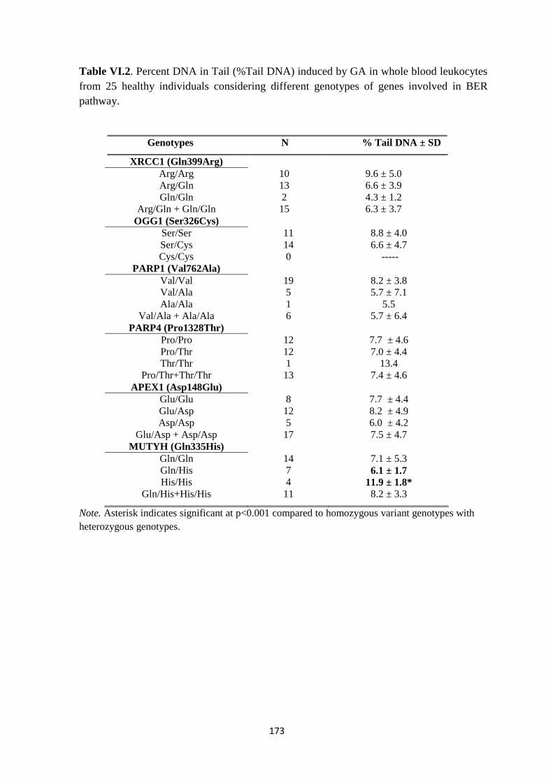
173
Table VI.2. Percent DNA in Tail (%Tail DNA) induced by GA in whole blood leukocytes
from 25 healthy individuals considering different genotypes of genes involved in BER
pathway.
Genotypes N % Tail DNA ± SD
XRCC1 (Gln399Arg)
Arg/Arg 10 9.6 ± 5.0
Arg/Gln 13 6.6 ± 3.9
Gln/Gln 2 4.3 ± 1.2
Arg/Gln + Gln/Gln 15 6.3 ± 3.7
OGG1 (Ser326Cys)
Ser/Ser 11 8.8 ± 4.0
Ser/Cys 14 6.6 ± 4.7
Cys/Cys 0 -----
PARP1 (Val762Ala)
Val/Val 19 8.2 ± 3.8
Val/Ala 5 5.7 ± 7.1
Ala/Ala 1 5.5
Val/Ala + Ala/Ala 6 5.7 ± 6.4
PARP4 (Pro1328Thr)
Pro/Pro 12 7.7 ± 4.6
Pro/Thr 12 7.0 ± 4.4
Thr/Thr 1 13.4
Pro/Thr+Thr/Thr 13 7.4 ± 4.6
APEX1 (Asp148Glu)
Glu/Glu 8 7.7 ± 4.4
Glu/Asp 12 8.2 ± 4.9
Asp/Asp 5 6.0 ± 4.2
Glu/Asp + Asp/Asp 17 7.5 ± 4.7
MUTYH (Gln335His)
Gln/Gln 14 7.1 ± 5.3
Gln/His 7 6.1 ± 1.7
His/His 4 11.9 ± 1.8*
Gln/His+His/His 11 8.2 ± 3.3
Note. Asterisk indicates significant at p<0.001 compared to homozygous variant genotypes with
heterozygous genotypes.

174
Table VI.3. Percent DNA in Tail (%Tail DNA) induced by GA in whole blood leukocytes
from 25 healthy individuals considering different genotypes of genes involved in NER
pathway.
Genotypes N % Tail DNA ± SD
ERCC2 (Lys751Gln)
Lys/Lys 8 8.2± 4.2
Lys/Gln 13 6.5 ± 3.5
Gln/Gln 4 10.0 ± 7.5
Lys/Gln+ Gln/Gln 17 7.3 ± 4.7
ERCC1 (Gln504Lys)
Gln/Gln 17 8.1 ± 4.9
Gln/Lys 6 7.3 ± 2.9
Lys/Lys 2 4.4 ± 5.9
Gln/Lys + Lys/Lys 8 6.6 ± 3.6
ERCC4 (Arg415Gln)
Arg/Arg 20 7.1 ± 4.3
Arg/Gln 3 10.8 ± 7.0
Gln/Gln 2 7.6 ± 0.7
Arg/Gln + Gln/Gln 5 9.5 ± 5.3
ERCC5 (His1104Asp)
His/His 15 7.9± 4.9
His/Asp 9 7.2 ± 4.2
Asp/Asp 1 5.5
His/Asp + Asp/Asp 10 7.1 ± 4.0
ERCC5 (Cys529Ser)
Cys/Cys 24 7.4 ± 4.5
Cys/Ser 1 10.9
Ser/Ser 0 -----
ERCC6 (Arg1230Pro)
Pro/Pro 22 7.3 ± 4.5
Pro/Arg 3 9.6 ± 4.6
Arg/Arg 0 -----
ERCC6 (Gln1413Arg)
Arg/Arg 17 7.6 ± 4.4
Arg/Gln 6 7.5 ± 5.9
Gln/Gln 2 7.6 ± 0.7
Arg/Gln + Gln/Gln 8 7.5 ± 5.0
RAD23B (Ala249Val)
Ala/Ala 19 7.1 ± 4.1
Ala/Val 6 9.1± 5.6
Val/Val 0 -----
XPC (Ala499Val)
Ala/Ala 2 6.9 ± 4.3
Ala/Val 10 6.6 ± 6.0
Val/Val 13 8.4 ± 3.2
Ala/Val + Val/Val
XPC (Lys939Gln)
Lys/Lys 11 7.4 ± 5.4
Lys/Gln 12 8.0 ± 4.0
Gln/Gln 2 6.2 ± 2.6
Lys/Gln + Gln/Gln 14 7.8 ± 3.8

175
Table VI.4. Percent DNA in Tail (%Tail DNA) induced by GA in whole blood
leukocytes from 25 healthy individuals considering different genotypes of genes
involved in HRR pathway.
Genotypes N % Tail DNA ± SD
RAD51 (5’UTR)
G/G 6 10.4 ± 5.7
G/T 11 6.9 ± 4.7
T/T 8 6.4 ± 2.3
G/T + T/T 19 6.7 ± 3.8
NBS (Glu185Gln)
Glu/Glu 15 7.3 ± 4.6
Glu/Gln 8 7.7 ± 5.0
Gln/Gln 2 9.2 ± 1.8
Glu/Gln + Gln/Gln 10 8.0 ± 4.5
XRCC3 (Thr241Met)
Thr/Thr 6 5.8 ± 3.7
Thr/Met 13 7.9 ± 4.4
Met/Met 6 8.6 ± 5.7
Thr/Met + Met/Met 19 8.1 ± 4.7
XRCC2 (Arg188His)
Arg/Arg 22 7.0 ± 4.5
Arg/His 3 11.6 ± 1.5
His/His 0 -----

176
Table VI.5. Percent DNA in Tail (%Tail DNA) induced by GA in whole blood
leukocytes from 25 healthy individuals considering different genotypes of genes
involved in NHEJ pathway.
Genotypes N % Tail DNA ± SD
XRCC4 Asn298Ser
Asn/Asn 17 8.4 ± 4.7
Asn/Ser 8 5.9 ± 3.6
Ser/Ser 0 ------
XRCC4 Thr134Ile
Ile/Ile 24 7.8 ± 4.5
Ile/Thr 1 3.5
Thr/Thr 0 -----
Lig4Thr9Ile
Thr/Thr 19 8.2 ± 4.7
Thr/Ile 6 5.7 ± 3.4
Ile/Ile 0 ------
Ku80Ex21-238G>A
G/G 12 7.4 ± 4.2
G/A 8 6.6 ± 4.4
A/A 5 9.8 ± 5.5
G/A+A/A 13 7.8 ± 4.9
Ku80Ex21+338T>C
T/T 20 7.8 ± 4.6
T/C 4 6.7 ± 5.4
C/C 1 7.2
T/C+C/C 5 6.8 ± 4.6
Ku80Ex21-352C>A
C/C 20 7.8 ± 4.6
C/A 4 6.7 ± 5.4
A/A 1 7.2
C/A+A/A 5 6.8 ± 4.6
Ku80Ex21+466A>G
A/A 20 7.8 ± 4.7
A/G 5 6.8 ± 4.0
G/G 0 -----

Table VI.6. XRCC1 Gln399Arg and MUTYH Gln335His association and DNA damage
XRCC1Gln399Arg / MUTYHGln335His
ArgArg+ArgGln/GlnGln+GlnHis ArgArg+ArgGln/HisHis* GlnGln/GlnGln+GlnHis** Gln/Gln/His/His
%Tail DNA ±SD (n) 7.0 ± 4.5 (19) 11.9 ± 1.8 (4) 4.3 ± 1.2 (2) n.d.
Note. Asterisk indicates significant difference at p=0.05 compared with ArgArg+ArgGln/GlnGln+GlnHis; Double asterisk indicates significant difference at
p<0.05 compared with ArgArg+ArgGln/HisHis; n.d. non detected
Table VI.7 XPC haplotypes and DNA damage
XPCAla499Val / XPCLys939Gln
AlaAla/LysLys AlaVal/LysLys AlaVal/LysGln* ValVal/LysLys ValVal/LysGln ValVal/GlnGln
%Tail DNA ±SD (n) 6.9 ± 4.3 (2) 7.6 ± 6.2 (8) 2.8 ± 3.7 (2) 6.7 (1) 9.1 ± 3.3 (10) 6.2 ± 2.6 (2)
Note Asterisk indicates significant difference at p<0.05 when compared AlaVal/LysGln with ValVal/LysGln

178
6.4. Discussion:
Acrylamide is a public health concern in terms of cancer risk assessment
justifying the need of reliable toxicological biomarkers. In this context, the comet assay
is actually an emerging tool to properly assess primary DNA damage either in vitro or
in vivo. Using this methodology, the in vitro genotoxic potential of AA and GA was
characterized by the % DNA in Tail in peripheral blood leukocytes from healthy donors.
Acrylamide 1-h exposure did not alter DNA damage at any concentration level
tested up to 1 mM. Our results are in agreement with Baum et al [38], who also did not
find AA genotoxicity in the comet assay with human blood cells at 1-4 h of incubation.
In addition, Hansen et al. [39] reported an absence of AA-induced DNA damage up to 5
mM in human peripheral PBL at 2 h. However, other studies with HepG2 [40, 41], V79
cells and Caco-2 cells [9] detected significant increases in DNA damage but only at
very high concentrations of AA (millimolar range). In contrast, Blasiak et al [42]
showed significant DNA damage in isolated human lymphocytes exposed to AA at low
concentrations, reinforcing the need for further evaluation of AA in human cells using
different experimental conditions and protocols. In contrast to the use of isolated human
lymphocytes, the present report was carried out using whole blood, an approach that can
be considered more closely related to the physiological situation, since it takes into
account the influence of other factors, such as red blood cells, platelets and plasma [43,
44]. Another important feature when whole blood is used is that AA reacts with
haemoglobin forming Hb adducts [2], rendering AA less available to a potential cellular
damaging effect.
There are convincing data showing that GA acts as the ultimate genotoxic agent
in AA exposure. Our results using the comet assay revealed that GA indeed produced
significant increases in terms of %Tail DNA, at concentrations starting from 250 µM.
These results are in agreement with the data published by Baum et al [38] who observed
that GA induced DNA damage for concentrations of 300 µM and highter. In addition,
the results presented here are generally in accordance with other investigations that also
found DNA damage for GA in the comet assay, although for higher GA concentrations
(≥ 500 µM) in human lymphoblastoid TK6 cells [45] and isolated lymphocytes [39].

179
The results from our study also demonstrated that PBL from healthy donors
respond differently in vitro to a given genotoxic concentration of GA. This is an
important point that has not yet been fully addressed. The heterogeneity of the response
observed in the comet assay using whole PBL from different donors may be in part
attributed to individual genome sensitivity [46]. A plausible explanation for the
interindividual variability noted herein may rely on individual genetic polymorphisms
associated with detoxification and repair pathways that are likely to influence the levels
of DNA damage after exposure to GA.
With respect to the detoxification polymorphisms in GSTs and EPHX1 no
relevant associations with GA-induced DNA damage were found. For a definitive
conclusion further studies need to be performed using different endpoints of DNA
damage, namely the induction of sister chromatid exchanges, which were shown to
correlate with GA-DNA adducts [20].
The predominant adduct N7-GA-Gua is promutagenic since it might undergo
spontaneous depurination [19, 47, 48]. The abasic site thus generated is likely to
promote incorporation of deoxyadenosine during DNA replication, leading to G→T
transversions [47]. DNA repair is therefore essential for the understanding of GA-
induced genotoxic effects, for which a number of different primary lesions are present,
including GA adducts, abasic sites and DNA breaks [20, 49]. Johansson et al using
DNA repair deficient cell lines, suggested that the repair of the lesions induced by GA
involves BER (short patch) and HRR, but not NER [49].
Concerning the BER pathway, MUTYH gene encodes a glycosylase involved in
the repair of DNA damage resulting from the oxidation of guanine nucleotides.
MUTYH protein can prevent transversions of a G:C base pair with a T:A base pair,
resulting from the oxidation product of guanine that mispairs to adenine [50]. There is
scarce information on the formation of reactive oxygen species by GA, although for AA
some reports suggest the involvement of oxidative stress [40, 51]. Our results suggest
that MUTYH (Gln335His) influences the genotoxicity induced by GA, since variant
individuals presented higher levels of DNA damage. Further studies should be
performed to clarify this point.
XRCC1 has multiple roles in repairing DNA base damage and single-strand
DNA breaks. Although XRCC1 does not have a known enzymatic activity, there is

180
evidence suggesting that it may act as a nucleating factor by bringing different BER
components together at the site of action [52]. The importance of XRCC1 Gln399Arg
polymorphism was previously identified in occupational studies using the comet assay.
Studies performed with welders [53], and healthy Japanese workers [54] associated with
higher DNA damage with XRCC1 variant allele. However, conflicting results have also
been published (e.g. fruit growers exposed to pesticides) [55].
Our results suggested a significant interaction between XRCC1 Gln399Arg and
MUTYH Gln335His polymorphisms, as PBL from individuals with the haplotype
ArgArg+ArgGln/HisHis were more prone to DNA damage than those with
ArgArg+ArgGln/GlnGln+GlnHis and with GlnGln/GlnGln+GlnHis. In view of this
MUTYH and XRCC1, seem to be involved in the genotoxicity induced by GA. Overall,
our results emphasize that the BER pathway may be operative in the repair of lesions
generated by GA. However, as a consequence of sample size, further confirmations
need to be made in a larger population.
Data presented here also suggest an association of global genome NER pathway,
specifically XPC, and GA-induced DNA damage. XPC is involved in DNA damage
recognition, and in DNA repair initiation. The binding of XPC to damaged DNA is the
rate-limiting step for NER [56]. Several epidemiological studies have been carried out
to evaluate the association of XPC polymorphisms with cancer risk at different organ
sites and in diverse populations (reviewed in [56]). Further, a study performed by Wang
et al [57] suggested that the polymorphisms of XPC genes might modulate the
genotoxicity by PAH in coke oven workers. Our results suggest that XPC
Ala499Val/Lys939Gln diplotype, might be associated with DNA damage induced by
GA.
Overall, this study shows that GA, but not AA, increases DNA damage as
measured by the comet assay. Using this methodology, the interindividual variation
observed in terms of GA-induced genotoxicity might be associated with polymorphic
genes involved in BER and NER pathways. Further studies should be performed to
reinforce these findings, including a larger number of individuals with different
genotypes. Further, functional studies are required in order to understand the underlying
mechanisms of the variability in the GA-induced DNA damage.

181
6.5. References
1. IARC: Acrylamide. In: Acrylamide In IARC monographs on the evaluation of
carcinogenic risk to humans Volume 60, edn.: International Agency for Research on
Cancer, Lyon; 1994: 389-433.
2. Carere A: Genotoxicity and carcinogenicity of acrylamide: a critical review.
AnnIstSuperSanita 2006, 42(2):144-155.
3. Friedman M: Chemistry, biochemistry, and safety of acrylamide. A review.
JAgricFood Chem 2003, 51(16):4504-4526.
4. Parzefall W: Minireview on the toxicity of dietary acrylamide. Food ChemToxicol
2008, 46(4):1360-1364.
5. Ruden C: Acrylamide and cancer risk--expert risk assessments and the public
debate. Food ChemToxicol 2004, 42(3):335-349.
6. Exon JH: A review of the toxicology of acrylamide. Journal of toxicology and
environmental health Part B, Critical reviews 2006, 9(5):397-412.
7. Rice JM: The carcinogenicity of acrylamide. MutatRes 2005, 580(1-2):3-20.
8. Stadler RH, Blank I, Varga N, Robert F, Hau J, Guy PA, Robert MC, Riediker S:
Acrylamide from Maillard reaction products. Nature 2002, 419(6906):449-450.
9. Puppel N, Tjaden Z, Fueller F, Marko D: DNA strand breaking capacity of
acrylamide and glycidamide in mammalian cells. Mutation research 2005, 580(1-
2):71-80.
10. Dybing E, Sanner T: Risk assessment of acrylamide in foods. ToxicolSci 2003,
75(1):7-15.
11. Tareke E, Rydberg P, Karlsson P, Eriksson S, Tornqvist M: Analysis of acrylamide, a
carcinogen formed in heated foodstuffs. Journal of agricultural and food chemistry
2002, 50(17):4998-5006.
12. Konings EJ, Baars AJ, van Klaveren JD, Spanjer MC, Rensen PM, Hiemstra M, van
Kooij JA, Peters PW: Acrylamide exposure from foods of the Dutch population and
an assessment of the consequent risks. Food and chemical toxicology : an
international journal published for the British Industrial Biological Research
Association 2003, 41(11):1569-1579.
13. Abou-Donia MB, Ibrahim SM, Corcoran JJ, Lack L, Friedman MA, Lapadula DM:
Neurotoxicity of glycidamide, an acrylamide metabolite, following intraperitoneal
injections in rats. Journal of toxicology and environmental health 1993, 39(4):447-
464.

182
14. Kirman CR, Gargas ML, Deskin R, Tonner-Navarro L, Andersen ME: A
physiologically based pharmacokinetic model for acrylamide and its metabolite,
glycidamide, in the rat. Journal of toxicology and environmental health Part A 2003,
66(3):253-274.
15. Ghanayem BI, McDaniel LP, Churchwell MI, Twaddle NC, Snyder R, Fennell TR,
Doerge DR: Role of CYP2E1 in the epoxidation of acrylamide to glycidamide and
formation of DNA and hemoglobin adducts. Toxicological sciences : an official
journal of the Society of Toxicology 2005, 88(2):311-318.
16. Ghanayem BI, Witt KL, Kissling GE, Tice RR, Recio L: Absence of acrylamide-
induced genotoxicity in CYP2E1-null mice: evidence consistent with a
glycidamide-mediated effect. Mutation research 2005, 578(1-2):284-297.
17. Glatt H, Schneider H, Liu Y: V79-hCYP2E1-hSULT1A1, a cell line for the sensitive
detection of genotoxic effects induced by carbohydrate pyrolysis products and
other food-borne chemicals. MutatRes 2005, 580(1-2):41-52.
18. Doerge DR, Young JF, McDaniel LP, Twaddle NC, Churchwell MI: Toxicokinetics of
acrylamide and glycidamide in Fischer 344 rats. Toxicology and applied
pharmacology 2005, 208(3):199-209.
19. Gamboa da Costa G, Churchwell MI, Hamilton LP, Von Tungeln LS, Beland FA,
Marques MM, Doerge DR: DNA adduct formation from acrylamide via conversion
to glycidamide in adult and neonatal mice. Chemical research in toxicology 2003,
16(10):1328-1337.
20. Martins C, Oliveira NG, Pingarilho M, Gamboa da CG, Martins V, Marques MM,
Beland FA, Churchwell MI, Doerge DR, Rueff J et al: Cytogenetic damage induced
by acrylamide and glycidamide in mammalian cells: correlation with specific
glycidamide-DNA adducts. ToxicolSci 2007, 95(2):383-390.
21. Burley VJ, Greenwood DC, Hepworth SJ, Fraser LK, de Kok TM, van Breda SG,
Kyrtopoulos SA, Botsivali M, Kleinjans J, McKinney PA et al: Dietary acrylamide
intake and risk of breast cancer in the UK women's cohort. BrJCancer 2010,
103(11):1749-1754.
22. Hogervorst JG, Schouten LJ, Konings EJ, Goldbohm RA, van den Brandt PA: A
prospective study of dietary acrylamide intake and the risk of endometrial,
ovarian, and breast cancer. Cancer EpidemiolBiomarkers Prev 2007, 16(11):2304-
2313.
23. Olesen PT, Olsen A, Frandsen H, Frederiksen K, Overvad K, Tjonneland A:
Acrylamide exposure and incidence of breast cancer among postmenopausal
women in the Danish Diet, Cancer and Health Study. IntJCancer 2008,
122(9):2094-2100.

183
24. Pelucchi C, Galeone C, Levi F, Negri E, Franceschi S, Talamini R, Bosetti C, Giacosa
A, La Vecchia C: Dietary acrylamide and human cancer. International journal of
cancer Journal international du cancer 2006, 118(2):467-471.
25. Wilson KM, Mucci LA, Cho E, Hunter DJ, Chen WY, Willett WC: Dietary
acrylamide intake and risk of premenopausal breast cancer. AmJEpidemiol 2009,
169(8):954-961.
26. Costa S, Coelho P, Costa C, Silva S, Mayan O, Santos LS, Gaspar J, Teixeira JP:
Genotoxic damage in pathology anatomy laboratory workers exposed to
formaldehyde. Toxicology 2008, 252(1-3):40-48.
27. Synowiec E, Stefanska J, Morawiec Z, Blasiak J, Wozniak K: Association between
DNA damage, DNA repair genes variability and clinical characteristics in breast
cancer patients. Mutation research 2008, 648(1-2):65-72.
28. Collins AR, Oscoz AA, Brunborg G, Gaivao I, Giovannelli L, Kruszewski M, Smith
CC, Stetina R: The comet assay: topical issues. Mutagenesis 2008, 23(3):143-151.
29. Burlinson B, Tice RR, Speit G, Agurell E, Brendler-Schwaab SY, Collins AR, Escobar
P, Honma M, Kumaravel TS, Nakajima M et al: Fourth International Workgroup on
Genotoxicity testing: results of the in vivo Comet assay workgroup. Mutation
research 2007, 627(1):31-35.
30. Singh NP, McCoy MT, Tice RR, Schneider EL: A simple technique for quantitation
of low levels of DNA damage in individual cells. Experimental cell research 1988,
175(1):184-191.
31. Busto N, Garcia B, Leal JM, Gaspar JF, Martins C, Boggioni A, Secco F: ACMA (9-
amino-6-chloro-2-methoxy acridine) forms three complexes in the presence of
DNA. Physical chemistry chemical physics : PCCP 2011, 13(43):19534-19545.
32. Fracasso ME, Doria D, Franceschetti P, Perbellini L, Romeo L: DNA damage and
repair capacity by comet assay in lymphocytes of white-collar active smokers and
passive smokers (non- and ex-smokers) at workplace. Toxicology letters 2006,
167(2):131-141.
33. Lin DX, Tang YM, Peng Q, Lu SX, Ambrosone CB, Kadlubar FF: Susceptibility to
esophageal cancer and genetic polymorphisms in glutathione S-transferases T1,
P1, and M1 and cytochrome P450 2E1. Cancer EpidemiolBiomarkers Prev 1998,
7(11):1013-1018.
34. Bastos HN, Antao MR, Silva SN, Azevedo AP, Manita I, Teixeira V, Pina JE, Gil OM,
Ferreira TC, Limbert E et al: Association of polymorphisms in genes of the
homologous recombination DNA repair pathway and thyroid cancer risk. Thyroid
2009, 19(10):1067-1075.

184
35. Conde J, Silva SN, Azevedo AP, Teixeira V, Pina JE, Rueff J, Gaspar JF: Association
of common variants in mismatch repair genes and breast cancer susceptibility: a
multigene study. BMCCancer 2009, 9:344.
36. Gomes BC, Silva SN, Azevedo AP, Manita I, Gil OM, Ferreira TC, Limbert E, Rueff J,
Gaspar JF: The role of common variants of non-homologous end-joining repair
genes XRCC4, LIG4 and Ku80 in thyroid cancer risk. OncolRep 2010, 24(4):1079-
1085.
37. Silva SN, Azevedo AP, Teixeira V, Pina JE, Rueff J, Gaspar JF: The role of GSTA2
polymorphisms and haplotypes in breast cancer susceptibility: a case-control study
in the Portuguese population. OncolRep 2009, 22(3):593-598.
38. Baum M, Fauth E, Fritzen S, Herrmann A, Mertes P, Merz K, Rudolphi M, Zankl H,
Eisenbrand G: Acrylamide and glycidamide: genotoxic effects in V79-cells and
human blood. Mutation research 2005, 580(1-2):61-69.
39. Hansen SH, Olsen AK, Soderlund EJ, Brunborg G: In vitro investigations of
glycidamide-induced DNA lesions in mouse male germ cells and in mouse and
human lymphocytes. Mutation research 2010, 696(1):55-61.
40. Cao J, Liu Y, Jia L, Jiang LP, Geng CY, Yao XF, Kong Y, Jiang BN, Zhong LF:
Curcumin attenuates acrylamide-induced cytotoxicity and genotoxicity in HepG2
cells by ROS scavenging. Journal of agricultural and food chemistry 2008,
56(24):12059-12063.
41. Jiang L, Cao J, An Y, Geng C, Qu S, Zhong L: Genotoxicity of acrylamide in human
hepatoma G2 (HepG2) cells. Toxicology in vitro : an international journal published
in association with BIBRA 2007, 21(8):1486-1492.
42. Blasiak J, Gloc E, Wozniak K, Czechowska A: Genotoxicity of acrylamide in human
lymphocytes. Chemico-biological interactions 2004, 149(2-3):137-149.
43. Ramos DL, Gaspar JF, Pingarilho M, Gil OM, Fernandes AS, Rueff J, Oliveira NG:
Genotoxic effects of doxorubicin in cultured human lymphocytes with different
glutathione S-transferase genotypes. MutatRes 2011, 724(1-2):28-34.
44. Rueff J, Chiapella C, Chipman JK, Darroudi F, Silva ID, Duverger-van BM, Fonti E,
Glatt HR, Isern P, Laires A et al: Development and validation of alternative
metabolic systems for mutagenicity testing in short-term assays. MutatRes 1996,
353(1-2):151-176.
45. Koyama N, Sakamoto H, Sakuraba M, Koizumi T, Takashima Y, Hayashi M, Matsufuji
H, Yamagata K, Masuda S, Kinae N et al: Genotoxicity of acrylamide and
glycidamide in human lymphoblastoid TK6 cells. Mutation research 2006,
603(2):151-158.

185
46. Kopjar N, Zeljezic D, Garaj-Vrhovac V: Evaluation of DNA damage in white blood
cells of healthy human volunteers using the alkaline comet assay and the
chromosome aberration test. Acta BiochimPol 2006, 53(2):321-336.
47. Besaratinia A, Pfeifer GP: Genotoxicity of acrylamide and glycidamide. JNatlCancer
Inst 2004, 96(13):1023-1029.
48. Segerback D, Calleman CJ, Schroeder JL, Costa LG, Faustman EM: Formation of N-
7-(2-carbamoyl-2-hydroxyethyl)guanine in DNA of the mouse and the rat
following intraperitoneal administration of [14C]acrylamide. Carcinogenesis 1995,
16(5):1161-1165.
49. Johansson F, Lundell T, Rydberg P, Erixon K, Jenssen D: Mutagenicity and DNA
repair of glycidamide-induced adducts in mammalian cells. Mutation research
2005, 580(1-2):81-89.
50. Nielsen M, van Steenbergen LN, Jones N, Vogt S, Vasen HF, Morreau H, Aretz S,
Sampson JR, Dekkers OM, Janssen-Heijnen ML et al: Survival of MUTYH-
associated polyposis patients with colorectal cancer and matched control colorectal
cancer patients. Journal of the National Cancer Institute 2010, 102(22):1724-1730.
51. Zeiger E, Recio L, Fennell TR, Haseman JK, Snyder RW, Friedman M: Investigation
of the low-dose response in the in vivo induction of micronuclei and adducts by
acrylamide. ToxicolSci 2009, 107(1):247-257.
52. Silva SN, Moita R, Azevedo AP, Gouveia R, Manita I, Pina JE, Rueff J, Gaspar J:
Menopausal age and XRCC1 gene polymorphisms: role in breast cancer risk.
Cancer detection and prevention 2007, 31(4):303-309.
53. Iarmarcovai G, Bonassi S, Botta A, Baan RA, Orsiere T: Genetic polymorphisms and
micronucleus formation: a review of the literature. MutatRes 2008, 658(3):215-233.
54. Weng H, Weng Z, Lu Y, Nakayama K, Morimoto K: Effects of cigarette smoking,
XRCC1 genetic polymorphisms, and age on basal DNA damage in human blood
mononuclear cells. Mutation research 2009, 679(1-2):59-64.
55. Wong RH, Chang SY, Ho SW, Huang PL, Liu YJ, Chen YC, Yeh YH, Lee HS:
Polymorphisms in metabolic GSTP1 and DNA-repair XRCC1 genes with an
increased risk of DNA damage in pesticide-exposed fruit growers. Mutation
research 2008, 654(2):168-175.
56. Qiu L, Wang Z, Shi X: Associations between XPC polymorphisms and risk of
cancers: A meta-analysis. Eur J Cancer 2008, 44(15):2241-2253.
57. Wang Y, Yang H, Li L, Wang H, Zhang C, Yin G, Zhu B: Association between
CYP2E1 genetic polymorphisms and lung cancer risk: a meta-analysis. Eur J
Cancer 2010, 46(4):758-764.


Chapter 7
Concluding remarks and future prospects


189
7.1. Concluding remarks and future prospects
Acrylamide is a well-known industrial chemical classified as probable human
carcinogen by IARC since 1994 [1] presented in stuff foods [2]. This carcinogen can be
formed by Maillard reaction between primarily the amino acid asparagine and reducing
sugars in food processing upon heating [3, 4]. Furthermore, AA can be found in
commonly consumed foods and beverages, such as processed cereals, French fries,
potato chips and coffee [2]. The average daily intake of AA was estimated to be about
0.5-1.0 µg/Kg bw in adults and up to 2-fold in 13 year-old children with a normal
western diet [2]. This has raised concerns about the positive health risks of AA for the
general population, even more because AA is a known rodent carcinogen [5].
Furthermore, the association of the increased risk of human cancer with AA dietary
consumption is still a matter of discussion [5, 6].
AA can be metabolized by cytochrome P450 (CYP2E1) to the genotoxic
epoxide glycidamide (GA), the ultimate genotoxin. However, the mechanisms of
genotoxicity of AA- and GA- dependent in mammalian cells are not yet fully
understood. In view of this, the evaluation of DNA damage induced by these two
compounds is of major importance. With this purpose, we resorted to the use of distinct
assays: chromosomal aberrations (CAs), sister chromatid exchange (SCEs),
quantification of DNA adducts and comet assay in V79 Chinese hamster cells and in
human lymphocytes.
V79 cells are a well established mammalian cell line widely used in cytotoxicity
and genotoxicity studies. These cells are very important in present study as they do not
express detectable levels of CYP2E1, the recognized cytochrome P450, responsible for
AA epoxidation [7, 8]. These cells revealed to be an adequate model to properly address
the mechanisms of genotoxicity by AA and GA. However, the use of human cells is
obviously more suitable for human risk assessment, and also provides the possibility to
address inter-individual variation in terms of DNA damage and the association with
individual susceptibility. The use of human cells from whole blood samples was chosen
in this work since it is considered to be more closely related to the physiological
situation than isolated lymphocytes, including the influence of others factors, such as
red blood cells, platelets and plasma [9].

190
In view of that, the cytotoxicity of AA and GA in V79 cells, evaluated by the
MTT reduction assay, was performed. The results showed that both AA and GA
induced dose-dependent cell death, being GA clearly more cytotoxic than AA for all the
concentrations studied, which is in agreement with data reported by others groups, using
different cell survival end points [10, 11].
The clastogenicity of these compounds were then analyzed by CAs assay in V79
cells. The results suggested that both AA and GA induced CAs, especially chromatid
breaks and gaps. AA and GA increased the % ACEG mainly for the higher
concentration studied (1000 and 2000 µM, for GA and AA, respectively).
The genotoxicity of AA and GA were also analyzed by SCEs assay in V79 cells
and in stimulated human lymphocytes. The results concerning SCE formation in V79
cells clearly showed that GA consistently induced SCEs for concentrations above 10
µM, increasing the background levels of SCEs by about 10-fold to levels of about 60
SCE/cell for the highest concentration tested. On the other hand, for AA-exposed
cultures, a significant increase in SCE/cell was observed only at a very high
concentration level (2000 µM). The induction of SCE in lymphocytes revealed results
that are in agreement with the previous one obtained in V79 cells, since the induction of
SCEs by AA was slight and specially associated with the very high and cytotoxic
concentration of 2000 µM and that GA markedly induced SCE in a dose-response
manner (up to 750 µM). These results reinforce the knowledge that GA is far more
genotoxic than AA and also highlight the high sensitivity of SCE to be used as a
toxicological biomarker in an AA exposure scenario.
The levels of N7-GA-Gua and N3-GA-Ade were also measured in V79 cells and
in stimulated lymphocytes exposed to AA and GA. Data showed that AA exposure in
V79 cells lead to very low levels of N7-GA-Gua, which were only observed for
concentrations higher than 1000 µM. These low levels of N7-GA-Gua stemming from
AA exposure might be related to either residual metabolism of AA in V79 cells or to a
small extent of spontaneous nonenzymatic oxidation to GA, since V79 cells are
essentially devoid of CYP2E1. On the other hand, GA showed to be a potent inducer of
N7-GA-Gua with linear dose-response dependence. The detection of this adducts was
observed for doses as low as 1 µM and 10 µM of GA, respectively in V79 cells and in
lymphocytes. The second more frequent adduct, N3-GA-Ade was only detected at very

191
high GA concentrations precluding the interest of its quantification in human dietary
studies. It is recognized that compounds that form DNA adducts are also strong SCEs
inducers. Our results showed, in fact, a very strong correlation between the levels of
N7-GA-Gua and SCE/cell for both cell types. This finding is very important in terms of
the understanding of the mechanisms involved in DNA damage signaling and repair.
These results suggested that the metabolism of AA to GA and the ensuing formation of
depurinating DNA lesions [12, 13] are responsible for SCE induction.
In view of these showed clearly differences between AA and GA genotoxicity, it
seemed important to clarify the genotoxicity mechanisms induced by AA per se. The
modulation of reduced glutathione (GSH) status could give additional insight into this
matter, as GSH is a key factor for mammalian cells homeostasis, with diverse functions
that include, among others, the conjugation of eletrophilic compounds, including AA
and GA, and the detoxification of products generated by oxidative stress. For this
purpose the cytotoxicity was analyzed by the MTT assay and clastogenicity evaluated
by CAs. This study evaluated the effect of GSH modulators, including the evaluation of
the effect of buthionine sulfoximine (BSO), of GSH-monoethyl ester (GSH-EE) and
also of GSH endogenously added to culture medium in V79 cells. BSO treatment alone
was associated with relatively low toxicity leading to a slight decrease in cell viability
and a moderate increase in the frequency of ACEG. In order to evaluate the potential
protective effect of GSH towards AA toxicity, the intracellular GSH enrichment was
performed by pre-treatment with GSH-EE. The results obtained fail to show the
protective effect of GSH-EE. Co-treatment with GSH exogenously added to the culture
medium revealed a protective effect either in terms of cytotoxicity or clastogenicity
induced by AA. This effect could be a consequence of the spontaneous conjugation of
AA with GSH in the extracellular medium. In this sense, the evaluation of the potential
conjugation of AA with GSH in the extracellular medium was performed. With this
purpose, we studied in a set of cell free experiments the intensity of fluorescence of the
conjugate GSH-monochorobimane (MCB), a fluorescent probe for GSH, after
incubation of AA with GSH. These results showed that spontaneous conjugation of AA
is favored when AA concentrations are higher than GSH concentrations and that
spontaneous conjugation is clearly time-dependent (1-h incubation decreased the
percentage of free GSH when compared with a 24-h incubation period). The results

192
presented here reinforce the role of GSH in the modulation of the cytotoxic and
clastogenic effects induced by AA.
The “single cell gel electrophoresis” or comet assay was also performed to study
the genotoxicity of AA and GA in human cells. We compared the DNA damage
potency of AA and its metabolite GA in the comet assay using human leukocytes
(whole blood samples) and the results revealed that AA was not genotoxic up to 1000
µM, while GA showed to significantly increase % Tail DNA at concentrations starting
from 250 µM.
Both SCE and comet assay showed some extent of inter-individual variability
that could be related with different polymorphisms in detoxification and DNA repair
genes. In view of this, the different donors were genotyped for detoxification (GSTM1,
GSTT1, GSTP1, GSTA2 and EPHX1) and DNA repair genes involved in BER (APEX1,
MUTYH, OGG1, PARP1, PARP4, and XRCC1), NER (ERCC1, ERCC2, ERCC4,
ERCC5, ERCC6, RAD23B and XPC), HRR (NBS, RAD51, XRCC2 and XRCC3) and
NHEJ (Ku80, Lig4 and XRCC4) pathways. These polymorphic genes were analyzed in
order to find out possible associations between genotypes and GA-induced SCE
frequency and % Tail DNA.
By combining DNA damage, assessed by SCE assay, in GA-treated
lymphocytes and polymorphisms data, associations between the induction of SCEs and
GSTP1 (Ile105Val) and GSTA2 (Glu210Ala) genotypes are suggested, but not the other
polymorphic genes associated with DNA repair pathways. Moreover, our results
suggested associations between DNA damage, assessed by the alkaline comet assay,
and polymorphisms of BER (MUTYH Gln335His and XRCC1 Gln399Arg) and NER
(XPC Ala499Val) genes, either alone or in combination. Further studies should be
performed in order to evaluate other polymorphisms and different combinations of
polymorphisms. These future studies necessarily need larger population samples and
will be important to reinforce these findings and understand the underlying mechanisms
of variability in GA-induced DNA damage.
The overall results showed that GA is far more genotoxic than AA in all the
endpoints studied and in both types of cell models used. All biomarkers developed in
the framework of this study revealed to be adequate to understand the mechanisms
triggered upon AA or GA exposure. Nonetheless, the use of specific N7-GA-Gua a GA-

193
DNA adduct revealed to be of utmost importance in terms of toxicological significance
in view of the high sensitivity, being much relevant its quantification in cultured cells
exposed to GA at concentrations as low as 1 µM. Taken together the results from this
thesis strongly point out that GA, the epoxide metabolite of AA is the responsible for
the genotoxic effects of AA.
In the future it would be important to validate these results with a group of
young Portuguese individuals, taking into account the amount of foodstuff consumed
containing AA. This study is already being performed by us and shall evaluate the
results obtained in a food frequency questionnaire and the basal levels of DNA damage
in this group using the cytogenetic biomarkers (SCEs, CAs, comet assay and DNA-
adducts) validated in this work.

194
7.2. References
1. IARC: Acrylamide. In: Acrylamide In IARC monographs on the evaluation of
carcinogenic risk to humans Volume 60, edn.: International Agency for Research on
Cancer, Lyon; 1994: 389-433.
2. Dybing E, Sanner T: Risk assessment of acrylamide in foods. ToxicolSci 2003,
75(1):7-15.
3. Stadler RH, Blank I, Varga N, Robert F, Hau J, Guy PA, Robert MC, Riediker S:
Acrylamide from Maillard reaction products. Nature 2002, 419(6906):449-450.
4. Friedman M: Chemistry, biochemistry, and safety of acrylamide. A review.
JAgricFood Chem 2003, 51(16):4504-4526.
5. Rice JM: The carcinogenicity of acrylamide. MutatRes 2005, 580(1-2):3-20.
6. Parzefall W: Minireview on the toxicity of dietary acrylamide. Food ChemToxicol
2008, 46(4):1360-1364.
7. Doehmer J, Dogra S, Friedberg T, Monier S, Adesnik M, Glatt H, Oesch F: Stable
expression of rat cytochrome P-450IIB1 cDNA in Chinese hamster cells (V79) and
metabolic activation of aflatoxin B1. Proceedings of the National Academy of
Sciences of the United States of America 1988, 85(16):5769-5773.
8. Glatt H, Schneider H, Liu Y: V79-hCYP2E1-hSULT1A1, a cell line for the sensitive
detection of genotoxic effects induced by carbohydrate pyrolysis products and
other food-borne chemicals. MutatRes 2005, 580(1-2):41-52.
9. Rueff J, Chiapella C, Chipman JK, Darroudi F, Silva ID, Duverger-van BM, Fonti E,
Glatt HR, Isern P, Laires A et al: Development and validation of alternative
metabolic systems for mutagenicity testing in short-term assays. MutatRes 1996,
353(1-2):151-176.
10. Baum M, Fauth E, Fritzen S, Herrmann A, Mertes P, Merz K, Rudolphi M, Zankl H,
Eisenbrand G: Acrylamide and glycidamide: genotoxic effects in V79-cells and
human blood. Mutation research 2005, 580(1-2):61-69.
11. Koyama N, Sakamoto H, Sakuraba M, Koizumi T, Takashima Y, Hayashi M, Matsufuji
H, Yamagata K, Masuda S, Kinae N et al: Genotoxicity of acrylamide and
glycidamide in human lymphoblastoid TK6 cells. Mutation research 2006,
603(2):151-158.
12. Ghanayem BI, McDaniel LP, Churchwell MI, Twaddle NC, Snyder R, Fennell TR,
Doerge DR: Role of CYP2E1 in the epoxidation of acrylamide to glycidamide and
formation of DNA and hemoglobin adducts. Toxicological sciences : an official
journal of the Society of Toxicology 2005, 88(2):311-318.
13. Ghanayem BI, Witt KL, Kissling GE, Tice RR, Recio L: Absence of acrylamide-
induced genotoxicity in CYP2E1-null mice: evidence consistent with a
glycidamide-mediated effect. Mutation research 2005, 578(1-2):284-297.
.

Supplement


197
Resumo
A acrilamida (AA) é um composto químico, utilizado principalmente como
agente floculante no processo de depuração de água potável e tratamento de águas
residuais urbanas e industriais, e como agente de controlo de fluxo em operações em
poços de petróleo. Por outro lado, a AA é utilizada em engenharia civil, em fundações,
como constituinte da argamassa utilizada na construção e reparação de esgotos, túneis e
barragens. A AA é ainda utilizada na estabilização de solos, síntese de corantes,
produção de materiais de embalagem, co-polímeros para lentes de contacto, cosméticos,
e em muitos laboratórios de biologia molecular e engenharia genética na preparação de
géis de electroforese.
A AA foi classificada como potencial carcinogéneo para os humanos, pela IARC
(International Agency for Research on Cancer), com base em resultados obtidos em
estudos com animais. Estudos de carcinogenicidade em ratos mostraram que a AA
aumenta o número de tumores em diferentes órgãos, incluindo a glândula mamária. Foi
também demostrado que a AA é neurotóxica em humanos e em experiências com
animais, e que apresenta propriedades mutagénicas em células somáticas e germinais. A
AA absorvida pode ser oxidada ao seu metabolito epóxido glicidamida (GA) através do
citocromo P450 2E1, podendo ambas, AA e GA, ser conjugadas com o glutationo
reduzido (GSH), originando conjugados de ácido mercaptúrico, que serão eliminados
pela urina. A GA, por sua vez, pode também ser conjugada com a epóxide hidrolase,
originando a gliceramida. Tanto a AA como a GA podem ligar-se a macromoléculas,
tais como a hemoglobina e o DNA originando aductos específicos. Estes aductos podem
ser usados como biomarcadores de dose efetiva de exposição à AA e GA.
Durante a década de 90 pensava-se que o homem estava exposto à AA apenas
através da exposição ocupacional, e também apenas em pequenas quantidades através
do consumo de água potável refinada com poliacrilamida, ou ainda pela inalação do
fumo de tabaco. No entanto, alguns estudos verificaram a existência de níveis elevados
de aductos de AA-hemoglobina em indivíduos não expostos, o que provocou uma forte
preocupação em termos de risco para a saúde humana, principalmente porque foram
descobertos níveis relativamente elevados de AA em diversos alimentos ricos em amido
quando sujeitos a elevado tratamento térmico. Os referidos produtos alimentares, além

198
de serem consumidos em larga escala, são especialmente consumidos por crianças e
jovens, sendo que nos mesmos se incluem por exemplo as batatas fritas, “snacks”,
cereais de pequeno-almoço, bolachas e café, tendo sido estimado que a dose total diária
de AA ingerida por adulto tendo como base uma dieta ocidental normal, se encontra na
ordem dos 1 µg/Kg por peso corporal, podendo ser 2 a 3 vezes superior em crianças e
adolescentes.
Desde 2002 as investigações sobre a presença de AA nos alimentos, e as
possíveis consequências para a saúde pública têm-se sucedido e vários progressos têm
sido alcançados. No entanto, até à data os resultados são escassos tendo em conta os
mecanismos de genotoxicidade da AA e GA. Por esse motivo, o objectivo desta tese
centra-se na avaliação das consequências genéticas da exposição à AA e GA,
recorrendo ao uso de linhas celulares e de linfócitos humanos.
Neste estudo procedeu-se inicialmente à avaliação da citotoxicidade e das lesões
de DNA induzidas pela AA e GA em células de hamster Chinês V79, uma linha celular
deficiente em CYP2E1, de modo a prevenir a conversão de AA a GA (Capítulo3). Para
avaliar a citoxicidade recorreu-se ao ensaio do MTT para concentrações compreendidas
entre 0.1 e 10 mM. A AA mostrou ser menos citotóxica do que a GA para todas as
concentrações testadas, apresentando baixos níveis de sobrevivência (<5%) para
concentrações elevadas de GA (≥ 4mM) e de AA (10 mM).
De modo a avaliar a genotoxicidade e a clastogenicidade da AA e GA em
células de mamífero, recorreu-se à utilização de dois testes citogenéticos diferentes: o
teste das aberrações cromossómicas (CAs), e o teste da troca de cromátides irmãs
(SCEs). Os resultados obtidos no teste das CAs em células V79, mostraram claramente
que tanto a AA como a GA, são compostos clastogénicos induzindo a formação de
aberrações cromossómicas, com uma dependência dose-resposta, especialmente
observada para as concentrações mais elevadas (1000 µM e 2000 µM). No entanto, para
concentrações equimolares a GA mostrou ser mais clastogénica do que a AA. Através
dos resultados obtidos no teste da troca de cromátides irmãs, na mesma linha celular,
pode-se afirmar que a GA é um potente indutor de SCEs para concentrações igual ou
superiores a 10 µM, com uma clara relação dose-resposta, aumentando a frequência
basal de troca de cromátides irmãs em cerca de 10 vezes, para níveis de
aproximadamente 60 SCE/célula para a concentração mais elevada (1000 µM). Em

199
contrapartida, a AA demonstrou ser um fraco indutor de SCEs, observando-se apenas
um aumento no nível de lesão para a concentração mais elevada (2000 µM). Os
resultados no geral, para todas as doses testadas, mostraram que a GA é mais citotóxica
e clastogénica do que a AA em células V79. Este estudo demonstrou também a elevada
sensibilidade do ensaio da troca de cromátides irmãs, de modo a poder ser utilizado
como biomarcador toxicológico no caso de um cenário de exposição à AA.
No âmbito desta tese efetuou-se também a quantificação de aductos específicos
de DNA, nomeadamente N7-(2-carbamoil-2-hidroxietil)guanina (N7-GA-Gua) e N3-(2-
carbamoil-2-hidroxietil)adenina (N3-GA-Ade) em células V79. Os resultados
mostraram que a exposição à AA origina níveis muito baixos de aductos N7-GA-Gua,
os quais foram apenas observados para concentrações superiores a 1000 µM. Estes
baixos níveis de aductos que são provenientes da exposição à AA devem estar
relacionados com a oxidação não enzimática espontânea da AA a GA, uma vez que as
células V79 são deficientes em CYP2E1. No entanto, a GA mostrou ser um potente
indutor de N7-GA-Gua, apresentando uma dose-resposta linear, sendo a detecção deste
aducto observada para concentrações tão baixas como 1 µM. O segundo aducto mais
frequente, N3-GA-Ade foi apenas detectado para concentrações muito elevadas de GA,
limitando o seu interesse em estudos sobre dieta humana. Adicionalmente, observou-se
que as concentrações de GA e os níveis de N7-GA-Gua apresentam uma relação dose-
resposta linear. Obteve-se também uma óptima correlação entre os níveis de N7-GA-
Gua e o nível de SCE/célula (r=0.987; p=1.25x10⁻¹²
).
Durante este trabalho procurou-se também compreender os mecanismos da
genotoxicidade induzida pela AA. Para tal estudaram-se os mecanismos de modulação
do glutationo reduzido (GSH), o qual possui vários papéis importantes nas células de
mamífero, incluindo a conjugação com compostos electrofílicos e a destoxificação de
produtos obtidos por stress oxidativo. Deste modo, foram efetuados ensaios onde se
averiguou o efeito da deplecção intracelular de GSH (pré-incubação com butionina
sulfoximina, BSO), o efeito do enriquecimento intracelular com GSH (pré-incubação
com GSH-monoetil estér, GSH-EE) e o papel do GSH adicionado extracelularmente
(através da co-incubação com GSH) em células V79 (Capítulo 4). Com este objectivo,
avaliaram-se diferentes parâmetros, tais como a citotoxicidade (ensaio do MTT) e a
clastogenicidade (ensaio das ACs). Nos ensaios das células V79 expostas a AA e com
pré-incubação com BSO, verificou-se um decréscimo na viabilidade celular e um

200
aumento na fequência das aberrações cromossómicas excluindo gaps (ACEG). Por
outro lado, através da pré-incubação com GSH-EE não se verificaram alterações
significativas na percentagem de sobrevivência das células V79 expostas à AA, nem na
frequência de ACEG. Por fim, no co-tratamento com GSH adicionado exogenamente ao
meio de cultura, verificou-se um efeito protectivo tanto em termos de viabilidade celular
quer em termos de clastogenicidade induzidas pela AA. Este efeito pode ser
consequência da conjugação expontânea da AA com GSH no meio extracelular. Deste
modo, foi posteriormente avaliado o potencial de conjugação da AA com o GSH com
base no teste da fluorescência do monoclorobimano (MCB), uma sonda fluorescente
que se liga ao GSH. Os resultados mostraram que a conjugação expontênea da AA é
favorecida quando existem concentrações superiores de AA em relação ao GSH e que
esta conjugação expontânea é claramente dependente do tempo de exposição (1h de
exposição diminuiu a percentagem de GSH livre quando comparado com o período de
24h de exposição). Globalmente, os resultados obtidos reforçaram o papel do GSH na
modulação da citotoxicidade e clastogenicidade induzidas pela AA.
Apesar do uso das células V79 ser adequado como modelo para inferir sobre a
genotoxicidade induzida pela AA e GA, o passo seguinte deste trabalho envolveu o
estudo em células humanas. A utilização deste tipo de células é obviamente mais
adequado para a avaliação de risco, permitindo também a possibilidade de investigar a
variabilidade individual em termos de lesão de DNA, e a sua correlação com a
susceptibilidade de cada individuo. Para tal recorreu-se à colheita de sangue periférico
de dadores saudáveis, não sujeitos a medicação e não fumadores. Os dadores foram
devidamente informados sobre o âmbito do estudo, e um consentimento informado foi
assinado pelos mesmos, antes de se ter procedido ao início dos trabalhos. Cumprirá
também salientar o facto de ter sido obtida uma aprovação do estudo pelo Comité de
Ética da Faculdade de Ciências Médicas da Universidade Nova de Lisboa. No referido
estudo recorreu-se ao uso de células humanas provenientes de sangue total, uma vez que
é considerado ser mais aproximado da situação fisiológica do que o uso de linfócitos
isolados, pois tem em conta a influência de outros factores, tais como glóbulos
vermelhos, plaquetas e plasma.
Deste modo, linfócitos estimulados com fitohemaglutinina foram expostos a
diferentes concentrações de AA e GA e as lesões de DNA quantificadas através do
ensaio das SCEs e dos níveis de aductos de DNA (Capítulo 5). A partir destes estudos e

201
em relação ao ensaio das SCEs, observou-se mais uma vez que a GA é muito mais
genotóxica do que a AA, aumentando a GA o nível de SCEs em cerca de 10 vezes,
quando comparada com o controlo, o que está de acordo com os resultados previamente
obtidos com células V79. Por outro lado, a AA só induziu SCE para a concentração
mais elevada (2000 µM). Do mesmo modo, a quantificação de aductos de DNA,
nomeadamente N7-GA-Gua e N3-GA-Ade, efectuados em linfócitos estimulados,
originaram resultados coerentes com os obtidos em células V79. Os resultados obtidos
mostram uma dose-resposta linear entre os níveis de N7-GA-Gua e as concentrações de
GA estudadas (até 750 µM), tendo sido este aducto detectado para a menor dose
estudada (10 µM). O aducto (N7-GA-Gua) determinado em linfócitos expostos à GA
apresentou uma correlação directa entre os níveis de aductos e a frequência de SCEs, tal
como anteriormente já se tinha verificado em células V79.
Por outro lado, foi também avaliado o potencial de lesão do DNA da AA e GA
em leucócitos humanos, recorrendo-se à técnica do ensaio do cometa alcalino (Capítulo
6). Os resultados mostraram que a GA aumentou significativamente a % de DNA na
cauda do cometa, para concentrações superiores a 250 µM, enquanto a AA não
provocou lesões de DNA significativas para as concentrações estudadas (até 1000 µM).
Com base nas curvas dose-resposta obtidas para o ensaio das SCEs e do cometa
alcalino, a concentração de 250 µM de GA foi escolhida para ser posteriormente
utilizada em ensaios com vários indivíduos, tanto no que diz respeito às SCEs (13
indivíduos) como ao ensaio do cometa (25 individuos). Ambos os ensaios mostraram
alguma variabilidade inter-individual que poderá estar relacionada com os diferentes
polimorfismos envolvidos na destoxificação e nas vias de reparação de DNA.
Deste modo, o presente estudo teve também como objectivo a identificação de
possíveis associações entre as lesões de DNA induzidas pela AA e GA quantificadas
através dos ensaios das SCEs e do ensaio do cometa, e biomarcadores de
susceptibilidade, considerando os polimorfismos genéticos individuais envolvidos nas
vias de destoxificação (GSTM1, GSTT1, GSTP1, GSTA2 e EPHX1), e nas vias de
reparação de DNA (Capítulo 5 e 6). No que diz respeito à reparação de DNA, foram
estudados diferentes genes envolvidos na via de reparação por excisão de bases (BER),
tais como APEX1, MUTYH, OGG1, PARP1, PARP4, e XRCC1, na reparação por
excisão de nucleótidos (NER), tais como ERCC1, ERCC2, ERCC4, ERCC5, ERCC6,

202
RAD23B e XPC, na via de reparação por recombinação homóloga (HRR), tais como
NBS, RAD51, XRCC2 e XRCC3, e na via de reparação por recombinação não homóloga
(NHEJ), tais como Ku80, Lig4 e XRCC4.
O nível de lesão de DNA determinado pela frequência de SCEs induzidas pela
GA, aponta para uma modulação pelos polimorfismos do GSTP1 (Ile105Val) e do
GSTA2 (Glu210Ala), mas não por outros polimorfismos associados com as vias de
reparação de DNA. Por outro lado, os resultados obtidos com base no ensaio do cometa
alcalino sugerem associações entre as lesões de DNA e polimorfismos da via BER
(MUTYH Gln335His e XRCC1 Gln39Arg) e da via NER (XPC Ala499val e
Lys939Gln), considerando os genes isoladamente ou combinados. No entanto, mais
estudos devem ser efetuados contemplando uma maior amostragem de indivíduos
saudáveis de modo a consolidar os resultados obtidos e compreender os mecanismos
envolvidos nas diferenças individuais de resposta a genotóxicos.
Os resultados globais mostraram que a GA é de facto mais genotóxica do que a
AA, tendo como referência todos os ensaios efectuados bem como os vários tipos de
células estudadas. Adicionalmente, todos os resultados apontam fortemente para que a
GA, o metabolito epóxido da AA, seja o principal responsável pelos efeitos genotóxicos
da AA. É de salientar também, que apesar dos vários tipos de ensaios utilizados neste
trabalho terem demonstrado ser adequados para a compreensão dos mecanismos
envolvidos na exposição à AA e GA, a quantificação de aductos de DNA por exposição
à GA, nomeadamente N7-GA-Gua, mostrou ser aquele que revela maior robustez em
termos de avaliação toxicológica devido à sua elevada sensibilidade. Assim sendo, este
biomarcador de dose-efectiva pode e deve ser usado em estudos epidemiológicos em
complementaridade com os questionários de frequência alimentar. Adicionalmente este
trabalho permitiu tirar conclusões acerca da variabilidade da susceptibilidade individual
relativa à destoxificação e reparação das lesões de DNA provocadas pela exposição a
estes xenobióticos alimentares.