universitÀ degli studi di padova -...
TRANSCRIPT

UNIVERSITÀ DEGLI STUDI DI
PADOVA
Facoltà di Scienze MM.FF.NN
Dipartimento di Biologia
DOTTORATO DI RICERCA IN
FISIOLOGIA MOLECOLARE E BIOLOGIA STRUTTURALE
CICLO XX
TOXIC EFFECTS OF THE OXIDIZED PRODUCTS
OF THE NEUROTRANSMITTER DOPAMINE ON MITOCHONDRIA
AND THEIR IMPLICATIONS IN PARKINSON’S DISEASE
Coordinatore : Ch.mo Prof. Benedetto Salvato
Supervisore : Ch.mo Prof. Luigi Bubacco
Dottorando : Irene Arduini
DATA CONSEGNA TESI 31 gennaio 2008


I
Table of contents
Index of figures..................................................................................IV
Summary..........................................................................................VII
Riassunto............................................................................................IX
Introduction ......................................................................................... 1
Parkinson’s Disease .............................................................................................1
Mitochondria and the Selective Vulnerability of SNpc Dopaminergic Neurons ................................................................................................................4
Mitochondrial dysfunction in PD.........................................................................5
Environmental Toxins of complex I: MPTP and rotenone ..............................5
Role of mitochondrial DNA in PD ...................................................................7
PD genes and Parkinson’s disease .......................................................................7
Parkin...............................................................................................................8
DJ-1................................................................................................................10
PINK1.............................................................................................................10
α-Synuclein ....................................................................................................11
LRRK2............................................................................................................12
DA synthesis and metabolism............................................................................13
Intraneuronal oxidation of dopamine ............................................................15
Statement of Purpose..........................................................................................17
Materials and Methods..................................................................... 19
Mitochondria Isolation .......................................................................................19
Rat brain mitochondria isolation...................................................................19
Mouse liver mitochondria isolation ...............................................................20
Biuret protein assay............................................................................................20
Measurements on Isolated Mitochondria ...........................................................21
Mitochondrial respiration analysis................................................................21
Mitochondrial swelling ..................................................................................23
Calcium Retention Capacity (CRC) experiments...........................................26
II
Pyridine Nucleotide Assay.............................................................................27
Complex I assay............................................................................................. 28
Complex I immunocapture (IP) ..................................................................... 29
Cell Biology....................................................................................................... 30
Cell Cultures.................................................................................................. 30
Trasnsfection ................................................................................................. 30
Cell count....................................................................................................... 31
Fluorescence Microscopy.............................................................................. 32
Biochemistry...................................................................................................... 32
Electrophoresis and Western Blot ................................................................. 32
SDS-PAGE................................................................................................32 Blue Native-PAGE ....................................................................................33
In-gel digestion of Proteins Separated by Polyacrylamide Gel Electrophoresis.............................................................................................. 35
Activity Stain for F1Fo ATPsynthase complex ............................................... 36
RESULTS...........................................................................................37
Part I: Permeability of mitochondria to DAQ and mitochondrial proteins modifications. ............................................37
Introduction ....................................................................................................... 37
Statement of purpose ......................................................................................... 40
Results ............................................................................................................... 40
DAQs enter the mitochondria........................................................................ 40
MS analysis of two proteins modified by DAQs ............................................ 45
VDAC1 is not one of the proteins modified by DAQs. .................................. 47
Independent experiments show that the γ subunit of the F1Fo ATP synthase complex is modified by DAQs......................................................... 48
Unaltered activity in the DAQs-modified F1Fo ATP synthase complex . ...... 49
Conclusions ....................................................................................................... 50
Part II: Pathological effects of oxidized products of dopamine in mitochondria. ................................................................................51
DAQs induce mitochondrial dysfunctions ........................................................ 51
Statement of purpose ......................................................................................... 52
Results ............................................................................................................... 53
DAQs Induce swelling in mitochondria ........................................................ 53
The early oxidized products of DA are more toxic compared with the late ones................................................................................................................ 55
DAQs toxicity involved the pyridine nucleotide pool. ................................... 58
DAQs oxidize the pyridine nucleotide in mitochondria. ............................... 60

III
DAQs depolarize mitochondria a cellular model. .........................................62
Conclusions........................................................................................................64
Part III: Complex I and Parkinson’s disease ................................. 67 Complex I...........................................................................................................67
Complex I is impaired in PD..............................................................................68
Statement of purpose..........................................................................................71
Results ................................................................................................................71
DAQs inhibit mitochondrial respiration. .......................................................71
DAQs inhibit Complex I of the electronic transport chain in mitochondria. .................................................................................................73
DAQs directly modify complex I subunits......................................................75
Rotenone-DAQs competition..........................................................................76
Cells resistant to rotenone show a decrease vulnerability to DAQs toxicity. ...........................................................................................................77
Conclusions........................................................................................................79
Discussion........................................................................................... 81
References .......................................................................................... 91

IV
Index of figures Figure 1 Clinical hallmarks of Parkinson’s disease............................................. 2
Figure 2 Oxidative damage arises from an unbalance between ROS production and ROS scavenging ........................................................... 3
Figure 3 Models of human PD-associated proteins ............................................. 9
Figure 4 Schematic diagram of DA synthesis and metabolism in dopaminergic neurons.......................................................................... 14
Figure 5 Byproducts of DA metabolism or DA oxidation are toxic for the neuron .................................................................................................. 15
Figure 6 The three state of mitochondrial respiration in oxygraphic measurements ...................................................................................... 21
Figure 7 Conditions that induce or inhibit the PT.............................................. 24
Figure 8 Explicative trace of a swelling experiment.......................................... 25
Figure 9 Example of Calcium Retention Capacity (CRC) experiments ............ 26
Figure 10 Dimensions of a hemacytometer ......................................................... 31
Figure 11 Separation of DDM-solubilized mitochondrial complexes by BN-PAGE............................................................................................ 33
Figure 12 The oxidative pathway of DA that leads to neuromelanine synthesis............................................................................................... 38
Figure 13 DAQs react and covalently modify cysteine residues of proteins....... 39
Figure 14 DAQs but not DA enter the mitochondria........................................... 41
Figure 15 DAQs can cross both the outer and the inner mitochondrial membrane ............................................................................................ 42
Figure 16 Energetic state of mitochondria does not influence DAQs entrance................................................................................................ 43
Figure 17 DAQ but not DA modify mitochondrial proteins................................ 44
Figure 18 DAQ modify mitochondrial proteins................................................... 45
Figure 19 Structure of the F1-ATPsynthase complex and the γ subunit.............. 46
Figure 20 Vdac1 KO mice show the same pattern of modifications of WT mice ..................................................................................................... 47
Figure 21 DAQs react and covalently modify complex V of the ETC................ 48
Figure 22 Complex V modified by DAQ at the γ subunit retain its F1Fo ATPase activity.................................................................................... 49

V
Figure 23 DAQs induce swelling in mitochondria...............................................55
Figure 24 Calcium retention capacity of mouse liver mitochondria treated with DA and tyrosinase........................................................................56
Figure 25 Mitochondrial swelling at different time point of the DA oxidation reaction: the early products are more toxic than the late ........................................................................................................57
Figure 26 DAQ induce swelling through alteration of the pyridine nucleotide (PN) pool in mitochondria..................................................59
Figure 27 The maintenance of PN pool in the reduced state protects toward DAQ-induced mitochondrial swelling .....................................60
Figure 28 DAQs oxidize the pyridine nucleotides in isolated mitochondria .......61
Figure 29 DAQs oxidize NADH in vitro..............................................................62
Figure 30 Changes of mitochondrial TMRM fluorescence induced by DAQs in SHSY-5Y neuroblastoma cells .............................................63
Figure 31 DAQ inhibit mitochondrial respiration ................................................73
Figure 32 DAQs inhibit Complex I ......................................................................74
Figure 33 Autoradiogram of Complex I isolated by Immunoprecipitation from mitochondria treated with 14C-DA and tyrosinase.....................76
Figure 34 Cells expressing the rotenone-insensitive Ndi1 subunits are protected from DAQs toxicity.

VI

VII
Summary
Parkinson’s disease (PD) is a chronic, progressive, neurodegenerative
disorder clinically characterized by motor symptoms such as tremor at rest, rigidity, slowness of movement (bradykinesia), and postural instability.
One pathological hallmark of the disease is the progressive and striking loss of dopaminergic neurons in the substantia nigra pars compacta (SNpc). These neurons reside in the midbrain and project axons rostrally to the forebrain where they release dopamine (DA) into the striatum. DA release in the striatum is critical for the coordination and initiation of movement.
It is unclear why SNpc neurons die during PD. However, several biochemical hallmarks of the disease exist, and likely reveal clues to the underlying etiology of PD. Mitochondrial dysfunctions, at the level of complex I of the electronic transport chain (ETC), have been reported in the SNpc of PD patients. Mitochondria are the main providers of cellular energy, which is generated through the flow of electrons down the electron transport chain (ETC) coupled with production of adenosine tri-phosphate (ATP) form Adenosine di-phosphate (ADP).
Deficits in complex I observed in PD patients are indicative of a bioenergetic defect and increased reactive oxygen species (ROS) production due to electron leak from ETC. Consistent with this, lipid peroxidation and protein carbonyls are present in striatum of post-mortem PD brains, indicative of exposure to ROS. Additionally, a specific decrease in the anti-oxidant glutathione (GSH) has been reported in the SNpc of PD patients, suggesting that these neurons may not be able to buffer the ROS. All these findings suggest that dopaminergic neurons are under oxidative stress in PD, and that the ability of dopaminergic neurons to scavenge free radicals and ROS is compromised. Taken together, mitochondrial dysfunction and oxidative stress are likely key factors that are responsible for the loss of SNpc neurons in PD.
It has been hypothesized that the reason dopaminergic neurons of SNpc are vulnerable in PD is due to the toxic properties of the DA itself. DA is a highly reactive molecule, normally stored in synaptic vesicles. The acidic environment of the synaptic vesicle prevents DA oxidation. However, DA is synthesized and metabolized in the cytoplasm and in this environment cytoplasmic DA can undergo spontaneous oxidation. DA oxidation gives arise to several toxic ROS and the dopamine quinone molecules (DAQs). Increasing cytosolic dopamine in neurons and its oxidized metabolites has many deleterious effects, including increases in oxidative stress, mitochondrial dysfunction, as well as the promotion and stabilization of a potentially toxic protein called α-synuclein. Therefore, the selective loss of SNpc DA neurons in PD may result from increased oxidation of cytoplasmic DA and the oxidative stress induced by ROS-mediated oxidative damage to the neurons. Moreover, the ability of DAQs to react and covalently

VIII
modify cellular molecules and macromolecules has been reported. In particular electrophilic DAQs have high affinity for free cysteine, reduced glutathione (GSH) and sulfhydryl groups of proteins to form cysteinyl-catechol conjugates
The overall aim of the research that this dissertation comprises is to identify the mechanism that contributes to the selective degeneration of dopaminergic neurons in PD. The research described herein utilizes in vitro and cellular model approaches to test the hypothesis that the oxidized products of the neurotransmitter DA (DAQs) induce toxic effects on mitochondria. If this hypothesis is correct any pathological event that impairs DA synthesis, storage or metabolism, leading to the cytoplasmic accumulation of DA can increase the presence of oxidized products of DA and their toxic effects in the cell with mitochondria as one of the target. Mitochondrial dysfunctions derived from exposure to DA-oxidized products can account for the specific vulnerability of dopaminergic neurons and their degeneration in PD.
Results from this research demonstrate that DAQs have the ability to enter the mitochondria crossing both the outer and the inner mitochondrial membranes (OMM, IMM). DAQs react and covalently modify several proteins within mitochondria and modifications of subunits of the complex I and complex V of the ETC were identified.
DAQs induce the permeability transition (PT) in mitochondria. PT is an increase in the permeability of the mitochondrial membranes to molecules of less than 1500 Daltons in molecular weight due to the opening a proteinaceous pore that is formed in the IMM and lead to mitochondrial swelling, disruption of the IMM structure and the release in the medium of the pro-apoptotic factors normally located in the inter-membrane space. The mechanism of DAQ-induced PT was studied in vitro. Oxidation of the pyridine nucleotide (PN) pool in DAQ-treated mitochondria was identified. The redox state of the PN is a key factor of PT ore conformation and oxidation of PN trigger PT.
The ability of DAQs to depolarize mitochondria by inducing PT in a cellular model is also reported.
DAQs can exert their toxic effects on mitochondria inhibiting mitochondrial respiration. The inhibitory effect of DAQ on isolated mitochondria complex I was determined. Toxic effect of this inhibition were compared with the ones of the specific complex I inhibitor rotenone in a cellular model expressing the rotenone-insensitive single subunit NADH dehydrogenase of Saccharomices cerevisiae (Ndi1). Ndi1 subunit allows electron transfer through the ETC avoiding electrons leakage and ROS production as consequence of rotenone inhibition. Ndi1-expressing cells are protected from DAQs toxicity suggesting that DAQs, as rotenone, can exert their toxic effects increasing electron leakage from ETC, at least, at the level of complex I and increasing ROS production.
Taken together the results described in this dissertation details a unique, novel, mechanism that may render SNpc DA neurons vulnerable to oxidative stress, ROS-mediated oxidative damage, and neurodegeneration in PD. The contribution of the neurotransmitter DA to oxidative stress represents a valid explanation for the peculiar vulnerability of these neurons in PD and also represents a logical therapeutic target for protecting these neurons.

IX
Riassunto
La malattia di Parkinson è una malattia cronica, progressiva, clinicamente caratterizzata da disfunzioni motorie come tremore a riposo, lentezza nei movimenti (bradicinesia) e instabilità posturale. L’analisi post mortem del cervello di pazienti affetti da malattia di Parkinson rivela la massiva e specifica degenerazione dei neuroni dopaminergici della Substantia nigra pars compacta (SNpc) e presenza di inclusioni proteiche nel citoplasma dei neuroni dopaminergici rimanenti. La perdita dei neuroni dopaminergici della SNpc provoca una diminuzione del rilascio del neurotrasmettitore dopamina (DA) nello striato specificamente espresso in questi neuroni. Il rilascio di DA nello striato è un evento chiave nell’inizio e nella coordinazione dei movimenti e la sua diminuzione nei pazienti affetti dalla malattia di Parkinson è la ragione dei sintomi motori.
La ragione per la quale i neuroni dopaminergici della SNpc degenerano nella malattia di Parkinson è tuttora sconosciuta. Evidenze biochimiche di disfunzioni mitocondriali nei neuroni della SNpc sono state riportate. In particolare, il complesso I della catena respiratoria è inibito in pazienti affetti dalla malattia di Parkinson. Inoltre i neuroni della SNpc di questi pazienti sono caratterizzati dalla presenza di markers per lo stress ossidativo, come aumentati perossi-lipidi, proteine carbonilate e glutatione ossidato. Tutte queste evidenze portano alla conclusione che i neuroni dopaminergici nella malattia di Parkinson sono esposti ad un elevato stress ossidativo e che la loro capacità di eliminare le molecole radicali o le specie reattive dell’ossigeno (ROS) è compromessa.
L’ipotesi che la molecola DA contribuisca allo stress ossidativo dei neuroni dopaminergici è stata formulata da molto tempo. La molecola DA è altamente reattiva e normalmente sequestrata in vescicole sinaptiche dov’è stabilizzata dall’ambiente acido.
Difetti nella sintesi o nel metabolismo della DA portano ad un suo accumulo nel citoplasma. Nel citoplasma l’anello catecolo della DA può essere ossidato e questa reazione porta alla formazione di ROS tra cui lo ione super-ossido, l’anione ossidrile e i chinoni della DA. Numerosi effetti citotossici sono stati riportati per i metaboliti ossidati della DA, tra cui aumento dello stress ossidativo, modificazione covalente e inibizione dell’attività di diverse proteine e disfunzioni mitocondriali.
Lo scopo generale della ricerca presentata in questa tesi è l’identificazione di meccanismi che contribuiscono alla morte selettiva dei neuroni dopaminergici nella malattia di Parkinson. L’ipotesi che i prodotti ossidati della DA (DAQ) inducano effetti tossici nei mitocondri è stata testata utilizzando sistemi in vitro e modelli cellulari. In questo scenario, qualsiasi evento patologico che alteri il catabolismo-metabolismo della DA – aumentandone la concentrazione nel citoplasma – all’aumento dei prodotti ossidati della DA nel citoplasma e, di

X
conseguenza, ad effetti citotossici. Uno dei target cellulari dei DAQ possono essere i mitocondri. Le disfunzioni mitocondriali derivanti dalla presenza dei DAQ nel citoplasma possono spiegare la peculiare vulnerabilità dei neuroni dopaminergici e la loro degenerazione nella malattia di Parkinson.
I risultati del lavoro di ricerca qui presentato mostrano che i DAQ sono in grado di penetrare entrambe le membrane mitocondriali e giungere nella matrice. I DAQ, inoltre, reagiscono e modificano covalentemente diverse proteine mitocondriali, tra queste subunità del complesso I e del complesso F1FO ATP sintasi (complesso V).
I DAQ, inoltre, sono in grado di indurre il fenomeno di transizione di permeabilità mitocondriale. Questo fenomeno è dovuto all’apertura di un poro costituito da proteine non ancora identificate nella membrana interna del mitocondrio. Quando questo poro si apre, la membrana interna diventa permeabile a soluti con massa inferiore ai 1500 Da e i mitocondri si rigonfiano per effetto osmotico. Questo rigonfiamento porta alla perdita della struttura ripiegata della membrana interna e alla rottura della membrana esterna, con rilascio dei fattori pro-apoptotici che normalmente risiedono nello spazio inter-membrana. La transizione di permeabilità è considerata uno dei fattori che scatenano la via mitocondriale di morte cellulare programmata. Il meccanismo di induzione di permeabilità da parte dei DAQ è stato studiato in vitro. I risultati di questi esperimenti indicano che i DAQ provocano l’ossidazione dei piridin nucleotidi nella matrice e, dato che lo stato redox dei piridin nucleotidi è uno dei fattori che regola la conformazione aperta/chiusa del poro di permeazione, questo effetto dei DAQ è suggerito come fattore decisivo per l’inizio della transizione di permeabilità. È stata inoltre descritta la capacità dei DAQ di depolarizzare i mitocondri e indurre la transizione di permeabilità in un sistema cellulare.
Data la capacità dei DAQ di legarsi covalentemente a proteine dei complessi della catena respiratoria, è stato studiato l’effetto dei DAQ sulla respirazione mitocondriale. I DAQ sono in grado di inibire la respirazione mitocondriale e, in particolare, è stato evidenziato un effetto inibitorio a livello del complesso I della catena respiratoria. Alcune tossine (come MPTP e rotenone), la cui azione specifica è quella di inibire il complesso I della catena respiratoria, sono usate per ottenere modelli animali della malattia di Parkinson. Un modello cellulare che esprime un complesso I resistente al rotenone è stato impiegato per comparare il meccanismo inibitorio dei DAQ e del rotenone. Queste cellule mostrano di essere protette, almeno in parte, anche dall’azione citotossica dei DAQ. Dato che la tossicità del rotenone risiede nella sua capacità di aumentare la produzione di ROS a livello del complesso I bloccando il normale flusso di elettroni nella catena respiratoria, si ipotizza che lo stesso meccanismo possa valere per i DAQ.
In conclusione, i risultati della ricerca presentati in questa tesi descrivono un meccanismo che può spiegare la ragione della specifica vulnerabilità dei neuroni dopaminerici della SNpc nella malattia di Parkinson. L’aumento delle specie reattive derivanti dall’ossidazione del neurotrasmettitore DA specificamente espresso da questi neuroni rappresenta una valida spiegazione per la vulnerabilità di questa famiglia di neuroni e rappresenta un logico target terapeutico per proteggere queste cellule.

1
Introduction Parkinson’s Disease
Parkinson’s disease (PD) is a chronic, progressive, neurodegenerative
disorder clinically characterized by motor symptoms such as tremor at rest,
rigidity, slowness of movement (bradykinesia), and postural instability (1). The
motor symptoms of the disease were first described in “An Essay on the Shaking
Palsy” by James Parkinson in 1817. It wasn’t until many years later that the
underlying pathology responsible for the development of the motor impairments
was revealed. One pathological hallmark of the disease is the progressive and
striking loss of dopaminergic neurons in the substantia nigra pars compacta
(SNpc). These neurons reside in the midbrain and project axons rostrally to the
forebrain where they release dopamine (DA) into the striatum. DA release in the
striatum is critical for the coordination and initiation of movement. The loss of
this DA input is primarily responsible for the manifestation of motor symptoms in
PD. A second pathological hallmark of PD is the presence of eosinophilic,
intracytoplasmic, proteinaceous inclusions called Lewy bodies (LB) found in
neurons at post-mortem analysis of the brain (1). Lewy bodies were first described
by Friedrich Lewy in 1912 as characteristic hyaline inclusions with a dense
eosinophilic core surrounded by a clear halo. They are found primarily in the
SNpc and another midbrain nucleus, the locus coeruleus (LC) in PD patients (2).
The presence of Lewy bodies in the midbrain is required for the post-mortem
diagnosis of PD.
It is unclear why SNpc neurons die during PD. However, several
biochemical hallmarks of the disease exist, and likely reveal clues to the
underlying etiology of PD. Mitochondrial dysfunctions, at the level of complex I
of the electronic transport chain (ETC), have been reported in the SNpc of PD
patients (3, 4). Mitochondria are the main providers of cellular energy, which is

2
Figure 1 Clinical hallmarks of Parkinson’s disease
(A) Schematic representation of the normal nigrostriatal pathway (in red). Dopaminergic neurons have the neuromelanin-containing cell bodies that reside in the substantia nigra pars compacta (SNpc) and they project (thick solid red lines) in the striatum (i.e., putamen and caudate nucleus). (B) Dopamineric neurons degenerate in PD (loss of dark-brown pigment neuromelanin; arrows). There is a marked loss of dopaminergicneurons that project to the putamen (dashed line) and much less of those that project to the caudate (thin red solid line). (C) Lewy bodies. Immunohistochemical labeling of intraneuronalinclusions, in SNpc dopaminergic neurons. Immunostaining with an antibody against α-synuclein (left photograpf) and against ubiquitin (right photograph). (from Dauer and Prezdborski 2003)

3
generated through the flow of electrons down the electron transport chain (ETC)
coupled with production of adenosine tri-phosphate (ATP) form adenosine di-
phosphate (ADP). The ETC is located on the inner mitochondrial membrane
(IMM) and consists of 4 membrane spanning enzyme complexes. These comprise
complex I (NADH–ubiquinone reductase), which oxidizes NADH, complex II
(succinate–ubiquinone oxidoreductase) which oxidizes FADH2, complex III
(ubiquinol cytochrome c oxidoreductase) and complex IV (cytochrome c oxidase).
The ETC transfers electrons through a series of oxidation–reduction reactions,
culminating in the generation of a proton gradient across the inner mitochondrial
membrane. Depolarization of this membrane potential is used to induce the
phosphorylation of ADP to ATP. Thus, the deficits in complex I observed in PD
patients are indicative of a bioenergetic defect and increased reactive oxygen
species (ROS) production due to electron leak from ETC. Consistent with this,
lipid peroxidation and protein carbonyls are present in striatum of post-mortem
PD brains (5), typical of exposure to ROS. Additionally, a specific decrease in the
anti-oxidant glutathione (GSH) has been reported in the SNpc of PD patients,
Figure 2 Oxidative damage arises from an unbalance between ROS production and ROS scavenging
Increase production of ROS or defects in ROS scavenging (i.e. supeoxide dismutase (SOD), catalase, glutathione (GSH)) lead to oxidative stress. This oxidative damage can induce cell death and its one of the main mechanism claimed for the degeneration of dopaminergic neurons in PD. DA itself, the neurotransmitter expressed by dopaminergic neurons, has been for long hypothesized as one cause of the increased oxidative stress that this neurons experience since reactive species are produced during its normal metabolism and DA can undergoes to spontaneous oxidation in the cytoplasm leading to the formation of ROS as superoxide ion and hydroxyl anion and DA quinone. (fromLotharius and Brundin 2002).

4
suggesting that these neurons may not be able to buffer the ROS (6). All these
findings imply that dopaminergic neurons are under oxidative stress in PD, and
that the ability of dopaminergic neurons to scavenge free radicals and ROS is
compromised. Taken together, mitochondrial dysfunction and oxidative stress are
likely key factors that are responsible for the loss of SNpc neurons in PD.
Mitochondria and the Selective Vulnerability of SNpc Dopaminergic Neurons
It is currently unknown why SNpc neurons selectively degenerate in PD.
While many factors may ultimately contribute to the degeneration of these
neurons, there appear to be two final common pathways that eventually result in
neuronal demise. First, the mitochondrial-stimulated programmed cell death
pathway is predominantly activated in the SN of patients with PD (7, 8).
Secondly, aggregation of misfolded proteins in the Lewy bodies is likely a
contributing factor. Nonetheless, the factors that render SNpc neurons vulnerable
likely lie upstream of these two pathological outcomes, since the mitochondrial-
stimulated apoptotic pathway is conserved among all the cells and cannot account
for the specific loss of SNpc dopaminergic neurons. Secondly, despite many
studies, it remains unclear whether Lewy bodies are toxic to or protective for
dopaminergic neurons. Postmortem studies of PD brains reveal high oxidative
damage and mitochondrial dysfunction with decreased complex I activity (3).
Whether mitochondrial dysfunction is a cause or consequence of the oxidative
stress in PD remains to be elucidated. However, neurotoxicity studies in animals
suggest that mitochondrial dysfunction and oxidative stress may be responsible
for the selective loss of SNpc neurons. Following exposure to the mitochondrial
complex I inhibitors, MPTP or rotenone, which affect many different neuronal
populations, there is a selective loss of SNpc DA neurons (9, 10). ATP depletion
and increased production of ROS due to leakage of electrons at the level of
complex I is the mechanism proposed for these toxins (8, 11). Therefore, the
selective vulnerability and loss of SNpc DA neurons in PD likely involves a
bioenergetic failure and increased exposure to ROS.

5
It has been hypothesized that the reason SNpc neurons are vulnerable in
PD is due to the toxic properties of the DA itself. DA is a highly reactive
molecule, normally stored in synaptic vesicles. The acidic environment of the
synaptic vesicle prevents DA oxidation. However, DA is synthesized and
metabolized in the cytoplasm and cytoplasmic DA can undergo to spontaneous
oxidation. DA oxidation gives rise to several toxic ROS among these super-oxide
anions (O2-), hydroxyl radicals (OH•), and the dopamine quinone molecules
(DAQs) (12). Increasing cytosolic dopamine in neurons and its oxidized
metabolites has many deleterious effects, including increases in oxidative stress,
mitochondrial dysfunction, as well as the promotion and stabilization of a
potentially toxic protein called α-synuclein (13, 14). Therefore, the selective loss
of SNpc DA neurons in PD may result from increased oxidation of cytoplasmic
DA and the oxidative stress induced by ROS-mediated oxidative damage to the
neurons.
The overall aim of the research that this dissertation comprises is to
identify the mechanism that contributes to the selective degeneration of
dopaminergic neurons in PD. The research described herein utilizes in vitro and
cellular model approaches to test the hypothesis that the oxidized products of the
neurotransmitter DA induce toxic effects on mitochondria. If this hypothesis is
correct any pathological event that impairs DA synthesis, storage or metabolism,
leading to the cytoplasmic accumulation of DA can increase the presence of
oxidized products of DA and their toxic effects in the cell with mitochondria as
one of the target. Mitochondrial dysfunctions derived from exposure to DA-
oxidized products can account for the specific vulnerability of dopaminergic
neurons and their degeneration in PD.
Mitochondrial dysfunction in PD
Environmental Toxins of complex I: MPTP and rotenone
Mitochondria were first implicated in PD in the 1980’s when several
designer-drug abusers developed an acute and irreversible parkinsonian syndrome

6
as result of accidental exposure to the environmental toxin 1-methyl-4-phenyl-
1,2,3,4-tetrahydropyridine (MPTP) (10). MPTP is able to cross the blood-brain
barrier, and is converted to the active metabolite N-methyl-4-phenylpyridinium
ion (MPP+) by MAO-B in the glial cells. MPP+ is released from glial cell by an
unknown mechanism and, due to its affinity for the dopamine transporter (DAT),
accumulates into dopaminergic neurons. Once in the neurons, MPP+ binds to
mitochondrial complex I, inhibiting its activity and increasing ROS production by
mitochondria (8). The bioenergetic deficit and ROS formation lead to the death of
the SNpc DA neurons.
Chronic administration of the highly selective complex I inhibitor
rotenone in rats induce a PD-like syndrome characterized by SNpc neuronal
degeneration, protein accumulation similar to Lewy bodies, and characteristic
motor impairments (9). Rotenone is a naturally occurring mitochondrial complex
I inhibitor developed for use as an insecticide. Rotenone is highly lipophilic and
can easily cross the blood-brain barrier and gain access to all cells. Following
systemic injection, rotenone is found evenly distributed throughout the brain (9).
Once rotenone enters cells, it accumulates in mitochondria, where it binds to
complex I and inhibits the transfer of electrons, thus inhibiting mitochondrial
respiration and ATP production. Additionally, binding to complex I inhibits the
transfer of electrons and promote the generation of super oxide ion and other ROS
(15).
Cellular toxicity following rotenone is due to ROS generation and ATP
depletion. Despite the ability of rotenone to enter all cell types, chronic
intravenous administration of the drug produces a relatively selective degeneration
of dopaminergic neurons and also produces intraneuronal inclusions similar to
Lewy-bodies (9). These results suggest that the primary pathological hallmarks of
PD, the unique vulnerability of dopaminergic neurons and Lewy-body formation,
can be produced as a result of systemic inhibition of mitochondrial complex I.
Also, the chronic nature of dopaminergic cell loss induced by rotenone more
accurately reflects the progressive nature of PD since it causes motor dysfunctions
including abnormal animal posture and decreased spontaneous movement.

7
Role of mitochondrial DNA in PD
Several lines of evidence point to a pathological role of the mitochondrial
DNA (mtDNA) itself in PD. mtDNA encodes 13 proteins, all of which are
components of the electron transport chain (ETC). To date, no specific mutations
in mtDNA have been found to be associated with PD, even though two mutations
have been associated with parkinsonism: a point mutation in 12S rRNA was found
in a family with parkinsonism, deafness, and neuropathy (16), and the G11778A
mutation in the mtDNA encoding the ND4 subunit of complex I was found in a
family with parkinsonism associated with Leber’s optic neuropathy (17).
Recently, mutations in the nuclear encoded gene, DNA polymerase γ (POLG),
which is responsible for mtDNA replication, were reported in families with
parkinsonism associated with progressive external ophthalmoplegia and multiple
mtDNA deletions (18). Finally, specific mtDNA polymorphisms or haplotypes
have been correlated with PD. Human mtDNA exhibits region-specific variations
in indigenous populations. Haplotypes J and K were found to reduce the incidence
of the disease by 50% in patients of European ancestry and the haplotype cluster
UKJT also exhibited a 22% decrease in risk of PD. Both these haplotypes are
associated with mild uncoupling of mitochondria, allowing adaptation to colder
climate, increasing heat generation and reducing ROS production (19).
PD genes and Parkinson’s disease
Recently several rare forms of inherited PD have been recognized. Inherited
mutations in at least five genes associated with familial parkinsonism
demonstrated that mutations in a single gene product could lead to SN neurons
degeneration and clinical manifestation of parkinsonism. Studies on the cellular
and physiological functions of these gene products has helped elucidate the
pathogenic mechanism underlying the selective degeneration of dopaminergic
neurons in PD. Interestingly, at least three of the genes associated with the
familial form of PD —parkin, DJ-1, and PINK1— have been linked to
mitochondria and oxidative stress-related survival pathways.

8
Even if such mutations account for a minority of cases of PD, it is
reasonable to think that a similar mechanism might underlie the inherited and
sporadic forms of the disease. A common site of action for these mutated gene
products seems to be the mitochondria. A description of the genes associated with
parkinsonism and their involvement in mitochondria pathophisyiology is presented
below.
Parkin
The parkin gene encodes a 465 amino acid protein containing an N-terminal
ubiquitin-like domain, a central linker region, and a C-terminus containing two Really
Interesting New Gene (RING) domains separated by an in-between RING domain (20).
Mammalian parkin is primarily localized in the cytoplasm of post-mitotic cells, but a
fraction appears to associate with the outer mitochondrial membrane (21). In
proliferating cells, parkin localizes to mitochondria and enhances mitochondrial
biogenesis (22). Parkin functions as an E3 ubiquitin protein ligase by targeting
misfolded proteins to the ubiquitin proteasome pathway for degradation (23). Mutations
in the parkin gene are a major cause of autosomal recessive early-onset PD (20, 23, 24).
The most accepted pathogenic mechanism of action for parkin mutations is that
mutated parkin is no longer able to degrade its unique subset of target proteins, and
these proteins subsequently accumulate in neurons. However, parkin-critical substrates
remain to be determined. Recently it has been suggested that parkin is a neuroprotective
protein crucial for neuronal survival (25). Consistent with this, parkin over-expression
is protective in several cellular models of apoptosis (21, 26, 27). It has been reported
that post-translational modification of parkin, due to covalent modification by
dopamine quinones, impairs its ubiquitin E3 ligase activity and compromises its
protective function (28). Analysis of Drosophila mutants that are deficient in an
orthologue of parkin reveal striking mitochondrial pathology (29). Wild-type parkin
also prevents mitochondrial swelling, cytochrome c release and caspase activation in
response to cytotoxic insults, and these functions are abrogated when parkin is mutated
(21). In flies, age-dependent dopaminergic neurodegeneration and motor impairments
are observed due to expression of human mutant parkin but not wild-type parkin,

9
suggesting a toxic gain-of-function mechanism (30). Consistent with this, parkin
knockout mice display little or no alterations in dopaminergic neurons (31-33).
Figure 3 Models of human PD-associated proteins.
(A) -Synuclein is a 140 amino acid protein belonging to a family of related synuclein that includes - and -synuclein. It has an N-terminal amphipatic region containing six imperfect repeats with a KTKEGV consensus sequence, a hydrophobic central region that contains the non-amyloid- component (NAC) domain, and a highly acidic C-terminal tail containing several phosphorylation sites. (B) Parkin is a 465 amino acid protein that functions as an E3 ubiquitin ligase. It contains an N-terminal ubiquitin-like (UBL) domain that binds to RPN10 subunit of the 26S proteasome system, a central linker region, and a C-terminal RING domain comprising two RING finger motifs (RING1 and RING2) separated by an in-between-RING (IBR) domain. (C) DJ-1 is a highly conserved 189 amino acid protein that is ubiquitously and abundantly expressed in most mammalian tissues and belongs to the DJ-1/ThiJ/PfpI superfamily. (D) PINK1 is a highly conserved 581 amino acid protein that is ubiquitously expressed. It localizes to the mitochondria via an N-terminal mitochondrion-targeting motif (MTS). Furthermore, it shares sequence similarity with Ca2+/calmodulin-dependent protein kinase I and contains a catalytic serine/threonine kinase domain. (E) LRRK2 is a 2537 amino acid complex multi-domain protein that consists of a ankyrin-repeat region (ANK), an N-terminal leucine-rich repeat domain (LRR), a GTPase Roc domain (Roc) followed by associated C terminal of Roc (COR), a mitogen-activated kinase kinase kinase domain, and C-terminal WD40 repeat (approximately 40 amino acid repeats that form a -propeller structure that might serve as a rigid scaffold for protein interactions). Approximate positions of missense mutations causing PD are indicated with arrows. (from Mandemarks W. et al., J. of Cell Sci.2007)

10
DJ-1
Loss-of-function mutations in the DJ-1 locus are associated with rare
forms of autosomal recessive early-onset parkinsonism (34). DJ-1 is a
homodimeric, highly conserved protein of 189 amino acids, ubiquitously
expressed in a variety of mammalian tissues including brain (35). Familial PD-
linked mutations in DJ-1 are considered to cause dopaminergic neuron
degeneration through loss-of-function mechanism consistent with a recessive
inheritance. Many lines of evidence suggest that DJ-1 functions as an antioxidant
protein. Oxidative stress leads to an acidic shift of DJ-1’s isoelectric point by
oxidation of its Cys106 residue (36). Also, given its propensity to undergo auto-
oxidation to eliminate H2O2, it may function as a scavenger of reactive oxygen
species (ROS) (36, 37). Mouse models lacking DJ-1 developed age-dependent
motor deficits, hypokinesia and dopaminergic dysfunction with no neuronal loss
(38, 39). Nigrostriatal dopaminergic neurons in these mice show increased
vulnerability to the parkinsonian toxin MPTP via an unknown mechanism (40).
Consistent with these findings DJ-1 over expression appears protective against a
number of oxidative toxic insults (36). DJ1 mitochondrial localization (41) and
hypersensitivity of DJ-1 KO mice to mitochondrial toxins like MPTP (40)
provides substantial evidence on its role in mitochondrial and oxidative stress-
mediated neurodegeneration. Moreover a link to age-dependence in sporadic PD
is further supported by oxidative inactivation of DJ-1 due to aging in flies (42).
PINK1
Discovery of PINK1, a mitochondrial serine/threonine kinase, mutated in
some rare familial forms of PD, provided a clear link between mitochondrial
dysfunction and neurodegeneration in PD. Further analysis confirmed that PINK1
protein accumulates within the intermembrane space of mitochondria (43). In
vitro studies suggest that over expression of wild type PINK1 can prevent
mitochondrial cytochrome c release and subsequent apoptosis and this function is
abolished in familial PD-linked PINK1 mutants (44). This is consistent with

11
increased vulnerability of dopaminergic SH-SY5Y cells to the mitochondrial
toxins rotenone and MPTP following PINK1 suppression by siRNA (45) or due to
expression of PINK1 mutants (46). In vivo loss of PINK1 function due to its
inactivation by siRNA or expression of PINK1 mutants leads to muscle and
dopaminergic neuron degeneration as a consequence of mitochondrial dysfunction
in flies (47). Interestingly, this phenotype was rescued by over expression of
parkin. These data strongly suggest a genetic pathway with parkin functioning
downstream PINK1 and implicate both parkin and PINK1 in the regulation of
mitochondrial physiology and survival in flies (47-49). Mutations at the
phosphorylation sites of the mitochondrial protease HtrA2/Omi have been found
in sporadic forms of PD moreover it has been shown in vitro the PINK ability to
phosphorylate HtrA2/Omi (50), and is tempting to speculate the presence of
PINK1 and HtrA2/Omi in the same survival pathway and that its impairment
leads to PD.
αααα-Synuclein
α-synuclein is a natively unfolded presynaptic protein of ~14-kDa that is
believed to play a role in synaptic vesicles recycling, storage, and
compartmentalization of neurotransmitters through its association with vesicular
and membranous structures (51-53). Structurally, α-synuclein consists of an N-
terminal amphipathic region, a hydrophobic middle region (containing a non-
amyloid-β component domain) and an acidic C-terminal region. Three missense
mutations in α-synuclein gene (A53T, A30P and E46K) as well as genomic
triplication of the α-synuclein gene region are associated with autosomal
dominant PD (54-57). α-synuclein has a propensity to aggregate due to its
hydrophobic non-amyloid-β component domain. The presence of fibrillar α-
synuclein as a major structural component of Lewy bodies in PD suggests a role
of aggregated α-synuclein in PD pathogenesis (58). Presently it is unclear whether
accumulation of misfolded proteins that lead to LB-like inclusions are toxic or
protective in PD (59). Mechanisms by which abnormal accumulation of α-

12
synuclein disrupts basic cellular functions and leads to dopaminergic
neurodegeneration are being intensely studied. One of the earliest defects
following α-synuclein accumulation in vivo is blockade of endoplasmic reticulum
(ER) to golgi vesicular trafficking leading to ER stress (60). The link between α-
synuclein and mitochondria is still unclear, however, several lines of evidence
suggest that the protein can affect mitochondrial function. Due to the ability of α-
synuclein to modulate synaptic vesicle formation and neurotransmitter storage it
has been suggested that α-synuclein can mediate levels of cytoplasmic DA, and
thus the rate of DA oxidation and ROS formation. Thus, α-synuclein may
indirectly affect mitochondrial function through ROS formation in SNpc neurons.
Additionally, administration of MPTP to mice over-expressing α-synuclein
produces enlarged and deformed mitochondria that are not observed in the control
mice (61). In addition, a recent study demonstrates that mice expressing human
A53T α-synuclein developed mitochondrial pathology and mitochondrial
dysfunction suggesting a crucial role for α-synuclein in modulating mitochondrial
functions in neurodegeneration (62). Conversely, mice lacking α-synuclein are
resistant to mitochondrial toxins like MPTP, 3-nitropropionic acid and malonate
(63). Another mechanism by which mutant α-synuclein can induce neuronal
toxicity is by increasing cytosolic catecholamine concentrations, which in turn
leads to increased levels of oxidized DA metabolites (64, 65).
LRRK2
Mutations in the leucin-rich repeat kinase 2 (LRRK2) cause autosomal
dominant PD (66, 67). LRRK2 encodes a 2527 amino acids multidomain of
280KDa protein belonging to the ROCO protein family and encompasses a
Rho/Ras-like GTPase domain, protein kinase domain of the MAPKKK family as
well as a WD40 repeat domain and leuchine-rich repeat domain (66, 67). An
additional domain C-terminal to the GTPase domain termed COR (carboxy-
terminal of Ras) is of unknown function. Point mutations have been identified in
almost all of the known domains and a single mis-sense allele of LRRK2, 2019S
may be associated with 1-2% of apparently “sporadic” PD cases (68-70). Kinase

13
activity appears to be required for the toxicity of 2019S LRRK2 in tissue culture
cell lines (71). The cellular and molecular mechanisms of LRRK2 toxicity remain
to described, however a role for LRRK2 as a modulator in a mitochondrial-
dependent cell death pathway has been suggested (66, 72, 73).
Taken together, these accumulating evidence derived from the toxic and
genetic models of PD provide a strong link between mitochondrial dysfunction
and oxidative stress in PD pathogenesis and for the selective vulnerability of
SNpc dopaminergic neurons. It has been for long hypothesized that vulnerability
of dopaminergic neurons to ROS-mediated oxidative damage is likely due to the
presence of DA itself. For this reason is compelling to understand the regulation
of cytoplasmic DA levels such as synthesis, release and metabolism in
dopaminergic neurons, since deregulation in these critical steps can play a critical
role in the dopaminergic neuron loss in PD.
DA synthesis and metabolism
DA is synthesized in the cytoplasm from the amino acid tyrosine. Dietary
tyrosine is taken up into axon terminals by the large neutral amino-acid
transporter (LNAA) and converted to L-DOPA by the cytoplasmic enzyme
tyrosine hydroxylase (TH) in the rate-limiting step of catecholamine synthesis
(74). L-DOPA is decarboxylated to DA by L-amino-acid decarboxylase (AADC)
(74). Newly synthesized DA is rapidly packaged into synaptic vesicles by the
vesicular monoamine transporter-2 (VMAT2) in a process ATP-dependent.
Additionally, newly synthesized DA that is not packed into vesicles can be
degraded to dihydroxyphenylacetic acid (DOPAC) through a two-step reaction
catalyzed by mitochondrial monoamine oxidase (MAO) and aldehyde
dehydrogenase (AD). Cytoplasmic DA inhibits the activity of TH via end-product
inhibition (75).
Action potential-induced Ca2+ entry into the axon terminal triggers the
release of vesicular DA into the synaptic cleft. DA released into the synaptic cleft
can bind to either postsynaptic D1 and D2 receptors, or the presynaptic D2 auto-

14
receptor. Activation of presynaptic D2 auto receptors inhibits further DA
synthesis. Synaptic DA is removed via the high affinity reuptake DA transporter
(DAT) that recaptures a vast majority of synaptic DA (76). Recaptured DA can
either be repackaged into synaptic vesicles for reuse, or metabolized to DOPAC
by mitochondrial MAO and AD (77). Cytosolic DA can also be imported into
lysosomes where it can auto-oxidize spontaneously to form the polymer
neuromelanin (77). A loss of regulation of DA metabolism or storage can lead to
an accumulation of DA in the cytoplasm. DA can give rise toxic species during its
normal metabolism and/or if DA accumulates in the cytoplasm. For these reasons,
the synthesis, storage, release, reuptake, and metabolism of DA are tightly
regulated in dopaminergic neurons.
Figure 4 Schematic diagram of DA synthesis and metabolism in dopaminergic neurons.
Tyrosine is taken up into the axon terminal by the large neutral amino-acid transporter (LNAA) and converted to DOPA by tyrosine hydroxylase (TH). DOPA is decarboxylated to DA by L-aromatic amino-acid decarboxylase (AADC). Newly synthesized DA is packaged by vesicular monoamine transporter-2 (VMAT2) into synaptic vesicles for release. Alternatively, newly synthesized DA can be degraded by mitochondrial (Mito) MAO or can feedback to inhibit TH activity. DA released into the synapse can bind to post-synaptic D1 or D2 receptors (DA-R) or D2 auto-receptors (D2-R). Synaptic DA is removed via the dopamine transporter (DAT). Once again in the cytoplasm, DA can be repackaged into synaptic vesicles or metabolized by MAO and aldehyde dehydrogenase (AD) to DOPAC. DOPAC is removed from the axon terminal through diffusion.

15
Intraneuronal oxidation of dopamine
DA metabolism by MAO-B and AD results in the formation of DOPAC
and hydrogen peroxide (H2O2) (Figure 4). H2O2 can be scavenged by GSH or
react with Fe2+ to form hydroxyl radical (OH•) by Fenton reaction. The MAO-B
product DOPAL is a highly toxic metabolite (78) and mitochondrial impairment
or AD inhibition can lead to its accumulation in dopaminergic neurons (78).
Noteworthy in the context of PD is the fact that mitochondrial complex I is
essential for DA metabolism since the byproduct of complex I (NAD+), is a co-
factors of AD. Impairment in complex I activity inhibit DA metabolism leading to
an increase in cytosolic DA.
Cytosolic DA can undergo spontaneous or enzymatic metabolic pathways
that give rise to cytotoxic molecules. This reaction leads to the formation of
several cytotoxic reactive oxygen species (ROS): super oxide anions (O2-),
Figure 5 Byproducts of DA metabolism or DA oxidation are toxic for the neuron
DA enzymatic (left) and non-enzymatic (right) metabolic pathways. The enzymatic metabolism of DA results in the formation of DOPAC and a hydrogen peroxide (H2O2) molecule. H2O2 can be scavenged by glutathione (GSH) or react with iron to form hydroxyl radical (OH-) through the Fenton reaction. DA auto-oxidation results in the formation of DA-quinone molecules and the reactive oxygen species H2O2 and superoxide (O2
-). H2O2 can produce OH- while O2- can react with nitric oxide to form
peroxynitrite (OONO-). ROS cause oxidative damage by reacting with lipids, DNA and proteins, and may ultimately result in neuronal death.

16
hydroxyl radicals (OH•), and dopamine quinones (DAQs)(12). This reaction is
spontaneous in the cytoplasm and is enhanced under oxidative conditions (79) and
in the presence of transition metals (80). Several enzymes can catalyze DAQ
synthesis, however the sub cellular location and the extent to which these enzymes
mediate this oxidative reaction are difficult to estimate. The enzyme tyrosinase
can catalyze the oxidation of DA to DA-o-quinone (DQ) and from
leukoaminochrome to aminochrome (AC) (81, 82), and tyrosinase’s presence in
brain tissues has been recently reported (82). Others enzymes present in the CNS
also have the ability to produce these toxic quinones. The ubiquitous enzyme
prostaglandin H synthase has been shown to oxidize DA to DA quinones via the
peroxidase activity of the enzyme (83). Lipoxygenase and xanthine oxidase have
also been shown to oxidize DA in presence of hydrogen peroxide (84, 85).
The toxicity of DAQs is related to their ability to react and covalently
modify cellular molecules and macromolecules. Electrophilic DAQs have high
affinity for free cysteine, reduced glutathione (GSH) and sulfhydryl groups of
proteins to form cysteinyl-catechol conjugates (85, 86). Reduced glutathione
(GSH) is the primary antioxidant defense against ROS-induced oxidative damage
in the neurons. The antioxidant defense capacity of dopaminergic neurons can be
overwhelmed by ROS and can also be impaired by depletion of GSH by DAQs.
Interestingly, GSH levels within dopaminergic neurons are depleted in PD
patients, suggesting that their ability to manage ROS is compromised in these
cells (87). Free cysteinyl-catechol and GSH-catechol derivatives have been
detected in vivo and are increased in the sustantia nigra of post-mortem brain
from PD patients (88). In addition to a decrease in cellular antioxidant defenses,
derivatives of GSH-DA conjugates have been found to be toxic to mitochondria
(89, 90).
DAQs have been shown to bind covalently to protein cysteinyl residues in
vitro and in vivo leading to the formation of 5-cysteyl-catechols (12, 13, 83, 91,
92). Sulfhydryl groups of cysteine are often found at the active site of proteins,
and covalent modification by DAQs can lead to inhibition of protein function with
toxic consequences for the cell (92).
So far it has been shown that, in the presence of tyrosinase, DA covalently
modifies and inactivates TH (93), DAT (94), glutamate transporter (95) and

17
parkin (28). Cytosolic quinones can also enter the nucleus and induce DNA base
modifications (96). Important for the pathogenesis of PD, DAQs can bind α-
synuclein and stabilize protofibrils of α-synuclein (α-syn), which are
hypothesized to be the toxic intermediates of α-syn fibril formation (97).
Statement of Purpose
The reasons for the specific loss of dopaminergic neurons in PD remain
obscure. Based on the anatomy of this neurodegeneration, a toxic role for the
neurotransmitter DA has been long hypothesized. DA in the cytoplasm can
undergo spontaneous or enzyme-catalyzed oxidative reactions that give rise to
several reactive toxic species including super oxide anion (O2 .-), hydroxyl radicals
(OH.) and reactive quinones (DAQs). For this reason the synthesis, storage,
release, and metabolism of DA are tightly regulated in dopaminergic neurons.
Impairments in DA storage or metabolism can lead to accumulation of DA in the
cytoplasm with subsequent formation of toxic species. If ROS production exceeds
the scavenging power of the cellular defense system, oxidative stress and
oxidative damage can ensue. Additionally, the oxidized products of DA have been
shown to bind and covalently modify proteins at the level of cysteinyl residues
leading to impairment of protein function and consequent cellular damage. These
products have been found in vivo and are increased in PD brains compared to age-
matched control brains. Impaired DA storage resulting from α-synuclein
mutations has been reported and this mechanism may contribute to the
pathogenesis of some rare forms of PD.
The presence of these toxic reactive species in the cytoplasm of
dopaminergic neurons can lead to several detrimental effects. The effect of the
oxidized products of DA on mitochondria was chosen as the subject of the
research that this dissertation comprises. The hypothesis that DAQs impaired
mitochondrial functions was tested in vitro and cellular models systems. These
toxic effects may contribute to the neurodegeneration of dopaminergic loss in PD
and account for the specific vulnerability of these cells to oxidative damage.

18

19
Materials and Methods Mitochondria Isolation
Rat brain mitochondria isolation
Brain mitochondria were prepared as described in Zoccarato et al. (2004)
(98). Specifically, the cerebral cortices of two 6–7-week-old male Sprague-
Dawley rats (300-350 g) were rapidly removed into 20 ml of ice-cold isolation
medium (320 mM sucrose, 5 mM MOPS, and 0.05 mM EGTA, pH 7.4) and
homogenized. The homogenate was centrifuged at 900 x g for 5 min at 4 °C. The
supernatant was centrifuged at 8500 x g for 10 min, and the resulting pellet was
resuspended in 1 ml of isolation medium. This was layered on a discontinuous
gradient consisting of 4 ml of 6% Ficoll, 1.5 ml of 9% Ficoll, and 4 ml of 12%
Ficoll (all prepared in isolation medium) and centrifuged at 75,000 x g for 30 min.
The myelin, synaptosomal, and free mitochondrial fractions formed above the 6%
layer, as a doublet within the 9% layer, and as a pellet, respectively. The pellet
was resuspended in 250 mM sucrose and 10 mM K-MOPS, pH 7.2, and
centrifuged at 8500 x g for 15 min before being resuspended in this last medium to
10–20 mg of protein/ml by the Gornall protein assay. Prior to the initiation of
every experiment, respiration rates of the mitochondrial preparation were
determined, and mitochondria were used for these studies when the ratio of state 3
respiration to state 4 respiration was determined to be between 3.5 and 7.0,
signifying healthy, well-coupled mitochondria. with glutamate and malate as
substrates.

20
Mouse liver mitochondria isolation
Liver mitochondria were prepared from liver homogenates by differential
centrifugation as described in Costantini et al. (1995) (99). Specifically liver was
readily removed from a male Winstar albino mice and placed in 50ml of ice-cold
isolation medium containing (320 mM sucrose, 5 mM Na-HEPES and 0.05 mM
EDTA, pH 7.4), and homogenized. The homogenized was transferred in two 50ml
centrifuge tubes, balanced with ice-cold isolation medium and centrifuged at 700
x g for 6 minutes at 4 °C. The supernatant was transferred in two new tubes and
centrifuged at 7000 x g for 6 minutes. The supernatant was discarded and the
pellet re-suspended in 30ml of ice-cold isolation medium and centrifuged again at
7000 x g for 6 minutes. The pellet from this last centrifuged was gently re-
suspended in 0.5-1ml of isolation buffer and the mg protein/ml was assessed by
the biuret protein assay. The mitochondria preparation was keep at 4 °C and the
respiration ratio was measured to assess the respiration control.
Biuret protein assay
In alkaline solutions ion copper (Cu2+) interacts with the peptide bonds
giving a “blue” complex. The amount of Cu2+-proteins complex is estimated
spectrophotometrically at 540nm and used to calculate the mg of mitochondrial
proteins/ml of every mitochondrial preparation. In a 3ml cuvette were added
0.5ml deoxycholate (DOC) 1% and sub sequentially 20µl of mitochondria, 1ml
mQ-H2O and 1.5ml biuret solution. The blank is a cuvette containing all the
reagents except the mitochondria. The cuvette were let warm in a beaker
containing 70-80 °C water for 3 minutes and cool down to room temperature on
ice. The absorbance of the sample respect to the blank was measured in a Perkin-
Elmer Life Sciences 650-40 spectrophotometer at the wavelength of 540nm. The
mg/ml concentration was calculated based on a standard curve made with
increasing amount of bovine serum albumin (BSA) and freshly prepared for every
new biuret solution. The biuret solution contains: for 500ml of solution, 4.5gr of
Sodium Potassium Tartarate, 1.5gr of copper sulphate pentahydrate, 2.5gr

21
potassium iodide were dissolved in 200ml of 0.2M NaOH. The final volume of
500ml was reached adding distilled H2O. The solution was kept at room
temperature.
Measurements on Isolated Mitochondria
Mitochondrial respiration analysis
Mitochondria oxidized substrates in the matrix to extract electrons that
flux through the four complexes of the mitochondrial electronic transport chain
(ETC). The electron passage in the ETC ends with the utilization of these
Figure 6 The three state of mitochondrial respiration in oxygraphic measurements
Mitochondrial respiration is assessed polarographically with a Clark oxygen electrode. To a medium containing oxidizing substrates for the ETC the mitochondria are added and the State 4 of the respiration, or resting state is measured. State 4 is defined as the respiration not coupled with ATP synthasys and it estimates the physiological state of the first four complex of the oxidative phosphorylation chain (OXPHOS). If ADP is added in the chamber the ETC ctivity is coupled with the ATP synthesis by the fifth complex of the OXPHOS, the F1FO ATP synthase complex. F1FO ATP synthase complex employ the proton gradient built by the ETC during the electron flux throught the first four complexes to synthesize ATP from ADP. Addition of a toxin that is able to dissipate the proton gradient lead to the uncoupling of the ETC from the complex V activity and ATP synthesis, an this state is called Uncoupled state where the oxygen consume is the highest. State 3/State 4 is called respiratory control ratio and for well-coupled mitochondria is between 5 and 12.

22
electrons by the complex IV to reduce molecular oxygen, O2, to water. Measure of
oxygen consumption by the mitochondria is used as index of the physiological
state of the ETC of the mitochondrial preparation.
Oxygen consumption was measured polarographically with a Clark oxygen
electrode in a closed 2-ml vessel equipped with magnetic stirring and thermostated
at 25 °C. Experiments were started with the addition of 1mg mg mitochondria in 2
ml of respiration buffer containing 0.25 M sucrose, 10 mM Tris-MOPS, 1 mM Pi-
Tris, 10 µM EGTA-Tris, pH 7.4. Two solutions were employed containing different
oxidizing substrates for the first two complexes of the mitochondrial ETC. For
complex I: 5 mM glutamate/2.5 mM malate were added to the medium, for
complex II: 5 mM succinate-Tris as substrate for complex II plus 2 µM rotenone to
completely inhibit complex I to avoid backward electron flux from complex II to
complex I in the ETC, were added. Two distinct chambers were used for the two
solutions. The measurement was started and the linear part of this trace (at least
200sec) was used to assess the State 4 (Figure 6) of the mitochondrial preparation.
100µM ADP (stock solution 0.1M) was added into the chamber with a Hamilton TM
syringe avoiding air bubbles. The measure was continuing until the trace returned
parallel to the initial State 4. With addition of ADP the respiration is coupled with
the phosphorylation of ADP to ATP by the last complex of the ETC, the F1FO
ATPsynthase complex, or complex V. This respiration state is conventionally called
State 3 and the State 3/State 4 ratio is used as index of the healthy state of the
mitochondria in the preparation. Well-coupled and healthy mitochondria show a
respiratory control State 3/State 4 between 5 and 12.
The maximal rate of oxidative phosphorylation (VADP) was measured after the
addition in the chamber of 25µM 2,4-Dinitrophenol (DNP) to measure the
uncoupled state. DNP is a molecule that dissipates the proton gradient, ∆Ψ, built
across the IMM between the matrix and the inter-membrane space by the complex
I, II and IV of the ETC. In this state the electron flux among the four complexes of
the ETC is no more coupled with the ATP production and the rate of oxygen
consumption by the complex IV is the highest since the ETC try to restore the
dissipated ∆Ψ. Data are recorded as mV during the time of the experiment.
Data were analyzed as follow: the ngAt of molecular oxygen per mg of
protein (ngAtO/mg) corresponding to 1mV in the oxyraph was calculated. The

23
average of all the initial values was calculated and the function (function 1),
1mV= [(485ng/ml) x ml in the chamber] / [average of starting point (mV) x mg
of protein] was used. 485ngAtO/ml correspond to O2 solubility at 25 °C. Titration
was 485ngAtO/ml ~200mV in the oxygraph. For every sample the results were
expressed in 485 ngAtO/ml multiplying the values expressed in mV for the
results of the function 1. The slope of the curve in the linear part for all the three
state was calculated and the results were expressed as 485ngAtO/[ml x min]. The
program Origin TM was used for the analysis of the data and the data are results of
triplicate experiments.
Mitochondrial swelling
Mitochondrial swelling was followed as the change of light scattering of the
mitochondrial suspension at 620 nm with a Perkin-Elmer Life Sciences 650-40
fluorescence spectrophotometer equipped with magnetic stirring and
thermostatic control. Mitochondrial swelling is a consequence of the opening
of the mitochondrial permeability transition pore (mPTP). The mPTP is a
proteinaceous pore that is formed in the inner membranes of mitochondria
(IMM) under certain pathological conditions such as traumatic brain injury and
stroke. Permeability transition (PT) is an increase in the permeability of the
mitochondrial membranes to molecules of less than 1500 Daltons in molecular
weight and lead to mitochondrial swelling, the disruption of the IMM structure
and the release in the medium of the pro-apoptotic factors normally located in
the inter-membrane space. Even if are not known so far the structure and the
components of the mPTP, nonetheless are well characterized conditions and
molecules that induce the PT or inhibit it (Figure 7). Noteworthy is the ability
of Ca2+ to induce PT and the molecule cyclosporine A (CsA) as one of the
most powerful inhibitor of the mPTP.

24
Mitochondria (0.5mg/ml) were incubated in 2 ml of medium containing
0.25 M sucrose, 10 mM Tris-MOPS, 1 mM Pi-Tris, 10 µM EGTA-Tris, , 5 mM
glutamate/2.5 mM malate or 5 mM succinate-Tris plus 2 µM rotenone. For every
mitochondrial preparation was found a Ca2+ concentration that is not able to
induce the PT per se’ but “sensibilize” the mPTP. 30µM CaCl2 were added (stock
10mM) to 1mg of mitochondria after 1min and the absorbance was registered for
10 minutes. Different amounts of calcium were tested to find the concentration
that is slightly under the amount that induces PT. In this condition is easier to
Figure 7 Conditions that induce or inhibit the PT.
Even if the structure of the mPTP has not been defined yet, several conditions and molecules are known to modulate mPTP conformation as inducer or inhibitors. A general reduced environment favorites the close conformation (-SH SH-, GSH) instead an oxidizing environment (-S-S-S, GSSG) promote opening of the mPTP. Noteworthy is the action of the molecule CsA, a specific and powerfull inhibitor of the PT, due to its interaction with the protein cyclophyllin D. Cyclophyllin D is so far the only PT modulator identified for sure. The ability of calcium ions to induce PT is also well characterized. Mitochondria are able to import calcium into the matrix by the Ca2+ uniporter and this is one of the cellular way to buffer cytoplasmic amount of Ca2+. When Ca2+ reach a treshold in the mitochondrial matrix it triggers the PT by interaction with still unidentified sites.

25
reveal if the molecule tested is an inducer or not of the PT since the mPTP
opening is faster. Usually the Ca2+ used were 50-55µM. Once found the [Ca2+]
that “sensibilize” the mPTP the swelling measures were carried out as follow: in
2ml of medium were added 1mg of mitochondria and the recording of the
absorbance was started. After 1 minute the [Ca2+] previously found was added.
After 1 minute were added DA and tyrosinase to generate DA quinines (DAQs) or
other compounds specified in the figure legends in the Results Chapters of this
thesis. When CsA (0.8µM) was used it was added in the medium before addition
of mitochondria (Figure 8).
Figure 8 Explicative trace of a swelling experiment
The decrease of absorbance of a mitochondrial suspension containing an inducer of the PT was followed at the spectrophotometer. When the inducer triggers the opening of the mPTP the IMM becomes permeable to sulutes <~ 1500Da and the mitochondria start to swell. The increase size of mitochondria lead to a decrease decrease of light scattering detectable at the spectrophotoeter. To verify that this fenomenon is due to the induction of the PT the experiments are repeated in the presence of the inducer and the specific inhibitor of PT CsA. If CsA is able to revert the inducer capacity of mPTP opening the

26
Calcium Retention Capacity (CRC) experiments
Extra-mitochondrial calcium concentration was monitored fluorimetrically
(Hitachi, F4500 or Ocean Optics SD2000 spectrofluorometer) using Calcium
Green-5N (excitation–emission: 505–535 nm). The incubation medium contained
0.25M sucrose, 10mM Tris-MOPS, 1mM Pi-Tris, 10µM EGTA-Tris, , and 1µM
Figure 9 Example of Calcium Retention Capacity (CRC) experiments.
Mitochondria are incubated in the presence of the fluorescent probe Calcium Green 5N (1µM). Calcium Green 5N is able to detect the Ca2+ present in the medium but not the one that is in the mitochondria. To this solution aliquots of Ca2+ (10-20µM ) are added (central trace in the Graph). Upward deflection of the traces reflects increases in extra-mitochondrial Ca2+ and downward deflections reflect mitochondrial Ca2+ uptake. Ca2+ is rapidly taken up by the mitochondria, and extra-mitochondrial Ca2+ levels rapidly returned to baseline until an aliquot of Ca2+ exceeded the mitochondrial Ca2+ retention capacity and the mitochondria released their calcium by opening the permeability transition pore (mPTP). If an inducer is present in the solution (Lower trace) the aliquots of Ca2+ that have to be added before inducing the mPTP are less than in the control, because the inducer “sensitize” mitochondria to the PT. That this phenomenon is due to the mPTP opening is verify repeating the experiments in presence of the inducer and the inhibitor of the PT cyclosporin A (CsA, upper trace). If CsA increase the CRC of the mitochondria in presence of the inducer it can be conclude that the inducer produce the calcium release by the opening of the PTP.

27
calcium green-5N. As oxidizing substrates were used 5 mM glutamate/2.5 mM
malate or 5 mM succinate-Tris plus 2 µM rotenone to completely block the
backward electron flux through complex I. Final volume was 2 ml, pH 7.4, 25 °C.
All the experiments were started with the addition of 0.5 mg/ml of mitochondria.
Calcium pulses (10µM) were then added at 1 min intervals until a Ca2+-induced
mitochondrial Ca2+ release was observed (Figure 9). Calcium retention capacity
(CRC) was taken as the total amount of Ca2+ accumulated by mitochondria prior
to the Ca2+ pulse triggering Ca2+ release. This value represents a reliable index of
the threshold [Ca2+] required to open the PTP in the whole mitochondrial
population studied.
Pyridine Nucleotide Assay
Mitochondria (6mg, 0.5mg/ml) were incubated 10min in presence of
different amount of DA and the enzyme tyrosinase to generate DAQs in the
standard medium containing 0.25 M sucrose, 10 mM Tris-MOPS, 1 mM Pi-Tris,
10 µM EGTA-Tris 5 mM succinate-Tris plus 2 µM rotenone and 0.8µM CsA. To
eliminate the colored quinines from this medium, mitochondria were spun 6min at
6000 x g in a benchtop centrifuge at 4 C. The mitochondria were then gently
resuspended in 3 ml of the medium and the pyridine nucleotides were extracted
from treated and untreated mitochondria. To 3ml of medium containing The
untreated mitochondria were processed as the same manner than the DAQ-
exposed mitochondria. To the mitochondria resuspended in 3ml of medium 0.6ml
of 1M EtOH-KOH were added and the suspension was kept 30min at room
temperature (RT). The samples were then cooled on ice for 10min and 1ml of
0.5M trietathanolamine (TEA)/K3PO4 were added and the suspension let 10min at
RT. The samples were centrifuge 10min at 18000 x rpm in a Sorvall centrifuge,
rotor SE12, and the supernatant transferred in new tubes. The supernatant was
then spun again 45min at 30000 x g and the supernatant collected and kept on ice.
The amount of NAD(P)H was assessed with two enzymatic assay at the
fluorimeter (Hitachi, F4500 ) as described in Beatrice et al. 1984. NAD(P)H have
a characteristic absorption peak at 340nm that is absent in the oxidized species.

28
Settings for the fluorimeter were excitation-emission: 340nm-460, slit excitation-
emission 15nm.
For NADH: to 2 ml of pyridine nucleotide extract were added 1µl of lactic
dehydrogenase enzyme and 50mM Na-Pyruvate were added and the oxidation of
NADH was followed, according to the reaction:
Piruvate + NADH + H+ ⇔ lactate + NAD+
(reaction catalyzed by lactic dehydrognase enzyme).
For NADPH: when the signal was stable 5mM of oxidized glutathione
(GSSG) and 2µl of glutathione reductase enzyme were added and the oxidation of
NADPH was followed according to the reaction:
GSSG + NADPH ⇔ GSH NADP+
(reaction catalyzed by the enzyme glutathione reductase).
A standard curve with known amounts of NADH and NADPH was made
and employed to express the ∆Φ of absorbance in µM NAD(P)H. Untreated
mitochondria were considered 100% NAD(P)H amounts in the graphs. All the
measures were repeated in triplicate and the data analyzed with the program
Origin TM.
Complex I assay
Complex I activity was assessed in isolated mitochondria as described in
Janssen et al. (2007) (100). In this assay dichloroindophenol (DCIP) is used as
terminal electron acceptor. Complex I oxidizes NADH, and the electrons are used
to reduce the artificial substrates decylubiquinone that subsequently delivers
electrons to DCIP. DCIP reduction is followed spectrophotometrically at 600nm.
The sensitivity of this method resides in the fact that other NADH-dehydrogenase
are not able to reduce decylubiquinone. 965µl of medium containing 25mM
potassium phosphate (KPO4, stock solution 100mM, pH 7.8), 3.5g/L BSA (stock

29
solution 70g/L), 60µM DCIP (stock solution 14mM in dimethil sulphoxide,
DMSO), 70µM decylubiquinone (stock solution 6mM in water) and 1µM
anticycine-A (stock solution 10mM in DMSO), pH 7.8 was pre-incubated with
25µl mitochondria (0.5mg of mitochondrial proteins) at 37 °C for 3 minutes. The
reaction was started with addition of 20µl of NADH (stock solution 10mM in
KPO4 buffer) and absorbance at 600nm was followed for 10 minutes every 30
seconds. 1µl of rotenone (stock solution 1mM in DMSO) was added and
absorbance measured for 1 minute every 30 seconds. Complex I activity was
expressed as U/mg protein, in which 1U complex I activity is equal to 1 µmol
DCIP reduced per min. Rotenone sensitivity was used to assess the assay
specificity. The experiments were repeated three times for every condition for
statistical analysis.
Complex I immunocapture (IP)
Complex I from isolated liver mitochondria was IP with MitoscienceTM
immunocapure antibody. To a mitochondrial membrane suspension at 5.5 mg/ml
protein in PBS 1/10 volume of 10% lauryl maltoside (final concentration of 1%)
was added, mixed and the suspension kept on ice for 30 minutes. The samples
were centrifuge at maximum speed in a benchtop microfuge for 30 minutes. The
supernatant was collected in new tubes. 5µl of solid beads cross-linked with the
antibody against complex I were added to the supernatants and the mix allowed to
turn overnight at 4 °C on a in a tube rotator. After the mixing step the beads were
collected by centrifugation for 1 minute at 1,000 x g on a benchtop microfuges
and the supernatant discard. The beads were washed with 100 volumes of PBS
buffer for 5 minutes on gently agitation to remove the non-specifically bound
proteins prior to elution.. Washing process was repeated twice. Elution was
performed adding SDS-PAGE loading buffer and boiling 10min at 96 °C before
loading the samples on a 15% pre-cast gel (Bio-Rad).

30
Cell Biology
Cell Cultures
SHSY-5Y human neuroblastoma cells (a generous gift of Dr. Tullio
Pozzan, Dept. of Biomedical Sciences, Padova, Italy) were grown in Dulbecco's
modified Eagle's medium supplemented with 2mM glutamine. The media were all
supplemented with 10% fetal calf serum, 50 units x ml–1 penicillin, and 50µg x
ml–1 streptomycin. Cells were kept in a humidified atmosphere of 95% air and 5%
CO2 at 37 °C in a Forma tissue culture water-jacketed incubator. Cells were
seeded 106 for every well of a 6wells plate or 2 x 10⁵/ml in flask with 15ml
medium and let grow until 75-80% confluency before re-seeding, transfection or
treatment.
Trasnsfection
SHSY-5Y cells were transfected with the NucleofectorTM technology
(amaxa biosystems). Cells were passage 3-4 days before transfection. Adherent
cells from a 75% confluence were washed with PBS and trypsin/EDTA was added
to cells. Trypsinization was stopped with additional 7 ml warm DMEM an aliquot
of cell suspension was used for count cells to determine cell density. 1x10⁶ cells
per sample were centrifuged at 90xg for 10 min and the supernatant discarded
completely. The pellet was resuspended 100µl Cell Line Nucleofector™ Solution
V and 2µg plasmid DNA (in 1-5 µl H2O or TE) was added. The sample was
transferred into an amaxa certified cuvette, avoiding air bubbles while pipetting.
The cuvette was inserted into the cuvette holder of the NucleofectorTM and the
NucleofectorTM programs G-04 was used to transfect the cells for high
transfection efficiency and analysis up to 24 hours.

31
Cell count
After treatment or before transfection cells were counted with a spelled
hemocytometer. Hemocytometer is an etched glass chamber with raised sides that
hold a quartz coverslip exactly 0.1 mm above the chamber floor. The counting
chamber is etched in a total surface area of 9 mm2 (see Figure 5). Calculation of
concentration is based on the volume underneath the cover slip. One large square
has a volume of 0.0001 ml (length x width x height; i.e., 0.1 cm x 0.1 cm x 0.01
cm). Before counting, cells were added with an equal volume of trypan blue
[0.4% (w/v) tyrypan blue in PBS] to facilitate visualization of the cells and to
determine live/dead count (dead cells are blue). A well-suspended mix of cells
was notch at the edge f the hemocytometer and fill slowly by capillarity. The
number of live and dead cells was count in the 4 outer squares and the cell
concentration was calculated as follows:
Cell concentration per ml = Total cell count in 4 squares x 2500 x dilution factor.
Figure 10 Dimensions of a hemacytometer.
32
Fluorescence Microscopy
One-hundred thousand cells were seeded onto 24-mm diameter round glass
coverslips in 6-well plates and grown for 1 day. The coverslips were then
transferred onto the stage of a Zeiss Axiovert 100TV inverted microscope
equipped with a HBO mercury lamp (100 watts), and epifluorescence was
detected with a 12-bit digital cooled CCD camera (Micromax, Princeton
Instruments). Cells were incubated in Hanks' balanced salt solution without
bicarbonate and allowed to equilibrate in the dark with 20 nM TMRM in the
presence of 1.6 µM CsH or 1 µM CsA for 30 min at 37 °C prior to further
additions. All the experiments were started with the addition of DA and tyrosinase
(100U). Fluorescence images were acquired with a 560-nm dichroic mirror using a
40x/1.3 oil immersion objective (Zeiss), with excitation at 546 ± 5 nm and
emission at 580 ± 15 nm. Exposure time was 80 ms, and data were acquired and
analyzed with the MetaMorph Metafluour Imaging Software. Clusters of several
mitochondria were identified as regions of interest, whereas background was taken
from fields not containing cells. Sequential digital images were acquired every 2
min for 60 min, and the average fluorescence intensity of all the regions of interest
and of the background was recorded and stored for subsequent analysis.
Mitochondrial fluorescence intensities minus background were normalized to the
initial fluorescence for comparative purposes.
Biochemistry
Electrophoresis and Western Blot
SDS-PAGE
Following determination of protein concentrations of mitochondria
samples the desired amount of mitochondria were placed in a fresh centrifuge
tubes and sun down at maximum rate in a benchtop centrifuge. The supernatant
was removed and loading buffer containing 250 mM Tris base pH 6.8, 20%
glycerol, 5% SDS, 0.01% bromophenol blue were added to samples to have a

33
final concentration of 5µg/µl. Samples were vortexed, boiled at 95°C for 10 min,
and cooled to 4°C.
The desired stacking and running Acrylamide/Bis-Acrylamide gels were
casted and run in a mini-vertical electrophoresis unit (SE250, Pharmacia)
following Laemmli et al. (1970) (101). The chamber was filled with 125 ml of
Laemmli running buffer (25 mM Tris base, 192 mM glycine, 0.1% SDS).
Mitochondrial protein samples were loaded into the wells within the gel. The
volume of each sample loaded was adjusted to ensure loading of equal
concentrations of protein. A standard containing proteins of known molecular
weights was loaded into the tenth lane. Mitochondrial proteins were separated by
applying a 100 mV current in the stacking gel and 150mV in the running gel..
Electrophoresis was stopped when the lightest band of the protein standard
reached the bottom of the gel. Proteins were then transferred to 0.45-µm
nitrocellulose membranes (Fisher Scientific) by electrophoresis at 30 mV for 12 h.
Blue Native-PAGE
Blue-Native PAGE was performed in a mini-vertical electrophoresis unit
(SE250, Pharmacia) as described in Wittig et al. (2006). This technique is used to
Figure 11 Separation of DDM-solubilized mitochondrial complexes by BN-PAGE
In BN-PAGE mitochondria are solubilized in the mild detergent DDM. Solubilized complexes are separated according to their masses on a linear 3.5-13% acrylamide gradient gel for BN-PAGE.

34
isolate mitochondrial complexes in their native and active form. In the present
research this technique was used to separate 14C-labelled proteins of the ETC
complexes and detected by autoradiography.
The 3.5-13% acrylamide gradient gel was casted as in Table 1.
Table 1 3.5-13% BN-PAGE gradient gel
Stacking gel Gradient Separation gel
3.5% acrylamide 4% acrylamide 13% acrylamide
Acryl Mix (30%, 37,5/1-Acylamide/Bis Acrylamide
0.44ml 1.5ml 3.9ml
Gel Buffer * 2ml 6ml 5ml
Glycerol - - 3g
Water 3.4ml 10.4ml 3ml
Total volume 6ml 18ml 15ml
10% APS 10µl 10µl 50µl
TEMED 1µl 1µl 10µl
* Gel Buffer:15M Aminocaproic acid, 150mM Bis-Tris pH 7.0
Mitochondria pellet (400µg proteins) were solubilized 10min at RT in
extraction buffer (EB), containing 0.75M aminocaproic acid, 50mM Bis-Tris pH
7, 10% nDodecyl-β-maltoside (12.5µl /100µl EB). The sample was centrifuge
30min at higher speed in a benchtop centrifuge and the supernatant transferred in
a new tube and 5% w/v coomassie brilliant blue G-250 in 0.5M Aminocaproic
acid (6.3µl/100µl EB) were added right before sample loading. Gel was placed in
the chamber of a mini-vertical electrophoresis unit (SE250, Pharmacia) and the
lower chamber was filled with Anode buffer containing 25mM Imidazole pH 7.0.
The running was start with the Cathode buffer B containing 50mM Tricine,
7,5mM Imidazole pH 7.0, 0.02% Coomassie blue G-250 and after the blue
running front has moved about one third of the total running distance the Cathode
buffer was replaced with the Cathode buffer 1/10 containing 0.002% Coomasie
blue G-250.
Running conditions were 100V until the sample has entered the gel and
50V overnight and until the end of the run. All the run was performed at 4 °C.

35
In-gel digestion of Proteins Separated by Polyacrylamide Gel Electrophoresis
Proteins were separated with SDS-PAGE and gel rinsed with water.
Gloves were worn in every passage of the preparation to avoid cheratines
contamination of the gel. The bands of interest were excised with a clean scalpel
making sure to cut as close as the edge of the band as possible. The bands were
chopped into cubes of ~1cm3 and transferred into a clean microcentrifuge tube.
Gel particles were washed with water for 5 min and spun down to remove the
liquid. Acetonitrile (3-4 volumes of the gel pieces) was added for 10-15 min until
gel pieces shrunk and they became white and stick together. The samples were
sun and liquid remove. The gel particles were then dried in a vacuum centrifuge.
To reduce protein10mM dithitreitol/0.1M NH4HCO3 was added enough to cover
the gel pieces and incubate 30 min at 56 °C. Gel particles were the spun down and
the excess of liquid removed. Acetonitrile was added until el pieces shrunk again
and replaced with 55mM iodoacetamide/0.1M NH4HCO3. The samples were let
20 min in the dark at RT. Iodoacetamide solution was then removed and gel
pieces washed with 150-200µl of 0.1M NH4HCO3 for 15 min. el pieces were spun
down and liquid remove. Acetonitrile was added again until gel particles shrunk
and then removed after centrifugation. Gel particles were dried in a vacuum
centrifuge.
Gel particles were dehydrated with 10µl of digestion buffer containing
50mM NH4HCO3 and 40-50ng/µl of trypsin. The samples were let 30-40 min on
ice checking after 15-20 min if the liquid was all absorbed by the gel pieces and
eventually more buffer was added. The excess of supernatant was removed and 5-
25µl of buffer without trypsin was added to cover the gel pieces. Digestion was
continued ON at 37 °C. After ON digestion the sample was spun down and 1-2µl
aliquot was used for MALDI analysis.
For ES MS/MS analysis the tryptic peptides were extracted from gel
particles. 20-15µl of 25mM NH4HCO3 and samples incubated at 37 °C for 15 min
with shacking. Gel particles were spun down and acetonitrile (1-2 volumes) were
added. Samples were incubated 15 min at 37 °C with shaking, then spun down
and the supernatants were collected in new tubes. To the gel particles 40-50µl of
36
5% formic acid was added and samples vortexed for 15 min at 37 °C. Gel
particles were spun down and the supernatant pulled together with the one already
collected. Extract were then dried in a vacuum centrifuge.
Activity Stain for F1Fo ATPsynthase complex
In this assay the ATPase activity of the F1Fo ATPsynthase complex is
assessed as described in Zerbetto et al. (1997) (102). The complex is separated by
BN-PAGE in its active form and both the dimmer and the monomer of the
complex can be resolved. In presence of ATP, F1Fo ATPsynthase complex
idrolizes ATP to ADP + Pi. Inorganic phosphate reacts with Pb2+ and Pi2Pb
precipitates in the gel in correspondence of the F1Fo ATPsynthase complex band.
In the BN-PAGE a Cathode buffer without Coomassie blue -250 was
added at half of the run to ameliorate detection of the bands of the active stain.
Once run until the end gel was rinsed with water briefly and a buffer containing
35mM Tris, 270mM glycine pH 7.4, 14mM MgSO4, 8mM ATP and 0.2% (w/v)
PbNO3 was added (20ml for a minigel). The gel was kept at RT until the bands of
the active stain appeared and picture at different time point of the reaction were
kept for comparison. Staining was stopped with water.

37
RESULTS Part I: Permeability of mitochondria to DAQ and mitochondrial proteins modifications. Introduction
One pathological hallmark of Parkinson disease (PD) is the degeneration
of dopaminergic neurons in the substantia nigra pars compacta (SNpc) (103). The
hypothesis that the neurotransmitter DA, specifically expressed by this type of
neurons, can play, besides its fundamental physiological functions, a cytotoxic
role that can contribute to the neurodegeneration of SNpc dopaminergic neurons
in PD was considered in this research. In particular, the toxic effects of the
oxidized products of DA on mitochondria were studied in detail. .
DA is synthesized in the cytoplasm and rapidly sequestered by the VMAT-2
transporter into synaptic vesicles where is maintained in the reduced state by the low
pH (104, 105). If DA synthesis, storage of metabolism is impaired DA can accumulate
in the cytoplasm of dopaminergic neurons. Cytoplasmic DA can undergo to
spontaneous oxidation in presence of molecular oxygen (91). This reaction leads to the
oxidation of the catechol ring of DA and the formation of several reactive oxygen
species (ROS) as super oxide anion (O2. -), hydroxyl radicals (OH.) and DA quinone
molecules (DAQs) (12). The oxidative reaction of DA involves multiple steps of
oxidation and spontaneous rearrangements of the intermediates as depicted in Figure
12. ROS can increase oxidative stress and if the scavenging systems are overwhelmed,
oxidative damage can lead to cell death. Besides toxic role of ROS, growing evidence
account for a detrimental role of the oxidation products of DA. Due to their
electrophilic nature DAQs are highly reactive species toward cellular nucleophiles as
the sulphydryl groups of free cysteines, glutathione (SH. Moreover, their ability to react
and bind covalently to cysteinyl residues of proteins both in vitro and in vivo has been

38
reported (28, 92, 94, 106-108). The reaction between dopamine-o-quinone and the
amino acid cysteine is depicted in Figure 13. Because these residues are often located
at the active site of a protein covalent modifications result in an impairment of protein
function with potentially deleterious effects on the cell, as described for the PD-gene
product parkin (92). In addition to protein, the oxidized products of DA can react and
covalently modify DNA (109). Several evidence account for a toxic effect of DAQs on
mitochondria. The ability of DAQs to inhibit mitochondrial respiration and in particular
Figure 12 The oxidative pathway of DA that leads to neuromelanine synthesis.
In the initial reaction DA is oxidized to dopamine-o-quinone (DQ). Neuromelanin is synthesize in the lysosomes that express the Vescicular monoamine transporter-2 (VMAT2) to import DA. Nonetheless DA oxidation is spontaneous in the cytoplasm and strongly favored at alkaline pH. Moreover several enzymes, found in the brain, can catalize this reaction as tyrosinase (Ty, Sanchez-Ferrer et al. 1995), Prostaglandin H synthase (Hastings et al. 1995), lipoxygenase (Blarzino et al. 1999), xanthine oxidase (Foppoli et al. 1997). In the lysosomes the pathway to neuromelanin proceed with the spountaneous cyclization of DQ to leukoaminochrome, this reaction is favored at lower pH and considered the rate-determinin step for the subsequent oxidation reaction of leukoaminochrome to Aminochrome AC). Aminochrome spountaneously rearrange to 5,6-dihydroxyndole (DHI) which can be oxidize o Indole-5,6-quinone (IQ) and polymerize to form neuromelanin

39
their toxicity toward complex I has been reported (110-112). DAQs are also able to
induce the permeability transition (PT) in brain mitochondria (110). Recently, their
ability to induce the electron leakage from the mitochondrial electronic transport chain
(ETC) at the level of complex I, increasing the amount of ROS normally produced by
mitochondria has been reported (113). Interestingly this is the same mechanism claimed
for the toxicity of the PD-inducing toxin rotenone (11)
Since in PD mitochondrial dysfunctions seem to play a central role in the
onset or progression of the pathogenesis of PD, the toxic effects of the oxidized
products of DA on mitochondria is a intriguing aspect to clarify and their
mechanism of interaction with the mitochondria organelle is subjects of this
chapter.
Figure 13 DAQs react and covalently modify cysteine residues of proteins
Oxidation of DA leads to the formation of DA quinone molecule and ROS as super oxide ion (O2
-•) and hydrogen peroxide (H2O2). H2O2 can react with iron abundant in the SN to form hydroxyl radical (OH-) through Fenton reaction. DA quinones are highly electrophiles and easily react with cellular nucleophiles as GSH, free cysteine or cysteine residues of proteins. With cysteine the C5 of the quinone ring react with the thinly of cysteine to give a 5-cysteynil-catechol molecule. Since cysteine residues are often found in active sites of proteins DA quinone-modification can lead to structure and function alteration of proteins. (from LaVoie et al. 2003)

40
Statement of purpose
The overall aim of the research that comprises this dissertation is to
enlighten toxic mechanisms of DA quinones toward mitochondria that contribute
to the degeneration of dopaminergic neurons in PD. The experiments described in
this chapter utilizes in vitro and cellular models approaches to assess the ability of
DAQs to enter the mitochondria and covalently modify mitochondrial proteins.
The attempt to identify two DAQs-modified proteins is also reported.
To generate DAQs the mitochondria were incubated with DA and the
enzyme tyrosinase (Ty). Ty is able to catalyze the two steps of the oxidation of
DA to melanin, in particular, the first oxidation that generate the dopamine-o-
quinone (DQ) and the oxidation of leukoaminochrome to aminochrome (AC) (See
also Figure 12).
Once proven the ability of DAQs to cross the outer mitochondrial
membrane (OMM) and the inner mitochondrial membrane (IMM), the capacity of
DAQs to bind covalently and modify mitochondrial proteins was assessed by
autoradiography techniques. Finally the attempts to identify two DAQs-modified
proteins with mass spectrometry analysis (MS) and the functional consequences
of these modifications are reported.
Results
DAQs enter the mitochondria
Several evidences in literature show the ability of the oxidized products of
the neurotransmitter DA to exert toxic effects toward the mitochondria such as
swelling, decreased activity of the electron transport chain (ETC) and altered
mitochondrial potential (89, 110-115).
To further characterize the toxic interaction of DAQs toward mitochondria
the ability of DAQs to cross both the outer and the inner mitochondrial
membranes (OMM and IMM) was examined. The model selected was the non-
synaptosomal fraction of mitochondria purified from rat brain. To be able to track

41
the presence DA inside mitochondria 14C-DA was used and scintillation or
autoradiography experiments were carried out. The oxidizing agent used to
generate DAQs from DA was the enzyme mushroom tyrosinase (EC 1.14.18.1,
from Sigma). In the medium both complex I and complex II substrates (glutamate-
malate and succinate, respectively), were present, to mimic a more physiological
situation. Once exposed the mitochondria to DAQs for 10 minutes the medium
was removed by filtration of mitochondria through silicone and the mitochondria
washed three times to remove the non-specifically bound DA and DAQs. Titration
experiments of several DA amounts and incubation times were carried out. The
amount of radioactivity accumulated in mitochondria was measured by
scintillation.
Data from these experiments (Figure 14) show that DAQs accumulate fast
in the mitochondria where they become 35 times more concentrated than in the
medium. DA itself is unable to enter the mitochondria, highlighting that the
active, and more probable toxic, species are the oxidized products of the
neurotransmitter.
Figure 14 DAQs but not DA enter the mitochondria.
Rat brain mitochondria were incubated with 14C-DA or 14DA and tyrosinase as described in the Materials and Methods. Samples at subsequent time points were taken and the DAQs accumulated were detected by scintillation. Radioactivity in the samples is expressed as pmoles of 14C-DA(Q)/ mg of proteins. As shown in the graph DAQs accumulate fast in the mitochondria on the contrary DA remains excluded.

42
To test further the ability of DA quinones to cross the IMM mitochondria
were treated with the mild detergent digitonin. Digitonin is able to remove the
outer mitochondrial membrane (OMM) generating the mitoplasts. Mitoplast are
mitochondria of which OMM has been removed and only the IMM and matrix
remain. Then the radioactivity accumulated in the mitoplast was measured. DAQs
accumulate also in the mitoplast, suggesting the ability of these compounds to
cross both OMM and IMM. (Figure 15). The mechanism by which the DAQs
cross the two membranes is not clarified so far, since an active transport seems
unconvincing, even if it cannot be excluded, and passive diffusion would be
hindered by the negative charge of the quinone compounds.
To test if the entrance of DAQs in mitochondria depends on the energetic
state of the organelle, and therefore the ∆Ψ among the IMM, DAQs-accumulation
in the presence or absence of substrate were compared.
No significant differences in the accumulation of the DAQs based on the
Figure 15 DAQs can cross both the outer and the inner mitochondrial membrane
Brain rat mitochondria were purified as described in Materials and Methods. To test if the oxidized products of DA can cross both the outer and the inner mitochondrial membrane (OMM, IMM) the OMM was removed with the mild detergent digitonin. Radioactivity corresponding to 14C-DAQ accumulated in the mitochondria or in the digitonin-generated mitoplast was measured by scintillation experiments. As shown in the graph DAQs can cross both mitochondrial membranes and accumulate in either in mitochondria and mitoplast. Radioactivity in the samples is expressed as pmoles of 14C-DAQ/ mg of proteins.

43
energetic state of the ETC were detectable (Figure 16). This result leads to the
exclude an active transport of DAQs into mitochondria, since an active transport
requires energy not available in absence of substrates. Interestingly, ∆Ψ seems not
influence the entrance of the negative charged DA-quinones molecules into the
mitochondria.
DAQs but not DA modify mitochondrial proteins
The ability of DAQs to bind and covalently modify mitochondrial proteins
was investigated. Rat brain mitochondria were purified and exposed to 14C-DA
alone or 14C-DA and tyrosinase to generate DAQs. At the end of the treatment the
medium was removed by centrifugation of mitochondria and mitochondrial
proteins separated by gel electrophoresis. The radioactivity incorporated in
Figure 16 Energetic state of mitochondria does not influence DAQs entrance.
Rat brain mitochondria purified as described in the Materials and Methods and incubated with 14C-DA and tyrosinase to generate DAQs. Radioactivity accumulated in the presence or absence of oxidizing substrates in the medium was compared. Radioactivity in the samples is expressed as pmoles of 14C-DAQ/ mg of proteins. As shown in the graph no significant differences are detectable between the two condition, excluding the energetic state of mitochondria as a important factor for DAQs entrance.

44
mitochondrial proteins was detected by scintillation (Figure 17). Results from
these experiments show that only in presence of tyrosinase radioactivity is
detectable in the mitochondria proteins.
In addition fluorography experiments were carried out to investigate the
ability of DAQs to modify mitochondrial proteins. In these experiments the 14C-
DAQ- modified proteins were revealed by autoradiography of X-rays exposure of
the mitochondrial proteins separated by SDS-PAGE. Data from these experiments
show that DAQs can bind and modify several mitochondria proteins and two bands
slightly above the 30kDa molecular weight are the most reactive (Figure 18).
Figure 17 DAQ but not DA modify mitochondrial proteins.
Rat brain mitochondria were incubated with 14C-DA or 14C-DA and tyrosinase. After 10 minutes of incubation mitochondria were spun and washed three times with fresh buffer. Proteins were extracted in loading buffer and isolated by gel electrophoresys. Every lane was cutted in 16 segments of 0.5cm each and the radioactivity present in the segments was detected by scintillation.

45
MS analysis of two proteins modified by DAQs
A Mass Spectrometry (MS) approach was employed to identify the
proteins highly reactive with DAQs. The two bands on the SDS-PAGE were
excised and processed for MS analysis. For the upper band the sequence identified
after passive elution was not unambiguous. A MALDI-TOF analysis of the Q-
V/I/F-L/E/A-E-D/G-I-K-Q/A/E-F/V-V/G peptide was necessary and three
different proteins were identified in the band: the voltage-dependent anion-
selective channel protein 1 (VDAC-1), Slc25a3 protein and Phosphate carrier
protein, mitochondrial precursor (PTP) (Solute carrier family 25 member 3). For
the lower band the γ-subunit ATP synthase complex was unambiguously
identified from the ATLKDITRXL peptide.
Figure 18 DAQ modify mitochondrial proteins
Purified mitochondria from brain, incubated with 10µM 14C-DA (1µCi) in the presence of the enzyme tyrosinase (100U) for 10 minutes. After the treatment the mitochondria were filtered trough silicone and washed three times with fresh buffer to remove the non specifically bound DA. Several amount of mitochondrial proteins (50µg-75µg-100µg-200µg) were separated by gel electrophoresis (coomassie stain, on the left) and the dried gel exposed to X-ray film to detect the labeled bands (autoradiogram on the right respect the coomassie). Several proteins are detectable in the autoradiogram and two bands slightly above the 30KDa molecular weight are hightly reactive.

46
The γ subunit of F1Fo ATP synthase protein complex is a structural
component of the complex V of the oxidative phosphorylation chain in the inner
mitochondrial membrane. Complex V synthesizes ATP from ADP using the
electrochemical potential made by the complex I, II and IV of the ETC. In
particular γ subunit is the stator that connect the functional F1Fo part of the
complex, facing the matrix, with the membrane-anchored part of the complex.
Structural analysis of the γ-subunit from the crystallographic structure of the F1
component of the F1Fo ATP-synthase complex (116) reveals that a cysteine
residue (Cys 78, PDB 1OHH) is present in the protein facing the matrix space
(Figure 19). Based on the well reported capacity of DAQs to react with cystein
residues in the proteins, this residue can be a reasonable candidate for DAQs
attack.
Figure 19 Structure of the F1-ATPsynthase complex and the γγγγ subunit
The structure of the matrix-protruding part of the ATP synthase complex from hearth bovine mitochondria (Ref 116) is shown (A). γ subunit is shown on the right (B). γ subunit is the “shaft” that connects the Fo component of the complex embedded in the IMM with the F1 component that protrude into the mitochondrial matrix. A cysteine residue (Cys78, PDB 1OHH) is highlighted (red balls) and can be a target for the attack of DAQs.

47
VDAC1 is not one of the proteins modified by DAQs.
To evaluate the role of VDAC-1 in DAQs modification we repeated the
experiment with mitochondria purified from Vdac-1-/- mice (kindly provided by
the laboratory of Prof. Paolo Bernardi at the Department of Biomedical Science of
the University of Padova). If VDAC-1 was the target of DAQs what was expected
was a disappear of the modified band in the autoradiogram of mitochondria
exposed to14C-DAQ extracted from Vdac1 KO mice.
Results from this experiment shown that the modification pattern from
Vdac-1-/- mitochondria is undistinguishable from the wild type. (Figure 20).
These result lead to exclude VDAC-1 as one of the target of the oxidized products
of dopamine and further analysis are necessary to identity the others DAQ-
modified proteins and the functional consequences of these modifications.
Figure 20 Vdac1 KO mice show the same pattern of modifications of WT mice
Mouse brain mitochondria from wt and Vdac-/- mice were purified by differential centrifugation. Mitochondria (0.5mg/ml) were incubated in 2 ml of medium containing 0.25 M sucrose, 10 mM Tris-MOPS pH 7.4, 1 mM Pi-Tris, 10 µM EGTA-Tris, , 5 mM glutamate/2.5 mM malate. Mitochondria were treated for 10 minutes with 10µM 14C-DA in presence of 120U tyrosinase. The protein were extracted in loading buffer and separated by SDS-PAGE. The pattern of protein modification by 14C-DAQ was compared with protein treated in the same way from wt animals
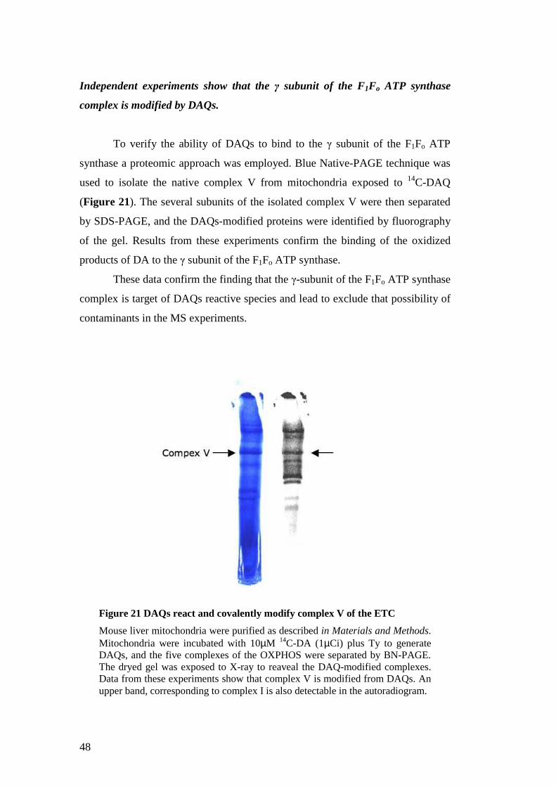
48
Independent experiments show that the γ subunit of the F1Fo ATP synthase
complex is modified by DAQs.
To verify the ability of DAQs to bind to the γ subunit of the F1Fo ATP
synthase a proteomic approach was employed. Blue Native-PAGE technique was
used to isolate the native complex V from mitochondria exposed to 14C-DAQ
(Figure 21). The several subunits of the isolated complex V were then separated
by SDS-PAGE, and the DAQs-modified proteins were identified by fluorography
of the gel. Results from these experiments confirm the binding of the oxidized
products of DA to the γ subunit of the F1Fo ATP synthase.
These data confirm the finding that the γ-subunit of the F1Fo ATP synthase
complex is target of DAQs reactive species and lead to exclude that possibility of
contaminants in the MS experiments.
Figure 21 DAQs react and covalently modify complex V of the ETC
Mouse liver mitochondria were purified as described in Materials and Methods. Mitochondria were incubated with 10µM 14C-DA (1µCi) plus Ty to generate DAQs, and the five complexes of the OXPHOS were separated by BN-PAGE. The dryed gel was exposed to X-ray to reaveal the DAQ-modified complexes. Data from these experiments show that complex V is modified from DAQs. An upper band, corresponding to complex I is also detectable in the autoradiogram.

49
Unaltered activity in the DAQs-modified F1Fo ATP synthase complex .
To assess if the Complex V modifications at the γ subunit by DAQs are
able to induce alterations in the enzyme activity an in gel activity assay for the
ATPase function of the complex V was carried out. The five complexes of the
mitochondria were isolated in the native and active state by Blue Native PAGE.
The ATPase activity of DAQs-modified and wild type Complex V was assessed
with an in-gel assay. The ability of the F1Fo ATP synthase complex to hydrolize
ADP was tested. ATP complex V cans idrolyze the molecule to ADP and Pi
inorganic. Pi reacts with Pb2+ ion to give the Pi2Pb that precipitates on the ATP-
synthase band in the gel. lead-phosphate. The activity of the isolated complex can
then be measured by densitometry analysis. The results from these experiments do
not show a decrease of the complex activity for the DAQs modified complex
respect to the control (Figure 22).
Nuclear Magnetic Resonance techniques were employed to investigate the
ATP synthase activity of the DAQs-modified F1Fo ATP synthase. With this
technique was possible to follow the ATP synthesis in the intact mitochondria
exposed to DAQs. As for the ATPase activity, also for the ATP synthase activity
Figure 22 Complex V modified by DAQ at the γγγγ subunit retain its F1Fo ATPase activity
Activity staining was performed on Complex V isolated from mitochondria incubated with DA and tyrosinase. Lane 1: control, 50µg proteins; lane 2: 50µg proteins from mitochondria exposed 10 minutes to 20µM DA plus 120U tyrosinase. To test if the latter oxidation products of DA show a different effect on ATPase activity DA and Ty were preincubated 5 minutes and after mitochondria were incubated with this mix for 10 minutes. 50µg of proteins from this sample were loaded in lane 3.

50
was not possible to detect a significative differences between DAQs-modified
complex V activity respect to the control.
Further studies are necessary to investigate the consequences of DAQs-
modification of γ subunit of the F1Fo ATP synthase complex. It is now clearly
established that dimerization of the F1Fo ATP synthase takes place in the
mitochondrial inner membrane. Interestingly, oligomerization of this enzyme
seems to be involved in cristae morphogenesis. As they were able to form
homodimers, subunits 4, ε, and γ have been proposed as potential ATP synthase
dimerization subunits. The hypothesis that DAQs-modified F1Fo ATP synthase
can alter mitochondrial morphology of the cristae is under study.
Conclusions
The present studies have shown that the oxidized products of the
neurotransmitter DA can enter the mitochondria crossing both the outer and the
inner membranes of the organelle. Moreover these reactive species are able to
bind and covalently modify several mitochondrial proteins. The ability of DAQs
to modify proteins at sulphydryl residues of proteins has been extensively
reported. Moreover several evidences of the ability of DAQs to bind and inhibit
cytosolic proteins as TH, DAT and parkin are present in literature. Evidence of
DAQs ability to enter and modify mitochondrial proteins is a crucial step in the
study of mitochondrial impairment in DA neurons in PD. This study has lead to
the identification of the γ subunit of the ATP synthase complex as one of the
protein modified by DAQs. Anyway assays to test the activity of modified ATP
synthase complex failed to shown impairments in ATP synthesis and
hydrolization activity of complex V. Further experiments are required in order to
understand if the binding of DAQs to the γ subunit of the ATP synthase complex
can impair other functions of complex V, as e.g. the structural role of complex V
in the formation of the cristae of the IMM. Moreover investigation to identity the
other DAQs-modified proteins in mitochondria are necessary and they can shed
light on the specific vulnerability of DA neurons and the mitochondrial
dysfunction in PD.

51
Part II: Pathological effects of oxidized products of dopamine in mitochondria.
DAQs induce mitochondrial dysfunctions
Mitochondria are key regulator of cell survival and death. A major role for
mitochondrial dysfunctions has been claimed in the pathogenesis of Parkinson’s
disease (PD). Mitochondrial dysfunction have been also assessed in PD patients,
and in particular defects in mitochondrial complex I of the respiratory chain has
been shown in the SNpc of sporadic PD (3). This defects seems to be present also
in other tissue of PD patients, since several evidence are reported for
mitochondrial impairment also in platelets and muscles (117).
A role of the neurotransmitter DA in the oxidative stress that lead to
dopamine neurons cell death in PD has been suggested for long time. As already
described in this thesis, the neurotransmitter DA can give rise to reactive species
in its normal metabolism or undergoes to spontaneous or catalyzed oxidative
reactions if accumulated in the cytoplasm of the cell. These reactive species can
lead to oxidative damage if not efficiently removed by antioxidant system of the
cell.
Previous studies on mitochondrial functions following exposure to
oxidized products of DA have shown that DAQs are able to induce several
mitochondrial damages as inhibition of mitochondrial respiration, increase of
ROS production and induction of apoptosis (89, 110-115). Interestingly the DAQs
ability to induce the mitochondrial PT in brain mitochondria has been also shown
(110).
Mitochondrial permeability transition (PT) is an increase in the
permeability of the mitochondrial membranes to molecules of less than 1500

52
Daltons in molecular weight. PT results from opening of mitochondrial
permeability transition pores (mPTP) (118-120). The mPT pore is a proteinaceous
pore that is formed in the inner membranes of mitochondria under certain
pathological conditions such as traumatic brain injury and stroke. Induction of the
permeability transition pore can lead to mitochondrial swelling and cell death and
plays an important role in some types of apoptosis (121-124). The MPTP was
proposed by Haworth and Hunter in 1979 and has since been found to be
involved in, among other things, neurodegeneration, a process that results in
damage and death of neurons (125). MPT is frequently studied in liver cells,
which have especially large number of mitochondria. In vitro PT is
accompanied by depolarization, matrix swelling, depletion of matrix pyridine
nucleotides (PN), outer membrane rupture and release of intermembrane proteins,
including cytochrome c (123, 126).
Statement of purpose
PT is claimed as one of the pathway of beginning for the mitochondrial
apoptosis. In PD the apoptotic pathway seems to be highly activated (7, 8). The
ability of DAQs to induce PT can account as one of the way that lead to the
massive loss of dopaminergic neurons in PD. Of great interest is then to
investigate the mechanism by which DAQs induce PT on mitochondria. As
shown in chapter I of this thesis DAQs have the ability to cross both mitochondria
membranes, enter the mitochondrial matrix, and covalently modify several
mitochondrial proteins.
The structure of the mPTP is hotly debated and the proteins that constitute
or regulate the PT are still not defined. Nonetheless several conditions and
molecules that modulate the mPTP conformation open/close are well
characterized as well as protocols to study the PT phenomenon (See also:
Materials and Methods, Mitochondrial Swelling) .

53
An analytical study on the ability of DAQs to induce PT in mitochondria and the
investigation of mechanism of its induction were carried out and the results are
explained in this chapter.
Results
DAQs Induce swelling in mitochondria
The characterization of the mechanism of DAQs induction of the
permeability transition (PT) in mitochondria was studied in collaboration with the
laboratory of the Prof. Paolo Bernardi at the Department of Biomedical Sciences
of the University of Padova.
Two types of experiments were used to evaluate the functional effects of
DAQs-mitochondrial swelling and modification of the calcium retention capacity
(CRC). Experimentally swelling of mitochondria can be followed at the
spectrophotometer as a decrease of light scattering of the mitochondrial
suspension due to the increase of mitochondria dimensions (127). The other
experimental protocol extensively used to study the induction of PT by DAQs is
the calcium retention capacity (CRC) experiment. Mitochondrial Ca2+ is
controlled by a transport cycle driven by the proton pumps of the respiratory
chain. The transport cycle is mediated by the Ca2+ uniporter, the Na+/Ca2+
antiporter and Na+/H+ antiporter of the inner membrane. Under physiological
conditions, mitochondrial Ca2+ controls key regulatory dehydrogenises in the
mitochondrial matrix. Under pathological conditions associated with cellular ATP
depletion and oxidative stress, mitochondrial Ca2+ triggers opening of the PT
pore (122). This is possible because Ca2+ binds to and activates Ca2+ binding sites
on the matrix side of the mPTP. The presence of free radicals, another result of
excessive intracellular calcium concentrations,
can also cause the mPT pore to open.
The amount of Ca2+ that mitochondria can store under different solution
conditions can be measure in the CRC experiments. In this fluorimetric assay

54
mitochondria are resuspended in presence of a fluorescent probe Calcium green-
5N that cannot permeate into the mitochondria. Small amounts of calcium are
then added and Ca2+ is sequestered by the functional mitochondria through the
Ca2+ uniporter. When the amount of calcium in the matrix reach a threshold of
excitotoxicity the mPT pore open with subsequent release of the calcium in the
medium. The fluorescent signal of the probe becomes then visible. Analysis of
the amount of calcium added to the mitochondria in different conditions allows
defining the Calcium Retention Capacity of mitochondria. A decrease of CRC of
treated mitochondria respect to the control can be read as an ability of the studied
compound to induce PT. Verification of this hypothesis can be carry on by
repeating the CRC experiments in the presence at the same time of the studied
compound and CsA. CsA is a powerful inhibitor of PT pore opening binding the
matrix protein cyclophillin D. Genetic inactivation of genes encoding putative
pore components have conclusively demonstrated the role of matrix
cyclophilin D as the mitochondrial receptor for the desensitizing effects of
cyclosporin A (118). Nevertheless the mechanism by which CsA keeps the mPTP
in the close conformation by binding cyclophillin D is still not fully understood.
Results from these experiments allow concluding that DAQs induce the
opening of the PTP in a dose dependant manner between 20-200µM DA (Figure
23). Moreover, the CRC experiments confirmed that DAQs are an act as inducers
of PT decreasing the CRC (Figure 24). For both these protocols controls
experiments with the specific inhibitor of the PTP Cyclosporine A (CsA) where
carried out. CsA is able to reverse the effect of DAQs on the PTP opening and
these results confirm that the mechanism by which DAQs induce the swelling of
mitochondria is opening the permeability transition pore.

55
The early oxidized products of DA are more toxic compared with the late ones.
The reaction of DA oxidation includes several steps that lead to melanin as
final product (81). Of great interest was to define which among the several
intermediates of the oxidation reaction has the more significant effect toward the
induction of mitochondrial permeability transition. The laboratory in Padova
invested a great deal of work in the chemical characterization of the several
oxidation products of dopamine. In the presence of the enzyme tyrosinase
dopamine is oxidized to dopamine-o-quinone (DAQ) that spontaneously
Figure 23 DAQs induce swelling in mitochondria
Mouse liver mitochondria (0.5mg/ml) were incubated with increasing amount of DA in presence of the enzyme tyrosinase. The decreasing in absorbance, correlated to the mitochondrial swelling, was followed spectrophotometrically at 620nm. Results from these experiments show that DAQs are able to induce the PT in mitochondria in a dose-dependant manner. To prove that this effect was due to the mitochondrial permeability transition pore (mPTP) opening, the same experiments were repeated in presence of the mPTP inhibitor CsA. As shown in the picture for the 100µM DA trace, CsA is able to completely revert the effect of DAQs.

56
ciclyzates to form the molecule leukoaminochrome. This catechol molecule can
be oxidized by tyrosinase to the quinone aminochrome (AC) that in a spontaneous
reaction rearrange to form the 5,6-dihydroxyindole (DHI). DHI can be oxidized
again by tyrosinase to indole-5, 6-quinone that combine with DHI to form the
polymer melanin (81).
The complexity of this reaction and the high reactivity of some of the
intermediates hinder the possibility to isolate these molecules and maintain them
stable in the physiologic buffers in order to single out the individual effect on
mitochondria. The strategy chosen to highlight the reactivity of the several
Figure 24 Calcium retention capacity of mouse liver mitochondria treated with DA and tyrosinase.
Mouse liver mitochondria (0.5mg/ml) were incubated in the presence of the fluorescent probe calcium green 5N, Calcium green 5N is fluorescent in presence of calcium and is excluded from mitochondria. Mitochondria are able to store Ca2+ in the matrix through the calcium uniporter. When Ca2+ reaches a threshold of toxicity it activates some still unknown sites triggering the mPTP opening and Ca2+ is released in the medium. In this protocol the amount of calcium that mitochondria can import in the presence of different amounts of the PT inducer DAQs is measured. Small mounts of calcium (10µM) are added every min until the PT happens with release of calcium in the medium visible at the fluorimeter as an increased of fluorescence of the probe calcium green 5N. The graph on the left shows that between 10 and 80µM DA (+120U tyrosinase) the calcium retention capacity of mitochondria decrease in a dose-dependant manner. The same experiments were repeated in the presence of the mPTP inhibitor CsA and results (on the right) show that CsA protect mitochondria from DAQs toxicity.

57
compounds was to incubate mitochondria with the products of subsequent time
points of the oxidative reaction, and compare the swelling induction effect.
Results from these experiments show that the earlier products of the DA
oxidation are more effective to induce the PT than the later ones. Since in the
reaction mix different products are present at the same time the identification of the
specific reactivity for the different products have to be investigated (Figure 25).
Figure 25 Mitochondrial swelling at different time point of the DA oxidation reaction: the early products are more toxic than the late.
Mitochondria purified from mouse liver (0.5mg/ml) were resuspended in medium containing glutamate and malate as oxidizing substrates for the ETC. Different time points of the oxidation reaction of 60µMDA plus tyrosinase were prepared separately and added to the mitochondria solution and the decreasing in absorbance at 620nm, corresponding to mitochondria swelling, was followed at the spectrophotometer. Results from these experiments show that the early products of DA oxidation reaction are more effective than the late ones to induce PT in liver mitochondria. I these conditions the first oxidation is catalized by the enzyme Ty and the spontaneous cyclization of the first product dopamine-o-quinone to leukoaminochrome is the rate limiting step for the formation of the second quinone aminochrome. Since cyclization is favored by acidic condition and the pH of the medium is 7.4, the dopamine-o-quinone, seems to be the most reactive species

58
DAQs toxicity involved the pyridine nucleotide pool.
In Costantini et al., 1996, authors describe two separate sites on the pore that
contains vicinal thiols (128). These thiols groups regulate PTP functions through
their redox state. Thiols/disulfide conformation on these residues depend on the
redox state of the matrix pool of pyridine nucleotide for one of site and on the
glutathione redox state for the other site (128). Authors show that selective
alkylation of this two sites with monothiol-binding compound makes possible to
point out if a molecule induce PT with a mechanism that involve redox state of PN
or GSH pool.
Based on the reported high capacity of DAQs to react with the free thiols
of GSH this protocol was employed to investigate the PT induction mechanism of
DAQs. The experiments were carried out as explained in Materials and Methods
of this thesis. The results show that DAQs ability of induce PT is reversed if the
thiols groups in equilibrium with the PN pool are blocked with the alkylating
reagent N-ethylmalemide (NEM) instead no differences are noticed if the thiols in
equilibrium with GSH pool are blocked (Figure 26). To further investigate the
nature of the interaction between DAQs and the pyridine nucleotides pool,
experiments with the molecule β-idroxybutirrate (BOH) where carried out. This
molecule is able increase the amounts of NADH in the matrix via the reaction
catalyzed by the enzyme acetate dehydrogenise. Acetate dehydrogenase use BOH
as oxidizing substrate to produce acetate and reduce NAD+ to NADH. Swelling
experiment where performed to test the protective effect of the BOH and results
from these experiments show that if the PN pool is maintained in the reduced state
mitochondria are protected from DAQs toxicity (Figure 27). These interesting
results let to the hypothesis that DAQs induce PT altering (directly or indirectly)
redox state of the PN pool in the mitochondrial matrix. The redox state of PN pool
has been prove to be one of the factors that affect the PTP state, in particular
oxidation of PN induce the pore opening (129). With these premises the state of
PN after mitochondria incubation with DAQs was assessed.

59
Figure 26 DAQ induce swelling through alteration of the pyridine nucleotide (PN) pool in mitochondria.
Mitochondria from mouse liver (0.5mg/ml) were incubated in medium containing glutamate and malate as oxidizing substrates. An amount of Ca2+ under the threshold necessary to trigger mPTP opening was added to accelerate mPTP opening by subsequent treatments. Alkylating reagents as monobromobimne (MBM) or N-ethylmaleimide (NEM) or reducing agent dithiotrehytol (DTT) were added after 1 minute. DA (100µM) and tyrosinase (120 units) were added to the solution after another minute and absorbance at 620nm was followed at the spectrofotometer. Results from these experiments reveal that if mitochondria are preincubated with the alkylating agen NEM, DAQs are not able to induce the PT. This result suggest that DAQs can induce PT with a echanism that involve thiols alkylation and in particular the thiols in equilibrium with the pyridine nucleotide (PN) pool in the matrix seem involved in this mechanism.

60
DAQs oxidize the pyridine nucleotide in mitochondria.
Experiments to evaluate the redox state of PN pool after DAQs treatment
of mitochondria were carried out. As explained in Materials and Methods section
the pyridine nucleotides were extracted from mitochondria after DAQs treatment
and an enzymatic assay was performed to estimate the amount of NADH and
NADPH separately. Data from these experiments demonstrate that NADH in
mitochondria exposed to small amounts of DAQs (20µ DA) is oxidized of 40%
respect to the control (Figure 28).
Figure 27 The maintenance of PN pool in the reduced state protects toward DAQ-induced mitochondrial swelling
Mitochondria from mouse liver (0.5mg/ml) were incubated in medium containing glutamate and malate as oxidizing substrates. An amount of calcium under the threshold that trigger PTP opening was added after 1 minute to accelerate PT in subsequent treatment. 30µM or 60µM DA were added to the mitochondrial suspension together with the enzyme tyrosinase (Ty, 120 units). β-idroxybutirrate (BOH) was added when indicated and the decrease of absorbance at 620nm corresponding to mitochondrial swelling was followed spectrofotometrically. Results from these experiments show that if NADH is maintained reduced by BOH presence, itochondria are protected from DAQ-induced swelling, reinforcing previous data of an involvement of PN oxidation in DAQ-induced PT in mitochndria.

61
These results show a toxic effect of DAQs toward mitochondria and lead
to hypothesize a mechanism by which DAQs induce the PT, since an oxidation of
the PN pool is recognized as inducing factor of mPTP opening (129). The
oxidation of PN can also lead to others detrimental effect in mitochondria, among
them an impairment of mitochondrial respiration, since PN are the electronic
donors of the complex I of the ETC. Further studies on this aspect of the toxicity
of DAQs in mitochondria can highlight the consequences of DAQs-induced PN
oxidation in mitochondria.
To point out if DAQs can directly react with NADH in vitro experiment
were carried out (Figure 29). The reaction between DA and DAQs with NADH
was followed specrophotometrically. The oxidation of NADH can be detected by
the decrease of the signal at 340nm since NADH has a characteristic peak of
Figure 28 DAQs oxidize the pyridine nucleotides in isolated mitochondria.
Mouse liver mitochondria were purified and exposed to DAQs as described in Materials and Methods. The amounts of reduced NADH and NADPH were assessed and compared to the untreated controls. Results from these experiments show that DAQs are able to decrease the reduced amounts of pyridine nucleotide in mitochondrial matrix. Further studies are necessary to establish if this oxidation is due to a direct redox reaction between the electrophiles DAQ and the reduced NAD(P)H or to other mechanism.

62
absorption at 340nm and NAD+ does not. Results from these experiments (Fig)
show that as DAQs is formed (signal at 480nm) NADH is rapidly oxidized (signal
340nm) claiming for a direct redox reaction between DAQs and NADH.
DAQs depolarize mitochondria a cellular model.
Since all the previous experiments were performed in in vitro systems, was of great
interest to explore if DAQs have the ability of induce PT in a cellular model. To
probe this toxic effect of DAQs could contribute to understand the unknown
mechanism of dopaminergic neurons loss in PD. PTP opening leads to
Figure 29 DAQs oxidize NADH in vitro
The reaction between DAQs and NADH was studied spectrophotometrically in vitro. Oxidation of NADH to NAD+ was followed at 340nm were NADH but not NAD+ has a characteristic absorption peak. Dopamine-o-quinone formation was followed at 480nm. As shown in the spectra above DA and NADH do not react instead when tyrosinase (Ty) enzyme is added in the cuvette the dopamine-o-quinone formation is accompanied by NADH oxidation, sign of a redox reaction between the electrophiles DAQs and the reduced NADH. These data suggest one possible meccanism of DAQs toxicity in mitochondria since NADH is one of the oxidizing substrates of complex I of the ETC and its depletion has been shown to induce PT. Both these events are detectable in mitochondria exposed to DAQs

63
mitochondrial membrane depolarization and this effect was followed in a
neuroblastoma SH-SY5Y cell line exposed to DA and tyrosinase. Mitochondrial
membrane potential was measured based on the accumulation of
tetramethylrhodamine methyl ester (TMRM) as described in the Materials and
Methods section of this thesis. Results from these experiments show that DAQs
induce depolarization of mitochondria in vivo (Figure 30). To prove that this effect
is due to PTP opening SHSY-5Y cell were exposed to DAQs in presence of the
PTP inhibitor CsA. CsA protects mitochondria from DAQs toxicity confirming that
the oxidized products of DA can be toxic toward mitochondria in vivo.
Figure 30 Changes of mitochondrial TMRM fluorescence induced by DAQs in SHSY-5Y neuroblastoma cells.
Neuroblastoma cell line SHSY-5Y were loaded with the fluorescent probe tetramethylrhodamine methyl ester (TMRM) and studied as described in the Materials and Methods. Oligomycin (Oligo) was added in every experiment to avoid depolarization due to the F1Fo ATPase complex. Cluster of mitochondria (10-30) were chosen as region of interest. Cells were exposed to 30µM DA and 100U of the enzyme tyrosinase (Ty). The same experiments in presence of the inhibitor of the mPTP CsA were carried out. Sequential digital images were acquired every 2 min, and the average fluorescence intensity of all relevant regions was recorded and stored for subsequent analysis. Results from these experiments show that DAQs depolarize mitochondria in vivo and that CsA protects at least in part, mitochondria from depolarization suggesting that DAQs-induce depolarization is due to induction of mPTP opening.

64
Conclusions
A critical role of mitochondrial dysfunction and oxidative damage has
been claimed in the pathogenesis of neurodegenerative diseases as Parkinson’s
disease (PD), Alzheimer’s disease, Huntington’s disease. Historically
mitochondria dysfunction has been associated with PD based on the observation
that accidental exposure to the environmental toxin 1-methyl-4-phenyl-1,2,3,4-
tetrathydropyridine (MPTP) results in development in parkinsonian syndrome
(10). MPTP is a weak inhibitor of mitochondrial complex I and its toxic
mechanism seems to involve oxidative damage by increase of ROS production
and DA oxidation (8). Moreover chronic exposure to the pesticide rotenone,
another complex I inhibitor reproduce the main pathological hallmarks of PD as
loss of dopaminergic neurons in the substantia nigra, α-synuclein positive
inclusions and the characteristic motor symptoms in mice (9). Mitochondrial
dysfunction has been reported in the SN of PD patients and in other tissues
outside the brain (3, 117) .
In the scenario of a global mitochondrial dysfunction in PD what becomes
interesting is to investigate what makes dopaminergic neurons more vulnerable to
a mitochondrial deficiency. A role for DA as endogenous toxin has been claimed
for long. Deregulation of DA synthesis, storage or metabolism can lead to DA
oxidation in the cytoplasm with accumulation of toxic products as ROS and DA
quinones. Taken together these evidence lead to the hypothesis that a
mitochondrial chronic defect or protein mutation can be exacerbated in
dopaminergic neurons by presence DA and its toxic metabolites.
The research presented in this chapter was focused on the characterization
of the toxic effects of the oxidized products of DA on mitochondria. DAQs are
able to cross both mitochondria membranes and covalently modify several
mitochondria proteins (Part I of this thesis). The pathological consequences, if
any, of this interaction were investigated. Previous evidence report the ability of
DAQs to induce PT in brain and liver mitochondria (110). This result was the
starting point for the analytical characterization of the ability of DAQs to induce
PT in liver mitochondria. Swelling experiments were carried out testing several
conditions of substrates and DA concentrations. CRC experiments were also

65
performed to quantify DAQs induction effect. The results presented in this chapter
show that the oxidized products of the neurotransmitter DA can induce the
permeability transition (PT) in a dose dependant manner between 5-100µM DA in
presence of tyrosinase enzyme as oxidizing agent of DA. On the contrary in the
same conditions DA or tyrosinase alone are not able to induce PT. CsA, a specific
inhibitor of PT, completely reverts DAQs effect proving that DAQs-induced
mitochondrial swelling is due to opening of the mPTP. Calcium retention capacity
experiments confirmed the PT induction ability of DAQs and allowed to quantify
the effect, in Ca2+ nmol that the treated mitochondria are able to store before
mPTP opening.
The mechanism by which DAQs induce PT was investigated and alteration
of the redox state of matrix pyridine nucleotide pool of mitochondria was shown.
Oxidation of NAD(P)H is one of the known mechanism to induce PT(129).
Finally the ability of DAQs to induce PT in a cellular model was shown. Since PT
has as final consequence the disruption of OMM and release of the pro-apoptotic
factors that resides in the intermembrane space, these data lead to the hypothesis
that DAQs-induced PT can contribute to the loss of dopaminergic neurons in PD.
Taken together, these data provide an additional biochemical mechanism by
which DAQs may affect cell function, and support the idea that mitochondrial
damage is an important determinant in DAQs toxicity.

66

67
Part III: Complex I and Parkinson’s disease Complex I
Complex I (reduced nicotinamide adenine dinucleotide–ubiquinone oxido-
reductase, or NADH:ubiquinone oxidoreductase) is the first complex of the
electron transport chain (ETC) in mitochondria. Among all the others four
complexes of the ETC it has the most complicated structure and genetics. It is the
largest complex of the respiratory chain and its relative molecular mass is ~980
000 Da. In mammals, it is composed at least of 45 subunits (identified so far), of
which 7 are encoded by the mitochondrial genome (mtDNA) and the remaining
by the nuclear genome(130). Complex I catalyzes the transfer of electrons from
NADH to coenzyme-Q10 or ubiquinone via a flavin mononucleotide cofactor and
iron sulfur centers. The pathway of electrons occurs as follows: NADH is
oxidized to NAD+, reducing flavin mononucleotide to FMNH2 in one two-electron
step. The next electron carrier is a Fe-S cluster, which can only accept one
electron at a time to reduce the ferric ion into a ferrous ion. In a convenient
manner, FMNH2 can be oxidized in only two one-electron steps, through a
semiquinone intermediate. The electron thus travels from the FMNH2 to the Fe-S
cluster, then from the Fe-S cluster to the oxidized Q to give the free-radical
(semiquinone) form of Q. This happens again to reduce the semiquinone form to
the ubiquinol form, QH2. Complex I also has proton-transporting activity over the
inner mitochondrial membrane to the intermembrane space, indeed during this
process, four protons are translocated across the inner mitochondrial membrane,
from the matrix to the intermembrane space. This creates a proton gradient that
will be later used to generate ATP from ADP through oxidative phosphorylation. .
Complex I is one of the main sites at which premature electron leakage to oxygen
occurs, thus being one of main sites of production of a harmful free radical called
super oxide.

68
The great complexity of mammal complex I in comparison with other
complex I from different species has made hypothesize that it can exerts
supplementary functions. Fontaine et al. have suggested that the electron flux
through complex I regulate the mitochondrial PTP (131), since when respiration is
supported by complex I substrates the PTP opened at much lower calcium loads
than with complex II and IV substrates. This effects was directly related to the rate
of electron flow through complex I and not to membrane potential or the redox
status of pyridine nucleotides in the matrix. Moreover rotenone, a specific
inhibitor of the electron flux through complex I, delays PTP opening and
increased CRC in mitochondria (132). Involvement of complex I in cell death
pathways has also been claimed, based on Cao’s laboratory observation that a
mammal complex I subunit, GRIM19, is released in the cytoplasm of apoptotic
cells (133).
A deficiency in complex I is the most frequently encountered cause f
mitochondrial disease and mutation in several nuclear DNA-encoded subunits
cause devastating neurological disorders as Leigh syndrome or Leber’s Hereditary
Optic Neuropathy (LHON) (134) . Milder defects of complex I has also been
claimed from long time to play a central role in several neurodegenerative
diseases as Parkinson’s disease, Alzheimer’s disease and Huntington’s disease.
Complex I is impaired in PD
Several lines of evidence point to an inhibition of complex I activity in
PD. The first evidence of complex I impairment in PD was reported in 1983 by
Langston and colleagues (10) when they identified 1-methyl-4-phenyl-1,2,3,6-
tetrahydropyridine (MPTP), a side-product of the synthesis of an heroin analog, as
the cause of development of acute, severe and permanent Parkinsonism in several
drug addicts in northern California. MPTP is a pro-toxin that enters the brain and
is then metabolized by monoamine oxidase B to 1-methyl-4-phenylpiridinium ion
(MPP+). MPP+ is accumulated in dopaminergic neurons and being an excellent
substrate of the dopamine transporter (DAT) and in these neurons accumulates in
mitochondria where inhibits the ETC at the level of complex I (8).

69
Other evidences stand for an inhibition of complex I in idiopathic PD.
Shapira and colleagues reported defects in complex I and reduced glutathione in
the substantia nigra of PD patients (3). Complex I deficits seems affect also tissue
outside the brain, several studies have reported reduced complex I activity in the
platelets of people with PD (135-138). Others studies with cells without mtDNA
(ρ0 cells) fused with platelets from PD patients (cybrid) show that the defect in
complex I activity can be stably transferred to ρ0 cells (4, 139). These studies
strongly suggest that complex I defects in PD might be due to mtDNA mutations
but efforts so far to identify these mutations have been unsuccessful. Upon these
findings a systemic defect of complex I in idiopathic PD has been hypothesized
The in vivo rotenone model of Parkinson’s disease is one powerful tool to
help to clarify this question (9). Rotenone is a high affinity inhibitor of complex I
used as organic pesticide. Moreover rotenone extreme lipophilic nature allows it
to cross easily biological membranes and enter the brain without specific
transporters. Systemic administration of rotenone in rats cause chronic and
uniform inhibition of complex I in several organs like brains, kidney hearth and
liver. Anyway after some weeks selective degeneration of neurons in the SN
occurred. Moreover many of the dying neurons have large cytoplasmatic
inclusions containing alpha-synuclein and ubiquitin as Lewy bodies in PD.
Dopaminergic degeneration is accompanied by marked increase in oxidative
protein damage only in affected brain region similar to which reported in PD.
Finally rotenone-infused animals developed symptoms of Parkinsonism including
bradikinesia and rigidity. A crucial implication of this work is that dopaminergic
neurons in the substantia nigra have intrinsic sensitivity to complex I defects,
since are the ones that die while complex I is uniformly inhibited throughout the
brain (and the rest of the body). Since the degeneration occurred at level of
complex I inhibition that don’t significantly impair respiration, the mechanism of
rotenone toxicity is not related to a bioenergetic deficit. The mechanism of
rotenone toxicity is an increased oxidative stress and oxidative damage of
mitochondria and the whole cell (11). Other experimental models of PD also
suggest oxidative damage as a key mechanism of dopaminergic degeneration (8).
Under physiological conditions complex I is one of the main sites at which
premature electron leakage to oxygen occurs, thus being one of main sites of
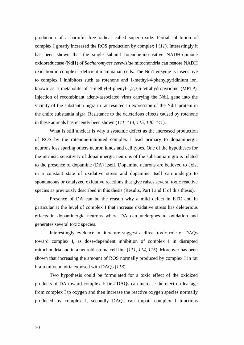
70
production of a harmful free radical called super oxide. Partial inhibition of
complex I greatly increased the ROS production by complex I (11). Interestingly it
has been shown that the single subunit rotenone-insensitive NADH-quinone
oxidoreductase (Ndi1) of Sacharomyces cerevisiae mitochondria can restore NADH
oxidation in complex I-deficient mammalian cells. The Ndi1 enzyme is insensitive
to complex I inhibitors such as rotenone and 1-methyl-4-phenylpyridinium ion,
known as a metabolite of 1-methyl-4-phenyl-1,2,3,6-tetrahydropyridine (MPTP).
Injection of recombinant adeno-associated virus carrying the Ndi1 gene into the
vicinity of the substantia nigra in rat resulted in expression of the Ndi1 protein in
the entire substantia nigra. Resistance to the deleterious effects caused by rotenone
in these animals has recently been shown (111, 114, 115, 140, 141).
What is still unclear is why a systemic defect as the increased production
of ROS by the rotenone-inhibited complex I lead primary to dopaminergic
neurons loss sparing others neuron kinds and cell types. One of the hypotheses for
the intrinsic sensitivity of dopaminergic neurons of the substantia nigra is related
to the presence of dopamine (DA) itself. Dopamine neurons are believed to exist
in a constant state of oxidative stress and dopamine itself can undergo to
spontaneous or catalyzed oxidative reactions that give raises several toxic reactive
species as previously described in this thesis (Results, Part I and II of this thesis).
Presence of DA can be the reason why a mild defect in ETC and in
particular at the level of complex I that increase oxidative stress has deleterious
effects in dopaminergic neurons where DA can undergoes to oxidation and
generates several toxic species.
Interestingly evidence in literature suggest a direct toxic role of DAQs
toward complex I, as dose-dependent inhibition of complex I in disrupted
mitochondria and in a neuroblastoma cell line (111, 114, 115). Moreover has been
shown that increasing the amount of ROS normally produced by complex I in rat
brain mitochondria exposed with DAQs (113)
Two hypothesis could be formulated for a toxic effect of the oxidized
products of DA toward complex I: first DAQs can increase the electron leakage
from complex I to oxygen and then increase the reactive oxygen species normally
produced by complex I, secondly DAQs can impair complex I functions

71
covalently bind and modify protein subunits of the complex leading to a loss of
structure and function of proteins and the entire complex.
Statement of purpose
The toxic effects of the oxidized products of DA toward complex I was
investigated. Several approach were pursued to characterize the interaction
between DAQs and complex I. The ability of DAQs to inhibit mitochondrial
respiration was tested by oxygraphic measures and a specific assay for complex I
activity was set. The ability of DAQs to bind and covalently modify complex I
subunits was also investigated. The toxic activity of DAQs was also compared to
the one of the specific inhibitor of complex I rotenone and the competition
between DAQs and rotenone to bind complex I subunits was compared.
Dopaminergic cells expressing the rotenone-insensitive complex I subunit
Ndi1 was used to point out the specificity of DAQs toward complex I (11). If cells
expressing Ndi1 subunit are resistant or protected from DAQs toxicity too, is
possible to hypothesize parallel effects of DAQs and rotenone toward
mitochondrial complex I.
Results
DAQs inhibit mitochondrial respiration.
NADH and NADPH are the electrons donor of the first complex of the
oxidative phosphorilation chain in mitochondria. As DAQs are able to oxidize the
pyridine nucleotide pool in a redox reaction (Results, Part II, of this thesis), is
reasonable to hypothesize that this effect leads to an impairment of the
mitochondrial respiration. Data present in literature (110) show the faculty of
DAQs to inhibit the state 3 of mitochondrial respiration. During mitochondrial
respiration, molecular oxygen is reduced to water by complex IV (cytochrome
oxidase complex), in the last step of the electronic transport chain.

72
Experimentally, mitochondria respiration is assessed by oxigraphic measurement
of the decrease of molecular oxygen dissolved in a mitochondria suspension.
Conventionally mitochondria respiration is divided in three states. State 4, or
resting respiration, is defined as oxygen consumption of isolated mitochondria in
the presence of oxidizing substrates and in the absence of ADP. State 3, or active
respiration, is ADP-stimulated respiration in presence of oxidizing substrates. The
uncoupled state is defined as a condition in which the rate of electron transport
can no longer be regulated by an intact chemiosmotic gradient, and is measured
after addition of an uncoupler agent as 2,4-dinitrophenol (DNP) or carbonyl
cyanide p-[rifluoromethoxyl]-phenyl-hydrozone (FCCP). DNP and FCCP act as
proton ionophore binding protons on one side of the membrane and being fat
soluble, drifting to the opposite side where they lose the protons, dissipating the
chemiosmotic gradient and leaving the electron transport system unhibited.
Oxygraphic measurements were carried out to evaluate the respiration capacity of
mitochondria. Accurate precautions were taken to remove the enzyme tyrosinase
and the non-reacted DA from the samples. Since tyrosinase requires molecular
oxygen as co-substrate in the oxidation of its substrates removal of tyrosinase is
crucial to avoid measurement of oxygen consumed in the tyrosinase oxidative
reaction.
Data from these experiments show that after incubation with DAQs all the
three states of mitochondrial respiration are inhibited. In particular state 3 and the
uncoupled state are almost equally inhibited leading to the hypothesis that
complex V is not inhibited by DAQs reinforcing previous findings of this thesis.
These findings do not permit to define which among the first four complexes of
the ETC is more affected by DAQs toxic effect respect to the others. Specific
assays for every complex of the ETC were planned to investigate at which point
of the mitochondrial respiration DAQs exert their inhibitory effect and the results
for the complex I are reported in the next paragraph (Figure 31).

73
DAQs inhibit Complex I of the electronic transport chain in mitochondria.
Since DAQs inhibit mitochondrial respiration, experiments to point out the
functional state of complex I after DAQs treatment were carried out. A specific
assay for complex I was employed (100) and the activity of the complex from
mitochondria exposed to increasing amount of DAQs was measured. In this assay
reduction of 2,6-dichloroindophenol (DCIP) by electrons accepted from
Figure 31 DAQ inhibit mitochondrial respiration
Mouse liver mitochondria were exposed to DA (20-60µM) and tyrosinase (Ty, 100 units) for 10 minutes as described in Materials and Methods. The medium contained glutamate and malate as oxidizing substrates and CsA (2µM) to inhibit mPTP opening and subsequent mitochondria depolarization. The three state of mitochondrial respiration were assessed by oxygraphic measurements with a Clark electrode chamber. Sate 4 was measured in absence of ADP. State 3 after ADP addition and the Uncoupled State after DNP addition. Non-reacted DA and Ty were removed by centrifugation before starting the respiration since Ty employs molecular oxygen for its oxidizing reaction. Results from these experiments show that DAQs are able to inhibit all the three states of mitochondrial respiration. State 3 and the Uncoupled state are inhibited almost at the same amount. This result leads to exclude the complex V as one of the target of DAQs inhibition.

74
decylubiquinol was measured as described in the Materials and Methods.
Decylubiquinol is reduced by complex I after oxidation of NADH.
Results from these experiments show an inhibitory effect of DAQs
treatment on complex I activity (Figure 32). Specific precautions were taken to
remove unreacted DA and the enzyme tyrosinase before starting the
measurements. This solution was necessary since NAD(P)H is readily oxidized by
DAQs in a redox reaction (Results, Part I of this thesis). After removal of both the
Figure 32 DAQs inhibit Complex I
A sensitive and specific assay for mitochondrial complex I activity was employed to test the inhibitory effect of DAQs on the first complex of the ETC. This method is based on measuring 2,6-dichloroindophenol (DCIP) reduction by electrons accepted from decylubiquinol reduced after oxidation of NADH by complex I. 0.5mg/ml of mitochondria were exposed to DA (0-150µM) and tyrosinase (Ty, 100U) to generate DAQs for 10 minutes. Mitochondria were gently spun down and resuspnded in 965µl of reaction buffer containing 25mM KPO4, 3.5g/L BSA, 70µM Decylubiquinone, 60µM Dichloroindophenol (DCIP), 1µM Antimycin A. The solution was let equilibrate for 3-5min at 37 °C and the reaction was started by adding 20µl of 10mM NADH solution. Absorbance at 600nm was measured every 30 seconds for 10 minutes. 1µl of rotenone 1mM was added and absorbance was measured every 30 seconds for 5 minutes. The data are expressed respect the control and the non specific DCIP reduction, if any, was detected after addition of rotenone and subtracted to every measure.
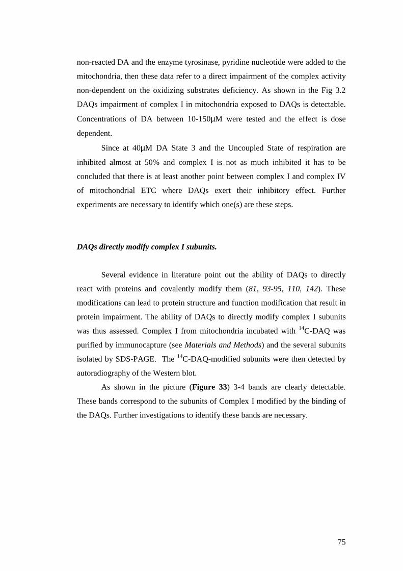
75
non-reacted DA and the enzyme tyrosinase, pyridine nucleotide were added to the
mitochondria, then these data refer to a direct impairment of the complex activity
non-dependent on the oxidizing substrates deficiency. As shown in the Fig 3.2
DAQs impairment of complex I in mitochondria exposed to DAQs is detectable.
Concentrations of DA between 10-150µM were tested and the effect is dose
dependent.
Since at 40µM DA State 3 and the Uncoupled State of respiration are
inhibited almost at 50% and complex I is not as much inhibited it has to be
concluded that there is at least another point between complex I and complex IV
of mitochondrial ETC where DAQs exert their inhibitory effect. Further
experiments are necessary to identify which one(s) are these steps.
DAQs directly modify complex I subunits.
Several evidence in literature point out the ability of DAQs to directly
react with proteins and covalently modify them (81, 93-95, 110, 142). These
modifications can lead to protein structure and function modification that result in
protein impairment. The ability of DAQs to directly modify complex I subunits
was thus assessed. Complex I from mitochondria incubated with 14C-DAQ was
purified by immunocapture (see Materials and Methods) and the several subunits
isolated by SDS-PAGE. The 14C-DAQ-modified subunits were then detected by
autoradiography of the Western blot.
As shown in the picture (Figure 33) 3-4 bands are clearly detectable.
These bands correspond to the subunits of Complex I modified by the binding of
the DAQs. Further investigations to identify these bands are necessary.

76
Rotenone-DAQs competition
Previous results shown in this research outline that the rotenone, a specific
complex I inhibitor, exacerbates the toxic effect of DAQ in mitochondria. To test
if the rotenone molecule and the oxidized products of DA compete for the same
sites of complex I and delineate their mechanism of interaction with the complex,
a competitive assay was carried out. Mitochondria were incubated with a large
amount of rotenone (2mM) and after 14C-DA and tyrosinase was added for 10
Figure 33 Autoradiogram of Complex I isolated by Immunoprecipitation from mitochondria treated with 14C-DA and tyrosinase.
Liver rat mitochondria were purified as described in Materials and Methods. Lane I: Mitochondria (5mg) were then resuspended in medium containing glutamate and malate as oxidizing substrates and exposed 10 minutes to 20µM 14C-DA, (1µCi), and tyrosinase (120 units) to generate DAQs. At the end of the treatment mitochondria were lysed and complex I immunocaptured following manufacture directions. Complex I subunits were then separated in a precast 12% gel by SDS-PAGE and the proteins transferred overnight in a PVDF membrane by Western blot. The membrane was the exposed to X-ray film and the autoradiogram developed 21 days later. Lane II as lane I, plus 2mM rotenone during DAQs tratment for 10 minutes. The Autoradiograms show that DAQs are able to react with at least three subunits of complex I instead rotenone completely revert this interaction.

77
minutes. The medium was then removed after centrifugation of mitochondria and
the mitochondria washed.
Complex I from this sample was isolated by immunocapture and the
subunits separated by SDS-PAGE. The proteins were transferred to a membrane
by Western blot and the membrane exposed to an X-ray film to detect the band
modified by the binding to DAQ.
The absence of detectable bands in the autoradiogram (Figure 33 lane II)
can suggest a direct competition between the molecules and a inability of DAQ to
bind to the complex I subunits sites already bound to rotenone molecules or a
conformation change in complex I structure due to rotenone that make the DAQ
binding not possible. This interesting result open new prospective of research in
both rotenone mechanism of complex I inhibition and DAQ modification ability
of complex I subunits.
Comparative experiments of DAQs and rotenone toxicity were then
planned to figure out if the toxic mechanism of these compounds is comparable in
vitro and in vivo.
Cells resistant to rotenone show a decrease vulnerability to DAQs toxicity.
To further investigate if DAQs toxicity depends on interaction with
complex I, the effect of DAQs in cells expressing the rotenone-insensitive single
subunit NADH dehydrogenase of Saccharomices cerevisiae (Ndi1) was analyzed.
This single subunit acts as a “replacement” for the entire complex I in mammalian
cells (Seo et al 200, Bai et al 2001, see Rotenone toxicity is caused by complex I
inhibition in Todd paper). This system was used to assess that rotenone toxicity
depended on direct interaction with complex I in previous findings from
Greenamyre’s laboratory (11). In this system, electrons from complex I substrates
(e.g., glutamate) are shunted through Ndi1 into downstream portions of the ETC,
thereby allowing mitochondrial respiration to bypass complex I inhibition. Since
cells expressing Ndi1 subunit are resistant to rotenone toxicity rotenone effects
can be entirely explained by the inhibition of complex I. This system was then
chosen to investigate the toxic effects of DAQs. If DAQs exert their toxic affect

78
toward complex I, cells expressing the Ndi1 subunit will be protected from DAQs
toxicity. Cells expressing Ndi1 subunit were treated with DAQs and the cells
availability after the treatment was assessed (See Material and Methods).
Interestingly cells expressing the rotenone-resistant subunit show an increased
resistance to DAQs toxicity too (Figura 34). This finding proves that DAQs exert
toxic effects from their interaction with complex I and if it is bypassed DAQs are
less toxic. Is tempting to speculate that DAQs exert their toxic effects on complex
I in a similar way as rotenone does. If this hypothesis is correct DAQs have the
ability to increase the electrons leakage from complex I increasing the production
of ROS and the oxidative damage. Evidences in literature support this speculation.
The ability of DAQs to increase the amount of H2O2 produced by mitochondria in
Figure 34 Cells expressing the rotenone-insensitive Ndi1 subunits are protected from DAQs toxicity.
Neuroblastoma SHSY-5Y cells were transfected with a NDI1 gene expressing plasmid as described in the Materials and Methods. The efficiency of the transfection was assessed at the microscope before starting the treatment. Cells were exposed to DA overnight and the cells viability was assessed at the microscope. Four fields were considered and the % of cell death is referred to the untreated control. As shown in the graph above the Ndi1 expressing cells are ore resistant to DAQs toxicity than the untreated cells. Ndi1 is a rotenone-insensitive single subunit NADH dehydrogenase of Saccharomices cerevisiae. These data suggest that one of the mechanism of DAQs toxicity can be similar to the rotenone with the specific inhibition of complex I and the increase of electrons leakage from this site.
79
presence of complex I oxidizing substrates has been shown, on the contrary the
effect is not detectable if complex I is bypassed with succinate, a complex II
substrate (113). Nonetheless cells expressing Ndi1 subunit are not fully protected
from DAQs toxicity their detrimental effect does not result solely from complex I
impairment. This result is expected from the high reactivity of DA oxidized
products and also from previous results from this thesis.
Conclusions
The research presented in this chapter show the ability of the oxidized
products of DA to inhibit mitochondrial respiration and the inhibitory effect at the
complex I level was studied further. The inhibition of the ETC was assessed by
oxygraphic measurements and the results show that all the three states of
mitochondrial respiration are inhibited after exposure to DAQs. Since the State 3
and the uncoupled State are equally inhibited (Figure 31) it can be excluded at
specific inhibition at the level of complex V of the mitochondria ETC. State 3 is
the respiration in presence of oxidizing substrates and ADP, so is an index of the
respiration coupled with ATP synthesis instead the Uncoupled State is the
maximum rate of the respiration not coupled with ATP synthesis, that is an index
of the function of the four complex of the ETC. Among them, the effect of DAQs
toward the first complex was investigated more in detail. Previous results from
this research suggest a possible inhibitory effect of DAQs on complex I can
derived from their ability to oxidized the pyridine nucleotide in mitochondrial
matrix (Figure 28) since complex I catalyzes the transfer of electrons from the
pyridine nucleotide NADH to ubiquinone. Results from these experiments lead to
conclude that his mechanism, even if present, is not the only way of DAQs
toxicity. Cells expressing the rotenone-insensitive single subunit NADH
dehydrogenase of Saccharomices cerevisiae (Ndi1) were a powerful tool to
investigate DAQs toxicity toward complex I. In this system NADH is still
oxidized by Ndi1 subunit but what is missing is the site of action of the complex I
inhibitor rotenone. In Ndi1-expressing cells DAQs are less toxic than in the
control, suggesting that besides the oxidation of the pyridine nucleotide there must

80
be another mechanism by which these cells are protected from DAQs toxicity. Is
tempting to speculate that the mechanism by Ndi1 cells are protected from DAQs
is the same they are protected from rotenone. Rotenone exerts its toxic effect
binding to complex I and increasing electron leakage from complex I, heightening
the amounts of ROS produced by complex I in mitochondria. There are already
evidence in literature that mitochondria exposed to DAQs increase the amount of
H2O2 produced respect to the control (113). In addition preincubation of
mitochondria with rotenone prevent DAQs to bind and covalently modify DAQs
subunits (Figure 33) suggesting that there can be a competition for the same
site(s) between these two species. Further investigation are necessary to clarify the
toxic mechanism of DAQs on mitochondria nonetheless evidences of their effects
to complex I is clearly suggested herein. Complex I is impaired in the
dopaminergic neurons of PD patients and studies in PD brain show a great
increase of oxidative stress in dopaminergic neurons. The results describe in this
chapter show a possible way of mitochondrial impairment and complex I
inhibition due to the oxidized products of DA.

81
Discussion
The research that comprises this dissertation concerns the toxic effects of
DA oxidation products on mitochondria. Further characterization of this
detrimental interaction may shed light on the still unknown mechanism of
dopaminergic neuron loss in Parkinson’s disease (PD). Based on studies using
human PD brains, animals models of PD, and in vitro cellular models, three
mechanisms seem to be primarily involved in the neurodegeneration of
dopaminergic neurons in PD: 1) increased oxidative stress, 2) mitochondrial
dysfunction (i.e. decrease of ATP production, Ca2+ mishandling and increase of
electrons leakage from ETC with consequent ROS production), and 3) impairment
of the ubiquitin-proteasome system with consequent accumulation of misfolded
proteins in the cytoplasm of neurons. Several factors have been shown to induce
neurodegeneration of dopaminerigc neurons, invoking one or more of these
mechanisms. It is particularly noteworthy that mitochondrial dysfunction can
exacerbate each of these pathogenic mechanisms. Additionally, since none of
these proposed pathogenic mechanisms adequately explains why dopaminergic
neurons are selectively vulnerable in PD, further characterizing the effects of DA
on mitochondria is paramount.
A combination of genetic susceptibilities and environmental factors seems
to play a central role in PD pathogenesis. In the last ten years, mutations in seven
genes have been reported to cause rare familial forms of PD. While these genetic
forms of PD represent a small portion of total PD cases, they highlight pathogenic
pathways that may be involved in all cases of PD. Of particular interest to this
dissertation, is that all mutations in genes related to PD exert an effect on
mitochondrial function.
Environmental toxins that inhibit mitochondrial complex I throughout the
body (MPTP and the pesticide rotenone) induce PD in humans and are considered
one of the factors involved in the development of sporadic forms of PD. What is
still not understood is why a defect or impairment that affects all the cells in the

82
body (for the rotenone) has as prime consequence the degeneration of a specific
family of neurons in the brain. However, similar to the genetic mutations, which
give rise to PD, these environmental toxins highlight the importance of
mitochondrial function and ROS-mediated oxidative stress in the death of
dopamineric neurons.
Based on the anatomy of this neurodegeneration, a central role of the
neurotransmitter DA in dopaminergic cell loss in PD has been for long proposed
(143). DA is a highly reactive molecule that can undergo spontaneous or
enzymatic catalyzed oxidative reactions in the cytoplasm, leading to the
production of highly reactive oxidized species. For this reason cytoplasmic DA is
normally stored in synaptic vesicles or readily metabolized to less toxic products
by cytosolic enzymes. If the synthesis or metabolism of DA is impaired, DA can
accumulate in the cytosol and give rise to several toxic reactive oxygen species
(ROS) such as superoxide anion, hydroxyl radical and DA quinones. Pathological
events that lead to DA catabolism/metabolism impairment can lead to
accumulation of ROS and DA quinone formation, increasing oxidative stress in
dopaminergic neurons. If cellular antioxidant systems that scavenge these toxic
species are overwhelmed, growing ROS production can induce cytotoxicity and
consequent cell death. Moreover, growing evidence suggests a toxic role
specifically for the DA quinone species. DA has a catechol ring that can be
oxidized to a quinone. Quinones are highly electrophilic and readily react with
cellular nucleophiles. It has been reported that DA quinones are able to react with
glutathione, and covalently modify proteins at cysteine residues in vitro and in
vivo (28, 94, 106, 108). Since cysteine amino acids are often found in the active
sites of proteins, binding of such residues by DA quinones can lead to protein
modification and impair function. DA quinones have been reported to modify and
inhibit several proteins, including tyrosine hydroxylase (TH) (108), dopamine
transporter (DAT) (94) and parkin (92). Moreover, DAQ-modification inhibits α-
synuclein fibrillation process and leads to accumulation of the more toxic
protofibrils species (97).
Evidence suggests that an alteration of DA metabolism occurs in PD with
consequent accumulation of DA in the cytoplasm (144, 145). Several mechanisms
have been suggested that lead to increased cytoplasmic DA, such as mitochondrial

83
dysfunction and a resulting decrease in ATP. This decrease in ATP production
can affect several ATP-dependent cellular processes, including the active
transport of DA molecules into synaptic vesicles (14). Other process that can
inhibit the transport of DA into vesicles have been reported, including a toxic
interaction between mutated α-synuclein and the synaptic vesicles (146) or MPTP
exposure (147). Moreover, free cysteinyl-catechol and GSH-catechol derivatives
have been found to be increased in post-mortem PD brains compared to age-
matched controls (85, 109). This growing body of evidence supports the theory
that DA oxidation and protein modification by DA quinones can be one of the key
explanations for why dopaminergic neurons are more vulnerable to insults in PD.
Based upon all these data, it was compelling to explore the possible
mechanisms that define the effects of DA quinones on mitochondria. Evidence in
the literature shows that the oxidized products of DA induce permeability
transition (PT) in isolated brain mitochondria and inhibit mitochondrial
respiration (110). In isolated and disrupted mitochondria, DAQs inhibit complex I
of the ETC. This effect can be reversed by GSH but not by ROS scavengers such
as catalase or α-tocopherol, suggesting that DAQs are the toxic species (114,
115).
In the research presented in this thesis, the toxic effects of DA quinones on
mitochondria were investigated using different approaches. The ability of the
oxidized products of DA to enter in the mitochondria was first investigated. The
results from these experiments show that DAQs but not DA are able to enter
mitochondria. The ability of DAQs to cross both the outer and inner
mitochondrial membranes of mitochondria was assessed. Within the
mitochondria, DAQs are able to react and covalently modify mitochondrial
proteins. DAQ-modified mitochondrial matrix proteins such as the γ-subunit of
F1Fo ATP synthase and complex I subunits were found. Interestingly the γ-subunit
of F1Fo ATP synthase contains a cysteine residues (Cys 78) in a loop exposed to
the matrix and this is a reasonable site for DAQs-binding and modification.
Experiments to verify whether the activity of the DAQ-modified F1Fo ATP
synthase is impaired were carried out. An in-gel activity assay for the ATPase
activity of the F1Fo ATP synthase complex did not reveal significant differences
when compared to the untreated complex. It is now clearly established that

84
dimerization of the F1Fo ATP synthase takes place in the mitochondrial inner
membrane. Interestingly, oligomerization of this enzyme seems to be involved in
cristae morphogenesis. As they were able to form homodimers, subunit γ has been
proposed as one of the potential ATP synthase dimerization subunits (148). The
hypothesis that DAQs-modified F1Fo ATP synthase can alter mitochondrial
morphology of the cristae is currently under study.
The mechanism by which DAQs enter the mitochondria was then
investigated. Results from these experiments have shown that DAQs enter the
mitochondria in a way that is independent of mitochondrial ∆Ψ. Experiments that
assess the ability of DAQs to enter the mitochondria were carried out in the
presence or absence of oxidizing substrates, however no significant differences
could be detected. From these experiments it can be concluded that DAQs enter
the mitochondria without an active transport, since absence of substrates for the
oxidative phosphorylation doesn’t alter their ability to enter the organelle.
DAQs were able to inhibit mitochondrial respiration and specific
inhibition of complex I isolated from mitochondria exposed to DAQs was found.
DAQ-modified complex I subunits were identified by autoradiography
experiments with 14C-DAQs and these modifications can account for complex I
inhibition. Another possible mechanism of DAQ-induced toxicity at the level of
complex I is suggested in Zoccarato et al. 2006 based on the capacity of the
electrophilic DAQs to remove electrons from complex I and increased ROS
production (113).
These in vitro findings point to a toxic role for the oxidized products of the
neurotransmitter DA on mitochondria, a scenario that is likely to occur if DA
accumulates in the cytoplasm of the dopaminergic neurons. Impaired storage or
metabolism of DA has been suggested in PD resulting from mitochondrial
impairments and decreased ATP production (143) or α-synuclein mutations (13).
Mitochondrial ETC dysfunction can lead to increased oxidative stress, and a
decrease of ATP production. Both these phenomena can lead to further
deregulation of DA synthesis, storage, and metabolism, thus reinforcing a vicious
cycle.
These results are also important for understanding the specific
vulnerability of DA neurons in PD, since this type of neuron selectively

85
expresses DA. Some dopaminergic neurons (in other regions of the CNS) are
spared in PD. This suggests that other mechanisms can be involved in this cell
loss. One possibility is that injury to SNpc neurons triggers compensatory
mechanisms in surviving neurons that increase DA release (149) and metabolism,
ultimately increasing DA oxidation as a negative side effect.
The ability of DAQs to induce permeability transition (PT) was
demonstrated in detail. PT is a phenomenon due to the opening of a proteinaceous
pore in the internal mitochondrial matrix (IMM) that makes the IMM permeable
to solutes with masses below ∼1.5kDa. A consequence of this transition is the loss
of structure of the cristae of the IMM and swelling of the mitochondria. The final
outcome is the disruption of the outer mitochondrial membrane and the release
into the cytosol of the pro-apoptotic factors that reside in the intermembrane
mitochondrial space.
Data from this thesis confirm previous findings (110) that the oxidized
products of DA induce PT in isolated mitochondria. Liver mitochondria were
chosen to characterize the DAQs ability to induce PT. The choice of this system
is based on practical reasons because PT transition in liver is better characterized,
the effect is easier to measure and the system is sensitive to cyclosporin A (CsA)
inhibition. The results from these studies show that DAQs are able to induce PT
in a dose-dependent manner at concentrations among 10 to 150µM.
Concentrations higher than 200µM were not tested since they are not considered
physiologically relevant. As expected, the same concentrations reduce the calcium
retention capacity of mitochondria, likewise in a dose-dependent manner. Both of
these effects are completely reversed when the specific inhibitor of the PT, CsA,
is added to the solution. These results let to conclude that the effect of DA
quinones is related to the opening of the PT pore.
Time-course experiments of DA oxidation reactions were carried out to
compare the reactivity of several products that arise from DA oxidation. The
earlier oxidation products exhibit the greatest ability to induce PT, suggesting that
the first quinone (dopamine-o-quinone) or the aminochrome formed are the most
reactive. Kinetic analyses of the DA oxidation process reveal that at physiological
pH the dopamine-o-quinone rearrange to form leukoaminochrome in kinetic very
fast (81). In the presence of –SH instead the electrophile attack and formation of

86
the 5-s-cystenil-catechol is favored several times respect the internal cyclization.
If –SH are not present the most reactive species seems to be the aminochrome,
due to its higher stability (81).
Interestingly, if the mPTP is kept in the closed conformation by the
addition of CsA, the various DAQs are equally able to enter the mitochondria.
Furthermore, the pattern of protein modification is the same regardless of whether
the mPTP is open (by Ca2+ action) or closed. Taken together these findings
suggest that DAQs are able to enter the mitochondria before PT induction and
only once inside the organelle DAQs are able induce PT.
The mechanism of DAQ-induced PT was then investigated. Even if the
structure and composition of the mPTP is not yet defined, several conditions are
known to induce PT or to maintain the pore in the closed conformation. The
pyridine nucleotides pool, [NAD(P)H/NAD(P)+], and glutathione (GSH/GSSG)
pool in the reduced state, promote the closed conformation, on the contrary, under
normal physiological (oxidizing) conditions the probability of PT induction
increases. The redox state of the pyridine nucleotide pool of mitochondria that
were exposed to DAQs was then estimated. Results from these experiments show
that DAQ exposure causes the oxidation of the pyridine nucleotide pool in the
mitochondrial matrix in a dose-dependent manner. These results suggest a
possible mechanism of toxicity of DAQs in the mitochondria that can lead to a
great variety of toxic effects. Pyridine nucleotide oxidation can directly affect the
activity of the ETC, particularly at the level of complex I since NADH is oxidized
by complex I in order to extract the electrons that flux through the ETC to create
the proton gradient that is used by the F1FO ATP synthase complex to produce
ATP from ADP. Additionally, the reduced state of the pyridine nucleotide pool is
required to scavenge ROS that are normally produced by the mitochondrion.
Mitochondria are the primary cellular site of ROS production, primarily due to the
electron leakage from the ETC, and for this reason several mechanism of ROS
scavenging are present in mitochondria. Reduced GSH, which is present in the
mitochondrial matrix in high amounts is one of the most important of these
antioxidant defenses. To maintain glutathione in the reduced state (GSH),
NADPH is oxidized by the enzyme glutathione reductase, meanwhile reducing
GSSG to GSH. If the pyridine nucleotide pool is oxidized, then the pool of
87
reduced glutathione cannot be regenerated, and this important antioxidant defense
is impaired, allowing for oxidative damage to the mitochondria. Lastly, the
reduced state of the pyridine nucleotide pool is one of several conditions that keep
the mPTP in the closed conformation, preventing PT. Under oxidizing conditions,
the oxidized pyridine nucleotide pool induces PT, and this effect is shown to play
a central role in DAQ-induced PT.
The ability of DAQs to induce PT transition in a cellular model has also
been shown. These results suggest a possible mechanism of cellular damage
induced by DAQs. As mentioned above, one consequence of PT is the disruption
of IMM structure and the subsequent disruption of the OMM with the release of
the pro-apoptotic factors that reside in the intermembrane space. The apoptotic
pathway seems to be highly activated in PD (7, 8) and cell death as a consequence
of DAQ-induced PT could account for the specific vulnerability of DA-containing
neurons in PD.
Another mechanism by which DAQs exert their toxic effects was
presented in the Part III of this thesis. DAQs are able to inhibit the principal
function of mitochondria, the ETC. In particular, the inhibitory activity of DAQs
on the first complex of the ETC was shown. Previous findings suggest that DAQ-
mediated inhibition of complex I is exerted by increasing of electron leakage from
the complex I, leading to increased ROS formation. This mechanism accounts for
the toxic effects of the environmental PD toxin, rotenone. Rotenone is a specific
inhibitor of complex I that has been shown to induce neurodegeneration of
dopaminergic neurons and is widely used to recapitulate PD in animals. Rotenone
toxicity is due principally to its ability of inhibit complex I and cause increased
ROS production (via electron leakage from this site) rather than by inducing a
mitochondrial bioenergetic defect with decreased ATP production, since cellular
toxicity still occurs when concentrations of rotenone that don’t alter ATP
production are used.
The toxicity of rotenone and DAQs was compared employing the Ndi1-
cellular model. In this model, neuroblastoma cells SH-SY5Y expressing the
rotenone-insensitive complex I subunit (Ndi1) were exposed to DAQs. Results
from these experiments show that the presence of Ndi1 subunits protects cells
from DAQs toxicity. Ndi1’s protective effect against rotenone relies on its ability

88
to oxidize NADH and maintain the electron flux through the ETC because it does
not have the rotenone binding site, thereby preventing rotenone from inducing
electron leakage from this complex and a resultant increase ROS production.
Since Ndi1-expressing cells are protected from the toxic effects of DAQs as well,
a possible mechanism of DAQ toxicity could be increasing ROS production in
mitochondria, in particular at complex I level.
One of the biochemical hallmarks of the PD brain is the significant
increase in oxidative modification to cellular macromolecules. These oxidative
end products include protein carbonyls, lipid peroxides, and oxidized DNA.
Alterations in DA catabolism and metabolism have long been claimed as one of
the factors that contribute to dopaminergic neurons loss in PD. If DA accumulates
in the cytoplasm, it can spontaneously undergo oxidation, resulting in DAQ
formation. Indeed, the presence of DAQ-modified proteins is increased in the SN
of PD brains.
Data from this thesis suggest a possible mechanism of DAQ toxicity that
involves mitochondria. DAQ can enter mitochondria and induce oxidative damage
by oxidizing the pyridine nucleotide pool in the mitochondrial matrix. Inhibition
of complex I promotes electron leakage and ROS formation, covalently modifying
and impairing the function of mitochondrial proteins, and inducing PT, ultimately
leading to cell death.
In conclusion, data presented in this work describe the toxic effects of the
oxidized products of the neurotransmitter DA on mitochondria both in vitro and in
vivo. The effects concern mitochondrial respiration, which leads to decreased
production of ATP as a final outcome with resulting energetic impairment for the
whole cell. DAQs are also shown to induce PT, which represents one of the
triggers of the mitochondrial pathway of cell death. Since in DAminergic neurons
the oxidation of cytoplasmatic DA occurs naturally, pathological conditions that
increase the accumulation of DA in the cytoplasm, such as an impairment in DA
storage and/or metabolism, can lead to increased formation of DAQs and their
downstream toxic effects.
In PD, the reason for the specific loss of dopaminergic neurons remains
elusive. In both sporadic and genetic forms of PD it is likely that mitochondrial
impairment underlies the vulnerability dopaminergic neurons of SNpc. Therefore,

89
the research described in this dissertation details a mechanism that may render
SNpc DA neurons specifically more vulnerable to oxidative stress and ROS-
mediated oxidative damage, and neurodegeneration in PD. The contribution of
the neurotransmitter DA to oxidative stress represents a valid explanation for the
peculiar vulnerability of these neurons in PD and also represents a logical
therapeutic target for protecting these neurons.
Several strategies have been developed to protect dopaminergic neurons from
DA-induce toxicity. One tricky issue in developing therapeutic solution for PD
patients is constituted by the fact that the severe motor symptoms that characterize
PD are alleviated increasing presence of DA neurotransmitter in the striatum of
PD patients. This goal is achieved by somministration of the DA precursor L-
DOPA or by inhibitor of the metabolic pathway of DA (i.e. inhibitors of
mitochondria MAO). These strategies, even if the most commonly used so far,
have detrimental side effects due to the increase of DA-derived oxidative stress.
Over the past few years several efforts has been spent to develop antioxidants that
decrease oxidative stress in dopaminergic neurons in PD.
The antioxidant efficacy of these molecules was increased considerably by
targeting these compounds to mitochondria. This was achieved by covalent
attachment of the antioxidant to lipophilic cation (150). Due to the large
mitochondrial potential, these cations accumulate several folds within
mitochondrial matrix protecting them from oxidative damage. Among these
molecules the natural antioxidant ubiquinone was modified with the lipophilic tri-
phenylphosphonium cation (MitoQ) and the small aromatic-cationic peptide (SS-
peptides Szeto-Schiller-peptides) seem the most promising. These compounds are
now tested for their potential therapeutic properties in animal model of PD (MPTP
mice) and they have reveled significant protection of striatal dopaminergic
neurons decreasing ROS production from mitochondria passing into the
cytoplasm.

90

91
References (1) Forno, L. S. (1996) Neuropathology of Parkinson's disease. J Neuropathol
Exp Neurol 55, 259-72.
(2) Pollanen, M. S., Dickson, D. W., and Bergeron, C. (1993) Pathology and biology of the Lewy body. J Neuropathol Exp Neurol 52, 183-91.
(3) Schapira, A. H., Cooper, J. M., Dexter, D., Clark, J. B., Jenner, P., and Marsden, C. D. (1990) Mitochondrial complex I deficiency in Parkinson's disease. J Neurochem 54, 823-7.
(4) Swerdlow, R. H., Parks, J. K., Miller, S. W., Tuttle, J. B., Trimmer, P. A., Sheehan, J. P., Bennett, J. P., Jr., Davis, R. E., and Parker, W. D., Jr. (1996) Origin and functional consequences of the complex I defect in Parkinson's disease. Ann Neurol 40, 663-71.
(5) Dexter, D. T., Carter, C. J., Wells, F. R., Javoy-Agid, F., Agid, Y., Lees, A., Jenner, P., and Marsden, C. D. (1989) Basal lipid peroxidation in substantia nigra is increased in Parkinson's disease. J Neurochem 52, 381-9.
(6) Sofic, E., Riederer, P., Heinsen, H., Beckmann, H., Reynolds, G. P., Hebenstreit, G., and Youdim, M. B. (1988) Increased iron (III) and total iron content in post mortem substantia nigra of parkinsonian brain. J Neural Transm 74, 199-205.
(7) Tatton, W. G., Chalmers-Redman, R., Brown, D., and Tatton, N. (2003) Apoptosis in Parkinson's disease: signals for neuronal degradation. Ann Neurol 53 Suppl 3, S61-70; discussion S70-2.
(8) Vila, M., and Przedborski, S. (2003) Targeting programmed cell death in neurodegenerative diseases. Nat Rev Neurosci 4, 365-75.
(9) Betarbet, R., Sherer, T. B., MacKenzie, G., Garcia-Osuna, M., Panov, A. V., and Greenamyre, J. T. (2000) Chronic systemic pesticide exposure reproduces features of Parkinson's disease. Nat Neurosci 3, 1301-6.
(10) Langston, J. W., Ballard, P., Tetrud, J. W., and Irwin, I. (1983) Chronic Parkinsonism in humans due to a product of meperidine-analog synthesis. Science 219, 979-80.
(11) Sherer, T. B., Betarbet, R., Testa, C. M., Seo, B. B., Richardson, J. R., Kim, J. H., Miller, G. W., Yagi, T., Matsuno-Yagi, A., and Greenamyre, J. T. (2003) Mechanism of toxicity in rotenone models of Parkinson's disease. J Neurosci 23, 10756-64.
92
(12) Graham, D. G., Tiffany, S. M., Bell, W. R., Jr., and Gutknecht, W. F. (1978) Autoxidation versus covalent binding of quinones as the mechanism of toxicity of dopamine, 6-hydroxydopamine, and related compounds toward C1300 neuroblastoma cells in vitro. Mol Pharmacol 14, 644-53.
(13) Lotharius, J., and Brundin, P. (2002) Pathogenesis of Parkinson's disease: dopamine, vesicles and alpha-synuclein. Nat Rev Neurosci 3, 932-42.
(14) Sulzer, D., Bogulavsky, J., Larsen, K. E., Behr, G., Karatekin, E., Kleinman, M. H., Turro, N., Krantz, D., Edwards, R. H., Greene, L. A., and Zecca, L. (2000) Neuromelanin biosynthesis is driven by excess cytosolic catecholamines not accumulated by synaptic vesicles. Proc Natl Acad Sci U S A 97, 11869-74.
(15) Betarbet, R., Sherer, T. B., and Greenamyre, J. T. (2002) Animal models of Parkinson's disease. Bioessays 24, 308-18.
(16) Thyagarajan, D., Bressman, S., Bruno, C., Przedborski, S., Shanske, S., Lynch, T., Fahn, S., and DiMauro, S. (2000) A novel mitochondrial 12SrRNA point mutation in parkinsonism, deafness, and neuropathy. Ann Neurol 48, 730-6.
(17) Simon, D. K., Pulst, S. M., Sutton, J. P., Browne, S. E., Beal, M. F., and Johns, D. R. (1999) Familial multisystem degeneration with parkinsonism associated with the 11778 mitochondrial DNA mutation. Neurology 53, 1787-93.
(18) Luoma, P., Melberg, A., Rinne, J. O., Kaukonen, J. A., Nupponen, N. N., Chalmers, R. M., Oldfors, A., Rautakorpi, I., Peltonen, L., Majamaa, K., Somer, H., and Suomalainen, A. (2004) Parkinsonism, premature menopause, and mitochondrial DNA polymerase gamma mutations: clinical and molecular genetic study. Lancet 364, 875-82.
(19) van der Walt, J. M., Nicodemus, K. K., Martin, E. R., Scott, W. K., Nance, M. A., Watts, R. L., Hubble, J. P., Haines, J. L., Koller, W. C., Lyons, K., Pahwa, R., Stern, M. B., Colcher, A., Hiner, B. C., Jankovic, J., Ondo, W. G., Allen, F. H., Jr., Goetz, C. G., Small, G. W., Mastaglia, F., Stajich, J. M., McLaurin, A. C., Middleton, L. T., Scott, B. L., Schmechel, D. E., Pericak-Vance, M. A., and Vance, J. M. (2003) Mitochondrial polymorphisms significantly reduce the risk of Parkinson disease. Am J Hum Genet 72, 804-11.
(20) Kitada, T., Asakawa, S., Hattori, N., Matsumine, H., Yamamura, Y., Minoshima, S., Yokochi, M., Mizuno, Y., and Shimizu, N. (1998) Mutations in the parkin gene cause autosomal recessive juvenile parkinsonism. Nature 392, 605-8.
(21) Darios, F., Corti, O., Lucking, C. B., Hampe, C., Muriel, M. P., Abbas, N., Gu, W. J., Hirsch, E. C., Rooney, T., Ruberg, M., and Brice, A. (2003) Parkin prevents mitochondrial swelling and cytochrome c release in mitochondria-dependent cell death. Hum Mol Genet 12, 517-26.

93
(22) Kuroda, Y., Mitsui, T., Kunishige, M., Shono, M., Akaike, M., Azuma, H., and Matsumoto, T. (2006) Parkin enhances mitochondrial biogenesis in proliferating cells. Hum Mol Genet 15, 883-95.
(23) Zhang, Y., Gao, J., Chung, K. K., Huang, H., Dawson, V. L., and Dawson, T. M. (2000) Parkin functions as an E2-dependent ubiquitin- protein ligase and promotes the degradation of the synaptic vesicle-associated protein, CDCrel-1. Proc Natl Acad Sci U S A 97, 13354-9.
(24) Shimura, H., Hattori, N., Kubo, S., Mizuno, Y., Asakawa, S., Minoshima, S., Shimizu, N., Iwai, K., Chiba, T., Tanaka, K., and Suzuki, T. (2000) Familial Parkinson disease gene product, parkin, is a ubiquitin-protein ligase. Nat Genet 25, 302-5.
(25) Feany, M. B., and Pallanck, L. J. (2003) Parkin: a multipurpose neuroprotective agent? Neuron 38, 13-6.
(26) Petrucelli, L., O'Farrell, C., Lockhart, P. J., Baptista, M., Kehoe, K., Vink, L., Choi, P., Wolozin, B., Farrer, M., Hardy, J., and Cookson, M. R. (2002) Parkin protects against the toxicity associated with mutant alpha-synuclein: proteasome dysfunction selectively affects catecholaminergic neurons. Neuron 36, 1007-19.
(27) Staropoli, J. F., McDermott, C., Martinat, C., Schulman, B., Demireva, E., and Abeliovich, A. (2003) Parkin is a component of an SCF-like ubiquitin ligase complex and protects postmitotic neurons from kainate excitotoxicity. Neuron 37, 735-49.
(28) LaVoie, M. J., Ostaszewski, B. L., Weihofen, A., Schlossmacher, M. G., and Selkoe, D. J. (2005) Dopamine covalently modifies and functionally inactivates parkin. Nat Med 11, 1214-21.
(29) Greene, J. C., Whitworth, A. J., Kuo, I., Andrews, L. A., Feany, M. B., and Pallanck, L. J. (2003) Mitochondrial pathology and apoptotic muscle degeneration in Drosophila parkin mutants. Proc Natl Acad Sci U S A 100, 4078-83.
(30) Sang, T. K., Chang, H. Y., Lawless, G. M., Ratnaparkhi, A., Mee, L., Ackerson, L. C., Maidment, N. T., Krantz, D. E., and Jackson, G. R. (2007) A Drosophila model of mutant human parkin-induced toxicity demonstrates selective loss of dopaminergic neurons and dependence on cellular dopamine. J Neurosci 27, 981-92.
(31) Goldberg, M. S., Fleming, S. M., Palacino, J. J., Cepeda, C., Lam, H. A., Bhatnagar, A., Meloni, E. G., Wu, N., Ackerson, L. C., Klapstein, G. J., Gajendiran, M., Roth, B. L., Chesselet, M. F., Maidment, N. T., Levine, M. S., and Shen, J. (2003) Parkin-deficient mice exhibit nigrostriatal deficits but not loss of dopaminergic neurons. J Biol Chem 278, 43628-35.

94
(32) Itier, J. M., Ibanez, P., Mena, M. A., Abbas, N., Cohen-Salmon, C., Bohme, G. A., Laville, M., Pratt, J., Corti, O., Pradier, L., Ret, G., Joubert, C., Periquet, M., Araujo, F., Negroni, J., Casarejos, M. J., Canals, S., Solano, R., Serrano, A., Gallego, E., Sanchez, M., Denefle, P., Benavides, J., Tremp, G., Rooney, T. A., Brice, A., and Garcia de Yebenes, J. (2003) Parkin gene inactivation alters behaviour and dopamine neurotransmission in the mouse. Hum Mol Genet 12, 2277-91.
(33) Perez, F. A., and Palmiter, R. D. (2005) Parkin-deficient mice are not a robust model of parkinsonism. Proc Natl Acad Sci U S A 102, 2174-9.
(34) Bonifati, V., Rizzu, P., van Baren, M. J., Schaap, O., Breedveld, G. J., Krieger, E., Dekker, M. C., Squitieri, F., Ibanez, P., Joosse, M., van Dongen, J. W., Vanacore, N., van Swieten, J. C., Brice, A., Meco, G., van Duijn, C. M., Oostra, B. A., and Heutink, P. (2003) Mutations in the DJ-1 gene associated with autosomal recessive early-onset parkinsonism. Science 299, 256-9.
(35) Bandopadhyay, R., Kingsbury, A. E., Cookson, M. R., Reid, A. R., Evans, I. M., Hope, A. D., Pittman, A. M., Lashley, T., Canet-Aviles, R., Miller, D. W., McLendon, C., Strand, C., Leonard, A. J., Abou-Sleiman, P. M., Healy, D. G., Ariga, H., Wood, N. W., de Silva, R., Revesz, T., Hardy, J. A., and Lees, A. J. (2004) The expression of DJ-1 (PARK7) in normal human CNS and idiopathic Parkinson's disease. Brain 127, 420-30.
(36) Canet-Aviles, R. M., Wilson, M. A., Miller, D. W., Ahmad, R., McLendon, C., Bandyopadhyay, S., Baptista, M. J., Ringe, D., Petsko, G. A., and Cookson, M. R. (2004) The Parkinson's disease protein DJ-1 is neuroprotective due to cysteine-sulfinic acid-driven mitochondrial localization. Proc Natl Acad Sci U S A 101, 9103-8.
(37) Taira, T., Saito, Y., Niki, T., Iguchi-Ariga, S. M., Takahashi, K., and Ariga, H. (2004) DJ-1 has a role in antioxidative stress to prevent cell death. EMBO Rep 5, 213-8.
(38) Chen, L., Cagniard, B., Mathews, T., Jones, S., Koh, H. C., Ding, Y., Carvey, P. M., Ling, Z., Kang, U. J., and Zhuang, X. (2005) Age-dependent motor deficits and dopaminergic dysfunction in DJ-1 null mice. J Biol Chem 280, 21418-26.
(39) Goldberg, M. S., Pisani, A., Haburcak, M., Vortherms, T. A., Kitada, T., Costa, C., Tong, Y., Martella, G., Tscherter, A., Martins, A., Bernardi, G., Roth, B. L., Pothos, E. N., Calabresi, P., and Shen, J. (2005) Nigrostriatal dopaminergic deficits and hypokinesia caused by inactivation of the familial Parkinsonism-linked gene DJ-1. Neuron 45, 489-96.
(40) Kim, R. H., Smith, P. D., Aleyasin, H., Hayley, S., Mount, M. P., Pownall, S., Wakeham, A., You-Ten, A. J., Kalia, S. K., Horne, P., Westaway, D., Lozano, A. M., Anisman, H., Park, D. S., and Mak, T. W. (2005) Hypersensitivity of DJ-1-deficient mice to 1-methyl-4-phenyl-1,2,3,6-tetrahydropyrindine (MPTP) and oxidative stress. Proc Natl Acad Sci U S A 102, 5215-20.
95
(41) Li, H. T., Lin, D. H., Luo, X. Y., Zhang, F., Ji, L. N., Du, H. N., Song, G. Q., Hu, J., Zhou, J. W., and Hu, H. Y. (2005) Inhibition of alpha-synuclein fibrillization by dopamine analogs via reaction with the amino groups of alpha-synuclein. Implication for dopaminergic neurodegeneration. Febs J 272, 3661-72.
(42) Meulener, M. C., Xu, K., Thomson, L., Ischiropoulos, H., and Bonini, N. M. (2006) Mutational analysis of DJ-1 in Drosophila implicates functional inactivation by oxidative damage and aging. Proc Natl Acad Sci U S A 103, 12517-22.
(43) Silvestri, L., Caputo, V., Bellacchio, E., Atorino, L., Dallapiccola, B., Valente, E. M., and Casari, G. (2005) Mitochondrial import and enzymatic activity of PINK1 mutants associated to recessive parkinsonism. Hum Mol Genet 14, 3477-92.
(44) Petit, A., Kawarai, T., Paitel, E., Sanjo, N., Maj, M., Scheid, M., Chen, F., Gu, Y., Hasegawa, H., Salehi-Rad, S., Wang, L., Rogaeva, E., Fraser, P., Robinson, B., St George-Hyslop, P., and Tandon, A. (2005) Wild-type PINK1 prevents basal and induced neuronal apoptosis, a protective effect abrogated by Parkinson disease-related mutations. J Biol Chem 280, 34025-32.
(45) Deng, H., Jankovic, J., Guo, Y., Xie, W., and Le, W. (2005) Small interfering RNA targeting the PINK1 induces apoptosis in dopaminergic cells SH-SY5Y. Biochem Biophys Res Commun 337, 1133-8.
(46) Tang, B., Xiong, H., Sun, P., Zhang, Y., Wang, D., Hu, Z., Zhu, Z., Ma, H., Pan, Q., Xia, J. H., Xia, K., and Zhang, Z. (2006) Association of PINK1 and DJ-1 confers digenic inheritance of early-onset Parkinson's disease. Hum Mol Genet 15, 1816-25.
(47) Yang, Y., Gehrke, S., Imai, Y., Huang, Z., Ouyang, Y., Wang, J. W., Yang, L., Beal, M. F., Vogel, H., and Lu, B. (2006) Mitochondrial pathology and muscle and dopaminergic neuron degeneration caused by inactivation of Drosophila Pink1 is rescued by Parkin. Proc Natl Acad Sci U S A 103, 10793-8.
(48) Clark, I. E., Dodson, M. W., Jiang, C., Cao, J. H., Huh, J. R., Seol, J. H., Yoo, S. J., Hay, B. A., and Guo, M. (2006) Drosophila pink1 is required for mitochondrial function and interacts genetically with parkin. Nature 441, 1162-6.
(49) Park, J., Lee, S. B., Lee, S., Kim, Y., Song, S., Kim, S., Bae, E., Kim, J., Shong, M., Kim, J. M., and Chung, J. (2006) Mitochondrial dysfunction in Drosophila PINK1 mutants is complemented by parkin. Nature 441, 1157-61.
(50) Plun-Favreau, H., Klupsch, K., Moisoi, N., Gandhi, S., Kjaer, S., Frith, D., Harvey, K., Deas, E., Harvey, R. J., McDonald, N., Wood, N. W., Martins, L. M., and Downward, J. (2007) The mitochondrial protease HtrA2 is regulated by Parkinson's disease-associated kinase PINK1. Nat Cell Biol 9, 1243-52.
96
(51) Abeliovich, A., Schmitz, Y., Farinas, I., Choi-Lundberg, D., Ho, W. H., Castillo, P. E., Shinsky, N., Verdugo, J. M., Armanini, M., Ryan, A., Hynes, M., Phillips, H., Sulzer, D., and Rosenthal, A. (2000) Mice lacking alpha-synuclein display functional deficits in the nigrostriatal dopamine system. Neuron 25, 239-52.
(52) Yavich, L., Jakala, P., and Tanila, H. (2006) Abnormal compartmentalization of norepinephrine in mouse dentate gyrus in alpha-synuclein knockout and A30P transgenic mice. J Neurochem 99, 724-32.
(53) Yavich, L., Tanila, H., Vepsalainen, S., and Jakala, P. (2004) Role of alpha-synuclein in presynaptic dopamine recruitment. J Neurosci 24, 11165-70.
(54) Singleton, A. B., Farrer, M., Johnson, J., Singleton, A., Hague, S., Kachergus, J., Hulihan, M., Peuralinna, T., Dutra, A., Nussbaum, R., Lincoln, S., Crawley, A., Hanson, M., Maraganore, D., Adler, C., Cookson, M. R., Muenter, M., Baptista, M., Miller, D., Blancato, J., Hardy, J., and Gwinn-Hardy, K. (2003) alpha-Synuclein locus triplication causes Parkinson's disease. Science 302, 841.
(55) Kruger, R., Kuhn, W., Muller, T., Woitalla, D., Graeber, M., Kosel, S., Przuntek, H., Epplen, J. T., Schols, L., and Riess, O. (1998) Ala30Pro mutation in the gene encoding alpha-synuclein in Parkinson's disease. Nat Genet 18, 106-8.
(56) Polymeropoulos, M. H., Lavedan, C., Leroy, E., Ide, S. E., Dehejia, A., Dutra, A., Pike, B., Root, H., Rubenstein, J., Boyer, R., Stenroos, E. S., Chandrasekharappa, S., Athanassiadou, A., Papapetropoulos, T., Johnson, W. G., Lazzarini, A. M., Duvoisin, R. C., Di Iorio, G., Golbe, L. I., and Nussbaum, R. L. (1997) Mutation in the alpha-synuclein gene identified in families with Parkinson's disease. Science 276, 2045-7.
(57) Zarranz, J. J., Alegre, J., Gomez-Esteban, J. C., Lezcano, E., Ros, R., Ampuero, I., Vidal, L., Hoenicka, J., Rodriguez, O., Atares, B., Llorens, V., Gomez Tortosa, E., del Ser, T., Munoz, D. G., and de Yebenes, J. G. (2004) The new mutation, E46K, of alpha-synuclein causes Parkinson and Lewy body dementia. Ann Neurol 55, 164-73.
(58) Spillantini, M. G., Crowther, R. A., Jakes, R., Hasegawa, M., and Goedert, M. (1998) alpha-Synuclein in filamentous inclusions of Lewy bodies from Parkinson's disease and dementia with lewy bodies. Proc Natl Acad Sci U S A 95, 6469-73.
(59) Bodner, R. A., Outeiro, T. F., Altmann, S., Maxwell, M. M., Cho, S. H., Hyman, B. T., McLean, P. J., Young, A. B., Housman, D. E., and Kazantsev, A. G. (2006) Pharmacological promotion of inclusion formation: a therapeutic approach for Huntington's and Parkinson's diseases. Proc Natl Acad Sci U S A 103, 4246-51.

97
(60) Cooper, A. A., Gitler, A. D., Cashikar, A., Haynes, C. M., Hill, K. J., Bhullar, B., Liu, K., Xu, K., Strathearn, K. E., Liu, F., Cao, S., Caldwell, K. A., Caldwell, G. A., Marsischky, G., Kolodner, R. D., Labaer, J., Rochet, J. C., Bonini, N. M., and Lindquist, S. (2006) Alpha-synuclein blocks ER-Golgi traffic and Rab1 rescues neuron loss in Parkinson's models. Science 313, 324-8.
(61) Song, D. D., Shults, C. W., Sisk, A., Rockenstein, E., and Masliah, E. (2004) Enhanced substantia nigra mitochondrial pathology in human alpha-synuclein transgenic mice after treatment with MPTP. Exp Neurol 186, 158-72.
(62) Stichel, C. C., Zhu, X. R., Bader, V., Linnartz, B., Schmidt, S., and Lubbert, H. (2007) Mono- and double-mutant mouse models of Parkinson's disease display severe mitochondrial damage. Hum Mol Genet 16, 3377-93.
(63) Klivenyi, P., Siwek, D., Gardian, G., Yang, L., Starkov, A., Cleren, C., Ferrante, R. J., Kowall, N. W., Abeliovich, A., and Beal, M. F. (2006) Mice lacking alpha-synuclein are resistant to mitochondrial toxins. Neurobiol Dis 21, 541-8.
(64) Hasegawa, T., Matsuzaki-Kobayashi, M., Takeda, A., Sugeno, N., Kikuchi, A., Furukawa, K., Perry, G., Smith, M. A., and Itoyama, Y. (2006) Alpha-synuclein facilitates the toxicity of oxidized catechol metabolites: implications for selective neurodegeneration in Parkinson's disease. FEBS Lett 580, 2147-52.
(65) Mosharov, E. V., Staal, R. G., Bove, J., Prou, D., Hananiya, A., Markov, D., Poulsen, N., Larsen, K. E., Moore, C. M., Troyer, M. D., Edwards, R. H., Przedborski, S., and Sulzer, D. (2006) Alpha-synuclein overexpression increases cytosolic catecholamine concentration. J Neurosci 26, 9304-11.
(66) Paisan-Ruiz, C., Jain, S., Evans, E. W., Gilks, W. P., Simon, J., van der Brug, M., Lopez de Munain, A., Aparicio, S., Gil, A. M., Khan, N., Johnson, J., Martinez, J. R., Nicholl, D., Carrera, I. M., Pena, A. S., de Silva, R., Lees, A., Marti-Masso, J. F., Perez-Tur, J., Wood, N. W., and Singleton, A. B. (2004) Cloning of the gene containing mutations that cause PARK8-linked Parkinson's disease. Neuron 44, 595-600.
(67) Zimprich, A., Biskup, S., Leitner, P., Lichtner, P., Farrer, M., Lincoln, S., Kachergus, J., Hulihan, M., Uitti, R. J., Calne, D. B., Stoessl, A. J., Pfeiffer, R. F., Patenge, N., Carbajal, I. C., Vieregge, P., Asmus, F., Muller-Myhsok, B., Dickson, D. W., Meitinger, T., Strom, T. M., Wszolek, Z. K., and Gasser, T. (2004) Mutations in LRRK2 cause autosomal-dominant parkinsonism with pleomorphic pathology. Neuron 44, 601-7.
(68) Di Fonzo, A., Rohe, C. F., Ferreira, J., Chien, H. F., Vacca, L., Stocchi, F., Guedes, L., Fabrizio, E., Manfredi, M., Vanacore, N., Goldwurm, S., Breedveld, G., Sampaio, C., Meco, G., Barbosa, E., Oostra, B. A., and Bonifati, V. (2005) A frequent LRRK2 gene mutation associated with autosomal dominant Parkinson's disease. Lancet 365, 412-5.

98
(69) Gilks, W. P., Abou-Sleiman, P. M., Gandhi, S., Jain, S., Singleton, A., Lees, A. J., Shaw, K., Bhatia, K. P., Bonifati, V., Quinn, N. P., Lynch, J., Healy, D. G., Holton, J. L., Revesz, T., and Wood, N. W. (2005) A common LRRK2 mutation in idiopathic Parkinson's disease. Lancet 365, 415-6.
(70) Nichols, W. C., Pankratz, N., Hernandez, D., Paisan-Ruiz, C., Jain, S., Halter, C. A., Michaels, V. E., Reed, T., Rudolph, A., Shults, C. W., Singleton, A., and Foroud, T. (2005) Genetic screening for a single common LRRK2 mutation in familial Parkinson's disease. Lancet 365, 410-2.
(71) Greggio, E., Jain, S., Kingsbury, A., Bandopadhyay, R., Lewis, P., Kaganovich, A., van der Brug, M. P., Beilina, A., Blackinton, J., Thomas, K. J., Ahmad, R., Miller, D. W., Kesavapany, S., Singleton, A., Lees, A., Harvey, R. J., Harvey, K., and Cookson, M. R. (2006) Kinase activity is required for the toxic effects of mutant LRRK2/dardarin. Neurobiol Dis 23, 329-41.
(72) Greggio, E., Lewis, P. A., van der Brug, M. P., Ahmad, R., Kaganovich, A., Ding, J., Beilina, A., Baker, A. K., and Cookson, M. R. (2007) Mutations in LRRK2/dardarin associated with Parkinson disease are more toxic than equivalent mutations in the homologous kinase LRRK1. J Neurochem 102, 93-102.
(73) Lewis, P. A., Greggio, E., Beilina, A., Jain, S., Baker, A., and Cookson, M. R. (2007) The R1441C mutation of LRRK2 disrupts GTP hydrolysis. Biochem Biophys Res Commun 357, 668-71.
(74) Kumer, S. C., and Vrana, K. E. (1996) Intricate regulation of tyrosine hydroxylase activity and gene expression. J Neurochem 67, 443-62.
(75) Haavik, J., Le Bourdelles, B., Martinez, A., Flatmark, T., and Mallet, J. (1991) Recombinant human tyrosine hydroxylase isozymes. Reconstitution with iron and inhibitory effect of other metal ions. Eur J Biochem 199, 371-8.
(76) Kilty, J. E., Lorang, D., and Amara, S. G. (1991) Cloning and expression of a cocaine-sensitive rat dopamine transporter. Science 254, 578-9.
(77) Sulzer, D., and Zecca, L. (2000) Intraneuronal dopamine-quinone synthesis: a review. Neurotox Res 1, 181-95.
(78) Burke, W. J., Li, S. W., Chung, H. D., Ruggiero, D. A., Kristal, B. S., Johnson, E. M., Lampe, P., Kumar, V. B., Franko, M., Williams, E. A., and Zahm, D. S. (2004) Neurotoxicity of MAO metabolites of catecholamine neurotransmitters: role in neurodegenerative diseases. Neurotoxicology 25, 101-15.
(79) Halliwell, B. (1992) Reactive oxygen species and the central nervous system. J Neurochem 59, 1609-23.
(80) Youdim, M. B. (1988) Iron in the brain: implications for Parkinson's and Alzheimer's diseases. Mt Sinai J Med 55, 97-101.

99
(81) Bisaglia, M., Mammi, S., and Bubacco, L. (2007) in J Biol Chem pp 15597-605.
(82) Greggio, E., Bergantino, E., Carter, D., Ahmad, R., Costin, G. E., Hearing, V. J., Clarimon, J., Singleton, A., Eerola, J., Hellstrom, O., Tienari, P. J., Miller, D. W., Beilina, A., Bubacco, L., and Cookson, M. R. (2005) Tyrosinase exacerbates dopamine toxicity but is not genetically associated with Parkinson's disease. J Neurochem 93, 246-56.
(83) Hastings, T. G. (1995) Enzymatic oxidation of dopamine: the role of prostaglandin H synthase. J Neurochem 64, 919-24.
(84) Blarzino, C., Mosca, L., Foppoli, C., Coccia, R., De Marco, C., and Rosei, M. A. (1999) Lipoxygenase/H2O2-catalyzed oxidation of dihdroxyindoles: synthesis of melanin pigments and study of their antioxidant properties. Free Radic Biol Med 26, 446-53.
(85) Fornstedt, B. (1990) Role of catechol autooxidation in the degeneration of dopamine neurons. Acta Neurol Scand Suppl 129, 12-4.
(86) Tse, D. C., McCreery, R. L., and Adams, R. N. (1976) Potential oxidative pathways of brain catecholamines. J Med Chem 19, 37-40.
(87) Jenner, P. (2003) Oxidative stress in Parkinson's disease. Ann Neurol 53 Suppl 3, S26-36; discussion S36-8.
(88) Spencer, J. P., Jenner, P., Daniel, S. E., Lees, A. J., Marsden, D. C., and Halliwell, B. (1998) Conjugates of catecholamines with cysteine and GSH in Parkinson's disease: possible mechanisms of formation involving reactive oxygen species. J Neurochem 71, 2112-22.
(89) Li, H., and Dryhurst, G. (1997) Irreversible inhibition of mitochondrial complex I by 7-(2-aminoethyl)-3,4-dihydro-5-hydroxy-2H-1,4-benzothiazine-3-carboxyli c acid (DHBT-1): a putative nigral endotoxin of relevance to Parkinson's disease. J Neurochem 69, 1530-41.
(90) Zhang, F., and Dryhurst, G. (1994) Effects of L-cysteine on the oxidation chemistry of dopamine: new reaction pathways of potential relevance to idiopathic Parkinson's disease. J Med Chem 37, 1084-98.
(91) Graham, D. G. (1978) Oxidative pathways for catecholamines in the genesis of neuromelanin and cytotoxic quinones. Mol Pharmacol 14, 633-43.
(92) LaVoie, M. J., and Hastings, T. G. (1999) Dopamine quinone formation and protein modification associated with the striatal neurotoxicity of methamphetamine: evidence against a role for extracellular dopamine. J Neurosci 19, 1484-91.
(93) Xu, Y., Stokes, A. H., Roskoski, R., Jr., and Vrana, K. E. (1998) Dopamine, in the presence of tyrosinase, covalently modifies and inactivates tyrosine hydroxylase. J Neurosci Res 54, 691-7.
(94) Whitehead, R. E., Ferrer, J. V., Javitch, J. A., and Justice, J. B. (2001) Reaction of oxidized dopamine with endogenous cysteine residues in the human dopamine transporter. J Neurochem 76, 1242-51.

100
(95) Berman, S. B., and Hastings, T. G. (1997) Inhibition of glutamate transport in synaptosomes by dopamine oxidation and reactive oxygen species. J Neurochem 69, 1185-95.
(96) Stokes, A. H., Hastings, T. G., and Vrana, K. E. (1999) Cytotoxic and genotoxic potential of dopamine. J Neurosci Res 55, 659-65.
(97) Conway, K. A., Rochet, J. C., Bieganski, R. M., and Lansbury, P. T., Jr. (2001) Kinetic stabilization of the alpha-synuclein protofibril by a dopamine-alpha-synuclein adduct. Science 294, 1346-9.
(98) Zoccarato, F., Cavallini, L., and Alexandre, A. (2004) Respiration-dependent removal of exogenous H2O2 in brain mitochondria: inhibition by Ca2+. J Biol Chem 279, 4166-74.
(99) Costantini, P., Petronilli, V., Colonna, R., and Bernardi, P. (1995) On the effects of paraquat on isolated mitochondria. Evidence that paraquat causes opening of the cyclosporin A-sensitive permeability transition pore synergistically with nitric oxide. Toxicology 99, 77-88.
(100) Janssen, A. J., Trijbels, F. J., Sengers, R. C., Smeitink, J. A., van den Heuvel, L. P., Wintjes, L. T., Stoltenborg-Hogenkamp, B. J., and Rodenburg, R. J. (2007) Spectrophotometric assay for complex I of the respiratory chain in tissue samples and cultured fibroblasts. Clin Chem 53, 729-34.
(101) Laemmli, U. K. (1970) Cleavage of structural proteins during the assembly of the head of bacteriophage T4. Nature 227, 680-5.
(102) Zerbetto, E., Vergani, L., and Dabbeni-Sala, F. (1997) Quantification of muscle mitochondrial oxidative phosphorylation enzymes via histochemical staining of blue native polyacrylamide gels. Electrophoresis 18, 2059-64.
(103) Hirsch, E., Graybiel, A. M., and Agid, Y. A. (1988) Melanized dopaminergic neurons are differentially susceptible to degeneration in Parkinson's disease. Nature 334, 345-8.
(104) Erickson, J. D., Eiden, L. E., and Hoffman, B. J. (1992) Expression cloning of a reserpine-sensitive vesicular monoamine transporter. Proc Natl Acad Sci U S A 89, 10993-7.
(105) Liu, Y., Peter, D., Roghani, A., Schuldiner, S., Prive, G. G., Eisenberg, D., Brecha, N., and Edwards, R. H. (1992) A cDNA that suppresses MPP+ toxicity encodes a vesicular amine transporter. Cell 70, 539-51.
(106) Hastings, T. G., Lewis, D. A., and Zigmond, M. J. (1996) Reactive dopamine metabolites and neurotoxicity: implications for Parkinson's disease. Adv Exp Med Biol 387, 97-106.
(107) Ito, S., Kato, T., and Fujita, K. (1988) Covalent binding of catechols to proteins through the sulphydryl group. Biochem Pharmacol 37, 1707-10.
(108) Kuhn, D. M., Arthur, R. E., Jr., Thomas, D. M., and Elferink, L. A. (1999) Tyrosine hydroxylase is inactivated by catechol-quinones and converted to a redox-cycling quinoprotein: possible relevance to Parkinson's disease. J Neurochem 73, 1309-17.

101
(109) Spencer, J. P., Whiteman, M., Jenner, P., and Halliwell, B. (2002) 5-s-Cysteinyl-conjugates of catecholamines induce cell damage, extensive DNA base modification and increases in caspase-3 activity in neurons. J Neurochem 81, 122-9.
(110) Berman, S. B., and Hastings, T. G. (1999) Dopamine oxidation alters mitochondrial respiration and induces permeability transition in brain mitochondria: implications for Parkinson's disease. J Neurochem 73, 1127-37.
(111) Brenner-Lavie, H., Klein, E., Zuk, R., Gazawi, H., Ljubuncic, P., and Ben-Shachar, D. (2007) Dopamine modulates mitochondrial function in viable SH-SY5Y cells possibly via its interaction with complex I: Relevance to dopamine pathology in schizophrenia. Biochim Biophys Acta.
(112) Jana, S., Maiti, A. K., Bagh, M. B., Banerjee, K., Das, A., Roy, A., and Chakrabarti, S. (2007) Dopamine but not 3,4-dihydroxy phenylacetic acid (DOPAC) inhibits brain respiratory chain activity by autoxidation and mitochondria catalyzed oxidation to quinone products: implications in Parkinson's disease. Brain Res 1139, 195-200.
(113) Zoccarato, F., Toscano, P., and Alexandre, A. (2005) Dopamine-derived dopaminochrome promotes H(2)O(2) release at mitochondrial complex I: stimulation by rotenone, control by Ca(2+), and relevance to Parkinson disease. J Biol Chem 280, 15587-94.
(114) Ben-Shachar, D., Zuk, R., Gazawi, H., and Ljubuncic, P. (2004) Dopamine toxicity involves mitochondrial complex I inhibition: implications to dopamine-related neuropsychiatric disorders. Biochem Pharmacol 67, 1965-74.
(115) Khan, F. H., Sen, T., Maiti, A. K., Jana, S., Chatterjee, U., and Chakrabarti, S. (2005) Inhibition of rat brain mitochondrial electron transport chain activity by dopamine oxidation products during extended in vitro incubation: implications for Parkinson's disease. Biochim Biophys Acta 1741, 65-74.
(116) Cabezon, E., Runswick, M. J., Leslie, A. G., and Walker, J. E. (2001) The structure of bovine IF(1), the regulatory subunit of mitochondrial F-ATPase. Embo J 20, 6990-6.
(117) Greenamyre, J. T., Sherer, T. B., Betarbet, R., and Panov, A. V. (2001) Complex I and Parkinson's disease. IUBMB Life 52, 135-41.
(118) Bernardi, P., and Forte, M. (2007) The mitochondrial permeability transition pore. Novartis Found Symp 287, 157-64; discussion 164-9.
(119) Bernardi, P., Krauskopf, A., Basso, E., Petronilli, V., Blachly-Dyson, E., Di Lisa, F., and Forte, M. A. (2006) The mitochondrial permeability transition from in vitro artifact to disease target. Febs J 273, 2077-99.
(120) Forte, M., and Bernardi, P. (2005) Genetic dissection of the permeability transition pore. J Bioenerg Biomembr 37, 121-8.
(121) Bernardi, P., Petronilli, V., Di Lisa, F., and Forte, M. (2001) A mitochondrial perspective on cell death. Trends Biochem Sci 26, 112-7.
102
(122) Crompton, M. (1999) The mitochondrial permeability transition pore and its role in cell death. Biochem J 341 ( Pt 2), 233-49.
(123) Crompton, M., Virji, S., Doyle, V., Johnson, N., and Ward, J. M. (1999) The mitochondrial permeability transition pore. Biochem Soc Symp 66, 167-79.
(124) Forte, M., and Bernardi, P. (2006) The permeability transition and BCL-2 family proteins in apoptosis: co-conspirators or independent agents? Cell Death Differ 13, 1287-90.
(125) Haworth, R. A., and Hunter, D. R. (1979) The Ca2+-induced membrane transition in mitochondria. II. Nature of the Ca2+ trigger site. Arch Biochem Biophys 195, 460-7.
(126) Bernardi, P., Colonna, R., Costantini, P., Eriksson, O., Fontaine, E., Ichas, F., Massari, S., Nicolli, A., Petronilli, V., and Scorrano, L. (1998) The mitochondrial permeability transition. Biofactors 8, 273-81.
(127) Petronilli, V., Cola, C., Massari, S., Colonna, R., and Bernardi, P. (1993) Physiological effectors modify voltage sensing by the cyclosporin A-sensitive permeability transition pore of mitochondria. J Biol Chem 268, 21939-45.
(128) Costantini, P., Chernyak, B. V., Petronilli, V., and Bernardi, P. (1996) Modulation of the mitochondrial permeability transition pore by pyridine nucleotides and dithiol oxidation at two separate sites. J Biol Chem 271, 6746-51.
(129) Chernyak, B. V., and Bernardi, P. (1996) The mitochondrial permeability transition pore is modulated by oxidative agents through both pyridine nucleotides and glutathione at two separate sites. Eur J Biochem 238, 623-30.
(130) Hatefi, Y. (1985) The mitochondrial electron transport and oxidative phosphorylation system. Annu Rev Biochem 54, 1015-69.
(131) Fontaine, E., Eriksson, O., Ichas, F., and Bernardi, P. (1998) Regulation of the permeability transition pore in skeletal muscle mitochondria. Modulation By electron flow through the respiratory chain complex i. J Biol Chem 273, 12662-8.
(132) Chauvin, C., De Oliveira, F., Ronot, X., Mousseau, M., Leverve, X., and Fontaine, E. (2001) Rotenone inhibits the mitochondrial permeability transition-induced cell death in U937 and KB cells. J Biol Chem 276, 41394-8.
(133) Huang, G., Lu, H., Hao, A., Ng, D. C., Ponniah, S., Guo, K., Lufei, C., Zeng, Q., and Cao, X. (2004) GRIM-19, a cell death regulatory protein, is essential for assembly and function of mitochondrial complex I. Mol Cell Biol 24, 8447-56.
(134) Robinson, B. H. (1998) Human complex I deficiency: clinical spectrum and involvement of oxygen free radicals in the pathogenicity of the defect. Biochim Biophys Acta 1364, 271-86.

103
(135) Haas, R. H., Nasirian, F., Nakano, K., Ward, D., Pay, M., Hill, R., and Shults, C. W. (1995) Low platelet mitochondrial complex I and complex II/III activity in early untreated Parkinson's disease. Ann Neurol 37, 714-22.
(136) Krige, D., Carroll, M. T., Cooper, J. M., Marsden, C. D., and Schapira, A. H. (1992) Platelet mitochondrial function in Parkinson's disease. The Royal Kings and Queens Parkinson Disease Research Group. Ann Neurol 32, 782-8.
(137) Parker, W. D., Jr., Boyson, S. J., and Parks, J. K. (1989) Abnormalities of the electron transport chain in idiopathic Parkinson's disease. Ann Neurol 26, 719-23.
(138) Yoshino, H., Nakagawa-Hattori, Y., Kondo, T., and Mizuno, Y. (1992) Mitochondrial complex I and II activities of lymphocytes and platelets in Parkinson's disease. J Neural Transm Park Dis Dement Sect 4, 27-34.
(139) Gu, M., Cooper, J. M., Taanman, J. W., and Schapira, A. H. (1998) Mitochondrial DNA transmission of the mitochondrial defect in Parkinson's disease. Ann Neurol 44, 177-86.
(140) Marella, M., Seo, B. B., Nakamaru-Ogiso, E., Greenamyre, J. T., Matsuno-Yagi, A., and Yagi, T. (2008) Protection by the NDI1 Gene against Neurodegeneration in a Rotenone Rat Model of Parkinson's Disease. PLoS ONE 3, e1433.
(141) Seo, B. B., Nakamaru-Ogiso, E., Flotte, T. R., Matsuno-Yagi, A., and Yagi, T. (2006) In vivo complementation of complex I by the yeast Ndi1 enzyme. Possible application for treatment of Parkinson disease. J Biol Chem 281, 14250-5.
(142) Asanuma, M., Miyazaki, I., and Ogawa, N. (2003) Dopamine- or L-DOPA-induced neurotoxicity: the role of dopamine quinone formation and tyrosinase in a model of Parkinson's disease. Neurotox Res 5, 165-76.
(143) Sulzer, D. (2007) Multiple hit hypotheses for dopamine neuron loss in Parkinson's disease. Trends Neurosci 30, 244-50.
(144) Ribeiro, M. J., Vidailhet, M., Loc'h, C., Dupel, C., Nguyen, J. P., Ponchant, M., Dolle, F., Peschanski, M., Hantraye, P., Cesaro, P., Samson, Y., and Remy, P. (2002) Dopaminergic function and dopamine transporter binding assessed with positron emission tomography in Parkinson disease. Arch Neurol 59, 580-6.
(145) Sossi, V., de La Fuente-Fernandez, R., Holden, J. E., Doudet, D. J., McKenzie, J., Stoessl, A. J., and Ruth, T. J. (2002) Increase in dopamine turnover occurs early in Parkinson's disease: evidence from a new modeling approach to PET 18 F-fluorodopa data. J Cereb Blood Flow Metab 22, 232-9.
(146) Lotharius, J., and Brundin, P. (2002) Impaired dopamine storage resulting from alpha-synuclein mutations may contribute to the pathogenesis of Parkinson's disease. Hum Mol Genet 11, 2395-407.

104
(147) Dauer, W., and Przedborski, S. (2003) Parkinson's disease: mechanisms and models. Neuron 39, 889-909.
(148) Paumard, P., Vaillier, J., Coulary, B., Schaeffer, J., Soubannier, V., Mueller, D. M., Brethes, D., di Rago, J. P., and Velours, J. (2002) The ATP synthase is involved in generating mitochondrial cristae morphology. Embo J 21, 221-30.
(149) Barrio, J. R., Huang, S. C., Melega, W. P., Yu, D. C., Hoffman, J. M., Schneider, J. S., Satyamurthy, N., Mazziotta, J. C., and Phelps, M. E. (1990) 6-[18F]fluoro-L-dopa probes dopamine turnover rates in central dopaminergic structures. J Neurosci Res 27, 487-93.
(150) Murphy, M. P. (2001) Development of lipophilic cations as therapies for disorders due to mitochondrial dysfunction. Expert Opin Biol Ther 1, 753-64.