university of groningen regulation of arabidopsis root ... · pdf fileregulation of...
TRANSCRIPT

University of Groningen
Regulation of Arabidopsis root development by receptor-like kinase RGIR1 and abiotic stressYu, Nana
IMPORTANT NOTE: You are advised to consult the publisher's version (publisher's PDF) if you wish to cite fromit. Please check the document version below.
Document VersionPublisher's PDF, also known as Version of record
Publication date:2017
Link to publication in University of Groningen/UMCG research database
Citation for published version (APA):Yu, N. (2017). Regulation of Arabidopsis root development by receptor-like kinase RGIR1 and abioticstress [Groningen]: University of Groningen
CopyrightOther than for strictly personal use, it is not permitted to download or to forward/distribute the text or part of it without the consent of theauthor(s) and/or copyright holder(s), unless the work is under an open content license (like Creative Commons).
Take-down policyIf you believe that this document breaches copyright please contact us providing details, and we will remove access to the work immediatelyand investigate your claim.
Downloaded from the University of Groningen/UMCG research database (Pure): http://www.rug.nl/research/portal. For technical reasons thenumber of authors shown on this cover page is limited to 10 maximum.
Download date: 11-05-2018
Regulation of Arabidopsis Root
Development by Receptor-like Kinase
RGIR1 and Abiotic Stress
Nana Yu
The research described in this thesis was carried out in the Plant Physiology cluster of the
Groningen Institute for Evolutionary life Sciences (GELIFES), Faculty of Science and
Engineering, University of Groningen, Groningen, The Netherlands. It was financially
supported by the China Scholarship Council (CSC) and the University of Groningen.
Cover design by Nana Yu
Cover photo by GeneMania and Nana Yu
Page layout by Nana Yu
Printed by Ridderprint, Ridderkerk, The Netherlands
ISBN (Printed book): 978-94-034-0185-0
ISBN (E-book): 978-94-034-0184-3
Copyright © 2017 by NanaYu, Groningen, The Netherlands
All rights reserved. No part of this thesis may be reproduced or transmitted in any form by
any means without permission of the author.
Regulation of Arabidopsis Root Development by Receptor-like Kinase
RGIR1 and Abiotic Stress
PhD thesis
to obtain the degree of PhD at the University of Groningen on the authority of the
Rector Magnificus Prof. E. Sterken and in accordance with
the decision by the College of Deans.
This thesis will be defended in public on
Friday 20 October 2017 at 14.30 hours
by
Nana Yu
born on 21 June 1985 in Shandong, China

Supervisor
Prof. J.T.M. Elzenga
Assessment Committee
Prof. J.D. van Elsas
Prof. R. Pierik
Prof. J.H.C. Cornelissen
Contents
Chapter1 General introduction 1
Chapter2 RGIR1 is a leucine-rich repeat kinase that involved in root
system architecture of Arabidopsis thaliana
23
Chapter3 Role of RGIR1 in controlling root system architecture of
Arabidopsis thaliana
41
Chapter4 Effects of Growth conditions on root growth patterns in
Arabidopsis
57
Chapter5 Salt stress and root-agar interactions affect the root skewing
behavior in Arabidopsis
71
Chapter6 General discussion 87
Chapter7 Summary 97
Chapter8 Samenvatting 101
References 105
Abbreviations 133
Acknowledgements 139


1
Chapter1
General introduction
Chapter1
2
Roots play vital roles in growth and development of plants. They function in
uptaking of water and nutrients in the soil, in the interaction with symbiotic fungi
and bacteria, in carbohydrates storage, and in maintenance of the rhizosphere (Zhu et
al. 2011; Petricka et al. 2012; Scheres et al. 2002; Morris and Walker 2003).
Environmental conditions greatly affect plant development and productivity. As
plants cannot escape from adverse environmental conditions, they utilize autologous
mechanisms to cope with environmental stresses. Among these mechanisms,
receptor-like kinases (RLKs) localized in the plasma membrane of plant cells, have
become the focus of more and more studies on signal perception and transduction in
various aspects of plant growth and development. Moreover, many identified RLKs
are also found to respond to environmental cues and trigger acclimation to cope with
different biotic and abiotic stresses.
Arabidopsis thaliana has been widely used in signal transduction studies since the
1980s because of it's short life circle and simple genome model for plant
physiological and genetic analyses (Smith and De Smet 2012; Osmont et al. 2007;
DeYoung and Clark 2008; Péret et al. 2009). Since the first plant receptor kinase, the
maize putative protein kinase-encoding cDNA clones (ZmPK1), was reported in
maize (Walker and Zhang 1990), more than 610 members of RLK genes have been
identified in Arabidopsis, representing nearly 2.5% of all Arabidopsis protein coding
genes (Shui and Bleecker 2001a). Unlike receptor tyrosine kinases (RTKs) found in
animals, which contain the tyrosine kinase catalytic domain, plants have the
serine/threonine signature, which is structurally related to the receptor tyrosine
kinases (Walker and Zhang 1990; Castells and Casacuberta 2007). Based on the
structure of the extracellular domain, all RLK genes are divided into three groups.
The transmembrane RLKs represents the largest group with more than 400 members,
which have a typical structure comprised of a signal peptide, an extracellular domain,
a serine/threonine transmembrane domain, and a cytoplasmic kinase domain. The
second group, the receptor like-cytoplasmic kinase (RLCK) family, which lacks the
extracellular domain, has 135 members (Shui and Bleecker 2001). The third group,
with 56 members, is the receptor-like proteins (RLPs) which lacking a cytoplasmic
domain (Wang et al. 2008).
The extracellular domains of RLKs are highly diverse. Based on the similarity of
these domains, RLKs are classified into more than 21 subfamilies, among which the
Leucine-rich repeat kinases (LRR-RLKs) represents the largest group in Arabidopsis
with more than 200 members (Shiu and Bleecker 2001b). The extracellular LRR
motif has a stretch of around 20-29 amino acids with conserved hydrophobic leucine
residues with the consensus sequence of LxxLxLxxNxL or LxxLxLxxCxxL, form a
short B-strand (Kobe and Deisenhofer 1994; Kobe and Kajava 2001). In this
consensus sequence, "x" represents the non-conserved residues, while "L" represents
Leucine, Isoleucine, Valine or Phenylalanine. "N" is Asparagine, Threonine, Serine,
or Cysteine, and "C" is Cysteine, Serine or Asparagine. The most common length for
an LRR is 24 residues, but repeats containing from 1 up to 32 residues can also be
found in the extracellular domain (Matsushima and Miyashita 2012; Matsushima et
al. 2010). Based on the amino acid sequence similarity between kinase domains, the
General introduction
3
LRR-RLKs can be subdivided into 14 subgroups, LRR I to XIV (Shiu and Bleecker
2003). In Arabidopsis there are 223 LRR-RLKs, but only about 60 have been
functionally described to date (Wu et al. 2016). Most of these characterized LRR-
RLKs are assumed to be involved in protein-protein interactions whereas other
motifs are implicated in binding to various carbohydrate substrates. An exceptional
type of substrate in implicated for the LRR-RLK, BRASSINOSTEROID
INSENSITIVE1 (BRI1), which may binds directly to a steroid hormone (Wang et al.
2001; She et al. 2011).
Receptor-like kinases in Arabidopsis root system architecture
Although the root system architecture varies among different species and can be
modulated by the conditions encountered in the soil environment, the basic root
system morphology is controlled by the inherent genetic blue print (Osmont et al.
2007). The Arabidopsis seedling displays a typical root system for dicotyledons,
consisting of one primary root (PR) that formed during embryogenesis, lateral roots
(LRs) branching out from PR, and root hairs (RHs) that originate from PR epidermal
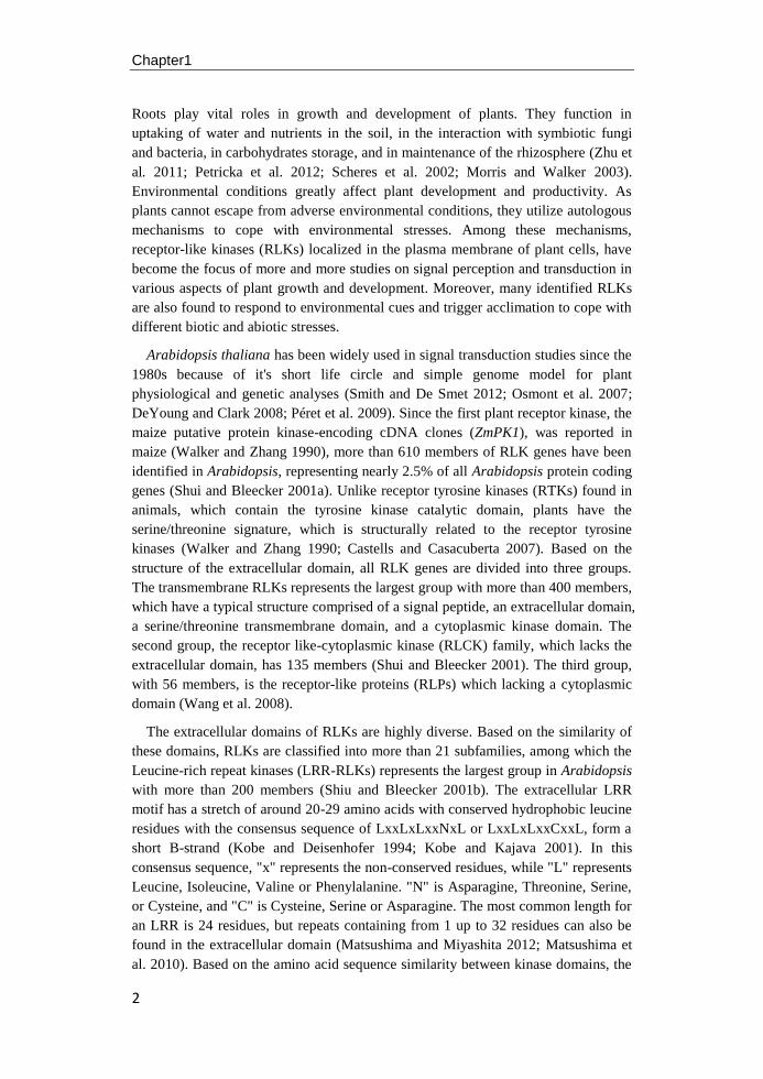
cells (Barrada et al. 2015). In the Arabidopsis primary root, a slowly dividing stem
cell pool of initial cells surrounds the quiescent center (QC), with three to four
infrequently dividing cells. The division of initial cell gives rise to the files of
distinct cell layers (tissues), including the epidermis, the cortex, the endodermis, the
pericycle and the stele that surrounds the vascular bundels (Figure 1). The
longitudinal axis of the PR demonstrates a developmental time line: within the apical
meristem zone, initials and their daughter cells divide multiple times producing
similar sized daughter cells, while in the transition zone only a few cells still divide
and the majority of cells start to elongate. Cell size in the elongation zone increases
sharply, compared with those cells in the transition zone, until they have reached
their final length. In cells that have reached their final size, polarized cell
enlargement leads to the formation of root hairs, demarcating the distal margin of the
maturation zone of a root.
Embryo receptor like kinases
The Arabidopsis zygote undergoes an asymmetric division to generate a smaller
apical cell and a larger basal cell. The apical cell-lineage generates an eight-cell
embryo proper with an apical domain (AD) and central domain (CD) after a series of
divisions. The AD generates the cotyledon and the shoot meristem, whereas the CD
produces part of the cotyledon, the hypocotyl and the root meristem initials (Mayer
et al. 1991; Jürgens et al. 1991; Slane et al. 2014; Meinke 1991). The basal cell
produces the suspensor that plays an important role during embryo development,
including (i) pushing the embryo proper into the endosperm cavity, (ii) transport of
molecules involved in nutrition and growth regulation and (iii) biosynthesis of plant
hormones (Kawashima and Goldberg 2010).
Chapter1
4
Figure 1. The longitudinal axis and root radial patterning of primary root of the model
flowering plant Arabidopsis thaliana. The primary root tip of wild-type Columbia is
consisted by three different zones, including the meristematic zone, the elongation zone, and
the maturation zone. The black contours of cortical cells highlight the increase of cell size (left)
and each color represents a different cell layer (right). Figure modified from Barrada et al.
(2015).
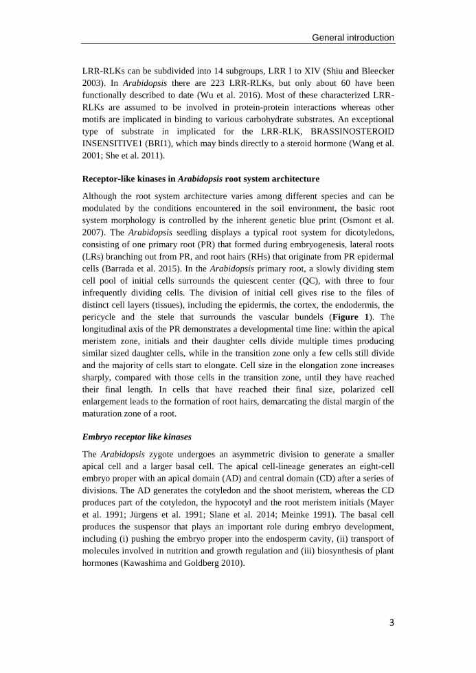
The development of the plant embryo is a complex process and experimental
evidence indicates that RLKs play major roles in the intercellular signaling in the
embryo development (Figure 2, reviewed by Nodine et al. 2011). The length of the
suspensor determines the speed of the development progression of the embryo in
Arabidopsis, and the SHORT SUSPENSOR (SSP) gene was the first RLCK
identified to functioning in the zygote (Bayer et al. 2009; Babu et al. 2013). Mutants
lacking a functional SSP gene fail to generate and elongate basal cells, resulting in a
short suspensor phenotype. Genetic analysis suggests that SSP acts upstream of the
YODA (YDA) MITOGEN-ACTIVATED PROTEIN (MAP) kinase cascade, which
is required for partitioning of the embryo and determine the extra embryonic fates
General introduction
5
(Lukowitz et al. 2004), but SSP regulates this pathway through a unique parental-
original effect (Bayer et al. 2009). In addition, the SSP is found related to another
group of RLCKs, the BRASSINOSTEROID-SIGNAL-KINASES (BSKs), which in
vitro are phosphorylated by BRI1 and which in vivo interact with BRI1 and regulate
cell elongation in Arabidopsis (Tang et al. 2008). However, no direct evidence was
found that SSP acts in the BRI1 pathway of controlling nuclear gene expression and
embryo development.
Figure 2. RLK and RLCK functions during embryogenesis of Arabidopsis. Dotted arrows
represent possible cell interaction and arrows with solid lines represent interactions that have
been confirmed by experiments. SSP acts as a signal to promote elongation and asymmetric
division of zygote via the YDA pathway MAP kinase cascade to regulate basal cell
development. RPK1/TOAD2 are redundantly required for maintaining protodermal cell fate
identity during the early globular stage of Arabidopsis embryo development. At the later
globular and transition stages, ACR4/ALE2 positively regulate protoderm gene expression
and the integrity of protoderm in the apical domain, which is required for normal cotyledon
emergency. GSO1/GSO2 are important for maintaining epidermal function from heart stage of
the embryo development. Figure adapted from Nodine et al. (2011).
In the early globular stage of Arabidopsis embryo development two closely related
RLKs, RECEPTOR PROTEIN KINASE1 (RPK1) and TOADSTOOL2 (TOAD2),
are required for the maintenance of protodermal cell fate identify in the central
domain (reviewed by Nodine et al. 2011). The localization of RPK1 and TOAD2
translational fusions of green fluorescent protein (GFP), together with the cell
specific markers in toadstool embryos, strongly indicates that RPK1 and TOAD2 are
redundantly required for Arabidopsis embryonic pattern formation. They are also
required together for cotyledon initiation during later embryonic stages (Nodine and
Tax 2008).
In maize, the CRINKLY4 (CR4) gene was found to encode a TNFR-like receptor-
like kinase that is involved in the leaf epidermis differentiation (Becraft et al. 1996).
Arabidopsis thaliana homologue of CR4 (ACR4), a putative receptor-like kinase

Chapter1
6
receptor of Arabidopsis, is homologous to the maize CR4 gene and required for
proper development of the embryo (Tanaka et al. 2002). At the globular stage during
early embryogenesis, ACR4 transcripts accumulate in both protoderm and inner cells
at comparable levels, but change at the heart stage with relative higher level in the
protoderm than in the inner cells, suggesting a role in epidermis differentiation. In
addition, the ABNORMAL LEAF SHAPE1 (ALE1) and the ABNORMAL LEAF
SHAPE2 (ALE2) that results in defects of cuticle formation, were found to act
together with ACR4 during early stages of embryogenesis (Tanaka et al. 2007;
Tanaka et al. 2001). These three genes play partially overlapping roles in positively
regulating protoderm-specific gene expression and the formation of cotyledon
through different modes of intercellular communications.
In addition, two other LRR-RLKs, GASSHO1 (GSO1) and GASSHO2 (GSO2)
are essential for the normal development of the epidermal surface in Arabidopsis
embryos (Tsuwamoto et al. 2008). Embryos of the double mutant GSO1/GSO2
display reverse bending of the embryo compared with the wild type embryo at the
heart-torpedo transition stage. No difference was apparent between wild type and the
GSO1/GSO2 double mutant embryos at the early heart stage, but in the mutant the
apical part of the embryo will stick to the peripheral tissue of the endosperm, which
is caused by abnormal development of the epidermis.
Based on microarray datasets from Keith Lindsey’s group (Spencer et al. 2007)
and John Harada-Robert Goldberg’s microarray data (NCBI GEO: GSE12404), more
than 300 expressed receptor-like genes were detected during different stages of
embryogenesis (Nodine et al. 2011). Apart from high expression of seven RLKs
(SSP, RPK1, TOAD2, ACR4, ALE2, GSO1, and GSO2) discussed above, a large
number of RLKs and RLCKs with known functions during adult growth and
development were detected during embryogenesis, including BARELY ANY
MERISTEM 1/2 (BAM1/2) (DeYoung and Clark 2008), SOMATIC
EMBRYOGENESIS RECEPTOR-LIKE KINASE1/2 (SERK1/2) (Fan et al. 2016;
Albrecht et al. 2005) that is involved in post-embryo development, BRI1 and BSKs
(Tang et al. 2008) that are involved in the brassinosteroids signal transduction
pathway, and RLK PEP1 RECEPTOR1 (PEPR1) (Yamaguchi et al. 2006) and the
RLCK AVRPPHB SUSCEPTIBLE1 (PBS1) (Swiderski and Innes 2001), with
known functions in pathogen defense responses. Thus, signaling via RLKs and
RLCKs is important during this part of the life cycle of a plant, and there are still a
large number of RLKs and RLCKs that are expressed during embryogenesis, but for
which a specific function during embryonic pattern formation still has to be
established.
Root apical meristem maintenance RLKs
Primary meristems of shoot and root are initiated during embryogenesis and control
plant growth along the main body axis. The CLAVATA pathway that acts in the
shoot apical meristem (SAM) to control shoot and floral meristem size in
Arabidopsis is a good illustration of study on the role of RLKs in signal transduction
General introduction
7
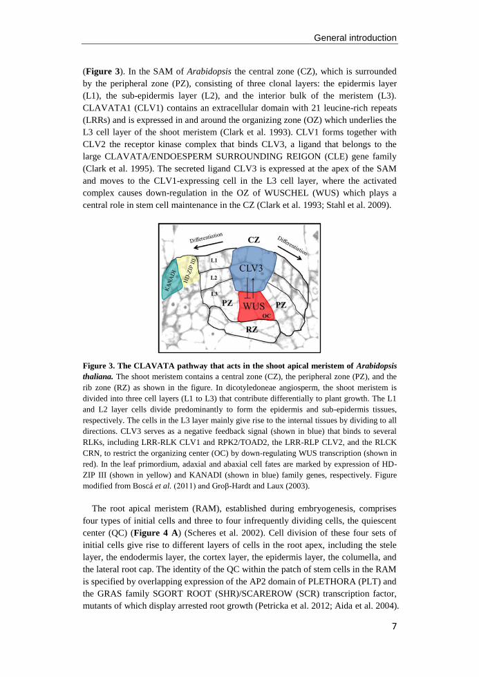
(Figure 3). In the SAM of Arabidopsis the central zone (CZ), which is surrounded
by the peripheral zone (PZ), consisting of three clonal layers: the epidermis layer
(L1), the sub-epidermis layer (L2), and the interior bulk of the meristem (L3).
CLAVATA1 (CLV1) contains an extracellular domain with 21 leucine-rich repeats
(LRRs) and is expressed in and around the organizing zone (OZ) which underlies the
L3 cell layer of the shoot meristem (Clark et al. 1993). CLV1 forms together with
CLV2 the receptor kinase complex that binds CLV3, a ligand that belongs to the
large CLAVATA/ENDOESPERM SURROUNDING REIGON (CLE) gene family
(Clark et al. 1995). The secreted ligand CLV3 is expressed at the apex of the SAM
and moves to the CLV1-expressing cell in the L3 cell layer, where the activated
complex causes down-regulation in the OZ of WUSCHEL (WUS) which plays a
central role in stem cell maintenance in the CZ (Clark et al. 1993; Stahl et al. 2009).
Figure 3. The CLAVATA pathway that acts in the shoot apical meristem of Arabidopsis
thaliana. The shoot meristem contains a central zone (CZ), the peripheral zone (PZ), and the
rib zone (RZ) as shown in the figure. In dicotyledoneae angiosperm, the shoot meristem is
divided into three cell layers (L1 to L3) that contribute differentially to plant growth. The L1
and L2 layer cells divide predominantly to form the epidermis and sub-epidermis tissues,
respectively. The cells in the L3 layer mainly give rise to the internal tissues by dividing to all
directions. CLV3 serves as a negative feedback signal (shown in blue) that binds to several
RLKs, including LRR-RLK CLV1 and RPK2/TOAD2, the LRR-RLP CLV2, and the RLCK
CRN, to restrict the organizing center (OC) by down-regulating WUS transcription (shown in
red). In the leaf primordium, adaxial and abaxial cell fates are marked by expression of HD-
ZIP III (shown in yellow) and KANADI (shown in blue) family genes, respectively. Figure
modified from Boscá et al. (2011) and Groβ-Hardt and Laux (2003).
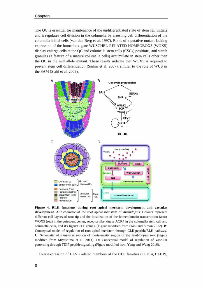
The root apical meristem (RAM), established during embryogenesis, comprises
four types of initial cells and three to four infrequently dividing cells, the quiescent
center (QC) (Figure 4 A) (Scheres et al. 2002). Cell division of these four sets of
initial cells give rise to different layers of cells in the root apex, including the stele
layer, the endodermis layer, the cortex layer, the epidermis layer, the columella, and
the lateral root cap. The identity of the QC within the patch of stem cells in the RAM
is specified by overlapping expression of the AP2 domain of PLETHORA (PLT) and
the GRAS family SGORT ROOT (SHR)/SCAREROW (SCR) transcription factor,
mutants of which display arrested root growth (Petricka et al. 2012; Aida et al. 2004).
Chapter1
8
The QC is essential for maintenance of the undifferentiated state of stem cell initials
and it regulates cell division in the columella by arresting cell differentiation of the
columella initial cells (van den Berg et al. 1997). Roots of a putative mutant lacking
expression of the homeobox gene WUSCHEL-RELATED HOMEOBOX5 (WOX5)
display enlarge cells at the QC and columella stem cells (CSCs) positions, and starch
granules (a feature of a mature columella cells) accumulate in stem cells other than
the QC in the null allele mutant. These results indicate that WOX5 is required to
prevent stem cell differentiation (Sarkar et al. 2007), similar to the role of WUS in
the SAM (Stahl et al. 2009).
Figure 4. RLK functions during root apical meristem development and vascular
development. A: Schematic of the root apical meristem of Arabidopsis. Colours represent
different cell layers of root tip and the localization of the homeodomain transcription factor
WOX5 (red) in the quiescent center, receptor like kinase ACR4 in the columella stem cell and
columella cells, and it's ligand CLE (blue). (Figure modified from Stahl and Simon 2012). B:
Conceptual model of regulation of root apical meristem through CLE peptide/RLK pathway.
C: Schematic of transverse section of meristematic region of the Arabidopsis root (Figure
modified from Miyashima et al. 2011). D: Conceptual model of regulation of vascular
patterning through TDIF peptide signaling (Figure modified from Yang and Wang 2016).
Over-expression of CLV3 related members of the CLE families (CLE14, CLE19,
General introduction
9
CLE20, and CLE40) results in arrested root growth, suggesting that the CLV-like
signaling pathway also operates in the root apical meristem (Meng and Feldman
2010; Stahl et al. 2009; Stahl and Simon 2009). ACR4 is expressed in the outer cell
layer of embryos and involved in proper embryogenesis (Tanaka et al. 2002). In the
RAM of plants transformed with the HISTONE2B::YFP fusion protein encoding
gene, ACR4 expression was observed in the QC, the columella initials and the
columella cells below the QC, the lateral root cap and the initial cells that give rise to
the epidermal tissue (Gifford et al. 2003). CLE40, which is also expressed in the
embryo, also acts as a secreted ligand of the ACR4 receptor in the columella and the
columella stem cells, where it up-regulates its own expression and that of CLV1,
restricting WOX5 expression to the QC (Pallakies and Simon 2014) (Figure 4 B).
Together, this indicates a WOX5-dependent mechanism of stem cell fate regulation
by CLE40, CLV1, and ACR4.
In addition to the CLE peptide-receptor pathway, the LRR-RLK BRI1 was
identified playing a specific role in the regulation of RAM through a steroid
hormone-RLK pathway (González-García et al. 2011; Hacham et al. 2011). The
BRI1 gene encodes a widely expressed putative receptor of the hormone
brassinosteroid (BR), which modulates cell elongation and division throughout
growth and development of a plant (Clouse et al. 1996; Li and Chory 1997). The
bri1 mutant displays a severely dwarfed phenotype and can't be rescued by BRs
treatment. Generally, treatment with the brassinosteroid brassinolide (BL) promotes
primary root growth at low levels and inhibits growth progressively at higher levels
(Clouse et al. 1996). Both gain- and loss-of function of BR-related Arabidopsis
mutants possess a reduced meristem size indicating a possible role for BRs in
optimal root growth. In fact, BRs act on the root stem cells by promoting the QC cell
renewal and controlling the cell cycle progression and differentiation necessary for
maintaining the meristem size (González-García et al. 2011). In addition, 4 nM BL
treatment of the bri1 mutant causes increased expression of WOX5 and SCR, and the
lack of SHR and WOX5 expression in the serk triple mutants (Du et al. 2012; Gou et
al. 2012), indicating that RLKs are candidate molecules to function as receptor or co-
receptors in the regulation of root apical meristem maintenance.
Stele and ground tissue RLKs
The vasculature of Arabidopsis root is organized into a central stele, comprised of
the xylem and phloem, and is formed in the RAM by initial cells that give rise to the
protoxylem, metaxylem and procambial cells, depending upon the direction of cell
division (Figure 4 C) (Ohashi-lto and Fukuda 2010; Scheres et al. 1994; Dolan et al.
1993). These tissues are originally formed from a set of pericycle/vascular initials
proximal to the QC, and they grow symmetrical along a central axis with protoxylem
at the poles and metaxylem in the center (Zhang et al. 2011).
The BREVIS RADIX (BRX) gene family of Arabidopsis is a class of transcription
factors that control growth and development throughout the plant and of which BRX
is the only gene that has a role in root system development of Arabidopsis (Mouchel
Chapter1
10
et al. 2004). BRX is expressed in the vasculature and the reduced root size phenotype
of brx mutant results from the reduced expression of a rate-limiting enzyme in
brassinosteroid biosynthesis pathway (Mouchel et al. 2006). Expression of BRX is
strongly induced by auxin and mildly repressed by brassinolide, indicating that BRX
mediates a feedback loop between brassinosteriod and auxin signaling enabling
optimal root growth. Recently, the LRR-RLK gene, BARLY ANY MERISTEM3
(BAM3) was identified as suppressor of root meristem growth and protophloem
development defects of brx mutant (Depuydt et al. 2013). While CLE45 treatment
severely inhibits root meristem growth in wild type roots, the roots of the bam3
mutant are insensitive to application of the CLE45 ligand. As expression of bam3 is
increased in both brx mutants and roots treated with CLE45 peptide, protophloem
differentiation in the transition zone of the root tip is caused by activation of BAM3
binding to CLE45, and BRX promotes protophloem differentiation through
inhibition of BAM3 expression.
In the Arabidopsis genome, thirty-two CLE genes have been identified to be
involved in many aspects of biological processes of plant growth and development
(Betsuyaku et al. 2011; Jun et al. 2008). Among them, CLE41 and CLE44 encode a
12-amino acid TRACHEARY ELEMENT DIFFERENTIATION INHIBITORY
FACTOR (TDIF) peptide. In the vascular meristem, the LRR-RLK PHLOEM
INTERCALATED WITH XYLEM/TDIF RECEPTOR (PXY/TDR), which shares
high level sequence similarity with CLV1, perceives the TDIF signals from phloem
to regulate the undifferentiated procambial cell fate during secondary growth
(Figure 4 D, Hirakawa et al. 2008; Ohyama et al. 2008; Yang and Wang 2016). The
TDR, localized in procambial cells, is activated by TDIF and then promotes cell
division of procambial cells and suppresses differentiation of the procambial cells
into xylem cells (Hirakawa et al. 2010). Additionally, expression of WOX4 increases
in the presence of TDIF in a TDR-dependent manner (Hirakawa et al. 2010). A
mutation in TDR causes both the suppression of procambial cell proliferation and the
enhancement of xylem differentiation, whereas a mutation in WOX4 only suppress
the proliferation of procambial cells, suggesting that TDIF-TDR signaling regulates
vascular stem cell fate by two independent pathways that appear to diverge early
after TDIF recognition.
Several other RLKs are also implicated in the development of the vascular system.
For instance, two members of the BRI1 family of plant steroid receptors, BRI1-
LIKE1 (BRL1) and BRI1-LIKE1 (BRL3), are predominantly expressed in the
vascular tissues and function specifically in provascular differentiation to maintain
xylem and phloem (Caño-Delgado et al. 2004). Another RLK, XYLEM
INTERMIEXD WITH PHLOEM1 (XIP1), displays an aberrant accumulation of
highly lignified cells and phloem cells adjacent to xylem cells in stem sections,
similar to the pxy mutant phenotype, indicating that XIP1 plays a role in
differentiation of phloem cells in vascular development (Bryan et al. 2012).
Moreover, MORE LATERAL GROWTH1 (MOL1) and REDUCED IN LATERAL
GROWTH1 (RUL1) were identified as opposing regulators of lateral expansion of
General introduction
11
plant growth axes, and they might function to recognize and communicate long or
short range signals to cambium cells (Agusti et al. 2011).
Regulation of epidermal cell fate and root hair formation
Root hairs are long cylindrical extensions of epidermal cells, and they are
responsible for uptake of nutrients, establishing plant-microbe interactions, and
helping plant anchoring to soil (Grierson et al. 2014). In Arabidopsis, epidermal cells
are divided into two groups, the root hair cells that can produce root hairs and the
non-hair cells, which lack root hairs. The Arabidopsis root epidermis is generated
from a set of epidermal/lateral root cap initial cells formed during embryogenesis,
and these initial cells can give rise to epidermal cells and cells of the lateral root cap,
in the proximal and distal direction of the QC, respectively (Petricka et al. 2012).
Like in many other members of the Brassicaceae, the epidermis of Arabidopsis
possesses a distinct position-dependent pattern of root hair cells and non-hair cells,
and how the identity of a newly formed epidermal cell, differentiating either into a
root hair cell or a non-hair cell, is established, has been studied extensively to
understand the regulation of cell type patterning in plants (Grierson et al. 2014;
Dolan et al. 1994; Galway et al. 1994).
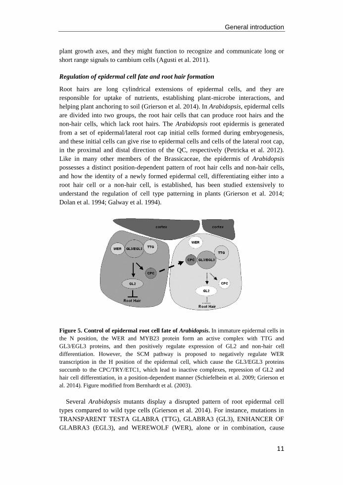
Figure 5. Control of epidermal root cell fate of Arabidopsis. In immature epidermal cells in
the N position, the WER and MYB23 protein form an active complex with TTG and
GL3/EGL3 proteins, and then positively regulate expression of GL2 and non-hair cell
differentiation. However, the SCM pathway is proposed to negatively regulate WER
transcription in the H position of the epidermal cell, which cause the GL3/EGL3 proteins
succumb to the CPC/TRY/ETC1, which lead to inactive complexes, repression of GL2 and
hair cell differentiation, in a position-dependent manner (Schiefelbein et al. 2009; Grierson et
al. 2014). Figure modified from Bernhardt et al. (2003).
Several Arabidopsis mutants display a disrupted pattern of root epidermal cell
types compared to wild type cells (Grierson et al. 2014). For instance, mutations in
TRANSPARENT TESTA GLABRA (TTG), GLABRA3 (GL3), ENHANCER OF
GLABRA3 (EGL3), and WEREWOLF (WER), alone or in combination, cause
Chapter1
12
plants to produce "hairy" roots, by changing non-hair cells in to root-hair cell
(Galway et al. 1994; Bernhardt et al. 2003; Lee and Schiefelbein et al. 1999). On the
other hand, CAPRICE (CPC), TRIPTYCHON (TRY), and ENHANCER OF TRY
AND CPC (ETC1) are required for establishing the root-hair cell identity and
mutation of these genes, alone or in combination, cause plants to produce "bald"
cells at the former root-hair cell position (Simon et al. 2007; Wada et al. 1997; Kirik
et al. 2004). Recently, an LRR-RLK SCRAMBLED (SCM) was discovered that
enables immature epidermal cells to detect a positional signal and establish an
appropriate cell-type pattern (Kwak et al. 2005). All these genetic findings to date
led to a possible model for cell type pattern formation in the root epidermis of
Arabidopsis (Figure 5) (Grierson et al. 2014).
Genetic analysis reveals discrete steps in the root hair development in Arabidopsis
(Péret et al. 2009). In the initiation stage, rop proteins are first localized at the site
where the root hair will be formed, before the hair begin to grow. Rop GTPases are
localized to the tips of root hairs and control polar growth of Arabidopsis (Molendijk
et al. 2001). Mutation of At3g51550, which encodes the FERONIA (FER) receptor-
like kinase, induces sever root hair defects and reduced levels of active RAC/ROPs,
indicating that FER assist in rop accumulation at the apical plasma membrane
domains in the root tip (Duan et al. 2010). Within a minute after localized rop
accumulation, the root hair cell wall begins to bugle out and the pH of the wall drops
to pH 4 - 4.5, which is thought to activate expansion proteins that catalyze cell wall
loosening (Grierson et al. 2014). As the bulge enlarges, large amount of endoplasmic
reticulum and filamentous (F) actin accumulate in the developing swelling. In the tip
growth stage the hairs grows to its final length by targeted secretion.
Lateral root development
In Arabidopsis, lateral roots are derived from the pericycle layer deep within the
differentiation zone of the primary root (De Smet 2012). The mature pericycle cells
along the xylem pole are stimulated to proliferate and re-differentiate into lateral root
primordia (LRP), which contain their own meristems when they mature (Malamy
and Benfey 1997). Histological studies showed that initiated LRP can then mature
through eight stages (stage I-VII and emergence) defined by specific anatomical
characteristics and cell divisions (Figure 6 A) (Malamy and Benfey 1997). Stage I of
LR development begins with increased anti-clinal (perpendicular orientation to the
root axis) divisions of cells in the pericycle layer. In stage II peri-clinal divisions
have led to an outer and an inner layer and further peri-clinal divisions in the out
layer result in a three layers primordium (outer layer1, outer layer2 and inner layer)
in stage III. A second round of peri-clinal divisions in the inner layer creates a four-
cell layer structure (layer1, outer layer2, inner layer1 and inner layer2). The LRP
then penetrates the parent endodermis at stage IV, the cortex by stage V and the
epidermis by stage VI. At stage VI and VII, the organization of the LRP shows
similarity to the primary root tip, with epidermis, cortex, endodermis layers
surrounding the stele and a root cap at the tip. Enlargement of the basal cells in the
General introduction
13
outer layer1 promotes the increase in length of the LRP and finally the new lateral
root emerges from the parent epidermis.
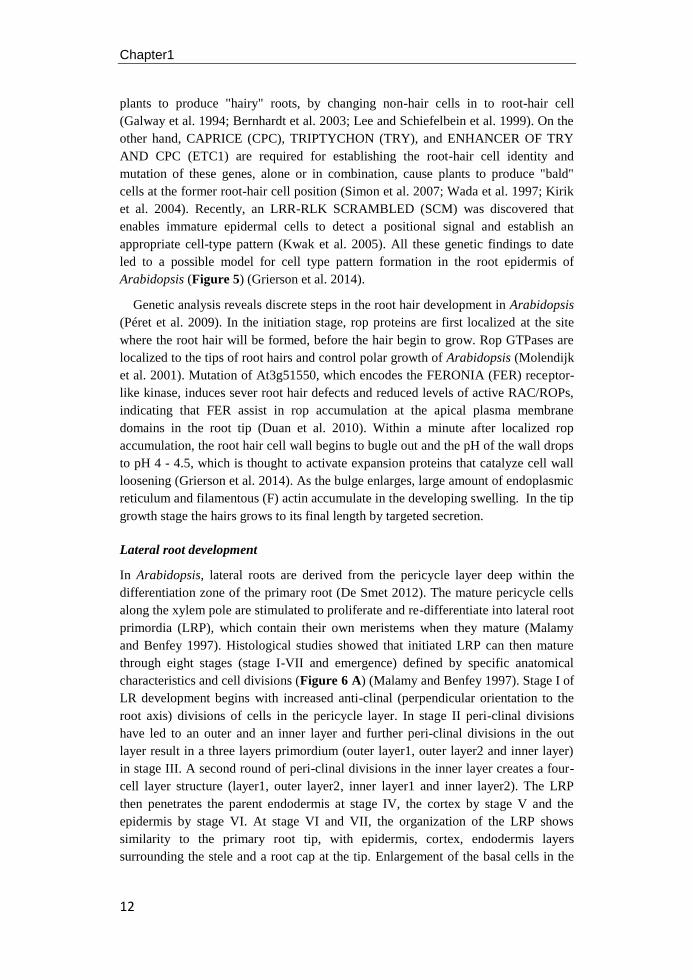
Figure 6. RLKs mediated pathways in the Arabidopsis lateral root development. A:
Lateral root formation consists of three stages including LR initiation, LRP development, and
LR emergence (Jung and McCouch 2013). B: Model of IDA-HAE/HSL2 signaling in lateral
root emergency (Kumpf et al. 2013). C: Model for auxin-dependent lateral root emergency
through auxin influx carrier LAX3 (Swarup et al. 2008).
During the development of LR auxin underpins each stage of the LRP
development (Figure 6) (Nibau et al. 2008). Auxin is crucial for the determination of
both the position and frequency of lateral root initiations and exogenous application
of auxin can activate the whole pericycle to form LRPs (Himanen et al. 2002). A
total of 1920 significantly differentially expressed genes were identified in auxin-
activated pericycle cells and 15 potential key regulator genes were found associated
with asymmetric cell division during lateral root initiation. Only one gene,
At3g59420, was identified by all of the different filters, and this encodes a
membrane localized receptor-like kinase ACR4 (De Smet et al. 2008). ACR4 is
expressed in the small daughter cells after the first asymmetric cell division in the
pericycle and mutants lacking ACR4 display additional cell divisions in the pericycle
cell adjacent to the lateral root initiation site, an uncommon position for the lateral
root meristems, or exhibit aberrant expression of the boundary marker (LBD5) and
auxin response marker (DR5). Thus, ACR4 might be required for the autonomous
specification of lateral root initials cell. As no ACR4 expression is observed in the
neighboring pericycle cells, ACR4 signaling might prevent neighboring pericycle
cells from being initiated as LRP, in a non-cell autonomously way. However, the
mechanism of how ACR4 acts is not known.

Chapter1
14
In the single mutant of INFLORESCENCE DEFICIENT IN ABSCISSION (IDA),
HAESA (HAE), HAE-LIKE2 (HSL2) and in the hae/hsl2 double mutant the density
of LRs is significantly reduced compared with wild type, suggesting that these genes
might play a role in LR development (Figure 6 B) (Kumpf et al. 2013). In
Arabidopsis, abscission of the floral organ is controlled by the ligand peptide IDA
through the receptor-like kinase receptors HAE and HLS2 (Cho et al. 2008).
Emergence of new lateral root primordia depends on cell separation in the overlaying
layers of the LR apex. At stage I and II, IDA expression in the overlaying cell layer
is induced by auxin derived from the LRP which then binds to the HAE and HLS2
receptors located in the cell membrane. The activated receptors trigger expression of
cell-wall-remodelling (CWR) genes and leads to cell wall separation in the
endodermis. In the overlaying cortex and epidermal layer, both IDA and the receptor
expression are coupled, in an ARF7-dependent manner, to the auxin influx carrier
LAX3 (Figure 6 C) (Swarup et al. 2008). IDA signaling through HAE induces the
expression of CWR enzymes to dissolve the cell walls and enable LR to penetrate
the cortex and epidermis tissue from the deep xylem. Although IDA expression is
100-fold increased by exogenous auxin, the HAE and HSL2 receptors function as the
limiting factor controlling cell separation (Kumpf et al. 2013). Thus, IDA-
HAE/HSL2 signaling module is crucial for root/shoot cell separation during plant
growth, but in different processes.
Other RLKs have been implicated in lateral LRP development and LR emergence
as well. The double and triple mutant combinations of the TRANSMEMBRANE
KINASE (TMK) subfamily of receptor-like kinases in Arabidopsis, tmk1, tmk3 and
tmk4, show a severe reduction in organ size and a related delay in growth stages (Dai
et al. 2013). Moreover, they show reduced lateral root density and are insensitive to
exogenously supplied auxin in root inhibition, suggesting that these RLKs might
play a role in the auxin-mediated signaling pathway of lateral root emergence and
development. In addition, other plant hormones also affect lateral roots development
in a complicated network by controlling auxin synthesis and/or transport. In
particular, BRs act synergistically with auxin to promote lateral root development
through increasing auxin acropetal transport (Bao et al. 2004). In bri1-119
background, the synthetic auxin-inducible promoter DR5 was severely decreased in
the lateral root, compared to the wild type. This promotor was also decreased when
treated with the BR biosynthesis inhibitor brassinozale, indicating that BRI1 is
probably affecting lateral roots development in an auxin-dependent manner.
However, currently there is not enough evidence to confirm the exact role of TMK,
or that of the BRI receptor, in the process of LRP development.
RLKs involved in plant stress responses
Endogenous stimuli, such as plant hormones and ROS, modulate the molecular and
biochemical mechanisms that increase the tolerance of plants to external stresses
(Petricka et al. 2012; Potters et al. 2007; Overvoorde et al. 2010; Walter et al. 2009;
Munns and Tester 2008). However, the external stress signals must first be perceived
by plant cells or organs in order to initiate the acclimation to the new conditions. In

General introduction
15
the case of molecular signals the perception is often by binding of the signaling
molecule to a receptor protein located in the plasma membrane. RLKs play
important roles in sensing the external stimuli and activating the down-stream
elements of the signaling pathway via their serine/threonine kinase domains (Shiu
and Bleecker 2001 a, b; Osakabe et al. 2013). Of the more than 610 genes that
encode RLKs and RLPs in Arabidopsis genome, only a fraction has been assigned to
the biological processes that they control (Diévart and Clark 2004).
The ERECTA family
The ERECTA family of LRR-RLKs, consisting of ERECTA (ER), ERECTA-like1
(ERL1), and ERECTA-like2 (ERL2), exhibits partial redundancy among these three
members and mediates cell fate specification during the development of the stomatal
complex (Pillitteri and Torii 2012). A mutant of TOO MANY MOUTHS (TMM)
was first isolated as a phenotype that has stomates formed in adjacent cells that in
wild type would have been developed into epidermal pavement cells (Shpak et al.
2005). The tmm plants exhibit an organ-dependent phenotype, with clustered stomata
in cotyledon and leaves, whereas hypocotyls and stem are devoid of stomata. TMM
thus can either influence stomata initiation in a positive or negative fashion (Geisler
et al. 1998; Yang and Sack 1995). Since TMM encodes an LRR-RLP lacking any
cytoplasmic effector domain by itself, it exerts its' effect by associating with
ERECTA family receptors as co-receptor to perceive their putative ligands,
EPFs/EPFLs (Pillitteri and Torii 2012).
The Epidermal Patterning Factor (EPF), EPF1 and EPF2, are three cysteine-rich
peptides (CRPs) that mediate divers aspects of cell-cell communication (Marshall et
al. 2011). ER is the main receptor for EPF2, together forming a ligand-pair in vivo,
governing the initial decision of protodermal cells to generate a stomatal complex by
asymmetric division. The EPF1-ERL1 pair acts to maintain stomatal cell activity and
suppress guard mother cell (GMC) differentiation (Hara et al. 2007; Lee et al. 2012;
Ohki et al. 2011). The signals received by kinase receptors are transmitted to the
downstream MAPK cascades consisting of YODA (MAPKKK), MKK4/5
(MAPKKs) and MPK3/6 (MAPKs) to inhibit phosphorylation of basic helix-loop-
helix (bHLH) transcription factors, such as SPEECHLESS (SPCH), MUTE and
FAMA, leading to a suppression of stomatal cell fate specification (Wang et al.
2007). Stresses such as low temperature, drought, wounding, pathogens and stress-
related molecules and hormones can either activate MAPK cascades or its target
bHLH transcription factors to control stomata development (Pillitteri and Torii 2012).
BRI1 kinase
Brassinosteroids (BRs) play crucial roles in various aspects of plant growth and
development, including cell elongation, photomorphogenesis, xylem differentiation,
and seed germination (González-García et al. 2011; Hacham et al. 2011; Howell et al.
2007). Brassinosteroid-insensitive1 (BRI1) has been identified as the plasma-
membranes receptor of brassinolide, the most active brassinosteroid (Wang et al.
Chapter1
16
2001; Li and Chory 1997). In this hormone-regulation pathway, BR binding to the
LRR-RLK BRI1 inactivates BIN2, a glycogen synthase kinase-3, through the
activation of phosphatase BSU1. By dephosphorylating transcription factor BZR1
and BES1, BSU1 positively regulates BR signaling, while BIN2 negatively regulates
BZR1 and BES1 by phosphorylating them (Tang et al. 2008). Although, both BRI1
ASSOCIATED KINASE1 (BAK1) and BR-signaling kinases (BSKs) interact with
BRI1 in vitro and in vivo, they play very distinct roles in BR signaling. BAK1
encodes an LRR-RLK that mainly mediates activation of BRI1 kinase, which
enhances signaling output though reciprocal BRI1 transphosphorylation (Li et al.
2002; Divi et al. 2010; Russinova et al. 2004; Fàbregas et al. 2013). In contrast,
BSKs directly mediate signal transduction from BRI1 to downstream BR responses
(Tang et al. 2008).
Another feature for BR is their potential to enhance tolerance in plants to a range
of abiotic stresses such as low/high temperature, drought stress, salt stress, and
pathogen attack (Krishna 2003; Elhiti et al. 2013). The molecular mechanism of BR-
induced stress tolerance is still largely unexplored (Elhiti et al. 2013). Recently, Kim
et al. (2012) reported that BR inhibits stomatal development by alleviating GSK3-
mediated inhibition of the MAPK module, leading to a decrease in stomatal density,
that effectively limits water loss under high salt stress conditions (Ryu and Cho
2015). Since BR interacts with other hormones, some molecular changes associated
with BR-induced stress tolerance result from BR cross talk with other hormones
(Krishna 2003). For instance, BR positively regulates the salicylic acid pathway
component NPR1 to promote thermo-tolerance (Divi et al. 2010; Ahammed et al.
2016), as well as WRKY70, which plays a pivotal role in salicylate-dependent and
jasmonate-dependent defense pathways (Li et al. 2004, 2006). Another example of
BR-other hormone cross talk is BR-enhanced salt tolerance which is established by
either an ethylene-dependent or an ethylene-independent pathway (Divi et al. 2010;
Ryu and Cho 2015). However, whether BRI1 participates in these stress responses
signaling is unknown, further studies need to be done in the future.
FLS2
In Arabidopsis, the leucine-rich receptor kinase FLAGELLIN-SENSITIVE2 (FLS2)
is involved in the recognition of flagellin, a protein that is part of the bacterial
flagella, as a signal of bacterial presence, binding of which leads to the activation of
defense responses (Gómez-Gómez and Boller 2000; Chinchilla et al. 2006). The fls2
mutant allele fls2-24 and fls2-17, with point mutations in the LRR motif of the
extracellular domain and kinase domain, respectively, were shown to confer
insensitivity to flg22. However, flg22 binding was restored in the transgenic fls2
plants expressing the wild-type FLS22 gene, indicating that both extracellular and
cytoplasmic domains of FLS2 protein are required for flagellin binding (Gómez-
Gómez et al. 2001; Chinchilla et al. 2006). Another RLK, BAK1 was shown to
interact with FLS2 in vivo, in a ligand-specific manner, suggesting a role for BAK1
in innate immunity of plants (Chinchilla et al. 2007; Lin et al. 2014; Heese et al.
2007). BAK1 has been considered a co-receptor for BR-binding BRI1, thus FLS2
General introduction
17
interacts with BAK1 which subsequently phosphorylates BIK1, a cytoplasmic kinase,
to control FLS2 signaling as a positive regulator (Wang 2012; Kemmerling et al.
2007; Schwessinger et al. 2011). Albrecht et al. (2008) reported that brassinosteroids
inhibit the FLS2-mediated immune signaling independent of the complex formation
with BAK1. BRs were also shown to inhibit downstream signaling triggered by the
BAK-independent recognition of the fungal PAMP chitin (Albrecht et al. 2012).
However, Belkhadir et al. (2011) provided evidence that BR acts antagonistically or
synergistically in the response to microbe-associated molecular pattern (MAMP)
through both BAK1-dependent and -independent mechanisms. It thus seems that the
relative levels of BR, BRI1 and BAK1 determine whether BAK1 has a positive or a
negative effect on FLS2-mediated signaling and that appropriate levels of
endogenous BR are required for optimal flg22 signaling.
RPK family
RECEPTOR-LIKE PROTEIN KINASE1 (RPK1) is an ABA-inducible LRR-RLK
isolated from Arabidopsis thaliana and is expressed ubiquitously in flower, stem,
leaves and roots. ABA is the plant hormone that is mainly associated with its role in
stress signaling (Malamy 2005). RPK1 expression is rapidly induced upon treatment
with ABA and by several environmental stresses, such as low temperature, high salt
and dehydration (Smith and De Smet 2012; Lucas et al. 2013), indicating that the
gene is involved in a general stress response. Loss of function of RPK1 resulted in
ABA insensitivity and decreased expression level of ABA-responsive genes
indicating that RPK1 functions as a positive regulator of ABA signal transduction
(Osakabe et al. 2005).
Reactive oxygen species (ROS) are sub-products of aerobic metabolism in plants
and other aerobic organisms (Apel and Hirt 2004). Various abiotic stresses lead to
the accumulation of ROS in plants and an elaborate plant ROS network, comprised
of efficient enzymatic and non-enzymatic antioxidant defense systems, is present to
maintain the ROS level low, to protect plant cells from oxidant damage (Gill and
Tuteja 2010; Gechev et al. 2006). ROS could also serve as signaling molecules to
modulate various processes, including plant stress responses, program cell death, and
stomatal behavior (Adler et al. 1999; Suzuki and Mittler 2006; Apel and Hirt 2004;
Gechev et al. 2006). In a microarray analysis, ROS genes were identified in the
RPK1 knockout mutants and antisense transgenic plants, as well as water stress-
responsive genes, which were also identified and up-regulated in Arabidopsis RPK1-
overexpressing plants (Osakabe et al. 2013; Osakabe et al. 2005). Therefore, RPK1
seems to controls ROS homeostasis and the related, ROS-mediated water-stress and
oxidative response pathways in Arabidopsis.
RECEPTOR-LIKE PROTEIN KINASE2 (RPK2), also known as RPK1/
TOADSTOOL2 (RPK1/TOAD2), is an important regulator in plant development,
controlling cell fate in anther development (Mizuno et al. 2007), regulating embryo
development during early globular stage (Nodine et al. 2011), and controlling plant
meristem maintenance through CLV3 signal independent of the two best known
Chapter1
18
pathways: the CLV-CLV3 homomers and CLV2-CRN/SOL2 heteromers (Kinoshita
et al. 2010).
PERK family
The Arabidopsis proline-rich extension-like receptor kinase (PERK) family consists
of 15 predicted receptor kinases, is related to the Brassica napus PERK1 and shares
sequence similarity with plant cell WALL-ASSOCIATED KINASEs (WAKs) (Silva
and Goring 2002; Nakhamchik et al. 2004; Osakabe et al. 2013). The Brassica napus
Bn-PERK1 was first reported to be involved in the early phase of perception and
response to wounding or exposure to pathogens (Silva and Goring 2002). Antisense
down-regulation of Bn-PERK expression in Arabidopsis results in phenotypic
changes, like loss of apical dominance, increased secondary branching and floral
organ defects (Humphrey et al. 2007; Haffani et al. 2006). PERK4 was identified as
a positive regulator in the ABA response and the perk4 T-DNA insertion mutant
plant shows decreased sensitivity to ABA for seedling growth and root tip growth.
Both [Ca2+
] channel currents and the cytosolic free calcium concentration are lower
in perk4 root cells than in wild type cells in the presence of ABA (Bai et al. 2009).
This implies that PERK4 functions in the early stage of the ABA signaling pathway
to modulate root cell elongation via [Ca2+
], a second messenger that participates in
many aspect of plant growth and development, as well as in the response of plants to
biotic and abiotic stresses (Bothwell and Ng 2004; Harper et al. 2004; Hetherington
and Brownlee 2004).
Other stress responsive RLKs
RLK7, belonging to the LRR-RLK XI subfamily, was identified to be involved in
the control of the timing of seed germination and tolerance to oxidative stress
(Pitorre et al. 2010). GUARD CELL HYDROGEN PEROXIDE-RESISTANT1
(GHR1) is a critical early component in ABA signaling and mediates ABA- and
H2O2-regulated stomatal movement in response to drought stress (Hua et al. 2012).
Srlk, a novel LRR-RLK gene forms the legume Medicago truncatula, is rapidly
induced by salt stress, and Srlk was shown to control the expression level of several
salt-responsive genes (de Lorenzo et al. 2009), suggesting that it is involved in the
adaptation of Medicago roots to salt tolerance. CRK36, a cysteine-rich RLK,
interacts with and phosphorylates ARCK1, a receptor-like cytosolic kinase gene
induced by abiotic stress, to form a complex that functions in a negative feedback
mechanism regulating ABA and osmotic stress responses (Tanaka et al. 2012;
Wrzaczek et al. 2010). However, as the ligands and kinase functions for these RLKs
have not been resolved, further studies are required to elucidate how they are
involved in sensing external signals and control downstream signaling pathways in
response to various stresses.
Plants have evolved complex processes to adapt to and tolerate environmental
stresses. The large membrane-anchored RLK protein families recognize these
extracellular signals at the cell surface and activate the downstream signaling
General introduction
19
pathways. The genome-wide collection of Arabidopsis insertion mutants presents us
with the opportunity to get insight into the biological role of RLKs in response to
these different environmental stimuli (Wang et al. 2008; Alonso et al. 2003).
Generally, RLKs perceive signals like hormones, small peptide or other molecules
and physical stimuli to trigger the intracellular downstream events of RLKs,
including the kinase MAPK, ROS signaling, cytosolic [Ca2+
] concentration, ABA
signaling and metabolic adjustment, leading to an acclimation to the environment
(Osakabe et al. 2013). Due to the functional redundancy between receptors on the
one hand and combination of functions for a single receptor on the other, the roles of
RLKs in plant development and defense responses appear complicated. Although,
more than 610 RLK genes were identified in the Arabidopsis thaliana genome, only
a fraction of them have been associated with biological functions yet, and even when
the function is known, the upstream and/or downstream targets are often not clear
(Afzal et al. 2008; Morris and Walker 2003). Mapping the intricate signaling web
will allow us to better understand how plant cells communicate with each other and
with their environments.
Chapter1
20
Aim and the outline of this thesis
Hidden from our view, root system is the first organ for plant sensing the adverse
signals in soil. Growing roots on the surface of a semi-solid agar medium greatly
facilitates the analysis of root system architecture under different abiotic stresses. To
date, most studies on RSA have been performed on two-dimensional images of roots
captured by digital cameras or scanners, and several sophisticated image analysis
programs have been designed to increase the accuracy of measuring specific RSA
traits, such as RootTrace (French et al. 2009), REGR analysis (Walter et al. 2002),
KineRoot (Basu et al. 2007), and RootflowRT (van der Weele et al. 2003). These
programs mainly focus on analyzing root growth from a time series of images, used
for kinematic or morphometric analysis of root. The second class of the RSA
program has been developed to quantify RSA traits across the entire root system,
including WinRHIZOTM
(http://www.regentinstruments.com/assets/winrhizo_softwa-
re.html), Delta-T-Scan (http://www.dianjianghk.com/v_1/272.aspx), WR-RIPL
(http://www.rootimages.msu. edu), RMS (Ingram and Leers 2001), and EZ-Rizo
(Armengaud et al. 2009).
Of the more than 610 members of the RLK family in Arabidopsis, only a fraction
has been firmly given a function. The collection of gene-knockout mutants provides
a direct route to determine the function of the gene product. Agrobacterium T-DNA
tagging has been proven to be a very efficient method of identifying a wide range of
gene-indexed loss-of-function mutants (Krysan et al. 1999; Feldmann 1993; Alonso
et al. 2003). After the isolation of a homozygous mutant with only one T-DNA
insertion present, the next step is to determine the consequences of the mutant gene
on growth and development compared with wild type. The aim of this thesis is to
determine the role of receptor-like kinases (RLKs) in the modifications of RSA
induced by environmental changes and in response to various biotic and abiotic
stresses. In the first chapter, we reviewed the structure and classification of plant
RLKs, their known functions during root growth and development, and the role of
RLKs in response to unfavorable stress conditions.
Chapter 2 provides an extensive description of the newly identified mutant of the
LRR-RLK gene, ROOT GROWTH IHIBITION RECEPTOR (RGIR1), which
displays a shorter primary root and less lateral roots when grown on agar medium
under optimal growth condition in Arabidopsis seedlings. Seed size, seed
germination, root phenotype and leaf phenotype of rgir1 knockout mutant plants
were measured to address a possible role for RGIR1 gene during plant growth and
development.
In chapter 3, a kinematic analysis of the growth of rgir1 mutant and wild type
seedlings was performed to identify the functions of RGIR1 on root growth. The root
system architecture, root growth rate and root branching of mutant and wild type
seedlings grown on medium supplied with salt, or grown in a low temperature
growth chamber of rgir1 mutants and wild type were analyzed. Our main aim was to
test whether RGIR1 acts in the chill-tolerance or salt-tolerance pathways of plant.

General introduction
21
Chapter 4 provides an overview of the common effects of growth medium
ingredients on RSA of plants and the responses of root morphology of rgir1 mutants
on 1/2 MS medium with low pH and medium with salt or without sulfur.
Chapter 5 describes the effects of the agar composition on root skewing behavior
and the interaction with salt and osmotic (high mannitol) stress. The impact of salt
and mannitol on root skewing behavior of plant, including the growth phenotypes of
root tip, the skewing direction, and the slanting angle of tip root deviation from the
vertical axis are described.
Finally, based on the outcome of the described experiments in the thesis, chapter 6
presents a general discussion of abiotic stresses and the surface of the media on RSA
of plant, and the role of LRR-RLK RGIR1 on root growth and development is
debated under control and stress treatments.

22
23
Chapter2
RGIR1 is a leucine-rich repeat kinase that involved in root system
architecture of Arabidopsis thaliana
Nana Yu, Marten Staal, J. Theo. M. Elzenga
Laboratory of Plant Ecophysiology, Groningen Institute for Evolutionary
Life Sciences, University of Groningen, The Netherlands.
Chapter2
24
Abstract: Receptor-like kinases are localized in the plasma membrane of plant cells
and have a wide range of functions during plant growth and development.
Previously, we identified the ROOT GROWTH INHIBITION RECEPTOR 1 (RGIR1),
which encodes a LRR-RLK, displayed distinct root phenotype in the insertional
mutagenized plants from Arabidopsis ecotype Columbia under standard growth
condition. To identify the biological functions of RGIR1, two T-DNA insertional
mutants (rgir1-1 and rgir1-2), were used in a detailed screen for alteration of root
system architecture under optimal growth condition. Whereas rgir1-1 and rgir1-2
have smaller seed size compared with wild type, no evidence was found of a direct
link between RGIR1 with control of seed size. Low temperature (15°C) and high
salinity (>100mM NaCl) delayed full germination from 1 to 5 days, but the final
germination percentage was not affected when compared with standard growth
conditions at 21°C. The only difference found between mutant and wild type during
the whole germination process, was a higher germination percentage of rgir1-2 on
day 1 at high temperature (25°C). Seedlings of rgir1-1 mutant showed a significantly
reduced root length and root surface area compared to wild type (P<0.05) on agar
plates, whereas no difference was found for rgir1-2 mutant seedlings grown on the
same plate. In addition, no difference was found in leaf phenotype both for rgir1-1
and rgir1-2 mutant plants. Taken together, our results indicate that RGIR1 only
functions in root elongation and development of plant.

RGIR1 is a putative LRR-I transmembrane receptor kinase protein
25
Introduction
Receptor-like kinases (RLKs) have been known to play major roles in integrating
environmental signals in plants (Shiu and Bleecker 2001 a, b; Osakabe et al. 2013).
After the first plant receptor kinase, ZmPK1, was reported in maize (Walker and
Zhang 1990), more than 610 members of RLK genes have been identified in
Arabidopsis, representing nearly 2.5% of all Arabidopsis protein coding genes (Shui
and Bleecker 2001a). Like in animals, RLKs are generally assumed to be localized in
the plasma membrane and can bind extracellular ligands. Based on the structure of
the extracellular domain, leucine-rich repeat (LRR) receptor-like kinases, form one
of the largest receptor gene families with more than 200 members, and are divided
into 15 subfamilies (LRR I to LRR XV) in Arabidopsis (Shiu and Bleecker 2003;
Gish and Clark 2011).
In Arabidopsis, some of the RLK genes are involved in the root apical
maintenance (Wierzba and Tax 2013). Key factors that controlling the distal
meristem during postembryonic root development including CLE40 peptide (Stahl et
al. 2009), receptor kinase encoding gene ACR4 (Tanaka et al. 2002; Gifford et al.
2003), homeobox gene WOX5 (Sarkar et al. 2007) and transcription factors
SCR/SHR (Helariutta et al. 2000; Laurenzio et al. 1996). ACR4, expressed in
columella cells and columella stem cells, is positively regulated by CLE40 from
mature columella cells, and the activated ACR4 up-regulates it’s own expression and
represses WOX5 expression, thereby restricting stem cell identity along the distal
axis to columella stem cells. Although the organization of the Arabidopsis shoot
meristem differs from the root meristem, and the regulatory genes for shoot and root
stem cell described so far are different, these genes were confirm to play parallel
roles in root and shoot apical meristem (RAM and SAM, respectively) maintenance.
Moreover, CLV1, a key receptor-like kinase, which perceives CLV3 to restrict the
expression of the homeodomain transcription factor WUS to the SAM (Clark et al.
1993; Clark et al. 1995), interacts with ACR4 to form a complex that binds CLE40
and reinforces the repression of WOX5 (Stahl et al. 2013). In addition to this CLE
peptide/receptor like kinase pathway, BRI1 expressed in the epidermis and SERKs
expressed throughout the root, promote WOX5 expression and cell cycle progression
via SCR and SHR, respectively (González-García et al. 2011; Petricka et al. 2012;
Du et al. 2012).
Lateral roots (LRs) of Arabidopsis, are derived from the pericycle cells within the
differentiation zone of the main root and contain their own apical meristem when
mature, through a series of seven distinguishable stages (Malamy and Benfey 1997;
De Smet 2012; De Smet et al. 2015). However, not all initiated lateral root
primordias (LRPs) can mature into a LR and emerge through the endodermis and
cortical cell layers of the mature root (Malamy 2005). One intrinsic pathway for LRs
development is controlled by the auxin-dependent induction of cell-wall-remodeling
(CWR) genes, which promote cell separation before LRP developing (Swarup et al.
2008; Péret et al. 2009). In endodermal cells, auxin derived from the LRP triggers
the expression of CWR enzymes by targeting auxin-dependent degradation of the
Chapter2
26
IAA/SHY2 repressor to initiate cell separation in this tissue. An auxin influx carrier,
LAX3, within the cortex is induced after the degradation of the IAA/SLR repressor.
Influx and accumulation of auxin causes up-regulation of CWR enzymes to initiate
cortex cell separation and induces the expression of LAX3 in the epidermal cells,
resulting the separation of epidermal cells.
Furthermore, the related LRR-RLKs HAE and HLS2 and their peptide signal IDA
have roles in cell separation in this auxin-regulation pathway during LR
development (Kumpf et al. 2013). IDA expression is induced by auxin, derived from
the young LRP, and then binds to it's receptor in the endodermal cells, to trigger
expression of CWR genes. Expression of IDA within the cortex and epidermal cells
overlaying LRP is dependent on key regulators of the LAX3 and ARF7, which
increase the expression IDA and the receptor genes, to trigger the expression of
CWR genes that dissolve cell wall of cortex and epidermal cells and ultimately
leading to the emergency of the lateral root emergency. Moreover, initially
characterization and BL treatment of mutants suggest that TMK family of LRR-RLK
and BRI1 are likely playing a role in the LR development (Dai et al. 2013; Kim et al.
2006; Bao et al. 2004; Yoshimitsu et al. 2011). However, detailed analysis need to be
done to help determine their specific function in this progress.
Root hairs are approximately cylindrical extension of epidermal cells that
important for plant anchoring to soil and nutrients acquisition (Grierson et al. 2014).
Like many other members of the Brassicaceae family, the root epidermis of
Arabidopsis possesses a distinct position-dependent pattern of root hair cells and
non-hair cells (Dolan et al. 1994; Galway et al. 1994). In the N position, the WER
and MYB23 proteins form an active complex together with TTG/GL3/EGL3
proteins, and the complex induces the expression of GL2 and non-hair cell
differentiation genes, leading to non-hair cell type of epidermis cells (Galway et al.
1994; Bernhardt et al. 2003; Lee and Schiefelbein 1999). However, a high level of
CPC is present in the immature epidermal cells in the H position, which forms an
inactive complex with TTG/GL3/EGL3 proteins instead of WER, leading to the
repression of GL2 and hair cell differentiation (Wada et al. 1997; Grierson et al.
2014). It is noteworthy that SCM, an LRR-RLK receptor, differs from these
preceding genes, is proposed to enable immature epidermal cells to detect a
positional signal and initiate differential accumulation of the WER and CPC
regulators (Kwak et al. 2005). In addition, the FERONIA (FER) receptor-like kinase
acts as upstream of the RAC/ROP-signaled pathways and controls the ROS-mediated
root hair development at the initiation stage (Molendijk et al. 2001; Duan et al. 2010;
Huang et al. 2013).
In an earlier study, 70 RLKs were identified in a proteomic analysis of plasma
membrane vesicles using an optimized 2D-LC MS approach. Here, we report the
isolation and characterization of one RLK gene At2g37050, named as ROOT
GROWTH INHIBITION RECEPTOR 1 (RGIR1), which encodes a putative leucine-
rich repeat receptor-like kinase in Arabidopsis. Two T-DNA insertional mutant lines
(rgir1-1 and rgir1-2) were used to study the biological function of RGIR1 during
RGIR1 is a putative LRR-I transmembrane receptor kinase protein
27
plant growth and development. Our results showed that low temperature and high
salinity directly delayed the germination of plant seeds both for wild type and rgir1
mutants. Whereas rgir1-1 and rgir1-2 have smaller seed size compared with wild
type, no evidence was found of a direct link between RGIR1 with control of seed
size in Arabidopsis. Moreover, roots of rgir1-1, but not rgir1-2, exhibit a reduced
root length, suggesting that the mutation didn’t affect the function of the kinase when
inserted after the kinase domain. Despite the distinct root phenotype of rgir1-1, no
difference was observed for cotyledon size on the agar and shoot phenotype in soil.
Thus, these results demonstrated that RGIR1 is a growth regulator that mainly
functions in processes involved in root development of plant.
Material and Methods
Isolation and sequence analysis of RGIR1
The gene with AGI code At2g37050 was identified in an earlier study on highly
expressed LRR-RLKs in Arabidopsis, as having a mutant phenotype with shorter
main roots and was named ROOT GROWTH INHIBITION RECEPTOR 1 (RGIR1).
The structural domains of LRR-RLK kinase encoded by RGIR1 were annotated by
using the SMART (Schultz et al. 2000) and Pfam (Sonnhammer et al. 1998)
algorithms and databases. Predicted protein interactions were identified tentatively
by consulting the STRING database (http://string-db.org/).
Plant material and growth conditions
The rgir1-1 (Salk_143700c) and rgir1-2 (Salk_071422c) mutant alleles are present
in the T-DNA express Collection at Salk institute (http://signal.salk.edu/cgi-
bin/tdnaexpress) and seeds of mutants were obtained from the Nottingham
Arabidopsis Stock Center (NASC, http://arabidopsis.info/). Homozygosity of the T-
DNA insertion for each allele was determined by a three-primer PCR method
designed by using the SIGnAl T-DNA Verification Primer Design Tool
(http://signal.salk.edu/tdnaprimers.2.html). The sequences of primers were: LP (5’-
TTGGACCCGTAAAAGAATTCC-3’) and RP (5’-GATAAATTTCGGGGC-
TGAAAG-3’) for rgir1-1, and LP (5’-CTTTTCTAATGGGGCCTCATC-3’) and RP
(5’-GAAAGCTTTGGTGTCAACTGC-3’) for rgir1-2. They shared the same T-DNA
left border primer LBb1.3: 5’-ATTTTGCCGATTTCGGAA-3’, as also
recommended by the SIGnAl Primer Design Tool.
Seeds of ecotype Columbia and the two mutant lines were sown in soil in a growth
chamber with 16 hours light (around ~120 μmol m-2
s-1
)/8 hours dark at 21 ºC during
day period and 18 ºC during night at 72% humidity, and then transferred to a green
house two weeks later. Leaf samples of 14 days old plants were used for verification
of homozygous T-DNA insertion and seeds of mutant plants were harvested only
when they were considered homozygous according to the result of the PCR reaction
system as described above.
Chapter2
28
RT-PCR analysis
For semi-quantitative RT-PCR analysis, shoot and root tissue of 24-d-old
Arabidopsis wild type plants (Col-0) and rgir1 mutant plants, cultured on 1/2 MS
medium with 1% sucrose, were harvested and frozen immediately in liquid nitrogen.
Total RNA from the different tissues was extracted with NucleoSpin® RNA plant kit
(Macherey-Nagel, Düren, Germany). cDNAs, which were synthesized from total
RNA with superscript 2 reverse transcripts (Fermentas) and Oligo (dT) primer
(Promega) in a total 20 μl reaction mixture, were PCR-amplified. For rgir1-1
transcription detection we used the following primers: RGIR1-1F (5'-
GGTCCTTAACTTACAGAATGAACC-3') and RGIR1-2R (5'-
CCATCAAGCCATAACTCAACC-3'). For rgir1-2 transcript detection we used the
specific primers: RGIR1-2F (5’-GTGTCAACTGCCGGAACATA-3’) and RGIR1-
2R (5’-GAGCTGTTGGCTGCAATACT-3’). The RT-PCR amplified reaction were
performed using the following program: 94 °C for 5 min, followed by 30 circles
consisting of 94 °C for 30 s, 60 °C for 30 s, and 72 °C for 90 s, followed by a 7 min
incubation at 72 °C. We followed the same protocol and conditions for the Actin2
gene with specific primers: Actin2-F (5’-GTTGGGATGAACCAGAAGGA-3’) and
Actin2-R (5’-GAACCACCGATCCAGACACT -3’).
Seed size and seed germination assay
The Columbia ecotype (Col-0), which is the background genotype for both rgir1
insertional mutant alleles, was used as control in this experiment. Seed size was
measured using a commercial scanner as described previously (Herridge et al. 2011).
Around five hundred seeds of wild type, rgir1-1 and rgir1-2 mutants were placed in
a glass petri dish and then imaged using a flatbed scanner (Epson, Québec, Canada).
The seed size was measured by the "particle analysis" macro of the image analysis
program ImageJ version 1.47 (National Institute of Health, USA) and each
experiment was repeated three times.
In order to avoid contamination by fungi and bacteria, seeds were gas-sterilized in
a desiccator by exposing them for 3 h to the fumes of a solution of 100 ml bleach (4%
NaClO) mixed with 5 ml of 37% HCl. Subsequently the seeds were transferred to
agar plates, consisting of 1/2 MS medium, supplemented with 1% sucrose, 1% agar,
2.5 mM MES (Sigma) and set to pH 5.7 with KOH (0.1 mM). After stratification for
3 days in dark at 4 °C, plates with seeds were transferred to growth chambers with
temperature of 15 °C, 21 °C and 25 °C, respectively, to test the effect of temperature
on seed germination. To test the effect of salinity on seed germination, seeds were
transferred to the same 1/2 MS medium plates, but supplemented with 0 mM, 50 mM,
100 mM, 150 mM and 200 mM NaCl, respectively, in a growth chamber at 21 °C
during day and night. All plates were placed vertically and all chambers had the
same photoperiod of 16 hours light/8 hours dark with a light level of ~120 μmol m-2
s-1
and a humidity of 72%.
RGIR1 is a putative LRR-I transmembrane receptor kinase protein
29
Characterization of RGIR1
All F1 seeds of wild type Columbia and rgir1 mutant lines were harvested at the
same time and were used to check root and leaf phenotype. In order to avoid
infection by fungi and bacteria, seeds were gas-sterilized as described above, and
then transferred to agar plates in petri dishes with 1/2 MS medium supplemented
with 1% sucrose, 2.5 mM MES (Sigma), 1% agar and pH was set at 5.7 with KOH
(0.1 mM). The seeds were vernalized at 4 °C for 3 days after sowing on agar plates,
subsequently all petri dishes were transferred to a growth chamber at 21 °C under
fluorescent light (16 hours light / 8 hours dark cycles, light level of~120 umol m-2
s-1
during day-time) with a 72% relative humidity.
Only seedlings that germinated at the same time were used for measuring the
phenotypic parameters of roots and shoots. Twelve day-old seedling of wild type and
rgir1 mutants cultured on agar were imaged with a flatbed scanner and images were
analyzed using the WinRHIZO software package (WinRHIZO 2009 a,b,c) connected
to the scanner. After growing for 17 days on agar plates the seedlings were
transferred to soil and moved to a greenhouse with natural light. After 27 days
growth in soil, leaves of wild type and mutant plants were photographed (Cannon
550d, 17-225nm lens) and the leaf surface area was determined using the Analyze
Particles module of the ImageJ software package.
EBL treatment
For epi-Brassinolide (EBL) treatment, wild type (Col-0), rgir1-1, and rgir1-2 seeds
were grown on the surface of solid media consisting of 1/2 MS basal salt medium,
2.5 mM MES, 1% agar, 1% sucrose, and supplied with EBL at concentrations of 1,
10 and 100 nM. The final concentration of 1 nM EBL was chosen to test sensitive
response of bri1-10 and rgir1 mutants grown on 1/2 MS medium as described above.
Seeds of Col-0, rgir1-1 and bri1-10 were directly germinated and grown on the
surface of the media for 19 days and then seedling were transferred to pots with
commercial potting soil (vermiculite) in the green house (16 hours light/8 hours dark)
for another 56 days before cutting to analysis shoot and root phenotype. Only demi-
water was used to water plants in pots in the green house.
Results
RGIR1 encodes an LRR-I receptor like protein kinase
The gene with AGI code At2G37050, here after termed RGIR1, contains 15 exons
and 14 introns, and encodes a protein of 934 amino acids with a predicted molecular
mass of 103.4 kD (Figure 1 A). To examine the function of RGIR1, two T-DNA
insertion lines, rgir1-1 (Salk_143700c) and rgir1-2 (Salk_071422c), from the
collections of T-DNA transformed Arabidopsis lines (ABRC), were characterized.
Right insertion of T-DNA in the RGIR1 gene gives a predicted and observed PCR
product around 700-bp using right and border primers, while the size of product in
the wild-type DNA is around 1100-bp with left and right primer (Figure 1 B). To
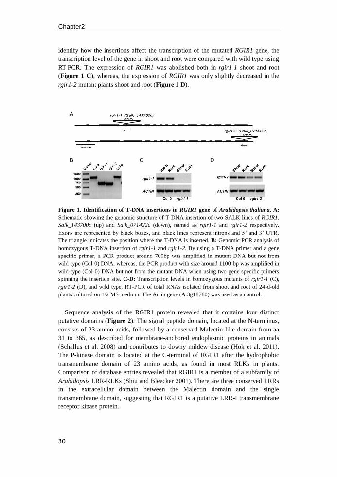
Chapter2
30
identify how the insertions affect the transcription of the mutated RGIR1 gene, the
transcription level of the gene in shoot and root were compared with wild type using
RT-PCR. The expression of RGIR1 was abolished both in rgir1-1 shoot and root
(Figure 1 C), whereas, the expression of RGIR1 was only slightly decreased in the
rgir1-2 mutant plants shoot and root (Figure 1 D).
Figure 1. Identification of T-DNA insertions in RGIR1 gene of Arabidopsis thaliana. A:
Schematic showing the genomic structure of T-DNA insertion of two SALK lines of RGIR1,
Salk_143700c (up) and Salk_071422c (down), named as rgir1-1 and rgir1-2 respectively.
Exons are represented by black boxes, and black lines represent introns and 5’ and 3’ UTR.
The triangle indicates the position where the T-DNA is inserted. B: Genomic PCR analysis of
homozygous T-DNA insertion of rgir1-1 and rgir1-2. By using a T-DNA primer and a gene
specific primer, a PCR product around 700bp was amplified in mutant DNA but not from
wild-type (Col-0) DNA, whereas, the PCR product with size around 1100-bp was amplified in
wild-type (Col-0) DNA but not from the mutant DNA when using two gene specific primers
spinning the insertion site. C-D: Transcription levels in homozygous mutants of rgir1-1 (C),
rgir1-2 (D), and wild type. RT-PCR of total RNAs isolated from shoot and root of 24-d-old
plants cultured on 1/2 MS medium. The Actin gene (At3g18780) was used as a control.
Sequence analysis of the RGIR1 protein revealed that it contains four distinct
putative domains (Figure 2). The signal peptide domain, located at the N-terminus,
consists of 23 amino acids, followed by a conserved Malectin-like domain from aa
31 to 365, as described for membrane-anchored endoplasmic proteins in animals
(Schallus et al. 2008) and contributes to downy mildew disease (Hok et al. 2011).
The P-kinase domain is located at the C-terminal of RGIR1 after the hydrophobic
transmembrane domain of 23 amino acids, as found in most RLKs in plants.
Comparison of database entries revealed that RGIR1 is a member of a subfamily of
Arabidopsis LRR-RLKs (Shiu and Bleecker 2001). There are three conserved LRRs
in the extracellular domain between the Malectin domain and the single
transmembrane domain, suggesting that RGIR1 is a putative LRR-I transmembrane
receptor kinase protein.

RGIR1 is a putative LRR-I transmembrane receptor kinase protein
31
Figure 2. RGIR1 encodes an LRR-I Receptor–like Protein Kinase. RGIR1 encodes a
polypeptide of 934 amino acids long and belongs to the LRR-I receptor-like kinase family of
Arabidopsis. The extracellular domain of this protein contains a signal peptide of 23 amino
acids, a Malectin-like structure, and 3 LRRs before the transmembrane region, followed by a
P-kinase cytoplasmic kinase domain. The numbers on the right side of the figure indicate the
position of all the amino acids residues, and the conserved residues of leucine-rich repeat are
highlighted in bold.
Predicted functions of RGIR1
To identify novel receptors that may function together with RGIR1, a search of the
Arabidopsis genome with the amino acid sequence of RGIR1 was conducted in the
STRING database (http://string-db.org/). Four Arabidopsis genes were found that
might be associated with RGIR1, including At5g08580 (EF-hand, calcium binding
motif-containing protein), AT5G01150 (an uncharacterized protein), At2g28060 (5'-
AMP-activated protein kinase beta-2 subunit protein), and AT2G23900 (Pectinlyase-
like protein). However, no evidence has been shown that RGIR1 has a direct
interaction with these identified genes.
Seed size and seed germination
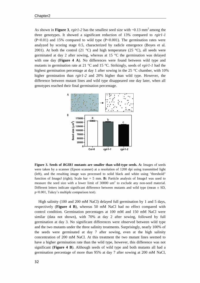
Chapter2
32
As shown in Figure 3, rgir1-2 has the smallest seed size with ~0.13 mm2 among the
three genotypes. It showed a significant reduction of 13% compared to rgir1-1
(P<0.01) and 15% compared to wild type (P<0.001). The germination rates were
analyzed by scoring stage 0.5, characterized by radicle emergence (Boyes et al.
2001). At both the control (21 °C) and high temperature (25 °C), all seeds were
germinated at day 2 after sowing, whereas at 15 °C the germination was delayed
with one day (Figure 4 A). No differences were found between wild type and
mutants in germination rate at 21 °C and 15 °C. Strikingly, seeds of rgir1-1 had the
highest germination percentage at day 1 after sowing in the 25 °C chamber, with 10%
higher germination than rgir1-2 and 20% higher than wild type. However, the
difference between mutant lines and wild type disappeared one day later, when all
genotypes reached their final germination percentage.
Figure 3. Seeds of RGIR1 mutants are smaller than wild-type seeds. A: Images of seeds
were taken by a scanner (Epson scanner) at a resolution of 1200 dpi using transmitted light
(left), and the resulting image was processed to solid black and white using "threshold"
function of ImageJ (right); Scale bar = 5 mm. B: Particle analysis of ImageJ was used to
measure the seed size with a lower limit of 30000 um2 to exclude any non-seed material.
Different letters indicate significant difference between mutants and wild type (mean ± SD,
p<0.001, Tukey’s multiple comparison test).
High salinity (100 and 200 mM NaCl) delayed full germination by 1 and 5 days,
respectively (Figure 4 B), whereas 50 mM NaCl had no effect compared with
control condition. Germination percentages at 100 mM and 150 mM NaCl were
similar (data not shown), with 70% at day 2 after sowing, followed by full
germination at day 3. No significant differences were observed between wild type
and the two mutants under the three salinity treatments. Surprisingly, nearly 100% of
the seeds were germinated at day 7 after sowing, even at the high salinity
concentration of 200 mM NaCl. At this treatment the two mutant lines seemed to
have a higher germination rate than the wild type, however, this difference was not
significant (Figure 4 B). Although seeds of wild type and both mutants all had a
germination percentage of more than 95% at day 7 after sowing at 200 mM NaCl,
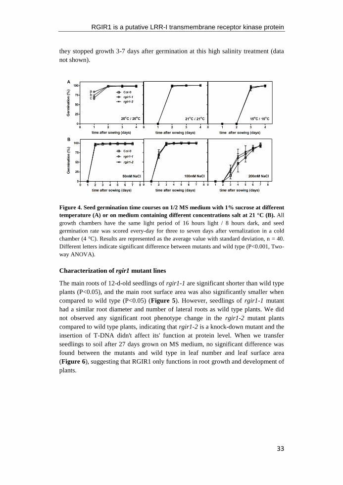
RGIR1 is a putative LRR-I transmembrane receptor kinase protein
33
they stopped growth 3-7 days after germination at this high salinity treatment (data
not shown).
Figure 4. Seed germination time courses on 1/2 MS medium with 1% sucrose at different
temperature (A) or on medium containing different concentrations salt at 21 °C (B). All
growth chambers have the same light period of 16 hours light / 8 hours dark, and seed
germination rate was scored every-day for three to seven days after vernalization in a cold
chamber (4 °C). Results are represented as the average value with standard deviation, n = 40.
Different letters indicate significant difference between mutants and wild type (P<0.001, Two-
way ANOVA).
Characterization of rgir1 mutant lines
The main roots of 12-d-old seedlings of rgir1-1 are significant shorter than wild type
plants (P<0.05), and the main root surface area was also significantly smaller when
compared to wild type (P<0.05) (Figure 5). However, seedlings of rgir1-1 mutant
had a similar root diameter and number of lateral roots as wild type plants. We did
not observed any significant root phenotype change in the rgir1-2 mutant plants
compared to wild type plants, indicating that rgir1-2 is a knock-down mutant and the
insertion of T-DNA didn't affect its' function at protein level. When we transfer
seedlings to soil after 27 days grown on MS medium, no significant difference was
found between the mutants and wild type in leaf number and leaf surface area
(Figure 6), suggesting that RGIR1 only functions in root growth and development of
plants.
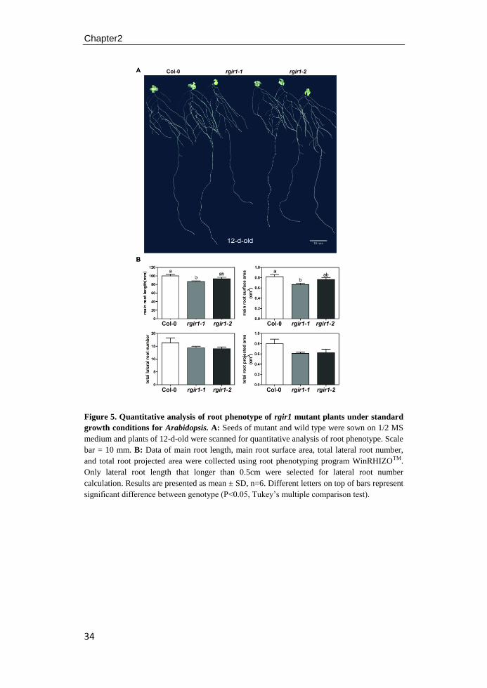
Chapter2
34
Figure 5. Quantitative analysis of root phenotype of rgir1 mutant plants under standard
growth conditions for Arabidopsis. A: Seeds of mutant and wild type were sown on 1/2 MS
medium and plants of 12-d-old were scanned for quantitative analysis of root phenotype. Scale
bar = 10 mm. B: Data of main root length, main root surface area, total lateral root number,
and total root projected area were collected using root phenotyping program WinRHIZOTM.
Only lateral root length that longer than 0.5cm were selected for lateral root number
calculation. Results are presented as mean ± SD, n=6. Different letters on top of bars represent
significant difference between genotype (P<0.05, Tukey’s multiple comparison test).
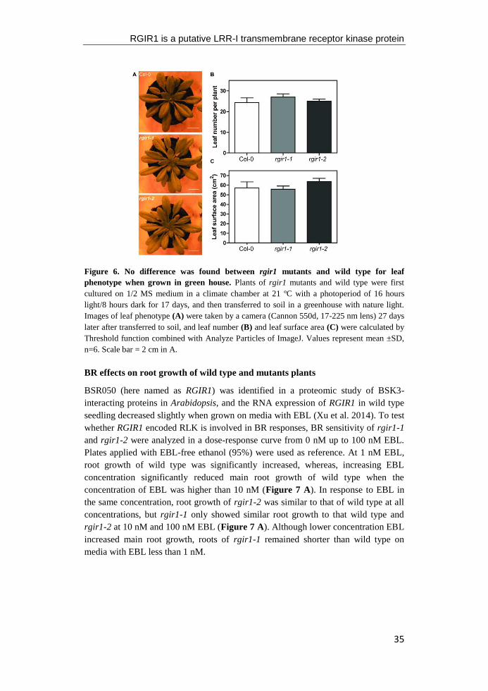
RGIR1 is a putative LRR-I transmembrane receptor kinase protein
35
Figure 6. No difference was found between rgir1 mutants and wild type for leaf
phenotype when grown in green house. Plants of rgir1 mutants and wild type were first
cultured on 1/2 MS medium in a climate chamber at 21 ºC with a photoperiod of 16 hours
light/8 hours dark for 17 days, and then transferred to soil in a greenhouse with nature light.
Images of leaf phenotype (A) were taken by a camera (Cannon 550d, 17-225 nm lens) 27 days
later after transferred to soil, and leaf number (B) and leaf surface area (C) were calculated by
Threshold function combined with Analyze Particles of ImageJ. Values represent mean ±SD,
n=6. Scale bar = 2 cm in A.
BR effects on root growth of wild type and mutants plants
BSR050 (here named as RGIR1) was identified in a proteomic study of BSK3-
interacting proteins in Arabidopsis, and the RNA expression of RGIR1 in wild type
seedling decreased slightly when grown on media with EBL (Xu et al. 2014). To test
whether RGIR1 encoded RLK is involved in BR responses, BR sensitivity of rgir1-1
and rgir1-2 were analyzed in a dose-response curve from 0 nM up to 100 nM EBL.
Plates applied with EBL-free ethanol (95%) were used as reference. At 1 nM EBL,
root growth of wild type was significantly increased, whereas, increasing EBL
concentration significantly reduced main root growth of wild type when the
concentration of EBL was higher than 10 nM (Figure 7 A). In response to EBL in
the same concentration, root growth of rgir1-2 was similar to that of wild type at all
concentrations, but rgir1-1 only showed similar root growth to that wild type and
rgir1-2 at 10 nM and 100 nM EBL (Figure 7 A). Although lower concentration EBL
increased main root growth, roots of rgir1-1 remained shorter than wild type on
media with EBL less than 1 nM.
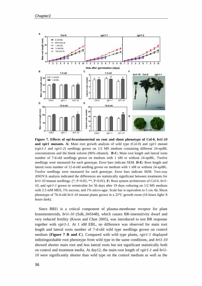
Chapter2
36
Figure 7. Effects of epi-brassinosteriod on root and shoot phenotype of Col-0, bri1-10
and rgir1 mutants. A: Main root growth analysis of wild type (Col-0) and rgir1 mutant
(rgir1-1 and rgir1-2) seedlings grown on 1/2 MS medium containing different 24-epiBL
concentrations and the blank solvent (96% ethanol). B-C: Main root length and lateral roots
number of 7-d-old seedlings grown on medium with 1 nM or without 24-epiBL. Twelve
seedlings were measured for each genotype. Error bars indicate SEM. D-E: Root length and
lateral roots number of 12-d-old seedling grown on medium with 1 nM or without 24-epiBL.
Twelve seedlings were measured for each genotype. Error bars indicate SEM. Two-way
ANOVA analysis indicated the differences are statistically significant between treatments for
bri1-10 mutant seedlings. (*, P<0.05; **, P<0.01). F: Root system architecture of Col-0, bri1-
10, and rgir1-1 grown in vermiculite for 56 days after 19 days culturing on 1/2 MS medium
with 2.5 mM MES, 1% sucrose, and 1% micro-agar. Scale bar is equivalent to 5 cm. G: Shoot
phenotype of 76-d-old bri1-10 mutant plants grown in a 22°C growth room (16 hours light/ 8
hours dark).
Since BRI1 is a critical component of plasma-membrane receptor for plant
brassinosteroids, bri1-10 (Salk_041648), which causes BR-insensitivity dwarf and
very reduced fertility (Kwon and Choe 2005), was introduced to test BR response
together with rgir1-1. At 1 nM EBL, no difference was observed for main root
length and lateral roots number of 7-d-old wild type seedlings grown on control
medium (Figure 7 B and C). Compared with wild type plants, rgir1-1 displayed
indistinguishable root phenotype from wild type in the same conditions, and bri1-10
showed shorter main root and less lateral roots but not significant statistically both
on control and treatment media. At day12, the main root length of rgir1-1 and bri1-
10 were significantly shorter than wild type on the control medium as well as the

RGIR1 is a putative LRR-I transmembrane receptor kinase protein
37
number for lateral roots (Figure 7 D and E). Treatment with 1 nM EBL enhanced
the main root growth and root branching both for 12-d-old wild type and mutant
plants, but the promotion was much stronger in the bri1-10 mutant plants.
The rgir1-1 had shoot phenotype similar to that of wild type when they were
grown in vermiculite for 56 days, but root system of rgir1-1 was shorter and smaller
than wild type in the identity condition (Figure 7 F). The bri1-10 showed a
significant dwarf shoot phenotype characterized by a dwarfed stature, dark green,
thicken leaves, and very lower fertility (Figure 7 G). Although BR seemed to
recover the root phenotype of bri1-10 seedlings on the medium, root system of these
two-month-old bri1-10 seedling was shorter and smaller compared with wild type
grown in vermiculites.
Discussion
There are more than 610 Receptor-like kinase genes in the Arabidopsis genome and
these genes play important roles in various aspects of plant life (Shiu and Bleecker
2001). Over the years, an increasing number of characterized RLKs have been
shown to play key roles in diverse and important biological processes, such as
hormone perception, morphology development and change, pattern development,
resistance to pathogens and abiotic stress tolerance (De Smet et al. 2009; Hazak and
Hardtke 2016; Diévart and Clark 2004; Osakabe et al. 2013; Wierzba and Tax 2013).
In this work, we present evidence that RGIR1, which encodes a putative leucine-rich
receptor like kinase protein, plays critical role in Arabidopsis root development.
Down-regulation of RGIR1 transcription of T-DNA insertion in the promote region
of rgir1-1 resulted in a significant decrease of main root length and root surface area,
whereas, the rgir1-2 mutant plants showed similar main root length with ecotype
Columbia (Figure 5). Sequence analysis showed that the T-DNA is inserted in the
third exon of RGIR1 gene in rgir1-1 mutant plants, but it is only 24bp upstream of
the end codon ATG in rgir1-2 (Figure 1). Combined with the RT-PCR result (Figure
1), we found that rgir1-2 is one knock-down mutant that has a somewhat lower
expression of RGIR1 both in shoot and root of the plants, but has a phenotype in both
root and shoot similar to the wild type.
Reverse genetics has been used as an effective approach to illuminate biological
functions of RLK genes of Arabidopsis (Alonso et al. 2003; Gou et al. 2010). In a
search of genomic-wide transcriptome analysis, RGIR1 was found involved in the
stress response of drought and freezing tolerance (Wituszyńska et al. 2013), in
protein phosphorylation in response to various plant hormones and corresponded
with quantitative trait loci that controls fiber length and lignin content of Arabidopsis
stems (Capron et al. 2012). Moreover, comparison of the NORK/SYMRK region of
legumes to other dicotyledonous plants showed that the closet homologues in
Arabidopsis were the two RLKs At1g67720 and At2g37050 (here named as RGIR1)
with a sequence identity of approximately of 33% (Stracke et al. 2002; Zhu et al.
2005; Kevei et al. 2005). Since SYMRK confirmed to function in early signal
transduction between nod factor perception and activation of the legheamoglobin

Chapter2
38
genes, homologous RGIR1 RLKs might also involved in signal transduction of early
root symbiotic.
Seed germination is controlled by environmental factors such as light, temperature,
humidity and applied chemicals in the culture medium, as well as by genetic
background. The size of the seed is determined by growth of both endosperm and
integument, and some genes have been found that directly affect seed size of
Arabidopsis, including IKU1 and IKU2 (Jofuku et al. 2005; Garcia et al. 2003),
MINI3 (Luo et al. 2005), SHB1 (Zhou et al. 2009), ARF2 (Schruff et al. 2006), and
TTG2 (Johnson et al. 2002). In this study, all genotypes had a germination
percentage higher than 97%, both under standard temperature condition and
higher/lower temperature, but the kinetics of the germination process was both
affected by genotype and temperature conditions (Figure 4). Although T-DNA
insertion mutant had smaller seed size than wild type before stratification, this did
not delay germination when compared to wild type under standard growth condition
and when exposed to salinity. Remarkably, the mutant rgir1-1 showed a higher
germination percentage at 25 °C at day1, indicating a possible role of gene RGIR1 in
seed germination process. Further studies are, however, needed to confirm a possible
role of RGIR1 in the process of seed germination.
One of the best-characterized LRR-RLKs in Arabidopsis is BRI1, which perceives
plant steroids hormone brassinosteroids at the cell surface (Li and Chory 1997), and
numerous alleles of bri1 were identified in a variety of independent screens (Clouse
2011). Recent evidences suggest that a large number of RLKs were confirmed to be
involved in the BRI1 hormone signal transduction pathway and several members of
BR-signaling kinases were phosphorylated by BRI1 before BR binding, which
appear to play a redundant role in BR signaling (Tang et al. 2008). RGIR1, together
with 11 transmembrane receptor-like kinases, was identified in a proteomics study of
BR-signaling kinases3 (BSK3)-interacting proteins (Xu et al. 2014). In the BR-
response experiment, the expression level of RGIR1 decreased slightly by increasing
concentration of EBL, indicating a role for RGIR1 in BR-regulated responses.
We found that BR is able to stimulate root elongation and growth in a dose-
dependent manner (Figure 7). Compared with other tissues of Arabidopsis, the
physiological response of different tissue varies in the concentration of exogenously
supplied EBL (Müssig et al. 2003; Bao et al. 2004; Yang et al. 2005). In response to
1 nM EBL, both root length and lateral formation were significantly promoted for
bri1-10 root, while it was only slight but not significantly increased for wild type and
rgir1-1 plant. Thus, bri1-10 might more sensitive to BR with shorter time treatment.
Plants of bri1-10 showed characteristic dwarf phenotype of BR-deficient and BR-
insensitive mutants when grown in the soil and only watered demi-water, and the
root system architecture was thinner and also decreased with shorter main root and
less lateral roots, compared with wild type (Figure 7, Vogler et al. 2014). rgir1-1
also displayed a smaller and compassed root system compared with wild type and
bri1-10, and no difference was observed for shoot phenotype of plants grown in soil

RGIR1 is a putative LRR-I transmembrane receptor kinase protein
39
(Figure 6), thus, the function of RGIR1 mutation might mainly acts on the growth of
main root.
Based on sequence similarity, BRL1 and BRL3 were identified possessing a
ligand binding island domain similar with BRI1 and encode functional BR receptors
to bind BL with high affinity (Caño-Delgado et al. 2004). Unlike BRI1 that is
expressed in most cells of the plant, the expression of BRL1 and BRL3 are mainly in
the vascular tissues, and they function redundantly with BRI1 in vascular
development in the Col-0 background. Using IP and LC/MS/MS techniques, RGIR1
together with an ATP-binding cassette-2 transporter were found to co-immuno-
precipitate with BRL3 in the provascular/stele tissues (Fàbregas et al. 2013),
supporting the deduction that RGIR1 is involved in root growth and development.
Taken together, our results indicate that LRR-RLK RGIR1 is involved in the root
growth and development at later stage of plant growth both on agar plates and in soil.
Although there is no direct evidence that RGIR1 is involved in the BR signal
transduction pathway during root growth and development, some results seem to
indicate such a role. The characterization of the rgir1-1 mutant root phenotype
provides a basis for further analysis of the role of gene RGIR1 in plant growth and
development.

40
41
Chapter3
Role of RGIR1 in controlling root system architecture of A. thaliana
Nana Yu, Jan Henk Venema, J. Theo M. Elzenga
Laboratory of Plant Ecophysiology, Groningen Institute for Evolutionary
Life Sciences, University of Groningen, The Netherlands.
Chapter3
42
Abstract: The ROOT GROWTH INHIBITION RECEPTOR 1 (RGIR1) gene encodes
a receptor-like kinase that is involved in Arabidopsis root growth under optimal
condition (Chapter2). In this study, kinematic and morphology parameters were
measured to quantify the spatial distribution of growth rate and the cell numbers and
size in the root tip of rgir1 mutants and wild type under control conditions and when
exposed to cold or salinity stress. Our results showed that the observed short root
phenotype of rgir1-1 mutant root was associated with a lower cell elongation rate
and decreased cortex cell number in the transition and elongation zone of the root tip.
In the presence of salt or cold stress, root growth and development in all genotypes
(mutants and wild type) were strongly affected with shorter main root length and less
lateral roots. The root phenotype of rgir1-2 was similar with wild type Col-0
seedling under both control and stress (salinity and temperature) conditions.
However, the rgir1-1 seedling had shorter main root and less lateral roots than wild
type under control condition. These differences between the rgir1-1 and wild type
were more pronounced at high temperature, but disappeared under cold and salinity
stress. These results indicate that RGIR1 is a positive regulator of root growth in
Arabidopsis under optimal growth condition and it seems not directly interact with
pathways of plants in response to cold and salinity stress.
Role of RGIR1 in controlling RSA of A.thaliana
43
Introduction
Roots play vital roles during plant growth and development, including the uptake of
water and nutrients, anchoring the plant into the soil, interacting with symbiotic
fungi and bacteria, carbohydrates storage and influencing the rhizosphere by
exudates, and have become more and more an indispensable part of the study on
plant-environment interactions (Zhu et al. 2011; Sánchez-Calderón et al. 2013).
Generally, the basic morphology of the root system is determined by inherent genetic
factors, but abiotic (stress) conditions can strongly modify the root system
architecture (RSA) through regulating primary root growth and lateral root initiation,
and via formation of adventitious roots and root hairs (Osmont et al. 2007). The RSA
is plastic and dynamic, allowing plants to respond to unfavorable environmental
conditions, including nutrient deficiency, extreme temperatures, flooding, and
salinity (Overvoorde et al. 2010; Galvan-Ampudia and Testerink 2011; González-
García et al. 2011; Gruber et al. 2013; Nagel et al. 2009; Koevoets et al. 2016).
However, the mechanism for the adaptation and alteration of the whole RSA of plant
by single or multiple stress factors remains unclear.
Cold is a major abiotic stress that adversely affects plant growth and crop
productivity (Yang et al. 2010). Low temperature not only decreases the elongation
rate of primary root tips, but also affects the RSA by inhibiting the formation of
lateral roots and the branching angle between primary and lateral roots (Nagel et al.
2009). The cold response of plants starts at the perception of the cold signal by
membrane-located proteins, and this signal is relayed to downstream signalling
components through a series of phosphorylation cascades resulting in an altered
transcription of several cold responsive genes (Rahman 2013; Kazan 2013). Auxin
plays major roles in the maintenance of cell division and expansion in the root apex,
and strongly influences the initiation and development of lateral roots (reviewed by
Nibau et al. 2008; Osmont et al. 2007; Péret et al. 2009; Petricka et al. 2012;
Shibasaki et al. 2009). Low temperature inhibits both cell number and meristem size
via the ARABIDOPSIS RESPONSE REGULATOR 1/12 (ARR1/12)-mediated
reduction in the auxin accumulation in apex root, and cytokinin-signalling is also
involved in the low temperature mediated inhibition of root growth (Zhu et al. 2015;
Yang et al. 2017). Moreover, CYTOKININ RESPONSE FACTOR 2 (CRF2) and
CRF3 are found to play an important role in regulating lateral root development in
response to cold stress in Arabidopsis (Jeon et al. 2016).
High salinity also modulates primary root growth, development of lateral roots
and/or root hairs, and the gravitropic growth of root tip (reviewed by Osmont et al.
2007; Galvan-Ampudia and Testerink 2011). Salinity stress is a combination of two
different processes, both requiring plant adaptation. The first is an, often rapid,
osmotic stress and the second is, a slower, ionic stress, caused by the toxic effect of
high Na+ concentration in the cytoplasm. Salinity tolerance mechanisms are a
combination of osmotic tolerance and Na+ and Cl
- exclusion, or Na
+ and Cl
-
sequestration in, for instance, the vacuole (reviewed by Munns and Tester 2008).
Many genes have been found to be involved in salinity tolerance mechanism, such as

Chapter3
44
SOS3 (with a role in sensing salinity in the root and transferring this signal to the
shoot (Qiu et al. 2003; Ishitani et al. 2000; Shi et al. 2000), HKT (that acts as Na+
transporter) (Rus et al. 2001; Rus et al. 2004; Mäser et al. 2002), and the vacuolar
Na+/H
+ exchanger AtNHX1 (that sequester Na
+ into the vacuole) (Møller and Tester
2007). In addition, plant hormones, which act as endogenous regulators of plant
development, function as central integrators in conferring tolerance to unfavorable
abiotic stress conditions including salinity stress (Ryu and Cho 2015; Iyer-Pascuzzi
et al. 2011; Colebrook et al. 2014; Kohli et al. 2013; Kazan 2015).
Recent studies suggest that receptor-like kinases play a major role in relaying
external abiotic stress signals to changes in gene transcription. For instance, a plasma
membrane anchored Calcium/CAM-regulated RLK (CRLK1) plays a role in bridging
calcium/calmodulin signalling and cold signalling and acts as a positive regulator of
cold-regulated genes expression (Yang et al. 2010). The Salt Overly Sensitive 2
(SOS2) gene encodes a functional serine/threonine-type protein kinase that is
predicted to be required for salt tolerance (Liu et al. 2000). Furthermore, other RLKs
have been implicated in controlling disease resistance and functioning in defence
responses, such as FLS2 (Gómez-Gómez and Boller 2000; Sun et al. 2013), PBS1
(Swiderski and Innes 2001), and WAK-like genes (Wagner and Kohorn 2001).
Although our understanding of the functions of RLKs in all aspects of plant growth
and development has increased in recent years, only a small number of members of
this big gene family have been established biological roles and not too many genetic
mutants of these related are available up to now.
Based on results of previous study in Chapter2 (not published), the RGIR1 gene is
believed to play a pivotal role in the root system architecture of Arabidopsis, with
shorter main root length under optimal growth condition. However, the mechanism
of how this newly identified RLK gene affects root growth or whether it is involved
in the tolerance of cold or salinity is not yet clear. In the present study, we firstly
performed a detailed root growth study of Arabidopsis ecotype Col-0 and rgir1
mutants (rgir1-1 and rgir1-2) at low or optimal growth temperature (21 °C) at
different concentrations of NaCl, quantifying the relative elongation rate in the
primary root growth zone with the aid of RootflowRT software (van der Weele et al.
2003). Our results indicate that RGIR1 inhibits root growth by reducing cell division
in the transition zone of the root tip at optimal growth condition. Cold and salinity
adversely affect root growth and lateral root development in wild type plants and
rgir1-1 and rgir1-2 alike, indicating that RGIR1 is not involved in the growth
response to low temperature or salinity.
Materials and methods
Plant materials and growth conditions
All homozygous F1 seeds of wild type Col-0 and two T-DNA insertion mutant lines
of At2G37050 (referred to as rgir1-1 and rgir1-2) were surface sterilized with
gaseous chlorine and sown on 1/2 MS medium agar plates as described in Chapter 2.
After sowing, plates with seeds were incubated at 4 ºC for three days and
Role of RGIR1 in controlling RSA of A.thaliana
45
subsequently transferred to growth chambers with a relative humidity of 70% and a
photoperiod of 16 hours light/ 8 hours dark. The irradiance level of light was about
110 µmol m-2
s-1
during daytime for all chambers. Effects of temperature treatment
on root growth and development were performed in three climate chambers at
constant temperatures of 15, 21, and 25 ºC, while the effect of salinity were
performed at 21 ºC. For effect of salinity stress on root growth and development,
seeds were germinated directly on 1/2 MS medium amended with NaCl to final
concentrations of 0, 50 and 100 mM.
The seedlings on agar filled petri dishes were scanned daily with a scanner (Epson,
Québec, Canada) to collect data on root system architecture analysis with
WinRIZHO software (2009 a, b, c). Since seeds germination time differs under
different treatments, day 0 was defined by radical emergence longer than 2 mm (48
hours after sowing at 25 and 21 ºC, and 72 hours at 15 ºC, whereas for salinity
treatments this was 48 hours for plates with 0 and 50 mM NaCl, and 72 hours for the
plates with 100 mM). Statistical analysis of data on main root length and lateral root
density was done with the Prism Graphpad software package (Version 5, GraphPad
Prism Software; San Diego, USA).
Kinematic analysis
To determine the effect of the RGIR1 mutation and salt stress (50 and 100 mM NaCl)
on cell expansion and division, we used a kinematic approach using the RootflowRT
software package (version 2.8, University of Missouri-Columbia), which identifies
the rate of expansion for different zones of apical part of the root (van der Weele et
al. 2003). Apart from the velocity of the different root zones, the position of the first
epidermal cell with a visible root bulge was determined. Six seedlings of both mutant
lines and the wild type were grown on a vertically placed petri dish and images of
the root tip under different culture conditions were taken with an Optikam B5 digital
camera (Ponteranica, Italy) that was installed on an optical microscope (CX41,
Olympus, Tokyo, Japan) using the 4X objective. For tip tracking, the tip was placed
in the central field of view and images were taken every 20 s for a total length of
time of up to 20 min. Nine images of each root tip were selected for calculation of
spatial velocity using RootflowRT software with the time interval of 60 s. Only data
with a reliable intermediate graph (automatically generated by the software) were
used for further statistical analysis.
The velocity profiles were fitted with a logistic function with following model:
Y=S+ (L-S) / (1+exp (-K*(X-X0)))
in which Y is the predicted value, X is the distance to the quiescent center, X0 is the
midpoint of X value, K is the steepness of the curve, S and L are the minimum and
maximum value of X at which breakpoint in the regression curve. The relative
elongation rate was calculated by taking the derivative of the root growth velocity in
relation to the distance from the root tip.
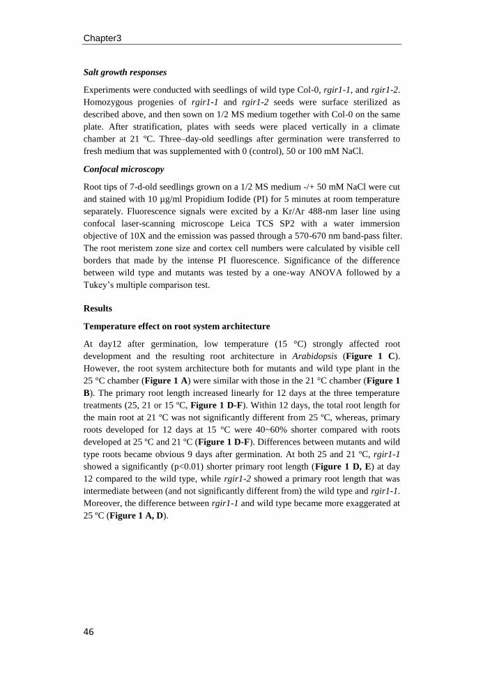
Chapter3
46
Salt growth responses
Experiments were conducted with seedlings of wild type Col-0, rgir1-1, and rgir1-2.
Homozygous progenies of rgir1-1 and rgir1-2 seeds were surface sterilized as
described above, and then sown on 1/2 MS medium together with Col-0 on the same
plate. After stratification, plates with seeds were placed vertically in a climate
chamber at 21 ºC. Three–day-old seedlings after germination were transferred to
fresh medium that was supplemented with 0 (control), 50 or 100 mM NaCl.
Confocal microscopy
Root tips of 7-d-old seedlings grown on a 1/2 MS medium -/+ 50 mM NaCl were cut
and stained with 10 µg/ml Propidium Iodide (PI) for 5 minutes at room temperature
separately. Fluorescence signals were excited by a Kr/Ar 488-nm laser line using
confocal laser-scanning microscope Leica TCS SP2 with a water immersion
objective of 10X and the emission was passed through a 570-670 nm band-pass filter.
The root meristem zone size and cortex cell numbers were calculated by visible cell
borders that made by the intense PI fluorescence. Significance of the difference
between wild type and mutants was tested by a one-way ANOVA followed by a
Tukey’s multiple comparison test.
Results
Temperature effect on root system architecture
At day12 after germination, low temperature (15 °C) strongly affected root
development and the resulting root architecture in Arabidopsis (Figure 1 C).
However, the root system architecture both for mutants and wild type plant in the
25 °C chamber (Figure 1 A) were similar with those in the 21 °C chamber (Figure 1
B). The primary root length increased linearly for 12 days at the three temperature
treatments (25, 21 or 15 ºC, Figure 1 D-F). Within 12 days, the total root length for
the main root at 21 ºC was not significantly different from 25 ºC, whereas, primary
roots developed for 12 days at 15 °C were 40~60% shorter compared with roots
developed at 25 ºC and 21 ºC (Figure 1 D-F). Differences between mutants and wild
type roots became obvious 9 days after germination. At both 25 and 21 ºC, rgir1-1
showed a significantly (p<0.01) shorter primary root length (Figure 1 D, E) at day
12 compared to the wild type, while rgir1-2 showed a primary root length that was
intermediate between (and not significantly different from) the wild type and rgir1-1.
Moreover, the difference between rgir1-1 and wild type became more exaggerated at
25 ºC (Figure 1 A, D).
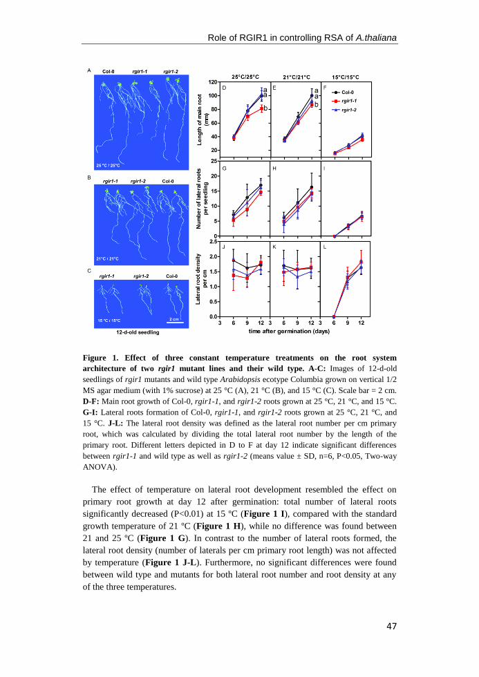
Role of RGIR1 in controlling RSA of A.thaliana
47
Figure 1. Effect of three constant temperature treatments on the root system
architecture of two rgir1 mutant lines and their wild type. A-C: Images of 12-d-old
seedlings of rgir1 mutants and wild type Arabidopsis ecotype Columbia grown on vertical 1/2
MS agar medium (with 1% sucrose) at 25 °C (A), 21 °C (B), and 15 °C (C). Scale bar = 2 cm.
D-F: Main root growth of Col-0, rgir1-1, and rgir1-2 roots grown at 25 °C, 21 °C, and 15 °C.
G-I: Lateral roots formation of Col-0, rgir1-1, and rgir1-2 roots grown at 25 °C, 21 °C, and
15 °C. J-L: The lateral root density was defined as the lateral root number per cm primary
root, which was calculated by dividing the total lateral root number by the length of the
primary root. Different letters depicted in D to F at day 12 indicate significant differences
between rgir1-1 and wild type as well as rgir1-2 (means value ± SD, n=6, P<0.05, Two-way
ANOVA).
The effect of temperature on lateral root development resembled the effect on
primary root growth at day 12 after germination: total number of lateral roots
significantly decreased (P<0.01) at 15 ºC (Figure 1 I), compared with the standard
growth temperature of 21 ºC (Figure 1 H), while no difference was found between
21 and 25 ºC (Figure 1 G). In contrast to the number of lateral roots formed, the
lateral root density (number of laterals per cm primary root length) was not affected
by temperature (Figure 1 J-L). Furthermore, no significant differences were found
between wild type and mutants for both lateral root number and root density at any
of the three temperatures.

Chapter3
48
Salinity effects on root system architecture
In accordance with the results of the temperature experiment, rgir1-1 showed a
significant shorter primary root length under control conditions (0 mM NaCl) at day
12 after germination, while no difference was found between the rgir1-2 mutant line
and wild type roots (Figure 2 A and D). At 50 mM NaCl, primary root growth was
strongly inhibited by ~50% and no differences were found between mutants and wild
type until day 12 (Figure 2 B and E). At 100 mM NaCl (Figure 2 C and F),
primary root growth was severely reduced (80%) throughout the 12 days treatment
when compared to conditions of 0 mM NaCl control treatment. Again, no differences
could be observed between mutants and the wild type at 100 mM NaCl during the
entire treatment period.
Figure 2. Effect of salinity stress on the root system architecture of two rgir1 mutant
lines and their wild type. A-C: Images for 12-d-old seedling of RGIR1 mutants and wild type
Arabidopsis ecotype Columbia grown on vertical 1/2 MS agar plates with 0 (A), 50 (B) or 100
mM (C) NaCl. Scale bar = 2 cm. D-F: Main root growth of Col-0, rgir1-1, and rgir1-2 roots
grown at 0, 50, and 100 mM NaCl treatment medium. G-I: Lateral roots formation of Col-0,
rgir1-1, and rgir1-2 roots grown at at 0, 50, and 100 mM NaCl treatment medium. J-L:
Lateral root density was defined as lateral roots number per cm of each primary root, which
was calculated by dividing the total lateral root number by the length of the primary root.
Different letters depicted in D at day 12 indicate significant differences between rgir1-1 and
the wild type as well as rgir1-2 (means value ± SD, n=6, P<0.05, Two-way ANOVA).
Role of RGIR1 in controlling RSA of A.thaliana
49
The total number of lateral roots without NaCl increased linearly during the 12
days treatment (Figure 2 G) and the lateral root density was between 1.5 to 2 per cm
after initiation of lateral roots (Figure 2 J) both for mutant and wild type plants.
Root branching was severely inhibited by the salinity treatment in a concentration
dependent matter (Figure 2 B, H, C, I). No differences were observed between the
two mutants and wild type in total lateral root number and lateral root density
(Figure 2 J-L).
Response of rgir1 mutants to salinity
To test the interaction between the RGIR1 mutation and salinity stress, seeds of
rgir1-1 and rgir1-2 were germinated on 1/2 MS medium, together with the wild type
on the same plate to exclude possible systematic errors. Seedlings were transferred to
new mediums with 0, 50 or 100 mM of NaCl after three days germination
/development on the control medium without NaCl. As shown in Figure 3 A, 100
mM NaCl caused severe growth inhibition of the primary root and induced a curly
root phenotype while seedlings at 0 and 50 mM NaCl were growing similarly and
without phenotypically abnormalities. In our experiments the growth of rgir1-2 and
wild type were indistinguishable under all conditions. However, rgir1-1 mutant
displayed a significantly shorter primary root length (p<0.05) compared to wild type
after 4 days grown on the new medium without salt. The difference became obscured
between rgir1-1 seedlings and wild type when grown on the medium in the presence
of NaCl, and no difference was observed in root growth rate between mutants and
wild type under different salt concentrations (Figure 3 C).
Velocity and strain rate profiles
The first epidermal cell with a visible root hair bulge is considered as a good
parameter to quantify cell elongation under different treatments (Le et al. 2001). For
a kinematic analysis of the root tip growth of rgir1 mutants and wild type, the
software package RootflowRT was used (van der Weele et al. 2003). The software
calculates the rate of expansion in the different zones behind the root tip from high-
resolution microscopic images taken with 20 s intervals. No visible root hair bulges
were found within 1500 µm from the root tip of 6-d-old germinated seedlings of wild
type and both mutants, whereas at day 14 root hairs already appeared at 1250 µm
distance from the tip (Figure 4 A and B). The wild type did not differ significantly
in the length between the root tip and the first visible root hair bulge at day 6 from
the two mutants, but root hair bulges of 14-d-old seedling of rgir1-1 emerged at a
distance around 1200±125 µm from the quiescent center, which was significantly
closer to the root tip than the root hair bulges of wild type and rgir1-2 seedlings (P<
0.001, Figure 4 B).
The kinematic analysis confirmed that zone of the primary root that exhibits
elongation growth at day 6 after germination encompassed the apical 1500 µm, while
the zone was limited to the apical 1250 µm at day 14 (Figure 4 C). The profile of the
elongation rate at day 6 peaks at about 800 µm from the apex, followed by a
gradually decrease to zero at 1500 µm. At day 6 there is no difference between the
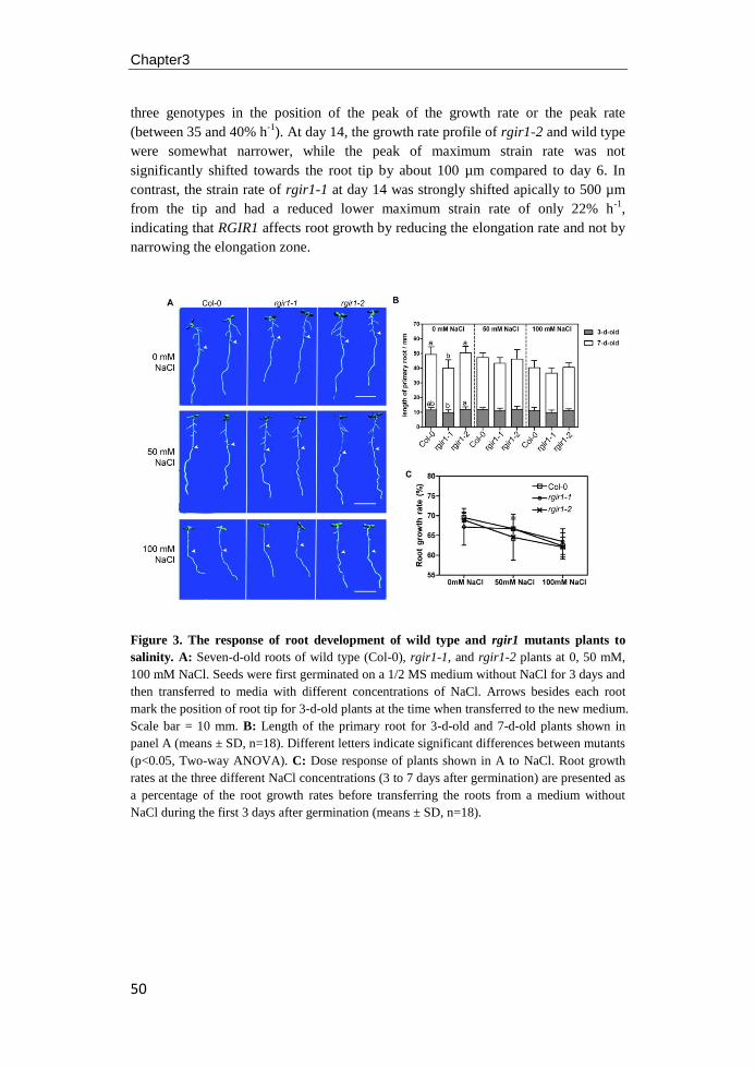
Chapter3
50
three genotypes in the position of the peak of the growth rate or the peak rate
(between 35 and 40% h-1
). At day 14, the growth rate profile of rgir1-2 and wild type
were somewhat narrower, while the peak of maximum strain rate was not
significantly shifted towards the root tip by about 100 µm compared to day 6. In
contrast, the strain rate of rgir1-1 at day 14 was strongly shifted apically to 500 µm
from the tip and had a reduced lower maximum strain rate of only 22% h-1
,
indicating that RGIR1 affects root growth by reducing the elongation rate and not by
narrowing the elongation zone.
Figure 3. The response of root development of wild type and rgir1 mutants plants to
salinity. A: Seven-d-old roots of wild type (Col-0), rgir1-1, and rgir1-2 plants at 0, 50 mM,
100 mM NaCl. Seeds were first germinated on a 1/2 MS medium without NaCl for 3 days and
then transferred to media with different concentrations of NaCl. Arrows besides each root
mark the position of root tip for 3-d-old plants at the time when transferred to the new medium.
Scale bar = 10 mm. B: Length of the primary root for 3-d-old and 7-d-old plants shown in
panel A (means ± SD, n=18). Different letters indicate significant differences between mutants
(p<0.05, Two-way ANOVA). C: Dose response of plants shown in A to NaCl. Root growth
rates at the three different NaCl concentrations (3 to 7 days after germination) are presented as
a percentage of the root growth rates before transferring the roots from a medium without
NaCl during the first 3 days after germination (means ± SD, n=18).
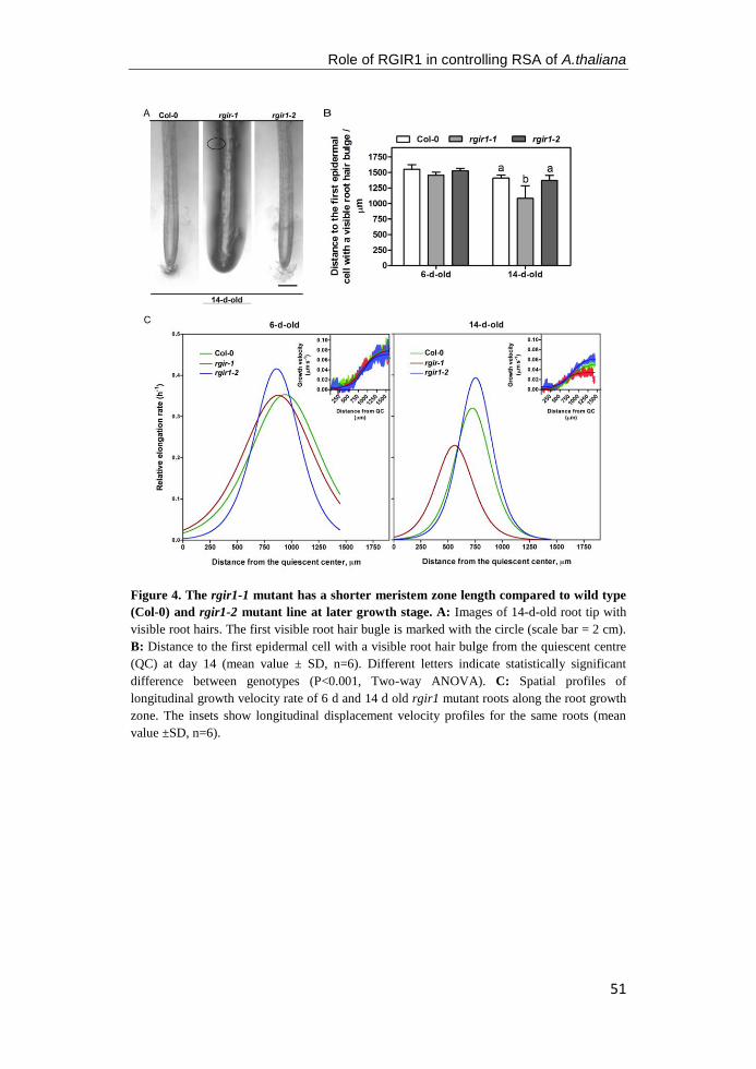
Role of RGIR1 in controlling RSA of A.thaliana
51
Figure 4. The rgir1-1 mutant has a shorter meristem zone length compared to wild type
(Col-0) and rgir1-2 mutant line at later growth stage. A: Images of 14-d-old root tip with
visible root hairs. The first visible root hair bugle is marked with the circle (scale bar = 2 cm).
B: Distance to the first epidermal cell with a visible root hair bulge from the quiescent centre
(QC) at day 14 (mean value ± SD, n=6). Different letters indicate statistically significant
difference between genotypes (P<0.001, Two-way ANOVA). C: Spatial profiles of
longitudinal growth velocity rate of 6 d and 14 d old rgir1 mutant roots along the root growth
zone. The insets show longitudinal displacement velocity profiles for the same roots (mean
value ±SD, n=6).
Chapter3
52
In the presence of salt in the MS medium root hairs were formed closer to the tip of
the root compared with control medium, in a concentration-dependent manner
(Figure 5 A). The first visible bulge of an emerging root hair of wild type emerged
at 750 µm from the QC in 100 mM NaCl, compared to 1500 µm in control medium
or medium supplied with 50 mM NaCl (Figure 5 B). Root growth of wild type was
severely prohibited under high salinity stress (Figure 2), thus the appearance of the
first root hair under salt stress gives a good correlation with the main root length.
While rgir1-2 resembles wild type (Figure 5 B), rgir1-1 showed a slight, non-
significant, reduction in the distance between first root hair bulge and QC. Seedling
of rgir1-1 plants older than 9 d had significantly shorter main root than wild type and
rgir1-2 under control conditions (Figure 2 A). No difference was observed in 6-d-
old between wild type and rgir1-1 (Figure 3 B), but at 12-d-old a difference was
obvious. Therefore, the lack of the correlation of root length with root hair formation
in rgir1-1 might be obscured by the lower growth rates and organ size in young
plants.
Salinity does not affect the maximum cell elongation rate (around 30% ~ 40% h-1
)
of wild type root, but the zone of maximal growth rate moved towards the QC by
200 µm and 300 µm under 50 mM and 100 mM NaCl treamtent, respectively,
compared with control condition (Figure 5 C). Since root growth was seriously
inhibited under high salinity stress, it seems that growth of main root doesn’t
correlate strongly with the distance between the QC and the zone of the maximum
growth rate both for different genotypes and for salt treatment. Although rgir1-1 had
the lowest elongation rate compared with wild type and rgir1-2, the zone for
elongation rate data and the zone for the maximum rate were not strongly affected
under salt stress, indicating that salt doesn’t have an additive inhibiting effect on the
RGIR1 mutation.
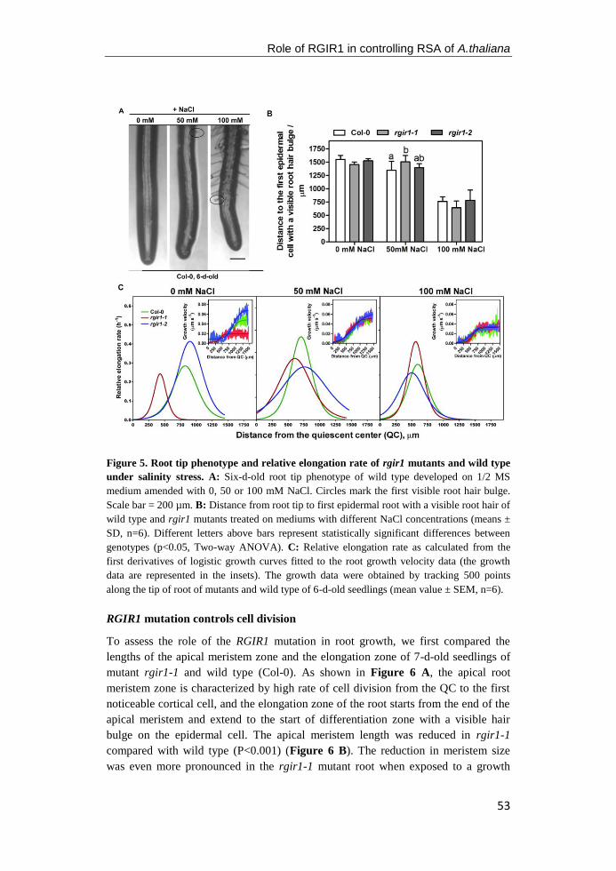
Role of RGIR1 in controlling RSA of A.thaliana
53
Figure 5. Root tip phenotype and relative elongation rate of rgir1 mutants and wild type
under salinity stress. A: Six-d-old root tip phenotype of wild type developed on 1/2 MS
medium amended with 0, 50 or 100 mM NaCl. Circles mark the first visible root hair bulge.
Scale bar = 200 µm. B: Distance from root tip to first epidermal root with a visible root hair of
wild type and rgir1 mutants treated on mediums with different NaCl concentrations (means ±
SD, n=6). Different letters above bars represent statistically significant differences between
genotypes (p<0.05, Two-way ANOVA). C: Relative elongation rate as calculated from the
first derivatives of logistic growth curves fitted to the root growth velocity data (the growth
data are represented in the insets). The growth data were obtained by tracking 500 points
along the tip of root of mutants and wild type of 6-d-old seedlings (mean value ± SEM, n=6).
RGIR1 mutation controls cell division
To assess the role of the RGIR1 mutation in root growth, we first compared the
lengths of the apical meristem zone and the elongation zone of 7-d-old seedlings of
mutant rgir1-1 and wild type (Col-0). As shown in Figure 6 A, the apical root
meristem zone is characterized by high rate of cell division from the QC to the first
noticeable cortical cell, and the elongation zone of the root starts from the end of the
apical meristem and extend to the start of differentiation zone with a visible hair
bulge on the epidermal cell. The apical meristem length was reduced in rgir1-1
compared with wild type (P<0.001) (Figure 6 B). The reduction in meristem size
was even more pronounced in the rgir1-1 mutant root when exposed to a growth
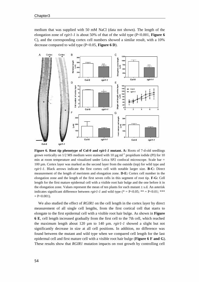
Chapter3
54
medium that was supplied with 50 mM NaCl (data not shown). The length of the
elongation zone of rgir1-1 is about 50% of that of the wild type (P<0.001, Figure 6
C), and the corresponding cortex cell numbers showed a similar result, with a 10%
decrease compared to wild type (P<0.05, Figure 6 D).
Figure 6. Root tip phenotype of Col-0 and rgir1-1 mutant. A: Roots of 7-d-old seedlings
grown vertically on 1/2 MS medium were stained with 10 µg ml-1 propidium iodide (PI) for 10
min at room temperature and visualized under Leica SP2 confocal microscope. Scale bar =
100 µm. Cortex layer was marked as the second layer from the outside (top) for wild type and
rgir1-1. Black arrows indicate the first cortex cell with notable larger size. B-C: Direct
measurement of the length of meristem and elongation zone. D-E: Cortex cell number in the
elongation zone and the length of the first seven cells in this segment of root tip. F-G: Cell
length for the first mature epidermal cell with a visible root hair bulge and the one before it in
the elongation zone. Values represent the mean of ten plants for each mutant ± s.d. An asterisk
indicates significant difference between rgir1-1 and wild type (* = P<0.05; ** = P<0.01; ***
= P<0.001).
We also studied the effect of RGIR1 on the cell length in the cortex layer by direct
measurement of all single cell lengths, from the first cortical cell that starts to
elongate to the first epidermal cell with a visible root hair bulge. As shown in Figure
6 E, cell length increased gradually from the first cell to the 7th cell, which reached
the maximum length about 120 µm to 140 µm. rgir1-1 showed a slight but not
significantly decrease in size at all cell positions. In addition, no difference was
found between the mutant and wild type when we compared cell length for the last
epidermal cell and first mature cell with a visible root hair bulge (Figure 6 F and G).
These results show that RGIR1 mutation impacts on root growth by controlling cell
Role of RGIR1 in controlling RSA of A.thaliana
55
division rather than cell expansion leading to less cell proliferation in the transition
zone of the root.
Discussion
Root growth is determined by a balance of cell proliferation and cell expansion in the
meristem and elongation zone of the root tip (Scheres et al. 2002). Cell numbers play
an important role in regulating growth of plant organ especially for Arabidopsis root
because of it's simple and stereotyped organization of cell types (Beemster and
Baskin 1998; Dolan et al. 1993; Wildwater et al. 2005; Sarkar et al. 2007), and the
first epidermal cell with a visible root hair bugle is generally accepted as the location
along the root where cell elongation no longer takes place (Le et al. 2001). The
kinematic results showed that inhibition of rgir1-1 root growth is caused by a
shortening of the division zone as indicated by the apical shift of the zone of
maximal growth rate, rather than a reduction of the maximum expansion rate
(Figure 4). According to the confocal results, the length of the cell division zone and
the elongation zone are decreased in the rgir1-1 mutant root while the number of
cortex cell layers was less in rgir1-1 than in wild type (Figure 6). However, the
average length of cortex cells was similar as those wild type roots suggesting that
RGIR1 reduces root growth by affecting cell divisions in the transition zone rather
than cell expansion in the elongation zone.
In roots of Arabidopsis seedlings, abiotic stress stimulates a stress–induced
morphogenic response (SIMR), which is characterized by proliferation of lateral
roots, inhibition of primary root and lateral root elongation, and alteration of cell
differentiation status in the root apex (Zolla et al. 2010; Potters et al. 2007). Results
from our study showed that both low temperature (15ºC) and salinity, 50 mM NaCl,
induced a change in the RSA of Arabidopsis characterized by a decrease in root
length and less lateral roots. The effect of salt stress on RSA was even more
pronounced under 100 mM salt treatment (Figure 2 and 3). When lateral root
density was measured as number of laterals per cm primary root length, no
difference was found between stress conditions at 15 ºC, nor at 50 mM NaCl
treatment, compared with control condition at 21 ºC, suggesting there is a balance
between growth and development in the intrinsic regulating pathways in response to
diverse mild abiotic stresses.
As sessile organism, plants development a wide assortment of mechanisms to cope
with the biotic and abiotic stress conditions in the surrounding environment
(Pasternak et al. 2005; Munns and Tester 2008; Ryu and Cho 2015; Schmidt et al.
2015; Atkin and Tjoelker 2003) and the RLKs, localized in the membrane of plants,
are important in the perception and transferring of signals during various aspects of
root growth and in response to diverse stresses. rgir1-1 mutant plants showed a
reduced primary root length under standard growth conditions at 21 ºC, but do not
exhibit a modified shoot phenotype (see Chapter 2, Figure 6). In the present
experiments, a significant difference was found between rgir1-1 mutant roots and its'
wild type at high temperature treatment (25 ºC), where plants have a much higher
Chapter3
56
growth rate (Figure 1 A). However, in the cold- and salt-stressed Arabidopsis roots,
there appeared to be no obviously correlation between the mutation of RGIR1 gene
with either cold (Figure 1 C and F) or salt stress (Figure 2 and 3) when root growth
rates were lower and inhibited by cold /salt stress.
In the presence of salt (Figure 3 and 5), the decrease of the growth rate for rgir1-
1 mutant roots matched by a reduction of growth in wild type and rgir1-2, obscuring
the phenotypic difference between rgir1-2 and wild type. One possible explanation
for the lack of difference in root growth under salinity stress between rgir1-1 and
wild type is that, the processes affected by salinity are no longer available for
modification in the rgir1-1 mutant background. Thus, the root growth phenotype for
rgir1-1 under control conditions is similar to those wild type plants under salinity
and low-temperature conditions. When these rgir1-1 roots are salt-stressed or
developed in a cold chamber, the cell division zone for rgir1-1 plants cannot be
shortened any further by an additional abiotic stress factor.
In conclusion, our data suggest that this newly identified RLK RGIR1, is a
positive growth regulator of root, and the short-root phenotype of the rgir1-1 mutant
is probably caused by reduction of the number of cortex cells in both the transition
and elongation zone of the root tip, without affecting cell size. Under low
temperature or salinity treatment, roots of rgir1-1 mutant were phenotypically and
physiologically similar as wild type, indicating that RGIR1 gene-associated
processes might partly overlap with the pathways involved in cold and salinity stress.
Another possibility is that the effect of the rgir1 mutant of abiotic stress leaves "no
room for further inhibition" of growth by abiotic stress. Thus, root system
architecture of Arabidopsis is controlled by a complex network and can be changed
under changing conditions. Future research is needed to understand the molecular
mechanism of RGIR1 and to find out what the specific role of RGIR1 in adaptation
of root system architecture under abiotic/biotic stresses.
57
Chapter4
Effects of Growth conditions on root growth patterns in Arabidopsis
Nana Yu, J. Theo. M. Elzenga
Laboratory of Plant Ecophysiology, Groningen Institute for Evolutionary
Life Sciences, University of Groningen, The Netherlands.
Chapter4
58
Abstract: Root system architecture of plants is regulated by both intrinsic and
environmental response pathways. The goal for this study was to determine which of
the components of the nutrient medium are having effects on root elongation or/and
root branching, and how roots respond to abiotic stresses and nutrient deficiency.
We found that sucrose induced a waving and skewing phenotype on hard agar, with
strongly reduced lateral root formation and an increased length of the main.
Although sucrose affected roots of wild type plants and rgir1 mutants in a similar
way, the rgir1-1 mutant, which exhibits altered root growth kinetics in the transition
zone, had a shorter main root. Inhibition of main root growth and root branching
were observed on media supplemented with salt or mannitol, or with a low pH, while
sulfur-deficiency did not affect main root length. In contrast, lateral root induction
was strongly stimulated in media lacking sulfur. Roots of rgir1 mutants reacted
similar to wild type to salt and osmotic stress, but S-deficiency medium induced
more lateral roots in rgir1-1 mutants than in wild type plants, indicating a possible
role for RGIR1 in the process of lateral root initiation or emergence.

Effects of growth conditions on root growth patterns
59
Introduction
The Arabidopsis root has been used as a model for root growth and development, as
it has a comparatively simple organization and can be easily cultured in non-soil
media (Petricka et al. 2012; Osmont et al. 2007). Structure and composition of the
growth medium can strongly modify root growth and architecture. When grown on
the surface of nutrient containing agar medium, the combined effect of gravity and
the contact between the medium and the root results in a characteristic skewing and
waving growth pattern (Oliva and Dunand 2007; Johnsson et al. 2009; Scherer and
Pietrzyk 2014), and this root surface-dependent behavior varies between different gel
patches (Vaughn and Masson 2011; Thompson and Holbrook 2004). Also the pH of
the root rhizosphere displays a heterogeneous patchiness when grown in vitro on
agar plates, as well as in natural soils (Walter et al. 2009; Blossfeld and Gansert
2007; Hinsinger et al. 2003). Moreover, the low pH of the growth medium can
severely inhibit root growth by decreasing the cytoplasmic pH of the root cells (Iuchi
et al. 2007; Yan et al. 1992).
There is a large body of literatures describing how the growth medium can alter
root system architecture in different ways (Malamy 2005). A major proportion for
these papers is referred to morphogenetic responses of plants to salinity stress. This
focus on salinity stress stems from the fact that this is one of the most significant
factors limiting crop production and is a threat for future food supply for human
(Kazama et al. 2013; Zhu et al. 2007; Boursiac et al. 2005; Møller and Tester 2007).
Other growth medium factors are nutrients, such as nitrate, phosphate, sulfate and
ion that act as environmental signals to trigger molecular mechanisms involved in
the cell division and proliferation of plant roots (Lopez-Bucio et al. 2003; Vidal et al.
2008; Gruber et al. 2013), and hormones, which play vital roles in the intrinsic
pathways that determine root system architecture (Muraro et al. 2014; González-
García et al. 2011; Petricka et al. 2012; Overvoorde et al. 2010; Ryu and Cho 2015).
In addition, osmotic stress caused by drought or high salinity in the medium also
affects plant root system growth and development (Deak and Malamy 2005; Nguyen
et al. 2016; Liu et al. 2014; Zwiewka et al. 2015; Osakabe et al. 2013).
Roots of Arabidopsis have developed a generic stress-induced morphogenetic
response under distinct stresses on the grown medium. For instance, exposure to a
mild salt stress causes a drastic reduction in main root and lateral root elongation, but
an increase in lateral root number. Furthermore, this stimulation of lateral roots
proliferation by salt stress is enhanced by nitrate in the medium (Zolla et al. 2010).
Additionally, nutrient varieties and the nutrient availability in the agar medium also
has a profound impact on root system architecture, by altering the length and
diameter of main roots, inhibiting or stimulating lateral roots and roots hairs, and
controlling root growth direction and branching angles of lateral roots (Singh et al.
2014; Booker et al. 2010; Malamy 2005; Gruber et al. 2013; Osmont et al. 2007).
However, distinct effects on root system architecture strongly depend on kind and
concentration of the nutrient involved (Gruber et al. 2013; Lopéz-Bucio et al. 2003;
Hodge 2004). Despite the diversity in phenotype changing under different abiotic
Chapter4
60
stress conditions, the generic stress-induced morphogenic response comprise of
inhibition of cell elongation, localized stimulation of cell proliferation, and alteration
of cell differentiation status (Potters et al. 2007).
Lineages and fate of cells in developing Arabidopsis roots show a stereotypical
pattern forming a set of concentric cylinders, consisting (starting from the outermost
one) of the epidermis, the cortex, the endodermis and the stele (consisting of
pericycle and vascular bundles), with the columella and lateral root cap providing
additional layers at the root tip (Wierzba and Tax 2013; Scheres et al. 2002; Petricka
et al. 2012; Yan et al. 1992). Each cell type in the root has its own transcriptional
profile and many biological functions are regulated in a cell–type-specific manner
(Birnbaum et al. 2003; Brady et al. 2007). Recently, comparison of gene expression
levels within cell types under standard conditions with those under certain stresses,
demonstrated that cell identity plays important roles in the stress responses of plants
(Iyer-Pascuzzi et al. 2011; Dinneny et al. 2008). Thus, the use of environmental
stimuli combined with the genomic-wide data sets allows the identification of
associated regulator genes within cell types, and those root-patterning factors, which
always are expressed at higher levels in the specific cell types regardless of the
outside environment.
We previously identified a receptor-like kinase mutant, rgir1-1, which had a
shorter main root and less lateral roots compared to the wild type Arabidopsis
ecotype Columbia. In a screen for common stress response genes in the whole root,
the gene expression of RGIR1 was enriched in the stele and endodermis layer under
low pH and sulfur deficiency stress conditions, respectively (Iyer-Pascuzzi et al.
2011). As detailed studies of wild type and rgir1 mutant root responses under low
pH and sulfur deficiency conditions are lacking, the specific role of RGIR1 in root
development is still not elucidated.
In this present study, we set out to understand how nutrient and growth media
affect root system architecture of Arabidopsis seedling grown on the surface agar
plate. Arabidopsis ecotype Col-0 and two RGIR1 alleles (rgir1-1 and rgir1-2) in Col-
0 background were used to investigate the response of root system architecture under
different abiotic stress conditions. In particular, our objective is to identify the
specific component of the growth media and to which extent that they affect root
elongation and/or root branching. Our data showed that elongation of primary root
was increased on the higher concentration agar while the average length of lateral
roots was inhibited for the same plant. The length of primary root was increased in
the presence of sucrose in the media, whereas, low pH and increasing salinity stress
dramatically repressed root elongation and root branching of plants. No difference
was observed between mutant seedlings and wild type seedling, when they had
reduced root system under stress conditions. Root elongation was not affected on the
sulfur deficiency media both for wild type and mutant seedlings. However, in rgir1-1
roots starved with sulfate, lateral roots formation formed earlier and closer to the tip
of the root compared to rgir1-2 and wild type.
Effects of growth conditions on root growth patterns
61
Materials and methods
Plant material and growth conditions
Arabidopsis (Arabidopsis thaliana) ecotype Columbia (Col-0), rgir1 mutants, rgir1-
1 (Salk_143700c) and rgir1-2 (Salk_071422c) were used in this experiment. All wild
type and mutant lines were in Columbia background and seeds were obtained from
NASC (http://arabidopsis.info/). Seeds of mutants and wild-type (Col-0) were gas-
sterilized in a desiccator for 3 hours with the fumes in a beaker with 100 ml of
bleach (4% NaClO) mixed with 5 ml 37% Hydrochloric Acid and then sown on agar
plates with nutrient media with different additives in 120×120 mm square Petri plate,
approximately 2 cm from the top edge. Plates were wrapped with parafilm and, after
two to four days vernalization at 4 °C, transferred to a growth chamber with 16 hours
light/ 8 hours dark at 21 °C. All plates were placed vertically in the growth chamber,
except when being scanned on a flatbed scanner for two to three minutes at the
indicated days.
Media composition
The standard medium consisted of 1/2 MS (Murashige and Skoog 1962) basal
medium, 2.5 mM MES, 1% agar, and adjusted to pH 5.7 with KOH (0.1 mM). In the
experiments to test the effect of the agar concentration on root growth and
development, the agar concentration was increased to 1.5% without any change for
the other ingredients. Sucrose (1%) was added to the standard medium to test the
effect of sucrose on root system architecture of plants grown on hard medium. For
salt and mannitol treatment experiments, plants were directly germinated on hard
(1.5%) standard medium with 1% sucrose and different concentrations of NaCl or
mannitol, as indicated.
The sulfur sufficient media contained 1.25 mM KNO3, 1.25 mM Ca(NO3)2, 0.25
mM KH2PO4, 0.5 mM MgSO4, 22.5 μM Fe3+
(EDTA), 11.6 μM H3BO3, 2.4 μM
MnCl2, 0.24 μM ZnSO4, 0.08 μM CuSO4, and 0.13 μM Na2MoO4. To make S-free
medium, MgSO4, ZnSO4, and CuSO4 were replaced by their respective chloride salts.
Low pH media is 1/2 MS salts with vitamins (Murashige and Skoog 1962), 2.5 mM
MES, 1% agar, and pH adjusted to 4.6 by KOH (0.1 mM).
Root system architecture analysis on solid media
Seedlings of Col-0, rgir1-1, and rgir1-2 that grown on different medias were
scanned everyday by a scanner connected to computer running image analysis
program WinRHIZO (van der Weele et al. 2003). The root architecture parameters,
including the root length, cotyledon length, and lateral roots number, were
determined. All data collected were statistically analyzed by a two-way ANOVA test
using GraphPad prism (http://www.graphpad.com/).
Chapter4
62
Results
Gel concentration affects root waving but not root growth when placed
vertically
To determine whether a higher concentration of agar in the medium affects the
architecture of surface-grown root system, seedlings of Arabidopsis ecotype
columbia and rgir1 mutants in the Col-0 background were grown on 1% and 1.5%
agar plates (without sucrose) and placed vertically or at 45°. When placed vertically,
seedlings of 7-d-old wild type grew relatively straight regardless of the agar
concentration, and no waves were observed near the tip of main root. Roots of rgir1
mutants similarly displayed a straight main root without waves, both on control and
hard medium when grown vertically. However, wild type and mutant roots skewed
to the right (observed form the bottom of the plate) and started to make coils when
the plates were placed at a 45° angle. The root phenotype of wild type and mutants
grown on 1% medium, were indistinguishable from those grown on hard medium
with 1.5% agar (data not shown).
We also compared root growth parameters between wild type and mutants on hard
medium placed vertically. rgir1-1 has significantly shorter main root length (P<0.05)
compared with wild type both at 1% and 1.5% agar medium (identical to our
previous results). The roots of rgir1-2 were indistinguishable from wild type on 1%
agar but significantly shorter (P<0.001) than wild type on vertical medium with 1.5%
agar (Figure 1 A), indicating a possible role for harder agar concentration on the
elongation of root. No differences between mutants and wild type both at control and
hard medium were found in the number of lateral roots (Figure 1 B) and cotyledon
length (Figure 1 C). However, rgir1-2 showed the shortest length of lateral roots on
1% medium placed vertically, a difference that disappeared when grown on hard
medium at 1.5% agar (Figure 1 D).
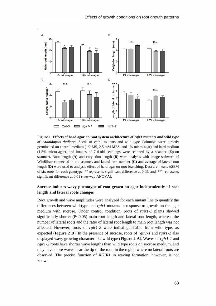
Effects of growth conditions on root growth patterns
63
Figure 1. Effects of hard agar on root system architecture of rgir1 mutants and wild type
of Arabidopsis thaliana. Seeds of rgir1 mutants and wild type Columbia were directly
germinated on control medium (1/2 MS, 2.5 mM MES, and 1% micro-agar) and hard medium
(1.5% micro-agar), and images of 7-d-old seedlings were scanned by a scanner (Epson
scanner). Root length (A) and cotyledon length (B) were analysis with image software of
WinRihzo connected to the scanner, and lateral root number (C) and average of lateral root
length (D) were used to analysis effect of hard agar on root branching. Data are means ±SEM
of six roots for each genotype. '*' represents significant difference at 0.05, and '**" represents
significant difference at 0.01 (two-way ANOVA).
Sucrose induces wavy phenotype of root grown on agar independently of root
length and lateral roots changes
Root growth and wave amplitudes were analyzed for each mutant line to quantify the
differences between wild type and rgir1 mutants in response to growth on the agar
medium with sucrose. Under control condition, roots of rgir1-1 plants showed
significantly shorter (P<0.05) main root length and lateral root length, whereas the
number of lateral roots and the ratio of lateral root length to main root length was not
affected. However, roots of rgir1-2 were indistinguishable from wild type, as
expected (Figure 2 B). In the presence of sucrose, roots of rgir1-1 and rgir1-2 also
displayed wavy growing character like wild type (Figure 2 A). Waves of rgir1-1 and
rgir1-2 roots have shorter wave lengths than wild type roots on sucrose medium, and
they have more waves near the tip of the root, in the region where no lateral roots are
observed. The precise function of RGIR1 in waving formation, however, is not
known.
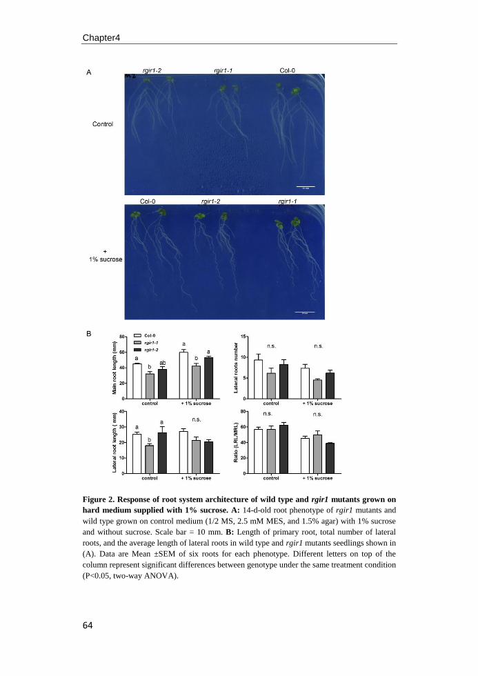
Chapter4
64
Figure 2. Response of root system architecture of wild type and rgir1 mutants grown on
hard medium supplied with 1% sucrose. A: 14-d-old root phenotype of rgir1 mutants and
wild type grown on control medium (1/2 MS, 2.5 mM MES, and 1.5% agar) with 1% sucrose
and without sucrose. Scale bar = 10 mm. B: Length of primary root, total number of lateral
roots, and the average length of lateral roots in wild type and rgir1 mutants seedlings shown in
(A). Data are Mean ±SEM of six roots for each phenotype. Different letters on top of the
column represent significant differences between genotype under the same treatment condition
(P<0.05, two-way ANOVA).
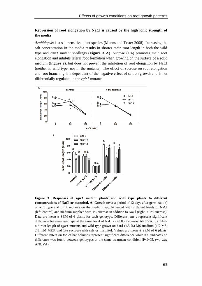
Effects of growth conditions on root growth patterns
65
Repression of root elongation by NaCl is caused by the high ionic strength of
the media
Arabidopsis is a salt-sensitive plant species (Munns and Tester 2008). Increasing the
salt concentration in the media results in shorter main root length in both the wild
type and rgir1 mutant seedlings (Figure 3 A). Sucrose (1%) promotes main root
elongation and inhibits lateral root formation when growing on the surface of a solid
medium (Figure 2), but does not prevent the inhibition of root elongation by NaCl
(neither in wild type, nor in the mutants). The effect of sucrose on root elongation
and root branching is independent of the negative effect of salt on growth and is not
differentially regulated in the rgir1 mutants.
Figure 3. Responses of rgir1 mutant plants and wild type plants to different
concentrations of NaCl or mannitol. A: Growth (over a period of 12 days after germination)
of wild type and rgir1 mutants on the medium supplemented with different levels of NaCl
(left, control) and medium supplied with 1% sucrose in addition to NaCl (right, + 1% sucrose).
Data are mean ± SEM of 6 plants for each genotype. Different letters represent significant
difference between genotype at the same level of NaCl (P<0.05, two-way ANOVA). B: 14-d-
old root length of rgir1 mtuants and wild type grown on hard (1.5 %) MS medium (1/2 MS,
2.5 mM MES, and 1% sucrose) with salt or mannitol. Values are mean ± SEM of 6 plants.
Different letters on top of bar columns represent significant difference while n.s. indicates no
difference was found between genotypes at the same treatment condition (P<0.05, two-way
ANOVA).
Chapter4
66
To determine whether the repression of root elongation was caused either by the
high osmotic value or the toxic ionic (Na+ and Cl
-) effect or both, we supplemented
the media with osmotically equivalent concentrations of mannitol. Mannitol
appeared to have no effect on root elongation (Figure 3 B), indicating that the
repression of root elongation on salt media is caused by the ionic effects. rgir1-1
displayed significantly shorter (P<0.05) main roots on control media, but this
difference disappeared both on mild and high salinity treatment media. Although
both salinity and RGIR1 mutation repress root elongation for Arabidopsis, salt does
not induce a further reduction of root growth in rgir1-1, possibly indicating an
interaction of salinity with gene function or suggesting that a further reduction of
growth in the rgir1-1 background by salt is not feasible.
Effects of Sulfur deficiency on root system architecture
In Arabidopsis seedlings grown on the surface of agar plates without SO42-
, lateral
roots formed closer to tip of the root and developed at a higher density, compared to
those grown on sulfate-sufficient medium (Lopez-Bucio et al. 2003). To assess the
effects of sulfur deficiency on root architectural traits of wild type and rgir1-1 and
rgir1-2 mutants, we first grew seedlings on control medium (+S) for 9 days and then
transferred to new medium with (+S) or without (-S) sulfate for 16 days. After 7 d
growth on the new medium, the primary root growth rate of wild type and the two
rgir1 alleles were not affected on the S-free medium (Figure 4 A and B). Root
growth of rgir1-2 was indistinguishable from wild type both on the +S medium and
–S medium. However, the rgir1-1 mutant displayed a 21% (Figure 4 A) and 41%
(Figure 4 C) reduction in main root length after 7 d and 16 d growth on new +S
medium. The reduction of main root length in rgir1-1 was 22% and 49% for 7 d and
16 d growth on -S medium, respectively, suggesting that sulfur does not interact with
RGIR1 in controlling elongation of root.
As shown in Figure 4 E, we carefully examined the formation of lateral roots in
the tip part of primary root in both the segment that formed on sulfur-sufficient
medium before transfer to new medium and the segment that was newly formed
while on the new +S or –S medium. After 7 d growth on the new medium, lateral
root formation was significantly increased by sulfur deprivation in both wild type
and rgir1 mutant roots (Figure 4 D). Although there were small differences between
rgir1-1, rgir1-2, and wild type, these were not statistically significant. The density of
lateral roots formed on the upper (proximal) segment was around 2-fold higher than
that of distal segment that formed on the new medium in wild type and mutant
seedlings (Figure 4 F). This pattern was identical in +S and –S roots. Since the
number of lateral roots in all genotypes is nearly the same, but the root length is
shorter in the rgir1-1 mutant, the lateral root density in this mutant is significantly
higher.
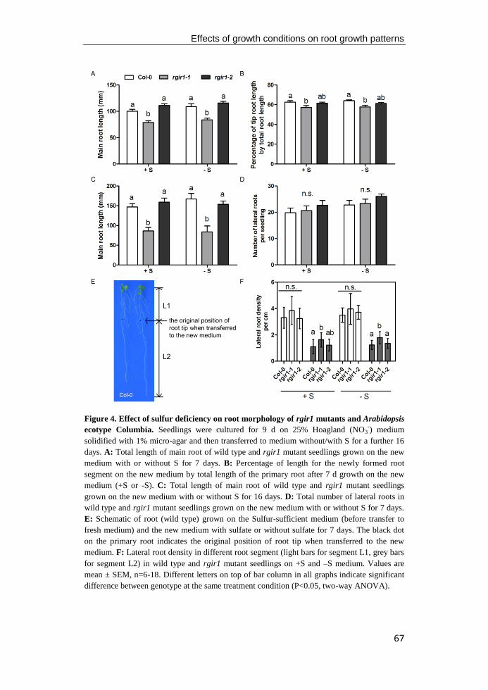
Effects of growth conditions on root growth patterns
67
Figure 4. Effect of sulfur deficiency on root morphology of rgir1 mutants and Arabidopsis
ecotype Columbia. Seedlings were cultured for 9 d on 25% Hoagland (NO3-) medium
solidified with 1% micro-agar and then transferred to medium without/with S for a further 16
days. A: Total length of main root of wild type and rgir1 mutant seedlings grown on the new
medium with or without S for 7 days. B: Percentage of length for the newly formed root
segment on the new medium by total length of the primary root after 7 d growth on the new
medium (+S or -S). C: Total length of main root of wild type and rgir1 mutant seedlings
grown on the new medium with or without S for 16 days. D: Total number of lateral roots in
wild type and rgir1 mutant seedlings grown on the new medium with or without S for 7 days.
E: Schematic of root (wild type) grown on the Sulfur-sufficient medium (before transfer to
fresh medium) and the new medium with sulfate or without sulfate for 7 days. The black dot
on the primary root indicates the original position of root tip when transferred to the new
medium. F: Lateral root density in different root segment (light bars for segment L1, grey bars
for segment L2) in wild type and rgir1 mutant seedlings on +S and –S medium. Values are
mean ± SEM, n=6-18. Different letters on top of bar column in all graphs indicate significant
difference between genotype at the same treatment condition (P<0.05, two-way ANOVA).

Chapter4
68
Response of roots morphology on low pH medium
Root growth and branching of both wild type and rgir1 mutants were markedly
reduced by lowering the pH of the growth medium from 5.7 (Figure 5 A) to 4.6
(Figure 5 B). Consistent with previous results, rgir1-1 had a shorter main root
compared to rgir1-2 and wild type after 5 d incubation at pH 5.7, but this difference
disappeared when grown at pH 4.6 (Figure 5 C). Seedlings of rgir1-1 that were 16
days old showed more lateral roots than rgir1-2 and wild type at pH 5.7. Low pH
strongly reduced lateral root density and the difference among wild type and mutants
plants was no longer visible (Figure 5 D). The ratio for the newly formed root on the
new medium of rgir1 mutants were similar with wild type Col-0 after 5 days grown
on the control medium, but it was significant decreased in the rgir1-1 seedling
compared with Col-0 and rgir1-2 at the same medium (Figure 5 E). The decrease of
ratio in rgir1-1 was even enlarged in the prolonged culture period on the fresh low
pH medium (Figure 5 F), indicating that low pH exacerbated the function of RGIR1
in controlling root growth of young seedlings.
Discussion
Plant growth is a complex and highly dynamic process, and the root system of plants
shows a high plasticity in growth and development in response to environmental
change (Sánchez-Calderón et al. 2013; Ryu and Cho 2015; Petricka et al. 2012;
Scheres et al. 2002; Walter et al. 2009). Our data reveal that the surface-dependent
growth behaviors of root were easily influenced by slight changes in the (nutrient)
properties of the medium.
Without sucrose, the wavy phenotype was not observed in wild type Columbia
seedlings and no difference was found between roots grown on standard medium (1%
agar) and on hard agar with 1.5% micro-agar (Figure 1). While seedlings showed
approximately straight root tip growth and had increased branching angles when
grown on the control medium without sucrose (Figure 2), vertically grown wild type
Col-0 seedlings displayed a waving phenotype combined with right skewing in the
presence of sucrose. In addition, sucrose (1%) induces a substantial increase in main
root growth and a decrease in the formation of lateral roots.
A stress-induced morphogenetic response in roots consists of inhibition of root
elongation and an increase or decrease in lateral roots number (Potters et al. 2007).
Arabidopsis is a relatively salt-sensitive species compared to other species, such as
rice, wheat, and barley (Munns and Tester 2008). Treating Arabidopsis wild type
Columbia with salt inhibits root elongation at relatively low concentrations of NaCl
(Figure 3 A) (see for instance also Zolla et al. 2010). Root system architecture
response to stress requires coordination of many genes and an intricate signal
transduction network of receptors, second messengers and transcription factors
(Scheres et al. 2002; Shiu and Bleecker 2001; Stahl and Simon 2012). Stress
responses in RSA are tightly coordinated with specific developmental processes and
some stress responses appear to show cell-type-specificity (Iyer-Pascuzzi et al. 2011;
Dinneny et al. 2008). The rgir1-1 mutants display a smaller root system with a

Effects of growth conditions on root growth patterns
69
shorter main root length and less lateral roots when cultured on standard MS medium
without sucrose (Figure 1). In response to sucrose and salt, roots of rgir1-2 were
indistinguishable from wild type on the same plate, but root elongation of rgir1-1
does not seem to be inhibited further by salt stress (Figure 3).
Figure 5. Responses of root morphology on medium with low pH. A-B: Root phenotype of
16-d-old rgir1 mutants and wild type seedlings grown on control medium (pH=5.7, A) and
low pH medium (pH=4.6, B). C: Growth of main root in wild type rgir1 mutants seedlings
grown on medium with pH 5.7 (red line) and 4.6 (blue lines). Value are mean±SEM, n=12. D:
Lateral roots number of 16-d-old seedlings in rgir1 mutants and wild type grown on control
medium and low pH medium. E-F: Ratio of the length of newly formed root on the new
medium for 5 days (E) and 11 days (F) to the total root length of plants under control medium
and low pH medium.Value are mean±SEM, n=12. Different letters on top of bar column
indicate significant difference between genotype at the same treatment condition (P<0.05,
two-way ANOVA).
At low pH expression level of rgir1 was increased in the transition zone of wild
type root tip stress (Iyer-Pascuzzi et al. 2011) and the main root elongation was
suppressed significantly, both in rgir1-1 mutant and wild type plants. The rgir1-1
mutant had more lateral roots than wild type at both pH 5.7 and 4.6, regardless of the
difference in growth rate of the main root. Therefore, despite the function as a

Chapter4
70
growth regulator in the root elongation zone RGIR1 might also play a role in lateral
root induction.
Nutrient availability exerts a profound impact on root system architecture and
plant roots exhibit strong morphological responses to different deficiencies (Gruber
et al. 2013; Hodge 2004). For instance, primary root length is significantly decreased
when grown under low P and K in the medium, but is slightly increased under
intermediate supplies of N and Fe (Zhang and Forde 2000; Williamson et al. 2001).
Nutrient deficiencies also induce changes in lateral root initiation (Gruber et al. 2013;
López-Bucio et al. 2002). Arabidopsis wild type seedlings produce a highly
branched root system with abundant lateral roots and a shorter primary root at low P,
K and S concentration medium (López-Bucio et al. 2002; Williamson et al. 2001;
Kutz et al. 2002), whereas the lateral root density is not affected under N and Ca
deficiency (Gruber et al. 2013). We observed that primary root length of wild type
was not affected by -S, but the lateral root formation was markedly increased
(Figure 4 C and F). Apart from the genetic control of the internal gene network of
plants, the total number of lateral roots depends on environmental factors (Malamy
and Ryan 2001). Expression of RGIR1 was strongly enriched in the endodermis
(Iyer-Pascuzzi et al. 2011), indicating a possible role for RGIR1 in controlling lateral
root initiation in the pericycle, the cell layer immediately inside the endodermis.
Indeed, the lateral root density of rgir1-1 plants was higher compared to wild type
and rgir1-2 both under control medium and the –S medium. It remains to be
confirmed that this increased lateral root density is the result of stimulated root
initiation or an effect of main root growth inhibition.
71
Chapter5
Salt stress and root-agar interactions affect the root skewing
Behavior in Arabidopsis
Nana Yu, J. Theo. M. Elzenga
Laboratory of Plant Ecophysiology, Groningen Institute for Evolutionary
Life Sciences, University of Groningen, The Netherlands.
Chapter5
72
Abstract: Arabidopsis thaliana roots display waving and skewing growth patterns
when grown on inclined medium, and the directional growth of roots is modulated
by endogenous factors (e.g. circumnutation) and various environmental signals (e.g.
gravity, water and nutrients in the medium). This work showed that the presence of
sucrose and salt in the medium extensively modulates directional root growth in
Arabidopsis seedlings. When grown on vertical agar medium, Col-0 displayed
rightward slanting roots and this slanted phenotype was enhanced in the presence of
1% sucrose. However, roots of Col-0 and rgir1 mutant seedlings developed
anticlockwise root coils on horizontal medium and a hooked root tip when culture
plates were inclined at an angle of 45°. High salt stress altered root directional
growth combined with severe suppression of main root length and lateral roots
formation in wild type and mutants, probably caused by uptake of sodium, but not
osmotic stress. In addition, high salinity (100 mM NaCl) induced strong right-hand
helical growth in wild type and mutants seedlings. Both loss of function mutant
rgir1-1 and knock-down mutant rgir1-2, displayed indistinguishable phenotypes in
the root direction growth when compared with wild type. Thus, sucrose and salt are
key regulators in the alteration of root surface growth of Arabidopsis roots, and
RGIR1 seems not involved in the root directional growth on the surface of the solid
medium except its role in controlling root elongation in the transition zone of the
root tip.
Effects of salt stress and root gel-interactions on root skewing behavior
73
Introduction
A common way to track root growth behavior and root system architecture of plants
is to germinate seedlings in a vertically placed petri dish on agar supplied with
nutrients. Primary roots of plant display a characteristic surface-growth pattern that
comprises of waves, coils and slanting towards one side from the gravity vector,
especially when the plate is inclined at 30° to 60° from the vertical plane (Migliaccio
and Piconese 2001; Mirza 1987; Simmons et al. 1995a; Simmons et al. 1995b;
Okada and Shimura 1990). Based on three-dimensional images and genetic analyses
of skewing/waving mutants, all these movements are assumed to be the result of the
interaction between positive gravitropism and negative thigmotropism, together with
the process of circumnutation, a more commonly found process in the above ground
parts of plants (Migliaccio et al. 2013; Migliaccio and Piconese 2001).
Plant organs display helical growth movements known as circumnutation and
produce tracks that are commonly elliptical or circular (Migliaccio et al. 2013;
Kitazawa et al. 2005; Kiss 2006). Roots of Arabidopsis seedlings grow in a circle
when the force of the gravity is excluded (i.e. in a Space Station) (Johnsson et al.
2009; Scherer and Pietrzyk 2014), but exhibit an oscillatory pattern called waving
under earth’s gravity when grown on inclined agar plates (Migliaccio and Piconese
2001; Migliaccio et al. 2013). This waving pattern was first published as being the
result of thigmotropism by Okada and Shimura (1990, 1992). They hypothesized that
roots grown on an inclined mediumsense both a touch experience and gravity and the
formation of waves would be the result of negative thigmotropism of the root tip
with agar surface and the positive gravitropism of plants. However, Simmons et al.
(1995) argued that this waving growth patterns results from the interaction between
the intrinsic circumnutation and gravitropism. A recent idea considers the
circumnutation of little importance in the formation of root waving, assuming that
the interaction between gravitropism and root tip impedence is sufficient for
generating the waving/coiling phenotype (Thompson and Holbrook 2004). Other
factors that are considered in this still open debate on the establishment of waving or
coiling phenotype are: possible involvement of hormone (s) (Vanneste and Friml
2009), light (Oyama et al. 1997) and anisotropic cellular growth (De Smet et al.
2007).
In addition to waving and/or coiling, roots tend to deviate their growth away from
the gravity vector, typically rightward for most Arabidopsis ecotypes when observed
from the back of plates, through the agar (Migliaccio and Piconese 2001; Oliva and
Dunand 2007). When grown on titled agar surfaces, the degree of slanting angle
differs in ecotypes of Arabidopsis plants, with little or no slanting for Columbia and
more slanting to the right for Landsberg and Wassileskija (Migliaccio and Piconese
2001; Migliaccio et al. 2013). Recently, mutants have been discovered that differ in
the root waving and skewing patterns on the inclined hard-agar plates (Migliaccio et
al. 2013). The wav1-1 mutant produces mostly straight root growth with few waves
on a titled plate (Okada and Shimura 1990), whereas, the wav2-1 and wav3-1 mutant
display enhanced wavy root growth on inclined agar surface with shorter-pitch
Chapter5
74
waves in their roots (Mochizuki et al. 2005; Sakai et al. 2012). Other mutants were
identified that had enhanced right skewing (i.e. sku1, 2 or lefty1, 2) or a pronounced
leftward skew (i.e. spr1, 2 or wvd2) (Rutherford and Masson 1996; Abe et al. 2004;
Thitamadee et al. 2002; Furutani et al. 2000; Yuen et al. 2003).
Many skewing mutants also display epidermis cell file rotations (CFRs) and
abnormal cortical microtubule array formation. In fact, re-arrangement of the
cytoskeleton could be an important process for epidermis CFRs and twisted growth
of plants (Ishida et al. 2007). At the molecular level, genes affecting skewing are
mainly involved in the arrangement of cytoskeleton structure, such as left1 and left2
(Thitamadee et al. 2002), SPR1 and SPR2 (Furutani et al. 2000), which encode
proteins that correlated with stability of the cytoskeleton in the epidermal cells. The
Arabidopsis sku mutant seedlings show an exaggerated right skewing phenotype on
agar (Rutherford and Masson 1996; Sedbrook et al. 2002; Sedbrook et al. 2004).
Cloning of genes of sku6 and spr1 revealed that they are the same gene, and
sku6/spr1 encodes a plus end-localized microtubule interacting protein that is
involved in directional expansion of cell walls (Sedbrook et al. 2004). In addition,
microtubule-depolymerizing molecules, such as propyzamide, oryzalin and taxol, or
microtubule-stabilizing compounds can provoke or suppress skewing (Sedbrook et al.
2004; Furutani et al. 2000), clearly supporting the effect of re-arrangement of the
cytoskeleton on root skewing and waving.
The mutant WAVE DAMPEND 2 (wvd2) roots skewed leftwards without waving
when grown on inclined agar with respect to its wild type ecotype (Nossen-0) (Yuen
et al. 2003), indicating that different processes regulate the waving and skewing
behavior of roots. Compared with skewing, the genes affecting waving seem to have
different functions. Most mutants discovered with abnormal waving phenotypes
were identified to be related to the gravitropic response. The first discovered waving
mutants (wav1-6) were almost all defective in the root gravitropic response (Okada
and Shimura 1990). Among them, some of the mutations were further identified as
allelic with genes mediating influx (wav5/aux1) and efflux (wav6/pin2) of the plant
hormone auxin (Müller et al. 1998; Rashotte et al. 2000). Auxin appears to be
involved in most of the tropic reactions studied, including gravitropism (Reviewed
by Vanneste and Friml 2009; Kleine-Vehn and Friml 2008; Rashotte et al. 2000;
Muday et al. 1993). De Smet et al. (2007) reported that application of the auxin
transport inhibitor naphthylphthalamic acid (NPA) blocks root waving and the
gravitropic response. Furthermore, the hybrid wag1/wag2 double mutant develops
pronounced and compressed waves even on vertical gar plates and its root are more
sensitive to NPA in the root curing processes (Santner and Watson 2006). Although
the precise functions of these genes are still lacking, they seem to mainly affect the
perception of gravity and have roles in transducing the gravity signal to produce
waves.
Recently, it was found that other hormones (i.e. ethylene, cytokinin) and the
contact between the root and medium also play important roles in the process of
waving and skewing (Buer et al. 2003; Thompson and Holbrook 2004). While our
Effects of salt stress and root gel-interactions on root skewing behavior
75
study of the loss function of gene in the rgir1 mutants was in progress, we found that
roots grown on the vertical medium always skewed to one side of the plate and
changed growth direction in the absence of osmotically active compounds in the
medium. The goal of this study was to assess whether RGIR1 is involved in
controlling the directional growth of the roots and how salt and growth medium
affect the root waving and slanting behavior on agar surfaces. Our results show that
roots display waving patterns and coils on tilted growth medium at different angles,
and that sucrose enhances the extent of skewing of roots on vertically placed agar
plates with control medium without other osmotic compounds. Furthermore, we have
also found that treatment of seedlings with NaCl, besides inhibiting root elongation,
induces leftward skewing (observed from the bottom of plate through the agar) at
higher concentration and that this NaCl-induced change of growth direction is not
due to the osmotic change alone. In the phenotypic analysis of rgir1 mutants, no
significant difference was found compared with wild type plants that were treated the
same, indicating that RGIR1 mainly affects root elongation, but is not involved in
controlling the direction of growth. Taken together, these results demonstrated that
sucrose and salt are key regulators in the alteration of root surface growth patterning
of Arabidopsis roots.
Material and methods
Plant materials and growth conditions
Arabidopsis thaliana wild type (Col-0) and T-DNA insertion mutants, rgir1-1 and
rgir1-2, were utilized to study the waving and skewing phenotype in this work. F1
seeds of wild type and homozygous mutants harvested at the same time were first
surface sterilized as described in chapter2, and then were sown on the surface of 5
mm thick medium containing 1/2 MS, 2.5 mM MES, 1% sucrose, and 1% micro-
agar, unless otherwise noted. Each plate was sealed with parafilm tape and seeds
were stratified on the plate for 3-4 days at 4 °C. Subsequently, plates with seeds were
transferred to a growth chamber at 21 °C. Light of the chamber was provided by
red/blue fluorescent lights with an average fluence rate of 120 μmol m-2
s-1
following
a photoperiod of 16 hours light/ 8 hours dark.
Effect of light, sucrose, and inclined medium on root skewing phenotype
Seeds were germinated on the plates that were directly placed vertical (θ=90°),
horizontal (θ=0°) or titled at a 45° angle. In order to study the effect of light on the
skewing root phenotype, one group of plates were placed vertically and covered by a
black box. For experiments testing the effect of sucrose on root directional growth,
seeds were germinated directly on 1/2 MS medium with 2.5 mM MES, 1% or 1.5%
micro-agar, and with or without 1% sucrose, and then placed vertically in the same
chamber at 21 °C. Images of roots were taken though the medium from the back of
the plate by a scanner (Epson scanner), and Day0 of the growth is defined as the time
when plates were transferred to the 21 °C growth chamber.
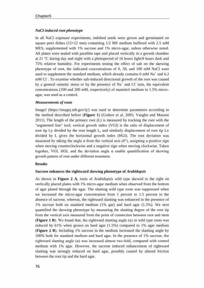
Chapter5
76
NaCl-induced root phenotype
In all NaCl exposure experiments, imbibed seeds were grown and germinated on
square petri dishes (12×12 mm) containing 1/2 MS medium buffered with 2.5 mM
MES, supplemented with 1% sucrose and 1% micro-agar, unless otherwise noted.
All plates were sealed with parafilm tape and placed vertically in a growth chamber
at 21 °C during day and night with a photoperiod of 16 hours light/8 hours dark and
75% relative humidity. For experiments testing the effect of salt on the skewing
phenotype of root, the indicated concentrations of 0, 50, and 100 mM NaCl were
used to supplement the standard medium, which already contains 0 mM Na+ and 6.2
mM Cl -. To examine whether salt-induced directional growth of the root was caused
by a general osmotic stress or by the presence of Na+ and Cl
- ions, the equivalent
concentrations (100 and 200 mM, respectively) of mannitol medium in 1.5% micro-
agar, was used as a control.
Measurements of roots
ImageJ (https://imagej.nih.gov/ij/) was used to determine parameters according to
the method described before (Figure 1) (Grabov et al. 2005; Vaughn and Masson
2011). The length of the primary root (L) is measured by tracking the root with the
"segmented line" tool; vertical growth index (VGI) is the ratio of displacement of
root tip Ly divided by the root length L, and similarly displacement of root tip Lx
divided by L gives the horizontal growth index (HGI). The root deviation was
measured by taking the angle α from the vertical axis (0°), assigning a positive sign
when moving counterclockwise and a negative sign when moving clockwise. Taken
together, VGI, HGI, and the deviation angle α enable quantification of skewing
growth pattern of root under different treatment.
Results
Sucrose enhances the rightward skewing phenotype of Arabidopsis
As shown in Figure 2 A, roots of Arabidopsis wild type skewed to the right on
vertically placed plates with 1% micro-agar medium when observed from the bottom
of agar plated through the agar. The slanting wild type roots was suppressed when
we increased the micro-agar concentration from 1 percent to 1.5 percent in the
absence of sucrose, whereas, the rightward slanting was enhanced in the presence of
1% sucrose both on standard medium (1% gar) and hard agar (1.5%). We next
quantified the skewing phenotype by measuring the slanting degree of the root tip
from the vertical axis measured from the point of connection between root and stem
(Figure 1 B). We found that, the rightward slanting angle (α) in wild type roots was
reduced by 61% when grown on hard agar (1.5%) compared to 1% agar medium
(Figure 2 B). Including 1% sucrose in the medium increased the slanting angle by
100% both for standard medium and hard agar. In the presence of 1% sucrose, the
rightward slanting angle (α) was increased almost two-fold, compared with control
medium with 1% agar. However, the sucrose induced enhancement of rightward
slanting was strongly reduced on hard agar, possibly caused by altered friction
between the root tip and the hard agar.
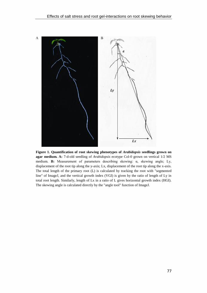
Effects of salt stress and root gel-interactions on root skewing behavior
77
Figure 1. Quantification of root skewing phenotypes of Arabidopsis seedlings grown on
agar medium. A: 7-d-old seedling of Arabidopsis ecotype Col-0 grown on vertical 1/2 MS
medium. B: Measurement of parameters describing skewing: α, skewing angle; Ly,
displacement of the root tip along the y-axis; Lx, displacement of the root tip along the x-axis.
The total length of the primary root (L) is calculated by tracking the root with "segmented
line" of ImageJ, and the vertical growth index (VGI) is given by the ratio of length of Ly in
total root length. Similarly, length of Lx in a ratio of L gives horizontal growth index (HGI).
The skewing angle is calculated directly by the "angle tool" function of ImageJ.
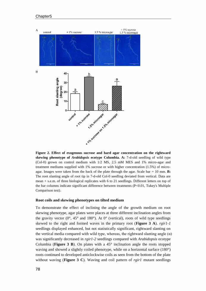
Chapter5
78
Figure 2. Effect of exogenous sucrose and hard agar concentration on the rightward
skewing phenotype of Arabidopsis ecotype Columbia. A: 7-d-old seedling of wild type
(Col-0) grown on control medium with 1/2 MS, 2.5 mM MES and 1% micro-agar and
treatment mediums supplied with 1% sucrose or with higher concentration (1.5%) of micro-
agar. Images were taken from the back of the plate through the agar. Scale bar = 10 mm. B:
The root slanting angle of root tip in 7-d-old Col-0 seedling deviated from vertical. Data are
mean + s.e.m. of three biological replicates with 6 to 21 seedlings. Different letters on top of
the bar columns indicate significant difference between treatments (P<0.01, Tukey's Multiple
Comparison test).
Root coils and skewing phenotypes on tilted medium
To demonstrate the effect of inclining the angle of the growth medium on root
skewing phenotype, agar plates were places at three different inclination angles from
the gravity vector (0°, 45° and 180°). At 0° (vertical), roots of wild type seedlings
skewed to the right and formed waves in the primary root (Figure 3 A). rgir1-1
seedlings displayed enhanced, but not statistically significant, rightward slanting on
the vertical media compared with wild type, whereas, the rightward slanting angle (α)
was significantly decreased in rgir1-2 seedlings compared with Arabidopsis ecotype
Columbia (Figure 3 B). On plates with a 45° inclination angle the roots stopped
waving and showed a slightly coiled phenotype, while on a horizontal surface (180°)
roots continued to developed anticlockwise coils as seen from the bottom of the plate
without waving (Figure 3 C). Waving and coil pattern of rgir1 mutant seedlings
Effects of salt stress and root gel-interactions on root skewing behavior
79
grown on tilted medium were similar as that of wild type (data not shown).
In an attempt to study the effect of light on the directional growth of root, seeds of
wild type and rgir1 mutants were germinated directly on vertical agar plates covered
by a black box. Waving movement of dark-grown seedlings was similar to those of
light-grown seedlings of wild type on the vertical plate in the same growth chamber
(Figure 3 D). However, the rightward slanting was reduced in the darkness and
seedlings developed shorter roots and longer hypocotyls compared with those grown
under 16 hours light/8 hours dark photoperiod, both in wild type and mutant
seedlings (data not shown).
Figure 3. Root growth phenotypes of wild-type (Col-0) and rgir1 mutants seedlings
grown on titled mediums. Wild type (Col-0) and rgir1 mutant seedlings grown 7 d on 1%
agar-solidified 1/2 MS medium placed vertically (A) or tilted at 45° and placed horizental (C)
in the growth chamber with light period of 16 hours light / 8 hours dark. B: The slanting angle
of wild type and rgir1 mutants seedling deviated from vertical y-axis that shown in (A). Data
are mean + s.e.m, n=29. Different letters on top of the bar columns indicate significant
difference between genotypes (P<0.05, Tukey's Multiple Comparison test). D: Wild-type
(Col-0) seedlings grown 9 d on 1% agar-solidified 1/2 MS medium in the same chamber as (A)
and (C), but in the dark. Note: all images were taken from the back of the plate, through the
agar. Scale bar = 1 cm in (A) and (D), and 2 mm in (C).
NaCl induced root growth direction changes independently of root length
change
When grown vertically on a hardagar (1.5% micro-agar) plates without salt, roots of
wild type and rgir1 mutants skewed to the right of the plate (Figure 4 A). In contrast
to the rightward skewing on the control medium, roots grew almost straight and
parallel to the vector of gravity, or even skewed to the left, when the medium was
supplied with 50 mM NaCl in wild type and rgir1 mutants (Figure 4 B). As shown
in Figure 4 C, high salinity (100 mM NaCl) induced a strong right-handed helical
arrangement of epidermal cell files (Figure 4 D), resulting in a more leftward
skewing in wild type and mutants seedlings (Figure 4 C) compared with those on
control medium (Figure 4 A) and 50 mM salt medium (Figure 4 B).
Chapter5
80
Figure 4. Root growth phenotypes of wild type (Col-0) and rgir1 mutant seedlings grown
on medium supplied with salt. Seedlings of wild type and rgir1 mutants were germinated
directly and grown 9 d on 1/2 MS hard medium (1.5% micro-agar) without salt (A) or with 50
mM (B), and 100 mM NaCl (C). Images were taken from back of the plate through the agar.
Scale bar = 1 cm in A to C. D: Root tip phenotype of 9-d-old seedlings in Col-0 grown under
different concentrations of NaCl. Arrows marked the first root hair bugle under 50 mM and
100 mM treatment and the twisting of the epidermal cell files under 100 mM salt treatment.
Scale bar = 200 μm.
As shown by the root slanting angle and the primary root length results (Figure 5
A and B), the length of Col-0, rgir1-1, and rgir1-2 was clearly suppressed in the
presence of high salinity (100 mM) and the root slanting angle was also strongly
exaggerated, but skewed to the opposite direction, when compared with growth
Effects of salt stress and root gel-interactions on root skewing behavior
81
conditions without NaCl. When seedlings were grown on medium containing 50 mM
NaCl, initially skewed root began to grow parallel to the vector of gravity or
leftwards in wild type and mutant seedlings. While rgir1-1 has the shortest primary
root length in control medium and in 50 mM NaCl medium (Figure 5 B), the
slanting angle of rgir1-1 is similar to those in wild type and rgir1-2 (Figure 5 B),
indicating that RGIR1 only affects the elongation of the cells in root tip, but is not
involved in helical growth of the root.
Figure 5. Quantification of root skewing phenotypes of wild type and rgir1 mutant
seedlings under salt treatment. Seedlings of wild type (Col-0) and rgir1 mutants were grown
for 9 days on salt medium without or with 1% sucrose. A-B: Effect of salt and sucrose on the
root slanting angle (A) and main root length (B). Letters at the right side of bars in B indicate
significant difference between genotype at the level of p<0.05 (n=13-16, Tukeys' Multiple
comparison Test). C-D: Dynamics of root vertical growth index (VGI, C) and horizental
growth index (HGI, D) in wild type (Col-0) and rgir1 mutant seedlings under different
treatments. No difference was observed between wild type and mutant seedlings for VGI and
HGI at the same treatment condition.
In the presence of 1% sucrose in the medium, the seedlings show a pronounced
rightward slanting with a concomitant increase in root length, both for wild type and
mutants seedlings. When grown on salt medium, adding sucrose affects root
elongation both at the lower (50 mM) and higher (100 mM) levels of NaCl in wild
type and mutants seedlings. However, the salt-induced leftward skewing is enhanced
Chapter5
82
in the 50 mM medium in the presence of 1% sucrose, whereas it was not affected on
the 100 mM medium.
The vertical growth index (VGI) and horizontal growth index (HGI) analysis is a
versatile method for quantifying the deviation of root tip from straight downward
vertical growth (Grabov et al. 2004). We next measured VGI and HGI to study the
dynamics of root development in different medias. Although the root length and root
slanting angles of Col-0 and rgir1 mutant seedlings were different under different
treatments (with or without sucrose; different concentrations of NaCl) we did not
detect a significant difference in HGI and VGI of those seedlings when treated
identically (Figure 5 C and D), which support the idea that VGI and HGI are
independent parameters of root development processes (Grabov et al. 2004). When
we only focus on the treatment effects, VGI in wild type and mutant seedlings was
lower in 1% sucrose, 100 mM NaCl or 100 mM NaCl with 1% sucrose, compared
with those grown on control medium (Figure 5 D). However, an increased was
observed in HGI for those seedlings with lower VGI, indicating the roots tend to
deviate to one side (left or right) from the vertical by positive HGI.
Compared with seedling treated with 100 mM NaCl, only 36% seedling skewed to
the left when treated with 50 mM NaCl in wild type seedling, while that is 33% and
50% in rgir1-1 and rgir1-2 seedlings, respectively. In the presence of 1% sucrose in
the 50 mM salt medium, the leftward skewing in wild type seedlings was increased
to 85%, while it was 79% for rgir1-1 and rgir1-2 mutant seedlings. However, the
VGI and HGI were not statistically different between wild type and mutants under
treatment with 50 mM NaCl. As shown in Figure 5 C and D, the VGI in wild type
and rgir1 mutant seedlings on the 50 mM salt medium was not affected by adding
sucrose, but HGI was significantly increased by including 1% sucrose in the medium.
Thus, the sucrose-induced enhancement of root skewing is mainly caused by an
increased growth rate and and not by an change in the skewing angle of root growth.
Salt-induced root growth direction changes are not merely due to osmotic stress
To determine which aspect of salt stress, osmolarity or ionic toxicity, affects the
skewing phenotype, we monitored root growth of seedlings on medium containing
osmotically equivalent concentrations of mannitol and of NaCl. When seedlings
were grown in the presence of NaCl at different concentrations, the initial rightward
slanting angle first decreased to 0° and then changed further into a leftward slant in a
dose dependent manner, with a concomitant decreased of root length, both in wild
type and mutants seedlings (Figure 6 A and C). In contrast, neither the main root
length nor the skewing angle was affected by osmotically equivalent concentrations
of mannitol up to 200 mM (Figure 6 B and D). The HGI and VGI were not affected
by mannitor in wild type or rgir1 mutant seedlings (Figure 6 F and H), whereas,
HGI of all genotypes were affected by NaCl (Figure 6 E). As expected, VGI of roots
in all genotypes were slightly affected by 50 mM NaCl in the medium, but decreased
strongly in 100 mM NaCl. The clear difference in effect of NaCl and mannitol
indicates that the suppression of root length and salt-induced changes in the growth
direction of the root is due to ionic toxicity of Na+ or Cl
-, but not to osmotic stress in
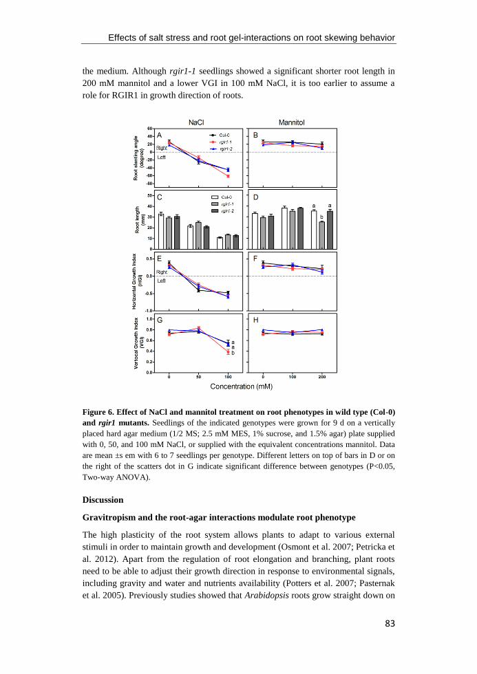
Effects of salt stress and root gel-interactions on root skewing behavior
83
the medium. Although rgir1-1 seedlings showed a significant shorter root length in
200 mM mannitol and a lower VGI in 100 mM NaCl, it is too earlier to assume a
role for RGIR1 in growth direction of roots.
Figure 6. Effect of NaCl and mannitol treatment on root phenotypes in wild type (Col-0)
and rgir1 mutants. Seedlings of the indicated genotypes were grown for 9 d on a vertically
placed hard agar medium (1/2 MS; 2.5 mM MES, 1% sucrose, and 1.5% agar) plate supplied
with 0, 50, and 100 mM NaCl, or supplied with the equivalent concentrations mannitol. Data
are mean ±s em with 6 to 7 seedlings per genotype. Different letters on top of bars in D or on
the right of the scatters dot in G indicate significant difference between genotypes (P<0.05,
Two-way ANOVA).
Discussion
Gravitropism and the root-agar interactions modulate root phenotype
The high plasticity of the root system allows plants to adapt to various external
stimuli in order to maintain growth and development (Osmont et al. 2007; Petricka et
al. 2012). Apart from the regulation of root elongation and branching, plant roots
need to be able to adjust their growth direction in response to environmental signals,
including gravity and water and nutrients availability (Potters et al. 2007; Pasternak
et al. 2005). Previously studies showed that Arabidopsis roots grow straight down on
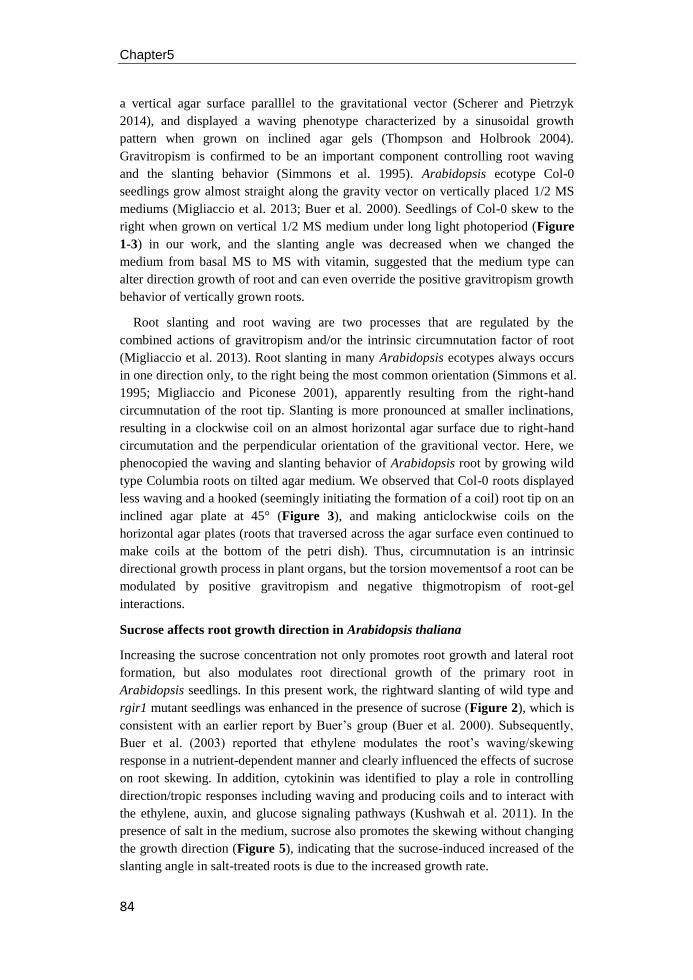
Chapter5
84
a vertical agar surface paralllel to the gravitational vector (Scherer and Pietrzyk
2014), and displayed a waving phenotype characterized by a sinusoidal growth
pattern when grown on inclined agar gels (Thompson and Holbrook 2004).
Gravitropism is confirmed to be an important component controlling root waving
and the slanting behavior (Simmons et al. 1995). Arabidopsis ecotype Col-0
seedlings grow almost straight along the gravity vector on vertically placed 1/2 MS
mediums (Migliaccio et al. 2013; Buer et al. 2000). Seedlings of Col-0 skew to the
right when grown on vertical 1/2 MS medium under long light photoperiod (Figure
1-3) in our work, and the slanting angle was decreased when we changed the
medium from basal MS to MS with vitamin, suggested that the medium type can
alter direction growth of root and can even override the positive gravitropism growth
behavior of vertically grown roots.
Root slanting and root waving are two processes that are regulated by the
combined actions of gravitropism and/or the intrinsic circumnutation factor of root
(Migliaccio et al. 2013). Root slanting in many Arabidopsis ecotypes always occurs
in one direction only, to the right being the most common orientation (Simmons et al.
1995; Migliaccio and Piconese 2001), apparently resulting from the right-hand
circumnutation of the root tip. Slanting is more pronounced at smaller inclinations,
resulting in a clockwise coil on an almost horizontal agar surface due to right-hand
circumutation and the perpendicular orientation of the gravitional vector. Here, we
phenocopied the waving and slanting behavior of Arabidopsis root by growing wild
type Columbia roots on tilted agar medium. We observed that Col-0 roots displayed
less waving and a hooked (seemingly initiating the formation of a coil) root tip on an
inclined agar plate at 45° (Figure 3), and making anticlockwise coils on the
horizontal agar plates (roots that traversed across the agar surface even continued to
make coils at the bottom of the petri dish). Thus, circumnutation is an intrinsic
directional growth process in plant organs, but the torsion movementsof a root can be
modulated by positive gravitropism and negative thigmotropism of root-gel
interactions.
Sucrose affects root growth direction in Arabidopsis thaliana
Increasing the sucrose concentration not only promotes root growth and lateral root
formation, but also modulates root directional growth of the primary root in
Arabidopsis seedlings. In this present work, the rightward slanting of wild type and
rgir1 mutant seedlings was enhanced in the presence of sucrose (Figure 2), which is
consistent with an earlier report by Buer’s group (Buer et al. 2000). Subsequently,
Buer et al. (2003) reported that ethylene modulates the root’s waving/skewing
response in a nutrient-dependent manner and clearly influenced the effects of sucrose
on root skewing. In addition, cytokinin was identified to play a role in controlling
direction/tropic responses including waving and producing coils and to interact with
the ethylene, auxin, and glucose signaling pathways (Kushwah et al. 2011). In the
presence of salt in the medium, sucrose also promotes the skewing without changing
the growth direction (Figure 5), indicating that the sucrose-induced increased of the
slanting angle in salt-treated roots is due to the increased growth rate.
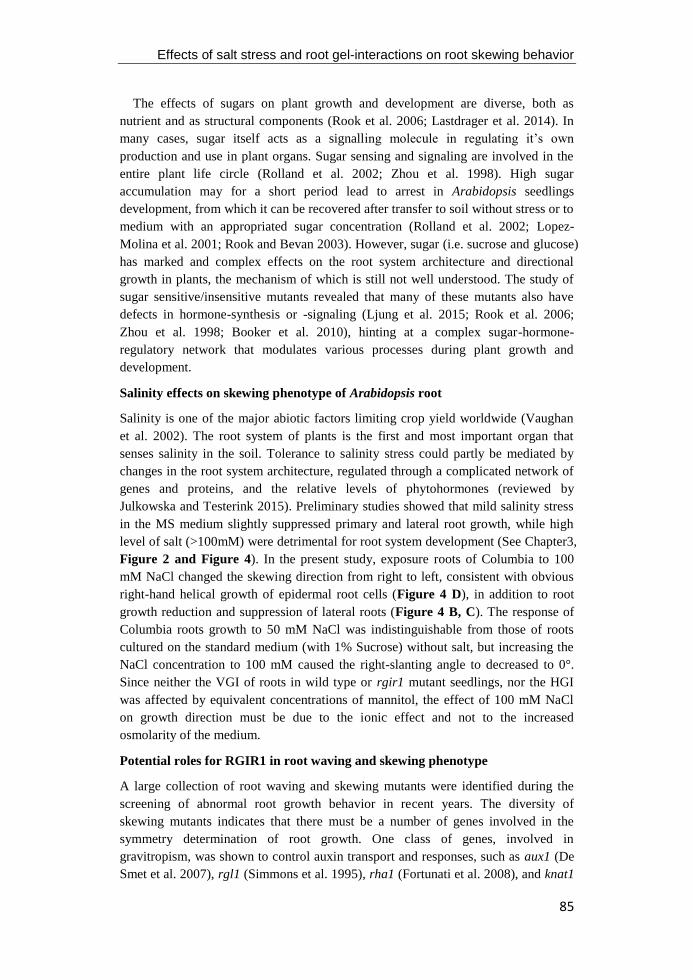
Effects of salt stress and root gel-interactions on root skewing behavior
85
The effects of sugars on plant growth and development are diverse, both as
nutrient and as structural components (Rook et al. 2006; Lastdrager et al. 2014). In
many cases, sugar itself acts as a signalling molecule in regulating it’s own
production and use in plant organs. Sugar sensing and signaling are involved in the
entire plant life circle (Rolland et al. 2002; Zhou et al. 1998). High sugar
accumulation may for a short period lead to arrest in Arabidopsis seedlings
development, from which it can be recovered after transfer to soil without stress or to
medium with an appropriated sugar concentration (Rolland et al. 2002; Lopez-
Molina et al. 2001; Rook and Bevan 2003). However, sugar (i.e. sucrose and glucose)
has marked and complex effects on the root system architecture and directional
growth in plants, the mechanism of which is still not well understood. The study of
sugar sensitive/insensitive mutants revealed that many of these mutants also have
defects in hormone-synthesis or -signaling (Ljung et al. 2015; Rook et al. 2006;
Zhou et al. 1998; Booker et al. 2010), hinting at a complex sugar-hormone-
regulatory network that modulates various processes during plant growth and
development.
Salinity effects on skewing phenotype of Arabidopsis root
Salinity is one of the major abiotic factors limiting crop yield worldwide (Vaughan
et al. 2002). The root system of plants is the first and most important organ that
senses salinity in the soil. Tolerance to salinity stress could partly be mediated by
changes in the root system architecture, regulated through a complicated network of
genes and proteins, and the relative levels of phytohormones (reviewed by
Julkowska and Testerink 2015). Preliminary studies showed that mild salinity stress
in the MS medium slightly suppressed primary and lateral root growth, while high
level of salt (>100mM) were detrimental for root system development (See Chapter3,
Figure 2 and Figure 4). In the present study, exposure roots of Columbia to 100
mM NaCl changed the skewing direction from right to left, consistent with obvious
right-hand helical growth of epidermal root cells (Figure 4 D), in addition to root
growth reduction and suppression of lateral roots (Figure 4 B, C). The response of
Columbia roots growth to 50 mM NaCl was indistinguishable from those of roots
cultured on the standard medium (with 1% Sucrose) without salt, but increasing the
NaCl concentration to 100 mM caused the right-slanting angle to decreased to 0°.
Since neither the VGI of roots in wild type or rgir1 mutant seedlings, nor the HGI
was affected by equivalent concentrations of mannitol, the effect of 100 mM NaCl
on growth direction must be due to the ionic effect and not to the increased
osmolarity of the medium.
Potential roles for RGIR1 in root waving and skewing phenotype
A large collection of root waving and skewing mutants were identified during the
screening of abnormal root growth behavior in recent years. The diversity of
skewing mutants indicates that there must be a number of genes involved in the
symmetry determination of root growth. One class of genes, involved in
gravitropism, was shown to control auxin transport and responses, such as aux1 (De
Smet et al. 2007), rgl1 (Simmons et al. 1995), rha1 (Fortunati et al. 2008), and knat1
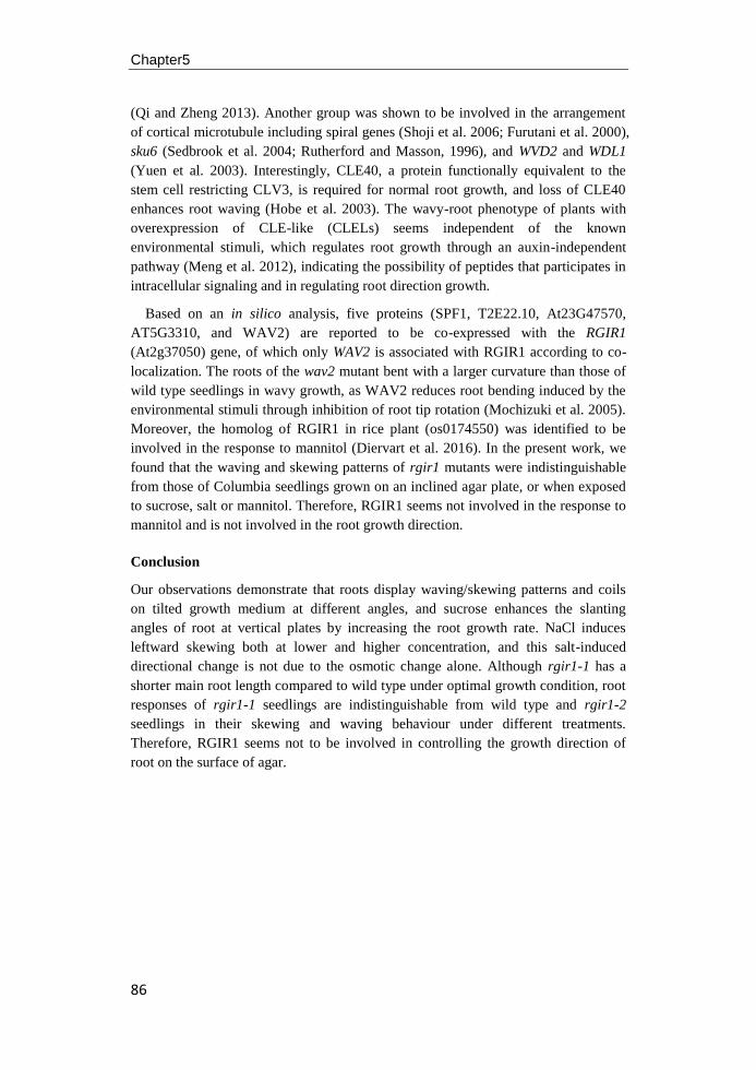
Chapter5
86
(Qi and Zheng 2013). Another group was shown to be involved in the arrangement
of cortical microtubule including spiral genes (Shoji et al. 2006; Furutani et al. 2000),
sku6 (Sedbrook et al. 2004; Rutherford and Masson, 1996), and WVD2 and WDL1
(Yuen et al. 2003). Interestingly, CLE40, a protein functionally equivalent to the
stem cell restricting CLV3, is required for normal root growth, and loss of CLE40
enhances root waving (Hobe et al. 2003). The wavy-root phenotype of plants with
overexpression of CLE-like (CLELs) seems independent of the known
environmental stimuli, which regulates root growth through an auxin-independent
pathway (Meng et al. 2012), indicating the possibility of peptides that participates in
intracellular signaling and in regulating root direction growth.
Based on an in silico analysis, five proteins (SPF1, T2E22.10, At23G47570,
AT5G3310, and WAV2) are reported to be co-expressed with the RGIR1
(At2g37050) gene, of which only WAV2 is associated with RGIR1 according to co-
localization. The roots of the wav2 mutant bent with a larger curvature than those of
wild type seedlings in wavy growth, as WAV2 reduces root bending induced by the
environmental stimuli through inhibition of root tip rotation (Mochizuki et al. 2005).
Moreover, the homolog of RGIR1 in rice plant (os0174550) was identified to be
involved in the response to mannitol (Diervart et al. 2016). In the present work, we
found that the waving and skewing patterns of rgir1 mutants were indistinguishable
from those of Columbia seedlings grown on an inclined agar plate, or when exposed
to sucrose, salt or mannitol. Therefore, RGIR1 seems not involved in the response to
mannitol and is not involved in the root growth direction.
Conclusion
Our observations demonstrate that roots display waving/skewing patterns and coils
on tilted growth medium at different angles, and sucrose enhances the slanting
angles of root at vertical plates by increasing the root growth rate. NaCl induces
leftward skewing both at lower and higher concentration, and this salt-induced
directional change is not due to the osmotic change alone. Although rgir1-1 has a
shorter main root length compared to wild type under optimal growth condition, root
responses of rgir1-1 seedlings are indistinguishable from wild type and rgir1-2
seedlings in their skewing and waving behaviour under different treatments.
Therefore, RGIR1 seems not to be involved in controlling the growth direction of
root on the surface of agar.

87
Chapter6
General discussion

Chapter6
88
Role of RGIR1 in plant growth and development
Reverse genetics procedures are now well-established methods to identify the
function of a gene. Analyzing the phenotypic characteristics caused by the mutation
of a particular gene with inserted elements, such as T-DNA of Agrobacterium or a
transposon (Bouché and Bouchez 2001; Feldmann 1991; Krysan et al. 1999) often
give strong indications of what the specific role the gene plays. The result of the
insertion of a T-DNA element in or near an Arabidopsis’ gene depends on the place
of insertion: the promoter, a coding region or a 3’ un-translated region. In some cases,
even a knockout mutant has no readily identifiable phenotype or displays a
distinguishable phenotype compared to its wild type ecotype under the same growth
conditions. The availability of large numbers of Arabidopsis T-DNA insertion lines
has facilitated the discovery of functions of newly identified genes or proteins.
Several steps are needed to characterize the phenotypic consequences of a particular
T-DNA induced mutation. The lack of alteration of the phenotype could be caused
by functional redundancy among members of a gene family and some other
mutations have phenotypes that are conditional and can only be observed under
specific physiological conditions.
Receptor-like kinases (RLKs) have emerged as a major component in the
intercellular signaling processes within Arabidopsis root development (Wierzba and
Tax 2013). Despite the large gene family of RLKs in the Arabidopsis genome, only a
few of the total 610+ RLKs and RLPs have clear, identified, functions in mediating
cell signaling during various stages of root development. Among these published
RLKs that have a clear function in Arabidopsis root development, LRR-receptor
kinases BRI1, BAK1, BRL1 and BRL3 reduce root length and root meristem size in
a BR-dependent pathway (Caño-Delgado et al. 2004; Hacham et al. 2011; González-
García et al. 2011). Besides interacting with BRI1 to control root growth via a BR-
dependent pathway, SERKs were identified to interact with another unknown RLK,
controlling root growth by regulating common target genes needed for root
development (Du et al. 2012). In addition to BR, the transmembrane kinase TMK
subfamily of RLKs show a reduced sensitivity to auxin and orchestrate plant growth
by regulating cell expansion and cell division, and some of its members could also
play a role in the auxin-mediated control of lateral roots development (Dai et al.
2013; Chang et al. 1992).
The Root-growth-inhibition-receptor 1 (RGIR1) belongs to the LRR I subfamily,
with three conserved LRRs in the extracellular domain between the Malectin domain
and the single transmembrane domain (Chapter 2). In the detailed root phenotypic
analysis of two homozygous RGIR1 alleles (rgir1-1 and rgir1-2), the rgir1-2 mutant,
which is caused by the invalid insertion position close to 3’-untranslated region, did
not exhibit visible changes under standard culture conditions when compared with
it's wild type ecotype Col-0. However, the knock-out mutant allele rgir1-1 has a
shorter main root and less lateral roots when grown on agar medium under optimal
growth condition (Chapter 2). Moreover, the transcripts of RGIR1 showed strong
tissues specificity with higher expression in the root and lower in the rosette in the
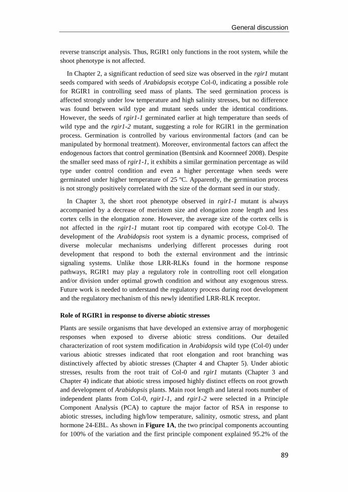
General discussion
89
reverse transcript analysis. Thus, RGIR1 only functions in the root system, while the
shoot phenotype is not affected.
In Chapter 2, a significant reduction of seed size was observed in the rgir1 mutant
seeds compared with seeds of Arabidopsis ecotype Col-0, indicating a possible role
for RGIR1 in controlling seed mass of plants. The seed germination process is
affected strongly under low temperature and high salinity stresses, but no difference
was found between wild type and mutant seeds under the identical conditions.
However, the seeds of rgir1-1 germinated earlier at high temperature than seeds of
wild type and the rgir1-2 mutant, suggesting a role for RGIR1 in the germination
process. Germination is controlled by various environmental factors (and can be
manipulated by hormonal treatment). Moreover, environmental factors can affect the
endogenous factors that control germination (Bentsink and Koornneef 2008). Despite
the smaller seed mass of rgir1-1, it exhibits a similar germination percentage as wild
type under control condition and even a higher percentage when seeds were
germinated under higher temperature of 25 ºC. Apparently, the germination process
is not strongly positively correlated with the size of the dormant seed in our study.
In Chapter 3, the short root phenotype observed in rgir1-1 mutant is always
accompanied by a decrease of meristem size and elongation zone length and less
cortex cells in the elongation zone. However, the average size of the cortex cells is
not affected in the rgir1-1 mutant root tip compared with ecotype Col-0. The
development of the Arabidopsis root system is a dynamic process, comprised of
diverse molecular mechanisms underlying different processes during root
development that respond to both the external environment and the intrinsic
signaling systems. Unlike those LRR-RLKs found in the hormone response
pathways, RGIR1 may play a regulatory role in controlling root cell elongation
and/or division under optimal growth condition and without any exogenous stress.
Future work is needed to understand the regulatory process during root development
and the regulatory mechanism of this newly identified LRR-RLK receptor.
Role of RGIR1 in response to diverse abiotic stresses
Plants are sessile organisms that have developed an extensive array of morphogenic
responses when exposed to diverse abiotic stress conditions. Our detailed
characterization of root system modification in Arabidopsis wild type (Col-0) under
various abiotic stresses indicated that root elongation and root branching was
distinctively affected by abiotic stresses (Chapter 4 and Chapter 5). Under abiotic
stresses, results from the root trait of Col-0 and rgir1 mutants (Chapter 3 and
Chapter 4) indicate that abiotic stress imposed highly distinct effects on root growth
and development of Arabidopsis plants. Main root length and lateral roots number of
independent plants from Col-0, rgir1-1, and rgir1-2 were selected in a Principle
Component Analysis (PCA) to capture the major factor of RSA in response to
abiotic stresses, including high/low temperature, salinity, osmotic stress, and plant
hormone 24-EBL. As shown in Figure 1A, the two principal components accounting
for 100% of the variation and the first principle component explained 95.2% of the
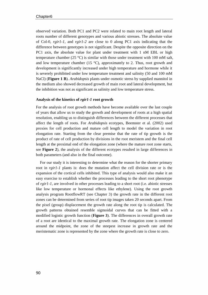
Chapter6
90
observed variation. Both PC1 and PC2 were related to main root length and lateral
roots number of different genotypes and various abiotic stresses. The absolute value
of Col-0, rgir1-1, and rgir1-2 are close to 0 along PC1 axis indicating that the
difference between genotypes is not significant. Despite the opposite direction on the
PC1 axis, the absolute value for plant under treatment with 1 nM EBL or high
temperature chamber (25 °C) is similar with those under treatment with 100 mM salt,
and low temperature chamber (15 °C), approximately to 2. Thus, root growth and
development is significantly increased under high temperature and hormone while it
is severely prohibited under low temperature treatment and salinity (50 and 100 mM
NaCl) (Figure 1 B). Arabidopsis plants under osmotic stress by supplied mannitol in
the medium also showed decreased growth of main root and lateral development, but
the inhibition was not as significant as salinity and low temperature stress.
Analysis of the kinetics of rgir1-1 root growth
For the analysis of root growth methods have become available over the last couple
of years that allow us to study the growth and development of roots at a high spatial
resolution, enabling us to distinguish differences between the different processes that
affect the length of roots. For Arabidopsis ecotypes, Beemster et al. (2002) used
proxies for cell production and mature cell length to model the variation in root
elongation rate. Starting from the clear premise that the rate of tip growth is the
product of rate of cell production by divisions in the root meristem and the final cell
length at the proximal end of the elongation zone (where the mature root zone starts,
see Figure 2), the analysis of the different ecotypes resulted in large differences in
both parameters (and also in the final outcome).
For our study it is interesting to determine what the reason for the shorter primary
root in rgir1-1 plants is: does the mutation affect the cell division rate or is the
expansion of the cortical cells inhibited. This type of analysis would also make it an
easy exercise to establish whether the processes leading to the short root phenotype
of rgir1-1, are involved in other processes leading to a short root (i.e. abiotic stresses
like low temperature or hormonal effects like ethylene). Using the root growth
analysis program RootflowRT (see Chapter 3) the growth rate in the different root
zones can be determined from series of root tip images taken 20 seconds apart. From
the pixel (group) displacement the growth rate along the root tip is calculated. The
growth patterns obtained resemble sigmoidal curves that can be fitted with a
modified logistic growth function (Figure 3). The differences in overall growth rate
of a root are identical to the maximal growth rate. The elongation zone is centered
around the midpoint, the zone of the steepest increase in growth rate and the
meristematic zone is represented by the zone where the growth rate is close to zero.
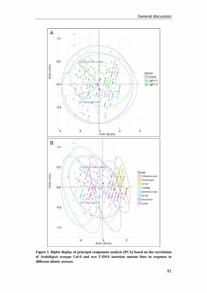
General discussion
91
Figure 1. Biplot display of principal component analysis (PCA) based on the correlation
of Arabidopsis ecotype Col-0 and two T-DNA insertion mutant lines in response to
different abiotic stresses.
Chapter6
92
Figure 2. Schematic overview of the different zones in the root tip where cell division and
cell expansion are mainly localized. The cortical cell files are derived from initials close to
the quiescent center. Cell division (and limited cell elongation) continues in the meristem zone
providing an ongoing "influx" of cells into the elongation zone, where cells no longer divide
and most of the cell elongation takes place. Proximal of the elongation zone the cells no longer
grow, the first root hairs are formed and the cell size has reached their maximal length (after
Beemster et al. 2002).
Figure 3. Family of theoretical curves for the growth rate along the root tip of roots with
normal and reduced growth to 70%. A: Curves of the overall growth relative to the
quiescent center (x=0) of normal growth (solid line), inhibited growth due to low expansion
rate in elongation zone (dotted line), inhibited growth due to shortening of the elongation zone
(dash-dotted line) and inhibited growth due to limited production of cells in the meristematic
zone. B: Curves representing the first derivative of the growth rate curves shown in A.
In Figure 3 the three basic processes that can lead to a reduced overall growth rate
in root tips are illustrated. The first process is a reduction in the number of cells that
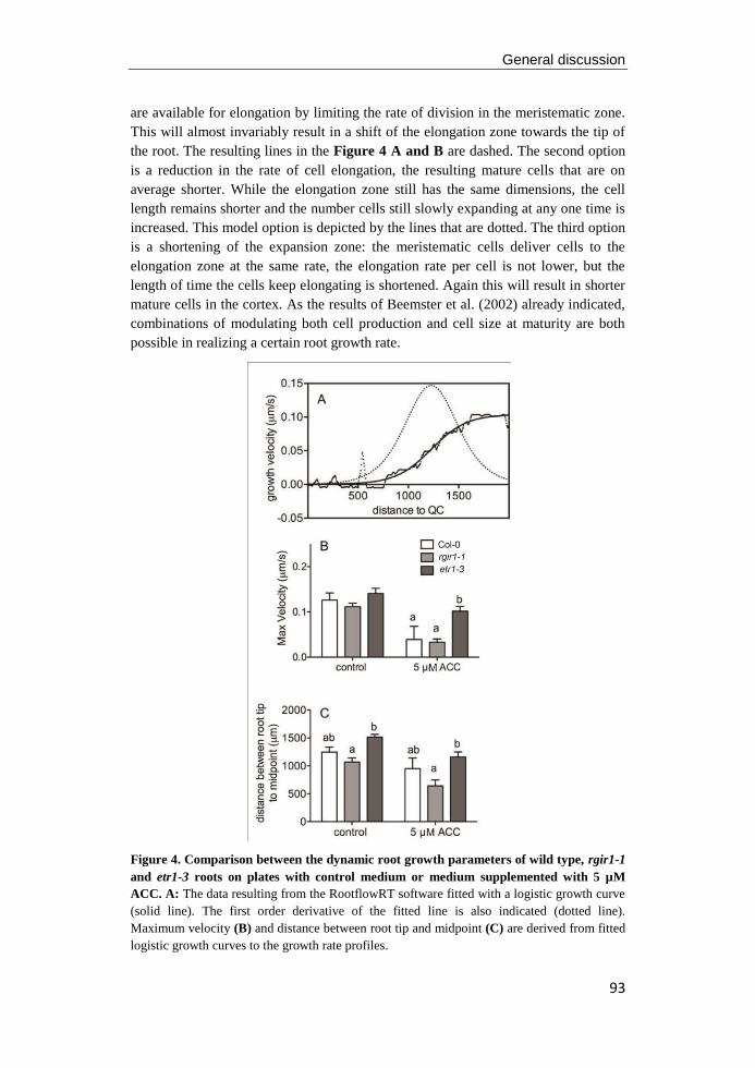
General discussion
93
are available for elongation by limiting the rate of division in the meristematic zone.
This will almost invariably result in a shift of the elongation zone towards the tip of
the root. The resulting lines in the Figure 4 A and B are dashed. The second option
is a reduction in the rate of cell elongation, the resulting mature cells that are on
average shorter. While the elongation zone still has the same dimensions, the cell
length remains shorter and the number cells still slowly expanding at any one time is
increased. This model option is depicted by the lines that are dotted. The third option
is a shortening of the expansion zone: the meristematic cells deliver cells to the
elongation zone at the same rate, the elongation rate per cell is not lower, but the
length of time the cells keep elongating is shortened. Again this will result in shorter
mature cells in the cortex. As the results of Beemster et al. (2002) already indicated,
combinations of modulating both cell production and cell size at maturity are both
possible in realizing a certain root growth rate.
Figure 4. Comparison between the dynamic root growth parameters of wild type, rgir1-1
and etr1-3 roots on plates with control medium or medium supplemented with 5 µM
ACC. A: The data resulting from the RootflowRT software fitted with a logistic growth curve
(solid line). The first order derivative of the fitted line is also indicated (dotted line).
Maximum velocity (B) and distance between root tip and midpoint (C) are derived from fitted
logistic growth curves to the growth rate profiles.

Chapter6
94
If sufficiently high resolution data from the dynamic analysis of root growth
profiles along the root tip could be obtained, distinguishing between the mechanism
of root growth inhibition would indeed be possible. In Figure 4 the results of a
comparison between short roots phenotypes of rgir1-1 and ACC-treated roots are
shown. From the comparison between the genotypes wild type, rgir1-1 and etr1-3 (a
mutant with a reduced sensitivity to ethylene) the slightly lower growth rate or rgir1-
1 and the slightly higher rate of etr1-3 are noted. In both short phenotype situations
(rgir1-1 and exposure to ACC, a precursor of ethylene) it is clear that the lower
growth rate correlates with a shift of the midpoint of the elongation zone towards the
root tip. The only two model options that are compatible with this shift of the
elongation zone are reduced cell production (reduced division rate in the
meristematic zone) and the shortening of the elongations zone. When the length of
the mature cells would be available for this experiment further distinction between
these two models would be possible. From the ANOVA that was performed on these
data it became clear that although the root length reduction in rgir1-1 resembles that
of ACC exposure, there was no statistically significant interaction between rgir1-1
and ACC effects, which indicates that both mechanisms probably act independently.
Root waving and skewing behavior of Arabidopsis
Arabidopsis ecotype Columbia root exhibit a sinusoidal waving pattern and skew to
one side of the plate when grown on inclined agar medium (Chapter 5). Since the
discovery of waving and skewing root growth patterns, different models have been
proposed to explain this surface-dependent root growth behavior (Roy and Bassham
2014). One widely accepted model explains waving and skewing as a result of
intrinsic circummnutation, positive gravitropism and negative thigmotropism, while
another interesting model considers the formation of these movements due to the
physical interaction of the root tip with the medium. Other factors that have been
implicated in regulating root movement on the surface of medium, including
hormones and environmental cues (e.g. light, humidity, and nutrients in the medium).
However, a unifying, generally accepted model for understanding the mechanism of
waving and skewing movements is still lacking.
In the last decade, the isolation of wavy and skewing mutants has been used to
discover genes implicated in skewing and waving behavior of the root (Oliva and
Dunand 2007). Mutants with aberrant skewing phenotypes also have defects in the
root cytoskeleton and cell wall modifications, indicating that function of genes
affecting skewing are mainly involved in the re-arrangement of cytoskeleton and cell
walls. The alteration of root skewing direction under high salinity and the enhanced
skewing angle in the presence of sucrose (Chapter 5) suggests the processes that lead
to the normal skewing response, based on the structure of the cytoskeletal elements,
can interact with other signaling pathways and create flexibility in the root growth
response to multiple environmental signals. The existence of mutations affecting
skewing, but not waving, indicating that these two root growth behaviors are
regulated by different processes. Compared with skewing, the process affecting
waving is more complex and more difficult to explain since a range of different
General discussion
95
factors is involved (Buer et al. 2003). Most discovered wavy mutants are defective in
root gravitropism and show altered waving dynamics, such as amplitude and
wavelength of waves, compared with wild type. Some mutants were identified
involved in mediating the influx and efflux of the plant hormone auxin, which plays
a fundamental role in the root gravitropic response. As lateral root initiation and the
gravitropic response are both affected by the redistribution and transportation of
auxin between different zones of the root tip, and lateral roots also space along the
primary root in a regular left-right pattern that correlates with gravitropic response-
mediated waves, this suggests that there is crosstalk between gravitropism, waving
and lateral root formation.
The roots of the wav2 mutant show wavy root growth with exaggerated curves
compared to wild type Arabidopsis. WAV2 is probably a negative regulator root
bending by inhibiting root tip rotation (Mochizuki et al. 2005). According to an in
silico proteomic data analysis, RGIR1 is co-expressed with SPF1, T2E22.10,
At3g47570, At5g43310 and associated with WAV2 based on co-localization,
indicating a possible role for RGIR1 in controlling the waving and skewing
phenotype of roots. However, the waving pattern of rgir1 mutant was
indistinguishable from that of Columbia seedlings grown on vertical medium plates
or on the inclined plates either at 45° or 180° (Chapter 5). Therefore, we conclude
that RGIR1 is involved in the regulation of root elongation, but that it has no effect
on root directional growth on vertical agar medium.

96

97
Chapter7
Summary
Chapter7
98
Receptor-like kinases (RLKs) have emerged as major components in intercellular
signaling during plant growth and development. Based on the similarity of the kinase
domain sequences, the RLK family is comprised of more than 610 members, but for
only a fraction we currently do know their function(s) during plant growth or in
response to various abiotic and biotic stresses (Chapter1). In a previously study, one
knock-out mutant of Arabidopsis gene At2g37050, here named ROOT GROWTH
INHIBITION RECEPTOR1 (RGIR1), displayed a distinctly shorter primary root and
less lateral roots. The RGIR1 gene encodes a protein of 934 amino acids with a
predicted molecular mass of 103.4 kD, which belongs to the LRR-I transmembrane
receptor-like protein kinase family, with three conserved LRRs in the extracellular
domain. Since most work published for RGIR1 was mainly focused on comparison
of transcriptome analyses under particular stress conditions, the role of RGIR1 in
plant growth and development was still lacking.
In Chapter2 seed germination of two T-DNA insertion mutants (rgir1-1 and rgir1-
2) under cold and salt stress was studied and a detailed screen for alteration of their
root system architecture under optimal growth condition was performed. Seed
germination was strongly affected by low temperature and salinity treatment both for
wild type and mutants. Whereas mutant seeds have a smaller seed size compared
with wild type, no evidence was found for a direct link between RGIR1 with control
of seed size and seed germination. Seedlings of rgir1-1 mutants showed a shorter
main root length and smaller root surface area on agar plates, while the leaf
phenotype was not affected on agar or in soil at optimal temperature, indicating that
RGIR1 only has a role in root development.
In Chapter3 root morphology, to quantify cell number and size, and kinematic
parameters of root elongation, to establish the location, size and activity of the
elongation zone, in wild type and rgri1-1 root tips were determined under optimal
condition and when exposed to cold or salinity stress. In the presence of salt or cold
stress, root growth and development were strongly affected with shorter main root
length and less lateral roots in Col-0 and rgir1-1mutant. The shorter root phenotype
of rgir1-1 seedlings is associated with a lower elongation rate and decreased cortex
cell number in the transition zone and elongation zone of the root tip under optimal
growth condition. Differences in main root and lateral roots between rgir1-1 and
wild type disappeared when exposed to cold and salinity, but were more pronounced
at high temperature, indicating that RGIR1 is a positive regulator in the process of
root growth and development under optimal growth condition. The pathways for
RGIR1 controlling root elongation seems to be independent of those involved in the
responses to cold and salinity.
In Chapter4 effects of culturing conditions on root growth patterns in Col-0 and
rgir1 mutants were studied. Sucrose (1.5%) induces a waving and skewing
phenotype on hard (1.5% micro-agar) medium, and strongly reduced lateral roots
formation and an increase length of the main root in both Col-0 wild type and rgir1
mutants. Main root growth and root branching in wild type and mutants were
inhibited under salt or osmotic stress, and at low pH. Sulfur-deficiency didn't affect
Summary
99
main root growth of Col-0 seedlings but lateral roots formation was strongly
stimulated compared with those grown on sulfur-sufficient medium. More lateral
roots developed in rgir1-1 roots than on Col-0 roots, grown on the same sulfur-
deficiency medium, indicating a possible role for RGIR1 in the process of lateral
root initiation or emergency.
In Chapter 5 we studied the effects of salt stress and root-agar interaction on root
skewing behavior in Arabidopsis thaliana. Root of Col-0 displayed rightward
slanting on vertically placed agar medium, and this slanted phenotype was enhanced
in the presence of 1% sucrose. Anti-clockwise root coils and hooked root tips were
identified in Col-0 seedlings when the agar plates were placed horizontally or
inclined at an angle of 45°, respectively. High salinity altered root skewing direction,
combined with severe suppression of main root elongation and lateral roots
formation in Col-0. These responses were observed when exposed to NaCl, but not
under osmotic stress. Roots of rgir1-1 seedlings responded the same as wild type,
both on inclined agar medium and on vertical medium with high salt or mannitol,
indicating that RGIR1 has a role controlling root elongation, but is not involved in
the directional growth of the root tip.
In summary, it can be concluded (Chapter 6) that RGIR1 is an LRR-I receptor-like
kinase which does have a function in the root system architecture of Arabidopsis.
However, changes in root morphology induced by high or low temperature or the
hormone EBL, or exposure to salinity, are not affected by mutating the RGIR1 gene.
The dynamic analysis of the root growth profile along the root tip in rgir1-1 and
etr1-3 (a mutant also with shorter main root length and reduced sensitivity to ACC),
does not show a statistically significant interaction between rgir1-1 and ACC
treatment, thus, both mechanisms controlling the reduction of root length probably
act independently. Although RGIR1 is associated with WAV2, which shows wavy
root growth and exaggerated curves compared to Arabidopsis wild type, no evidence
was found for RGIR1 in controlling the directional growth of root in this thesis.

100

101
Chapter8
Samenvatting
Chapter8
102
Receptor-like kinases (RLK's) zijn belangrijke componenten in de intercellulaire
signaaloverdrachtin zowel dieren als planten en spelen een belangrijke rol in groei en
ontwikkeling. Op basis van overeenkomsten in de de kinase domein sequenties
onderscheiden we 610 eiwitten die tot de de RLK familie behoren.Van slechts een
klein deel van deze 610 RLK’s weten we de precieze functie tijdens de groei van
planten of in hun reactie op verschillende abiotische / biotische stress
omstandigheden (hoofdstuk 1). In een eerder onderzoek werd een
mutantgeïdentificeerd (At2g37050, in dit proefschrift ROOT GROWTH
INHIBITION RECEPTOR1 (RGIR1) genoemd), die een duidelijke kortere primaire
wortel had en ook minder zijwortels vertoonde.Het RGIR1 gen codeert voor een
eiwit van 934 aminozuren met een voorspelde moleculaire massa van 103,4 kD, dat
behoort tot de LRR-I transmembrane receptor-like kinase familie, met drie
geconserveerde leucine-rich repeats (LRRs) in het extracellulaire domein. Aangezien
het meeste dat al bekend was over RGIR1 voornamelijk was bebaseerd op
vergelijking van genexpressieonder verschillendestressomstandigheden, ontbrak een
duidelijke rol voor het RGIR1 gen.
In hoofdstuk 2 worden zaadkiemingsexperimentan van twee T-DNA
insertiemutanten van RGIR1 (rgir1-1 en rgir1-2) onder lage temperatuur en bij
blootstelling aan een hoge zoutconcentratie beschreven. Tevens werd een
gedetailleerd analyse gemaakt van de verandering in de structuur van wortelstelsel
van RGIR1 mutanten ten opzichte van wildtype planten.De zaadkieming werd sterk
beïnvloed door een lage temperatuur en door een hoog zoutgehalte, zowel voor
wildtype als mutanten. Terwijl de zaden van mutantenwel kleiner zijndan die van
het wildtype, werd geen bewijs gevonden dat RGIR1 een rol heeft in de controle op
zaadgrootte en -vorming. Zaailingen van rgir1-1 mutant vertoonden een kortere
lengte van de primaire wortel en een kleiner worteloppervlakte, maar de
ontwikkeling van de spruit of de bladeren was identiek in wildtype en mutanten, wat
impliceert dat RGIR1 alleen een rol heeft in de ontwikkeling van de wortel.
In hoofdstuk 3 werden morfologie (celgroote en –aantal) en kinematische
parameters (locatie, omvang en activiteit van de groeizone) van de worteltop
gemeten in de wildtype en rgri1-1 planten onderzowel optimale omstandigheden,
lage temperatuur en hoge zoutconcentratie. In aanwezigheid van zout of bij lage
temperatuur werden wortelgroei en -ontwikkeling sterk beïnvloed, resulterend in een
kortere hoofdwortellengte en minder zijwortels in Col-0 en rgir1-1 mutant. Het
kortere wortelfenotype van rgir1-1-zaailing is geassocieerd met een lagere
celstrekking en een lager aantalcortexcellen in de overgangszone en de
strekkingszonevan de wortel onder optimale groeiconditie. Verschillen tussen rgir1-1
en wildtype inwortellengte en aantallen laterale wortels verdwenen wanneer de
planten werden blootgesteld aan koude en zoutg, maar werden meer uitgesproken bij
hoge temperaturen.Dit duidt erop dat RGIR1 een positieve regulator is van het
proces van wortelgroei en –ontwikkeling onder optimale omstandigheden. Het
regulatiemechanismewaarmee RGIR1 de wortellengte beinvloedt, lijkt onafhankelijk
het mechanisme waarmee lage temperatuur en zout de wortellengte veranderen.
Samenvatting
103
In hoofdstuk 4 werden effecten van groeiomstandighedenop de structuur van het
wortelstelvan van Col-0 en rgir1 mutanten bestudeerd. Sucrose (1.5%) induceert een
golvend en ‘scheef’fenotype op hard (1,5% micro-agar) medium, een sterk
verminderde aantal laterale wortels en een langere hoofdwortel in zowel wildtype
(Col-0), als rgir1 mutant planten. Blootstelling aan zout, een lage pH, of een hoge
osmotische waarde remt de lengte groei en de ontwikkeling van zijwortels in zowel
wildtype als in de mutanten. Zwavel-deficiëntie had geen invloed op de lengte van
de hoofdwortel van Col-0-zaailingen, maar de vorming van laterale wortels werd
sterk gestimuleerd in vergelijking met de planten opmedium met voldoende zwavel.
Laterale wortels van rgir1-1 zaailingen waren sterker gestimuleerd dan die van
wildtype planten op hetzelfde zwavel-deficiëntie medium, wat mogelijke duidt op
een rol voor RGIR1 in de intiatie van laterale wortels.
In hoofdstuk 5 bestuderen we de effecten van zoutstress en de interactie van de
wortel met het oppervlakte van agarmediumop het wortelfenotype van Arabidopsis
thaliana. Wortels van Col-0 op verticaal geplaatste agar platen hebben een duidelijk
neiging om scheef naar rechts te groeien, en dit schuine fenotype wordt versterkt in
aanwezigheid van 1% sucrose. Op platen die horizontaal of onder een hoek van 45
graden worden geplaatst vertone een wortels een ant-iclockwise kurketrekker-achtige
groei waarbij de wortelpunt een krul vertoont.Een hoog zoutgehalte verandert de
groeirichting van de wortelpunt, terwijl tegelijkertijd de lengtegroei van de
hoofdwortel en de ontwikkeling van zijwortels sterk wordt geremd in Col-0. Deze
remming wordt veroorzaakt door NaCl, en niet door de hogere osmotische waarde.
Wortels van rgir1-1 mutant zaailingen reageerden op een vergelijkbare manier: op
vertikaal geplaatste platen en onder NaCl en osmotische (mannitol) stress reageert
rgir-1 zoals wildtype. Dit lijkt erop te wijzen dat RGIR1 alleen een rol heeft bij de
lengtegroeivan de wortel, maar niet op de groeirichting van de wortelpunt.
Samenvattend kan worden geconcludeerd (hoofdstuk 6) dat RGIR1 een LRR-I
receptor-likekinase is diealleen in de ontwikkeling van de wortel van Arabidopsis
een rol van betekenis speelt en geen effect heeft op de ontwikkeling van de spruit of
van het blad. Wortelgroei en -ontwikkeling wordt aanzienlijk verhoogd onder hoge
temperatuur en behandleing met het hormoonEBL, terwijl lage temperatuur en een
hoog zoutgehalte wortelgroei remmen, in zowel wildtype en mutanten. RGIR1 lijkt
geen functie te hebben in de respons op deze abiotische stressen en
hormoonbehandeling. Uit de dynamische analyse van het wortelgroei profiel langs
de wortelpunt in rgir1-1 en etr1-3 (een mutant ook met kortere wortellengte en
verminderde gevoeligheid voor ACC), waren er geen statistisch significante
interacties in rgir1-1 en ACC behandeling, dus beide mechanismen die de
vermindering van de wortellengte beinvloeden, treden onafhankelijk op. Hoewel
RGIR1 is geassocieerd met WAV2, die een golvende wortelgroei en overdreven
wortelcurvatuur veroorzaakt in vergelijking met Arabidopsis wildtype, werdin dit
proefschriftgeen bewijs gevonden voor een rol van RGIR1 de regulatie van de
richtinggroei van de wortelpunt.

104

105
References

106
Abe, T., Thitamadee, S., & Hashimoto, T. (2004). Microtubule defects and cell morphogenesis in the lefty1/lefty2 tubulin mutant of Arabidopsis thaliana. Plant and Cell Physiology, 45(2), 211-220.
Adler, V., Yin, Z., Tew, K. D., & Ronai, Z. (1999). Role of redox potential and reactive oxygen species in stress signaling, 6104–6111.
Afzal, A. J., Wood, A. J., & Lightfoot, D. a. (2008). Plant receptor-like serine threonine kinases: roles in signaling and plant defense. Molecular Plant-Microbe Interactions : MPMI, 21(5), 507–517.
Agusti, J., Lichtenberger, R., Schwarz, M., Nehlin, L., & Greb, T. (2011). Characterization of transcriptome remodeling during cambium formation identifies MOL1 and RUL1 as opposing regulators of secondary growth. PLoS Genetics, 7(2).
Ahammed, G. J., Li, X., Zhou, J., Zhou, Y. H., & Yu, J. Q. (2016). Role of hormones in plant adaptation to heat stress. In Plant Hormones under Challenging Environmental Factors (pp. 1-21). Springer Netherlands.
Aida, M., Beis, D., Heidstra, R., Willemsen, V., Blilou, I., Galinha, C., ... & Scheres, B. (2004). The PLETHORA genes mediate patterning of the Arabidopsis root stem cell niche. Cell, 119(1), 109-120.
Albrecht, C., Boutrot, F., Segonzac, C., Schwessinger, B., Gimenez-Ibanez, S., Chinchilla, D., … Zipfel, C. (2012). Brassinosteroids inhibit pathogen-associated molecular pattern-triggered immune signaling independent of the receptor kinase BAK1. Proceedings of the National Academy of Sciences, 109(1), 303–308.
Albrecht, C., Russinova, E., Hecht, V., Baaijens, E., & de Vries, S. (2005). The Arabidopsis thaliana SOMATIC EMBRYOGENESIS RECEPTOR-LIKE KINASES1 and 2 control male sporogenesis. The Plant Cell, 17(12), 3337–3349.
Alonso, J. M. (2003). Genome-Wide Insertional Mutagenesis of Arabidopsis thaliana. Science, 301(5633), 653–657.
Apel, K., & Hirt, H. (2004). REACTIVE OXYGEN SPECIES: Metabolism, Oxidative Stress, and Signal Transduction. Annual Review of Plant Biology, 55(1), 373–399.
Armengaud, P., Zambaux, K., Hills, A., Sulpice, R., Pattison, R. J., Blatt, M. R., & Amtmann, A. (2009). EZ-Rhizo: Integrated software for the fast and accurate measurement of root system architecture. Plant Journal, 57(5), 945–956.
Atkin, O. K., & Tjoelker, M. G. (2003). Thermal acclimation and the dynamic response of plant respiration to temperature. Trends in Plant Science, 8(7), 343–351.
107
Babu, Y., Musielak, T., Henschen, A., & Bayer, M. (2013). Suspensor Length Determines Developmental Progression of the Embryo in Arabidopsis. Plant Physiology, 162(3), 1448–1458.
Bai, L., Zhang, G., Zhou, Y., Zhang, Z., Wang, W., Du, Y., … Song, C. P. (2009). Plasma membrane-associated proline-rich extensin-like receptor kinase 4, a novel regulator of Ca2+ signalling, is required for abscisic acid responses in Arabidopsis thaliana. Plant Journal, 60(2), 314–327.
Bao, F., Shen, J., Brady, S. R., Muday, G. K., Asami, T., & Yang, Z. (2004). Brassinosteroids interact with auxin to promote lateral root development in Arabidopsis. Plant physiology, 134(4), 1624-1631.
Barrada, A., Montané, M. H., Robaglia, C., & Menand, B. (2015). Spatial regulation of root growth: Placing the plant TOR pathway in a developmental perspective. International Journal of Molecular Sciences, 16(8), 19671–19697.
Basu, P., Pal, A., Lynch, J. P., & Brown, K. M. (2007). A Novel Image- Analysis Technique for Kinematic Study of Growth and Curvature. Plant Physiology, 145(2), 305–316.
Bayer, M., Nawy, T., Giglione, C., Galli, M., Meinnel, T., & Lukowitz, W. (2009). Paternal control of embryonic patterning in Arabidopsis thaliana. Science, 323(5920), 1485–1488.
Becraft, P. W., Stinard, P. S., & McCarty, D. R. (1996). CRINKLY4: A TNFR-like receptor kinase involved in maize epidermal differentiation. Science, 273(5280), 1406–1409.
Beemster, G. T., & Baskin, T. I. (1998). Analysis of cell division and elongation underlying the developmental acceleration of root growth in Arabidopsis thaliana. Plant Physiology, 116(4), 1515–1526.
Beemster, G. T., De Vusser, K., De Tavernier, E., De Bock, K., & Inzé, D. (2002). Variation in growth rate between Arabidopsis ecotypes is correlated with cell division and A-type cyclin-dependent kinase activity. Plant Physiology, 129(2), 854-864.
Belkhadir, Y., Jaillais, Y., Epple, P., Balsemão-Pires, E., Dangl, J. L., & Chory, J. (2012). Brassinosteroids modulate the efficiency of plant immune responses to microbe-associated molecular patterns. Proceedings of the National Academy of Sciences, 109(1), 297-302.
Bentsinka, L., & Koornneef, M. (2008). Seed Dormancy and Germination. The Arabidopsis Book 6: e0119. 2008 , 6.
Bernhardt, C., Lee, M. M., Gonzalez, A., Zhang, F., Lloyd, A., & Schiefelbein, J. (2003). The bHLH genes GLABRA3 (GL3) and ENHANCER OF GLABRA3 (EGL3) specify
108
epidermal cell fate in the Arabidopsis root. Development, 130(1), 6431–6439.
Betsuyaku, S., Sawa, S., & Yamada, M. (2011). The function of the CLE peptides in plant development and plant-microbe interactions. The Arabidopsis Book, e0149.
Birnbaum, K., Shasha, D. E., Wang, J. Y., Jung, J. W., Lambert, G. M., Galbraith, D. W., & Benfey, P. N. (2003). A gene expression map of the Arabidopsis root. Science, 302(5652), 1956–1960.
Blossfeld, S., & Gansert, D. (2007). A novel non-invasive optical method for quantitative visualization of pH dynamics in the rhizosphere of plants. Plant, Cell and Environment, 30(2), 176–186.
Booker, K. S., Schwarz, J., Garrett, M. B., & Jones, A. M. (2010). Glucose attenuation of auxin-mediated bimodality in lateral root formation is partly coupled by the heterotrimeric G Protein Complex. PLoS ONE, 5(9), 1–6.
Boscá, S., Knauer, S., & Laux, T. (2011). (2011). Embryonic development in Arabidopsis thaliana: from the zygote division to the shoot meristem. Frontiers in Plant Science, 2(12), 1–6.
Bothwell, J. H. F., & Ng, C. K. Y. (2005). The evolution of Ca2+ signalling in photosynthetic eukaryotes. New Phytologist, 166(1), 21–38.
Bouché, N., & Bouchez, D. (2001). Arabidopsis gene knockout: Phenotypes wanted. Current Opinion in Plant Biology, 4(2), 111–117.
Boursiac, Y., Chen, S., Luu, D.-T., Sorieul, M., Van Den Dries, N., Maurel, C., … Maurel, C. (2005). Early Effects of Salinity on Water Transport in Arabidopsis Roots . Molecular and Cellular Features of Aquaporin Expression 1. Plant Physiology, 139(October), 790–805.
Boyes, D. C., Zayed, A. M., Ascenzi, R., McCaskill, A. J., Hoffman, N. E., Davis, K. R., & Görlach, J. (2001). Growth stage-based phenotypic analysis of Arabidopsis: a model for high throughput functional genomics in plants. The Plant Cell, 13(7), 1499–510.
Brady, S. M., Orlando, D. A., Lee, J. Y., Wang, J. Y., Koch, J., Dinneny, J. R., ... & Benfey, P. N. (2007). A high-resolution root spatiotemporal map reveals dominant expression patterns. Science, 318(5851), 801-806.
Bryan, A. C., Obaidi, A., Wierzba, M., & Tax, F. E. (2012). XYLEM INTERMIXED WITH PHLOEM1, a leucine-rich repeat receptor-like kinase required for stem growth and vascular development in Arabidopsis thaliana. Planta, 235(1), 111–122.
Buer, C. S., Masle, J., & Wasteneys, G. O. (2000). Growth conditions modulate root-wave phenotypes in Arabidopsis. Plant & Cell Physiology, 41(10), 1164–1170.
109
Buer, C. S., Wasteneys, G. O., & Masle, J. (2003). Ethylene modulates root-wave responses in Arabidopsis. Plant Physiology, 132(2), 1085–1096.
Capron, A., Chang, X. F., Hall, H., Ellis, B., Beatson, R. P., & Berleth, T. (2012). Identification of quantitative trait loci controlling fibre length and lignin content in Arabidopsis thaliana stems. Journal of experimental botany, 64(1), 185-197.
Caño-Delgado, A., Yin, Y., Yu, C., Vafeados, D., Mora-García, S., Cheng, J.-C., … Chory, J. (2004). BRL1 and BRL3 are novel brassinosteroid receptors that function in vascular differentiation in Arabidopsis. Development, 131(21), 5341–5351.
Castells, E., & Casacuberta, J. M. (2007). Signalling through kinase-defective domains: The prevalence of atypical receptor-like kinases in plants. Journal of Experimental Botany, 58(13), 3503–3511.
Chang, C., Schaller, G. E., Patterson, S. E., Kwok, S. F., Meyerowitz, E. M., & Bleecker, A. B. (1992). The Tmk1-Gene from Arabidopsis Codes for a Protein with Structural and Biochemical Characteristics of a Receptor Protein-Kinase. Plant Cell, 4(10), 1263–1271.
Chinchilla, D., Bauer, Z., Regenass, M., Boller, T., & Felix, G. (2006). The Arabidopsis receptor kinase FLS2 binds flg22 and determines the specificity of flagellin perception. The Plant Cell, 18(2), 465-476.
Chinchilla, D., Zipfel, C., Robatzek, S., Kemmerling, B., Nürnberger, T., Jones, J. D. G., … Boller, T. (2007). A flagellin-induced complex of the receptor FLS2 and BAK1 initiates plant defence. Nature, 448(7152), 497–500.
Cho, S. K., Larue, C. T., Chevalier, D., Wang, H., Jinn, T.-L., Zhang, S., & Walker, J. C. (2008). Regulation of floral organ abscission in Arabidopsis thaliana. Proceedings of the National Academy of Sciences of the United States of America, 105(40), 15629–15634.
Clark, S. E., Running, M. P., & Meyerowitz, E. M. (1993). CLAVATA1, a regulator of meristem and flower development in Arabidopsis. Development, 119(2), 397–418.
Clark, S., Running, M., & Meyerowitz, E. (1995). CLAVATA3 is a specific regulator of shoot and floral meristem development affecting the same processes as CLAVATA1. Development, 121(7), 2057–2067.
Clouse, S. D. (2011). Brassinosteroids. The Arabidopsis Book, 9, e0151.
Clouse, S. D., Langford, M., & Mcmorris, T. C. (1996). A Brassinosteroid-lnsensitive Mutant in Arabidopsis thaliana Exhibits Multiple Defects in Growth and Development. Plant Physiol, 11(1), 671–678.
110
Colebrook, E. H., Thomas, S. G., Phillips, A. L., & Hedden, P. (2014). The role of gibberellin signalling in plant responses to abiotic stress. The Journal of Experimental Biology, 217(Pt 1), 67–75.
Dai, N., Wang, W., Patterson, S. E., & Bleecker, A. B. (2013). The TMK Subfamily of Receptor-Like Kinases in Arabidopsis Display an Essential Role in Growth and a Reduced Sensitivity to Auxin. PLoS ONE, 8(4), 1–12.
Deak, K. I., & Malamy, J. (2005). Osmotic regulation of root system architecture. The Plant Journal : For Cell and Molecular Biology, 43(1), 17–28.
de Lorenzo, L., Merchan, F., Laporte, P., Thompson, R., Clarke, J., Sousa, C., & Crespi, M. (2009). A Novel Plant Leucine-Rich Repeat Receptor Kinase Regulates the Response of Medicago truncatula Roots to Salt Stress. The Plant Cell Online, 21(2), 668–680.
De Smet, I. (2012). Lateral root initiation: one step at a time. New Phytologist, 193(4), 867-873.
De Smet, I., Tetsumura, T., De Rybel, B., Frei dit Frey, N., Laplaze, L., Casimiro, I., … Beeckman, T. (2007). Auxin-dependent regulation of lateral root positioning in the basal meristem of Arabidopsis. Development, 134(4), 681–690.
De Smet, I., Vassileva, V., De Rybel, B., Levesque, M. P., Grunewald, W., Van Damme, D., … Beeckman, T. (2008). Receptor-like kinase ACR4 restricts formative cell divisions in the Arabidopsis root. Science, 322(5901), 594–597.
De Smet, I., Voss, U., Jürgens, G., & Beeckman, T. (2009). Receptor-like kinases shape the plant. Nature Cell Biology, 11(10), 1166–1173.
De Smet, S., Cuypers, A., Vangronsveld, J., & Remans, T. (2015). Gene Networks Involved in Hormonal Control of Root Development in Arabidopsis thaliana: A Framework for Studying its Disturbance by Metal Stress. International Journal of Molecular Sciences, 16(8), 19195–19224.
Depuydt, S., Rodriguez-Villalon, A., Santuari, L., Wyser-Rmili, C., Ragni, L., & Hardtke, C. S. (2013). Suppression of Arabidopsis protophloem differentiation and root meristem growth by CLE45 requires the receptor-like kinase BAM3. Proceedings of the National Academy of Sciences of the United States of America, 110(17), 7074–7079.
DeYoung, B. J., & Clark, S. E. (2008). BAM receptors regulate stem cell specification and organ development through complex interactions with CLAVATA signaling. Genetics, 180(2), 895–904.
Diévart, A., & Clark, S. E. (2004). LRR-containing receptors regulating plant development and defense. Development, 131(2), 251–261.
111
Dievart, A., Perin, C., Hirsch, J., Bettembourg, M., Lanau, N., Artus, F., … Guiderdoni, E. (2016). The phenome analysis of mutant alleles in Leucine-Rich Repeat Receptor-Like Kinase genes in rice reveals new potential targets for stress tolerant cereals. Plant Science, 242, 240–249.
Dinneny, J. R., Long, T. A., Wang, J. Y., Jung, J. W., Mace, D., Pointer, S., ... & Benfey, P. N. (2008). Cell identity mediates the response of Arabidopsis roots to abiotic stress. Science, 320(5878), 942-945.
Divi, U. K., Rahman, T., & Krishna, P. (2010). Brassinosteroid-mediated stress tolerance in Arabidopsis shows interactions with abscisic acid, ethylene and salicylic acid pathways. BMC Plant Biology, 10, 151.
Dolan, L., Duckett, C. M., Grierson, C., Linstead, P., Schneider, K., Lawson, E., … Roberts, K. (1994). Clonal relationships and cell patterning in the root epidermis of Arabidopsis. Development, 2474, 2465–2474.
Dolan, L., Janmaat, K., Willemsen, V., Linstead, P., Poethig, S., Roberts, K., & Scheres, B. (1993). Cellular organisation of the Arabidopsis thaliana root. Development, 119(1), 71–84.
Du, J., Yin, H., Zhang, S., Wei, Z., Zhao, B., Zhang, J., … Li, J. (2012). Somatic Embryogenesis Receptor Kinases Control Root Development Mainly via Brassinosteroid-Independent Actions in Arabidopsis thaliana. Journal of Integrative Plant Biology, 54(6), 388–399.
Duan, Q., Kita, D., Li, C., Cheung, A. Y., & Wu, H.-M. (2010). FERONIA receptor-like kinase regulates RHO GTPase signaling of root hair development. Proceedings of the National Academy of Sciences of the United States of America, 107(41), 17821–17826.
Elhiti, M., Stasolla, C., & Wang, A. (2013). Molecular regulation of plant somatic embryogenesis. In Vitro Cellular & Developmental Biology - Plant, 49(6), 631–642.
Fàbregas, N., Li, N., Boeren, S., Nash, T. E., Goshe, M. B., Clouse, S. D., ... & Caño-Delgado, A. I. (2013). The BRASSINOSTEROID INSENSITIVE1–LIKE3 signalosome complex regulates Arabidopsis root development. The Plant Cell, 25(9), 3377-3388.
Fan, M., Wang, M., & Bai, M. Y. (2016). Diverse roles of SERK family genes in plant growth, development and defense response. Science China Life Sciences, 59(9), 889-896.
Feldmann, K. A. (1991). T‐DNA insertion mutagenesis in Arabidopsis: mutational spectrum. The Plant Journal, 1(1), 71-82.
Feldmann, K. A. (1993). T-DNA insertion mutagenesis in Arabidopsis : seed infection
112
/ transformation. Methods in Arabidopsis Research, 1(1), 274–289.
Fortunati, A., Piconese, S., Tassone, P., Ferrari, S., & Migliaccio, F. (2008). A new mutant of Arabidopsis disturbed in its roots, right-handed slanting, and gravitropism defines a gene that encodes a heat-shock factor. Journal of Experimental Botany, 59(6), 1363–1374.
French, A., Ubeda-Tomas, S., Holman, T. J., Bennett, M. J., & Pridmore, T. (2009). High-Throughput Quantification of Root Growth Using a Novel Image-Analysis Tool. Plant Physiology, 150(August), 1784–1795.
Furutani, I., Watanabe, Y., Prieto, R., Masukawa, M., Suzuki, K., & Naoi, K. (2000). The SPIRAL genes are required for directional control of cell elongation in Arabidopsis thaliana, 4453, 4443–4453.
Galvan-Ampudia, C. S., & Testerink, C. (2011). Salt stress signals shape the plant root. Current Opinion in Plant Biology, 14(3), 296–302.
Galway, M. E., Masucci, J. D., Lloyd, A. M., Walbot, V., Davis, R. W., & Schiefelbein, J. W. (1994). The TTG gene is required to specify epidermal cell fate and cell patterning in the Arabidopsis root. Developmental biology, 166(2), 740-754.
Garcia, D., Saingery, V., Chambrier, P., Mayer, U., Ju, G., Lyon, F., & France, D. G. (2003). Arabidopsis haiku Mutants Reveal New Controls of Seed Size by Endosperm 1, 131(April), 1661–1670.
Gechev, T. S., Van Breusegem, F., Stone, J. M., Denev, I., & Laloi, C. (2006). Reactive oxygen species as signals that modulate plant stress responses and programmed cell death. BioEssays, 28(11), 1091–1101.
Geisler, M., Yang, M., & Sack, F. D. (1998). Divergent regulation of stomatal initiation and patterning in organ and suborgan regions of the Arabidopsis mutants too many mouths and four lips. Planta, 205(4), 522–530.
Gifford, M. L., Dean, S., & Ingram, G. C. (2003). The Arabidopsis ACR4 gene plays a role in cell layer organisation during ovule integument and sepal margin development. Development, 130(18), 4249–4258.
Gill, S. S., & Tuteja, N. (2010). Reactive oxygen species and antioxidant machinery in abiotic stress tolerance in crop plants. Plant Physiology and Biochemistry, 48(12), 909–930.
Gish, L. A., & Clark, S. E. (2011). The RLK/Pelle family of kinases. Plant Journal, 66(1), 117–127.
Gómez-Gómez, L., & Boller, T. (2000). FLS2: An LRR Receptor-like Kinase Involved in the Perception of the Bacterial Elicitor Flagellin in Arabidopsis. Molecular Cell, 5(6), 1003–1011.

113
Gómez-Gómez, L., Bauer, Z., & Boller, T. (2001). Both the extracellular leucine-rich repeat domain and the kinase activity of FLS2 are required for flagellin binding and signaling in Arabidopsis. The Plant Cell, 13(5), 1155-1163.
González-García, M.-P., Vilarrasa-Blasi, J., Zhiponova, M., Divol, F., Mora-García, S., Russinova, E., & Caño-Delgado, A. I. (2011). Brassinosteroids control meristem size by promoting cell cycle progression in Arabidopsis roots. Development (Cambridge, England), 138(5), 849–859.
Gou, X., Yin, H., He, K., Du, J., Yi, J., Xu, S., … Li, J. (2012). Genetic evidence for an indispensable role of somatic embryogenesis receptor kinases in brassinosteroid signaling. PLoS Genetics, 8(1).
Grabov, a, Ashley, M. K., Rigas, S., Hatzopoulos, P., Dolan, L., & Vicente-Agullo, F. (2004). Morphometric analysis of root shape. The New Phytologist, 165(2), 641–651.
Grierson, C., Nielsen, E., Ketelaarc, T., & Schiefelbein, J. (2014). Root hairs. The Arabidopsis Book, 12, e0172.
Groß-Hardt, R., & Laux, T. (2003). Stem cell regulation in the shoot meristem. Journal of cell science, 116(9), 1659-1666.
Gruber, B. D., Giehl, R. F. H., Friedel, S., & von Wirén, N. (2013). Plasticity of the Arabidopsis root system under nutrient deficiencies. Plant Physiology, 163(1), 161–179.
Hacham, Y., Holland, N., Butterfield, C., Ubeda-Tomas, S., Bennett, M. J., Chory, J., & Savaldi-Goldstein, S. (2011). Brassinosteroid perception in the epidermis controls root meristem size. Development, 138(5), 839–848.
Haffani, Y. Z., Silva-Gagliardi, N. F., Sewter, S. K., Grace Aldea, M., Zhao, Z., Nakhamchik, A., … Goring, D. R. (2006). Altered Expression of PERK Receptor Kinases in Arabidopsis Leads to Changes in Growth and Floral Organ Formation. Plant Signaling & Behavior, 1(5), 251–260.
Hara, K., Kajita, R., Torii, K. U., Bergmann, D. C., & Kakimoto, T. (2007). The secretory peptide gene EPF1 enforces the stomatal one-cell-spacing rule. Genes & development, 21(14), 1720-1725.
Harper, J. F., Breton, G., & Harmon, A. (2004). Decoding Ca2+ signals through plant protein kinases. Annual Review of Plant Biolology, 55, 263-288.
Hazak, O., & Hardtke, C. S. (2016). CLAVATA 1-type receptors in plant development. Journal of Experimental Botany, 67(16), 4827–4833.
Heese, A., Hann, D. R., Gimenez-Ibanez, S., Jones, A. M. E., He, K., Li, J., … Rathjen, J. P. (2007). The receptor-like kinase SERK3/BAK1 is a central regulator of innate
114
immunity in plants. Proceedings of the National Academy of Sciences of the United States of America, 104(29), 12217–12222.
Helariutta, Y., Fukaki, H., Wysocka-Diller, J., Nakajima, K., Jung, J., Sena, G., … Benfey, P. N. (2000). The SHORT-ROOT gene controls radial patterning of the Arabidopsis root through radial signaling. Cell, 101(5), 555–567.
Herridge, R. P., Day, R. C., Baldwin, S., & Macknight, R. C. (2011). Rapid analysis of seed size in Arabidopsis for mutant and QTL discovery. Plant Methods, 7(1), 3.
Hetherington, A. M., & Brownlee, C. (2004). The generation of Ca2+ signals in plants. Annual Review of Plant Biology, 55, 401.
Himanen, K., Boucheron, E., Vanneste, S., de Almeida Engler, J., Inzé, D., & Beeckman, T. (2002). Auxin-mediated cell cycle activation during early lateral root initiation. The Plant Cell, 14(10), 2339-2351.
Hinsinger, P., Plassard, C., Tang, C., & Jaillard, B. (2003). Origins of root-mediated pH changes in the rhizosphere and their responses to environmental constraints: a review. Plant and soil, 248(1-2), 43-59.
Hirakawa, Y., Kondo, Y., & Fukuda, H. (2010). TDIF peptide signaling regulates vascular stem cell proliferation via the WOX4 homeobox gene in Arabidopsis. Plant Cell, 22(8), 2618–2629.
Hirakawa, Y., Shinohara, H., Kondo, Y., Inoue, A., Nakanomyo, I., Ogawa, M., … Fukuda, H. (2008). Non-cell-autonomous control of vascular stem cell fate by a CLE peptide/receptor system. Proceedings of the National Academy of Sciences of the United States of America, 105(39), 15208–15213.
Hobe, M., Müller, R., Grünewald, M., Brand, U., & Simon, R. (2003). Loss of CLE40, a protein functionally equivalent to the stem cell restricting signal CLV3, enhances root waving in Arabidopsis. Development Genes and Evolution, 213(8), 371–381.
Hodge, A. (2004). The plastic plant: Root responses to heterogeneous supplies of nutrients. New Phytologist, 162(1), 9–24.
Hok, S., Danchin, E. G. J., Allasia, V., Panabières, F., Attard, A., & Keller, H. (2011). An Arabidopsis (malectin-like) leucine-rich repeat receptor-like kinase contributes to downy mildew disease. Plant, Cell and Environment, 34(11), 1944–1957.
Howell, W. M., Keller, G. E., Kirkpatrick, J. D., Jenkins, R. L., Hunsinger, R. N., & McLaughlin, E. W. (2007). Effects of the plant steroidal hormone, 24-epibrassinolide, on the mitotic index and growth of onion (Allium cepa) root tips. Genetics and Molecular Research, 6(1), 50–58.
115
Hua, D., Wang, C., He, J., Liao, H., Duan, Y., Zhu, Z., … Gong, Z. (2012). A Plasma Membrane Receptor Kinase, GHR1, Mediates Abscisic Acid- and Hydrogen Peroxide-Regulated Stomatal Movement in Arabidopsis. The Plant Cell, 24(6), 2546–2561.
Huang, G. Q., Li, E., Ge, F. R., Li, S., Wang, Q., Zhang, C. Q., & Zhang, Y. (2013). Arabidopsis RopGEF4 and RopGEF10 are important for FERONIA-mediated developmental but not environmental regulation of root hair growth. New Phytologist, 200(4), 1089–1101.
Humphrey, T. V., Bonetta, D. T., & Goring, D. R. (2007). Sentinels at the wall: Cell wall receptors and sensors. New Phytologist, 176(1), 7–21.
Ingram, K. T., & Leers, G. A. (2001). Software: Software for measuring root characters from digital images. Agronomy Journal, 93(4), 918–922.
Ishida, T., Thitamadee, S., & Hashimoto, T. (2007). Twisted growth and organization of cortical microtubules. Journal of Plant Research, 120(1), 61–70.
Ishitani, M., Liu, J., Halfter, U., Kim, C. S., Shi, W., & Zhu, J. K. (2000). SOS3 function in plant salt tolerance requires N-myristoylation and calcium binding. The Plant Cell, 12(9), 1667-1677.
Iuchi, S., Koyama, H., Iuchi, A., Kobayashi, Y., Kitabayashi, S., Kobayashi, Y., … Kobayashi, M. (2007). Zinc finger protein STOP1 is critical for proton tolerance in Arabidopsis and coregulates a key gene in aluminum tolerance. Proceedings of the National Academy of Sciences of the United States of America, 104(23), 9900–9905.
Iyer-Pascuzzi, A. S., Jackson, T., Cui, H., Petricka, J. J., Busch, W., Tsukagoshi, H., & Benfey, P. N. (2011). Cell Identity Regulators Link Development and Stress Responses in the Arabidopsis Root. Developmental Cell, 21(4), 770–782.
JEON, J., Cho, C., Lee, M. R., Binh, N. Van, & Kim, J. (2016). CYTOKININ RESPONSE FACTOR2 (CRF2) and CRF3 Regulate Lateral Root Development and Response to Cold Stress in Arabidopsis. The Plant Cell, 28(August), tpc.00909.2015.
Jofuku, K. D., Omidyar, P. K., Gee, Z., & Okamuro, J. K. (2005). Control of seed mass and seed yield by the floral homeotic gene APETALA2. Proceedings of the National Academy of Sciences of the United States of America, 102(8), 3117–3122.
Johnsson, A., Solheim, B. G. B., & Iversen, T. H. (2009). Gravity amplifies and microgravity decreases circumnutations in Arabidopsis thaliana stems: Results from a space experiment. New Phytologist, 182(3), 621–629.
Julkowska, M. M., & Testerink, C. (2015). Tuning plant signaling and growth to survive salt. Trends in Plant Science, 20(9), 586–594.
116
Jun, J. H., Fiume, E., & Fletcher, J. C. (2008). The CLE family of plant polypeptide signaling molecules. Cellular and Molecular Life Sciences, 65(5), 743-755.
Jung, J. K., & McCouch, S. (2013). Getting to the roots of it: Genetic and hormonal control of root architecture. Fronts in Plant Science, 4(June), 186.
Jürgens, G., Mayer, U., Torres Ruiz, R. A., Berleth, T., & Miséra, S. (1991). Genetic analysis of pattern formation in the Arabidopsis embryo. Development Supplement 1, 27–38.
Kawashima, T., & Goldberg, R. B. (2010). The suspensor: not just suspending the embryo. Trends in Plant Science, 15(1), 23–30.
Kazama, D., Itakura, M., Kurusu, T., Mitsuda, N., Ohme-Takagi, M., & Tada, Y. (2013). Identification of Chimeric Repressors that Confer Salt and Osmotic Stress Tolerance in Arabidopsis. Plants, 2(4), 769–785.
Kazan, K. (2013). Auxin and the integration of environmental signals into plant root development. Annals of Botany, 112(9), 1655–1665.
Kazan, K. (2015). Diverse roles of jasmonates and ethylene in abiotic stress tolerance. Trends in Plant Science, 20(4), 219–229.
Kemmerling, B., Schwedt, A., Rodriguez, P., Mazzotta, S., Frank, M., Qamar, S. A., ... & Thomma, B. P. (2007). The BRI1-associated kinase 1, BAK1, has a brassinolide-independent role in plant cell-death control. Current Biology, 17(13), 1116-1122.
Kevei, Z., Seres, A., Kereszt, A., Kaló, P., Kiss, P., Tóth, G., … Kiss, G. B. (2005). Significant microsynteny with new evolutionary highlights is detected between Arabidopsis and legume model plants despite the lack of macrosynteny. Molecular Genetics and Genomics, 274(6), 644–657.
KIM, H., PARK, P.-J., HWANG, H.-J., LEE, S.-Y., OH, M.-H., & KIM, S.-G. (2006). Brassinosteroid Signals Control Expression of the AXR3/IAA17 Gene in the Cross-Talk Point with Auxin in Root Development. Bioscience, Biotechnology, and Biochemistry, 70(4), 768–773.
Kim, T.-W., Michniewicz, M., Bergmann, D. C., & Wang, Z.-Y. (2012). Brassinosteroid regulates stomatal development by GSK3-mediated inhibition of a MAPK pathway. Nature, 482(7385), 419–422.
Kinoshita, A., Betsuyaku, S., Osakabe, Y., Mizuno, S., Nagawa, S., Stahl, Y., … Sawa, S. (2010). RPK2 is an essential receptor-like kinase that transmits the CLV3 signal in Arabidopsis. Development, 137(22), 3911–3920.
Kirik, V., Simon, M., Huelskamp, M., & Schiefelbein, J. (2004). The ENHANCER of TRY and CPC1 gene acts redundantly with TRIPTYCHON and CAPRICE in trichome
117
and root hair cell patterning in Arabidopsis. Developmental Biology, 268(2), 506–513.
Kiss, J. Z., & Scr, T. (2006). To Environmental Stimuli, 103(51), 829–830.
Kitazawa, D., Hatakeda, Y., Kamada, M., Fujii, N., Miyazawa, Y., Hoshino, A., … Takahashi, H. (2005). Shoot circumnutation and winding movements require gravisensing cells. Proceedings of the National Academy of Sciences of the United States of America, 102(51), 18742–18747.
Kleine-Vehn, J., & Friml, J. (2008). Polar targeting and endocytic recycling in auxin-dependent plant development. Annual Review of Cell and Developmental Biology, 24, 447–473.
Kobe, B., & Deisenhofer, J. (1994). The leucine-rich repeat: a versatile binding motif. Trends in Biochemical Sciences, 19(10), 415–421.
Kobe, B., & Kajava, A. V. (2001). The leucine-rich repeat as a protein recognition motif. Proteins, 11, 725–732.
Koevoets, I. T., Venema, J. H., Elzenga, J. T. M., & Testerink, C. (2016). Roots Withstanding their Environment: Exploiting Root System Architecture Responses to Abiotic Stress to Improve Crop Tolerance. Frontiers in Plant Science, 7(August), 1335.
Kohli, A., Sreenivasulu, N., Lakshmanan, P., & Kumar, P. P. (2013). The phytohormone crosstalk paradigm takes center stage in understanding how plants respond to abiotic stresses. Plant Cell Reports, 32(7), 945–957.
Krishna, P. (2003). Brassinosteroid-Mediated Stress Responses. Journal of Plant Growth Regulation, 22(4), 289–297.
Krysan, P. J., Young, J. C., & Sussman, M. R. (1999). T-DNA as an insertional mutagen in Arabidopsis. The Plant Cell, 11(12), 2283–2290.
Kumpf, R. P., Shi, C.-L., Larrieu, A., Sto, I. M., Butenko, M. a., Peret, B., … Aalen, R. B. (2013). Floral organ abscission peptide IDA and its HAE/HSL2 receptors control cell separation during lateral root emergence. Proceedings of the National Academy of Sciences, 110(13), 5235–5240.
Kushwah, S., Jones, A. M., & Laxmi, A. (2011). Cytokinin interplay with ethylene, auxin, and glucose signaling controls Arabidopsis seedling root directional growth. Plant Physiology, 156(4), 1851–1866.
Kutz, A., Müller, A., Hennig, P., Kaiser, W. M., Piotrowski, M., & Weiler, E. W. (2002). A role for nitrilase 3 in the regulation of root morphology in sulphur-starving Arabidopsis thaliana. Plant Journal, 30(1), 95–106.

118
Kwak, S.-H., Shen, R., & Schiefelbein, J. W. (2005). Positional Signaling Mediated by a Receptor-like Kinase in Arabidopsis. Science, 307(5712), 1111–1113.
Kwon, M., & Choe, S. (2005). Brassinosteroid biosynthesis anddwarf mutants. Journal of Plant Biology, 48(1), 1.
Lastdrager, J., Hanson, J., & Smeekens, S. (2014). Sugar signals and the control of plant growth and development. Journal of Experimental Botany, 65(3), 799–807.
Laurenzio, L. Di, Wysocka-Diller, J., Malamy, J. E., Pysh, L., & Helariutta, Y. (1996). The SCARECROW Gene Regulates an Asymmetric Cell Division That Is Essential for Generating the Radial Organization of the Arabidopsis Root. Cell, 86, 423–433.
Le, J., Vandenbussche, F., Straeten, D. Van Der, Le, J., Vandenbussche, F., Straeten, D. Van Der, & Verbelen, J. (2001). In the Early Response of Arabidopsis Roots to Ethylene , Cell Elongation Is up- and Down- Regulated and Uncoupled from Differentiation Linked references are available on JSTOR for this article : In the Early Response of Arabidopsis Roots to Ethylene , Cel, 125(2), 519–522.
Lee, J. S., Kuroha, T., Hnilova, M., Khatayevich, D., Kanaoka, M. M., McAbee, J. M., ... & Torii, K. U. (2012). Direct interaction of ligand–receptor pairs specifying stomatal patterning. Genes & development, 26(2), 126-136.
Lee, M. M., & Schiefelbein, J. (1999). WEREWOLF, a MYB-related protein in arabidopsis, is a position-dependent regulator of epidermal cell patterning. Cell, 99(5), 473–483.
Li, J., Brader, G., Kariola, T., & Tapio Palva, E. (2006). WRKY70 modulates the selection of signaling pathways in plant defense. Plant Journal, 46(3), 477–491.
Li, J., Brader, G., & Palva, E. T. (2004). The WRKY70 Transcription Factor: A Node of Convergence for Jasmonate-Mediated and Salicylate-Mediated Signals in Plant Defense. The Plant Cell, 16(2), 319–331.
Li, J., & Chory, J. (1997). A putative leucine-rich repeat receptor kinase involved in brassinosteroid signal transduction. Cell, 90(5), 929-938.
Li, J., Wen, J., Lease, K. A., Doke, J. T., Tax, F. E., & Walker, J. C. (2002). BAK1, an Arabidopsis LRR receptor-like protein kinase, interacts with BRI1 and modulates brassinosteroid signaling. Cell, 110(2), 213–222.
Lin, W., Li, B., Lu, D., Chen, S., Zhu, N., He, P., & Shan, L. (2014). Tyrosine phosphorylation of protein kinase complex BAK1/BIK1 mediates Arabidopsis innate immunity. Proceedings of the National Academy of Sciences, 111(9), 3632-3637.
119
Liu, J., Ishitani, M., Halfter, U., Kim, C. S., & Zhu, J. K. (2000). The Arabidopsis thaliana SOS2 gene encodes a protein kinase that is required for salt tolerance. Proceedings of the National Academy of Sciences, 97(7), 3730-3734.
Liu, J., Wang, B., Zhang, Y., Wang, Y., Kong, J., Zhu, L., … Zha, G. (2014). Microtubule dynamics is required for root elongation growth under osmotic stress in Arabidopsis. Plant Growth Regulation, 74(2), 187–192.
Ljung, K., Nemhauser, J. L., & Perata, P. (2015). New mechanistic links between sugar and hormone signalling networks. Current Opinion in Plant Biology, 25, 130–137.
Lopez-Bucio, J. (2002). Phosphate Availability Alters Architecture and Causes Changes in Hormone Sensitivity in the Arabidopsis Root System. Plant Physiology, 129(1), 244–256.
López-Bucio, J., Cruz-Ramírez, A., & Herrera-Estrella, L. (2003). The role of nutrient availability in regulating root architecture. Current Opinion in Plant Biology, 6(3), 280–287.
Lopez-Molina, L., Mongrand, S., & Chua, N. H. (2001). A postgermination developmental arrest checkpoint is mediated by abscisic acid and requires the ABI5 transcription factor in Arabidopsis. Proceedings of the National Academy of Sciences of the United States of America, 98(8), 4782–4787.
Lucas, W. J., Groover, A., Lichtenberger, R., Furuta, K., Yadav, S. R., Helariutta, Y., … Kachroo, P. (2013). The Plant Vascular System: Evolution, Development and Functions. Journal of Integrative Plant Biology, 55(4), 294–388.
Lukowitz, W., Roeder, A., Parmenter, D., & Somerville, C. (2004). A MAPKK Kinase Gene Regulates Extra-Embryonic Cell Fate in Arabidopsis. Cell, 116(1), 109–119.
Luo, M., Dennis, E. S., Berger, F., Peacock, W. J., & Chaudhury, A. (2005). MINISEED3 (MINI3), a WRKY family gene, and HAIKU2 (IKU2), a leucine-rich repeat (LRR) KINASE gene, are regulators of seed size in Arabidopsis. Proceedings of the National Academy of Sciences of the United States of America, 102(48), 17531–17536.
Malamy, J. E. (2005). Intrinsic and environnemental response pathways that regulate root system architecture. Plant, Cell & Environment, 28(1), 67–77.
Malamy, J. E., & Ryan, K. S. (2001). Environmental Regulation of Lateral Root Initiation in Arabidopsis 1. Plant Physiology, 127(November), 899–909.
Malamy, J. E., & Benfey, P. N. (1997). Organization and cell differentiation in lateral roots of Arabidopsis thaliana. Development, 124(1), 33-44.
120
Marshall, E., Costa, L. M., & Gutierrez-Marcos, J. (2011). Cysteine-Rich Peptides (CRPs) mediate diverse aspects of cell-cell communication in plant reproduction and development. Journal of Experimental Botany, 62(5), 1677–1686.
Mäser, P., Eckelman, B., Vaidyanathan, R., Horie, T., Fairbairn, D. J., Kubo, M., ... & Robertson, W. (2002). Altered shoot/root Na+ distribution and bifurcating salt sensitivity in Arabidopsis by genetic disruption of the Na+ transporter AtHKT1. FEBS letters, 531(2), 157-161
Matsushima, N., & Miyashita, H. (2012). Leucine-Rich Repeat (LRR) Domains Containing Intervening Motifs in Plants. Biomolecules, 2(2), 288–311.
Matsushima, N., Miyashita, H., Mikami, T., & Kuroki, Y. (2010). A nested leucine rich repeat (LRR) domain: the precursor of LRRs is a ten or eleven residue motif. BMC Microbiology, 10, 235.
Mayer, U., & Ruiz, R. A. T. (1991). Mutations affecting body organization in the Arabidopsis embryo. Nature, 353(6343), 402.
Meinke, D. (1991). Perspectives on Genetic Analysis of Plant Embryogenesis. The Plant Cell, 3(9), 857–866.
Meng, L., Buchanan, B. B., Feldman, L. J., & Luan, S. (2012). CLE-like (CLEL) peptides control the pattern of root growth and lateral root development in Arabidopsis. Proceedings of the National Academy of Sciences of the United States of America, 109(5), 1760–1765.
Meng, L., & Feldman, L. J. (2010). CLE14/CLE20 peptides may interact with CLAVATA2/CORYNE receptor-like kinases to irreversibly inhibit cell division in the root meristem of Arabidopsis. Planta, 232(5), 1061–1074.
Migliaccio, F., & Piconese, S. (2001). Spiralizations and tropisms in Arabidopsis roots. Trends in plant science, 6(12), 561-565.
Migliaccio, F., Tassone, P., & Fortunati, A. (2013). Circumnutation as an autonomous root movement in plants. American Journal of Botany, 100(1), 4–13.
Mirza, J. I. (1987). The Effects of Light and Gravity on the Horizontal Curvature of Roots of Gravitropic and Agravitropic Arabidopsis thaliana L. Plant Physiology, 83(1), 118–120.
Miyashima, S., Koi, S., Hashimoto, T., & Nakajima, K. (2011). Non-cell-autonomous microRNA165 acts in a dose-dependent manner to regulate multiple differentiation status in the Arabidopsis root. Development, 138(11), 2303–2313.
Mizuno, S., Osakabe, Y., Maruyama, K., Ito, T., Osakabe, K., Sato, T., … Yamaguchi-
121
Shinozaki, K. (2007). Receptor-like protein kinase 2 (RPK 2) is a novel factor controlling anther development in Arabidopsis thaliana. Plant Journal, 50(5), 751–766.
Mochizuki, S., Harada, A., Inada, S., Sugimoto-Shirasu, K., Stacey, N., Wada, T., ... & Sakai, T. (2005). The Arabidopsis WAVY GROWTH 2 protein modulates root bending in response to environmental stimuli. The Plant Cell,17(2), 537-547.
Molendijk, A. J., Bischoff, F., Rajendrakumar, C. S. V, Friml, J., Braun, M., Gilroy, S., & Palme, K. (2001). Arabidopsis thaliana Rop GTPases are localized to tips of root hairs and control polar growth. EMBO Journal, 20(11), 2779–2788.
Møller, I. S., & Tester, M. (2007). Salinity tolerance of Arabidopsis: a good model for cereals? Trends in Plant Science, 12(12), 534–540.
Morris, E. R., & Walker, J. C. (2003). Receptor-like protein kinases: The keys to response. Current Opinion in Plant Biology, 6(4), 339–342.
Mouchel, C. F., Briggs, G. C., & Hardtke, C. S. (2004). Natural genetic variation in Arabidopsis identifies BREVIS RADIX, a novel regulator of cell proliferation and elongation in the root. Genes & Development, 18(6), 700–714.
Mouchel, C. F., Osmont, K. S., & Hardtke, C. S. (2006). BRX mediates feedback between brassinosteroid levels and auxin signalling in root growth. Nature, 443(7110), 458–461.
Muday, G. K., Brunn, S. A., Haworth, P., & Subramanian, M. (1993). Evidence for a single napthylphthalamic acid binding site on the zucchini plasma membrane. Plant Physiology, 103, 449–456.
Müller, A., Guan, C., Gälweiler, L., Tänzler, P., Huijser, P., Marchant, A., … Palme, K. (1998). AtPIN2 defines a locus of Arabidopsis for root gravitropism control. The EMBO Journal, 17(23), 6903–6911.
Munns, R., & Tester, M. (2008). Mechanisms of salinity tolerance. Annual Review of Plant Biology, 59, 651–681.
Muraro, D., Mellor, N., Pound, M. P., Help, H., Lucas, M., Chopard, J., … Bishopp, A. (2014). Integration of hormonal signaling networks and mobile microRNAs is required for vascular patterning in Arabidopsis roots. Proceedings of the National Academy of Sciences, 111(2), 857–862.
Murashige, T., & Skoog, F. (1962). A revised medium for rapid growth and bio assays with tobacco tissue cultures. Physiologia plantarum, 15(3), 473-497.
Müssig, C. (2003). Brassinosteroids Promote Root Growth in Arabidopsis. Plant Physiology, 133(3), 1261–1271.
122
Nagel, K., Kastenholz, B., Jahnke, S., van Dusschoten, D., Aach, T., Muhlich, M., … Schurr, U. (2009). Temperature responses of roots: impact on growth, root system archetecture and implications for phenotyping. Functional Plant Biology, 36, 947–959.
Nakhamchik, A., Zhao, Z., Provart, N. J., Shiu, S.-H., Keatley, S. K., Cameron, R. K., & Goring, D. R. (2004). A comprehensive expression analysis of the Arabidopsis proline-rich extensin-like receptor kinase gene family using bioinformatic and experimental approaches. Plant & Cell Physiology, 45(12), 1875–1881.
Nguyen, X. C., Kim, S. H., Hussain, S., An, J., Yoo, Y., Han, H. J., … Chung, W. S. (2016). A positive transcription factor in osmotic stress tolerance, ZAT10, is regulated by MAP kinases in Arabidopsis. Journal of Plant Biology, 59(1), 55–61.
Nibau, C., Gibbs, D. J., & Coates, J. C. (2008). Branching out in new directions: the control of root architecture by lateral root formation. The New Phytologist, 179(3), 595–614.
Nodine, M. D., Bryan, A. C., Racolta, A., Jerosky, K. V., & Tax, F. E. (2011). A few standing for many: Embryo receptor-like kinases. Trends in Plant Science, 16(4), 211–217.
Nodine, M. D., & Tax, F. E. (2008). Two receptor-like kinases required together for the establishment of Arabidopsis cotyledon primordia. Developmental Biology, 314(1), 161–170.
Ohashi-Ito, K., & Fukuda, H. (2010). Transcriptional regulation of vascular cell fates. Current Opinion in Plant Biology, 13(6), 670–676.
Ohki, S., Takeuchi, M., & Mori, M. (2011). The NMR structure of stomagen reveals the basis of stomatal density regulation by plant peptide hormones. Nature Communications, 2, 512.
Ohyama, K., Ogawa, M., & Matsubayashi, Y. (2008). Identification of a biologically active, small, secreted peptide in Arabidopsis by in silico gene screening, followed by LC-MS-based structure analysis. Plant Journal, 55(1), 152–160.
Okada, K., & Shimura, Y. (1990). Reversible root tip rotation in Arabidopsis seedlings induced by obstacle-touching stimulus. Science (New York, N.Y.), 250(4978), 274–276.
Okada, K., & Shimura, Y. (1992). Aspects of recent developments in mutational studies of plant signaling pathways. Cell, 70(3), 369–372.
Oliva, M., & Dunand, C. (2007). Waving and skewing: How gravity and the surface of growth media affect root development in Arabidopsis. New Phytologist, 176(1), 37–43.
123
Osakabe, Y., Maruyama, K., Seki, M., Satou, M., Shinozaki, K., & Yamaguchi-Shinozaki, K. (2005). Leucine-rich repeat receptor-like kinase1 is a key membrane-bound regulator of abscisic acid early signaling in Arabidopsis. The Plant Cell, 17(4), 1105-1119.
Osakabe, Y., Arinaga, N., Umezawa, T., Katsura, S., Nagamachi, K., Tanaka, H., … Yamaguchi-Shinozaki, K. (2013). Osmotic stress responses and plant growth controlled by potassium transporters in Arabidopsis. Plant Cell, 25(2), 609–624.
Osakabe, Y., Yamaguchi-Shinozaki, K., Shinozaki, K., & Tran, L. S. P. (2013). Sensing the environment: Key roles of membrane-localized kinases in plant perception and response to abiotic stress. Journal of Experimental Botany, 64(2), 445–458.
Osmont, K. S., Sibout, R., & Hardtke, C. S. (2007). Hidden branches: developments in root system architecture. Annual Review of Plant Biology, 58, 93–113.
Overvoorde, P., Fukaki, H., & Beeckman, T. (2010). Auxin control of root development. Cold Spring Harbor perspectives in biology, 2(6), a001537.
Oyama, T., Shimura, Y., & Okada, K. (1997). The Arabidopsis HY5 gene encodes a bZIP protein that regulates stimulus-induced development of root and hypocotyl. Genes & Development, 11(22), 2983-2995.
Pallakies, H., & Simon, R. (2014). The CLE40 and CRN/CLV2 signaling pathways antagonistically control root meristem growth in arabidopsis. Molecular Plant, 7(11), 1619–1636.
Pasternak, T., Rudas, V., Potters, G., & Jansen, M. A. K. (2005). Morphogenic effects of abiotic stress: Reorientation of growth in Arabidopsis thaliana seedlings. Environmental and Experimental Botany, 53(3), 299–314.
Péret, B., De Rybel, B., Casimiro, I., Benková, E., Swarup, R., Laplaze, L., … Bennett, M. J. (2009). Arabidopsis lateral root development: an emerging story. Trends in Plant Science, 14(7), 399–408.
Péret, B., Larrieu, A., & Bennett, M. J. (2009). Lateral root emergence: A difficult birth. Journal of Experimental Botany, 60(13), 3637–3643.
Petricka, J. J., Winter, C. M., & Benfey, P. N. (2012). Control of Arabidopsis Root Development. Annual Review of Plant biology,63,563–590.
Pillitteri, L. J., & Torii, K. U. (2012). Mechanism of stomatal development. Annual Review of Plant Biology, 63, 591-614.
Pitorre, D., Llauro, C., Jobet, E., Guilleminot, J., Brizard, J. P., Delseny, M., & Lasserre, E. (2010). RLK7, a leucine-rich repeat receptor-like kinase, is required for
124
proper germination speed and tolerance to oxidative stress in arabidopsis thaliana. Planta, 232(6), 1339–1353.
Potters, G., Pasternak, T. P., Guisez, Y., Palme, K. J., & Jansen, M. A. K. (2007). Stress-induced morphogenic responses: growing out of trouble? Trends in Plant Science, 12(3), 98–105.
Qi, B., & Zheng, H. (2013). Modulation of root-skewing responses by KNAT1 in Arabidopsis thaliana. Plant Journal, 76(3), 380–392.
Qiu, Q.-S., Barkla, B. J., Vera-Estrella, R., Zhu, J., & Schumaker, K. S. (2003). Na+ /H+ Exchange Activity in the Plasma Membrane of Arabidopsis. Plant Physiology, 132(June), 1041–1052.
Rahman, A. (2013). Auxin: A regulator of cold stress response. Physiologia Plantarum, 147(1), 28–35.
Rashotte, a M., Brady, S. R., Reed, R. C., Ante, S. J., & Muday, G. K. (2000). Basipetal auxin transport is required for gravitropism in roots of Arabidopsis. Plant Physiology, 122(2), 481–490.
Rolland, F., Moore, B., & Sheen, J. (2002). Sugar sensing and signaling in plants. The plant cell, 14(suppl 1), S185-S205.
Rook, F., & Bevan, M. W. (2003). Genetic approaches to understanding sugar-response pathways. Journal of Experimental Botany, 54(382), 495–501.
Rook, F., Hadingham, S. A., Li, Y., & Bevan, M. W. (2006). Sugar and ABA response pathways and the control of gene expression. Plant, Cell and Environment, 29(3), 426–434.
Roy, R., & Bassham, D. C. (2014). Root growth movements: Waving and skewing. Plant Science, 221–222, 42–47.
Rus, A., Lee, B. H., Muñoz-Mayor, A., Sharkhuu, A., Miura, K., Zhu, J. K., ... & Hasegawa, P. M. (2004). AtHKT1 facilitates Na+ homeostasis and K+ nutrition in planta. Plant physiology, 136(1), 2500-2511.
Rus, a, Yokoi, S., Sharkhuu, a, Reddy, M., Lee, B. H., Matsumoto, T. K., … Hasegawa, P. M. (2001). AtHKT1 is a salt tolerance determinant that controls Na+ entry into plant roots. Proceedings of the National Academy of Sciences of the United States of America, 98(24), 14150–14155.
Russinova, E., Borst, J.-W., Kwaaitaal, M., Caño-Delgado, A., Yin, Y., Chory, J., & de Vries, S. C. (2004). Heterodimerization and Endocytosis of Arabidopsis Brassinosteroid Receptors BRI1 and AtSERK3 (BAK1). The Plant Cell Online, 16(12), 3216–3229.
125
Rutherford, R., & Masson, P. H. (1996). Arabidopsis thaliana sku mutant seedlings show exaggerated surface-dependent alteration in root growth vector. Plant Physiology, 111(5), 987–998.
Ryu, H., & Cho, Y.-G. (2015). Plant hormones in salt stress tolerance. Journal of Plant Biology, 58(3), 147–155.
Sánchez-Calderón, L., Ibarra-Cortés, M. E., & Zepeda-Jazo, I. (2013). Root development and abiotic stress adaptation. In Abiotic Stress-Plant Responses and Applications in Agriculture. InTech.
Santner, A. A., & Watson, J. C. (2006). The WAG1 and WAG2 protein kinases negatively regulate root waving in Arabidopsis. Plant Journal, 45(5), 752–764.
Sakai, T., Mochizuki, S., Haga, K., Uehara, Y., Suzuki, A., Harada, A., ... & Okada, K. (2012). The wavy growth 3 E3 ligase family controls the gravitropic response in Arabidopsis roots. The Plant Journal, 70(2), 303-314.
Sarkar, A. K., Luijten, M., Miyashima, S., Lenhard, M., Hashimoto, T., Nakajima, K., … Laux, T. (2007). Conserved factors regulate signalling in Arabidopsis thaliana shoot and root stem cell organizers. Nature, 446(7137), 811–814.
Schallus, T., Jaeckh, C., Fehér, K., Palma, A. S., Liu, Y., Simpson, J. C., ... & Pieler, T. (2008). Malectin: a novel carbohydrate-binding protein of the endoplasmic reticulum and a candidate player in the early steps of protein N-glycosylation. Molecular biology of the cell, 19(8), 3404-3414.
Scherer, G. F. E., & Pietrzyk, P. (2014). Gravity-dependent differentiation and root coils in Arabidopsis thaliana wild type and phospholipase-A-I knockdown mutant grown on the International Space Station. Plant Biology, 16(SUPPL.1), 97–106.
Scheres, B., Wolkenfelt, H., Willemsen, V., Terlouw, M., Lawson, E., Dean, C., & Weisbeek, P. (1994). Embryonic origin of the Arabidopsis primary root and root meristem initials. Development, 2487, 2475–2487.
Scheres, B., Benfey, P., Dolan, L., B. (2002). Root Development. The Arabidopsis Book, e0101.
Shibasaki, K., Uemura, M., Tsurumi, S., & Rahman, A. (2009). Auxin response in Arabidopsis under cold stress: underlying molecular mechanisms. The Plant Cell, 21(12), 3823-3838.
Schiefelbein, J., Kwak, S. H., Wieckowski, Y., Barron, C., & Bruex, A. (2009). The gene regulatory network for root epidermal cell-type pattern formation in arabidopsis. Journal of Experimental Botany, 60(5), 1515–1521.
Schmidt, J., Messmer, M., & Wilbois, K. P. (2015). Beneficial microorganisms for
126
soybean (Glycine max (L.) Merr), with a focus on low root-zone temperatures. Plant and Soil, 397(1–2), 411–445.
Schruff, M. C., Spielman, M., Tiwari, S., Adams, S., Fenby, N., & Scott, R. J. (2006). The AUXIN RESPONSE FACTOR 2 gene of Arabidopsis links auxin signalling, cell division, and the size of seeds and other organs. Development, 133(2), 251–61.
Schultz, J., Copley, R. R., Doerks, T., Ponting, C. P., & Bork, P. (2000). SMART: a web-based tool for the study of genetically mobile domains. Nucleic acids research, 28(1), 231-234.
Schwessinger, B., Roux, M., Kadota, Y., Ntoukakis, V., Sklenar, J., Jones, A., & Zipfel, C. (2011). Phosphorylation-dependent differential regulation of plant growth, cell death, and innate immunity by the regulatory receptor-like kinase BAK1. PLoS Genetics, 7(4).
Sedbrook, J. C., Carroll, K. L., Hung, K. F., Masson, P. H., & Somerville, C. R. (2002). The Arabidopsis SKU5 Gene Encodes an Extracellular Glycosyl Phosphatidylinositol-Anchored Glycoprotein Involved in Directional Root Growth. The Plant Cell Online, 14(7), 1635–1648.
Sedbrook, J. C., Ehrhardt, D. W., Fisher, S. E., & Somerville, C. R. (2004). The Arabidopsis SKU6 / SPIRAL1 Gene Encodes a Plus End – Localized Microtubule-Interacting Protein Involved in Directional Cell Expansion. The Plant Cell, 16(June), 1506–1520.
She, J., Han, Z., Kim, T.-W., Wang, J., Cheng, W., Chang, J., … Chai, J. (2011). Structural insight into brassinosteroid perception by BRI1. Nature, 474(7352), 472–476.
Shi, H., Ishitani, M., Kim, C., & Zhu, J. K. (2000). The Arabidopsis thaliana salt tolerance gene SOS1 encodes a putative Na+/H+ antiporter. Proceedings of the National Academy of Sciences of the United States of America, 97(12), 6896–6901.
Shiu, S., & Bleecker, A. B. (2003). Expansion of the Receptor-Like Kinase / Pelle Gene Family and Receptor-Like Proteins in Arabidopsis. Plant Physiology, 132(June), 530–543.
Shiu, S. H., & Bleecker, A. B. (2001a). Plant receptor-like kinase gene family: diversity, function, and signaling. Science’s Signal Transduction Knowledge Environment, 2001(December), 1–13.
Shiu, S. H., & Bleecker, A. B. (2001b). Receptor-like kinases from Arabidopsis form a monophyletic gene family related to animal receptor kinases. Proceedings of the National Academy of Sciences of the United States of America, 98(19), 10763–10768.
127
Shoji, T., Suzuki, K., Abe, T., Kaneko, Y., Shi, H., Zhu, J. K., … Hashimoto, T. (2006). Salt stress affects cortical microtubule organization and helical growth in Arabidopsis. Plant and Cell Physiology, 47(8), 1158–1168.
Shpak, E. D., McAbee, J. M., Pillitteri, L. J., & Torii, K. U. (2005). Stomatal patterning and differentiation by synergistic interactions of receptor kinases. Science, 309(5732), 290–293.
Silva, N. F., & Goring, D. R. (2002). The proline-rich, extensin-like receptor kinase-1 (PERK1) gene is rapidly induced by wounding. Plant Molecular Biology, 50(4–5), 667–685.
Simmons, C., Migliaccio, F., Masson, P., Caspar, T., & Söll, D. (1995a). A novel root gravitropism mutant of Arabidopsis thaliana exhibiting altered auxin physiology. Physiologia Plantarum, 93(4), 790-798.
Simmons, C., Söll, D., & Migliaccio, F. (1995b). Circumnutation and gravitropism cause root waving in Arabidopsis thaliana. Journal of Experimental Botany, 46(282), 143–150.
Simon, M., Lee, M. M., Lin, Y., Gish, L., & Schiefelbein, J. (2007). Distinct and overlapping roles of single-repeat MYB genes in root epidermal patterning. Developmental Biology, 311(2), 566–578.
Singh, M., Gupta, A., & Laxmi, A. (2014). Glucose control of root growth direction in Arabidopsis thaliana. Journal of Experimental Botany, 65(12), 2981–2993.
Slane, D., Kong, J., Berendzen, K. W., Kilian, J., Henschen, A., Kolb, M., … Jürgens, G. (2014). Cell type-specific transcriptome analysis in the early Arabidopsis thaliana embryo. Development (Cambridge, England), 141(24), 4831–4840.
Smith, S., & De Smet, I. (2012). Root system architecture: insights from Arabidopsis and cereal crops. Philosophical Transactions of the Royal Society B: Biological Sciences, 367(April), 1441–1452.
Sonnhammer, E. L., Eddy, S. R., Birney, E., Bateman, A., & Durbin, R. (1998). Pfam: multiple sequence alignments and HMM-profiles of protein domains. Nucleic acids research, 26(1), 320-322.
Spencer, M. W. B., Casson, S. A., & Lindsey, K. (2007). Transcriptional Profiling of the Arabidopsis Embryo. Plant Physiology, 143(2), 924–940.
Stahl, Y., Grabowski, S., Bleckmann, A., Kühnemuth, R., Weidtkamp-Peters, S., Pinto, K. G., ... & Felekyan, S. (2013). Moderation of Arabidopsis root stemness by CLAVATA1 and ARABIDOPSIS CRINKLY4 receptor kinase complexes. Current Biology, 23(5), 362-371.
Stahl, Y., & Simon, R. (2009). Is the Arabidopsis root niche protected by
128
sequestration of the CLE40 signal by its putative receptor ACR4? Plant Signaling & Behavior, 4(7), 634–635.
Stahl, Y., & Simon, R. (2012). Peptides and receptors controlling root development. Philosophical Transactions of the Royal Society of London. Series B, Biological Sciences, 367(1595), 1453–60.
Stahl, Y., Wink, R. H., Ingram, G. C., & Simon, R. (2009). A Signaling Module Controlling the Stem Cell Niche in Arabidopsis Root Meristems. Current Biology, 19(11), 909–914.
Stracke, S., Kistner, C., Yoshida, S., Mulder, L., Sato, S., Kaneko, T., … Parniske, M. (2002). A plant receptor-like kinase required for both bacterial and fungal symbiosis. Nature, 417, 959–962.
Sun, Y., Li, L., Macho, A. P., Han, Z., Hu, Z., Zipfel, C., ... & Chai, J. (2013). Structural basis for flg22-induced activation of the Arabidopsis FLS2-BAK1 immune complex. Science, 342(6158), 624-628.
Suzuki, N., & Mittler, R. (2006). Reactive oxygen species and temperature stresses: A delicate balance between signalling and destruction. Physiologia Plantarum, 126, 41–51.
Swarup, K., Benková, E., Swarup, R., Casimiro, I., Péret, B., Yang, Y., ... & Levesque, M. P. (2008). The auxin influx carrier LAX3 promotes lateral root emergence. Nature cell biology, 10(8), 946.
Swiderski, M. R., & Innes, R. W. (2001). The Arabidopsis PBS1 resistance gene encodes a member\nof a novel protein kinase subfamily. The Plant Journal, 26(1), 101–112.
Tanaka, H., Watanabe, M., Watanabe, D., Tanaka, T., Machida, C., & Machida, Y. (2002). ACR4, a putative receptor kinase gene of Arabidopsis thaliana, that is expressed in the outer cell layers of embryos and plants, is involved in proper embryogenesis. Plant and Cell Physiology, 43(4), 419-428.
Tanaka, H., Onouchi, H., Kondo, M., Hara-Nishimura, I., Nishimura, M., Machida, C., & Machida, Y. (2001). A subtilisin-like serine protease is required for epidermal surface formation in Arabidopsis embryos and juvenile plants. Development, 128(23), 4681–4689.
Tanaka, H., Osakabe, Y., Katsura, S., Mizuno, S., Maruyama, K., Kusakabe, K., … Yamaguchi-Shinozaki, K. (2012). Abiotic stress-inducible receptor-like kinases negatively control ABA signaling in Arabidopsis. Plant Journal, 70(4), 599–613.
Tanaka, H., Watanabe, M., Sasabe, M., Hiroe, T., Tanaka, T., Tsukaya, H., … Machida, Y. (2007). Novel receptor-like kinase ALE2 controls shoot development by specifying epidermis in Arabidopsis. Development (Cambridge, England),
129
134(9), 1643–52.
Tang, W., Kim, T.-W., Oses-Prieto, J. A., Sun, Y., Deng, Z., Zhu, S., … Wang, Z.-Y. (2008). BSKs mediate signal transduction from the receptor kinase BRI1 in Arabidopsis. Science, 321(5888), 557–560.
Thitamadee, S., Tuchihara, K., & Hashimoto, T. (2002). Microtubule basis for left-handed helical growth in Arabidopsis. Nature, 417(6885), 193–196.
Thompson, M. V, & Holbrook, N. M. (2004). Root-gel interactions and the root waving behavior of Arabidopsis. Plant Physiology, 135(July), 1822–1837.
Tsuwamoto, R., Fukuoka, H., & Takahata, Y. (2008). GASSHO1 and GASSHO2 encoding a putative leucine-rich repeat transmembrane-type receptor kinase are essential for the normal development of the epidermal surface in Arabidopsis embryos. Plant Journal, 54(1), 30–42.
van den Berg, C., Willemsen, V., Hendriks, G., Weisbeek, P., & Scheres, B. (1997). Short-range control of cell differentiation in the Arabidopsis root meristem. Nature, 390(6657), 287.
van der Weele, C. M., Jiang, H. S., Palaniappan, K. K., Ivanov, V. B., Palaniappan, K., & Baskin, T. I. (2003). A new algorithm for computational image analysis of deformable motion at high spatial and temporal resolution applied to root growth. Roughly uniform elongation in the meristem and also, after an abrupt acceleration, in the elongation zone. Plant Physiology, 132(3), 1138–1148.
Vanneste, S., & Friml, J. (2009). Auxin: A Trigger for Change in Plant Development. Cell, 136(6), 1005–1016.
Vaughan, L. V., MacAdam, J. W., Smith, S. E., & Dudley, L. M. (2002). Root growth and yield of differing alfalfa rooting populations under increasing salinity and zero leaching. Crop Science, 42(6), 2064–2071.
Vaughn, L. M., & Masson, P. H. (2011). A QTL study for regions contributing to Arabidopsis thaliana root skewing on tilted surfaces. G3: Genes, Genomes, Genetics, 1(2), 105-115.
Vidal, E. A., & Gutiérrez, R. A. (2008). A systems view of nitrogen nutrient and metabolite responses in Arabidopsis. Current Opinion in Plant Biology, 11(5), 521–529.
Vogler, F., Schmalzl, C., Englhart, M., Bircheneder, M., & Sprunck, S. (2014). Brassinosteroids promote Arabidopsis pollen germination and growth. Plant Reproduction, 27(3), 153–167.
Wada, T., Tachibana, T., Shimura, Y., & Okada, K. (1997). Epidermal cell differentiation in Arabidopsis determined by a Myb homolog,
130
CPC. Science,277(5329), 1113-1116.
Wagner, T. A., & Kohorn, B. D. (2001). Wall-Associated Kinases Are Expressed throughout Plant Development and Are Required for Cell Expansion. The Plant Cell, 13(February), 303–318.
Walker, J. C., & Zhang, R. (1990). Relationship of a putative receptor protein kinase from maize to the S-locus glycoproteins of Brassica. Nature, 345(6277), 743–746.
Walter, A., Silk, W. K., & Schurr, U. (2009). Environmental effects on spatial and temporal patterns of leaf and root growth. Annual Review of Plant Biology, 60, 279–304.
Walter, A., Spies, H., Terjung, S., Küsters, R., Kirchgeßner, N., & Schurr, U. (2002). Spatio‐temporal dynamics of expansion growth in roots: automatic quantification of diurnal course and temperature response by digital image sequence processing. Journal of experimental botany, 53(369), 689-698.
Wang, Z. Y. (2012). Brassinosteroids modulate plant immunity at multiple levels. Proceedings of the National Academy of Sciences, 109(1), 7-8.
Wang, G., Ellendorff, U., Kemp, B., Mansfield, J. W., Forsyth, A., Mitchell, K., … Thomma, B. P. H. J. (2008). A Genome-Wide Functional Investigation into the Roles of Receptor-Like Proteins in Arabidopsis. Plant Physiol., 147(2), 503–517.
Wang, H., Ngwenyama, N., Liu, Y., Walker, J. C., & Zhang, S. (2007). Stomatal development and patterning are regulated by environmentally responsive mitogen-activated protein kinases in Arabidopsis. The Plant Cell, 19(1), 63–73.
Wang, S., Kurepa, J., Hashimoto, T., & Smalle, J. A. (2011). Salt stress–induced disassembly of Arabidopsis cortical microtubule arrays involves 26S proteasome–dependent degradation of SPIRAL1. The Plant Cell, 23(9), 3412-3427.
Wang, Z. Y., Seto, H., Fujioka, S., Yoshida, S., & Chory, J. (2001). BRI1 is a critical component of a plasma-membrane receptor for plant steroids. Nature,410(6826), 380.
Wierzba, M. P., & Tax, F. E. (2013). Notes from the Underground: Receptor-like kinases in arabidopsis root development. Journal of Integrative Plant Biology, 55(12), 1224–1237.
Wildwater, M., Campilho, A., Perez-Perez, J. M., Heidstra, R., Blilou, I., Korthout, H., … Scheres, B. (2005). The RETINOBLASTOMA-RELATED gene regulates stem cell maintenance in Arabidopsis roots. Cell, 123(7), 1337–1349.
Williamson, L. C. (2001). Phosphate Availability Regulates Root System Architecture
131
in Arabidopsis. Plant Physiology, 126(2), 875–882.
Wituszyoska, W., Ślesak, I., Vanderauwera, S., Szechyoska-Hebda, M., Kornaś, a, Van Der Kelen, K., … Karpioski, S. (2013). LSD1, EDS1 and PAD4 Conditionally Regulate Cellular Signaling Homeostasis, Photosynthesis, Water Use Efficiency and Seed Yield in Arabidopsis. Plant Physiol, 161(April), 1795–1805.
Wrzaczek, M., Brosché, M., Salojärvi, J., Kangasjärvi, S., Idänheimo, N., Mersmann, S., … Kangasjärvi, J. (2010). Transcriptional regulation of the CRK/DUF26 group of receptor-like protein kinases by ozone and plant hormones in Arabidopsis. BMC Plant Biology, 10, 95.
Wu, Y., Xun, Q., Guo, Y., Zhang, J., Cheng, K., Shi, T., … Li, J. (2016). Genome-Wide Expression Pattern Analyses of the Arabidopsis Leucine-Rich Repeat Receptor-Like Kinases. Molecular Plant, 9(2), 289–300.
Xu, P., Xu, S. L., Li, Z. J., Tang, W., Burlingame, A. L., & Wang, Z. Y. (2014). A brassinosteroid-signaling kinase interacts with multiple receptor-like kinases in arabidopsis. Molecular Plant, 7(2), 441–444.
Yamaguchi, Y., Pearce, G., & Ryan, C. A. (2006). The cell surface leucine-rich repeat receptor for AtPep1, an endogenous peptide elicitor in Arabidopsis, is functional in transgenic tobacco cells. Proceedings of the National Academy of Sciences, 103(26), 10104-10109.
Yan, F., Schubert, S., & Mengel, K. (1992). Effect of low root medium pH on net proton release, root respiration, and root growth of corn (Zea mays L.) and broad bean (Vicia faba L.). Plant Physiology, 99(2), 415-421.
Yang, J. H., & Wang, H. (2016). Molecular Mechanisms for Vascular Development and Secondary Cell Wall Formation. Frontiers in Plant Science, 7(March), 356.
Yang, M., & Sack, F. D. (1995). The too many mouths and four lips mutations affect stomatal production in Arabidopsis. The Plant Cell, 7(12), 2227–2239.
Yang, T., Chaudhuri, S., Yang, L., Du, L., & Poovaiah, B. W. (2010). A calcium/calmodulin-regulated member of the receptor-like kinase family confers cold tolerance in plants. Journal of Biological Chemistry, 285(10), 7119–7126.
Yang, X., Dong, G., Palaniappan, K., Mi, G., & Baskin, T. I. (2017). Temperature-compensated cell production rate and elongation zone length in the root of Arabidopsis thaliana. Plant, Cell & Environment, 264–276.
Yang, X., Xu, Z., & Xue, H. (2005). Arabidopsis Membrane Steroid Binding Protein 1 Is Involved in Inhibition of Cell Elongation. Cell, 17(1), 116–131.
Yoshimitsu, Y., Tanaka, K., Fukuda, W., Asami, T., Yoshida, S., Hayashi, K. ichiro, …
132
Okamoto, S. (2011). Transcription of DWARF4 plays a crucial role in auxin-regulated root elongation in addition to brassinosteroid homeostasis in arabidopsis thaliana. PLoS ONE, 6(8).
Yuen, C. Y. L., Pearlman, R. S., Silo-Suh, L., Hilson, P., Carroll, K. L., & Masson, P. H. (2003). WVD2 and WDL1 modulate helical organ growth and anisotropic cell expansion in Arabidopsis. Plant Physiol., 131(2), 493–506.
Zhang, H., & Forde, B. G. (2000). Regulation of Arabidopsis root development by nitrate availability. Journal of experimental botany, 51-59.
Zhang, J., Elo, A., & Helariutta, Y. (2011). Arabidopsis as a model for wood formation. Current Opinion in Biotechnology, 22(2), 293–299.
Zhou, L., Jang, J.-C., Jones, T. L., & Sheen, J. (1998). Glucose and ethylene signal transduction crosstalk revealed by an Arabidopsis glucose-insensitive mutant. Plant Biology, 95(August), 10294–10299.
Zhou, Y., Zhang, X., Kang, X., Zhao, X., Zhang, X., & Ni, M. (2009). SHORT HYPOCOTYL UNDER BLUE1 associates with MINISEED3 and HAIKU2 promoters in vivo to regulate Arabidopsis seed development. The Plant Cell, 21(January), 106–117.
Zhu, H., Choi, H.-K., Cook, D. R., & Shoemaker, R. C. (2005). Bridging model and crop legumes through comparative genomics. Plant Physiology, 137(4), 1189–1196.
Zhu, J., Ingram, P. A., Benfey, P. N., & Elich, T. (2011). From lab to field, new approaches to phenotyping root system architecture. Current Opinion in Plant Biology, 14(3), 310–317.
Zhu, J. J.-K., Fu, X., Koo, Y. D., Zhu, J. J.-K., Jenney, F. E., Adams, M. W. W., … Bressan, R. a. (2007). An enhancer mutant of Arabidopsis salt overly sensitive 3 mediates both ion homeostasis and the oxidative stress response. Molecular and Cellular Biology, 27(14), 5214–5224.
Zhu, J., Zhang, K. X., Wang, W. S., Gong, W., Liu, W. C., Chen, H. G., … Lu, Y. T. (2015). Low temperature inhibits root growth by reducing auxin accumulation via ARR1/12. Plant and Cell Physiology, 56(4), 727–736.
Zolla, G., Heimer, Y. M., & Barak, S. (2010). Mild salinity stimulates a stress-induced morphogenic response in Arabidopsis thaliana roots. Journal of Experimental Botany, 61(1), 211–224.
Zwiewka, M., Nodzyoski, T., Robert, S., Vanneste, S., & Friml, J. (2015). Osmotic Stress Modulates the Balance between Exocytosis and Clathrin-Mediated Endocytosis in Arabidopsis thaliana. Molecular Plant, 8(8), 1175–1187

133
Abbreviation
134
ACR4 Arabidopsis thaliana homologue of CR4
AD apical domain
ALE1 ABNORMAL LEAF SHAPE1
ALE2 ABNORMAL LEAF SHAPE2
BAK1 BRI1 ASSOCIATED KINASE1
BAM1/2 BARELY ANY MERISTEM 1/2
bHLH basic helix-loop-helix
BL brassinolide
BR brassinosteroid
BRI1 BRASSINOSTEROID INSENSITIVE1
BRL1 BRI1-LIKE1
BRL3 BRI1-LIKE1
BSKs BR-signaling kinases
CD central domain
CFRs cell file rotations
CLE CLAVATA/ENDOESPERM SURROUNDING REIGON
CLV1 CLAVATA1
CPC CAPRICE
CR4 CRINKLY4
CRF2 CYTOKININ RESPONSE FACTOR 2
CRLK1 Calcium/CAM-regulated RLK
CRPs cysteine-rich peptides
CSCs columella stem cells
CWR cell-wall-remodelling
CZ central zone
EBL epi-Brassinolide
EGL3 ENHANCER OF GLABRA3
EPF Epidermal Patterning Factor
ER ERECTA
ERL1 ERECTA-like1
ERL2 ERECTA-like2
ETC1 ENHANCER OF TRY AND CPC
135
FER FERONIA
FLS2 FLAGELLIN-SENSITIVE2
GFP green fluorescent protein
GHR1 GUARD CELL HYDROGEN PEROXIDE-RESISTANT1
GMC guard mother cell
GSO1 GASSHO1
GSO2 GASSHO2
HAE HAESA
HGI horizontal growth index
HSL2 HAE-LIKE2
IDA INFLORESCENCE DEFICIENT IN ABSCISSION
LRPs lateral root primordias
LRRs leucine-rich repeats
LRs lateral roots
MAMP microbe-associated molecular pattern
MAP MITOGEN-ACTIVATED PROTEIN
MOL1 MORE LATERAL GROWTH1
NPA naphthylphthalamic acid
OC organizing center
OZ organizing zone
PBS1 AVRPPHB SUSCEPTIBLE1
PEPR1 PEP1 RECEPTOR1
PERK proline-rich extension-like receptor kinase
PI Propidium Iodide
PLT PLETHORA
PR primary root
PXY/TDR PHLOEM INTERCALATED WITH XYLEM/TDIF RECEPTOR
PZ peripheral zone
QC quiescent center
QTL quantitative trait locus
RAM root apical meristem
REGR relative elemental growth rate
136
RGIR1 Root Growth Inhibition Receptor
RHs root hairs
RLCK receptor like-cytoplasmic kinase
RLKs Receptor-like kinases
RLPs receptor-like proteins
ROS Reactive oxygen species
RPK1 RECEPTOR PROTEIN KINASE1
RPK2 RECEPTOR-LIKE PROTEIN KINASE2
RSA root system architecture
RTKs receptor tyrosine kinases
RUL1 REDUCED IN LATERAL GROWTH1
RZ rib zone
SAM shoot apical meristem
SCAREROW WUSCHEL-RELATED HOMEOBOX5
SCM SCRAMBLED
SCR SCAREROW
SERK1/2 SOMATIC EMBRYOGENESIS RECEPTOR-LIKE KINASE1/2
SHR SGORT ROOT
SIMR stress-induced morphogenic response
SOS2 Salt Overly Sensitive 2
SPCH SPEECHLESS
SSP SHORT SUSPENSOR
TDIF TRACHEARY ELEMENT DIFFERENTIATION INHIBITORY FACTOR
TMK TRANSMEMBRANE KINASE
TMM TOO MANY MOUTHS
TOAD2 TOADSTOOL2
TRY TRIPTYCHON
TTG TRANSPARENT TESTA GLABRA
VGI vertical growth index
WAKs WALL-ASSOCIATED KINASEs
WER WEREWOLF
137
WOX5 WUSCHEL-RELATED HOMEOBOX5
WUS WUSCHEL
XIP1 XYLEM INTERMIEXD WITH PHLOEM1
YDA YODA
ZmPK1 the maize putative protein kinase-encoding cDNA clones

138

139
Acknowledgements
140
Finally, I come to the last part of the thesis and also the end of the PhD period. It
would have been impossible for me to reach the final point without of help from my
colleagues and friends. Here, I would like to offer my sincere thanks to all of you
who helped to turn this thesis into reality.
First and foremost, I would like to give my most sincere gratitude to my promoter
and supervisor Prof. dr. Theo Elzenga for giving me the opportunity to be a PhD
candidate in the group of plant ecophysiology, and also for your continuous guidance,
motivation, patient, and the freedom you gave to me over the entire PhD period.
Thank you for such a super excellent supervisor to encourage me to be more
confident in science and your insightful advice always gave me the inspiration to
find the way out. You are such a sagacity and amiable supervisor, I really learned a
lot from you inside and outside of academia.
Further, I would like to thank Jan henk and Marten for your valuable contributions
to this thesis. Jan Henk, thanks for your useful suggestions and advices during the
weekly work discussion, and also grateful for your critical opinion and guidance on
Chapter3 of this thesis. Marten, big thanks to you for all your technical supports in
the lab and for keeping my plants alive when I was ill and absent for vacations in
China. I really admire your excellent skill for the scientific techniques and appreciate
your kindness to be always available to help me for different aspects no matters in
work or life.
I also would like to thank Dr. Luit J. De Kok for your insightful discussion and
suggestions for my project. I am grateful for the opportunity you offered to me to
join the conference in Italy and met so many talent professors and scientists from all
over the world. Thanks also give to you and your wife for the dinner invitation at
your place.
To my fellow PhD students and employees in the lab: Martin, Jan Spodel, Tahereh,
Dharmendra, Parvathi, Désirée and Casper, thanks for being such nice colleagues,
and for all the support and discussions during work. Although we had quite different
projects, for all the discussions on the science question helped me to open my eye on
the problems. The time we spent together during coffee breaks, courses, and
conferences will be precious memory that I will never forget. Special thanks for
Eelco and Jan Warmink, thank you for allowing me to use your own buffers and
facilities in the lab, and for your valuable discussions and suggestions when I worked
with molecular biology experiments.
I am grateful to the secretary of Jannie and Adelr, for assisting me in handling all
kinds of paper files during the whole PhD period. Thanks also give to Ger, thank you
for your friendly assistance for managing the growth chambers I needed for my
plants and these financial matters for attending courses and conferences.
Maria and Jacqueline, thanks for your completely trust and invited me to be the
house keeper and cat keeper when you were away for fieldwork in Antarctic. It was
joyful for the delighted time spent together with Snoopy at your place, and I will
never forget the night when this naughty boy carried a live mouse inside of the room

141
to show off in front of me. But that, unfortunately, I jumped into the sofa right away
and begged him to take it away. I don't like mice at all.
Arike, I am profoundly grateful for your hospitality during my stay at your house
at the beginning and at the end for my study in Netherland when I was homeless in
Groningen. Also thanks to you for taking me to the hospital the second day after my
accident, and for all the assistance you offered during the past years. You are so kind
and nice to me and it's more like my family. Words cannot express my gratitude to
you and I really hope we will stay in contact.
Special thanks for my Chinese friends during my stay in Groningen: Lei Zhu,
Xiaoyun Zhou, Guowei Li, Yanping Geng, Qiuyan Yang, Youtao Liu, Wenjun
Wang & Tao Zhang, Jingyi Huang & Shuo Yang, He Sun & Jiawei Rong, Linlin Li
& Jintao Shen, Xiaoqiong Lin & Wei Yuan, Honghao Ren, Zhiyuan Zhao, Ming Li,
Xiangfeng Meng, Wenxuan Zhang, Siwen Tang, Yanglei Yi, Miao Wang, Pu Yang,
and many others. Thank you for all those wonderful Chinese get-togethers. I will
remember all the cherished moments that we spent together. Now most of you
already started new life in other cities of the world, I wish the best for all your new
life in the new places and success for your work in science or other institutes.
Liping Wang and Qian Chen, I am so glad that we chose the same lab to study
abroad and I guess there would be more Chinese students joining the group in the
future. It is a pity that we couldn't work together in the coming years and I have to
leave earlier. Hope you all will enjoy the time in the group and achieve fruitful
results for your research work.
Much gratitude goes to the CSC for the financial support for the first four years,
and Prof. Lugang Zhang and Maixia Xi for being my guarantees, which helped me a
lot to get the scholarship successfully. I would also like to thank the members of the
doctoral dissertation: Prof. J.D. van Elsas, Prof. R. Pierik, and Prof. J.H.C.
Cornelissen for taking your time to evaluate my thesis. Marten and Qian also
thanked for accepted to be my paranymphs. Your kind help in the whole process for
the PhD defense promotion are much appreciated.
Last but not the least, sincere gratitude to my family, especially for my beloved
husband Zishun Hou. Darling, thank you for being such an excellent and thoughtful
boyfriend in the long-distance relationship. Life was tuff at that period when we
lived in two time zones for five years, but we made it and married finally. As a
husband, you took all the responsibilities not only for our small family but also for
the two big families. No matter how tired and stressful you were, you never
complained about difficulties and always encouraged me to pursue my dreams.
Thank you for all you had done for me. I am so lucky to have known you and my
achievement is partly yours. I would like to also thank my parents, brother, parents-
in-law, and brother- and sister-in-laws here. Without your understanding and
supports, it is impossible for me to reach this point and to make all these done.
Life is equal to everybody. You unfortunately lost something or maybe sacrificed
part of the life at some moment, but you never know what you will get in the future.

142
Thanks for the beautiful idyllic scenery of Netherlands, and thanks to all the
interesting people I met in the journey and stories happened in Groningen.
Nana Yu
2017-9-18