unraveling a cytoplasmic role for hnrnp d in the in vivo...
TRANSCRIPT

Unraveling a cytoplasmic rolefor hnRNP D in the in vivo mRNAdestabilization directedby the AU-rich elementPaul Loflin, Chyi-Ying A. Chen, and Ann-Bin Shyu1
Department of Biochemistry and Molecular Biology, The University of Texas Houston Health Science Center, MedicalSchool, Houston, Texas 77030 USA
AU-rich RNA-destabilizing elements (AREs) have become a paradigm for studying cytoplasmic mRNAturnover in mammalian cells. Though many RNA-binding proteins have been shown to bind to AREs in vitro,trans-acting factors that participate in the in vivo destabilization of cytoplasmic RNA by AREs remainsunknown. Experiments were performed to investigate the cellular mechanisms and to identify potentialtrans-acting factors for ARE-directed mRNA decay. These experiments identified hnRNP D, a heterogeneousnuclear ribonucleoprotein (hnRNP) capable of shuttling between the nucleus and cytoplasm, as an RNAdestabilizing protein in vivo in ARE-mediated rapid mRNA decay. Our results show that the AREdestabilizing function is dramatically impeded during hemin-induced erythroid differentiation and not inTPA-induced megakaryocytic differentiation of human erythroleukemic K562 cells. A sequestration of hnRNPD into a hemin-induced protein complex, termed hemin-regulated factor or HRF, correlates well with the lossof ARE-destabilizing function in the cytoplasm. Further experiments show that in hemin-treated cells, ectopicexpression of hnRNP D restores the rapid decay directed by the ARE. The extent of destabilizing effect variesamong the four isoforms of hnRNP D, with p37 and p42 displaying the most profound effect. These resultsdemonstrate a specific cytoplasmic function for hnRNP D as an RNA-destabilizing protein in ARE-mediateddecay pathway. These in vivo findings support an emerging idea that shuttling hnRNP proteins have not onlya nuclear but also a cytoplasmic function in mRNA metabolism. The data further imply that shuttling hnRNPproteins define, at least in part, the nuclear history of individual mRNAs and thereby influence theircytoplasmic fate.
[Key Words: AU-rich element; hnRNP protein; mRNA turnover; tetracycline-regulatory system; hematopoieticdifferentiation]
Received April 27, 1999; accepted in revised form June 1, 1999.
AU-rich RNA destabilizing elements (AREs) are found inmRNAs encoding proteins with diversified functionsand synthesized under a vast variety of physiologicalconditions (Chen and Shyu 1995), suggesting that AREsare a key player in controlling gene expression post-tran-scriptionally. The potent RNA-destabilizing ability ofAREs coupled with transient transcription of the corre-sponding genes is a prerequisite for achieving a tighttemporal and spatial regulation of a transient expressionof mRNA (Treisman 1985; Schiavi et al. 1992; Ross1995). Whereas AREs are found in many different labilemRNAs, they are most commonly found in the cytokinemRNAs whose half-lives change in cells undergoing astress response, an immune response, and responding to
tissue repair (Caput et al. 1986; Shaw and Kamen 1986;Greenberg and Belasco 1993; Chen and Shyu 1995). It isnow clear that the regulation of cytoplasmic mRNAturnover plays a critical role in determining the durationand level of expression of many cytokines. Recently,much has been learned concerning the key sequence fea-tures of AREs that are necessary for exerting their desta-bilizing function (Chen and Shyu 1995; Xu et al. 1997).Increasing reports have also been made on how alter-ations of certain signaling transduction pathways inlymphoid or myeloid cell lines lead to changes of thestability of cytokine mRNAs via mechanisms that re-quire AREs, for example, IL-2, IL-3, and IL-8 mRNAs(Sirenko et al. 1997; Chen et al. 1998; Ming et al. 1998; R.Winzen, M. Kracht, B. Ritten, A. Wilhelm, C.-Y.A. Chen,A.-B. Shyu, M. Muller, M. Gaestel, K. Resch, and H.Holtmann, in prep.). However, relatively little is knownconcerning the trans-acting factors that participate in or
1Corresponding author.E-MAIL [email protected]; FAX (713) 500-0652.
1884 GENES & DEVELOPMENT 13:1884–1897 © 1999 by Cold Spring Harbor Laboratory Press ISSN 0890-9369/99 $5.00; www.genesdev.org
Cold Spring Harbor Laboratory Press on January 20, 2020 - Published by genesdev.cshlp.orgDownloaded from

modulate the ARE-directed rapid mRNA turnover invivo.
By use of in vitro assays, for example, RNA gel mobil-ity shift and UV cross-linking, many protein factors thatare capable of forming an RNA–protein complex with anARE have been described previously. These includeAUF1 (Zhang et al. 1993), 3-oxoacyl-CoA thiolase(Nanbu et al. 1993), glyceraldehyde-3-phosphate-dehy-drogenase (GAPDH) (Nagy and Rigby 1995), hnRNP A1(Hamilton et al. 1993), hnRNP C (Hamilton et al. 1993),AUH with enoyl-CoA hydratase activity (Nakagawa etal. 1995), and the ELAV family of RNA-binding proteins(for review, see Antic and Keene 1997). However, thefunctional consequences and the physiological signifi-cance of the observed in vitro RNA–protein interactionsin vivo remain largely unknown. Recent in vivo and invitro evidence has shown that the ELAV family of pro-teins, in particular HuR, have the ability to inactivatethe RNA-destabilizing function of AREs (Jain et al. 1997;Fan and Steitz 1998; Levy et al. 1998; Peng et al. 1998;Ford et al. 1999). Nevertheless, trans-acting factors thatparticipate in mRNA destabilization by AREs in vivohave not been identified, although tristetraprolin (TTP)has been suggested recently to play such a role in theturnover of TNFa mRNA, which bears a potent ARE inits 38 UTR (untranslated region) (Carballo et al. 1998).
Among all of the ARE-binding proteins, AUF1 is ofparticular interest because there is a large body of cor-relative evidence for a functional role of AUF1 in ARE-mediated decay (Buzby et al. 1996; DeMaria and Brewer1996; Pende et al. 1996; Lafon et al. 1998). It was origi-nally copurified with the fractions of a protein purifica-tion that display ARE-binding activity and enhance invitro decay of c-myc mRNA with cell lysates made fromhuman erythroleukemia K562 cells (Brewer and Ross1989; Brewer 1991). Nonetheless, it remains to be proventhat AUF1 has an in vivo function in the ARE-mediatedmRNA decay, especially given the observation thatAUF1 is mainly present in the nucleus. Following thecDNA cloning for AUF1, it was realized that the AUF1 isa known member of hnRNP proteins, namely, thehnRNP D (Zhang et al. 1993; Kajita et al. 1995). Subse-quent cloning and characterization of the genomicclones indicate that hnRNP D/AUF1 (hnRNP D, thereof)gene is transcribed into a pre-mRNA that undergoes al-ternative pre-mRNA splicing to give rise to four differentprotein isoforms with apparent molecular masses of 37,40, 42, and 45 kD (Wagner et al. 1998). The observationthat hnRNP D may be able to shuttle between thenucleus and cytoplasm suggests that it has a function inthe cytoplasm (Pinol-Roma and Dreyfuss 1991, 1992;Dreyfuss et al. 1993).
As an effort to identify the destabilizing protein fac-tor(s) and further delineate how ARE function may beregulated, we have chosen to investigate whether andhow ARE function may be controlled during hematopoi-etic differentiation with a human K562 erythroleukemiccell line as a model system. Proliferating K562 cells canundergo megakaryocytic differentiation when stimu-lated with the phorbol ester (TPA), whereas hemin pro-
motes erythroid differentiation of these cells (Rutherfordet al. 1979, 1981; Alitalo 1990). To begin to investigatethe regulation of ARE-mediated mRNA decay in K562cells undergoing cell differentiation, we have developedrecently a new transcriptional pulsing strategy by intro-ducing a tetracycline-based promoter system, the so-called Tet-off system, into the K562 cell line (Xu et al.1998). By use of this new strategy, it is possible to carryout time-course experiments to monitor kinetics ofmRNA decay under a vast variety of physiological con-ditions of cells without using transcription inhibitors(Loflin et al. 1999).
Here, we report that hnRNP D can function in vivo asan RNA-destabilizing factor in the ARE-mediated decaypathway. We show that ectopic expression of the p37 andp42 isoforms releases the inhibition of ARE-mediatedrapid decay of cytoplasmic RNA as a result of hemin-induced erythroid differentiation of K562 cells, whereasthe p40 and p45 partially restore the rapid decay of theARE-containing transcripts. Thus, different isoforms ofhnRNP D appear to display differential destabilizing ef-fects. The hemin-induced RNA stabilization effect islikely specific for hemin-induced erythroid differentia-tion as the phorbol ester, TPA, that induces megakaryo-cytic differentiation of K562 cells, but does not elicit anysignificant change in ARE function. Our results suggestthat the stabilization of ARE-containing mRNA by he-min is accomplished via a mechanism that causes thespecific assembly of a protein complex, which we havetermed hemin-regulated factor or HRF involving hnRNPD proteins, on the ARE.
Results
Experimental approach
In the past we have used the serum-inducible c-fos pro-moter system and NIH-3T3 cells to study the ARE-me-diated mRNA turnover (Chen et al. 1994; Chen and Shyu1994; Shyu et al. 1996). However, the c-fos promoter isnot able to respond to serum stimulation effectively fora transient burst of mRNA synthesis in K562 cells (un-published observation). In an effort to address how ARE-destabilizing function may be modulated in proliferatingK562 cells or in K562 cells induced to differentiate, weused a new transcriptional pulsing strategy that used theTet-regulatory promoter system, to monitor kinetics orrates of mRNA turnover (Xu et al. 1998; Loflin et al.1999). Briefly, a stable K562 cell transfectant, designatedK562 III-2, was established that constitutively expressedtTA, a trans-activator that, in the absence of tetracy-cline, recognizes and activates transcription from genesbearing the tetracycline-regulatory promoter. Prior tothe test of candidate AREs for their ability to function asRNA-destabilizing elements in K562 cells, we first char-acterized the decay of a reporter transcript, the rabbitb-globin mRNA, whose expression is under the controlof the Tet-regulatory promoter. These experiments wereperformed in proliferating cells and in cells undergoingerythroid differentiation by hemin or megakaryocyticdifferentiation by TPA.
In vivo RNA decay mediated by hnRNP D through AREs
GENES & DEVELOPMENT 1885
Cold Spring Harbor Laboratory Press on January 20, 2020 - Published by genesdev.cshlp.orgDownloaded from

Following Tet-controlled transcriptional pulse, a tran-sient burst of the reporter mRNA synthesis from theTet-regulatory promoter was achieved. The results dem-onstrated that neither hemin nor TPA treatment of K562cells changes the stability of b-globin mRNA (Fig. 1A).b-Globin mRNA appeared in the cytoplasm as a tightband and remained stable for the first 16 hr in all threecell states. During the 16-hr period the poly(A) tail wasgradually shortened to around 60 nucleotides in lengthbefore the b-globin mRNA was quickly degraded.Poly(A) shortening appeared to precede the decay of theRNA body, supporting that shortening of the poly(A) tailis a prerequisite for degrading the stable b-globin mRNAin K562 cells. The anticipated effects of hemin and TPAon cell differentiation were confirmed by hybridizingRNA blots for respective transferrin receptor (TfR) andfor PDGF b mRNA expression (Alitalo et al. 1987, 1988;Miyamoto et al. 1990). The results showed that hemin
down-regulated TfR mRNA expression and TPA inducedthe megakaryocyte-related expression of PDGF b mRNA(data not shown). Taken together, these experimentsdemonstrated that our model system for controlling thedifferentiation and transcriptional states of K562 cellswas sufficient for studying the in vivo mechanism andregulation of mRNA decay during hematopoietic differ-entiation.
Stabilization of ARE-containing mRNAs duringhemin-induced erythroid differentiation
Next, we set out to address whether the RNA-destabi-lizing function of all three classes of AREs (I, II, and III)observed in proliferating cells are modulated in hemin-induced erythrocytic differentiation and/or in TPA-in-duced megakaryocytic differentiation of K562 cells. Wefirst examined the decay of the reporter b-globin mRNA
Figure 1. ARE-mediated RNA destabiliza-tion in proliferating K562 cells is inhibitedduring hemin-induced erythroid differentia-tion. K562 III-2 cells were electroporated withpTetBBB (A) or pTetBBB+ARE bearing various38-UTR ARE’s (BD). Cells were kept in me-dium with 45 ng/ml tetracycline for 22 hr fol-lowed by treatment with no drug (proliferat-ing), with 50 µM hemin (erythrocytic differen-tiation), or with 20 nM TPA (megakaryocyticdifferentiation) for an additional 24 hr in thepresence of 45 ng/ml of tetracycline (+Tet).After transcriptional pulsing (see Materialsand Methods), cytoplasmic RNA was isolatedimmediately for the zero time point or 500ng/ml Tet was added for the various time in-tervals as indicated before cytoplasmic RNAwas extracted. RNA samples were analyzedby Northern blotting. Poly(A)− RNA was pre-pared in vitro by treating the zero time pointsample with oligo(dT) and RNase H. (BBB)b-Globin mRNA; (BBB+ARE) b-globin mRNAbearing an ARE; (GAPDH) glyceraldehyde-3-phosphate dehydrogenase mRNA served as aninternal control.
Loflin et al.
1886 GENES & DEVELOPMENT
Cold Spring Harbor Laboratory Press on January 20, 2020 - Published by genesdev.cshlp.orgDownloaded from

bearing an ARE in K562-III cells undergoing erythroiddifferentiation. The b-globin mRNA bearing one of thefollowing ARE representatives (pTet-BBB+ARE) wastested individually, including c-fos and c-myc AREs forclass I, GM-CSF, and TNFa AREs for class II, and c-junARE for class III (Chen and Shyu 1995). Transientlytransfected cells were treated with hemin for 24 hr toinduce erythroid differentiation. A short burst of mRNAsynthesis was then induced by manipulating Tet concen-trations in the medium, and the time course experi-ments were performed to measure mRNA deadenylationand decay. Remarkably, there was a dramatic stabiliza-tion of transcripts bearing all three classes of AREs in
hemin-treated cells when compared with proliferatingcells (Figs. 1 and 2). mRNAs bearing class I and class IIAREs remained at the same level over the time course,whereas the class III-containing mRNA decreasedslightly by approximately twofold after 10 hr. It appearedthat the deadenylation step was not affected. Instead,hemin impaired the decay of the RNA body followingpoly(A) shortening. In contrast to the hemin treatment,TPA treatment for 24 hr did not cause any significantchange of the stability of mRNAs bearing all threeclasses of AREs. All three classes of AREs remained ca-pable of directing rapid degradation of b-globin mRNA,although the transcript bearing class II AREs (GM-CSF
Figure 2. Kinetic analysis of the decay ofb-globin mRNA with or without an ARE.Northern blot analysis depicted in the leg-end to Fig. 1 was analyzed on a digitalscanner. The corresponding signals forb-globin mRNA were normalized againstthe corresponding signals observed forGAPDH. The identities of the AREs testedare indicated at the top of each graph.Symbols correspond to the following treat-ments: (open oval) proliferating; (solid tri-angle) 20 nM TPA; (solid oval) 50 µM he-min.
In vivo RNA decay mediated by hnRNP D through AREs
GENES & DEVELOPMENT 1887
Cold Spring Harbor Laboratory Press on January 20, 2020 - Published by genesdev.cshlp.orgDownloaded from

and TNF-a) were slightly retarded compared with prolif-erating cells (Figs. 1 and 2). These results indicate thatthe pathway for ARE-mediated mRNA decay in K562cells is impeded by hemin treatment.
Formation of a specific cytoplasmic ARE/protein supercomplex in response to hemin-inducederythroid differentiation
To begin to elucidate the mechanism underlying thisdramatic stabilization of transcripts bearing AREs by he-min, we sought to identify trans-acting factor(s) thatmay be involved. We chose to focus on the c-fos ARE insubsequent experiments because it was the best-charac-terized ARE. Gel mobility shift assays were carried outusing 32P-labeled c-fos ARE as the RNA substrate. c-fosARE probe was incubated with cytoplasmic lysates indi-vidually prepared from proliferating, hemin-treated, orTPA-treated K562 cells. Figure 3A showed that whenproliferating lysate was used, two major and several mi-nor RNA–protein complexes were detected. However,when cytoplasmic lysate prepared from hemin-treatedcells was used, a significant reduction of nearly all of thecomplexes was observed, which concomitantly resultedin the appearance of a slow-migrating super complex. Incontrast, the patterns of mobility shift were nearly iden-tical between proliferating cells and TPA-treated cellswith a couple of minor bands loosing their intensity inTPA-treated cells. To demonstrate that formation of thehemin-induced complex was ARE-specific, competitionexperiments were performed. Figure 3B showed that in-creasing amounts of unlabeled homologous c-fos AREcould specifically and readily abolish the formation ofhemin-induced super complex, whereas increasingamounts of the nonspecific unlabeled b-globin RNA hadlittle effect. Together, these experiments suggest the ex-istence of a functional correlation between hemin-in-duced inactivation of ARE-mediated decay and the for-mation of a hemin-induced ARE–protein super complex.
The hemin-induced stabilization of ARE-containingmRNA is time dependent and correlates with thetime-dependent formation of the hemin-inducedsuper complex
To seek further evidence to support the aforementionedcorrelation, we examined the time frame in which he-min was able to induce the super complex formation andwhether this complex formation coincided with the lossof ARE-destabilizing function. Cytoplasmic lysates wereprepared from either proliferating K562 cells or cellstreated with hemin for 3, 6, 9, 12, 16, and 24 hr. In theparallel experiments, decay of BBB+AREfos mRNA wasalso monitored in K562 cells pretreated with 50 µM he-min for 3, 9, 12, 16, and 24 hr. Gel mobility shift assaysshowed the first appearance of the super complex at ∼9hr of hemin treatment with an increase in intensity be-tween 12 and 24 hr of hemin treatment (Fig. 4A). Thisgradual and time-dependent appearance of the hemin-induced super complex coincided with a time-dependentinactivation of ARE destabilizing function. It took ∼9 hrof hemin treatment to begin to observe some RNA sta-bilization effect. A nearly complete stabilization effectby hemin was seen when cells were treated with heminfor 16 or 24 hr (Fig. 4B). Therefore, a correlation can beestablished between the time-dependent formation ofthe hemin-induced ARE–protein super complex and thetime-dependent loss of ARE function. Taken together,the above experiments suggest a potential role for thishemin-induced ARE–protein super complex, which wetermed hemin-regulated factor or HRF, in down-regulat-ing ARE destabilizing function.
hnRNP D is an ARE-binding component in the heminregulated complex, HRF
Having established a correlation between the appearanceof HRF and the RNA stabilization by hemin, we nextasked what RNA-binding protein(s) may be responsible
Figure 3. Formation of a specific cyto-plasmic ARE/protein super complex in re-sponse to hemin-induced erythroid differ-entiation. (A) Cytoplasmic lysates pre-pared from proliferating cells (Pr), fromcells treated with 20 nM TPA for 24 hr(TPA), or from cells treated with 50 µM
hemin for 24 hr were incubated for 15 minwith the fos ARE RNA substrate labeledwith [32P]UTP. The samples were thentreated with RNase T1 for 20 min and theRNA–protein complexes were separatedon a nondenaturing 6% polyacrylamidegel. (B) Competition analysis was carriedout by incubating cytoplasmic lysate fromhemin-treated cells with [32P]UTP-labeledfos–ARE RNA substrate in the presence ofincreasing amounts of nonlabeled fos ARERNA (specific) or a b-globin coding regionRNA (nonspecific) at the various molarexcess as indicated.
Loflin et al.
1888 GENES & DEVELOPMENT
Cold Spring Harbor Laboratory Press on January 20, 2020 - Published by genesdev.cshlp.orgDownloaded from

for the ARE-binding activity in the HRF. In the past fewyears, there have been many reports identifying ARE-binding activity in crude cell extracts; at least 11 of themhave been either cloned or found to be identical toknown gene products (see Introduction). There is a largebody of correlative evidence for a potential role of thehnRNP D or HuR in the ARE-mediated decay. Therefore,we have focused our initial effort on addressing whetherHuR or hnRNP D participates in the hemin-inducedRNA stabilization. Antibodies raised against HuR orhnRNP D were used to perform antibody supershift as-says. Antibodies were added in two ways, either before(Fig. 5, Bf) or after (Fig. 5, Af) RNA substrate was mixedwith lysate. Experiments with lysates prepared from pro-liferating cells (Fig. 5) showed that a significant portionof the ARE–protein complexes were supershifted by anti-AUF1 polyclonal antibody and not by preimmune se-rum. More importantly, the hemin-induced ARE–pro-tein super complex or HRF was further shifted by theanti-AUF1 antiserum but not by preimmune serum,demonstrating the presence of hnRNP D in HRF (Fig. 5).These experiments also demonstrated that hnRNP Dwas part of normal ARE–protein complexes detected in
proliferating lysate and becomes part of the HRF on pro-longed hemin treatment. In contrast, similar supershiftswere not observed for the anti-HuR antibody, suggestingthat HuR was not part of the ARE–protein complexes(Fig. 5).
Hemin induces time-dependent changes of relativedistribution of hnRNP D isoforms betweenthe nucleus and cytoplasm
Previously, it has been reported that hnRNP D has fourdistinct isoforms resulting from alternative RNA splic-ing (Wagner et al. 1998), termed p37, p40, p42, and p45(Fig. 6A). Therefore, we asked whether there exists anisoform-specific formation of ARE–protein complexes inboth proliferating and hemin-treated cells and whetherhemin treatment changes relative distribution of theseisoforms between the nucleus and cytoplasm. Cytoplas-mic and nuclear extracts were prepared from proliferat-ing, TPA-treated, and hemin-treated cells and subjectedto Western blot analysis with a monoclonal antibody,5B9, to a common epitope present in the RNA-bindingdomain II of all isoforms of hnRNP D (Kajita et al. 1995).
Figure 4. The time frame for hemin-in-duced super complex formation correlateswith that of hemin-induced stabilizationof the ARE-containing mRNA. (A) Gelmobility shift analyses of cytoplasmic ly-sates prepared from K562 cells treatedwith 50 µM hemin for the various time in-tervals indicated. Cytoplasmic lysateswere incubated with a [32P]UTP-labeledfos–ARE RNA, after which RNase T1 di-gestion was performed. Complexes werethen analyzed by electrophoresis througha 6% polyacrylamide nondenaturing gel.(B) Kinetic analysis of Northern blots pre-formed on K562 III-2 cells transfected withpTetBBB+AREfos and treated with 50 µM
hemin for various time intervals. The re-sults were normalized and plotted as de-
scribed in the legend to Fig. 2 and are depicted as no hemin (open oval), 3 hr hemin treatment (solid oval), 9 hr hemin treatment (opentriangle), 12 hr hemin treatment (solid triangle), 16 hr hemin treatment (open square) and 24 hr hemin treatment (solid square).
Figure 5. hnRNP D is part of ARE–protein complexesdetected in proliferating lysate and becomes an integralpart of a hemin super complex induced by hemin. Gelmobility shift assays with a [32P]UTP-labeled fos–AREprobe were performed as described in the legend to Fig. 2.Antibodies against hnRNP D/AUF (a-AUF) or HuR (a-HuR) were added in two ways for antibody super-shiftassays, either before (Bf) or after (Af) RNA substrate wasmixed with lysate. Cytoplasmic lysates from proliferatingK562 III-2 cells (left) or cells treated with 50 µM hemin(right) were as indicated. RNA/lysate/antibody mixtureswere then analyzed by nondenaturing 6% polyacrylamidegel electrophoresis. (PI) Preimmune serum.
In vivo RNA decay mediated by hnRNP D through AREs
GENES & DEVELOPMENT 1889
Cold Spring Harbor Laboratory Press on January 20, 2020 - Published by genesdev.cshlp.orgDownloaded from
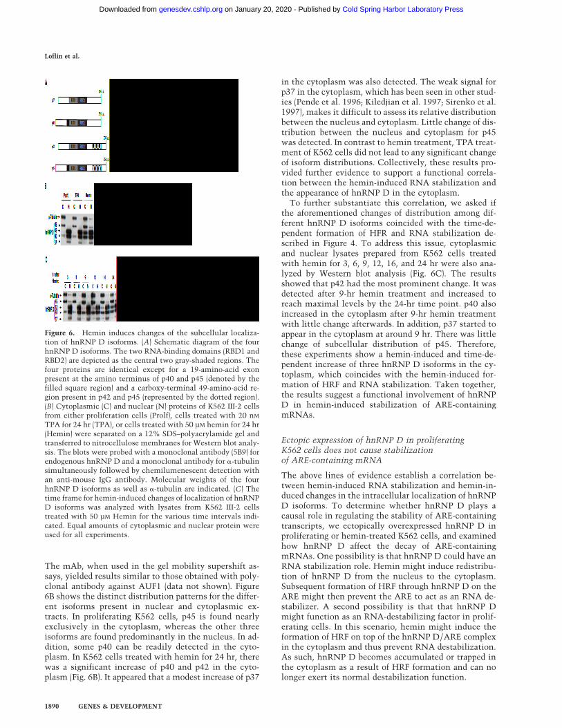
The mAb, when used in the gel mobility supershift as-says, yielded results similar to those obtained with poly-clonal antibody against AUF1 (data not shown). Figure6B shows the distinct distribution patterns for the differ-ent isoforms present in nuclear and cytoplasmic ex-tracts. In proliferating K562 cells, p45 is found nearlyexclusively in the cytoplasm, whereas the other threeisoforms are found predominantly in the nucleus. In ad-dition, some p40 can be readily detected in the cyto-plasm. In K562 cells treated with hemin for 24 hr, therewas a significant increase of p40 and p42 in the cyto-plasm (Fig. 6B). It appeared that a modest increase of p37
in the cytoplasm was also detected. The weak signal forp37 in the cytoplasm, which has been seen in other stud-ies (Pende et al. 1996; Kiledjian et al. 1997; Sirenko et al.1997), makes it difficult to assess its relative distributionbetween the nucleus and cytoplasm. Little change of dis-tribution between the nucleus and cytoplasm for p45was detected. In contrast to hemin treatment, TPA treat-ment of K562 cells did not lead to any significant changeof isoform distributions. Collectively, these results pro-vided further evidence to support a functional correla-tion between the hemin-induced RNA stabilization andthe appearance of hnRNP D in the cytoplasm.
To further substantiate this correlation, we asked ifthe aforementioned changes of distribution among dif-ferent hnRNP D isoforms coincided with the time-de-pendent formation of HFR and RNA stabilization de-scribed in Figure 4. To address this issue, cytoplasmicand nuclear lysates prepared from K562 cells treatedwith hemin for 3, 6, 9, 12, 16, and 24 hr were also ana-lyzed by Western blot analysis (Fig. 6C). The resultsshowed that p42 had the most prominent change. It wasdetected after 9-hr hemin treatment and increased toreach maximal levels by the 24-hr time point. p40 alsoincreased in the cytoplasm after 9-hr hemin treatmentwith little change afterwards. In addition, p37 started toappear in the cytoplasm at around 9 hr. There was littlechange of subcellular distribution of p45. Therefore,these experiments show a hemin-induced and time-de-pendent increase of three hnRNP D isoforms in the cy-toplasm, which coincides with the hemin-induced for-mation of HRF and RNA stabilization. Taken together,the results suggest a functional involvement of hnRNPD in hemin-induced stabilization of ARE-containingmRNAs.
Ectopic expression of hnRNP D in proliferatingK562 cells does not cause stabilizationof ARE-containing mRNA
The above lines of evidence establish a correlation be-tween hemin-induced RNA stabilization and hemin-in-duced changes in the intracellular localization of hnRNPD isoforms. To determine whether hnRNP D plays acausal role in regulating the stability of ARE-containingtranscripts, we ectopically overexpressed hnRNP D inproliferating or hemin-treated K562 cells, and examinedhow hnRNP D affect the decay of ARE-containingmRNAs. One possibility is that hnRNP D could have anRNA stabilization role. Hemin might induce redistribu-tion of hnRNP D from the nucleus to the cytoplasm.Subsequent formation of HRF through hnRNP D on theARE might then prevent the ARE to act as an RNA de-stabilizer. A second possibility is that that hnRNP Dmight function as an RNA-destabilizing factor in prolif-erating cells. In this scenario, hemin might induce theformation of HRF on top of the hnRNP D/ARE complexin the cytoplasm and thus prevent RNA destabilization.As such, hnRNP D becomes accumulated or trapped inthe cytoplasm as a result of HRF formation and can nolonger exert its normal destabilization function.
Figure 6. Hemin induces changes of the subcellular localiza-tion of hnRNP D isoforms. (A) Schematic diagram of the fourhnRNP D isoforms. The two RNA-binding domains (RBD1 andRBD2) are depicted as the central two gray-shaded regions. Thefour proteins are identical except for a 19-amino-acid exonpresent at the amino terminus of p40 and p45 (denoted by thefilled square region) and a carboxy-terminal 49-amino-acid re-gion present in p42 and p45 (represented by the dotted region).(B) Cytoplasmic (C) and nuclear (N) proteins of K562 III-2 cellsfrom either proliferation cells (Prolf), cells treated with 20 nM
TPA for 24 hr (TPA), or cells treated with 50 µM hemin for 24 hr(Hemin) were separated on a 12% SDS–polyacrylamide gel andtransferred to nitrocellulose membranes for Western blot analy-sis. The blots were probed with a monoclonal antibody (5B9) forendogenous hnRNP D and a monoclonal antibody for a-tubulinsimultaneously followed by chemilumenescent detection withan anti-mouse IgG antibody. Molecular weights of the fourhnRNP D isoforms as well as a-tubulin are indicated. (C) Thetime frame for hemin-induced changes of localization of hnRNPD isoforms was analyzed with lysates from K562 III-2 cellstreated with 50 µM Hemin for the various time intervals indi-cated. Equal amounts of cytoplasmic and nuclear protein wereused for all experiments.
Loflin et al.
1890 GENES & DEVELOPMENT
Cold Spring Harbor Laboratory Press on January 20, 2020 - Published by genesdev.cshlp.orgDownloaded from
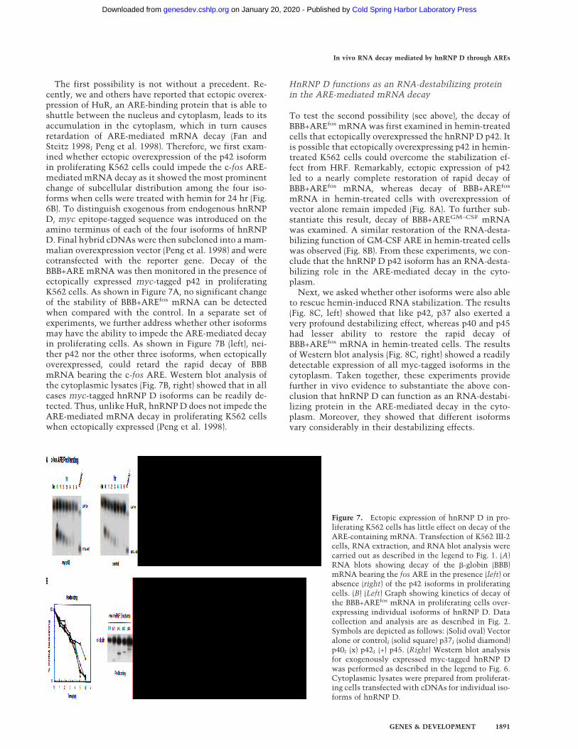
The first possibility is not without a precedent. Re-cently, we and others have reported that ectopic overex-pression of HuR, an ARE-binding protein that is able toshuttle between the nucleus and cytoplasm, leads to itsaccumulation in the cytoplasm, which in turn causesretardation of ARE-mediated mRNA decay (Fan andSteitz 1998; Peng et al. 1998). Therefore, we first exam-ined whether ectopic overexpression of the p42 isoformin proliferating K562 cells could impede the c-fos ARE-mediated mRNA decay as it showed the most prominentchange of subcellular distribution among the four iso-forms when cells were treated with hemin for 24 hr (Fig.6B). To distinguish exogenous from endogenous hnRNPD, myc epitope-tagged sequence was introduced on theamino terminus of each of the four isoforms of hnRNPD. Final hybrid cDNAs were then subcloned into a mam-malian overexpression vector (Peng et al. 1998) and werecotransfected with the reporter gene. Decay of theBBB+ARE mRNA was then monitored in the presence ofectopically expressed myc-tagged p42 in proliferatingK562 cells. As shown in Figure 7A, no significant changeof the stability of BBB+AREfos mRNA can be detectedwhen compared with the control. In a separate set ofexperiments, we further address whether other isoformsmay have the ability to impede the ARE-mediated decayin proliferating cells. As shown in Figure 7B (left), nei-ther p42 nor the other three isoforms, when ectopicallyoverexpressed, could retard the rapid decay of BBBmRNA bearing the c-fos ARE. Western blot analysis ofthe cytoplasmic lysates (Fig. 7B, right) showed that in allcases myc-tagged hnRNP D isoforms can be readily de-tected. Thus, unlike HuR, hnRNP D does not impede theARE-mediated mRNA decay in proliferating K562 cellswhen ectopically expressed (Peng et al. 1998).
HnRNP D functions as an RNA-destabilizing proteinin the ARE-mediated mRNA decay
To test the second possibility (see above), the decay ofBBB+AREfos mRNA was first examined in hemin-treatedcells that ectopically overexpressed the hnRNP D p42. Itis possible that ectopically overexpressing p42 in hemin-treated K562 cells could overcome the stabilization ef-fect from HRF. Remarkably, ectopic expression of p42led to a nearly complete restoration of rapid decay ofBBB+AREfos mRNA, whereas decay of BBB+AREfos
mRNA in hemin-treated cells with overexpression ofvector alone remain impeded (Fig. 8A). To further sub-stantiate this result, decay of BBB+AREGM–CSF mRNAwas examined. A similar restoration of the RNA-desta-bilizing function of GM-CSF ARE in hemin-treated cellswas observed (Fig. 8B). From these experiments, we con-clude that the hnRNP D p42 isoform has an RNA-desta-bilizing role in the ARE-mediated decay in the cyto-plasm.
Next, we asked whether other isoforms were also ableto rescue hemin-induced RNA stabilization. The results(Fig. 8C, left) showed that like p42, p37 also exerted avery profound destabilizing effect, whereas p40 and p45had lesser ability to restore the rapid decay ofBBB+AREfos mRNA in hemin-treated cells. The resultsof Western blot analysis (Fig. 8C, right) showed a readilydetectable expression of all myc-tagged isoforms in thecytoplasm. Taken together, these experiments providefurther in vivo evidence to substantiate the above con-clusion that hnRNP D can function as an RNA-destabi-lizing protein in the ARE-mediated decay in the cyto-plasm. Moreover, they showed that different isoformsvary considerably in their destabilizing effects.
Figure 7. Ectopic expression of hnRNP D in pro-liferating K562 cells has little effect on decay of theARE-containing mRNA. Transfection of K562 III-2cells, RNA extraction, and RNA blot analysis werecarried out as described in the legend to Fig. 1. (A)RNA blots showing decay of the b-globin (BBB)mRNA bearing the fos ARE in the presence (left) orabsence (right) of the p42 isoforms in proliferatingcells. (B) (Left) Graph showing kinetics of decay ofthe BBB+AREfos mRNA in proliferating cells over-expressing individual isoforms of hnRNP D. Datacollection and analysis are as described in Fig. 2.Symbols are depicted as follows: (Solid oval) Vectoralone or control; (solid square) p37; (solid diamond)p40; (x) p42; (+) p45. (Right) Western blot analysisfor exogenously expressed myc-tagged hnRNP Dwas performed as described in the legend to Fig. 6.Cytoplasmic lysates were prepared from proliferat-ing cells transfected with cDNAs for individual iso-forms of hnRNP D.
In vivo RNA decay mediated by hnRNP D through AREs
GENES & DEVELOPMENT 1891
Cold Spring Harbor Laboratory Press on January 20, 2020 - Published by genesdev.cshlp.orgDownloaded from

DiscussionIn this report we have used a new transcriptional pulsingstrategy, which made it possible to monitor kinetics ofmRNA decay in human erythroleukemia K562 cellswithout using transcription inhibitors (Xu et al. 1998;Loflin et al. 1999). Therefore, the new system has offeredan opportunity to study the regulation of ARE functionduring hematopoietic differentiation. We show thatwhen K562 cells were induced by hemin to differentiatedown the erythroid lineage, ARE-containing transcriptsare dramatically stabilized. In contrast, when proliferat-ing K562 cells were induced by TPA to undergo mega-karyocytic differentiation, there was little change of sta-bility of all three classes of ARE-containing transcripts.The stabilization engendered by hemin was not due to ablockade in deadenylation, as shown by our experimentsexamining poly(A) tail loss after shut-off of transcription.It appears that decay of the body of poly(A) shortenedmRNA is profoundly impeded. It is striking that this lossof rapid RNA turnover in hemin-treated cells can be sig-nificantly rescued by ectopically overexpressing hnRNPD. This rescue appears to be more profound for p37 andp42, than for p40 or p45 (see below). Our findings pro-
vide, to our knowledge, the first in vivo evidence thatidentifies a destabilizing protein for the ARE-mediatedmRNA turnover. Furthermore, they also demonstrate acytoplasmic role for hnRNP D as a destabilizing protein,thus extending the roles played by hnRNP proteins fromthe nucleus to the cytoplasm.
Diversified roles and functions in RNA biogenesis andtranscription have been described for hnRNP proteins inthe nucleus (Dreyfuss et al. 1993, 1996). The findingsthat hnRNP proteins, including hnRNP A1, D, and K,shuttle between the nucleus and the cytoplasm haveraised the possibility that shuttling hnRNP proteins maybe involved in the export of mature mRNAs out of thenucleus with shuttling signals like the M9 or the KNS(Weighardt et al. 1996; Izaurralde et al. 1997; Michael etal. 1997; Siomi et al. 1997). Moreover, they also have ledto the idea that RNA-binding proteins that initially bindRNA in the nucleus, such as shuttling hnRNP proteins,can influence the fate of mRNA in the cytoplasm (Pinol-Roma and Dreyfuss 1992; Dreyfuss et al. 1996). Severalrecent reports have lent support for such cytoplasmicfunctions for hnRNP proteins. For example, hnRNP Khas been shown to be part of a cytoplasmic complex that
Figure 8. Ectopic expression of hnRNP D in he-min-treated K562 cells restore the rapid decay ofARE-containing transcripts. Transfection ofK562III-2 cells, RNA extraction, and RNA blotanalysis were carried out as described in the leg-end to Fig. 1. RNA blots showing decay of theb-globin (BBB) mRNA bearing the fos ARE (A) orthe GM-CSF ARE (B) in the presence (left) or ab-sence (right) of the p42 isoforms in K562 cellstreated with hemin for 24 hr. (C) (Left) Graphshowing kinetics of decay of the BBB+AREfos
mRNA in hemin-treated cells overexpressing in-dividual isoforms of hnRNP D. Data collectionand analysis are as described in the legend to Fig.2. Symbols are depicted as follows: (Solid oval)Vector alone; (solid square) p37; (solid diamond)p40; (x) p42; (+) p45. (Right) Western blot analysisfor exogenously expressed myc-tagged hnRNP Dwas performed as described in the legend to Fig. 6.Cytoplasmic lysates were prepared from hemin-treated cells transfected with cDNAs for indi-vidual isoforms of hnRNP D.
Loflin et al.
1892 GENES & DEVELOPMENT
Cold Spring Harbor Laboratory Press on January 20, 2020 - Published by genesdev.cshlp.orgDownloaded from

is formed on a 38 UTR element termed DICE of 15-li-poxygenase mRNA and regulates its translation duringerythroid differentiation (Ostareck et al. 1997). HnRNP Land I both have been implicated in the control of cap-independent translation of various RNAs bearing an in-ternal ribosomal entry site (Svitkin et al. 1996; Visa et al.1996; Hahm et al. 1998). HnRNP A1, which is able tointeract with AU-rich sequences, has been shown to beassociated with cytoplasmic poly(A)+ mRNAs and isfound to be present abundantly in the cytoplasm of hu-man T lymphocytes (Hamilton et al. 1997). HnRNP Dhas been shown recently to be an integral component ofa cytoplasmic complex, termed mRNA-stabilizinga-complex that is formed on a CU-rich region in the 38UTR of the a-globin mRNA (Kiledjian et al. 1991). Al-though the a-complex is necessary for maintaining thestability of a-globin mRNA in the cytoplasm during ery-throid differentiation (Kiledjian et al. 1995; Wang andLiebhaber 1996), the role played by hnRNP D remainsunclear. It will be important to test directly whether andhow hnRNP D may participate in the regulation of a-glo-bin mRNA stability in vivo. Nevertheless, our studyidentifies a new role for shuttling hnRNP proteins in thecytoplasm as an RNA-destabilizing protein.
How do cells achieve differential regulation of ARE-destabilizing function by different isoforms of hnRNPD? Our results show that p37 and p42 are more capableof releasing hemin-induced stabilization of transcriptsbearing an ARE than p40 and p45 (Fig. 8). These proper-ties correlate with the in vitro RNA-binding affinitiesdisplayed by the four isoforms, p37>p42>p45>p40 (De-Maria and Brewer 1996; DeMaria et al. 1997). Recentstudies showed that several distinct structural determi-nants in hnRNP D are required for high-affinity bindingto AREs. HnRNP D and A1 share a common structuralbackbone that consists of two canonical RRMs followedby carboxy-terminal RGG motifs (Zhang et al. 1993;Kajita et al. 1995). Although the two RRMs are necessaryfor RNA binding, they are not sufficient for high affinityfor AREs (DeMaria et al. 1997). Both an alanine-rich re-gion of the amino terminus and a short glutamine-richregion in the carboxyl terminus are required to achievethe highest affinity binding of hnRNP D to an ARE. It isinteresting to note that unlike p40 and p45, both p37 andp42 lack a small peptide (19 amino acids) insertion at theamino terminus that may have interfered with the par-ticipation of the amino-terminal alanine-rich region inthe ARE binding (DeMaria and Brewer 1996; DeMaria etal. 1997). This may explain the diminished affinity ofp40 and p45 for AREs. Another possibility is that eachisoform may elicit interaction with distinct proteins toform different complexes that help to fulfill multipleroles in the cytoplasm, for example, in the ARE-medi-ated decay or in the stabilization of a-globin mRNA.
Given the observations that the carboxy-terminalRGG-containing domain of hnRNP can be methylated,glycosylated, and phosphorylated (e.g., Soulard et al.1993; Pype et al. 1994; Bosser et al. 1995; Shen et al.1998; Valentini et al. 1999), one could imagine thatthrough various extents of post-translational modifica-
tions among the isoforms of hnRNP D, a change of iso-forms-binding specificity and affinity for a given sub-strate may be achieved. Therefore, differential post-translational modifications of different isoforms ofhnRNP D could offer more versatility to its function. Forexample, they can enhance, reduce, or eliminate thebinding specificity or affinity for AREs by altering thenucleocytoplasmic distribution of hnRNP D or by alter-ing their interactions with auxiliary protein factors thatare necessary for substrate recognition and binding.Whereas the exact relationship between post-transla-tional modifications and changes of functionality ofhnRNP D isoforms await further experimentation, sev-eral studies suggest the existence of such a functionalcorrelation. It is clear that shuttling hnRNP proteins,including A1, D, F/H and K, exhibit differential distri-bution in a cell-type dependent and tissue-specific man-ner (Faura et al. 1995; Kamma et al. 1995). Moreover, thedistribution pattern of an individual hnRNP protein var-ies during proliferation or differentiation. It has beenshown that arginine methylation of RGG motifs facili-tates the nuclear export of hnRNP proteins (Shen et al.1998). Therefore, it will be interesting to determine thefunctional consequence in the ARE-mediated decay bymutating these sites in hnRNP D, which are potentialtargets for post-translational modifications.
One of the critical issues remaining to be addressed isthe nature of the hemin-induced mRNA stabilization inK562 cells. The lack of any immediate stabilization ef-fect by hemin suggests the RNA stabilization is relatedto the prolonged treatment of K562 cells with hemin.The time-frame for hemin to exhibit profound RNA-sta-bilization effect coincides with the time required for dif-ferentiation of K562 cells further toward erythrocytes(Baliga et al. 1993; Nakajima et al. 1997). In addition,hemin is known to induce stress response in K562 cellsthat leads to some significant changes of gene expression(Theodorakis et al. 1989; Sistonen et al. 1992). Theseobservations suggest that hemin-induced RNA stabiliza-tion may be related to erythroid differentiation, physi-ological changes as a result of prolonged stress, or both.Preliminary experiments with various pharmacologicaldrugs that are capable of inducing further erythroid dif-ferentiation do not show the mRNA stabilization effect(unpublished data). Several lines of evidence we havegathered indicate that the hemin-induced stabilizationwe described here may be a consequence of stress re-sponse induced by prolonged hemin treatment (unpub-lished data). We have identified, along with hnRNP D,heat-shock proteins in HRF, and have shown that a re-ductant, when added to the culture medium, can abro-gate the stabilization effect by hemin. If the hemin-in-duced stabilization is related to stress response, the ques-tions become if stress-activated signaling pathways, forexample, p38 map kinase and JUK kinase pathways,might be involved and if hemin as a whole or iron orprotoporphyrin alone is sufficient for the effect. It is in-teresting to note that several recent reports have shownthat either signal pathway can regulate the ARE-desta-bilizing function, depending on the systems and the na-
In vivo RNA decay mediated by hnRNP D through AREs
GENES & DEVELOPMENT 1893
Cold Spring Harbor Laboratory Press on January 20, 2020 - Published by genesdev.cshlp.orgDownloaded from

ture of stress evoked (Sirenko et al. 1997; Chen et al.1998; Ming et al. 1998).
Finally, how may hnRNP D be involved in ARE-me-diated mRNA destabilization in proliferating cells andhow does hemin abolish its function? It is possible thathnRNP D may be first assembled on the ARE while themessage is still in the nucleus. Once mature ARE-bear-ing mRNP is formed, hnRNP D may then function toescort this ARE-containing mRNP complex to the cyto-plasm (Muller et al. 1992). On reaching the cytoplasm,hnRNP D may gain new functions. For example, it maydirect the mRNA to a specific location, release it fordegradation, and then return to the nucleus. The otherpossibility is that it may be required to elicit an assem-bly of a decay complex, through protein–protein interac-tions, on the ARE for RNA degradation. In this latterscenario, hnRNP D may play an active and direct role inRNA decay. Once RNA is degraded, it is released fromthe complex and then returned to the nucleus. We fur-ther hypothesize that when a cell is treated with hemin,it induces an assembly of the HRF on the ARE throughinteraction with hnRNP D in the cytoplasm and thusinterferes with the RNA destabilizing function ofhnRNP D, leading to mRNA stabilization. This is con-sistent with our observations that hnRNP D is part ofthe HRF and that hnRNP D still retains its ARE-bindingability when associated with other factors to form HRF(Fig. 5). Because ectopic expression of hnRNP D alone issufficient to release the hemin-induced RNA stabiliza-tion, it is tempting to speculate that excess and freehnRNP D in the cytoplasm compete with the HRF andreplace its binding at the ARE to resume rapid mRNAdecay by AREs. It is interesting to note that deadenyla-tion rates appear not to be affected in hemin-inducedcells. Given the observations that in ARE-mediated de-cays deadenylation always precedes the decay of theRNA body, it is possible that the hemin-induced com-plex does not abrogate the stimulatory effect of the AREon deadenylation but does impede its stimulatory effecton the decay of the RNA body following poly(A) re-moval. This would imply that hnRNP D participates inthe second step of the ARE-mediated mRNA decay.
In summary, our studies have identified the first de-stabilizing protein in the ARE-mediated decay pathwayin vivo. The system we describe here will offer an op-portunity to conduct in vivo dissection and characteriza-tion of structural features of hnRNP D proteins that arefunctionally important for the ARE-mediated decaypathway and more importantly its regulation. An eluci-dation of a potential role of stress-activated signalingpathways in this case may provide further insight intomechanisms underlying the regulation of ARE functionduring cell growth, differentiation, and immune re-sponse. Moreover, the formation of HRF and its correla-tion with the loss of ARE-destabilizing ability suggestsan experimental basis for identifying other protein fac-tors that also participate in the ARE-mediated mRNAdecay in vivo. Characterization of the protein composi-tion of HRF should shed light on this process. Given thata wide variety of changes in physiological conditions
lead to changes of the stability of transcripts bearing anARE, our studies suggest that hnRNP D has a generaland key role in mediating or manifesting those physi-ological responses via AREs at the level of message sta-bility.
Materials and methods
Plasmid constructions
When necessary, DNA with 58- or 38-protruding ends wastreated with Klenow fragment or T4 DNA polymerase to makeends blunt. The construction of plasmids pTet-BBB and pTet-BBB+ARE has been described previously (Xu et al. 1998). Briefly,plasmid pBBB(Stu) was created by first using site directed mu-tagenesis to insert a StuI site at a position immediately up-stream of the transcription start site of rabbit b-globin gene inplasmid pBBB. pBBB(Stu) was then digested with StuI (fill in) andKpnI (fill in) and the fragment containing the b-globin sequencewas subcloned between the EcoRI (fill in) and NotI (fill in) sitesof plasmid pTet-splice (GIBCO-BRL) to create pTet-BBB. Frag-ments containing various AREs were synthesized by PCR asdescribed previously (Chen and Shyu 1994; Xu et al. 1997), withvarious pBBB+ARE plasmids as templates. These ARE frag-ments were flanked by a BamHI site at the 58 end and a BglII siteat the 38 end. Following BamHI and BglII digestion, the frag-ment was inserted into the unique BglII site in pTet-BBB tocreate pTet-BBB+ARE. To construct pSVmyc45, which ex-presses myc-tagged hnRNP D isoform p45, a p45 cDNA frag-ment was first prepared by NdeI and EcoRI digestions of plasmidpET-21(c)-cDx9-His6 (kindly provided by F. Ishikawa, Depart-ment of Bioengineering, Tokyo Institute of Technology, Yoko-hama, Japan), blunt ended, and then inserted into the SalI(blunt-ended) site of plasmid pMyc-over (Peng et al. 1998). Plas-mid pMyc-over is a pCAT3 (Promega) derivative in which theCAT-coding sequence has been replaced by a sequence encodingthe myc epitope tag. The His epitope tag present in the carboxylterminus of p45 was subsequently removed by replacing theXcmI–EcoRI fragment spanning the His-tag region with a XcmI–ApaI (blunt-ended) fragment of the plasmid pEx10x-cDx9,which is kindly provided by F. Ishikawa (Kajita et al. 1995). Toconstruct pSVmyc42, a 711-bp PCR product amplified from aplasmid carrying a full-length AUF1-p37 cDNA (Kiledjian et al.1997), kindly provided by Gary Brewer (Wake Forest UniversitySchool of Medicine, Winston, Salem, NC), was first subclonedinto pKS Bluscript (Stratagene). A SalI fragment spanning thePCR-amplified hnRNP D/AUF1 cDNA was then subclonedinto the SalI site of pTet-Myc-over (Peng et al. 1998) to createpTetMycAUF1. A StuI fragment of pSVmyc45 containing the 58
portion of p45 cDNA was then replaced by a corresponding StuIfragment of pTetMycAUF1 to generate pSVmyc42. PlasmidpSVmyc40 was constructed by removing the BstXI fragmentfrom pSVmyc45 that carries the 49-amino-acid insertion at thecarboxyl terminus of p45 isoform. Plasmid pSVmyc37 was cre-ated by replacing the StuI fragment of pSVmyc40 containing the58 portion of p40 cDNA with a corresponding StuI fragmentfrom pTetMycAUF1. Plasmid pT7b38C was constructed by firstexcising a unique 150-bp BamHI–BglII fragment from the 608-bp PCR-amplified rabbit b-globin cDNA and then inserting itinto the unique BamHI site of plasmid T3–T7a18 (GIBCO-BRL).
Analysis of mRNA decay and deadenylation
Establishment of a stable K562 cell transfectant III-2 harboringthe gene for tTA (tetracycline-controlled transcriptional activa-
Loflin et al.
1894 GENES & DEVELOPMENT
Cold Spring Harbor Laboratory Press on January 20, 2020 - Published by genesdev.cshlp.orgDownloaded from
tor) was described previously (Xu et al. 1998; Loflin et al. 1999).Cell culture, DNA transfection, isolation of total cytoplasmicRNA, and Northern blot analysis were conducted as describedpreviously (Xu et al. 1998; Loflin et al. 1999). Briefly, cells werecultured at 37°C, 8% CO2 in RPMI1640 (GIBCO-BRL) with 10%fetal bovine serum (FBS) in the presence of tetracycline (500ng/ml) to a density of 106 cells/ml, harvested, washed with PBS,and resuspended in RPMI at a density of 3 × 10⁷ cells/0.4 mlbefore transfection cells. Cells (3 × 10⁷) were transfected byelectroporation in a 2-mm gap cuvette with 30 µg of pTetBBB orpTetBBB+ARE and 90 µg of pMyc-over or its derivatives carry-ing hnRNP D cDNA (Loflin et al. 1999). After electroporation,cells were maintained in RPMI/10%FBS containing 45 ng/ml oftetracycline (Sigma) for ∼16 hr. When needed, cells were treatedwith 50 µM hemin (Sigma) or 20 nM TPA (LC Laboratories, Bos-ton, MA) for various time intervals as indicated in each experi-ment (see Figures). Cells were then transferred to fresh mediumwithout Tet for 3–4 hr to resume transcription, followed by theaddition of 500 ng/ml Tet to block further transcription. Totalcytoplasmic RNA was isolated at various time intervals afterthe addition of Tet (500 ng/ml), and Northern blot analysis wasperformed with a probe spanning 60 bp of the rabbit b-globin38-UTR for detection of the transfected b-globin message with-out (BBB) or with an ARE (BBB+ARE). A 300-bp fragment span-ning the second exon of GAPDH was used for detection of en-dogenous GAPDH, which was used as an internal control. Thequantitation of data was obtained by scanning the radioactiveblots with an imager (Packard). All experiments described inthis manuscript have been performed in duplicate or triplicate.
Preparation of K562 cytoplasmic and nuclear extracts
Cytoplasmic and nuclear lysates were prepared as described pre-viously (Peng et al. 1998). Briefly, cytoplasmic lysates were pre-pared from K562 cells by lysis at 4°C in a lysis/extraction buffercontaining 10 mM HEPES (pH 7.6), 3 mM MgCl2, 40 mM KCl, 2mM DTT, 5% glycerol, 0.5% NP-40, 8 µg/ml aprotinin, 8 µg/mlleupeptin, and 100 µg/ml PMSF. Nuclei were removed by cen-trifugation (1250g) at 4°C for 5 min. Nuclei were then resus-pended and spun down in the lysis/extraction buffer twice toavoid contamination of cytoplasmic proteins. Nuclear extractswere prepared from the pelleted nuclei in nuclear extractionbuffer containing 10 mM HEPES (pH 7.9), 0.1 mM EGTA, 1.5 mM
MgCl2, 420 mM NaCl, 0.5 mM DTT, 0.5 mM PMSF, and 25%glycerol. After incubation on ice for 20 min, cellular debris wasremoved by centrifugation (1250g) at 4°C for 5 min. Proteinconcentration was analyzed by the BCA protein assay reagent(Pierce).
Gel mobility shift assay and supershift analysis
Preparation of RNA probes by in vitro transcription and analysisof RNA–protein interaction were described previously (You etal. 1992). Transcription reactions were performed according toPromega instructions, with T3 or T7 RNA polymerase. LabeledRNA transcripts were produced by inclusion of [a-32P]UTP (Du-Pont, 800 Ci/mmole) in the reactions. The c-fos ARE probe is asense transcript synthesized from HindIII-linearized pT3ARE(You et al. 1992) with T3 polymerase. The b-globin RNA used asa nonspecific competitor in Figure 3 is an antisense transcriptspanning the 150-nucleotide 38 portion of the protein codingregion of the rabbit b-globin mRNA and is synthesized fromBamHI-linearized pT7b38C with T7 polymerase. Cytoplasmiclysate (6 µg of protein) and 32P-labeled RNA (1 ng) were incu-bated at room temperature for 15 min in a buffer containing 10mM HEPES (pH 7.6), 3 mM MgCl2, 40 mM KCl, 2 mM DTT, 5%glycerol, and 0.5% NP-40. Heparin (5 µg/ml, final concentra-
tion) and yeast total RNA (200 µg/ml, final concentration) wereadded to reduce nonspecific binding. The final volume of eachreaction was 10 µl. Subsequently, unbound RNA was digestedfor 20 min by 0.6 units of RNase T1 (Calbiochem) at roomtemperature. RNA–protein complexes were resolved on 6%nondenaturing polyacrylamide gels. Gel mobility supershiftanalysis was performed by the addition of antibody into thebinding reaction (final volume, 10 µl; 1:20 dilution for antibodyto AUF1, and 1:10 dilution for antibody to HuR), which waspreincubated for 15 min at room temperature to allow RNA–protein interactions to occur, and the antibody–RNA–proteinmixture was then incubated at room temperature for another 15min. The RNA–protein–antibody complexes were resolved on6% nondenaturing polyacrylamide gels. The purified monoclo-nal antibody against the myc-tag (0.1 µg of IgG/µl) was pur-chased from Calbiochem. The polyclonal antibody to AUF1 waskindly provided by G. Brewer (Pende et al. 1996; Kiledjian et al.1997; Sirenko et al. 1997) and the antibody to human HuR wasa gift from H. Furneaux (Memorial Sloan Kettering Cancer Cen-ter, NY) and was raised against the peptide sequence spanningthe first 13 amino acids of human HuR (Peng et al. 1998).
Western blotting analysis
Cytoplasmic and nuclear lysates were resolved on a 12% SDS–polyacrylamide gel and analyzed by Western blotting with anECL Western-blotting kit (Amersham). The blots were probedwith specific antibodies as described in the legends to the fig-ures. The purified monoclonal antibody against a-tubulin(DM1A) was purchased from Sigma and was used at a 1:20,000dilution. The antibody to myc tag was as described above andwas used at a 1:100 dilution. The monoclonal antibody tohnRNP D (5B9) was kindly provided by G. Dreyfuss (HowardHughes Medical Institute, University of Pennsylvania School ofMedicine, Philadelphia, PA) and was used at 1:10,000 dilution(Faura et al. 1995; Kamma et al. 1995; Kiledjian et al. 1997).
Acknowledgments
We thank M. Blackburn, G. Cote, and M. Wilkinson for criticalreading of the manuscript and their valuable comments, N. Xufor technical assistance and discussion, G. Brewer and G. Drey-fuss for the antibodies against hnRNP D/AUF1, G. Brewer, F.Ishikawa, and M. Kiledjian for the AUF1 and hnRNP D cDNAclones. This work was supported by a grant from National In-stitutes of Health (RO1 GM 46454) and in part by a grant fromthe Council of Tobacco Research-USA, Inc. A.-B.S. is the recipi-ent of an American Heart Association Established InvestigatorAward.
The publication costs of this article were defrayed in part bypayment of page charges. This article must therefore be herebymarked ‘advertisement’ in accordance with 18 USC section1734 solely to indicate this fact.
References
Alitalo, R. 1990. Induced differentiation of K562 leukemia cells:A model for studies of gene expression in early megakaryo-blasts. Leuk. Res. 14: 501–514.
Alitalo, R., L.C. Andersson, C. Betsholtz, K. Nilsson, B. Wester-mark, C.H. Heldin, and K. Alitalo. 1987. Induction of plate-let-derived growth factor gene expression during megakaryo-blastic and monocytic differentiation of human leukemiacell lines. EMBO J. 6: 1213–1218.
Alitalo, R., T.P. Makela, P. Koskinen, L.C. Andersson, and K.Alitalo. 1988. Enhanced expression of transforming growthfactor beta during megakaryoblastic differentiation of K562
In vivo RNA decay mediated by hnRNP D through AREs
GENES & DEVELOPMENT 1895
Cold Spring Harbor Laboratory Press on January 20, 2020 - Published by genesdev.cshlp.orgDownloaded from

leukemia cells. Blood 71: 899–906.Antic, D. and J.D. Keene. 1997. Insights from model systems-
Embryonic lethal abnormal visual RNA-binding proteins in-volved in growth, differentiation, and posttranscriptionalgene expression. Am. J. Hum. Genet. 61: 273–278.
Baliga, B.S., M. Mankad, A.K. Shah, and V.N. Mankad. 1993.Mechanism of differentiation of human erythroleukaemiccell line K562 by hemin. Cell Prolif. 26: 519–529.
Bosser, R., M. Faura, J. Serratosa, J. Renau-Piqueras, M. Pruschy,and O. Bachs. 1995. Phosphorylation of rat liver hetero-geneous nuclear ribonucleoproteins A2 and C can be modu-lated by calmodulin. Mol. Cell. Biol. 15: 661–670.
Brewer, G. 1991. An A + U-rich element RNA-binding factorregulates c-myc mRNA stability in vitro. Mol. Cell. Biol.11: 2460–2466.
Brewer, G. and J. Ross. 1989. Regulation of c-myc mRNA sta-bility in vitro by a labile destabilizer with an essentialnucleic acid component. Mol. Cell. Biol. 9: 1996–2006.
Buzby, J.S., S.M. Lee, P. Van Winkle, C.T. DeMaria, G. Brewer,and M.S. Cairo. 1996. Increased granulocyte-macrophagecolony-stimulating factor mRNA instability in cord versusadult mononuclear cells is translation-dependent and asso-ciated with increased levels of A + U-rich element bindingfactor. Blood 88: 2889–2897.
Caput, D., B. Beutler, K. Hartog, R. Thayer, S. Brown-Shimer,and A. Cerami. 1986. Identification of a common nucleotidesequence in the 38-untranslated region of mRNA moleculesspecifying inflammatory mediators. Proc. Natl. Acad. Sci.83: 1670–1674.
Carballo, E., W.S. Lai, and P.J. Blackshear. 1998. Feedback inhi-bition of macrophage tumor necrosis factor-alpha produc-tion by tristetraprolin. Science 281: 1001–1005.
Chen, C.Y. and A.B. Shyu. 1994. Selective degradation of early-response-gene mRNAs: Functional analyses of sequence fea-tures of the AU-rich elements. Mol. Cell. Biol. 14: 8471–8482.
———. 1995. AU-rich elements: Characterization and impor-tance in mRNA degradation. Trends Biochem. Sci. 20: 465–470.
Chen, C.Y., T.M. Chen, and A.B. Shyu. 1994. Interplay of twofunctionally and structurally distinct domains of the c-fosAU-rich element specifies its mRNA-destabilizing function.Mol. Cell. Biol. 14: 416–426.
Chen, C.Y., F. Del Gatto-Konczak, Z. Wu, and M. Karin. 1998.Stabilization of interleukin-2 mRNA by the c-Jun NH2-ter-minal kinase pathway. Science 280: 1945–1949.
DeMaria, C.T. and G. Brewer. 1996. AUF1 binding affinity toA + U-rich elements correlates with rapid mRNA degrada-tion. J. Biol. Chem. 271: 12179–12184.
DeMaria, C.T., Y. Sun, L. Long, B.J. Wagner, and G. Brewer.1997. Structural determinants in AUF1 required for high af-finity binding to A + U-rich elements. J. Biol. Chem.272: 27635–27643.
Dreyfuss, G., M.J. Matunis, S. Pinol-Roma, and C.G. Burd. 1993.hnRNP proteins and the biogenesis of mRNA. Annu. Rev.Biochem. 62: 289–321.
Dreyfuss, G., M. Hentze, and A.I. Lamond. 1996. From tran-script to protein. Cell 85: 963–972.
Fan, X.C. and J.A. Steitz. 1998. Overexpression of HuR, anuclear-cytoplasmic shuttling protein, increases the in vivostability of ARE-containing mRNAs. EMBO J. 17: 3448–3460.
Faura, M., J. Renau-Piqueras, O. Bachs, and R. Bosser. 1995.Differential distribution of heterogeneous nuclear ribonu-cleoproteins in rat tissues. Biochem. Biophys. Res. Comm.217: 554–560.
Ford, L.P., J. Watson, J.D. Keene, and J. Wilusz. 1999. ELAVproteins stabilize deadenylated intermediates in a novel invitro mRNA deadenylation/degradation system. Genes &Dev. 13: 188–201.
Greenberg, M.E. and J.G. Belasco. 1993. Control of the decay oflabile protooncogene and cytokine mRNAs. In Control ofmessenger RNA stability (ed. J.G. Belasco and G. Brawer-man), pp. 199–218. Academic Press, San Diego, CA.
Hahm, B., Y.K. Kim, J.H. Kim, T.Y. Kim, and S.K. Jang. 1998.Heterogeneous nuclear ribonucleoprotein L interacts withthe 38 border of the internal ribosomal entry site of hepatitisC virus. J. Virol. 72: 8782–8788.
Hamilton, B.J., E. Nagy, J.S. Malter, B.A. Arrick, and W.F. Rigby.1993. Association of heterogeneous nuclear ribonucleopro-tein A1 and C proteins with reiterated AUUUA sequences. J.Biol. Chem. 268: 8881–8887.
Hamilton, B.J., C.M. Burns, R.C. Nichols, and W.F.C. Rigby.1997. Modulation of AUUUA response element binding byheterogeneous nuclear ribonucleoprotein A1 in human Tlymphocytes. The roles of cytoplasmic location, transcrip-tion, and phosphorylation. J. Biol. Chem. 272: 28732–28741.
Izaurralde, E., A. Jarmolowski, C. Beisel, I.W. Mattaj, G. Drey-fuss, and U. Fischer. 1997. A role for the M9 transport signalof hnRNP A1 in mRNA nuclear export. J. Cell. Biol. 137: 27–35.
Jain, R.G., L.G. Andrews, K.M. McGowan, P.H. Pekala, and J.D.Keene. 1997. Ectopic expression of Hel-N1, an RNA-bindingprotein, increases glucose transporter (GLUT1) expression in3T3-L1 adipocytes. Mol. Cell. Biol. 17: 954–962.
Kajita, Y., J. Nakayama, M. Aizawa, and F. Ishikawa. 1995. TheUUAG-specific RNA binding protein, heterogeneousnuclear ribonucleoprotein D0. Common modular structureand binding properties of the 2xRBD-Gly family. J. Biol.Chem. 270: 22167–22175.
Kamma, H., D.S. Portman, and G. Dreyfuss. 1995. Cell type-specific expression of hnRNP proteins. Exp. Cell. Res.221: 187–196.
Kiledjian, M., X. Wang, and S.A. Liebhaber. 1995. Identificationof two KH domain proteins in the alpha-globin mRNP sta-bility complex. EMBO J. 14: 4357–4364.
Kiledjian, M., C.T. DeMaria, G. Brewer, and K. Novick. 1997.Identification of AUF1 (heterogeneous nuclear ribonucleo-protein D) as a component of the alpha-globin mRNA sta-bility complex. Mol. Cell. Biol. 17: 4870–4876.
Lafon, I., F. Carballes, G. Brewer, M. Poiret, and D. Morello.1998. Developmental expression of AUF1 and HuR, two c-myc mRNA binding proteins. Oncogene 16: 3413–3421.
Levy, N.S., S. Chung, H. Furneaux, and A.P. Levy. 1998. Hyp-oxic stabilization of vascular endothelial growth factormRNA by the RNA-binding protein HuR. J. Biol. Chem.273: 6417–6423.
Loflin, T.L., C.-Y.A. Chen, N. Xu, and A.-B. Shyu. 1999. Tran-scriptional pulsing approaches for analysis of mRNA turn-over in mammalian cells. Methods: Companion MethodsEnzymol. 17: 11–20.
Michael, W.M., P.S. Eder, and G. Dreyfuss. 1997. The K nuclearshuttling domain: A novel signal for nuclear import andnuclear export in the hnRNP K protein. EMBO J. 16: 3587–3598.
Ming, X.F., M. Kaiser, and C. Moroni. 1998. c-jun N-terminalkinase is involved in AUUUA-mediated interleukin-3mRNA turnover in mast cells. EMBO J. 17: 6039–6048.
Miyamoto, Y., M. Kosaka, Y. Eto, H. Shibai, and S. Saito. 1990.Effect of erythroid differentiation factor on erythroid differ-entiation and proliferation of K-562 cells. Biochem. Biophys.Res. Comm. 168: 1149–1156.
Loflin et al.
1896 GENES & DEVELOPMENT
Cold Spring Harbor Laboratory Press on January 20, 2020 - Published by genesdev.cshlp.orgDownloaded from
Muller, W.E., H. Slor, K. Pfeifer, P. Huhn, A. Bek, S. Orsulic, H.Ushijima, and H.C. Schroder. 1992. Association of AUUUA-binding protein with A+U-rich mRNA during nucleo-cyto-plasmic transport. J. Mol. Biol. 226: 721–733.
Nagy, E. and W.F. Rigby. 1995. Glyceraldehyde-3-phosphate de-hydrogenase selectively binds AU-rich RNA in the NAD(+)-binding region (Rossmann fold). J. Biol. Chem. 270: 2755–2763.
Nakagawa, J., H. Waldner, S. Meyer-Monard, J. Hofsteenge, P.Jeno, and C. Moroni. 1995. AUH, a gene encoding an AU-specific RNA binding protein with intrinsic enoyl-CoA hy-dratase activity. Proc. Natl. Acad. Sci. 92: 2051–2055.
Nakajima, O., S. Iwasaki, and Y. Hashimoto. 1997. Hemin-in-duced erythroid differentiation of human myeloleukemiaK562 cell line and its modification by bioresponse modifiers.Cell. Mol. Biol. (Noisy-le-grand) 43: 115–134.
Nanbu, R., T. Kubo, T. Hashimoto, and S. Natori. 1993. Purifi-cation of an AU-rich RNA binding protein from Sarcophagaperegrina (flesh fly) and its identification as a Thiolase. J.Biochem. (Tokyo) 114: 432–437.
Ostareck, D.H., A. Ostareck-Lederer, M. Wilm, B.J. Thiele, M.Mann, and M.W. Hentze. 1997. mRNA silencing in ery-throid differentiation: hnRNP K and hnRNP E1 regulate 15-lipoxygenase translation from the 38 end. Cell 89: 597–606.
Pende, A., K.D. Tremmel, C.T. DeMaria, B.C. Blaxall, W.A. Mi-nobe, J.A. Sherman, J.D. Bisognano, M.R. Bristow, G.Brewer, and J. Port. 1996. Regulation of the mRNA-bindingprotein AUF1 by activation of the beta-adrenergic receptorsignal transduction pathway. J. Biol. Chem. 271: 8493–8501.
Peng, S.-P., C.-Y. Chen, N. Xu, and A.-B. Shyu. 1998. RNA sta-bilization by the AU-rich element binding protein, HuR, anELAV protein. EMBO J. 17: 3461–3470.
Pinol-Roma, S. and G. Dreyfuss. 1991. Transcription-dependentand transcription-independent nuclear transport of hnRNPproteins. Science 253: 312–314.
———. 1992. Shuttling of pre-mRNA binding proteins betweennucleus and cytoplasm. Nature 355: 730–732.
Pype, S., H. Slegers, L. Moens, W. Merlevede, and J. Goris. 1994.Tyrosine phosphorylation of a M(r) 38,000 A/B-type hnRNPprotein selectively modulates its RNA binding. J. Biol.Chem. 269: 31457–31465.
Ross, J. 1995. mRNA stability in mammalian cells. Microbiol.Rev. 59: 423–450.
Rutherford, T.R., J.B. Clegg, and D.J. Weatherall. 1979. K562human leukaemic cells synthesize embryonic haemoglobinin response to haemin. Nature 280: 164–165.
Rutherford, T., J.B. Clegg, D.R. Higgs, R.W. Jones, J. Thompson,and D.J. Weatherall. 1981. Embryonic erythroid differentia-tion in the human leukemic cell line K562. Proc. Natl. Acad.Sci. 78: 348–352.
Schiavi, S.C., J.G. Belasco, and M.E. Greenberg. 1992. Regula-tion of proto-oncogene mRNA stability. Biochim. Biophys.Acta 1114: 95–106.
Shaw, G. and R. Kamen. 1986. A conserved AU sequence fromthe 38 untranslated region of GM-CSF mRNA mediates se-lective mRNA degradation. Cell 46: 659–667.
Shen, E.C., M.F. Henry, V.H. Weiss, S.R. Valentini, P.A. Silver,and M.S. Lee. 1998. Arginine methylation facilitates thenuclear export of hnRNP proteins. Genes & Dev. 12: 679–691.
Shyu, A.-B., J.A. Garcia-Sanz, and E. Mullner. 1996. Analysis ofmRNA decay in mammalian cells. In The immunologymethods manual (ed. I. Lefkovits), pp. 450–456. AcademicPress, London, UK.
Siomi, M.C., P.S. Eder, N. Kataoka, L. Wan, Q. Liu, and G.Dreyfuss. 1997. Transportin-mediated nuclear import of het-
erogeneous nuclear RNP proteins. J. Cell. Biol. 138: 1181–1192.
Sirenko, O.I., A.K. Lofquist, C.T. DeMaria, J.S. Morris, G.Brewer, and J.S. Haskill. 1997. Adhesion-dependent regula-tion of an A+U-rich element-binding activity associatedwith AUF1. Mol. Cell. Biol. 17: 3898–3906.
Sistonen, L., K.D. Sarge, B. Phillips, K. Abravaya, and R.I. Mo-rimoto. 1992. Activation of heat shock factor 2 during he-min-induced differentiation of human erythroleukemiacells. Mol. Cell. Biol. 12: 4104–4111.
Soulard, M., V. Della Valle, M.C. Siomi, S. Pinol-Roma, P.Codogno, C. Bauvy, M. Bellini, J.C. Lacroix, G. Monod, G.Dreyfuss et al. 1993. hnRNP G: Sequence and characteriza-tion of a glycosylated RNA-binding protein. Nucleic AcidsRes. 21: 4210–4217.
Svitkin, Y.V., L.P. Ovchinnikov, G. Dreyfuss, and N. Sonenberg.1996. General RNA binding proteins render translation capdependent. EMBO J. 15: 7147–7155.
Theodorakis, N.G., D.J. Zand, P.T. Kotzbauer, G.T. Williams,and R.I. Morimoto. 1989. Hemin-induced transcriptional ac-tivation of the HSP70 gene during erythroid maturation inK562 cells is due to a heat shock factor-mediated stress re-sponse. Mol. Cell. Biol. 9: 3166–3173.
Treisman, R. 1985. Transient accumulation of c-fos RNA fol-lowing serum stimulation requires a conserved 58 elementand c-fos 38 sequences. Cell 42: 889–902.
Valentini, S.R., V.H. Weiss, and P.A. Silver. 1999. Argininemethylation and binding of Hrp1p to the efficiency elementfor mRNA 38-end formation. RNA 5: 272–280.
Visa, N., A.T. Alzhanova-Ericsson, X. Sun, E. Kiseleva, B. Bjork-roth, T. Wurtz, and B. Daneholt. 1996. A pre-mRNA-bindingprotein accompanies the RNA from the gene through thenuclear pores and into polysomes. Cell 84: 253–264.
Wagner, B.J., C.T. DeMaria, Y. Sun, G.M. Wilson, and G.Brewer. 1998. Structure and genomic organization of the hu-man AUF1 gene: Alternative pre-mRNA splicing generatesfour protein isoforms. Genomics 48: 195–202.
Wang, X. and S.A. Liebhaber. 1996. Complementary change incis determinants and trans factors in the evolution of anmRNP stability complex. EMBO J. 15: 5040–5051.
Weighardt, F., G. Biamonti, and S. Riva. 1996. The roles of het-erogeneous nuclear ribonucleoproteins (hnRNP) in RNAmetabolism. BioEssays 18: 747–756.
Winzen, R., S. Kafert, B. Preiss, H.A. Mylius-Spencker, K. Resch,and H. Holtmann. 1996. Interaction between the mRNA ofthe 55-kDa tumor necrosis factor receptor and cellular pro-teins. Possible involvement in post-transcriptional regula-tion of receptor expression. J. Biol. Chem. 271: 13461–13467.
Xu, N., C.-Y.A. Chen, and A.-B. Shyu. 1997. Modulation of thefate of cytoplasmic mRNA by AU-rich elements: Key se-quence features controlling mRNA deadenylation and de-cay. Mol. Cell. Biol. 17: 4611–4621.
Xu, N., P. Loflin, C.-Y.A. Chen, and A.-B. Shyu. 1998. A broaderrole for AU-rich element-mediated mRNA turnover revealedby a new transcriptional pulse strategy. Nucleic Acids Res.26: 558–565.
You, Y., C.Y. Chen, and A.B. Shyu. 1992. U-rich sequence-bind-ing proteins (URBPs) interacting with a 20-nucleotide U-richsequence in the 38 untranslated region of c-fos mRNA maybe involved in the first step of c-fos mRNA degradation. Mol.Cell. Biol. 12: 2931–2940.
Zhang, W., B.J. Wagner, K. Ehrenman, A.W. Schaefer, C.T. De-Maria, D. Crater, K. DeHaven, L. Long, and G. Brewer. 1993.Purification, characterization, and cDNA cloning of an AU-rich element RNA-binding protein, AUF1. Mol. Cell. Biol.13: 7652–7665.
In vivo RNA decay mediated by hnRNP D through AREs
GENES & DEVELOPMENT 1897
Cold Spring Harbor Laboratory Press on January 20, 2020 - Published by genesdev.cshlp.orgDownloaded from
13:1999, Genes Dev. Paul Loflin, Chyi-Ying A. Chen and Ann-Bin Shyu destabilization directed by the AU-rich elementUnraveling a cytoplasmic role for hnRNP D in the in vivo mRNA
References
http://genesdev.cshlp.org/content/13/14/1884.full.html#ref-list-1
This article cites 70 articles, 42 of which can be accessed free at:
License
ServiceEmail Alerting
click here.right corner of the article or
Receive free email alerts when new articles cite this article - sign up in the box at the top
Cold Spring Harbor Laboratory Press
Cold Spring Harbor Laboratory Press on January 20, 2020 - Published by genesdev.cshlp.orgDownloaded from