uva-dare (digital academic repository) the amsterdam ... · hyperthyroidism (jod–basedow effect)...
TRANSCRIPT
UvA-DARE is a service provided by the library of the University of Amsterdam (http://dare.uva.nl)
UvA-DARE (Digital Academic Repository)
The Amsterdam autoimmune thyroid disease cohort
Strieder, T.G.A.
Link to publication
Citation for published version (APA):Strieder, T. G. A. (2008). The Amsterdam autoimmune thyroid disease cohort.
General rightsIt is not permitted to download or to forward/distribute the text or part of it without the consent of the author(s) and/or copyright holder(s),other than for strictly personal, individual use, unless the work is under an open content license (like Creative Commons).
Disclaimer/Complaints regulationsIf you believe that digital publication of certain material infringes any of your rights or (privacy) interests, please let the Library know, statingyour reasons. In case of a legitimate complaint, the Library will make the material inaccessible and/or remove it from the website. Please Askthe Library: https://uba.uva.nl/en/contact, or a letter to: Library of the University of Amsterdam, Secretariat, Singel 425, 1012 WP Amsterdam,The Netherlands. You will be contacted as soon as possible.
Download date: 30 Dec 2019
Invited Interview
European Journal of Endocrinology
2004; 150: 605-618
Mark F Prummel
Thea Strieder
Wilmar M Wiersinga
2|The environment
and
AutoImmune Thyroid Diseases

The environment and autoimmune thyroid diseases
19
Chapter 2
The environment and autoimmune thyroid diseases
Abstract
Genetic factors play an important role in the pathogenesis of autoimmune thyroid disease
(AITD) and it has been calculated that 80% of the susceptibility to develop Graves’ disease is
attributable to genes. The concordance rate for AITD among monozygotic twins is, however,
well below 1 and environmental factors thus must play an important role. We have attempted
to carry out a comprehensive review of all the environmental and hormonal risk factors
thought to bring about AITD in genetically predisposed individuals. Low birth weight, iodine
excess and deficiency, selenium deficiency, parity, oral contraceptive use, reproductive span,
fetal microchimerism, stress, seasonal variation, allergy, smoking, radiation damage to the
thyroid gland, viral and bacterial infections all play a role in the development of autoimmune
thyroid disorders. The use of certain drugs (lithium, interferon-a, Campath-1H) also
increases the risk of the development of autoimmunity against the thyroid gland.
Further research is warranted into the importance of fetal microchimerism and of viral
infections capable of mounting an endogenous interferon-a response.
Chapter 2
20
Introduction
Graves’ hyperthyroidism, Hashimoto’s hypothyroidism and post-partum thyroid dysfunction
are common disorders. They have an autoimmune origin and are therefore also alluded to as
autoimmune thyroid disease (AITD). Like other organ-specific autoimmune endocrinopathies,
e.g. type I diabetes mellitus (IDDM), they have a multifactorial etiology. Genes are certainly
involved and in order to develop AITD a subject will have a certain genetic susceptibility,
probably involving multiple genes of which only a few have been identified. Most notably,
certain human leukocyte antigen (HLA)-DR genes determine this genetic susceptibility, but
there are other genes involved and AITD thus has a polygenetic background. Nevertheless,
non-genetic (environmental, hormonal) factors must also play an important etiologic role,
because the concordance rate for AITD in monozygotic twins is not 100%. Another argument
is that immigrants coming from countries with a low incidence of autoimmune diseases will
adopt the incidence rate of the new country. For instance, type I diabetes mellitus is 10 times
more frequent in Pakistanis living in the UK than in those living in Pakistan (1).
In recent years, a number of excellent reviews have been published on the genetic background
of AITD (2–6). Here we will attempt to review the environmental factors that may be
involved in the development of AITD (Table 1). In this review we will consider both Graves’
Table 1. Environmental factors involved in the etiology of AITD
Environmental factor
Mechanism
Phenotype
Low birth weight Insufficient thymic maturation TPO antibodies
Iodine excess No escape from Wolff-Chaikoff effect HT
Jod-Basedow GD
Selenium deficiency Unknown: viral infections? HT
Longer reproductive span Estradiol effect? HT
Oral contraceptives Protective TPO antibodies
Fetal michrochimerism Male cells in thyroid elicit antithyroid attack HT and GD
Stress Upregulation HPA axis GD
Allergy Unknown: high IgE levels GD
Smoking Hypoxia? High IgE levels GD; esp. GO
Yersinia enterocolitica infection
Moleculair mimicry GD
See text for explanation and references. GD, Graves’ disease; HT, Hashimoto’s thyroiditis; GO, Graves’ ophthalmopathy.
disease and Hashimoto’s thyroiditis. Despite their different phenotype, they do share some
homology. First, autoantibodies against thyroid peroxidase (TPO) are common in both
diseases. Secondly, Graves’ disease and Hashimoto’s thyroiditis appear to run in the same
families and thus share a common genetic background (7, 8).
The environment and autoimmune thyroid diseases
21
Fetal growth
Reduced fetal growth is a risk factor for several common disorders, such as chronic heart
disease (9), and famine exposure during fetal life is associated with subsequent glucose
intolerance during adult life (10). Prenatal malnutrition is associated with a lower thymic and
splenic weight (11), and this may cause earlier maturation of the thymus resulting in a decline
in T suppressor cells (12). Indeed, Phillips et al. (12) found that among 305 women aged 60–
71 years born in the UK the presence of TPO antibodies was positively related to lower birth
weights (but not to weight at 1 year of age). The prevalence of TPO antibodies was 2.4 times
higher in women with a birth weight of less than 5.5 lbs (2.49 kg) compared with those with a
higher birth weight. In a twin study, the same group found that among monozygous twins, the
smaller twin had higher levels of TPO antibodies (13). Because of the genetic identity of
monozygous twins, this study strongly suggests that certain intrauterine factors causing
reduced fetal growth are the first environmental risk factors for AITD in later life. However,
this was not confirmed in another twin study where birth weight was not found to be a
determinant for clinically overt AITD (14).
Iodine intake
Iodine intake seems to influence the prevalence rates of hyper- and hypothyroidism. In areas
with sufficient iodine intake, hypothyroidism is more common than in iodine-deficient
regions (15), whereas the overall prevalence of thyrotoxicosis is greater in iodine-deficient
areas (16). Looking at the different causes of hyperthyroidism, Graves’ hyperthyroidism as
the cause of thyrotoxicosis is seen more frequently in iodine-replete areas (17), and TPO
antibodies as a marker for impending thyroid failure are more prevalent in iodine-deficient
regions (18).
Excessive iodine intake can cause dysthyroidism, especially in patients with underlying
autoimmune thyroiditis (19). Due to a failure to escape from the Wolff–Chaikoff effect,
iodine excess can cause hypothyroidism and/or goiter, but if autonomously functioning
nodules or a subclinical form of Graves’ disease are present, it can also induce
hyperthyroidism (Jod–Basedow effect) (20, 21). Both phenomena are thought to lead to some
thyroid destruction and hence presentation of thyroidal antigens to the immune system leading
to an autoimmune reaction (22). It thus appears that iodine intake is indeed a risk factor for
the development of AITD. This is in agreement with animal studies showing that a high
Chapter 2
22
iodine intake aggravates autoimmune thyroiditis in several genetically susceptible animal
strains (23–25).
Selenium intake
Selenium is a trace mineral and an essential nutrient for selenocysteine synthesis and is also
called the 21st amino acid. It is incorporated into 35 selenoproteins, mostly enzymes (26).
Selenium also has a marked influence on the immune system and selenium deficiency is
associated with a greater susceptibility for viral infections such as the Coxsackie virus (27),
possibly because T-lymphocytes have an important functional need for selenium (26). In
addition, selenium acts as an antioxidant and reduces free radical formation. It plays an
essential role in thyroid hormone synthesis, because two enzymes involved in thyroid
hormone production are selenoproteins: the deiodinases and glutathione peroxidase (28).
Selenium deficiency leads to a variety of symptoms including a higher miscarriage rate (29)
and a higher cancer mortality rate (26).
Selenium intake in Europe is lower than in the United States and in many countries it is below
the UK reference nutrient intake of 75 mg/day. Sources of selenium are crab, other shellfish
and fish, but alternative sources such as wheat are relatively low in selenium content because
of the low selenium availability in European soils (26).
Low selenium blood levels are associated with increased thyroid volume and with thyroid
hypoechogenicity, a marker for lymphocytic infiltration (30). In agreement with this finding,
a recent double-blind randomized trial in patients with subclinical hypothryoidism showed
that treatment with 200 mg sodium selenite caused a significant decrease in TPO antibody
titers (as well as an increase in quality of life), without affecting thyroid hormone status (31).
In another randomized trial in patients with subclinical hypothyroidism who were treated with
thyroxine supplementation, addition of 200 mg selenium methionine led to a significant
decrease in TPO antibody concentrations (32).
Hormonal influences:
Female sex
One of the most striking characteristics of organspecific autoimmune diseases is its female
preponderance. The female:male ratio for Graves’ disease and Hashimoto thyroiditis is 5–
10:1 (33). The reason for this is unclear and genetic factors must play a role, although it is
The environment and autoimmune thyroid diseases
23
noteworthy that Hashimoto’s thyroiditis is very prevalent among girls with Turner’s
syndrome (XO karyotype), but not in men with Klinefelter’s syndrome (XXY karyotype) (6).
The influence of the X chromosome is thus limited and hormonal influences may also be
operative in the induction of AITD. It is interesting to note the female preponderance in
nonautoimmune-mediated thyroid disease, such as multinodular goiter, but this is outside the
scope of this review.
Oral contraceptives
Another link to explain the sex difference would be the use of oral contraceptives or hormone
replacement therapy (HRT). The latter, however, was found not to be associated with either
subclinical hypothyroidism or the presence of TPO antibodies (39), although in one case
report an exacerbation of eye symptoms was seen in a woman with Graves’ ophthalmopathy
starting HRT (40). As for oral contraceptives, used by over 100 million women worldwide
(41), there are remarkably few studies on their use and the development of AITD and in
contrast to what one intuitively may think their use seems to protect against AITD. In an early
large study among 46 000 women, cases of hypo- or hyperthyroidism together were seen less
frequently among oral contraceptive users than in controls (relative risk (RR), 0.68; 95%
confidence interval (CI), 0.52–0.85) (42). Two large population-based studies found that
thyroid volume was smaller in oral contraceptive users than in controls (43, 44). We found
that estrogen use protected against the development of hyperthyroidism, independently of the
number of previous pregnancies (7). This is in agreement with the observation that the use of
contraceptives had a protective effect for the development of Graves’ disease (odds ratio
(OR), 0.68; 95% CI, 0.49–0.93), but not for Hashimoto’s thyroiditis (45).
Parity
Silent thyroiditis frequently occurs in the post-partum period, hence the name post-partum
thyroid dysfunction, but Graves’ disease is also often seen in the first months post-partum.
During pregnancy, the immune system is suppressed with a fall in the T-helper/suppressor-
cell ratio, whereas in the first post- partum months T-cell activation occurs and thyroid
autoantibody production rises (34). The immune suppression during pregnancy suggests that
high levels of estradiol (E2) may prevent autoimmunity, which is indeed true in several
animal models for T-helper (Th)-mediated diseases (35). This immune suppression is
associated with a decrease in the severity of Th1-mediated autoimmune diseases such as type
I diabetes mellitus, rheumatoid arthritis and multiple sclerosis, whereas systemic lupus
Chapter 2
24
erythematosus (SLE) often worsens or remains unchanged during pregnancy (36). This has
been attributed to a shift in the Th1/Th2 balance towards Th2 immunity to protect the fetus.
This paradigm has recently been challenged and does not explain why Graves’ disease, as a
clearly autoantibodymediated disease, also abates during pregnancy.
On the other hand, the hyperprolactinemia of the post-partum period suggests that prolactin
may act as an immunostimulant, although prolactin levels are also clearly elevated during
pregnancy. In a large survey among 1877 subjects, hyperprolactinemia was found not to be
associated with AITD (37).
It is possible, therefore, that parity itself is responsible for the gender difference in AITD, but
no relation could be found between Hashimoto’s thyroiditis and parity (38). In this study,
however, a lower risk for Hashimoto’s thyroiditis was found in subjects with a later age at
menarche (≥15 years) and a higher risk with a later age at menopause (≥51 years), resulting
in a higher risk for Hashimoto’s thyroiditis in women with longer reproductive spans.
Fetal microchimerism
Lastly, a new concept has emerged that may explain the female preponderance: fetal
microchimerism. This involves the transfer of fetal cells into the maternal circulation. These
fetal cells can persist for a long time (46), and the consequences of the presence of semi-
allogeneic cells for autoimmunity are currently being explored, also in the field of AITD (47).
Imaizumi et al. (48) found fetal cells in the thyroid glands of 12/46 (46%) of Tg-immunized
pregnant mice as compared with only a small number in 2/10 (20%) of control pregnant mice.
The same group then found that fetal cells were more often present in thyroid glands of
patients affected by Graves’ disease than in nodular thyroids (49). Klintschar et al. (50) found
intrathyroidal fetal cells in 8/17 (47%) Hashimoto patients compared with only 1/25 controls.
This is an exciting new discovery, and it may be that these engrafted semi-allogeneic cells
trigger autoimmunity towards the organ in which they live and it has now been implicated in
several other autoimmune diseases including systemic sclerosis and Sjögren’s syndrome (47,
51).
Stress
Stress has a profound influence on the immune system through neuroendocrine networks (52,
53). During stress the hypothalamo–pituitary–adrenal (HPA) axis becomes activated, which
The environment and autoimmune thyroid diseases
25
would imply that stress as an immunosuppressive effect. However, it is becoming clear that
stress and corticosteroids have a differential effect on Th1 and Th2 cells, driving the immune
system towards a Th2 response. It thus suppresses cellular immunity and facilitates the
persistent presence of certain viruses (such as Coxsackie B), while humoral immunity is
enhanced. This may explain why certain autoimmune diseases are often preceded by severe
stress (54, 55), and Graves’ disease seems to be one of them.
The possible relation between stress and Graves’ hyperthyroidism was noted in the early
descriptions by Parry, Graves and von Basedow. Later it was noted that there was always a
major increase in the occurrence of Graves’ disease during wartime, a condition called
‘Kriegsbasedow’ (56). For example, the incidence of Graves’ disease in Denmark became 4-
fold higher in 1942 as compared with 1940 (57). A good recent example for this is the
increase in Graves’ disease during the civil war in Yugoslavia (58). However, there are
exceptions because no increased frequency of Graves’ disease was found in Belfast during the
civil unrest there (59). Apart from war, the association has also been studied in a number of
formal case–control studies. The first study from Sweden established an association between
negative life events in the year preceding the diagnosis of Graves’ hyperthyroidism (60). This
was later confirmed by various other studies (61–63). However, these case–control studies
can and have been criticized because of their retrospective nature, the influence of recall bias
and the fact that hyperthyroidism itself is associated with increased anxiety (64, 65).
Nevertheless, treatment with a benzodiazepine reduced the relapse rate in a retrospective
study from 74% in untreated patients to 29% in treated patients (66). In a recent prospective
study, it was shown that four personality traits (hypochondria, depression, paranoia and
mental fatigue) were positively related to the relapse rate after antithyroid drugs in Graves’
disease, and that stressful life events correlated with the titer of thyroid-stimulating hormone
(TSH)-receptor antibodies (67). Another case– control study found that Graves’ disease
patients had had a significantly greater number of stressful life events than patients with toxic
nodular goiter or controls (the latter two groups were not different from each other in terms of
stressful life events) (68).
Whether stress is also related to Hashimoto’s disease is unknown, but we could not find a
relationship between stressful life events and daily hassles with the presence of TPO
antibodies in euthyroid subjects (203).
Seasonal variation
Chapter 2
26
The incidence of myxedema coma is higher in the winter (provoked by lower ambient
temperatures), whereas thyrotoxicosis is more often diagnosed in the warmer periods of the
year (69, 70). The seasonality of thyrotoxicosis may not be related to the warmer
temperatures (71), but to the fact that milk (in the UK the major source of iodine) contains
more iodine in winter than in summer (72). Another factor responsible for seasonal
differences may be the seasonal variation in viral infections or in allergen exposure.
Allergy
Allergic diseases (being Th2 disorders) and autoimmune diseases (Th1 mediated) are usually
considered as the opposites in immune reactions, but this contention is now less evident
because allergy-associated mechanisms can contribute to the pathogenesis of autoimmune
diseases such as multiple sclerosis (73). A recent study showed that there is an association
between the presence of wheezing as a measure of asthma and the occurrence of type I
diabetes (74). Similarly, an association was found between an allergic constitution (asthma,
atopic eczema) and AITD with OR values of 2.54 (95% CI, 1.16–5.57) and 2.95 (95% CI,
1.37–6.34) (75). Furthermore, there is a correlation between elevated levels of
immunoglobulin E (IgE) and a slower decrease in TSH-receptor autoantibody levels in
patients with Graves’ disease (76). Patients with elevated IgE levels also have a lower chance
of remission of Graves’ disease after antithyroid drug treatment: remission levels of 20/41
(49%) versus 53/66 (80%; P ¼ 0.0014) were reported in patients with elevated and normal
levels of IgE respectively (77). In addition, patients with a relapse of Graves’ hyperthyroidism
had a higher rate of allergic rhinitis attacks (34%) than those who went into remission (7%)
(78). The same authors reported on a TPO-antibody-positive patient who developed Graves’
disease shortly after a severe allergic rhinitis due to an allergy to Japanese cedar pollen, with a
concomitant rise in IgE levels, and suggested that allergic rhinitis is another risk factor for
Graves’ disease (79).
There is also an association between another allergic disease, chronic urticaria, and
Hashimoto’s thyroiditis (80). TPO and/or Tg autoantibodies were found more frequently in
patients with chronic urticaria and angioedema (11.7%) than in controls (3.7%) (81),
confirming an earlier report that found that 14% of urticaria patients had evidence for thyroid
autoimmunity, more than statistically expected (82).
The environment and autoimmune thyroid diseases
27
Smoking
Apart from being a risk factor for cardiovascular diseases and lung carcinoma, cigarette
smoking also has an influence on the immune system. Smoking induces a polyclonal
activation of both B and T cells enhancing interleukin (IL)-2 production (83); it can also
stimulate the HPA axis (84). Smoking (including passive smoking) increases serum IgE
levels (85) and increases the risk of allergic symptoms (86). smoking may also increase the
presentation of antigens by damaging cells and this mechanism has been proposed in the
pathogenesis of Goodpasture’s syndrome (83). It may also explain why anti-heat shock
protein (hsp)72 antibodies are more frequently found in smokers than in non-smokers (87).
Smoking also appears to induce the production of several cytokines such as soluble IL (sIL)-
2-receptor (88), sIL-1-receptor antagonist (89), soluable Intracellular Adhesion Molecule
(sICAM)-1 (90), and IL-4 but not interferon-γ (IFN-γ) (91).
Smoking is linked to autoimmune diseases and increases the risk for rheumatoid arthritis,
with an RR of 3.8 (92). It is also associated with Graves’ hyperthyroidism with an RR of 2.62
(95% CI, 2.01–3.38) (93), but it is especially related to Graves’ ophthalmopathy as was first
reported by Hägg & Asplund (94). In our own study (95), we found an RR for
ophthalmopathy of 7.7 (95% CI, 4.3–13.7), and the RR increased significantly from 2.5 for
mild eye disease to 27.2 for severe eye disease (95). Similar results were obtained by others,
with an RR for ophthalmopathy of 4.66 (95% CI, 3.46–6.27) in Italy (96) and 8.15 (95% CI,
2.81–23.64) in Taiwan (97). In most studies a dose–response relationship between smoking
and disease severity was found (98–101). In a recent meta-analysis, the overall OR associated
with smoking was 4.40 (95% CI, 2.88–6.73) (93).
If smoking increases the risk for Graves’ ophthalmopathy via immunological mechanisms,
one would expect it to be also related to autoimmune hypothyroidism. Although one study
found an RR of 3.9 (95% CI, 1.6–9.1) (102), a meta-analysis could not confirm this: OR,
1.71 (95% CI, 0.87–3.39) (93). On the other hand, smoking was found to be a risk factor for
the development of post-partum thyroid dysfunction: OR, 1.97 (95% CI, 1.23–3.17) (93). We
recently found that smoking is negatively associated with the presence of TPO antibodies in
euthyroid females and thus seems to protect against autoimmune thyroiditis (7).
The association between smoking and Graves’ disease is further underscored by the fact that
smoking increases the risk for a relapse of Graves’ hyperthyroidism (103, 104). Smoking also
increases the chances of an exacerbation of the eye disease after treatment with 131I, and it
Chapter 2
28
reduces the efficacy of radiotherapy and corticosteroid treatment of the ophthalmopathy (105,
106).
The reason for the strong association of smoking with Graves’ Ophthalmopathy is largely
unknown (107). Hypoxia may play a role (108), because fibroblasts show a significant
increase in proliferation and glycosaminoglycan production when cultured under hypoxic
circumstances (109). Nicotine itself may also be involved, since nicotine addition to cultured
orbital fibroblasts increased the expression of HLA-DR (110).
Drugs
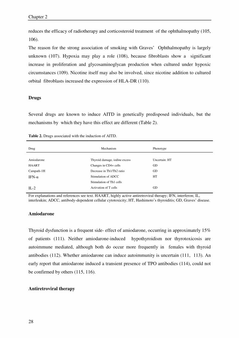
Several drugs are known to induce AITD in genetically predisposed individuals, but the
mechanisms by which they have this effect are different (Table 2).
Table 2. Drugs associated with the induction of AITD.
Drug
Mechanism
Phenotype
Amiodarone
Thyroid damage, iodine excess
Uncertain: HT
HAART Changes in CD4+ cells GD
Campath-1H Decrease in Th1/Th2 ratio GD
IFN-α Stimulation of ADCC
Stimulation of Th1 cells
HT
IL-2 Activation of T cells GD
For explanations and references see text. HAART, highly active antiretroviral therapy; IFN, interferon; IL,
interleukin; ADCC, antibody-dependent cellular cytotoxicity; HT, Hashimoto’s thyroiditis; GD, Graves’ disease.
Amiodarone
Thyroid dysfunction is a frequent side- effect of amiodarone, occurring in approximately 15%
of patients (111). Neither amiodarone-induced hypothyroidism nor thyrotoxicosis are
autoimmune mediated, although both do occur more frequently in females with thyroid
antibodies (112). Whether amiodarone can induce autoimmunity is uncertain (111, 113). An
early report that amiodarone induced a transient presence of TPO antibodies (114), could not
be confirmed by others (115, 116).
Antiretroviral therapy
The environment and autoimmune thyroid diseases
29
Highly active antiretroviral therapy (HAART) has been found to be associated with Graves’
disease, occurring 16–19 months after initiation of different combinations of indinavir,
stavudine, lamivudine and ritonavir (117). It may be related to HAART- induced changes in
CD4 T cells (118).
Campath-1H
This humanized anti-CD52 monoclonal antibody induced Graves’ disease in one-third of
patients with multiple sclerosis treated with this compound (119). The reason for this is
unknown, but since multiple sclerosis is not associated with AITD and the patients in whom
Graves’ disease occurred were not predisposed to the development of AITD (they lacked TPO
antibodies), the effect should be related to the antibody. Campath-1H suppresses Th1
lymphocytes and thus shifts the Th1/Th2 balance towards antibody production and hence
apparently towards a humoral immune response against the TSH-receptor (22).
IFN-αααα
IFN-α is widely used in the treatment of hepatitis C virus infection (120). Unlike IFN-g (121),
it is strongly associated with the induction of AITD (122). Risk factors for the development of
autoimmune thyroid dysfunction include the female sex (RR, 4.4; 95% CI, 3.2–5.9) and the
pretreatment presence of TPO antibodies (RR, 3.9; 95% CI, 1.9–8.1) (122). IFN-a treatment
can induce three types of thyroid dysfunction: autoimmune hypothyroidism, destructive
thyroiditis and hyperthyroidism. These can occur at any time after the start of treatment with a
median of 17 weeks (123). Hypothyroidism is slightly more frequent than thyrotoxicosis, and
in the large majority of cases it is of autoimmune origin leading to permanent thyroid failure
in approximately 60% of patients (124, 125). Graves’ hyperthyroidism is the cause of
thyrotoxicosis in about half of the patients; the rest suffer from silent thyroiditis.
IFN-α is a type I interferon (like IFN-β, but not IFN-γ which is a type II IFN) and stimulates
Th1 development (126). It has strong antiviral activity by promoting HLA-I class I expression
leading to recognition of virus-infected cells by cytotoxic T-lymphocytes (127). It also
enhances antibody-dependent cell-mediated immunity by upregulating Fc-receptor density on
lymphoid cells (128). Since infections with various viruses stimulate endogenous IFN-α
Chapter 2
30
production (129), we postulated that viral infections may also precipitate AITD via this IFN
pathway (see below) (122).
IL-2
IL-2 is used in the treatment of HIV infection and in metastatic renal carcinoma and
melanoma. Its pleiotropic immune effects include activation of T cells and among them
autoreactive lymphocytes (130). IL-2 is involved in autoimmunity and it was shown recently
that labeled IL-2 could be used to visualize sites of autoimmune inflammation in the pancreas
of pre-diabetics and in Hashimoto’s thyroiditis patients (131). Shortterm IL-2 administration
induces an increase in serum thyroxine (T4), 3,5,30-triiodothyronine (T3) and TSH levels,
probably via a direct central stimulation of the pituitary (132); long-term use is associated
with hypothyroidism, occurring in as many as 16% of patients (130). However, in most of
these patients other therapies (lymphokine-activated killer (LAK) cell infusion) were used
concomitantly and the hypothyroidism was not always of autoimmune origin (133, 134).
Granulocyte-macrophage colony-stimulating factor (GM-CSF)
GM-CSF may activate mature lymphocytes and hence aggravate or induce autoimmunity
against the thyroid (130). Nevertheless, such a side-effect has been reported only rarely
(135). In one study among 25 patients, only two patients with pre-existing TPO antibodies
suffered from transient hypothyroidism (136). In another study, however, no thyroid
dysfunction was found among 20 patients treated with GM-CSF despite the fact that two had
positive antithyroidal antibodies (137).
Irradiation
Irradiation of the thyroid gland may expose thyroidal antigens to the immune system and thus
induce autoimmunity by stimulation of dendritic cells (138). Both external irradiation and
internal irradiation by 131I are associated with AITD.
External irradiation
The environment and autoimmune thyroid diseases
31
External irradiation is a clear risk factor for the induction of thyroid cancer, but also of
hypothyroidism. In a large series of 1677 patients irradiated to the neck because of Hodgkin’s
disease, hypothyroidism was found in 47% after a median of 4.0 (0.2–23.7) years after
treatment (139). This is probably caused by damage to the gland and is not autoimmune
mediated. However, external irradiation is also associated with Graves’ disease, occurring in
3.3% of the patients in the same study. This was confirmed in another study, where Graves’
disease was diagnosed in 5% of 1791 irradiated patients; an 8-fold greater incidence rate than
in controls (140). The reason for this association may lie in the exposure of TSH-receptor
protein to the immune system and this may also be the reason that external neck irradiation
also enhances the risk for Graves’ ophthalmopathy (141–144).
131I therapy
Radioactive iodine is frequently used in the treatment of Graves’ hyperthyroidism and
multinodular goiter. In the last decade, it has become clear that it can induce the occurrence
of Graves’ hyperthyroidism in patients treated for (non-)toxic multinodular goiter (145, 146).
This complication typically occurs after 3–6 months and is seen in 4–5% of cases; it occurs
in parallel to an increase in TSH-receptor autoantibodies (147, 148). Interestingly, TSH-
receptor antibodies were not induced in ten patients without this complication, indicating that
this induction only occurs in otherwise – genetically – predisposed individuals (147).
Environmental radiation (nuclear fall-out)
In addition, environmental radiation exposure such as occurred after the dropping of the
nuclear bombs on Nagasaki and Hiroshima, or the Chernobyl nuclear plant accident, may
also damage the thyroid and expose antigen to the immune system. Indeed, the survivors of
the atomic bomb on Nagasaki not only have an increased risk of thyroid cancer, but also of
antibody- positive hypothyroidism (149). The same appears to be true for the people exposed
to the Chernobyl fallout. In one case–control study, the OR for the development of TPO
antibodies was 6.89 (95% CI, 3.17–14.99) and was higher in girls (9.64) than in boys (4.19)
(150). This was confirmed in another case– control study, where 18.9% of children in the
exposed area had TPO antibodies versus only 5% of controls from a non-exposed region in
southwestern Russia (151). There was no difference in thyroid volume or function. However,
there are also a number of studies that failed to find an association with TPO antibodies
Chapter 2
32
(152–155). Nevertheless, when Eheman et al. (156) reviewed the literature they concluded
that low-dose environmental radiation exposure may be associated with the development of
AITD.
Viral infections
In view of the association between IFN-α and AITD, it has been suggested that viruses
causing high endogenous IFN-α levels may also be associated with the induction of AITD.
One such virus is the Coxsackie B virus, which has been implicated in the induction of type I
or insulin-dependent diabetes mellitus (IDDM). Evidence of a recent Coxsackie B infection
was found more frequently in children who developed IDDM than in controls (157–159). In
another study, 39/56 (70%) patients with IDDM of recent onset had high IFN-α levels and in
half of them the Coxsackie B virus could be detected, while the virus was absent in IDDM
patients with low IFN-α levels (160). In line with this, IFN-α induction by injection of
polyinosinic polycytidylic acid (Poly IC) could induce IDDM in a rat strain that does not
spontaneously develop IDDM (161).
IFN-α may thus act as a non-specific stimulus of the induction of autoimmunity. However,
whether AITD is associated with viral infections is unknown since no studies like those
mentioned above have been done in this field. Only congenital rubella infection, a strong risk
factor for IDDM (162), is known to be associated with the presence of TPO antibodies in
children, but this syndrome is very rare (163). In addition, there have been reports on the
presence of retroviral sequences and proteins in thyroid glands from patients with AITD such
as the gag protein from the human foamy virus (HFV) (164). The importance of this virus is
doubtful, because HFV sequences can be found in blood lymphocytes from both Graves’
disease patients and healthy controls (165). Viruses are thought to induce De Quervain’s
thyroiditis; however, this is not an utoimmune condition but rather an inflammatory disorder
with high levels of C-reactive protein (166).
Bacterial infections
Several autoimmune diseases have been linked to bacterial infections, including Graves’
disease (Table 3) (167). There are several hypotheses to explain this association. The first
implies molecular mimicry (168). Bacterial pathogens can have an antigen sharing homology
with a self-antigen and an immune reaction against the bacterial antigen may then lead to a
The environment and autoimmune thyroid diseases
33
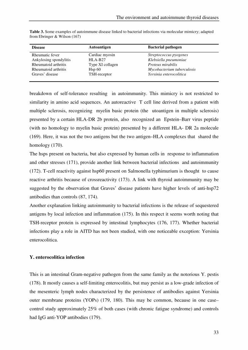
Table 3. Some examples of autoimmune disease linked to bacterial infections via molecular mimicry; adapted
from Ebringer & Wilson (167)
Disease Autoantigen Bacterial pathogen
Rheumatic fever Cardiac myosin Streptococcus pyogenes
Ankylosing spondylitis HLA-B27 Klebsiella pneumoniae
Rheumatoid arthritis Type XI collagen Proteus mirabilis
Rheumatoid arthritis Hsp 60 Mycobacterium tuberculosis
Graves’ disease TSH-receptor Yersinia enterocolitica
breakdown of self-tolerance resulting in autoimmunity. This mimicry is not restricted to
similarity in amino acid sequences. An autoreactive T cell line derived from a patient with
multiple sclerosis, recognizing myelin basic protein (the utoantigen in multiple sclerosis)
presented by a certain HLA-DR 2b protein, also recognized an Epstein–Barr virus peptide
(with no homology to myelin basic protein) presented by a different HLA- DR 2a molecule
(169). Here, it was not the two antigens but the two antigen–HLA complexes that shared the
homology (170).
The hsps present on bacteria, but also expressed by human cells in response to inflammation
and other stresses (171), provide another link between bacterial infections and autoimmunity
(172). T-cell reactivity against hsp60 present on Salmonella typhimurium is thought to cause
reactive arthritis because of crossreactivity (173). A link with thyroid autoimmunity may be
suggested by the observation that Graves’ disease patients have higher levels of anti-hsp72
antibodies than controls (87, 174).
Another explanation linking autoimmunity to bacterial infections is the release of sequestered
antigens by local infection and inflammation (175). In this respect it seems worth noting that
TSH-receptor protein is expressed by intestinal lymphocytes (176, 177). Whether bacterial
infections play a role in AITD has not been studied, with one noticeable exception: Yersinia
enterocolitica.
Y. enterocolitica infection
This is an intestinal Gram-negative pathogen from the same family as the notorious Y. pestis
(178). It mostly causes a self-limiting enterocolitis, but may persist as a low-grade infection of
the mesenteric lymph nodes characterized by the persistence of antibodies against Yersinia
outer membrane proteins (YOPs) (179, 180). This may be common, because in one case–
control study approximately 25% of both cases (with chronic fatigue syndrome) and controls
had IgG anti-YOP antibodies (179).
Chapter 2
34
In the 1970s two studies reported a higher prevalence of Y. enterocolitica (especially
serotype O:3) antibodies in Graves’ disease patients (50 and 66% respectively) than in
controls (28 and 8% respectively) (181, 182). These findings prompted an investigation into
the possibility of shared antigens with the thyroid and it was found that Y. enterocolitica had
specific binding sites for TSH in the 1028M range (183). These binding sites were also
recognized by TSH-receptor autoantibodies (184). Antibodies against YOPs raised in rabbits
displaced TSH from binding to TSH-receptor protein, and these antibodies stained thyroid
epithelial cells in immunohistochemistry (185, 186). Cellular immunity is also involved,
because Y. enterocolitica can inhibit the migration of lymphocytes from patients with
Graves’ disease (181), and in a mouse model Y. enterocolitica acts as a superantigen (187).
The cross-reacting protein(s), at first thought to be the TSH-receptor itself, has not been
identified yet, but appears to have conformational homology with the TSH-receptor, and one
may be hsp70 (188). Others have found two low molecular weight envelope proteins (of 5.5
and 8 kDa) that are cross-reactive with the extracellular part of the TSH-receptor (189). The
protein(s) do not seem to be Y. enterocolitica specific, since TSH binding sites were also
found on other intestinal pathogens (190).
Y. enterocolitica infections are common. In a large Danish study, 8.3% of 48857 patients with
bacterial enteritis had a Y. enterocolitica infection (191). In Canada, the annual incidence of
Y. enterocolitica infections is 3/100 000 subjects (192); in The Netherlands the yearly
incidence is 1.2/100 000 inhabitants (193). In view of the high incidence of AITD, Y.
enterocolitica infections may thus play a role in its development. With more specific assays
using YOPs, there is indeed an association between antibodies against YOPs and AITD. IgA
antibodies are thought to indicate that the primary immune response is mounted in the gut,
and not in the thyroid, suggesting the Y. enterocolitica infection is causative (194). In a
German study, IgG class antibodies were found in 72% of Graves’ patients and in 66% of
patients with Hashimoto’s thyroiditis as compared with 35% in controls; IgA antibodies were
found in respectively 33, 37 and 11% (195). In Greece, 25% of Hashimoto patients had IgG
antibodies and 2.8% had IgA antibodies, compared with 2 and 0% respectively in controls
(196). A higher incidence of Y. enterocolitica antibodies in Graves and Hashimoto patients
than in controls was also found in Japan and Turkey (197, 198). However, there are also
studies that could not confirm these findings and found a similar rate of seropositivity in
AITD patients and controls (199, 200). We recently found that 40% of 803 female relatives
of patients with documented AITD had IgG antibodies against Y. enterocolitica YOPs (22%
had IgA antibodies), as compared with only 24% of controls (13% had IgA antibodies), but
The environment and autoimmune thyroid diseases
35
the presence of these antibodies was unrelated to the presence of thyroid autoimmunity (201).
We hypothesized that this high rate of probably persisting, low-grade Y. enterocolitica
infections in relatives of AITD patients is related to a particular genetic make-up facilitating
Y. enterocolitica infections independently from conferring a risk for AITD.
Concluding remarks
AITD is a polygenetic disease and currently only a few genes have been identified as causing
AITD, all with a rather low RR which is seldom higher than 3.0. Nevertheless, it has been
calculated that 79% of the susceptibility to develop Graves’ disease can be attributed to
genetic factors, leaving 21% for environmental factors (202). Reviewing these non-genetic
factors, it appears that multiple environmental factors are involved in the induction of AITD
in genetically predisposed individuals (Table 4).
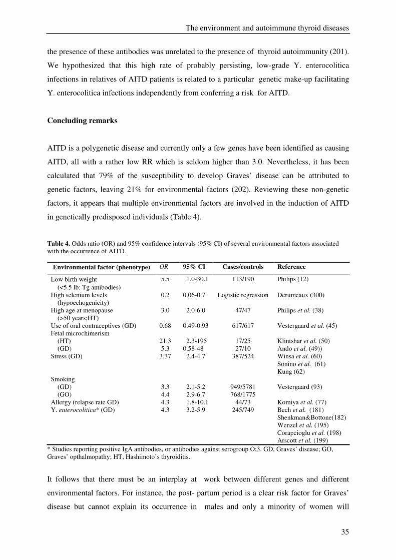
Table 4. Odds ratio (OR) and 95% confidence intervals (95% CI) of several environmental factors associated
with the occurrence of AITD.
Environmental factor (phenotype) OR 95% CI Cases/controls Reference
Low birth weight
(<5.5 lb; Tg antibodies)
5.5 1.0-30.1 113/190 Philips (12)
High selenium levels
(hypoechogenicity)
0.2 0.06-0.7 Logistic regression Derumeaux (300)
High age at menopause
(>50 years;HT)
3.0 2.0-6.0 47/47 Philips et al. (38)
Use of oral contraceptives (GD) 0.68 0.49-0.93 617/617 Vestergaard et al. (45)
Fetal microchimerism
(HT)
(GD)
21.3
5.3
2.3-195
0.58-48
17/25
27/10
Klintshar et al. (50)
Ando et al. (49))
Stress (GD) 3.37 2.4-4.7 387/524 Winsa et al. (60)
Sonino et al. (61)
Kung (62)
Smoking
(GD)
(GO)
3.3
4.4
2.1-5.2
2.9-6.7
949/5781
768/1775
Vestergaard (93)
Allergy (relapse rate GD) 4.3 1.8-10.1 44/73 Komiya et al. (77)
Y. enterocolitica* (GD) 4.3 3.2-5.9 245/749 Bech et al. (181)
Shenkman&Bottone(182)
Wenzel et al. (195)
Corapcioglu et al. (198)
Arscott et al. (199)
* Studies reporting positive IgA antibodies, or antibodies against serogroup O:3. GD, Graves’ disease; GO,
Graves’ opthalmopathy; HT, Hashimoto’s thyroiditis.
It follows that there must be an interplay at work between different genes and different
environmental factors. For instance, the post- partum period is a clear risk factor for Graves’
disease but cannot explain its occurrence in males and only a minority of women will
Chapter 2
36
develop Graves’ disease in the post partum period. In other words, one gene may predispose
for AITD in general while a second gene may dictate whether childbirth will precipitate its
onset or not, while in another woman with the same first susceptibility gene the trigger may
lie in a stress-coping gene. This would explain the rather low OR values of individual –
genetic and environmental – risk factors: a specific environmental risk factor may have a
very large RR in a person with a certain genetic make- up. This implies that the true
importance of both genes and environment can only be discerned when studied in
conjunction. Such an approach requires a much larger sample size and probably multi-center
cooperation. The good news is, however, that we now have powerful computers to perform
the necessary multivariate analyses. It also means that we need a much more rigorous
phenotype definition. Environmental risk factors are more likely to be important in older
patients with AITD than in younger ones, their influence may also differ between patients
who come from a family of AITD patients and isolated cases, or between males and females.
This does not mean that a further search for specific risk factors is useless. When reviewing
all factors, one of the most promising is fetal microchimerism. To date this has been limited
to the search for remnants of male fetuses (the Y chromosome), but female fetuses are likely
to have the same impact. This implies further studies into the genetic make-up of partners of
AITD patients. A second area holding promise is the importance of viral infections in the
induction of AITD. They appear to be of importance in the induction of IDDM, lead to an
endogenous surge in IFN-a (a clear risk factor for AITD when administered as a drug) and
are more likely to occur in selenium deficiency (which is itself another risk factor for AITD).
The ultimate goal of this research is to find a feasible way of preventing the occurrence of
AITD; for this we will need progress both in delineating the genetic background and in
clarifying the precipitating environmental factors.
The environment and autoimmune thyroid diseases
37
References
1. Bach JF. The effect of infections on susceptibility to autoimmune and allergic diseases.
New England Journal of Medicine 2002 347 911–920.
2. Gough SCL. The immunogenetics of Graves’ disease. Current Opinion in Endocrinology
and Diabetes 1999 6 270–276.
3. Tait KF & Gough SC. The genetics of autoimmune endocrine disease. Clinical
Endocrinology 2003 59 1–11.
4. Vaidya B, Kendall-Taylor P & Pearce SH. The genetics of autoimmune thyroid disease.
Journal of Clinical Endocrinology and Metabolism 2002 87 5385–5397.
5. Gough SC. The genetics of Graves’ disease. Endocrinology and Metabolism Clinics of
North America 2000 29 255–266.
6. Barbesino G & Chiovato L. The genetics of Hashimoto’s disease. Endocrinology and
Metabolism Clinics of North America 2000 29 357–374.
7. Strieder TG, Prummel MF, Tijssen JG, Endert E & Wiersinga WM. Risk factors for and
prevalence of thyroid disorders in a cross-sectional study among healthy female relatives
of patients with autoimmune thyroid disease. Clinical Endocrinology 2003 59 396–401.
8. Dayan CM & Daniels GH. Chronic autoimmune thyroiditis. New England Journal of
Medicine 1996 335 99–107.
9. Forsen T, Eriksson JG, Tuomilehto J, Osmond C & Barker DJ. Growth in utero and
during childhood among women who develop coronary heart disease: longitudinal study.
British Medical Journal 1999 319 1403–1407.
10. Roseboom TJ, van der Meulen JH, Ravelli AC, Osmond C, Barker DJ & Bleker OP.
Effects of prenatal exposure to the Dutch famine on adult disease in later life: an
overview. Molecular and Cellular Endocrinology 2001 185 93–98.
11. Winick M & Noble A. Cellular response in rats during malnutrition at various ages.
Journal of Nutrition 1966 89 300–306.
12. Phillips DI, Cooper C, Fall C, Prentice L, Osmond C, Barker DJ et al. Fetal growth and
autoimmune thyroid disease. Quarterly Journal of Medicine 1993 86 247–253.
13. Phillips DI, Osmond C, Baird J, Huckle A & Rees-Smith B. Is birthweight associated
with thyroid autoimmunity? A study in twins. Thyroid 2002 12 377–380.
14. Brix TH, Kyvik KO & Hegedus L. Low birth weight is not associated with clinically
overt thyroid disease: a population based twin case-control study. Clinical Endocrinology
2000 53 171–176.
15. Laurberg P, Pedersen KM, Hreidarsson A, Sigfusson N, Iversen E & Knudsen PR. Iodine
intake and the pattern of thyroid disorders: a comparative epidemiological study of
thyroid abnormalities in the elderly in Iceland and in Jutland, Denmark. Journal of
Clinical Endocrinology and Metabolism 1998 83 765–769.
16. Laurberg P, Nohr SB, Pedersen KM, Hreidarsson AB, Andersen S, Bulow P et al.
Thyroid disorders in mild iodine deficiency. Thyroid 2000 10 951–963.
17. Laurberg P, Pedersen KM, Vestergaard H & Sigurdsson G. High incidence of
multinodular toxic goitre in the elderly population in a low iodine intake area vs. high
incidence of Graves’ disease in the young in a high iodine intake area: comparative
surveys of thyrotoxicosis epidemiology in East-Jutland Denmark and Iceland. Journal of
Internal Medicine 1991 229 415–420.
18. Pedersen IB, Knudsen N, Jorgensen T, Perrild H, Ovesen L & Laurberg P. Thyroid
peroxidase and thyroglobulin autoantibodies in a large survey of populations with mild
and moderate iodine deficiency. Clinical Endocrinology 2003 58 36–42.
Chapter 2
38
19. Wiersinga WM & Braverman LE. Iodine-induced thyroid disease. In Contemporary
Endocrinology: Diseases of the Thyroid, edn 2,pp 347–362. Ed. LE Braverman. Totowa,
NJ: Humana Press Inc., 2003.
20. Roti E & Uberti ED. Iodine excess and hyperthyroidism. Thyroid 2001 11 493–500.
21. Markou K, Georgopoulos N, Kyriazopoulou V & Vagenakis AG. Iodine-induced
hypothyroidism. Thyroid 2001 11 501–510.
22. Weetman AP. Autoimmune thyroid disease: propagation and progression. European
Journal of Endocrinology 2003 148 1–9.
23. Ruwhof C & Drexhage HA. Iodine and thyroid autoimmune disease in animal models.
Thyroid 2001 11 427–436.
24. Sundick RS, Bagchi N & Brown TR. The role of iodine in thyroid autoimmunity: from
chickens to humans: a review. Autoimmunity 1992 13 61–68.
25. Lam-Tse WK, Lernmark A & Drexhage HA. Animal models of endocrine/organ-specific
autoimmune diseases: do they really help us to understand human autoimmunity?
Springer Seminars in Immunopathology 2002 24 297–321.
26. Rayman MP. The importance of selenium to human health. Lancet 2000 356 233–241.
27. Beck MA, Shi Q, Morris VC & Levander OA. Rapid genomic evolution of a non-
virulent coxsackievirus B3 in selenium-deficient mice results in selection of identical
virulent isolates. Nature Medicine 1995 1 433–436.
28. Zimmermann MB & Kohrle J. The impact of iron and selenium deficiencies on iodine
and thyroid metabolism: biochemistry and relevance to public health. Thyroid 2002 12
867–878.
29. Barrington JW, Lindsay P, James D, Smith S & Roberts A. Selenium deficiency and
miscarriage: a possible link? British Journal of Obstetrics and Gynaecology 1996 103
130–132.
30. Derumeaux H, Valeix P, Castetbon K, Bensimon M, Boutron-Ruault MC, Arnaud J et al.
Association of selenium with thyroid volume and echostructure in 35- to 60-year-old
French adults. European Journal of Endocrinology 2003 148 309–315.1
31. Gartner R, Gasnier BC, Dietrich JW, Krebs B & Angstwurm MW. Selenium
supplementation in patients with autoimmune thyroiditis decreases thyroid peroxidase
antibodies concentrations. Journal of Clinical Endocrinology and Metabolism 2002 87
1687–1691.
32. Duntas LH, Mantzou E & Koutras DA. Effects of a six month treatment with
selenomethionine in patients with autoimmune thyroiditis. European Journal of
Endocrinology 2003 148 389–393.
33. Vanderpump MP, Tunbridge WM, French JM, Appleton D, Bates D, Clark F et al. The
incidence of thyroid disorders in the community: a twenty-year follow-up of the
Whickham Survey. Clinical Endocrinology 1995 43 55–68.
34. Stagnaro-Green A, Roman SH, Cobin RH, el Harazy E, Wallenstein S & Davies TF. A
prospective study of lymphocyte-initiated immunosuppression in normal pregnancy:
evidence of a T-cell etiology for postpartum thyroid dysfunction. Journal of Clinical
Endocrinology and Metabolism 1992 74 645–653.
35. Draca S. Is pregnancy a model how we should control some autoimmune diseases?
Autoimmunity 2002 35 307–312.
36. Beagley KW & Gockel CM. Regulation of innate and adaptive immunity by the female
sex hormones oestradiol and progesterone. FEMSImmunology and Medical
Microbiology 200338 13–22.
37. Vanderpump MP, French JM, Appleton D, Tunbridge WM & Kendall-Taylor P. The
prevalence of hyperprolactinaemia and association with markers of autoimmune thyroid
The environment and autoimmune thyroid diseases
39
disease in survivors of the Whickham Survey cohort. Clinical Endocrinology 1998 48
39–44.
38. Phillips DI, Lazarus JH & Butland BK. The influence of pregnancy and reproductive
span on the occurrence of autoimmune thyroiditis. Clinical Endocrinology 1990 32 301–
306.
39. Massoudi MS, Meilahn EN, Orchard TJ, Foley TP Jr, Kuller LH, Costantino JP et al.
Prevalence of thyroid antibodies among healthy middle-aged women. Findings from the
thyroid study in healthy women. Annals of Epidemiology 1995 5 229–233.
40. Ogard CG, Ogard C & Almdal TP. Thyroid-associated orbitopathy developed during
hormone replacement therapy. Acta Ophthalmologica Scandinavica 2001 79 426–427.
41. Petitti DB. Clinical practice. Combination estrogen–progestin oral contraceptives. New
England Journal of Medicine 2003 349 1443–1450.
42. Frank P & Kay CR. Incidence of thyroid disease associated with oral contraceptives.
British Medical Journal 1978 2 1531.
43. Knudsen N, Bulow I, Laurberg P, Perrild H, Ovesen L & Jorgensen T. Low goitre
prevalence among users of oral contraceptives in a population sample of 3712 women.
Clinical Endocrinology 2002 57 71– 76.
44. Barrere X, Valeix P, Preziosi P, Bensimon M, Pelletier B, Galan P et al. Determinants of
thyroid volume in healthy French adults participating in the SU.VI.MAX cohort.
Clinical Endocrinology 2000 52 273–278.
45. Vestergaard P, Rejnmark L, Weeke J, Hoeck HC, Nielsen HK, Rungby J et al. Smoking
as a risk factor for Graves’ disease, toxic nodular goiter, and autoimmune
hypothyroidism. Thyroid 2002 12 69–75.
46. Bianchi DW, Zickwolf GK, Weil GJ, Sylvester S & DeMaria MA. Male fetal progenitor
cells persist in maternal blood for as long as 27 years postpartum. PNAS 1996 93 705–
708.
47. Khosrotehrani K & Bianchi DW. Fetal cell microchimerism: helpful or harmful to the
parous woman? Current Opinion in Obstetrics and Gynecology 2003 15 195–199.
48. Imaizumi M, Pritsker A, Unger P & Davies TF. Intrathyroidal fetal microchimerism in
pregnancy and postpartum. Endocrinology 2002 143 247–253.
49. Ando T, Imaizumi M, Graves PN, Unger P & Davies TF. Intrathyroidal fetal
microchimerism in Graves’ disease. Journal of Clinical Endocrinology and Metabolism
2002 87 3315–3320.
50. Klintschar M, Schwaiger P, Mannweiler S, Regauer S & Kleiber M. Evidence of fetal
microchimerism in Hashimoto’s thyroiditis. Journal of Clinical Endocrinology and
Metabolism 2001 86 2494–2498.
51. Badenhoop K. Intrathyroidal microchimerism in Graves’ disease or Hashimoto’s
thyroiditis: regulation of tolerance or alloimmunity by fetal–maternal immune
interaction. European Journal of Endocrinology 2003 (in press).
52. Besedovsky HO & del Rey A. Immune-neuro-endocrine interactions: facts and
hypotheses. Endocrine Reviews 1996 17 64–102.
53. Sternberg EM. Neuroendocrine regulation of autoimmune/inflammatory disease. Journal
of Endocrinology 2001 169 429–435.
54. Elenkov IJ & Chrousos GP. Stress hormones, Th1/Th2 patterns, pro/anti-inflammatory
cytokines and susceptibility to disease. Trends in Endocrinology and Metabolism 1999
10 359–368.
55. Elenkov IJ & Chrousos GP. Stress hormones, proinflammatory and antiinflammatory
cytokines, and autoimmunity. Annals of the New York Academy of Sciences 2002 966
290–303.
56. Rosch PJ. Stressful life events and Graves’ disease. Lancet 1993 342 566–567.
Chapter 2
40
57. Gorman CA. A critical review of the role of stress in hyperthyroidism. In The Thyroid
Gland, Environment and Autoimmunity, pp 191–200. Eds HA Drexhage, JJM de Vijlder
&WM Wiersinga. Amsterdam: Elsevier Science Publishers, 1990.
58. Paunkovic N, Paunkovic J, Pavlovic O & Paunovic Z. The significant increase in
incidence of Graves’ disease in eastern Serbia during the civil war in the former
Yugoslavia (1992 to 1995).Thyroid 1998 8 37– 41.
59. Hadden DR & McDevitt DG. Environmental stress and thyrotoxicosis.Absence of
association. Lancet 974 2 577–578.
60. Winsa B, Adami HO, Bergstrom R, Gamstedt A, Dahlberg PA, Adamson U et al.
Stressful life events and Graves’ disease. Lancet 1991 338 1475–1479.
61. Sonino N, Girelli ME, Boscaro M, Fallo F, Busnardo B & Fava GA. Life events in the
pathogenesis of Graves’ disease. A controlled study. Acta Endocrinologica (Copenh)
1993 128 293–296.
62. Kung AW. Life events, daily stresses and coping in patients with Graves’ disease.
Clinical Endocrinology 1995 42 303–308.
63. Radosavljevic VR, Jankovic SM & Marinkovic JM. Stressful life events in the
pathogenesis of Graves’ disease. European Journal of Endocrinology 1996 134 699–701.
64. Dayan CM. Stressful life events and Graves’ disease revisited. Clinical Endocrinology
2001 55 13–14.
65. Chiovato L & Pinchera A. Stressful life events and Graves’ disease. European Journal of
Endocrinology 1996 134 680–682.
66. Benvenga S. Benzodiazepine and remission of Graves’ disease. Thyroid 1996 6 659–660.
67. Fukao A, Takamatsu J, Murakami Y, Sakane S, Miyauchi A, Kuma K et al. The
relationship of psychological factors to the prognosis of hyperthyroidism in antithyroid
drug-treated patients with Graves’ disease. Clinical Endocrinology 2003 58 550–555.
68. Matos-Santos A, Nobre EL, Costa JG, Nogueira PJ, Macedo A, Galvao-Teles A et al.
Relationship between the number and impact of stressful life events and the onset of
Graves’ disease and toxic nodular goitre. Clinical Endocrinology 2001 55 15–19.
69. Wiersinga WM. Environmental factors in autoimmune thyroid disease. Experimental and
Clinical Endocrinology and Diabetes 1999 107 (Suppl 3) S67–S70.
70. Westphal SA. Seasonal variation in the diagnosis of Graves’ disease. Clinical
Endocrinology 1994 41 27–30.
71. Phillips DI, Barker DJ & Morris JA. Seasonality of thyrotoxicosis. Journal of
Epidemiology and Community Health 1985 39 72–74.
72. Phillips DI, Nelson M, Barker DJ, Morris JA & Wood TJ. Iodine in milk and the
incidence of thyrotoxicosis in England. Clinical Endocrinology 1988 28 61–66.
73. Pedotti R, De Voss JJ, Steinman L & Galli SJ. Involvement of both ‘allergic’ and
‘autoimmune’ mechanisms in EAE, MS and other autoimmune diseases. Trends in
Immunology 2003 24 479–484.
74. Stene LC & Nafstad P. Relation between occurrence of type 1 diabetes and asthma.
Lancet 2001 357 607–608.
75. Moens HJ, Wiersinga WM & Drexhage HA. Association between autoimmune thyroid
disease, atopy, and urticaria? Lancet 1984 2 582–583.
76. Sato A, Takemura Y, Yamada T, Ohtsuka H, Sakai H, Miyahara Y et al. A possible role
of immunoglobulin E in patients with hyperthyroid Graves’ disease. Journal of Clinical
Endocrinology and Metabolism 1999 84 3602–3605.
77. Komiya I, Yamada T, Sato A, Kouki T, Nishimori T & Takasu N. Remission and
recurrence of hyperthyroid Graves’ disease during and after methimazole treatment
when assessed by IgE and interleukin 13. Journal of Clinical Endocrinology and
Metabolism 2001 86 3540–3544.
The environment and autoimmune thyroid diseases
41
78. Hidaka Y, Amino N, Iwatani Y, Itoh E, Matsunaga M & Tamaki H. Recurrence of
thyrotoxicosis after attack of allergic rhinitis in patients with Graves’ disease. Journal of
Clinical Endocrinology and Metabolism 1993 77 1667–1670.
79. Hidaka Y, Masai T, Sumizaki H, Takeoka K, Tada H & Amino N. Onset of Graves’
thyrotoxicosis after an attack of allergic rhinitis. Thyroid 1996 6 349–351.
80. Rottem M. Chronic urticaria and autoimmune thyroid disease: is there a link?
Autoimmunity Review 2003 2 69–72.
81. Turktas I, Gokcora N, Demirsoy S, Cakir N & Onal E. The association of chronic
urticaria and angioedema with autoimmune thyroiditis. International Journal of
Dermatology 1997 36 187–190.
82. Leznoff A & Sussman GL. Syndrome of idiopathic chronic urticaria and angioedema
with thyroid autoimmunity: a study of 90 patients. Journal of Allergy and Clinical
Immunology 1989 84 66–71.
83. George J, Levy Y & Shoenfeld Y. Smoking and immunity: an additional player in the
mosaic of autoimmunity. Scandinavian Journal of Immunology 1997 45 1–6.
84. McAllister-Sistilli CG, Caggiula AR, Knopf S, Rose CA, Miller AL & Donny EC. The
effects of nicotine on the immune system. Psychoneuroendocrinology 1998 23 175–187.
85. Ronchetti R, Macri F, Ciofetta G, Indinnimeo L, Cutrera R, Bonci E et al. Increased
serum IgE and increased prevalence of eosinophilia in 9-year-old children of smoking
parents. Journal of Allergy and Clinical Immunology 1990 86 400–407.
86. Weitzman M, Gortmaker S, Walker DK & Sobol A. Maternal smoking and childhood
asthma. Pediatrics 1990 85 505–511.
87. Prummel MF, Van Pareren Y, Bakker O & Wiersinga WM. Antiheat shock protein
(hsp)72 antibodies are present in patients with Graves’ disease (GD) and in smoking
control subjects. Clinical and Experimental Immunology 1997 110 292–295.
88. Prummel MF, Wiersinga WM, Van der Gaag R, Mourits MP & Koornneef L. Soluble IL-
2 receptor levels in patients with Graves’ ophthalmopathy. Clinical and Experimental
Immunology 1992 88 405–409.
89. Hofbauer LC, Muhlberg T, Konig A, Heufelder G, Schworm HD & Heufelder AE.
Soluble interleukin- 1 receptor antagonist serum levels in smokers and nonsmokers with
Graves’ ophthalmopathy undergoing orbital radiotherapy. Journal of Clinical
Endocrinology and Metabolism 1997 82 2244–2247.
90. Wakelkamp IM, Gerding MN, van der Meer JW, Prummel MF & Wiersinga WM.
Smoking and disease severity are independent determinants of serum adhesion molecule
levels in Graves’ ophthalmopathy. Clinical and Experimental Immunology 2002 127
316–320.
91. Byron KA, Varigos GA & Wootton AM. IL-4 production is increased in cigarette
smokers. Clinical and Experimental Immunology 1994 95 333–336.
92. Heliovaara M, Aho K, Aromaa A, Knekt P & Reunanen A. Smoking and risk of
rheumatoid arthritis. Journal of Rheumatology 1993 20 1830–1835.
93. Vestergaard P. Smoking and thyroid disorders–a meta-analysis. European Journal of
Endocrinology 2002 146 153–161.
94. Hagg E & Asplund K. Is endocrine ophthalmopathy related to smoking? British Medical
Journal 1987 295 634–635.
95. Prummel MF & Wiersinga WM. Smoking and risk of Graves’ disease. Journal of the
American Medical Association 1993 269 479–482.
96. Bartalena L, Martino E, Marcocci C, Bogazzi F, Panicucci M, Velluzzi F et al. More on
smoking habits and Graves’ ophthalmopathy. Journal of Endocrinological Investigation
1989 12 733–737.
Chapter 2
42
97. Chen YL, Chang TC & Chen CJ. Influence of smoking on Graves’ disease with or
without ophthalmopathy and nontoxic nodular goiter in Taiwan. Journal of the Formosan
Medical Association 1994 93 40–44.
98. Shine B, Fells P, Edwards OM & Weetman AP. Association between Graves’
ophthalmopathy and smoking. Lancet 1990 335 1261–1263.
99. Winsa B, Mandahl A & Karlsson FA. Graves’ disease, endocrine ophthalmopathy and
smoking. Acta Endocrinologica 1993 128 156–160.
100. Tellez M, Cooper J & Edmonds C. Graves’ ophthalmopathy in relation to cigarette
smoking and ethnic origin. Clinical Endocrinology 1992 36 291–294.
101. Pfeilschifter J & Ziegler R. Smoking and endocrine ophthalmopathy:impact of smoking
severity and current vs lifetime cigarette consumption. Clinical Endocrinology 1996 45
477–481.
102. Nystrom E, Bengtsson C, Lapidus L, Petersen K & Lindstedt G. Smoking–a risk factor
for hypothyroidism. Journal of Endocrinological Investigation 1993 16 129–131.
103. Orgiazzi J & Madec AM. Reduction of the risk of relapse after withdrawal of medical
therapy for Graves’ disease. Thyroid 2002 12 849–853.
104. Glinoer D, de Nayer P & Bex M. Effects of l-thyroxine administration,TSH-receptor
antibodies and smoking on the risk of recurrence in Graves’ hyperthyroidism treated
with antithyroid drugs: a double- blind prospective randomized study. European Journal
of Endocrinology 2001 144 475–483.
105. Bartalena L, Marcocci C, Tanda ML, Manetti L, Dell’Unto E, Bartolomei MP et al.
Cigarette smoking and treatment outcomes in Graves ophthalmopathy. Annals of
Internal Medicine 1998 129 632–635.
106. Eckstein A, Quadbeck B, Mueller G, Rettenmeier AW, Hoermann R, Mann K et al.
Impact of smoking on the response to treatment of thyroid associated ophthalmopathy.
British Journal of ophthalmology 2003 87 773–776.
107. Bartalena L, Pinchera A & Marcocci C. Management of Graves’ ophthalmopathy: reality
and perspectives. Endocrine Reviews 2000 21 168–199.
108. Weetman AP. Determinants of autoimmune thyroid disease. Nature Immunology 2001 2
769–770.
109. Metcalfe RA & Weetman AP. Stimulation of extraocular muscle fibroblasts by cytokines
and hypoxia: possible role in thyroid-associated ophthalmopathy. Clinical
Endocrinology 1994 40 67–72.
110. Mack WP, Stasior GO, Cao HJ, Stasior OG & Smith TJ. The effect of cigarette smoke
constituents on the expression of HLA-DR in orbital fibroblasts derived from patients
with Graves ophthalmopathy. Ophthalmic and Plastic Recon structive Surgery 1999 15
260–271.
111. Wiersinga WM. Amiodarone and the thyroid. In Handbook of Experimental
Pharmacology, vol 128, pp 225–287. Eds AP Weetman & A Grossman. Berlin:
Springer-Verlag, 1997.
112. Trip MD, Wiersinga W & Plomp TA. Incidence, predictability, and pathogenesis of
amiodarone- induced thyrotoxicosis and hypothyroidism. American Journal of Medicine
1991 91 507–511.
113. Daniels GH. Amiodarone-induced thyrotoxicosis. Journal of Clinical Endocrinology and
Metabolism 2001 86 3–8.
114. Monteiro E, Galvao-Teles A, Santos ML, Mourao L, Correia MJ, Lopo TJ et al.
Antithyroid antibodies as an early marker for thyroid disease induced by amiodarone.
British Medical Journal 1986 292 227–228.
The environment and autoimmune thyroid diseases
43
115. Trip MD, Duren DR & Wiersinga WM. Two cases of amiodaroneinduced thyrotoxicosis
successfully treated with a short course of antithyroid drugs while amiodarone was
continued. British Heart Journal 1994 72 266–268.
116. Weetman AP, Bhandal SK, Burrin JM, Robinson K & McKennaW. Amiodarone and
thyroid autoimmunity in the United Kingdom. British Medical Journal 1988 297 33.
117. Gilquin J, Viard JP, Jubault V, Sert C & Kazatchkine MD. Delayed occurrence of
Graves’ disease after immune restoration with HAART. Highly active antiretroviral
therapy. Lancet 1998 352 1907–1908.
118. Weetman AP. Graves’ disease. New England Journal of Medicine 2000 343 1236–1248.
119. Coles AJ, Wing M, Smith S, Coraddu F, Greer S, Taylor C et al. Pulsed monoclonal
antibody treatment and autoimmune thyroid disease in multiple sclerosis. Lancet 1999
354 1691–1695.
120. Lauer GM & Walker BD. Hepatitis C virus infection. New England Journal of Medicine
2001 345 41–52.
121. Bhakri H, Sriskandan K, Davis T, Pettingale K & Tee D. Recombinant gamma interferon
and autoimmune thyroid disease. Lancet 1985 2 457.
122. Prummel MF & Laurberg P. Interferon-alpha and autoimmune thyroid disease. Thyroid
2003 13 547– 551.
123. Okanoue T, Sakamoto S, Itoh Y, Minami M, Yasui K, Sakamoto M et al. Side effects of
high-dose interferon therapy for chronic hepatitis C. Journal of Hepatology 1996 25
283–291.
124. Fattovich G, Giustina G, Favarato S & Ruol A. A survey of adverse events in 11,241
patients with chronic viral hepatitis treated with alfa interferon. Journal of Hepatology
1996 24 38–47.
125. Koh LK, Greenspan FS & Yeo PP. Interferon-alpha induced thyroid dysfunction: three
clinical presentations and a review of the literature. Thyroid 1997 7 891–896.
126. Farrar JD & Murphy KM. Type I interferons and T helper development. Immunology
Today 2000 21 484–489.
127. Parkin J & Cohen B. An overview of the immune system. Lancet 2001 357 1777–1789.
128. Burman P, Totterman TH, Oberg K & Karlsson FA. Thyroid autoimmunity in patients on
long term therapy with leukocyte-derived interferon. Journal of Clinical Endocrinology
and Metabolism 1986 63 1086–1090.
129. Biron CA. Interferons alpha and beta as immune regulators–a new look. Immunity 2001
14 661–664.
130. Vial T & Descotes J. Immune-mediated side-effects of cytokines in humans.Toxicology
1995 105 31–57.
131. Signore A, Picarelli A, Annovazzi A, Britton KE, Grossman AB, Bonanno E et al. 123I-
interleukin-2: biochemical characterization and in vivo use for imaging autoimmune
diseases. Nuclear Medicine Communications 2003 24 305–316.
132. Witzke O, Winterhagen T, Saller B, Roggenbuck U, Lehr I, Philipp T et al. Transient
stimulatory effects on pituitary-thyroid axis in patients treated with interleukin-2.
Thyroid 2001 11 665–670.
133. Atkins MB, Mier JW, Parkinson DR, Gould JA, Berkman EM & Kaplan MM.
Hypothyroidism after treatment with interleukin- 2 and lymphokine-activated killer
cells. New England Journal of Medicine 1988 318 1557–1563.
134. Meloni G, Trisolini SM, Capria S, Torelli GF, Baldacci E, Torromeo C et al. How long
can we give interleukin-2? Clinical and immunological evaluation of AML patients after
10 or more years of IL2 administration. Leukemia 2002 16 2016–2018.
Chapter 2
44
135. Hansen PB, Johnsen HE & Hippe E. Autoimmune hypothyroidism and granulocyte-
macrophage colony-stimulating factor. European Journal of Haematology 1993 50 183–
184.
136. Hoekman K, von Blomberg-van der Flier BM, Wagstaff J, Drexhage HA & Pinedo HM.
Reversible thyroid dysfunction during treatment with GM-CSF. Lancet 1991 338 541–
542.
137. Van Hoef ME & Howell A. Risk of thyroid dysfunction during treatment with G-CSF.
Lancet 1992 340 1169–1170.
138. Goodnow CC. Pathways for self-tolerance and the treatment of autoimmune diseases.
Lancet 2001 357 2115–2121.
139. Hancock SL, Cox RS & McDougall IR. Thyroid diseases after treatment of Hodgkin’s
disease. New England Journal of Medicine 1991 325 599–605.
140. Sklar C, Whitton J, Mertens A, Stovall M, Green D, Marina N et al. Abnormalities of the
thyroid in survivors of Hodgkin’s disease: data from the Childhood Cancer Survivor
Study. Journal of Clinical Endocrinology and Metabolism 2000 85 3227–3232.
141. Jacobson DR & Fleming BJ. Graves’ disease with ophthalmopathy following
radiotherapy for Hodgkin’s disease. American Journal of the Medical Sciences 1984 288
217–220.
142. Wasnich RD, Grumet FC, Payne RO & Kriss JP. Graves’ ophthalmopathy following
external neck irradiation for nonthyroidal neoplastic disease. Journal of Clinical
Endocrinology and Metabolism 1973 37 703–713.
143. Jackson R, Rosenberg C, Kleinmann R, Vagenakis AG & Braverman LE.
Ophthalmopathy after neck irradiation therapy for Hodgkin’s disease. Cancer Treament
Reviews 1979 63 1393–1395.
144. Loeffler JS, Tarbell NJ, Garber JR & Mauch P. The development of Graves’ disease
following radiation therapy in Hodgkin’s disease. International Journal of Radiation
Oncology, Biology, Physics 1988 14 175–178.
145. Soule J & Mayfield R. Graves’ disease after 131I therapy for toxic nodule. Thyroid 2001
11 91–92.
146. Niepomniszcze H, Pitoia F, Goodall C, Manavela M & Bruno OD. Development of
Graves’ hyperthyroidism after radioiodine treatment for a toxic nodule: is the
hyperthyroidism always triggered by 131I therapy? Thyroid 2001 11 991.
147. Nygaard B, Knudsen JH, Hegedus L, Scient AV & Hansen JE. Thyrotropin receptor
antibodies and Graves’ disease, a sideeffect of 131I treatment in patients with nontoxic
goiter. Journal of Clinical Endocrinology and Metabolism 1997 82 2926–2930.
148. Huysmans AK, Hermus RM, Edelbroek MA, Tjabbes T, Oostdijk, Ross HA et al.
Autoimmune hyperthyroidism occurring late after radioiodine treatment for volume
reduction of large multinodular goiters. Thyroid 1997 7 535–539.
149. Nagataki S, Shibata Y, Inoue S, Yokoyama N, Izumi M & Shimaoka K. Thyroid diseases
among atomic bomb survivors in Nagasaki. Journal of the American Medical
Association 1994 272 364–370.
150. Pacini F, Vorontsova T, Molinaro E, Kuchinskaya E, Agate L, Shavrova E et al.
Prevalence of thyroid autoantibodies in children and adolescents from Belarus exposed
to the Chernobyl radioactive fallout. Lancet 1998 352 763–766.
151. Vermiglio F, Castagna MG, Volnova E, Lo PV, Moleti M, Violi MA et al. Post-
Chernobyl increased prevalence of humoral thyroid autoimmunity in children and
adolescents from a moderately iodine- deficient area in Russia. Thyroid 1999 9 781–786.
152. Kerber RA, Till JiE, Simon SL, Lyon JL, Thomas DC, Preston- Martin S et al. A cohort
study of thyroid disease in relation to fallout from nuclear weapons testing. Journal of
the American Medical Association 1993 270 2076–2082.
The environment and autoimmune thyroid diseases
45
153. Yoshimoto Y, Ezaki H, Etoh R, Hiraoka T & Akiba S. Prevalence rate of thyroid
diseases among autopsy cases of the atomic bomb survivors in Hiroshima, 1951–1985.
Radiation Research 1995 141 278–286.
154. Morimoto I, Yoshimoto Y, Sato K, Hamilton HB, Kawamoto S, Izumi M et al. Serum
TSH, thyroglobulin, and thyroidal disorders in atomic bomb survivors exposed in youth:
30-year follow-up study. Journal of Nuclear Medicine 1987 28 1115–1122.
155. Fujiwara S, Carter RL, Akiyama M, Akahoshi M, Kodama K, Shimaoka K et al.
Autoantibodies and immunoglobulins among atomic bomb survivors. Radiation
Research 1994 137 89–95.
156. Eheman CR, Garbe P & Tuttle RM. Autoimmune thyroid disease associated with
environmental thyroidal irradiation. Thyroid 2003 13 453–464.
157. Lonnrot M, Korpela K, Knip M, Ilonen J, Simell O, Korhonen S et al. Enterovirus
infection as a risk factor for beta-cell autoimmunity in a prospectively observed birth
cohort: the Finnish Diabetes Prediction and Prevention Study. Diabetes 2000 49 1314–
1318.
158. Lonnrot M, Salminen K, Knip M, Savola K, Kulmala P, Leinikki P et al. Enterovirus
RNA in serum is a risk factor for beta-cell autoimmunity and clinical type 1 diabetes: a
prospective study. Childhood Diabetes in Finland (DiMe) Study Group. Journal of
Medical Virology 2000 61 214–220.
159. Sadeharju K, Hamalainen AM, Knip M, Lonnrot M, Koskela P, Virtanen SM et al.
Enterovirus infections as a risk factor for type I diabetes: virus analyses in a dietary
intervention trial. Clinical and Experimental Immunology 2003 132 271–277.
160. Chehadeh W, Weill J, Vantyghem MC, Alm G, Lefebvre J, Wattre P et al. Increased
level of interferon-alpha in blood of patients with insulin-dependent diabetes mellitus:
relationship with coxsackievirus B infection. Journal of Infectious Diseases 2000 181
1929–1939.
161. Ellerman KE & Like AA. Susceptibility to diabetes is widely distributed in normal class
IIu haplotype rats. Diabetologia 2000 43 890–898.
162. Moriyama H & Eisenbarth GS. Genetics and environmental factors in endocrine/organ-
specific autoimmunity: have there been any major advances? Springer Seminars in
Immunopathology 2002 24 231–242.
163. Tomer Y & Davies TF. Infection, thyroid disease, and autoimmunity. Endocrine Reviews
1993 14 107–120.
164. Wick G, Trieb K, Aguzzi A, Recheis H, Anderl H & Grubeck-Loebenstein B. Possible
role of human foamy virus in Graves’ disease. Intervirology 1993 35 101–107.
165. 165 Lee H, Kim S, Kang M, Kim W & Cho B. Prevalence of human foamy virus-related
sequences in the Korean population. Journal of the Biomedical Sciences 1998 5 267–
273.
166. Pearce EN, Farwell AP & Braverman LE. Thyroiditis. New England Journal of Medicine
2003 348 2646–2655.
167. Ebringer A & Wilson C. HLA molecules, bacteria and autoimmunity. Journal of Medical
Microbiology 2000 49 305–311.
168. Albert LJ & Inman RD. Molecular mimicry and autoimmunity. New England Journal of
Medicine 1999 341 2068–2074.
169. Lang HL, Jacobsen H, Ikemizu S, Andersson C, Harlos K, Madsen L et al. A functional
and structural basis for TCR cross-reactivity in multiple sclerosis. Nature Immunology
2002 3 940–943.
170. Wekerle H & Hohlfeld R. Molecular mimicry in multiple sclerosis. New England Journal
of Medicine 2003 349 185–186.
Chapter 2
46
171. Pockley AG. H eat shock proteins as regulators of the immune response. Lancet 2003
362 469–476.
172. Purcell AW, Todd A, Kinoshita G, Lynch TA, Keech CL, Gething MJ et al. Association
of stress proteins with autoantigens: a possible mechanism for triggering autoimmunity?
Clinical and Experimental Immunology 2003 132 193–200.
173. Lo WF,Woods AS, DeCloux A, Cotter RJ, Metcalf ES & Soloski MJ. Molecular
mimicry mediated by MHC class Ib molecules after infection with gram-negative
pathogens. Nature Medicine 2000 6 215– 218.
174. Paggi A, Di Prima MA, Paparo BS, Pellegrino C, Faralli AR, Sinopoli MT et al. Anti 70
kDa heat shock protein antibodies in sera of patients affected by autoimmune and non-
autoimmune thyroid diseases. EndocrineResearch 1995 21 555–567.
175. Davidson A & Diamond B. Autoimmune diseases. New England Journal of Medicine
2001 345 340– 350.
176. Wang J, Whetsell M & Klein JR. Local hormone networks and intestinal T cell
homeostasis. Science 1997 275 1937–1939.
177. Shanahan F. A gut reaction: lymphoepithelial communication in the intestine. Science
1997 275 1897–1898.
178. Bottone EJ. Yersinia enterocolitica: overview and epidemiologic correlates. Microbes
and Infection 1999 1 323–333.
179. Swanink CM, Stolk-Engelaar VM, van der Meer JW, Vercoulen JH, Bleijenberg G,
Fennis JF et al. Yersinia enterocolitica and the chronic fatigue syndrome. Journal of
Infection 1998 36 269–272.
180. de Koning J, Heesemann J, Hoogkamp-Korstanje JA, Festen JJ, Houtman PM & van
Oijen PL. Yersinia in intestinal biopsy specimens from patients with seronegative
spondyloarthropathy: correlation with specific serum IgA antibodies. Journal of
Infectious Diseases 1989 159 109–112.
181. Bech K, Clemmensen O, Larsen JH, Thyme S & Bendixen G. Cellmediated immunity of
Yersinia enterocolitica serotype 3 in patients with thyroid diseases. Allergy 1978 33 82–
88.
182. Shenkman L & Bottone EJ. Antibodies to Yersinia enterocolitica in thyroid disease.
Annals of Internal Medicine 1976 85 735–739.
183. Weiss M, Ingbar SH, Winblad S & Kasper DL. Demonstration of a saturable binding site
for thyrotropin in Yersinia enterocolitica. Science 1983 219 1331–1333.
184. Heyma P, Harrison LC & Robins-Browne R. Thyrotrophinv (TSH) binding sites on
Yersinia enterocolitica recognized by immunoglobulins from humans with Graves’
disease. Clinical and Experimental Immunology 1986 64 249–254.
185. Wenzel BE, Peters A & Zubaschev I. Bacterial virulence antigens and the pathogenesis
of autoimmune thyroid diseases (AITD). Experimental Clinical Endocrinology and
Diabetes 1996 104 (Suppl 4) 75–78.
186. Wenzel E, Franke TF, Heufelder AE & Heesemann J. Autoimmune thyroid diseases and
enteropathogenic Yersinia enterocolitica. Autoimmunity 1990 7 295–303.
187. Stuart PM & Woodward JG. Yersinia enterocolitica produces superantigenic activity.
Journal of Immunology 1992 148 225–233.
188. Wenzel BE, Heesemann J, Heufelder A, Franke TF, Grammerstorf S, Stemerowicz R et
al. Enteropathogenic Yersinia enterocolitica and organ-specific autoimmune diseases in
man. Contributions to Microbiology and Immunology 1991 12 80–88.
189. Zhang H, Kaur I, Niesel DW, Seetharamaiah GS, Peterson JW, Justement LB et al.
Yersinia enterocolitica envelope proteins that are crossreactive with the thyrotropin
receptor (TSHR) also have B- cell mitogenic activity. Journal of Autoimmunity 1996 9
509–516.
The environment and autoimmune thyroid diseases
47
190. Byfield PG, Davies SC, Copping S, Barclay FE & Borriello SP. Thyrotrophin (TSH)-
binding proteins in bacteria and their cross-reaction with autoantibodies against the
human TSH receptor. Journal of Endocrinology 1989 121 571–577.
191. Helms M, Vastrup P, Gerner-Smidt P & Molbak K. Short and long term mortality
associated with foodborne bacterial gastrointestinal infections: registry based study.
British Medical Journal 2003 326 357.
192. Lee MB & Middleton D. Enteric illness in Ontario, Canada, from 1997 to 2001. Journal
of Food Protection 2003 66 953–961.
193. van Pelt W, de Wit MA,Wannet WJ, Ligtvoet EJ, Widdowson MA & van Duynhoven
YT. Laboratory surveillance of bacterial gastroenteric pathogens in The Netherlands,
1991–2001. Epidemiology and Infection 2003 130 431–441.
194. Toivanen P & Toivanen A. Does Yersinia induce autoimmunity? International Archives
of Allergy and Immunology 104 107–111.
195. Wenzel BE, Heesemann J, Wenzel KW & Scriba PC. Antibodies to plasmid-encoded
proteins of enteropathogenic Yersinia in patients with autoimmune thyroid disease.
Lancet 1988 1 56.
196. Chatzipanagiotou S, Legakis JN, Boufidou F, Petroyianni V & Nicolaou C. Prevalence of
Yersinia plasmid-encoded outer protein (Yop) class-specific antibodies in patients with
Hashimoto’s thyroiditis. Clinical Microbiology and Infection 2001 7 138–143.
197. Asari S, Amino N, Horikawa M & Miyai K. Incidences of antibodies to Yersinia
enterocolitica: high incidence of serotype O5 in autoimmune thyroid diseases in Japan.
Endocrinology Journal 1989 36 381– 386.
198. Corapcioglu D, Tonyukuk V, Kiyan M, Yilmaz AE, Emral R, Kamel N et al.
Relationship between thyroid autoimmunity and Yersinia enterocolitica antibodies.
Thyroid 2002 12 613–617.
199. Arscott P, Rosen ED, Koenig RJ, Kaplan MM, Ellis T, Thompson Net al.
Immunoreactivity to Yersinia enterocolitica antigens in patients with autoimmune
thyroid disease. Journal of Clinical Endocrinology and Metabolism 1992 75 295–300.
200. Resetkova E, Notenboom R, Arreaza G, Mukuta T, Yoshikawa N & Volpe R.
Seroreactivity to bacterial antigens is not a unique phenomenon in patients with
autoimmune thyroid diseases in Canada. Thyroid 1994 4 269–274.
201. Strieder TG, Wenzel BE, Prummel MF, Tijssen JG & Wiersinga WM. Increased
prevalence of antibodies to enteropathogenic Yersinia enterocolitica virulence proteins
in relatives of patients with autoimmune thyroid disease. Clinical and Experimental
Immunology 2003 132 278–282.
202. Brix TH, Kyvik KO, Christensen K & Hegedus L. Evidence for a major role of heredity
in Graves’ disease: a population-based study of two Danish twin cohorts. Journal of
Clinical Endocrinology and Metabolism 2001 86 930–934
203. Strieder TG, Prummel MF, Tijssen JG, Brosschot JF, Wiersinga WM. Stress is not
associated with thyroid peroxidase autoantibodies in euthyroid women. Brain Behav
Immun. 2005 193 203-6.