world journal of - bsdwebstorage.blob.core.windows.net · ping-heng tan, kaohsiung chien-kun ting,...
TRANSCRIPT

World Journal of AnesthesiologyWorld J Anesthesiol 2016 November 27; 5(3): 44-72
Published by Baishideng Publishing Group Inc
ISSN 2218-6182 (online)

EDITOR-IN-CHIEFZhiyi Zuo, Charlootesville
GUEST EDITORIAL BOARD MEMBERSDer-Yang Cho, TaichungChih-Cheng Chen, TaipeiJen-Kun Cheng, TaipeiYuan-Yi Chia, KaohsiungFu-Tsai Chung, TaoyuanJia-You Fang, TaoyuanChung-Hsi Hsing, TainanShih-Tzu Huang, TaichungBruno Jawan, KaohsiungMei-Chuan Ko, TaipeiWen-Jinn Liaw, TaipeiJaung-Geng Lin, TaichungYi-Wen Lin, TaichungBai C Shyu, TaipeiWei-Zen Sun, TaipeiPing-Heng Tan, KaohsiungChien-Kun Ting, TaipeiChih-Shung Wong, TaipeiKar-Lok Wong, TaichungSheng-Nan Wu, TainanHuang-Ping Yu, TaoyuanHui-Bih Yuan, Taipei
MEMBERS OF THE EDITORIAL BOARD
ArgentinaDaniel P Cardinali, Buenos Aires
ArmeniaRemy V Hakobyan, Yerevan
AustraliaDirk F van Helden, CallaghanManal I Kassab, Irbid
AustriaAndre Ewers, SalzburgRudolf Likar, KlagenfurtGerhard Litscher, GrazThomas J Luger, Innsbruck
BelgiumKarel M Allegaert, LeuvenSteven Droogmans, BrusselsAli Gholamrezaei, LeuvenMarcel Vercauteren, Edegem
BrazilLeonardo F Fraceto, SorocabaJanetti N Francischi, Belo HorizonteRenato S Gomez, Belo HorizonteLuiz E Imbelloni, Joao PessoaOmero B Poli-Neto, Ribierao PretoAndre P Schmidt, Porto Alegre
Silvia RDT Siqueira, Sao Paulo
CanadaSeth NP Davis, MontrealStephane Elkouri, QuébecMichael Namaka, WinnipegHarsha Shanthanna, HamiltonPrabhat K Sinha, CampbelltonAlex W Thomas, LondonYu-Feng Xie, TorontoHaibo Zhang, Toronto
ChileIván S Galdames, TalcaGuillermo E Lema, SantiagoJosé Lorenzoni, Santiago
ChinaJun-Li Cao, XuzhouChi W Cheung, Hong KongSheng-Hua Chu, ShanghaiRu-Ping Dai, ChangshaYanguo Hong, FuzhouYi-Ping Hou, LanzhouWen-Jin Ji, GuangzhouEn-You Li, HarbinJing Li, Xi'anJun-Fa Li, BeijingMing Li, Shanghai
I
Editorial Board2016-2019
The World Journal of Anesthesiology Editorial Board consists of 375 members, representing a team of worldwide experts in anesthesiology. They are from 52 countries, including Argentina (1), Armenia (1), Australia (2), Austria (4), Belgium (4), Brazil (7), Canada (8), Chile (3), China (48), Croatia (3), Czech Republic (3), Denmark (3), Egypt (5), Finland (2), France (4), Germany (8), Greece (5), India (21), Iran (16), Ireland (3), Israel (5), Italy (26), Jamaica (1), Japan (12), Jordan (1), Kosovo (1), Lebanon (4), Luxembourg (1), Mexico (2), Netherlands (2), New Zealand (1), Nigeria (1), Norway (1), Poland (1), Portugal (1), Romania (1), Russia (1), Saint Kitts and Nevis (1), Saudi Arabia (5), Serbia (1), Singapore (2), South Africa (1), South Korea (12), Spain (3), Sweden (4), Switzerland (4), Thailand (2), Tunisia (1), Turkey (24), United Arab Emirates (1), United Kingdom (12), and United States (89).
February 28, 2016WJA|www.wjgnet.com
World Journal of AnesthesiologyW J A

Xiao-Li Li, BeijingKe-Xuan Liu, GuangzhouTao Luo, WuhanFan Qu, HangzhouFu-Zhou Wang, NanjingXuan Wang, ShanghaiGordon TC Wong, Hong KongGuo-Gang Xing, BeijingGuang-Yin Xu, SuzhouJian-Guo Xu, NanjingFu-Shan Xue, BeijingJian-Jun Yang, NanjingZuo-Zhang Yang, KunmingWei-Feng Yu, ShanghaiSheng-Mei Zhu, Hangzhou
CroatiaSlavica Kvolik, OsijekKata Sakic, OsijekAlan Sustic, Rijeka
Czech RepublicPavel Michalek, PragueLadislav Novotny, CeperkaJosef Velisek, Vodnany
DenmarkMads Carstensen, HerlevCarl-Johan Jakobsen, Aarhus Troels H Lunn, Copenhagen
EgyptOmar ME Abdel-Salam, CairoYasser M Amr, TantaHussein I El-Subbagh, CairoYasser A Khadrawy, CairoSherif K Mohamed, Cairo
FinlandSubas Neupane, TampereJyrki J Tenhunen, Tampere
FranceJacques H Abraini, CaenChrystel Becker, ParisGilles Dhonneur, ParisClement Dubost, Saint Mande
GermanyMussarat Afzal, DammamKamayni Agarwal, StadeSascha Meyer, Homburg
M Javad Mirzayan, HannoverMichael Noll-Hussong, UlmRainer Sabatowski, DresdenSebastian Straube, GoettingenStefan Wirz, Bonn
GreeceArgyro Fassoulaki, AthensEvangelos A Konstantinou, AthensAnna Mavroforou, LarissaIoannis K Toumpoulis, AthensTheodoros Xanthos, Athens
IndiaVivek Aggarwal, New DelhiSanjay Agrawal, DehradunSushma Bhatnagar, New DelhiSubhagata Chattopadhyay, KolkataAnusha Cherian, PondicherrySarbani Hazra, KolkataDebasish Hota, BhubaneswarKalpesh Jani, VadodaraNishant Kumar, New DelhiPramod V Lokhande, PuneNeeti Makhija, New DelhiSukanya Mitra, ChandigarhMedha Mohta, DelhiDeb S Nag, JamshedpurGoverdhan D Puri, ChandigarhGirija P Rath, New DelhiSubrata B Ray, New DelhiRajeev Sharma, New DelhiUma Srivastava, AgraRajeshwar N Srivastava, LucknowAsha Tyagi, New Delhi
IranAsghar Akbari, ZahedanSayed A Ebneshahidi, IsfahanPayam Eghtesadi-Araghi, TehranSina Ghaffaripour, ShirazRoja Hadianamrei, SariAlireza R Jafari, TehranMohammad-Reza Jafari, ZanjanZahid H Khan, TehranPatricia Khashayar, TehranJalil Makarem, TehranShahram Nafisi, TehranJalal Pourahmad, TehranMohammadreza Safavi, IsfahanParvin Sajedi, IsfahanFarid Zand, ShirazNasrin Zand, Tehran
IrelandMuhammad Ajmal, DublinIan Conrick-Martin, Dublin
Brian O'Donnell, Cork
IsraelAbraham J Domb, JerusalemDoron Kopelman, HaifaMicha Y Shamir, JerusalemEyal Sheiner, Beer ShevaAlexander Zlotnik, Beer Sheva
ItalyCaterina Aurilio, NapoliCarlo V Bellieni, SienaPaolo Boffano, AostaGiampiero Campanelli, MilanoMassimiliano Carassiti, RomeFranco Cavaliere, RomeCosimo Chelazzi, FlorenceFlaminia Coluzzi, LatinaGermano De Cosmo, RomePasquale De Negri, Rionero in Vulture Ivano Dones, MilanoAlfio Ferlito, UdineDario Galante, FoggiaAndrea Gentili, BolognaLuca La Colla, ParmaGiovanni Landoni, MilanoMarco Luchetti, LeccoSabatino Maione, NaplesMaurizio Marandola, RomePaolo Marchettini, MilanoMichele Papa, NapoliGiuseppe Simone, RomeStefano Tamburin, VeronaAndrea Tinelli, LecceGabriele Tonni, GuastallaJorge H Villafane, Piossasco
JamaicaHariharan Seetharaman, Mona
JapanYoung-Chang P Arai, AichiSei Fukui, OtsuSatoshi Hagiwara, YufuKazuo Higa, FukuokaNozomi Majima, TakatsukiTomoki Nishiyama, TokyoShinji Osada, GifuMio Ozawa, HiroshimaKazi Rafiq, Miki-ChoTsukasa Sakurada, FukuokaYasuhito Uezono, TokyoTakeshi Yano, Miyazaki
JordanNijmeh Al-Atiyyat, Amman
II February 28, 2016WJA|www.wjgnet.com

III February 28, 2016WJA|www.wjgnet.com
KosovoAntigona Hasani, Pristina
LebanonChakib M Ayoub, BeirutSharbel D Fahed, BeirutJohn J Haddad, BeirutFreda C Richa, Beirut
LuxembourgFernand Anton, Luxembourg
MexicoCarlos R Camara-Lemarroy, MonterreySergio RZ Hernandez, Santo Tomas
NetherlandsAntoinette IM van Laarhoven, NijmegenAndre P Wolff, Nijmegen
New ZealandKelly PA Byrne, Hamilton
NigeriaMisauno M Ayedima, Jos
NorwayHarald Breivik, Oslo
PolandTomasz J Stefaniak, Gdansk
PortugalFrancisco Almeida Lobo, Porto
RomaniaDaniela Ionescu, Cluj-Napoca
RussiaAndrey Bokov, Nizhniy Novgorod
Saint Kitts and NevisIgnacio Lizarraga, Basseterre
Saudi ArabiaWadha M Al Otaibi, RiyadhRoshdi R Al-Metwalli, Al-KhobarLaiche Djouhri, Al-AhssaHany A Mowafi, Al-KhobarAhmed M Omar, Dhahran
SerbiaSlobodan D Spasojevic, Novi Sad
SingaporeNicholas HL Chua, SingaporeKalindi DeSousa, Singapore
South AfricaLinzette D Morris, Tygerberg
South KoreaDong-Kuk Ahn, DeaguChong W Bak, SeoulHwan-Soo Jang, DaeguSung J Jung, SeoulDuk-Kyung Kim, SeoulJang-Hern Lee, SeoulKi-Young Lee, SeoulIl-Ok Lee, SeoulSun S Min, DaejeonJu-Tae Sohn, JinjuKyung Y Yoo, GwangjuMyung H Yoon, Gwangju
SpainPedro E Bermejo, MadridManuel Giner, MadridBernardino Roca, Castellón de la Plana
SwedenRobert G Hahn, SodertaljeGabriel Sandblom, StockholmHari S Sharma, UppsalaFolke Sjoberg, Linkoping
SwitzerlandChristoph K Hofer, ZurichKonstantinos Kalimeris, ZurichHeinz-Theo Lubbers, ZurichBernhard Schaller, Therwil
ThailandVarut Lohsiriwat, Bangkok
Sasikaan Nimmaanrat, Songkhla
TunisiaIsmail Bejia, Monastir
TurkeyMurat Alemdar, SakaryaGokcen Basaranoglu, IstanbulAzize Bestas, ElazigOzgur Canbay, SihhiyeSaliha E Demir, IstanbulOzlem K Dilmen, IstanbulEmine Efe, AntalyaYusuf Ergün, KahramanmarasNermin K Girgin, BursaEnsari M Guneli, IzmirVolkan Hanci, IzmirNurten Inan, AnkaraCetin Kaymak, AnkaraHakan Kulacoglu, AnkaraOner Mentes, AnkaraTufan Mert, AdanaDilek Ozcengiz, AdanaMurat Ozgoren, IzmirNesrin B Ozyilkan, AdanaOzlem Sagir, BalikesirMehmet A Taskaynatan, AnkaraAhmet Ulugol, EdirneIsin Unal-Cevik, AnkaraGokhan Yagci, Ankara
United Arab EmiratesAhmed A Shorrab, Sharjah
United KingdomOlu-muyiwa Bamgbade, ManchesterAndrea E Cavanna, BirminghamFrancis A Fatoye, ManchesterAlastair J Glossop, SheffieldAnthony KP Jones, SalfordSandeep Kapur, DudleyDaqing Ma, LondonIstvan Nagy, LondonFaraz Shafiq, North Yorkshire Saowarat Snidvongs, LondonAsquad Sultan, KetteringMalcolm Woollard, West Midlands
United StatesClaude Abdallah, WashingtonBasem Abdelmalak, ClevelandSalahadin Abdi, HoustonMatthew S Abrahams, PortlandShamsuddin Akhtar, New HavenChristian C Apfel, LouisvilleErman Aytac, Cleveland

IV February 28, 2016WJA|www.wjgnet.com
Alex Bekker, New YorkSergio D Bergese, ColumbusLauren C Berkow, BaltimoreAlexandra S Bullough, Ann ArborKenneth D Candido, ChicagoArvind Chandrakantan, Stony BrookWei Chao, BaltimoreJacques E Chelly, PittsburghJianguo Cheng, ClevelandConstantinos Chrysostomou, PittsburghMichael A Cucciare, Menlo ParkRivat Cyril, SeattleD John Doyle, ClevelandAyse Ozcan Edeer, OrangeburgLawrence J Epstein, New YorkKyota Fukazawa, MiamiSimon Gelman, BostonChris R Giordano, GainesvilleAllan Gottschalk, BaltimoreThomas M Halaszynski, New HavenMaged Hamza, RichmondPhilip M Hartigan, BostonPhilip E Hess, BostonIbtesam Hilmi, PittsburghJanean E Holden, Ann ArborJeffrey Huang, Winter ParkBilly K Huh, Durham
Corey W Hunter, New YorkSamuel A Irefin, ClevelandPiotr K Janicki, HersheyStephen Kishner, New OrleansHelena Knotkova, New YorkMatthew D Koff, LebanonPei F Lim, Chapel HillChing-Yi Lin, ClevelandHong Lium, SacramentoHenry Liu, New OrleansJames F Mayhew, Oklahoma CityCraig D McClain, BostonDonald D McGeary, San AntonioMichael J Murray, PhoenixSri Nalamachu, Overland ParkDevi E Nampiaparampil, New YorkAshok Nimgade, BostonMehmet S Ozcan, ChicagoHui-Lin Pan, HoustonPaul Park, Ann ArborJohn F Peppin, LexingtonJoseph V Pergolizzi, NaplesRaymond M Planinsic, PittsburghSrinivas Pyati, DurhamDouglas E Raines, BostonJacob Raphael, CharlottesvilleArra S Reddy, Boston
Joseph G Reves, CharlestonMeg A Rosenblatt, New YorkXiulu Ruan, MobileAlfred Sacchetti, CamdenLuiz C Santos, IthacaMichael E Schatman, MiddletownJan D Schmitto, BostonRoman Schumann, BostonAdrian Sculptoreanu, SeattleAshish C Sinha, PhiladelphiaChao Tai, SeattleDouglas K Taylor, AtlantaMohamed Tiouririne, CharlottesvilleJoseph D Tobias, ColumbusChuanyao Tong, Winston-SalemAlparslan Turan, ClevelandJohannes van den Anker, WashingtonJacob M Vigil, AlbuquerqueCheng Wang, JeffersonHong Wu, MilwaukeeZhongcong Xie, BostonFadi Xu, AlbuquerqueZhongjin Yang, SyracuseRuixin Zhang, BaltimoreJun-Ming Zhang, CincinnatiWei Zhu, New YorkJunya Zhu, Boston

MINIREVIEWS44 Anaesthesiaforpatientswitharrhythmogenicrightventriculardysplasia
Blaskovics I, Valchanov K
54 Anestheticconsiderationsforliverdiseasesuniquetopregnancy
Gunaydin B, Tuna AT
62 Awarenessduringanesthesia:CurrentstatusinJapan
Morimoto Y
ORIGINAL ARTICLE
Retrospective Study
67 Intrathecalmorphinevsfemoralnerveblockforpostoperative-analgesiaaftertotalkneearthroplasty:A
two-yearretrospectiveanalysis
DeSousa K, Chandran R
World Journal of AnesthesiologyW J A
Contents Four-monthly Volume 5 Number 3 November 27, 2016
IWJA|www.wjgnet.com November 27, 2016|Volume 5|Issue 3|
REVIEW

ContentsWorld Journal of Anesthesiology
Volume 5 Number 3 November 27, 2016
EDITORS FOR THIS ISSUE
Responsible Assistant Editor: Xiang Li Responsible Science Editor: Shui QiuResponsible Electronic Editor: Huan-Liang Wu Proofing Editorial Office Director: Xiu-Xia SongProofing Editor-in-Chief: Lian-Sheng Ma
Fang-Fang Ji, Vice DirectorWorld Journal of AnesthesiologyBaishideng Publishing Group Inc8226 Regency Drive, Pleasanton, CA 94588, USATelephone: +1-925-2238242Fax: +1-925-2238243E-mail: [email protected] Desk: http://www.wjgnet.com/esps/helpdesk.aspxhttp://www.wjgnet.com
PUBLISHERBaishideng Publishing Group Inc8226 Regency Drive, Pleasanton, CA 94588, USATelephone: +1-925-223-8242Fax: +1-925-223-8243E-mail: [email protected] Desk: http://www.wjgnet.com/esps/helpdesk.aspxhttp://www.wjgnet.com
PUBLICATIONDATENovember 27, 2016
COPYRIGHT© 2016 Baishideng Publishing Group Inc. Articles published by this Open-Access journal are distributed under the terms of the Creative Commons Attribution Non-commercial License, which permits use, distribution, and reproduction in any medium, provided the original work is properly cited, the use is non commercial and is otherwise in compliance with the license.
SPECIALSTATEMENTAll articles published in journals owned by the Baishideng Publishing Group (BPG) represent the views and opinions of their authors, and not the views, opinions or policies of the BPG, except where otherwise explicitly indicated.
INSTRUCTIONSTOAUTHORShttp://www.wjgnet.com/bpg/gerinfo/204
ONLINESUBMISSIONhttp://www.wjgnet.com/esps/
IIWJA|www.wjgnet.com
ABOUT COVER
AIM AND SCOPE
FLYLEAF
November 27, 2016|Volume 5|Issue 3|
NAMEOFJOURNALWorld Journal of Anesthesiology
ISSNISSN 2218-6182 (online)
LAUNCHDATEDecember 27, 2011
FREQUENCYFour-monthly
EDITOR-IN-CHIEFZhiyi Zuo, MD, PhD, Professor, Department of Anesthesiology, University of Virginia, Charlottes-ville, PO Box 800710, VA 22908, United States
EDITORIALBOARDMEMBERSAll editorial board members resources online at http://www.wjgnet.com/2218-6182/editorialboard.htm
EDITORIALOFFICEXiu-Xia Song, Director
EditorialBoardMemberofWorldJournalofAnesthesiology ,KyotaFukazawa,
MD,AssociateProfessor,DivisionofSolidOrganTransplantation,Department
ofAnesthesiology,PerioperativeMedicineandPainManagement,University
ofMiamiMillerSchoolofMedicineandJacksonMemorialHospital,Miami,FL
33136,UnitedStates
World Journal of Anesthesiology (World J Anesthesiol, WJA, online ISSN 2218-6182, DOI: 10.5313) is a peer-reviewed open access academic journal that aims to guide clinical prac-tice and improve diagnostic and therapeutic skills of clinicians.
WJA covers topics concerning general anesthesia, local anesthesia, obstetric anesthesia, pediatric anesthesia, neurosurgical anesthesia, cardiovascular anesthesia, organ transplanta-tion anesthesia, anesthesia complications, anesthesia monitoring, new techniques, quality control, airway management, volume therapy, pain diagnosis and treatment, and intensive care, as well as, evidence-based medicine, epidemiology and nursing. The current columns of WJA include editorial, frontier, diagnostic advances, therapeutics advances, field of vision, mini-reviews, review, topic highlight, medical ethics, original articles, case report, clinical case conference (Clinicopathological conference), and autobiography.
We encourage authors to submit their manuscripts to WJA. We will give priority to manuscripts that are supported by major national and international foundations and those that are of great basic and clinical significance.
World Journal of Anesthesiology is now indexed in China National Knowledge Infrastructure
(CNKI).
I-IV EditorialBoard
INDExING/ABSTRACTING

Ivett Blaskovics, Kamen Valchanov
MINIREVIEWS
44 November 27, 2016|Volume 5|Issue 3|WJA|www.wjgnet.com
Anaesthesia for patients with arrhythmogenic right ventricular dysplasia
Ivett Blaskovics, Kamen Valchanov, Department of Anaesthesia and Intensive Care, Papworth Hospital, Cambridge CB23 3RE, United Kingdom
Author contributions: Both authors contributed to this manuscript.
Conflict-of-interest statement: Authors declare no conflict of interests for this article.
Open-Access: This article is an open-access article which was selected by an in-house editor and fully peer-reviewed by external reviewers. It is distributed in accordance with the Creative Commons Attribution Non Commercial (CC BY-NC 4.0) license, which permits others to distribute, remix, adapt, build upon this work non-commercially, and license their derivative works on different terms, provided the original work is properly cited and the use is non-commercial. See: http://creativecommons.org/licenses/by-nc/4.0/
Manuscript source: Invited manuscript
Correspondence to: Kamen Valchanov, MD, FRCA, FFICM, Department of Anaesthesia and Intensive Care, Papworth Hospital, Papworth Everard, Cambridge CB23 3RE, United Kingdom. [email protected]: +44-1480-364406Fax: +44-1480-364936
Received: July 20, 2016Peer-review started: July 21 2016First decision: September 5, 2016Revised: October 13, 2016Accepted: November 1, 2016Article in press: November 2, 2016Published online: November 27, 2016
AbstractArrhythmogenic right ventricular dysplasia (ARVD) is an inherited heart muscle disease. Myocyte apoptosis and fibro-fatty scar tissue predisposes patients to malignant ventricular arrhythmias. Patients may present to variety of surgical procedures with diagnosed ARVD. Surgical insult, catecholamine surge and physiological
disturbance can be hazardous on the vulnerable myo-cardium and may result in life-threatening ventricular tachycardia or sudden cardiac death in the perioperative period. Anaesthetists have particular role in perioperative management of this patient population, meticulous perioperative planning, close haemodynamic monitoring and maintenance of physiological stability throughout helps to avoid devastating perioperative loss.
Key words: Arrhythmogenic right ventricular dysplasia; Arrhytmogenic cardiomyopathy; Anaesthesia
© The Author(s) 2016. Published by Baishideng Publishing Group Inc. All rights reserved.
Core tip: Arrhythmogenic right ventricular dysplasia (ARVD) is an inherited disease of the cardiac muscle, characterised by progressive myocyte death and scarring of the myocardium associated with ventricular tachycardia and sudden cardiac death. Electrical instability is exacerbated by physiological changes induced by surgical insult and may lead to unexpected perioperative death. Careful anaesthetic management can minimise stress response and reduce the incidence of malignant ventricular arrhythmias in the perioperative period. In this article we discuss the available literature with the aim to provide some guidance for the clinical anaesthetist encountering patient with ARVD.
Blaskovics I, Valchanov K. Anaesthesia for patients with arrhythmogenic right ventricular dysplasia. World J Anesthesiol 2016; 5(3): 44-53 Available from: URL: http://www.wjgnet.com/2218-6182/full/v5/i3/44.htm DOI: http://dx.doi.org/10.5313/wja.v5.i3.44
INTRODUCTIONArrhythmogenic right ventricular dysplasia (ARVD) is inherited cardiomyopathy characterised by progressive
Submit a Manuscript: http://www.wjgnet.com/esps/Help Desk: http://www.wjgnet.com/esps/helpdesk.aspxDOI: 10.5313/wja.v5.i3.44
World J Anesthesiol 2016 November 27; 5(3): 44-53ISSN 2218-6182 (online)
© 2016 Baishideng Publishing Group Inc. All rights reserved.
World Journal of AnesthesiologyW J A

45 November 27, 2016|Volume 5|Issue 3|WJA|www.wjgnet.com
Blaskovics I et al . Anaesthesia for patients with ARVD
death of the myocytes most commonly in the right ventricle. Fibro-fatty replacement of the myocardium forms scar tissue causes malignant ventricular arrhythmias, sudden cardiac death (SDC), or cardiac failure. In the late stages it affects both left and right ventricles. Early diagnosis could offer control over progress of the disease and prevention of malignant arrhythmias and cardiac arrest. Once arrhythmia control is achieved AVRD could become a stable condition[1,2].
Patients with ARVD undergo variety of surgical procedures in their life span. This is why the condition is of a particular interest for anaesthetists and peri-operative physicians. Physiological changes and medi-cation in the perioperative period may have proarrhy-tmogenic effect. Surgical insult activates stress response with catecholamine surge while adverse effects of anaesthetic agents induce significant cardiovascular instability.
AVRD have autosomal dominant inheritance pattern with an estimated prevalence rate of 1:5000 that may be underestimated. Forensic autopsy following unexpected perioperative death in young individuals proved ARVD in eighteen out of fifty patients[3]. It is common enough for most anaesthetists to encounter a patient with ARVD at some point in their career either: (1) In the subclinical stage, leading to sudden perioperative death in seemingly healthy patients undergoing low-risk surgical intervention. In such cases post mortem examination could confirm the diagnosis of ARVD; or (2) in patients with established diagnosis of ARVD presenting for elective or emergency surgical procedure. Disease may be in the early or advanced stage at the time of presentation, symptoms may be well or poorly controlled.
Literature on the management of ARVD in the anaesthetic practice is based on a small number of individual case reports describing anaesthetic technique used in patients with subclinical or diagnosed ARVD. Straightforward link between anaesthetic agents and perioperative mortality is difficulty to establish. Previously uneventful general or regional anaesthesia does not exclude ARVD. It could be that perioperative death is associated with physiological instability, rather than administration individual anaesthetic agent. Majority of the recommendations on perioperative management of these patients are based on case reports when many things have gone wrong, and it is difficult to untangle whether a particular medication was a culprit. Not many anaesthetists have had frequent exposure to ARVD patients to be able to scientifically approach the management. In this review we will discuss the literature on presentation and perioperative management of these patients, and add some of the experience of our centre where there is an unusually high concentration of ends stage cardiac failure ARVD patients.
PATHOGENESIS AND GENETICSARVD in an heritable cardiac muscle disease, most
commonly follows autosomal dominant inheritance with variable penetrance in families and wide spectrum of disease severity[4]. AVRD does not present at birth. Symptoms develop in adolescence, and diagnosis is commonly made in the second and third decade of life. ARVD affects young men three times more frequently than women and an important cause of SCD in young adults and athletes[5-7].
ARVD incidence remains unclear, prevalence is estimated between 1 in 2000-5000 showing high variability between geographical regions. Positive family history presents in up to 50% of the cases. High incidence was observed in Vento region of Italy where forensic autopsy proved ARVD behind SCD in 20% of the cases[8,9]. Autosomal recessive forms are less common, Naxos disease is associated with palmoplantar keratosis and severe cardiac features[10].
AVRD develops due to desmosomal and non-des-mosomal gene mutation. Gene mutation alters protein synthesis. Desmosomes (also called macula densa) are the structural and functional units of the heart muscle cell, connecting cells in the intercalate discs and mediating intracellular signal conduction. Disrupted desmosomal structure cannot withstand physical activity and shear forces and induces cardiomyocyte death. Apoptotic cells are replaced with fibro-fatty tissue causing increased excitability and structural abnormality. Initially focal process becomes generalised leaving extensive scar tissue with scattered residual myocytes within the thin ventricular wall. Gene mutation alters the structure of the following five desmosomal proteins: Plakoglobin, Desmoplakin, Plakophillin, Desmoglein and Desmocollin.
Non-desmosomal gene mutations are also found to be linked to AVRD. Ryanodine receptor-2 mutation causes ryanodine receptor dysfunction and alter intra-cellular calcium release from the sarcoplasmatic reticu-lum. Transforming growth factor-B3 gene mutation may have a role in myocardial fibrosis whilst TMEM43 gene mutation may cause dysregulation in adipogenic pathway and induces fibro-fatty replacement of the cardiomyocytes[2,11,12].
DIAGNOSISAVRD is difficult to diagnose when asymptomatic. First presentation is cardiac arrest in up to 50% of the clinical cases. Post mortem examination confirms fibro-fatty histology. Screening of family members is important to identify and risk stratify genetically affected relatives to prevent SDC.
Diagnostic test specific for ARVD has not been invented. In 1994, the International Task Force proposed the first diagnostic criteria system and combined multiple diagnostic information (structural, histological, arrhythmic, echocardiographic, genetic and familiar features). This system highly specific but failed to identify early, asymptomatic and familiar cases. Inter-national Task Force Criteria has been modified in 2010. Diagnosis was based on the combination of major and

46 November 27, 2016|Volume 5|Issue 3|WJA|www.wjgnet.com
minor criteria[13]. Electrocardiogram (ECG) abnormalities observed
include T-wave inversion in anterior precordial leads, left axis deviation, wide QRS complexes and epsilon waves. Ventricular tachy-arrhythmias may present in a wide spectrum on resting ECG from premature ventricular extrasystoles, non-sustained ventricular tachycardia and monomorphic ventricular tachyarrhythmia with left bundle branch block.
Echocardiography may detect regional or global dysfunction, predominantly affecting the RV. RV wall thinning and dilation with tricuspid regurgitation are the features of advanced right heart failure.
Cardiac magnetic resonance imaging (MRI) is a non-specific diagnostic modality, however allows reliable quantification of the right ventricle volume however may fail to detect the ventricular adipose tissue and wall thinning accurately.
Right ventricular angiogram is considered a reliable invasive method for evaluating regional right ventricular wall motional abnormality. Cardiac MRI and 3D Echo-cardiography are non-invasive and preferred over right ventricular angiography for assessing morphology and functional abnormalities of the right ventricle[14].
Ventricular biopsy may confirm the suspected dia-gnosis of ARVD however negative biopsy should be interpreted with caution. Myocardial sample may be unreliable as AVRD often does not involve the subendo-cardium whilst the myocardium exhibits patchy involve-ment. Negative biopsy does not exclude the diagnosis of ARVD[15]. Involvement of a specialist pathologist experienced in this condition is important.
Immunohistochemical analysis: Reference diagnostic test highly sensitive and specific to ARVD would provide accurate early diagnosis in asymptomatic cases. Immunohistochemical analysis of plakoglobin in the intercalate discs found reduced signal level in patients with ARVD and may be of value in diagnosing ARVD[16,17]
(Figure 1).
TREATMENTCurrent data suggest that asymptomatic patients with established ARVD do not benefit from prophylactic therapeutic interventions.
Once the clinical diagnosis of ARVD is established, treatment modalities target three main goals.
Life-style modificationClinical evidence suggests the strenuous physical exercise accelerates myocyte apoptosis and increase the risk of SDC in competitive athletes. Physical exercise may also be responsible for “hot disease phase” when myocyte atrophy accelerates. This phase is characterised by increased excitability[18,19]. Restriction from strenuous exercise and competitive sports is thought to be slow down myocardial cell death.
Prevention or effective termination of recurrent arrhythmiasVentricular arrhythmias manifest in a wide spectrum. Sporadic premature ventricular extrasystole may progress to non-sustained or sustained ventricular tachycardia and VF. VF leads to sudden cardiac arrest. Long term arrhythmia control may be achieved with implantable cardioverter defibrillator (ICD) alone or in combination with antiarrhythmic drugs.
The effect of β-blockers or Amiodarone in treatment of sustained VT has been proven. Sotalol provides the most effective arrhythmia control. Amiodarone is usually preserved for advance heart failure. In combination with B-blocker its efficacy is comparable to Sotalol[20,21].
ICD is considered for primary prevention in high risk cases and proved to be successful when cardiac dysfunction occurs. However ICD for AVRD treatment will require long term follow up and may be complicated with adverse effects associated with procedures. When cardiac failure is a problem the ICD can be also used as a biventricular re-synchronisation therapy too[22].
Figure 1 Twelve-lead electrocardiogram leads with regular sinus rhythm and an epsilon waves (arrow) in leads V1-V3.
Blaskovics I et al . Anaesthesia for patients with ARVD

47 November 27, 2016|Volume 5|Issue 3|WJA|www.wjgnet.com
Catheter ablation: Developing fibro-fatty scar tissue with scattered myocytes increases the risk of re-entrant tachycardia. Catheter ablation is reserved for patients with recurrent ventricular arrhythmia not responsive to pharmacological treatment as it only offers short-term success, long-term efficacy remained debatable[23].
Cardiac failureSymptomatic heart failure is rare and occurs predo-minantly in advanced stage. Cardiac failure should be treated in conjunction with conventional heart failure guidelines, most commonly with angiotensin-converting enzyme inhibitors, diuretics, digoxin and anticoagulants. In the rare occasions, when heart failure is unresponsive to medical management, heart transplantation may be necessary.
ANAESTHETIC CONSIDERATIONSGiven a prevalence rate of 1:5000, most anaesthetists will encounter a patient with ARVD during their practice. SDC during general anaesthesia is unresponsive to cardiopulmonary resuscitation. Disease awareness, understanding pathophysiology and hazardous conditions help improve safety in these patients. Perioperative management is divided in two groups: For patients with already diagnosed ARVD, and for patients who present with malignant arrhythmias and SDC.
Preoperative managementPreoperative evaluation of patients with AVRD should include collecting medical history, documenting physical findings and obtaining specialized diagnostic tests. All elective patients must be consulted with a cardiologist with experience with the disease. History of provoked arrhythmias and safe medication is essential.
Subclinical stage of ARVD is symptomless or chara-cterised by non-specific clinical symptoms such as palpitation, chest pain and syncope. Linking these fea-tures to a rare cardiac muscle disease may be difficult. Previous uneventful general or regional anaesthesia does not exclude ARVD[24]. ECG is not used for routine assessment in young and healthy patients.
Patient with confirmed diagnosis of ARVD should undergo regular follow up to evaluate efficacy of arrhy-thmia control and cardiac function. Review of case notes and discussion with a cardiologist would enable the anaesthetist to gain information about the course of the disease. Physical examination carries limited significance in early stages, signs and symptoms of systemic or pulmonary congestion become apparent in advanced stage. Twenty-four hour holter-monitor is a reliable non-invasive method to discover sustained and non-sustained ventricular tachycardia. Echocardiography is available in most institutions to offer a quick insight to cardiac function. Preoperative optimisation carries high impact on perioperative mortality. Multidisciplinary team approach including cardiologist, anaesthetist and operating surgeon facilitate safe perioperative planning.
Elective procedure should be postponed until ventricular arrhythmia controlled and heart failure is optimised. Regular anti-arrhytmic drugs should be continued until surgery. Patient with ICD should undergo device check to assess optimal function and number of appropriate ICD discharges. Electrical safety may warrant inacti-vation of the device in the immediate preoperative period[25].
Intraoperative managementMaintaining haemodynamic state near to the normal physiological conditions is likely to offer best arrhythmia protection. Close haemodynamic monitoring is essential for early recognition of physiological instability and assessment cardiovascular response to intervention. Routine monitoring with ECG, oxygen saturation probe, non-invasive blood pressure cuff and capnography may be supplemented with invasive arterial blood pressure cannula even in case of minor surgical pro-cedures. Invasive arterial pressure monitoring will allow for blood pressure monitoring even in the face of arrhythmia, when NIBP could be unreliable. Major interventions warrant more invasive monitoring. Central venous pressure provides information about right heart function and filling status. The role of pulmonary artery catheters may be controversial outside specialised cardiothoracic institutions due to induction of malignant ventricular arrhythmia and ventricular perforation during placement. However, intraoperative transoesophageal echocardiography offers superior diagnostic and avoids risks of arrhythmia. Less invasive forms of continuous cardiac output monitoring allow assessment of contra-ctility, ventricular filling.
Malignant ventricular arrhythmia and SCD could occur at induction of general anaesthesia, during the surgical procedure or in the immediate postoperative stage. Therefore, appropriate post-operative monitoring and care locations must be chosen according to institutional organisation.
Safe anaesthetic management is facilitated by the following two principlesUnderstanding the pharmacokinetics and adverse effects of the anaesthetic agents: Hemodynamic variation during anaesthesia is related to specific effects of the anaesthetic agents on the sympathetic nervous system. Patient with extensive sympathetic blockade are prone to develop reduced systemic vascular resis-tance, reduced cardiac output and severe arterial hypotension.
Midazolam causes little direct myocardial depression and was reported to be safe for sedation in conjunction with opioid analgesics or for induction of general anaesthesia in combination with a different induction agent[26]. However in two case reports administration of Midazolam was associated with perioperative death[27,28]. Propofol is known to induce significant arterial hypo-tension and myocardial suppression. Judicious dosing or slow infusion for induction may help to overcome these
Blaskovics I et al . Anaesthesia for patients with ARVD

48 November 27, 2016|Volume 5|Issue 3|WJA|www.wjgnet.com
unfavourable pharmacological properties making it one of the commonly used induction agent. Maintenance of general anaesthesia using Propofol gains increasing popularity. Case reports describe Propofol as a safe agent for induction[27] and maintenance of general anaesthesia[26]. Ketamine has a stable haemodynamic profile however anticholinergic effect can result in sinus tachycardia.
Opioid analgesia should be selected carefully. Perioperative death in patients with diagnosed ARVD was reported after administration of Sufentanyl[28]. Fentanyl is considered to be safe[28,29]. High dose of Fentanyl was used in a cardiothoracic patient popu-lation[26].
Volatile anaesthetic agents should be administered cautiously to patients with ARVD as these are known to induce dose-dependent myocardial depression, tachycardia and arterial hypertension. Isoflurane was associated with intraoperative SCD but paradoxically, uneventful anaesthesia was also described[27-31].
Muscle relaxants have several unfavourable side effects on the circulation. Careful selection helps to avoid haemodynamic instability. Perioperative death was observed in a patient receiving Suxamethonium and Atracurium[32]. This had been explained by histamine release followed haemodynamic instability. Cisatracurium has minimal effect on the cardiovascular system and was associated with positive patient outcome[31]. Pan-curonium is likely to induce sinus tachycardia due to its vagolytic effect and thought to be contraindicated in patients with ARVD. However Pancuronium was the muscle relaxant of choice in successful case series of intraoperative management of patients with advance cardiac failure ARVD patients undergoing orthotopic cardiac transplantation. There were no deleterious effects reported[26,33].
Anticipating and promptly responding physiological disturbance. These are the principles of balanced anaesthesia for all operations. However, there are no more important in any other group of patients other than ARVD.
Arterial perfusion pressureHypertension increase afterload on the diseased ventricle. Hypertension may be treated with deepening the plane of anaesthesia. Intravenous Nicardipine has been described as a safe choice. Although there is lack of experience, it is possible that other systemic blood pressure lowering agents are safe too.
Hypotension could diminish coronary perfusion and oxygen delivery. Although direct vasoactive drugs can provoke arrhythmias, these may need to be used to maintain haemodynamic parameters. Vasopressor infusions have been previously described. Dopamine is known to increase the incidence arrhythmias whilst used to treat arterial hypotension[34], however safe administration in patients with ARVD was reported in the literature[26].
Heart rate and rhythmTachycardia increases the incidence of premature ventricular complexes and ventricular extrasystole predisposing to malignant arrhythmias. During general anaesthesia tachycardia may be explained by for several factors. Pain control, adequate depth of anaesthesia and muscle relaxation should be maintained at all time. Agents with positive dromotropic and batmotropic effects have to be avoided where possible. Controlling electrolyte levels of potassium and magnesium can offer some protection. Persistent tachycardia may be treated with short acting intravenous β-blocker (Esmolol). When premature ventricular extrasystole increases in number, Amiodarone infusion should be commenced.
Volume statusExcessive intravenous fluid administration is hazardous in case of ventricular dysfunction as it can distend the heart. Hypovolaemia secondary to dehydration or large fluid shift during complex abdominal and thoracic procedures should be treated intravenous crystalloid. Anticipating and promptly responding to haemorrhage helps to avoid decompensated blood. Compensatory tachycardia to maintain cardiac out and oxygen delivery predisposes to malignant tachyarrhythmia.
Normoxia and normocapniaControlled ventilation maintains normal arterial carbon-dioxide partial pressure (pCO2). Tachycardia may be the first manifestation of increased pCO2. Muscle relaxation in major surgical cases could offer also reduction of oxygen demand due to reduced striated muscle oxygen demand.
NormothermiaHypothermia increases the risk of shivering and increased oxygen consumption and predispose to tachyarrhythmia.
Electrolyte and acid-base balanceCalcium, magnesium and potassium play important role in regulation of cardiac function and vessel tone. Deranged plasma level can precipitate haemodynamic instability. Metabolic acidosis has deleterious effect on cardiac contractility and reduces sensitivity to endogenous and exogenous catecholamines.
Postoperative carePostoperative care plays an important role, malignant arrhythmia in the postoperative phase must be antici-pated and predisposing factors prevented or promptly eliminated. ARVD may present exclusion criteria for day surgery even following low risk surgical interventions. Patient with ARVD should be cared for in a clinical area where close monitoring can be maintained.
Maintaining physiological parameters near to the normal values decreases the incidence of malignant arrhythmias and SDC.
Blaskovics I et al . Anaesthesia for patients with ARVD

49 November 27, 2016|Volume 5|Issue 3|WJA|www.wjgnet.com
Adequate arterial perfusion pressure is essential to maintain coronary perfusion pressure, myocardial oxygen supply and end-organ perfusion. Perioperative clinicians should have low threshold to treat bradycardia and tachycardia. Close monitoring of fluid balance and early recognition of significant aim to maintain haemodynamic stability.
Supplemental oxygen may help to avoid arrhythmias induced by hypoxia, however, importance of adequate carbon-dioxide removal must not be overlooked.
Adequate pain control reduces anxiety, maintains autonomic stability and prevents further catecholamine surge. Multimodal approach with non-opioid analgesic drugs may be appropriate choice after minor surgical interventions. Opioids analgesics have significant side effects on the cardiovascular and respiratory systems and their use is reserved to treat severe postoperative pain after. Regional and neuroaxial blockade may be useful after major surgical interventions.
Satisfactory treatment of postoperative nausea and vomiting eliminates vagotonic effect. Electrolyte balance maintains electrical stability of the diseased myocardium.
When malignant supraventricular or ventricular arrhythmia occurs, pharmacological treatment or electrical cardioversion should not be delayed. Prompt consideration of reversible causes and electrolyte abnormalities may help to restore cardiovascular stability. Early involvement of experienced cardiologist should be thought. Management of cardiovascular collapse follows Adult Life Support (ALS) guidelines
(Table 1).
SPECIAL FIELDS OF ANAESTHESIAObstetric anaesthesiaIn pregnancy all maternal organ system undergoes physiological changes in order to adapt to increased metabolic demand of the foetus and prepare the maternal body for delivery. Cardiovascular changes occur throughout pregnancy: Systemic vascular resistance decreases initially due to progesterone effect and low resistance placental vascular bed later on. Cardiac output may increase up to 50% at term due to left ventricular hypertrophy. Intravascular volume expansion induces physiological anaemia. Healthy gravidas tolerate adaptation well, women with structural heart disease may experience worsening symptoms while pregnant.
Limited available data describes pregnancy safe and well-tolerated in women with mild to moderate form of ARVD, however all women with ARVD in child-bearing age should receive counselling before pregnancy.
In an ideal world all pregnant women with ARVD should be assessed by multidisciplinary team including specialist cardiologist, obstetrician and anaesthetist as soon as pregnancy discovered. Early assessment enables the medical team to plan essential investigation, maintain effective control of malignant arrhythmias and establish a robust plan for delivery and the postpartum period.
Anaesthetists are likely to encounter the following issues whilst managing pregnant women with AVRD: (1)
Preoperative management History Symptomatic arrhythmia, cardiac drug history, cardiology follow-up Physical findings Signs of systemic and pulmonary congestion Specific diagnostic tests ECG, Holter monitor, Echocardiography Preoperative optimisation Multidisciplinary approach involving cardiologist, surgeons, anaesthetists and intensivists Intraoperative management Monitoring Minor surgical intervention: ECG, non-invasive blood pressure monitoring, SpO2, capnography
Major surgical interventions: Central venous pressure, pulmonary artery catheter and cardiac output monitoring, temperature monitoring, intraoperative transoesophageal echocardiography
Haemodynamic stability Maintaining adequate depth of anaesthesia and analgesiaAdequate arterial perfusion pressure
Avoiding tachycardia, bradycardia and arrhythmiasAvoiding blood loss, hypovolaemia and fluid overload
Maintaining adequate gas exchangeTemperature controlElectrolyte balanceAcid-base balance
Choice of anaesthetic agents and vasoactive drugs
Understanding pharmacokinetics and side effects of the individual anaesthetic agents and anticipating cardiovascular effects
Postoperative management Cardiovascular stability: Avoiding hypo- and hypertension, tachycardia, bradycardia and arrhythmiasPrompt recognition of blood loss and hypovolaemia
Normoxia and normocapniaTemperature control
Adequate pain controlEarly treatment of postoperative nausea and vomiting
Electrolyte and acid-base balance
Table 1 Anaesthetic management of patients with arrhythmogenic right ventricular dysplasia
ECG: Electrocardiogram.
Blaskovics I et al . Anaesthesia for patients with ARVD

50 November 27, 2016|Volume 5|Issue 3|WJA|www.wjgnet.com
discontinuation of pharmacological control of malignant arrhythmias: Omission of regular antiarrhytmic drugs have been reported with minimal effect[35]; (2) amio-darone is associated with severe adverse effect on the foetus therefor it needs to be used only to treat life-treating arrhythmias. β-blockers may be associated with intra-uterine growth retardation however Bisoprolol is considered to be safe[36]; (3) alteration of heart failure medications due to their teratogenic effect: While Warfarin and ACE-inhibitors may be associated with faulty foetal organogenesis, most diuretics, digoxin and low molecule weight heparin can be used without having an adverse effect on the foetus[36,37]; and (4) worsening cardiac function and symptomatic ventricular arrhythmia: Despite of continuous rhythm control, women may experience increased incidence of palpitation. Evaluation of cardiac function with echocardiograph and holter monitor is described to be beneficial.
Labour and deliveryARVD is not a contraindication of vaginal delivery, however significant deterioration of pre-existing heart failure might indicate surgical delivery. Planned delivery may provide a better control of perinatal events.
In labour cardiac output increases 85% above the non-pregnant level. This is due to increased venous return from the placental vessels with each uterine contraction. Furthermore, anxiety and pain results in sympathetic stimulation and increase heartrate. Low dose spinal or effective epidural anaesthesia achieves good pain relief and blunts autonomic response but also warrants higher level of haemodynamic monitoring to avoid cardiovascular instability.
Successful surgical delivery of healthy new born has been described both under regional and general anaesthesia[35,36,38]. Suitable technique is determined by maternal choice and the degree of heart failure. There is no evidence to supports one technique over the other. The ultimate goal is to maintain cardiovascular stability. Haemodynamic instability is more common with spinal anaesthesia, epidural anaesthesia allows better control over instability and more time for adaptation.
Most commonly used oxytocin has deleterious side effects such as hypotension and tachycardia, dilution and slow administration may overcome. It is important to judge whether use of oxytocin is needed for the individual ARVD patient at all, rather than follow the routine protocol for all patients. Involvement of experienced obstetrician in these cases is important.
Postnatal periodIncreased incidence of malignant arrhythmias has been reported in the third trimester and the postpartum period. High level of monitoring is recommended in the early postpartum phase. Lactation raises concern about electrolyte derangement however there is no report of ventricular tachyarrhythmia in women who opted for breast feeding. Evaluation of cardiac function 3 mo after
delivery provides good guide regarding the need for modifying pharmacological arrhythmia control.
Regional anaesthesiaLocal anaesthetic drugs exert effect by blocking voltage sensitive sodium channels and inhibit propagation of action potential via the nerve fibres.
Regional blocks may provide anaesthesia as a sole technique or used as an adjunct to general anaesthesia to facilitate adequate analgesia and blunt stress res-ponse to pain.
Cardiovascular changes are the most important effects of central neural block. These responses are due to arterial vasodilation and decreased contractility due to sympathetic denervation. Reflex tachycardia aims to maintain cardiac output however significant decrease in systemic vascular resistance due to sympathectomy reduces venous return and paradoxically induces brady-cardia or asystole.
Anaesthetists should anticipate these changes when planning central neuraxial blockade for a patient with ARVD. Remembering of non-specific symptoms such as nausea, vomiting and syncope may be helpful to avoid cardiovascular collapse.
Epidural anaesthesia provides better controlled onset of the blockade with less marked effect on cardio-vascular system. However, it also uses large doses of local anaesthetics, which can cause arrhythmias after their systemic absorption.
Local anaesthetics exhibit dose dependent sodium channel blockage on the cardiac muscle, inducing prolongation of the PR interval and widening the QRS complexes. Direct cardiac toxicity depends on the potency. Bupivacaine is the most commonly used local anaesthetic agent however its higher plasma level is associated with severe adverse effect on the cardiovascular system.
There is no sufficient data to support any local anaesthetic agent being superior to other, Bupivacaine was associated with perioperative death in patients with ARVD and used successfully during surgical delivery[29,38]. In fact, it may support the concept that perioperative death is associated with deleterious haemodynamic instability rather than the choice of the local anaesthetic agent.
Ephedrine is frequently administered concomitantly with local anaesthetic drugs with the aim to prevent rapid absorption to the circulation and avoid toxic plasma level. It also increases the duration of the blockade. Subcutaneous and extravascular administration is associated with minimal cardiovascular sequels. Intravascular use could induce hypertension, tachycardia and increased myocardial oxygen consumption predisposing patients with ARVD to malignant ventricular arrhythmias and SDC.
Laparoscopic surgeryLaparoscopic surgery an important diagnostic and
Blaskovics I et al . Anaesthesia for patients with ARVD

51 November 27, 2016|Volume 5|Issue 3|WJA|www.wjgnet.com
therapeutic tool and gaining increased popularity over the past two decades. Growing surgical experience and better understanding of the physiological effect of pneumoperitoneum lead to that more and more major surgical interventions are being performed laparos-copically.
Laparoscopy offers lower perioperative morbidity, better wound healing, fewer wound infection and better pain control allowing early mobilisation and shorter hospital stay.
Anaesthetists should be aware of the physiological changes induced by laparoscopic surgery whilst preparing a patient with diagnosed ARVD for surgery: Mechanical effect of pneumoperitoneum has significant cardiovascular consequences: Reduced venous return due to compression of the inferior vena cava, increased myocardial afterload and bradycardia secondary to increased systemic vascular resistance. Compensatory tachycardia precipitates malignant arrhythmias. Carbon-dioxide insufflation may induce respiratory acidosis. Perforation of the solid organs or great vessels precipi-tates decompensated blood loss.
Laparoscopy does not have absolute contraindications, however patients with diagnosed ARVD or advanced heart disease need to be assessed on an individual basis. Operative planning should involve surgeon, anaesthetist and specialist cardiologist. Experienced surgeon can reduce intraoperative complication and the duration of the surgical intervention. Anaesthetist should anticipate and respond to cardiovascular changes. Invasive haemodynamic monitoring may be necessary for minor procedures. Postoperative nausea and vomiting are common, prevention with combined antiemetic drugs is recommended. Persisting haemodynamic concerns should prompt early discussion with the operating surgeon and conversion to open procedure should be thought.
MANAGEMENT OF PERIOPERATIVE CARDIOVASCULAR COLLAPSE IN PATIENTS WITH SUSPECTED ARVD Undiagnosed ARVD often presents as unexpected perioperative death in young and fit patient undergoing minor surgical intervention. Cardiovascular collapse is commonly unresponsive to cardiopulmonary resusci-tation.
It such cases all anaesthetists ask for help, and it is the culture of perioperative case. Once cardiovascular collapse occurs, management should follow the ALS guidelines. Defibrillation is almost always needed. Administration of Amiodarone bolus and infusion should follow ALS guidelines. Other reversible causes should be considered, intravascular volume, electrolytes should be replaced.
Patent airway and controlled ventilation aim to maintain adequate arterial oxygen and carbon-dioxide
pressure and avoid arrhythmia induced by hypoxia and hypercarbia.
PRACTICAL CONSIDERATIONSAVRD is a rare form of cardiomyopathy, subclinical stage may be asymptomatic or characterised by non-specific symptoms such as palpitation or chest pain. Awareness amongst anaesthetists may help to raise the suspicion. When malignant tachycardia occurs is healthy individual in the perioperative period, differential diagnosis should include ARVD. Early recognition and prompt treatment of these arrhythmias may help to avoid cardiovascular collapse.
Patients with established diagnosis of ARVD should be managed by a multidisciplinary team. Robust perioperative plan should include preoperative workup and optimisation involving a cardiologist. Surgical intervention must be performed by an experienced consultant surgeon with the least haemostatic distur-bance. Experienced consultant anaesthetist in charge for the anaesthetic management should understand the pathophysiology of ARVD and physiological anomalies predisposing to adverse events. Cardiovascular stability should be maintained throughout. Malignant arrhythmias and cardiovascular collapse can occur at any time during the perioperative care therefor emergency vasoactive and anti-arrhythmia drugs and defibrillator should be available. Postoperative recovery is the best to take place in a clinical area with high level of monitoring. Stable haemodynamic parameters, adequate analgesia and temperature control, fluid and electrolyte balance aim to avoid arrhythmias and facilitate early recovery, mobilisation and hospital discharge. Once adverse event occurs at any point during perioperative care, experience cardiologist can offer valuable input.
CONCLUSIONMost anaesthetists would encounter patients with subclinical or diagnosed ARVD. Available literature on management of ARVD in the anaesthetic practice is not sufficient to establish evidence-based recommendations. Clinical data remains conflicting about the safety of the individual pharmacological agents. Careful perioperative assessment and solid management plan focusing on the maintenance of physiological stability may help to avoid perioperative loss and provide the highest level of safety in this patient population.
REFERENCES1 Dalal D, Nasir K, Bomma C, Prakasa K, Tandri H, Piccini J, Roguin
A, Tichnell C, James C, Russell SD, Judge DP, Abraham T, Spevak PJ, Bluemke DA, Calkins H. Arrhythmogenic right ventricular dysplasia: a United States experience. Circulation 2005; 112: 3823-3832 [PMID: 16344387 DOI: 10.1161/CIRCULATIONAHA.105.542266]
2 Thiene G, Basso C. Arrhythmogenic right ventricular cardiomyo-pathy: An update. Cardiovasc Pathol 2001; 10: 109-117 [PMID:
Blaskovics I et al . Anaesthesia for patients with ARVD

52 November 27, 2016|Volume 5|Issue 3|WJA|www.wjgnet.com
11485854 DOI: 10.1016/S1054-8807(01)00067-9]3 Tabib A, Loire R, Miras A, Thivolet-Bejui F, Timour Q, Bui-Xuan
B, Malicier D. Unsuspected cardiac lesions associated with sudden unexpected perioperative death. Eur J Anaesthesiol 2000; 17: 230-235 [PMID: 10866005]
4 Corrado D, Fontaine G, Marcus FI, McKenna WJ, Nava A, Thiene G, Wichter T. Arrhythmogenic right ventricular dysplasia/cardiomyopathy: need for an international registry. Study Group on Arrhythmogenic Right Ventricular Dysplasia/Cardiomyopathy of the Working Groups on Myocardial and Pericardial Disease and Arrhythmias of the European Society of Cardiology and of the Scientific Council on Cardiomyopathies of the World Heart Federation. Circulation 2000; 101: E101-E106 [PMID: 10725299 DOI: 10.1161/01.CIR.101.11.e101]
5 Borjesson M, Pelliccia A. Incidence and aetiology of sudden cardiac death in young athletes: an international perspective. Br J Sports Med 2009; 43: 644-648 [PMID: 19734497 DOI: 10.1136/bjsm.2008.054718]
6 Maron BJ. Sudden death in young athletes. N Engl J Med 2003; 349: 1064-1075 [PMID: 12968091 DOI: 10.1056/NEJMra022783]
7 Corrado D, Basso C, Schiavon M, Thiene G. Screening for hyper-trophic cardiomyopathy in young athletes. N Engl J Med 1998; 339: 364-369 [PMID: 9691102 DOI: 10.1056/NEJM199808063390602]
8 Matsumori A, Furukawa Y, Hasegawa K, Sato Y, Nakagawa H, Morikawa Y, Miura K, Ohno Y, Tamakoshi A, Inaba Y, Sasayama S. Epidemiologic and clinical characteristics of cardiomyopathies in Japan: results from nationwide surveys. Circ J 2002; 66: 323-336 [PMID: 11954944 DOI: 10.1253/circj.66.323]
9 Peters S, Trümmel M, Meyners W. Prevalence of right ventricular dysplasia-cardiomyopathy in a non-referral hospital. Int J Cardiol 2004; 97: 499-501 [PMID: 15561339 DOI: 10.1016/j.ijcard.2003.10.037]
10 Protonotarios N, Tsatsopoulou A, Patsourakos P, Alexopoulos D, Gezerlis P, Simitsis S, Scampardonis G. Cardiac abnormalities in familial palmoplantar keratosis. Br Heart J 1986; 56: 321-326 [PMID: 2945574]
11 Murray B. Arrhythmogenic right ventricular dysplasia/cardio-myopathy (ARVD/C): a review of molecular and clinical literature. J Genet Couns 2012; 21: 494-504 [PMID: 22426942 DOI: 10.1007/s10897-012-9497-7]
12 Kapplinger JD, Landstrom AP, Salisbury BA, Callis TE, Pollevick GD, Tester DJ, Cox MG, Bhuiyan Z, Bikker H, Wiesfeld AC, Hauer RN, van Tintelen JP, Jongbloed JD, Calkins H, Judge DP, Wilde AA, Ackerman MJ. Distinguishing arrhythmogenic right ventricular cardiomyopathy/dysplasia-associated mutations from background genetic noise. J Am Coll Cardiol 2011; 57: 2317-2327 [PMID: 21636032 DOI: 10.1016/j.jacc.2010.12.036]
13 Marcus FI, McKenna WJ, Sherrill D, Basso C, Bauce B, Bluemke DA, Calkins H, Corrado D, Cox MG, Daubert JP, Fontaine G, Gear K, Hauer R, Nava A, Picard MH, Protonotarios N, Saffitz JE, Sanborn DM, Steinberg JS, Tandri H, Thiene G, Towbin JA, Tsatsopoulou A, Wichter T, Zareba W. Diagnosis of arrhythmogenic right ventricular cardiomyopathy/dysplasia: proposed modification of the task force criteria. Circulation 2010; 121: 1533-1541 [PMID: 20172911 DOI: 10.1161/CIRCULATIONAHA.108.840827]
14 Tavano A, Maurel B, Gaubert JY, Varoquaux A, Cassagneau P, Vidal V, Bartoli JM, Moulin G, Jacquier A. MR imaging of arrhythmogenic right ventricular dysplasia: What the radiologist needs to know. Diagn Interv Imaging 2015; 96: 449-460 [PMID: 25736879 DOI: 10.1016/j.diii.2014.07.009]
15 Kiès P, Bootsma M, Bax J, Schalij MJ, van der Wall EE. Arrhythmogenic right ventricular dysplasia/cardiomyopathy: screening, diagnosis, and treatment. Heart Rhythm 2006; 3: 225-234 [PMID: 16443541 DOI: 10.1016/j.hrthm.2005.10.018]
16 Munkholm J, Christensen AH, Svendsen JH, Andersen CB. Usefulness of immunostaining for plakoglobin as a diagnostic marker of arrhythmogenic right ventricular cardiomyopathy. Am J Cardiol 2012; 109: 272-275 [PMID: 22036107 DOI: 10.1016/j.amjcard.2011.08.044]
17 Asimaki A, Tandri H, Huang H, Halushka MK, Gautam S, Basso C,
Thiene G, Tsatsopoulou A, Protonotarios N, McKenna WJ, Calkins H, Saffitz JE. A new diagnostic test for arrhythmogenic right ventricular cardiomyopathy. N Engl J Med 2009; 360: 1075-1084 [PMID: 19279339 DOI: 10.1056/NEJMoa0808138]
18 Sen-Chowdhry S, Morgan RD, Chambers JC, McKenna WJ. Arrhythmogenic cardiomyopathy: etiology, diagnosis, and treatment. Annu Rev Med 2010; 61: 233-253 [PMID: 20059337 DOI: 10.1146/annurev.med.052208.130419]
19 Saguner AM, Brunckhorst C, Duru F. Arrhythmogenic ventricular cardiomyopathy: A paradigm shift from right to biventricular disease. World J Cardiol 2014; 6: 154-174 [PMID: 24772256 DOI: 10.4330/wjc.v6.i4.154]
20 Wichter T, Paul TM, Eckardt L, Gerdes P, Kirchhof P, Böcker D, Breithardt G. Arrhythmogenic right ventricular cardiomyopathy. Antiarrhythmic drugs, catheter ablation, or ICD? Herz 2005; 30: 91-101 [PMID: 15875097 DOI: 10.1007/s00059-005-2677-6]
21 Ermakov S, Scheinman M. Arrhythmogenic Right Ventricular Cardiomyopathy - Antiarrhythmic Therapy. Arrhythm Electrophysiol Rev 2015; 4: 86-89 [PMID: 26835106 DOI: 10.15420/aer.201-5.04.02.86]
22 Mugnai G, Tomei R, Dugo C, Tomasi L, Morani G, Vassanelli C. Implantable cardioverter-defibrillators in patients with arrhy-thmogenic right ventricular cardiomyopathy: the course of electronic parameters, clinical features, and complications during long-term follow-up. J Interv Card Electrophysiol 2014; 41: 23-29 [PMID: 24928487 DOI: 10.1007/s10840-014-9920-0]
23 Dalal D, Jain R, Tandri H, Dong J, Eid SM, Prakasa K, Tichnell C, James C, Abraham T, Russell SD, Sinha S, Judge DP, Bluemke DA, Marine JE, Calkins H. Long-term efficacy of catheter ablation of ventricular tachycardia in patients with arrhythmogenic right ventricular dysplasia/cardiomyopathy. J Am Coll Cardiol 2007; 50: 432-440 [PMID: 17662396 DOI: 10.1016/j.jacc.2007.03.049]
24 Staikou C, Chondrogiannis K, Mani A. Perioperative management of hereditary arrhythmogenic syndromes. Br J Anaesth 2012; 108: 730-744 [PMID: 22499746 DOI: 10.1093/bja/aes105]
25 Alexoudis AK, Spyridonidou AG, Vogiatzaki TD, Iatrou CA. Anaesthetic implications of arrhythmogenic right ventricular dysplasia/cardiomyopathy. Anaesthesia 2009; 64: 73-78 [PMID: 19087010 DOI: 10.1111/j.1365-2044.2008.05660.x]
26 Valchanov K, Goddard M, Ghosh S. Anesthesia for heart trans-plantation in patients with arrhythmogenic right ventricular dysplasia. J Cardiothorac Vasc Anesth 2014; 28: 355-357 [PMID: 23994174 DOI: 10.1053/j.jvca.2013.02.025]
27 Bastien O, Guérin JM, Artru F, Lehot JJ. Arrhythmogenic right ventricular dysplasia: an underestimated cause of perioperative death? J Cardiothorac Vasc Anesth 2002; 16: 357-358 [PMID: 12073212 DOI: 10.1053/jcan.2002.124149]
28 Bonnet F, Samain E, Bocquet R, Le Corre F, Marty J. Perioperative management of severe head injury in a patient with arrhythmogenic right ventricular dysplasia. Anesthesiology 2001; 95: 255-256 [PMID: 11465565]
29 Houfani B, Meyer P, Merckx J, Roure P, Padovani JP, Fontaine G, Carli P. Postoperative sudden death in two adolescents with myelomeningocele and unrecognized arrhythmogenic right ventricular dysplasia. Anesthesiology 2001; 95: 257-259 [PMID: 11465566 DOI: 10.1097/00000542-200107000-00037]
30 Martínez Torrente F, Orts Castro A, García-Montoto F, Pérez-Cerdá F. Patient with right ventricular arrhythmogenic dysplasia, ascites and ulcerative colitis: anesthetic management during major abdominal surgery. Rev Esp Anestesiol Reanim 2005; 52: 631-633 [PMID: 16435619]
31 Morita K, Takeuchi M, Oe K, Iwasaki T, Taga N, Hirakawa M, Sano S. Perioperative management of Fontan operation for two patients with arrhythmogenic right ventricular dysplasia. J Anesth 2002; 16: 169-172 [PMID: 14517671 DOI: 10.1007/s005400200015]
32 Toh KW, Nadesan K, Sie MY, Vijeyasingam R, Tan PS. Postoperative death in a patient with unrecognized arrhythmogenic right ventricular dysplasia syndrome. Anesth Analg 2004; 99: 350-352, table of contents [PMID: 15271703 DOI: 10.1213/01.ANE.0000132996.12248.B0]
Blaskovics I et al . Anaesthesia for patients with ARVD

53 November 27, 2016|Volume 5|Issue 3|WJA|www.wjgnet.com
33 Massen H, Cosnay JP, Bouzaher M, Mercat C. Arrhythmogenic right ventricular dysplasia revealed during anesthesia. Ann Fr Anesth Reanim 1986; 5: 436-440 [PMID: 2430492 DOI: 10.1016/S0750-7658(86)80014-4]
34 De Backer D, Biston P, Devriendt J, Madl C, Chochrad D, Aldecoa C, Brasseur A, Defrance P, Gottignies P, Vincent JL. Comparison of dopamine and norepinephrine in the treatment of shock. N Engl J Med 2010; 362: 779-789 [PMID: 20200382 DOI: 10.1056/NEJMoa0907118]
35 Hodes AR, Tichnell C, Te Riele AS, Murray B, Groeneweg JA, Sawant AC, Russell SD, van Spaendonck-Zwarts KY, van den Berg MP, Wilde AA, Tandri H, Judge DP, Hauer RN, Calkins H, van Tintelen JP, James CA. Pregnancy course and outcomes in
women with arrhythmogenic right ventricular cardiomyopathy. Heart 2016; 102: 303-312 [PMID: 26719359 DOI: 10.1136/heartjnl-2015-308624]
36 Krul SP, van der Smagt JJ, van den Berg MP, Sollie KM, Pieper PG, van Spaendonck-Zwarts KY. Systematic review of pregnancy in women with inherited cardiomyopathies. Eur J Heart Fail 2011; 13: 584-594 [PMID: 21482599 DOI: 10.1093/eurjhf/hfr040]
37 Burt C, Durbridge J. Management of cardiac disease in pregnancy. BJA Education 2009; 9: 44-47 [DOI: 10.1093/bjaceaccp/mkp005]
38 Bauce B, Daliento L, Frigo G, Russo G, Nava A. Pregnancy in women with arrhythmogenic right ventricular cardiomyopathy/dysplasia. Eur J Obstet Gynecol Reprod Biol 2006; 127: 186-189 [PMID: 16337730 DOI: 10.1016/j.ejogrb.2005.10.011]
P- Reviewer: Amornyotin S, Shorrab AA S- Editor: Ji FF L- Editor: A E- Editor: Wu HL
Blaskovics I et al . Anaesthesia for patients with ARVD

Berrin Gunaydin, Ayca Tas Tuna
MINIREVIEWS
54 November 27, 2016|Volume 5|Issue 3|WJA|www.wjgnet.com
Anesthetic considerations for liver diseases unique to pregnancy
Berrin Gunaydin, Department of Anesthesiology, Gazi University School of Medicine, 06500 Ankara, Turkey
Ayca Tas Tuna, Department of Anesthesiology, Sakarya University School of Medicine, 54100 Sakarya, Turkey
Author contributions: Both authors contributed to this manuscript.
Conflict-of-interest statement: We, the authors, declare and sign that we do not have any conflict of interest.
Open-Access: This article is an open-access article which was selected by an in-house editor and fully peer-reviewed by external reviewers. It is distributed in accordance with the Creative Commons Attribution Non Commercial (CC BY-NC 4.0) license, which permits others to distribute, remix, adapt, build upon this work non-commercially, and license their derivative works on different terms, provided the original work is properly cited and the use is non-commercial. See: http://creativecommons.org/licenses/by-nc/4.0/
Manuscript source: Invited manuscript
Correspondence to: Berrin Gunaydin, MD, PhD, Professor of Anesthesia, Department of Anesthesiology, Gazi University School of Medicine, Besevler, 06500 Ankara, Turkey. [email protected]: +90-312-2025318
Received: April 27, 2016 Peer-review started: April 28, 2016 First decision: June 16, 2016Revised: July 13, 2016 Accepted: July 29, 2016Article in press: August 1, 2016Published online: November 27, 2016
AbstractLiver diseases that are most unique to pregnancy consist of hyperemesis gravidarum, acute fatty liver of pregnancy, intrahepatic cholestasis of pregnancy,
and hemolysis, elevated liver enzymes and low plate-lets syndrome. In this review, risk factors, etiology, symptoms, diagnosis, prognosis and treatment of each entity followed by principles of anesthetic management based on the case reports or retrospective records will be addressed.
Key words: Liver disease; Pregnancy; Anesthesia
© The Author(s) 2016. Published by Baishideng Publishing Group Inc. All rights reserved.
Core tip: Liver diseases like hyperemesis gravidarum, acute fatty liver of pregnancy, intrahepatic cholestasis of pregnancy and hemolysis, elevated liver enzymes and low platelets are challenging for anesthesiologists because of the increased risk of morbidity and mortality. Therefore, general and specific anesthetic management strategies are of utmost important. In this review, the risk factors, etiology, symptoms, diagnosis, prognosis and treatment of these liver diseases during pregnancy and general principles of anesthetic management based on our case report and retrospective records will be addressed.
Gunaydin B, Tuna AT. Anesthetic considerations for liver diseases unique to pregnancy. World J Anesthesiol 2016; 5(3): 54-61 Available from: URL: http://www.wjgnet.com/2218-6182/full/v5/i3/54.htm DOI: http://dx.doi.org/10.5313/wja.v5.i3.54
INTRODUCTIONMost unique liver diseases that occur during pregnancy are hyperemesis gravidarum (HG), acute fatty liver of pregnancy (AFLP), intrahepatic cholestasis of pregnancy (IHCP), and hemolysis, elevated liver enzymes and low platelets (HELLP) syndrome[1,2]. We aim to present risk factors, etiology, symptoms, diagnosis, and prognosis briefly and as well as discuss the treatment of each
Submit a Manuscript: http://www.wjgnet.com/esps/Help Desk: http://www.wjgnet.com/esps/helpdesk.aspxDOI: 10.5313/wja.v5.i3.54
World J Anesthesiol 2016 November 27; 5(3): 54-61ISSN 2218-6182 (online)
© 2016 Baishideng Publishing Group Inc. All rights reserved.
World Journal of AnesthesiologyW J A

55 November 27, 2016|Volume 5|Issue 3|WJA|www.wjgnet.com
Gunaydin B et al . Anesthesia for liver diseases in pregnancy
entity, principles of anesthetic management based on the current literature.
HG HG, generally occurs generally at 4-10 wk of gestation up to 20 weeks’ gestation[3-5]. There are risk factors such as history of HG, hyperthyroidism, psychiatric ill-ness, molar pregnancy, preexisting diabetes, multiple gestations, multiparity, increased body mass index, and excessive daily intake of saturated fat before pregnancy are some of the common risk factors[1]. Female fetus and maternal Helicobacter pylori infection might be considered as additional risk factors[6]. The incidence, etiology, symptoms, diagnosis and treatment of the disease were summarized in Table 1[5,7-17].
We would like to underline our established thera-peutic plan based on a 21-year-old woman (G1, P0) with a diagnosed HG without remarkable medical history. She was admitted to our unit at 19[1] weeks’ gestation. She was suffering from nausea, vomiting and weight loss refractory to medical treatment though she was treated in another hospital before admission. Her current weight was 56 kg (Pre-pregnancy weight 73 kg), and BMI was 18.3 kg/m2. Her vital signs including blood pressure and heart rate were 90/60 mmHg and 90 beat/min, respectively. Blood and urine laboratory results and thyroid function tests were within normal clinical limits. Obstetric and abdominal USG were unremarkable. Medical treatment was started with metochlopramide and vitamin B during IV total parenteral nutrition. Fluid intake and urine output were adjusted accordingly. Because of persistent refractory nausea and vomiting, metoclopramide was switched to domperidon. For differential diagnosis, endoscopy was planned but 1st endoscopy attempt without sedation was unsuccessful. Therefore, 2nd endoscopy was planned using propofol under monitored anesthesia care (MAC). The endoscopy revealed minimal superficial gastritis and motility dysfunction (with no pyloric stenosis and helicobacteria presence). Although there was rarely need for anesthesia in HG since that disease usually seen during early pregnancy period, we performed sedation with propofol which was a safe intravenous anesthetic agent in liver diseases under MAC.
AFLP The AFLP is observed 1 in 10000-15000 pregnancies[18]. It often develops between 27-40 weeks’ gestation, but may be undiagnosed until the postpartum period[1].
Advanced maternal age, primiparity, multiple pre-gnancies, preeclampsia, male fetus, being underweight, the use of non-steroidal anti-inflammatory drugs and previous AFLP are considered to be some of the risk factors[1,19]. The incidence of AFLP is high in women with a genetic mutation. Basically, mitochondrial fatty acid oxidation pathway is affected. Fetus has a long-chain 3-hydroxyacyl-coenzyme A dehydrogenase defici-
ency[7,20]. The incidence, etiology, symptoms, diagnosis and treatment of AFLP were shown in Table 1[7,18,20-29].
Primary obstetric management is to make immediate delivery decision since recovery before delivery is not possible. The anesthesia technique either regional or general must be discussed. General anesthesia is required in patients with coagulopathy because of the concern for regional anesthesia related hematoma risk[1]. Most of the patients recover within 48-72 h after delivery with improved aminotransferase levels[22]. However, patients with coagulopathy, encephalopathy, or hypoglycemia require intensive care admission[26].
Perioperative care includes establishing adequate intravenous accesses readily available for cross-mat-ched blood and blood products against increased risk of postpartum hemorrhage (PPH)[28].
Clinical and laboratory findings, anesthetic manage-ments with maternal and neonatal outcomes of 28 cases from Shanghai Public Health Center were retrospectively reviewed over 5 years. Cesarean delivery was performed under either neuraxial (n = 16) or general anesthesia (n = 12) with rapid sequence induction (RSI). Two maternal deaths (7.1%) without fetal deaths were recorded. As a result of this retrospective study, recommendation of general anesthesia with RSI in case of severe coagulopathy was reconfirmed[29].
IHCP The incidence of IHCP is approximately 1-2 in 1000 pregnancies. Generally it manifests either in the second or third trimester, around 30 weeks’ gestation. After delivery, symptoms generally resolve[30]. According to the recent reports, the incidence varies between 1.5%-4%[1].
Accused common risk factors are advanced mater-nal age, multiparity, family history, preexisting liver disease, or history of cholestasis while taking oral contraceptives[31,32].
The incidence, etiology, symptoms, diagnosis and treatment of IHCP were also shown in Table 1[1,30-51].
Diagnosis is based on clinical signs and laboratory tests. Elevated bilirubin levels (< 6 mg/dL), and elevated transaminases (approximately 20 times than normal values). The most sensitive diagnostic biomarker is the elevation in the fasting serum bile acid level. Parturients generally have bile acid levels higher than 10 μmol/L. The degree of disease as mild, moderate or severe is made according to bile acid levels (Mild: 10-39 μmol/mL, moderate: 40-99 μmol/mL and severe: ≥ 100 μmol/mL)[38].
We have retrospectively documented maternal, fetal and neonatal outcomes of parturients with IHCP delivered in Gazi University. Maternal outcomes were generally good as indicated in many previous reports[28,37,47,48]. Twenty-seven percent of our cases had normal spontaneous vaginal delivery, while the rest (73%) underwent cesarean delivery. Approximately

56 November 27, 2016|Volume 5|Issue 3|WJA|www.wjgnet.com
18.5% of cesarean deliveries were not elective or planned. When anesthesia and/or analgesia choices were evaluated in whole, we performed combined spinal epidural (CSE) for labor analgesia only in 1 patient. According to the records of cesarean deliveries,
rates of neuraxial and general anesthesia were 85% and 15%, respectively. Among neuraxial anesthesia choices, spinal anesthesia (96%) was the mostly preferred one. Regarding neonatal outcomes, birth weight and Apgar scores were good as well. However,
Disease Incidence Etiology Symptoms Diagnosis Prognosis Treatment
Hyperemesis gravidarum[3-17]
1-20/1000 pregnancies
(< 2%)[5]
Psychological predisposition
Hormones (human chorionic
gonadotropin, estradiol)[7]
Severe nausea and vomiting
DehydrationMalnutrition
Poor weight gain[1,7]
Diagnosis by clinical presentation (persistent
vomiting, acute starvation and weight loss)Increaed levels
of liver enzymes (aminotransferase,
alkaline phosphatase and amylase)[1,3,8-12]
Rarely, liver biopsy is needed[1,11,12]
Unchanged maternal and fetal outcomes
after use of safe antiemetics
Increased risk of low-birth-weight
infants, preterm birth, preeclampsia, and
placental abruption in the 2nd trimester[8,16,17]
Avoid nausea triggering substances
Medical and supportive therapy (ginger,
multivitamin or Vit B6 with H1 receptor antagonist
doxylamine)Treatment of dehydration (intravenous infusion of
fluids, metoclopramide or promethazine with another
H1 receptor antagonist dimenhydramine)[1,8,13-15]
Acute fatty liver of pregnancy[18-29]
1/10000-15000 pregnancies[18]
Mutations in LCHAD[1,21]
Nausea, vomiting, anorexia lethargy, abdominal pain,
ascites, progressive jaundice
Polyuria and polydipsia due to transient diabetes
insipidusAcute renal failure
Hepatic encephalopathy Hypertension,
proteinuriaand edema[1,4,22,23]
Diagnosis by clinical and laboratory findings
(increased levels of aminotransferases, ammonia, bilirubin,
leukocytosis, hypoglycemia,
thrombocytopenia, neutrophilia,
coagulopathy, renal dysfunction)[20,22,24]
Tomography and ultrasonography are
unremarkable[7]
Liver biopsy reveals microvesicular steatosis[25]
Liver function improves within a week to months[27]
Preterm delivery (75%) approximately at 34 wk gestation
Check all mothers with AFLP for defects in
fatty acid oxidation[1]
Immediate hospitalization Supportive measures
(glucose infusion, readily availbale blood products)
Prompt delivery
Intrahepatic cholestasis of pregnancy[1,30-51]
1-2/1000 pregnancies[1]
Multifactorialgenetic (mutations in the MDR3 gene)
HormonalExogenous factors (e.g.,
progesterone)Abnormal biliary
transport[25,33-35]
Generalized peripheral pruritus
(1st sign)Chills and
abdominal painDiarrhea or
steatorrhea[36,37]
Diagnosis by clinical symptoms and/or
laboratory tests (increased levels of fasting serum bile acid and elevated bilirubin and transaminase levels)[38]
Liver biopsy is needed only in severe cases and
biopsy reveals cholestasis with minimal or no
inflammatory changes[39]
Good maternal outcome
(laboratory results resolve within 2-8 wk
postpartum)[7] Compromised fetal
outcome (spontaneous preterm labor,
meconium-stained fluid with some
perinatal mortality) in moderate and severe
forms[1,42-46]
Symptomatic medical treatment (ursodeoxycholic
acid: UDCA which is B Class safe drug for pregnancy and
breastfeeding by FDA)[40,41]
Optimal timing of delivery at the best possible fetal
maturity[45]
HELLP syndrome[1, 51-62]
0.1%-0.6%[1,52] Usually presents with preeclampsia
(4%-12% are with severe
preeclampsia)Endothelial injury with fibrin deposit is the underlying mechanism of the
disease[54]
Right upper quadrant or
epigastric painNausea and
vomitingMalaise
Nonspecific viral-like symptoms
headachevisual symptoms[55]
Liver biopsy is not necessary but if performed sinusoidal fibrin thrombin,
hemorrhage, and hepatocellular necrosis might be observed[25] Laboratory findings
Platelet count < 100000/μLSerum aspartate
aminotransferase > 70 U/LSerum lactic
dehydrogenase > 600 U/L[56]
Maternal death (1%)Perinatal death
(7.4%-20.4%)Pulmonary edemaAcute renal failure
DICAbruptio placenta
Liver hemorrhage or failureARDS
Retinal detachmentStroke
Blood transfusion related complications
Provide transfer to tertiary care center after confirmed
diagnosisDelivery > 34 weeks’
gestation is recommended if possible
Prophylaxis of seizures with magnesium during labor and 24 h postpartum[58]
Table 1 The incidence, etiology, symptoms, diagnosis and treatment of the liver disease unique to pregnancy
HELLP: Hemolysis, elevated liver enzymes and low platelets; LCHAD: Long-chain 3-hydroxyacyl-coenzyme A dehydrogenase; ARDS: Acute respiratory distress syndrome; AFLP: Acute fatty liver of pregnancy; FDA: Food and Drug Administration.
Gunaydin B et al . Anesthesia for liver diseases in pregnancy

57 November 27, 2016|Volume 5|Issue 3|WJA|www.wjgnet.com
there were 2 preterm births and 2 preterm labors from mild and severe IHCP cases (one from each). Hepatitis in 2 newborns was observed in parturients with mild IHCP. But the worst fetal outcome observed in a parturient having severe IHCP was perinatal fetal death at the 34 weeks’ of gestation. Anesthesia choices for delivery might be challenging in IHCP. Due to the physiologic decrease in gall bladder con-tractility, pregnant women tend to have a sort of physiologic cholestasis. The cholestasis might lead to malabsorption of vitamin K. Vitamin K is a cofactor responsible from synthesis of coagulation factors II, VII, IX and X. Therefore, coagulation abnormalities might be expected in parturients with IHCP. Additionally, increased liver enzymes; aspartate aminotransferase (AST) and predominantly ALT are considered to be determinants of liver diseases[1,28,37,47-49]. However, in a recent retrospective study investigating the incidence of coagulopathy in parturients with IHCP, no abnormal coagulation studies were found even in the presence of significantly increased liver enzymes[50]. The authors reported that the presence of coagulopathy in parturients with isolated IHCP was low and the routine coagulation studies were not necessary except patients having IHCP with coexisting preeclampsia[50]. Similarly, we had neither abnormal coagulation parameters nor we came across any patient with coexisting pree-clampsia in our study.
Severity of IHCP, especially bile acid levels higher than 40 μmol/L, may affect pregnancy outcomes[46,51]. Based on these studies, we similarly classified our cases having bile acid level ≥ 10-39 μmol/L, 40-99 μmol/L and ≥ 100 μmol/L as mild, moderate and severe IHCP, respectively. In the present study the rates of mild, moderate and severe IHCP were 65%, 21% and 14%, respectively. When we compared our studies in this regard, the incidence of mild IHCP was higher but severe IHCP was less than that of study by Brouwers et al[51]. In mild cases obstetric management includes delivery at 38 weeks’ gestation but early delivery at 36 weeks’ can be considered in severe cases due to the high risk of fetal distress/death, jaundice or unbearable maternal pruritis despite ursodeoxy cholic acid (UDC) treatment[28,37,47-51]. In contrast to the 14% of CS rate in Brouwers et al[51]’s study, we documented much higher CS rate, which was 73%. However, our high CS rate was consistent with 65% rate of DeLeon et al[50]. Currently, we have observed adverse outcomes including preterm labor and birth and perinatal fetal death in severe IHCP class parturients.
Pregnancy associated liver diseases and/or abnor-malities in conjunction with their interpretation have been extensively studied[2,25,26,32]. Our maternal and fetal-neonatal outcomes according to the elevated bile acid levels were comparable to recent retrospec-tive studies[50,51]. However, the present retrospective records including 37 cases with IHCP delivered in Gazi University in one-year period might be helpful to provide better understanding of anesthetic management.
Consequently, parturients with IHCP having normal coagulation parameters despite increased liver enzymes preoperatively underwent cesarean delivery mostly under spinal anesthesia uneventfully. Although maternal outcomes were generally good, adverse fetal and neonatal outcomes may occur more likely in severe IHCP.
HELLP SYNDROMEHELLP syndrome is commonly associated with hyper-tension, proteinuria, and edema develop either in the second or third trimester. HELLP syndrome has an incidence of 0.1%-0.6% that develops usually in the 3rd trimester. The rate of HELLP patients with severe preeclampsia is 4%-12%[52]. While 70% of patients with HELLP syndrome present before delivery, 30% of them develop in the postpartum period[1].
Some of the risk factors for HELLP syndrome are nulliparity and advanced maternal age[53]. The incidence, etiology, symptoms, diagnosis and treatment of HELLP syndrome were indicated in Table 1 as well[1,51-62].
Fifty percent of patients with HELLP syndrome might be free of all diagnostic criteria. Maternal morbidity varies due to the degree of thrombocytopenia. According to the Mississippi classification for HELLP, class 1 corresponds to platelet count ≤ 50000/μL, class 2 corresponds to platelet count greater than > 50000 but ≤ 100000/μL, while class 3 corresponds to platelet count 100000-≤ 150000/μL[57].
HELLP syndrome often progresses and may eventually compromise maternal and fetal outcome. If diagnosis is controversial, hypertension should be controlled by available intravenous antihypertensive drugs (hydralazine or labetalol)[58].
Delivery is recommended ≥ 34 weeks’ gestation. Intravenous magnesium sulfate (4-g loading dose followed by 2 g/h) should be administered for seizure prophylaxis during labor and for 24 h postpartum[58]. In case of active labor, a vaginal delivery may be proceeded if there is no fetal distress or risk of disseminated intra-vascular coagulopathy. Additionally, in case of coexis-tence with multi-organ dysfunction, renal failure, or abruption, immediate cesarean delivery should be performed because induction of labor is not indicated.
Patients with HELLP syndrome generally receive platelet transfusion if the platelet count is < 20000/μL, if < 50000/μL and cesarean delivery is mandatory. There is no need to transfuse platelet more than once, since thrombocytopenia improves after delivery[59].
For choosing method of analgesia, epidural block is generally contraindicated if platelet count is less than 75000/μL but is also up to the experience of the anesthesiologist. Patients should be monitored for at least 48 h in the postpartum period to avoid pulmonary edema. Laboratory abnormalities usually regress 24 h postpartum and almost completely recover 48 h postpartum[1].
HELLP syndrome is associated with increased risk of maternal and fetal morbidity and mortality. Rate
Gunaydin B et al . Anesthesia for liver diseases in pregnancy

58 November 27, 2016|Volume 5|Issue 3|WJA|www.wjgnet.com
of maternal death is approximately 1%. Noteworthy maternal complications include pulmonary edema, acute renal failure, DIC, abruptio placenta, liver hemorr-hage or failure, ARDS, retinal detachment, stroke and adverse events due to blood transfusion[1].
The rate of perinatal death varies between 7.4%-20.4%, depending on the gestational age and concurrent factors related to the pregnancy. The highest morbidity and mortality rates are observed < 28 weeks’ gestation[60]. Most perinatal morbidity is due to prematurity that may cause to RDS, bronchopulmonary dysplasia, intracerebral hemorrhage, and necrotizing enterocolitis.
General anesthesia for CS has been the most commonly preferred technique in HELLP syndrome. However, high rate of use of regional anesthesia for CS has been documented in a review of 102 cases with preterm HELLP syndrome[61]. According to review of 102 charts, number of antepartum and postpartum HELLP were 95 and 7, respectively. Mean gestational age was 30.6 ± 2.7 (23-36) wk. Most of the parturients underwent regional anesthesia (n = 65). Cases having preoperative mean platelet count of 113000/μL (n = 53) underwent CS under CSE, while spinal anesthesia was performed in pregnant women (n = 12) having preoperative platelet count of 95000/μL. Interestingly, 2 patients with mean platelet count < 50000/μL underwent CS with CSE. One of these patients received platelet transfusion immediately before CSE. Luckily, epidural hematoma has not been reported in none of the patients received regional anesthesia[61].
However, subarachnoid hematoma following spinal anesthesia in a 39-year-old having G3, P2 (151 cm and 52 kg) with severe preeclampsia associated HELLP delivered at 27 weeks’ gestation was reported. A 23 G Quincke spinal needle was used to inject 2.5 mL of hyperbaric bupivacaine for spinal block. Apgar scores of a 696 g of baby were 6 and 9 at 1 and 5 min, respectively. Duration of surgery was approximately 37 min with approximately 400 mL blood loss. Complete recovery of motor block was achieved 5 h after spinal anesthesia. Although preoperative platelet count was 91000/µL, it declined progressively to 30000/μL postoperatively (2nd day). Patient was suffering from numbness on the posterior aspect of her thigh and the toes of her right leg and her bladder was insensitive to fullness (urinary retention). Neurologic examination revealed flaccid paraparesis. Power in her right hip was 4/5, while it was 3/5 in her right knee, ankle and toes. There was sensory deficit in her right leg between L3-S5 dermatomes, while it was between L4-S5 dermatomes in her left leg. Spinal subarachnoid hematoma com-pressing cauda equina was observed in the magnetic resonance imaging. Medical treatment including Vit B12 and PG E1 against neurologic deficit, oral neostigmine + bladder exercise for urinary retention, flurbiprofen for relieving headache and neck pain and rehabilitation for paraparesis were performed. Complete recovery was observed 3 mo after conservative treatment (hematoma
regression)[62].
PRINCIPLES OF ANESTHETIC MANAGEMENTRegional anesthesia is recommended in patients with advanced liver disease whenever possible because of the less systemic effects of the locally administered drugs. Regional anesthesia is superior than general anesthesia due to possible liver dysfunction leading to delayed metabolism of general anesthetic drugs. Either central or peripheral blocks could be considered in liver dysfunction and/or failure. Total drug dose used in peripheral block should be cautiously calculated and close monitoring is advocated for side effects[49]. Regarding local anesthetic drugs, t1/2elim of lidocaine increases 3 fold from 108 to 296 min. Meanwhile, increased volume of distribution (Vd) of lidocaine offers some protection against toxicity. α1 acid GP is synthesized even in end stage liver disease which provides some protection against toxicity as well. However, clearance of ropivacaine is less in the end stage liver disease than normal. For ester type local anesthetic drugs, though pseudocholin esterase enzyme production in the liver may decrease in disease state, overall clearance of chloroprocaine is unclear.
Although coagulopathy is an absolute contraindication for regional anesthesia, it can be recommended in selected patients having acceptable coagulation profile. Regional anesthesia blunts hemodynamic effects of stress hormones in the circulation. These hormones depress immune function as well[63]. In all cases, arterial blood pressure should be maintained and sympathetic stimulation should be avoided.
Considering general anesthesia, isoflurane seems to be a better choice[64]. When sevoflurane or desflurane were compared with isoflurane, sevoflurane could have some advantages over others without significant differences among them[65,66]. Nitrous oxide was used to be an inhalation anesthetic despite risk of accumulation of gas in the closed spaces leading to distension[49]. However, liver cell injury with xenon anesthesia has been shown to be impossible which might be a pro-mising alternative agent[67].
Successful use of opioids has been reported in liver disease despite there were concerns with delayed drug clearance and prolonged half-life. Fentanyl, if used in relatively moderate doses, is a good choice without affecting oxygen supply, or requirements of the liver[68,69].
The rate of oddi sphincter spasm was nearly 3% due to opioids. Medical treatment can be provided with atropine, naloxane, glucagon, nitroglycerin, volatile anesthetics, or antispasmodic drugs. Anesthesia induction and maintenance are provided with possibly safe drugs and pulmonary and cardiovascular measures should be maintained. Anesthetic management using inhalation agents (isoflurane, desflurane or sevoflurane), alone or in combination with small doses of fentanyl
Gunaydin B et al . Anesthesia for liver diseases in pregnancy

59 November 27, 2016|Volume 5|Issue 3|WJA|www.wjgnet.com
seems to be reasonable. Anesthetist must consider the altered pharmacokinetics in liver disease. Half-life of lidocaine and benzodiazepines may increase by more than 300% and 100%, respectively. Drugs, like sodium thiopentone, highly bound to albumin have a decreased Vd. Therefore, doses should be adjusted accordingly. Among intravenous anesthetic agents, propofol is the most favorable one for liver diseases. It has a short half-life even in cirrhosis. However, either edema or increased gamma globulin resulting in the increased Vd may require to increase the first effective dose of many drugs[49].
Long acting narcotics and sedatives should be avoided in cirrhotic patients. Fentanyl or sufentanil and oxazepam or lorazepam, in conjunction with sevoflurane or propofol are recommended[40,70].
For non-depolarizing muscle relaxants, vecuronium and atracurium are recommended because these muscle relaxants are not metabolized mainly in the liver. Clearance and elimination half-life of atracurium are preserved even in impaired liver or renal function. Because of larger Vd, elimination half-life is shorter in severe hepatorenal dysfunction than that of healthy individuals. Therefore, muscle relaxants are used guided by neuromuscular block monitoring[49].
Suxamethonium which is a depolarizing muscle relaxant is used for providing RSI. It has a prolonged half-life due to decreased serum cholinesterase con-centrations in liver dysfunction and during pregnancy.
Both vecuronium and rocuronium have prolonged elimination in severe cases. Either atracurium or cisatar-curium may be a better option because of independent metabolism of liver and kidney[71].
Consequently, rational selection of anesthetic drugs and close monitoring are the key factors for providing safe anesthesia.
POSTOPERATIVE CAREEven though delayed clearance is a concern in severe liver disease, intravenous opioids can be administered for postoperative analgesia. Neuraxial opioids, esp-ecially a single dose of morphine, may obviate any accumulation issues. Advanced liver disease can lead to hepatic encephalopathy. Neurologic deterioration in the postoperative period may result from the residual effects of anesthetic agents, acute liver decompensation, or an intracranial process. Neurologic observation and liver function monitoring is essential to the proper postoperative management of pregnant women with advanced liver disease[49].
SPECIFIC ANESTHETIC CONSIDERATIONS Pregnant woman with either mild IHCP or uncom-plicated liver transplantation may be managed in the same manner as a healthy parturient assuming that
hepatic synthetic and metabolic functions were intact. Only coagulopathy should be excluded or corrected before regional anesthesia if possible[1].
According to a retrospective cohort study including 319 parturients with or without coagulation tests, no neuraxial hematoma was observed even in case of liver enzyme elevations > 5 times than normal. Postpartum hemorrhage after vaginal or cesarean delivery was 2.4% and 6.3%, respectively. Based on these results, routine coagulation test monitoring in IHCP to minimize neuraxial anesthesia complications or predict postpartum hemorrhage was not necessary. Therefore, neuraxial anesthesia is not necessarily be delayed or avoided in pregnant patient with IHCP[50].
Anesthesia selection for AFLP should be indivi-dualized. General anesthesia with RSI is recommended in case of severe coagulopathy. Perioperative anesthetic care includes establishing adequate iv access with readily available cross matched blood and blood pro-ducts since PPH is anticipated[49].
In summary, particularly spinal anesthesia is the 1st choice for patients with HELLP syndrome scheduled to undergo CS if there is no progressive thrombo-cytopenia. Close patient monitoring is a must against hemorrhagic complications, DIC or ecclampsia at all times. If neuraxial anesthesia is contraindicated, general anesthesia with RSI should be performed especially in hypertensive patients. Additionally, risk of aspiration due to full stomach and/or difficult airway should be taken into account. General anesthesia is indicated as a 1st option when platelets < 80000/μL (class 1 or partially class 2 HELLP syndrome)[71].
In conclusion, selection of either general or regional, or MAC alone may not be sufficient enough for good survival and better outcomes because induction of a safe anesthesia in this specific group needs special attention and care with rational choice of drugs under constant careful monitoring. Prevention of further liver injury can be provided by optimizing hepatic blood flow and oxygenation. Effects of anesthesia type and local and general anesthetic drugs on the liver should be carefully considered as well.
REFERENCES1 Sasaki KJ. Liver Disease and Pregnancy. [updated 2015 Aug 27].
Available from: URL: http://emedicine.medscape.com/article/188143-overview#a1
2 Hepburn IS, Schade RR. Pregnancy-associated liver disorders. Dig Dis Sci 2008; 53: 2334-2358 [PMID: 18256934 DOI: 10.1007/s10620-007-0167-9]
3 Riely CA. Liver disease in the pregnant patient. American College of Gastroenterology. Am J Gastroenterol 1999; 94: 1728-1732 [PMID: 10406228 DOI: 10.1111/j.1572-0241.1999.01199.x]
4 Lee NM, Brady CW. Liver disease in pregnancy. World J Gastroenterol 2009; 15: 897-906 [PMID: 19248187 DOI: 10.3748/wjg.15.897]
5 Fairweather DV. Nausea and vomiting in pregnancy. Am J Obstet Gynecol 1968; 102: 135-175 [PMID: 4877794 DOI: 10.1016/0002-9378(68)90445-6]
6 Golberg D, Szilagyi A, Graves L. Hyperemesis gravidarum
Gunaydin B et al . Anesthesia for liver diseases in pregnancy

60 November 27, 2016|Volume 5|Issue 3|WJA|www.wjgnet.com
and Helicobacter pylori infection: a systematic review. Obstet Gynecol 2007; 110: 695-703 [PMID: 17766620 DOI: 10.1097/01.AOG.0000278571.93861.26]
7 Schutt VA, Minuk GY. Liver diseases unique to pregnancy. Best Pract Res Clin Gastroenterol 2007; 21: 771-792 [PMID: 17889807 DOI: 10.1016/j.bpg.2007.05.004]
8 American College of Obstetrics and Gynecology. ACOG (American College of Obstetrics and Gynecology) Practice Bulletin: nausea and vomiting of pregnancy. Obstet Gynecol 2004; 103: 803-814 [PMID: 15051578 DOI: 10.1097/00006250-200404000-00045]
9 Adams RH, Gordon J, Combes B. Hyperemesis gravidarum. I. Evidence of hepatic dysfunction. Obstet Gynecol 1968; 31: 659-664 [PMID: 5646397 DOI: 10.1097/00006250-196805000-00011]
10 Larrey D, Rueff B, Feldmann G, Degott C, Danan G, Benhamou JP. Recurrent jaundice caused by recurrent hyperemesis gravidarum. Gut 1984; 25: 1414-1415 [PMID: 6510771 DOI: 10.1136/gut.25.12.1414]
11 Abell TL, Riely CA. Hyperemesis gravidarum. Gastroenterol Clin North Am 1992; 21: 835-849 [PMID: 1478739]
12 Hay JE. Liver disease in pregnancy. Hepatology 2008; 47: 1067-1076 [PMID: 18265410 DOI: 10.1002/hep.22130]
13 Matthews A, Haas DM, O’Mathúna DP, Dowswell T. Interventions for nausea and vomiting in early pregnancy. Cochrane Database Syst Rev 2015; (9): CD007575 [PMID: 26348534 DOI: 10.1002/14651858.CD007575]
14 Sahakian V, Rouse D, Sipes S, Rose N, Niebyl J. Vitamin B6 is effective therapy for nausea and vomiting of pregnancy: a randomized, double-blind placebo-controlled study. Obstet Gynecol 1991; 78: 33-36 [PMID: 2047064]
15 Quinla JD, Hill DA. Nausea and vomiting of pregnancy. Am Fam Physician 2003; 68: 121-128 [PMID: 12887118]
16 Bolin M, Åkerud H, Cnattingius S, Stephansson O, Wikström AK. Hyperemesis gravidarum and risks of placental dysfunction disorders: a population-based cohort study. BJOG 2013; 120: 541-547 [PMID: 23360164 DOI: 10.1111/1471-0528.12132]
17 Veenendaal MV, van Abeelen AF, Painter RC, van der Post JA, Roseboom TJ. Consequences of hyperemesis gravidarum for offspring: a systematic review and meta-analysis. BJOG 2011; 118: 1302-1313 [PMID: 21749625 DOI: 10.1111/j.1471-0528.2011.03023.x]
18 Watson WJ, Seeds JW. Acute fatty liver of pregnancy. Obstet Gynecol Surv 1990; 45: 585-591 [PMID: 2204851 DOI: 10.1097/00006254-199009000-00004]
19 Ibdah JA. Acute fatty liver of pregnancy: an update on pathogenesis and clinical implications. World J Gastroenterol 2006; 12: 7397-7404 [PMID: 17167825 DOI: 10.3748/wjg.v12.i46.7397]
20 Browning MF, Levy HL, Wilkins-Haug LE, Larson C, Shih VE. Fetal fatty acid oxidation defects and maternal liver disease in pregnancy. Obstet Gynecol 2006; 107: 115-120 [PMID: 16394048 DOI: 10.1097/01.AOG.0000191297.47183.bd]
21 Benjaminov FS, Heathcote J. Liver disease in pregnancy. Am J Gastroenterol 2004; 99: 2479-2488 [PMID: 15571598 DOI: 10.1111/j.1572-0241.2004.30231.x]
22 Nelson DB, Yost NP, Cunningham FG. Acute fatty liver of pregnancy: clinical outcomes and expected duration of recovery. Am J Obstet Gynecol 2013; 209: 456.e1-456.e7 [PMID: 23860212 DOI: 10.1016/j.ajog.2013.07.006]
23 Minakami H, Morikawa M, Yamada T, Yamada T, Akaishi R, Nishida R. Differentiation of acute fatty liver of pregnancy from syndrome of hemolysis, elevated liver enzymes and low platelet counts. J Obstet Gynaecol Res 2014; 40: 641-649 [PMID: 24428400 DOI: 10.1111/jog.12282]
24 Ko H, Yoshida EM. Acute fatty liver of pregnancy. Can J Gastroenterol 2006; 20: 25-30 [PMID: 16432556 DOI: 10.1155/2006/638131]
25 Joshi D, James A, Quaglia A, Westbrook RH, Heneghan MA. Liver disease in pregnancy. Lancet 2010; 375: 594-605 [PMID: 20159293 DOI: 10.1016/S0140-6736(09)61495-1]
26 Kia L, Rinella ME. Interpretation and management of hepatic abnormalities in pregnancy. Clin Gastroenterol Hepatol 2013; 11: 1392-1398 [PMID: 23707777 DOI: 10.1016/j.cgh.2013.05.016]
27 Knight M, Nelson-Piercy C, Kurinczuk JJ, Spark P, Brocklehurst P. A prospective national study of acute fatty liver of pregnancy in the UK. Gut 2008; 57: 951-956 [PMID: 18332072 DOI: 10.1136/gut.2008.148676]
28 Kamimura K, Abe H, Kawai H, Kamimura H, Kobayashi Y, Nomoto M, Aoyagi Y, Terai S. Advances in understanding and treating liver diseases during pregnancy: A review. World J Gastroenterol 2015; 21: 5183-5190 [PMID: 25954092 DOI: 10.3748/wjg.v21.i17.5183]
29 Zhou G, Zhang X, Ge S. Retrospective analysis of acute fatty liver of pregnancy: twenty-eight cases and discussion of anesthesia. Gynecol Obstet Invest 2013; 76: 83-89 [PMID: 23796980 DOI: 10.1159/000351565]
30 Knox TA, Olans LB. Liver disease in pregnancy. N Engl J Med 1996; 335: 569-576 [PMID: 8678935 DOI: 10.1056/NEJM199608223350807]
31 Reyes H. Sex hormones and bile acids in intrahepatic cholestasis of pregnancy. Hepatology 2008; 47: 376-379 [PMID: 18220280 DOI: 10.1002/hep.22139]
32 Than NN, Neuberger J. Liver abnormalities in pregnancy. Best Pract Res Clin Gastroenterol 2013; 27: 565-575 [PMID: 24090943 DOI: 10.1016/j.bpg.2013.06.015]
33 Jacquemin E, Cresteil D, Manouvrier S, Boute O, Hadchouel M. Heterozygous non-sense mutation of the MDR3 gene in familial intrahepatic cholestasis of pregnancy. Lancet 1999; 353: 210-211 [PMID: 9923886 DOI: 10.1016/S0140-6736(05)77221-4]
34 Reyes H, Simon FR. Intrahepatic cholestasis of pregnancy: an estrogen-related disease. Semin Liver Dis 1993; 13: 289-301 [PMID: 8235718 DOI: 10.1055/s-2007-1007357]
35 Meng LJ, Reyes H, Axelson M, Palma J, Hernandez I, Ribalta J, Sjövall J. Progesterone metabolites and bile acids in serum of patients with intrahepatic cholestasis of pregnancy: effect of ursodeoxycholic acid therapy. Hepatology 1997; 26: 1573-1579 [PMID: 9398000 DOI: 10.1002/hep.510260627]
36 Kondrackiene J, Kupcinskas L. Intrahepatic cholestasis of pregnancy-current achievements and unsolved problems. World J Gastroenterol 2008; 14: 5781-5788 [PMID: 18855975 DOI: 10.3748/wjg.14.5781]
37 Bacq Y. Liver diseases unique to pregnancy: a 2010 update. Clin Res Hepatol Gastroenterol 2011; 35: 182-193 [PMID: 21310683 DOI: 10.1016/j.clinre.2010.11.011]
38 Lata I. Hepatobiliary diseases during pregnancy and their management: An update. Int J Crit Illn Inj Sci 2013; 3: 175-182 [PMID: 24404454 DOI: 10.4103/2229-5151.119196]
39 Steven MM. Pregnancy and liver disease. Gut 1981; 22: 592-614 [PMID: 7021334 DOI: 10.1136/gut.22.7.592]
40 Liu Y, Qiao F, Liu H, Liu D. Ursodeoxycholic acid in the treatment of intraheptic cholestasis of pregnancy. J Huazhong Univ Sci Technolog Med Sci 2006; 26: 350-352 [PMID: 16961291 DOI: 10.1007/BF02829573]
41 Kondrackiene J, Beuers U, Kupcinskas L. Efficacy and safety of ursodeoxycholic acid versus cholestyramine in intrahepatic cholestasis of pregnancy. Gastroenterology 2005; 129: 894-901 [PMID: 16143129 DOI: 10.1053/j.gastro.2005.06.019]
42 Rioseco AJ, Ivankovic MB, Manzur A, Hamed F, Kato SR, Parer JT, Germain AM. Intrahepatic cholestasis of pregnancy: a retrospective case-control study of perinatal outcome. Am J Obstet Gynecol 1994; 170: 890-895 [PMID: 8141222 DOI: 10.1016/S0002-9378(94)70304-3]
43 Falconer JD, Smith AN, Eastwood MA. The effects of bile acids on colonic motility in the rabbit. Q J Exp Physiol Cogn Med Sci 1980; 65: 135-144 [PMID: 6902962 DOI: 10.1113/expphysiol.1980.sp002497]
44 Kirwan WO, Smith AN, Mitchell WD, Falconer JD, Eastwood MA. Bile acids and colonic motility in the rabbit and the human. Gut 1975; 16: 894-902 [PMID: 1193418 DOI: 10.1136/gut.16.11.894]
45 Roncaglia N, Arreghini A, Locatelli A, Bellini P, Andreotti C, Ghidini A. Obstetric cholestasis: outcome with active management. Eur J Obstet Gynecol Reprod Biol 2002; 100: 167-170 [PMID: 11750958 DOI: 10.1016/S0301-2115(01)00463-8]
46 Glantz A, Marschall HU, Mattsson LA. Intrahepatic cholestasis
Gunaydin B et al . Anesthesia for liver diseases in pregnancy

61 November 27, 2016|Volume 5|Issue 3|WJA|www.wjgnet.com
of pregnancy: Relationships between bile acid levels and fetal complication rates. Hepatology 2004; 40: 467-474 [PMID: 15368452 DOI: 10.1002/hep.20336]
47 Ahmed KT, Almashhrawi AA, Rahman RN, Hammoud GM, Ibdah JA. Liver diseases in pregnancy: diseases unique to pregnancy. World J Gastroenterol 2013; 19: 7639-7646 [PMID: 24282353 DOI: 10.3748/wjg.v19.i43.7639]
48 Goel A, Jamwal KD, Ramachandran A, Balasubramanian KA, Eapen CE. Pregnancy-related liver disorders. J Clin Exp Hepatol 2014; 4: 151-162 [PMID: 25755551 DOI: 10.1016/j.jceh.2013.03.220]
49 Rahimzadeh P, Safari S, Faiz SH, Alavian SM. Anesthesia for patients with liver disease. Hepat Mon 2014; 14: e19881 [PMID: 25031586 DOI: 10.5812/hepatmon.19881]
50 DeLeon A, De Oliveira GS, Kalayil M, Narang S, McCarthy RJ, Wong CA. The incidence of coagulopathy in pregnant patients with intrahepatic cholestasis: should we delay or avoid neuraxial analgesia? J Clin Anesth 2014; 26: 623-627 [PMID: 25439411 DOI: 10.1016/j.jclinane.2014.04.013]
51 Brouwers L, Koster MP, Page-Christiaens GC, Kemperman H, Boon J, Evers IM, Bogte A, Oudijk MA. Intrahepatic cholestasis of pregnancy: maternal and fetal outcomes associated with elevated bile acid levels. Am J Obstet Gynecol 2015; 212: 100.e1-100.e7 [PMID: 25046809 DOI: 10.1016/j.ajog.2014.07.026]
52 Mihu D, Costin N, Mihu CM, Seicean A, Ciortea R. HELLP syndrome - a multisystemic disorder. J Gastrointestin Liver Dis 2007; 16: 419-424 [PMID: 18193124]
53 Fitzpatrick KE, Hinshaw K, Kurinczuk JJ, Knight M. Risk factors, management, and outcomes of hemolysis, elevated liver enzymes, and low platelets syndrome and elevated liver enzymes, low platelets syndrome. Obstet Gynecol 2014; 123: 618-627 [PMID: 24499757 DOI: 10.1097/AOG.0000000000000140]
54 Groot E, de Groot PG, Fijnheer R, Lenting PJ. The presence of active von Willebrand factor under various pathological conditions. Curr Opin Hematol 2007; 14: 284-289 [PMID: 17414220 DOI: 10.1097/MOH.0b013e3280dce531]
55 Audibert F, Friedman SA, Frangieh AY, Sibai BM. Clinical utility of strict diagnostic criteria for the HELLP (hemolysis, elevated liver enzymes, and low platelets) syndrome. Am J Obstet Gynecol 1996; 175: 460-464 [PMID: 8765269 DOI: 10.1016/S0002-9378(96)70162-X]
56 Rose CH, Thigpen BD, Bofill JA, Cushman J, May WL, Martin JN. Obstetric implications of antepartum corticosteroid therapy for HELLP syndrome. Obstet Gynecol 2004; 104: 1011-1014 [PMID: 15516393 DOI: 10.1097/01.AOG.0000143262.85124.e8]
57 Martin JN, Blake PG, Lowry SL, Perry KG, Files JC, Morrison JC. Pregnancy complicated by preeclampsia-eclampsia with the syndrome of hemolysis, elevated liver enzymes, and low platelet count: how rapid is postpartum recovery? Obstet Gynecol 1990; 76: 737-741 [PMID: 2216215 DOI: 10.1097/00006250-199011000-000
01]58 Padden MO. HELLP syndrome: recognition and perinatal
management. Am Fam Physician 1999; 60: 829-36, 839 [PMID: 10498110]
59 Geary M. The HELLP syndrome. Br J Obstet Gynaecol 1997; 104: 887-891 [PMID: 9255078 DOI: 10.1111/j.1471-0528.1997.tb14346.x]
60 Habli M, Eftekhari N, Wiebracht E, Bombrys A, Khabbaz M, How H, Sibai B. Long-term maternal and subsequent pregnancy outcomes 5 years after hemolysis, elevated liver enzymes, and low platelets (HELLP) syndrome. Am J Obstet Gynecol 2009; 201: 385.e1-385.e5 [PMID: 19716544 DOI: 10.1016/j.ajog.2009.06.033]
61 Palit S, Palit G, Vercauteren M, Jacquemyn Y. Regional anaesthesia for primary caesarean section in patients with preterm HELLP syndrome: a review of 102 cases. Clin Exp Obstet Gynecol 2009; 36: 230-234 [PMID: 20101854]
62 Koyama S, Tomimatsu T, Kanagawa T, Sawada K, Tsutsui T, Kimura T, Chang YS, Wasada K, Imai S, Murata Y. Spinal subarachnoid hematoma following spinal anesthesia in a patient with HELLP syndrome. Int J Obstet Anesth 2010; 19: 87-91 [PMID: 19945267 DOI: 10.1016/j.ijoa.2009.05.007]
63 Lautt WW. The 1995 Ciba-Geigy Award Lecture. Intrinsic regulation of hepatic blood flow. Can J Physiol Pharmacol 1996; 74: 223-233 [PMID: 8773400 DOI: 10.1139/y96-029]
64 Gelman S. General anesthesia and hepatic circulation. Can J Physiol Pharmacol 1987; 65: 1762-1779 [PMID: 3319112 DOI: 10.1139/y87-276]
65 Armbruster K, Nöldge-Schomburg GF, Dressler IM, Fittkau AJ, Haberstroh J, Geiger K. The effects of desflurane on splanchnic hemodynamics and oxygenation in the anesthetized pig. Anesth Analg 1997; 84: 271-277 [PMID: 9024014 DOI: 10.1097/00000539-199702000-00007]
66 O’Connor CJ, Rothenberg DM, Tuman KJ. Anesthesia and the hepatobiliary system. In: Miller RD, editor. Miller’s Anesthesia. 6th ed. Philadelphia: Churchill Livingstone/Elsevier, 2005: 2209-2229
67 Dabbagh A, Rajaei S. Xenon: a solution for anesthesia in liver disease? Hepat Mon 2012; 12: e8437 [PMID: 23300498 DOI: 10.5812/hepatmon.8437]
68 Hoetzel A, Ryan H, Schmidt R. Anesthetic considerations for the patient with liver disease. Curr Opin Anaesthesiol 2012; 25: 340-347 [PMID: 22450699 DOI: 10.1097/ACO.0b013e3283532b02]
69 Kiamanesh D, Rumley J, Moitra VK. Monitoring and managing hepatic disease in anaesthesia. Br J Anaesth 2013; 111 Suppl 1: i50-i61 [PMID: 24335399 DOI: 10.1093/bja/aet378]
70 Kurosawa M, Unno T, Aikawa Y, Yoneda M. Neural regulation of hepatic blood flow in rats: an in vivo study. Neurosci Lett 2002; 321: 145-148 [PMID: 11880193 DOI: 10.1016/S0304-3940(01)02509-5]
71 del-Rio-Vellosillo M, Garcia-Medina JJ. Anesthetic considerations in HELLP syndrome. Acta Anaesthesiol Scand 2016; 60: 144-157 [PMID: 26446688 DOI: 10.1111/aas.12639]
P- Reviewer: De Cosmo G, Wong KL S- Editor: Ji FF L- Editor: A E- Editor: Wu HL
Gunaydin B et al . Anesthesia for liver diseases in pregnancy

Yasuhiro Morimoto
MINIREVIEWS
62 November 27, 2016|Volume 5|Issue 3|WJA|www.wjgnet.com
Awareness during anesthesia: Current status in Japan
Yasuhiro Morimoto, Department of Anesthesia, Ube Industries Central Hospital, Yamaguchi 755-0151, Japan
Author contributions: Morimoto Y wrote all manuscript and figures.
Conflict-of-interest statement: There are no conflicts of interest to report. There is no conflict of interest associated with the author.
Open-Access: This article is an open-access article which was selected by an in-house editor and fully peer-reviewed by external reviewers. It is distributed in accordance with the Creative Commons Attribution Non Commercial (CC BY-NC 4.0) license, which permits others to distribute, remix, adapt, build upon this work non-commercially, and license their derivative works on different terms, provided the original work is properly cited and the use is non-commercial. See: http://creativecommons.org/licenses/by-nc/4.0/
Manuscript source: Invited manuscript
Correspondence to: Yasuhiro Morimoto, MD, Department of Anesthesia, Ube Industries Central Hospital, 750 Nishikiwa Ube, Yamaguchi 755-0151, Japan. [email protected]: +81-836-222291
Received: July 23, 2016 Peer-review started: July 29, 2016First decision: September 6, 2016Revised: October 3, 2016 Accepted: October 22, 2016Article in press: October 24, 2016Published online: November 27, 2016
AbstractIntraoperative awareness is the explicit recall of sensory perceptions during general anesthesia. I presume the epidemiology and characteristics of intraoperative awareness from these surveys in Japan. A questionnaire survey was conducted via the Internet. The first survey was conducted in 2008. Our survey showed 17% of anesthesiologists experienced definite or possible awareness. The second survey was conducted to
evaluate the first survey in detail in 2008. A total of 172 anesthesiologists answered. The total number of reported anesthetic cases was 85156. Twenty-four cases of definite or possible awareness were reported by 21 anesthesiologists. The most surprising finding was total intravenous anesthesia (TIVA) was used in 21 of the 24 cases. The third survey was conducted in 2011 as a continuous survey. Six cases of definite or possible awareness were reported by six anesthesiologists (7%). Two cases were maintained by TIVA, and 2 cases were sevoflurane. The survey showed 76% anesthesiologists routinely use bispectral index (BIS) for TIVA, but for sevoflurane only 27% anesthesiologists routinely use BIS. The incidence of intraoperative awareness decreased in the third survey. The continuous survey revealed the current status of daily anesthesia and the results might be used to prevent the awareness during general anesthesia.
Key words: Awareness; Bispectral index; Sevoflurane; Total intravenous anesthesia
© The Author(s) 2016. Published by Baishideng Publishing Group Inc. All rights reserved.
Core tip: The epidemiology and characteristics of intraoperative awareness from these surveys in Japan are reviewed in this manuscript. The incidence of intraoperative awareness decreased in the third survey. The continuous survey revealed the current status of daily anesthesia and the results might be used to prevent the awareness during general anesthesia.
Morimoto Y. Awareness during anesthesia: Current status in Japan. World J Anesthesiol 2016; 5(3): 62-66 Available from: URL: http://www.wjgnet.com/2218-6182/full/v5/i3/62.htm DOI: http://dx.doi.org/10.5313/wja.v5.i3.62
INTRODUCTIONIntraoperative awareness is a rare but still serious
Submit a Manuscript: http://www.wjgnet.com/esps/Help Desk: http://www.wjgnet.com/esps/helpdesk.aspxDOI: 10.5313/wja.v5.i3.62
World J Anesthesiol 2016 November 27; 5(3): 62-66ISSN 2218-6182 (online)
© 2016 Baishideng Publishing Group Inc. All rights reserved.
World Journal of AnesthesiologyW J A

63 November 27, 2016|Volume 5|Issue 3|WJA|www.wjgnet.com
Morimoto Y. Awareness in Japan
problem in clinical anesthesia. It is defined as ann explicit recall of sensory and auditory awareness during general anesthesia. The incidence of intraoperative awareness has been reported as 0.1%-0.2%[1,2]. Although this problem is rare, some patients might suffer prolonged psychological side effects, including post-traumatic stress disorders.
Widespread changes have occurred after the intro-duction of remifentanil in Japan in 2007. This caused a reduction of the maintenance dose or concentrations in volatile anesthesia. These changes might induce the increase in the incidence of intraoperative awareness. Therefore, we conducted three separate questionnaire surveys of anesthesiologists from 2008 through 2011. From the results of these surveys, we were able to make presumptions about the epidemiology and characteristics of intraoperative awareness, and discuss recommendations to decrease its incidence.
THE FIRST SURVEYRemifentanil was released in Japan in 2007, followed by widespread changes in anesthesiology procedures. The doses of anesthetics used in combination with remifentanil might decrease. Moreover, the choice of total intravenous anesthesia (TIVA) increased. These changes might have increased the incidence of awareness.
The incidence of awareness during general anes-thesia has not been evaluated prospectively in Japan. Therefore, we conducted a questionnaire survey in 2008[3]. The purpose of the survey was to assess the status of anesthesia after the release of remifentanil and the incidence of intraoperative awareness during remifentanil anesthesia.
Letters were sent to the chief staff anesthesiologists in Japanese hospitals to response the survey. The responders answered the survey questions anonymously on an open access website. It was designed to acquire the information concerning daily clinical anesthesia, choice of anesthetics, and experiences of intraoperative awareness during 2007.
Awareness cases in the first surveyA total of 145 anesthesiologists responded to the survey. Five (3%) anesthesiologists reported cases of definite awareness, and 21 (14%) anesthesiologists reported cases of possible awareness.
The relationship between daily anesthesia practice and experience with intraoperative awareness was evaluated. Concerning the standard maintenance con-centration of sevoflurane in combination with remifentanil, 7% of the anesthesiologists used 0.7%, 44% used 1%, 30% used 1.2%, and 16% used 1.5%. These low concentrations might be explained by the manufacturer’s usage guide for remifentanil stating that sevoflurane should be used at a 1.0% concentration in combination with remifentanil. The choice of sevoflur-
ane concentrations might also have been influenced by a report by Manyam et al[4] that recommended 0.75% sevoflurane in combination with remifentanil. Figure 1 shows the relationship between cases of awareness reported by the anesthesiologists and their standard maintenance sevoflurane concentration. The anesthesiologists who experienced definite awareness cases maintained sevoflurane concentrations below 1.0%. Cases of intraoperative awareness were signi-ficantly lower for anesthesiologists who maintained sevoflurane concentrations above 1.2% than for those who maintained concentrations below 1.0%. This result is compatible with a study by Avidan et al[5] that found the incidence of awareness in patients maintained with end-tidal volatile anaesthetic concentrations above 0.7 minimum alveolar concentration (1.2%-1.4% sevoflurane) was 0.1%, which is compatible with the incidence in patients maintained at target bispectral index (BIS) values between 40 and 60. Our results show that, after the release of remifentanil, the inci-dence of intraoperative awareness might have been higher because 17% of anesthesiologists experienced definite or possible awareness cases in one year. Maintaining sevoflurane concentrations below 1.0% might be associated with a high risk for intraoperative awareness. Our results indicate that maintaining volatile anesthetic concentrations is the most important factor in preventing intraoperative awareness.
THE SECOND SURVEYTo get more detailed information, the second survey was planned[6]. The second survey was planned to obtain information concerning the cases involving intraoperative awareness in 2008. The survey method was similar to that of the first survey. A total of 172 anesthesiologists responded to the second survey. The total number of general anesthesia cases in 2008 was 85156 in this survey. Twenty-one anesthesiologists reported 24 cases of definite or possible awareness, of which 14 were cases of definite awareness and 10 were possible awareness. One patient experienced definite awareness twice, and another patient suffered possible awareness three times. The incidence of intraoperative awareness, including cases of possible awareness, was 0.028%. Many previous studies demonstrate a higher incidence of about 0.2%[1,2]. However, the results of a retrospective study revealed that the actual incidence of awareness is underestimated. For example, a recent report[7] from the Fifth National Audit Project (NAP5), the national United Kingdom survey of intraoperative awareness during general anesthesia based on volun-tary reporting by doctors, estimated that the incidence of awareness in general anesthesia was as low as 1:15414. Our survey results are consistent with the results of a retrospective study reported by Mashour et al[8]. The results of our study indicate that the incidence of awareness during anesthesia in Japan is compatible

64 November 27, 2016|Volume 5|Issue 3|WJA|www.wjgnet.com
with the incidence reported in other countries.
Awareness cases in the second surveyThe most surprising finding from the second survey was that propofol was used as the anesthetic in TIVA in 21 of the 24 cases. Therefore, our results provide information about the characteristics of awareness in TIVA. Based on the data about daily anesthesia acquired from the survey, 16% of anesthesiologists reported using mostly propofol, 17% used more propofol than volatile anesthetics, 9% used propofol and volatile anesthetics equally, 26% used more volatile anesthetics than propofol, and 32% used mainly volatile anesthetics. This result indicates that volatile anesthetics were chosen more commonly than propofol. Our survey indicates that TIVA might be a risk for intraoperative awareness.
TIVA’s role as a risk for intraoperative awareness is still controversial. No prospective studies evaluated the incidence of awareness between the anesthetics. However, some studies have stated to TIVA as a risk factor for awareness[9]. One reported risk is associated with the failures of infusion pumps. The absence of real-time feedback of blood anesthetic concentrations might also be a risk factor. Moreover, the inter-individual variability in need of anesthetics essential to prevent movement during the noxious stimulation may be not as much of volatile anesthetics than TIVA[10]. The target-controlled infusion system predicts the plasma anesthetics concentrations, which might be different from the actual concentration. Therefore, the use of brain monitoring, including BIS, is essential. In our survey, BIS monitoring was used in seven (33%) out of 21 awareness cases that were maintained by TIVA. This shows that the use of a BIS monitor could not prevent all cases of awareness. The usefulness of BIS to prevent awareness in TIVA should be evaluated in a future study.
In our survey, 16 (67%) of the patients experiencing awareness during general anesthesia were below 50 years of age, and the majority of patients were female (Figure 1). Similarly, previous studies showed that the incidence of awareness is higher in female patients[11]. Moreover, women recovered from propofol anesthesia
more rapidly than men[12]. This might indicate that women are less sensitive to propofol.
The reported surgical procedures included one craniotomy, four facial operations, seven neck and chest wall operations, one cardiac operation, three gynecologic operations, three operations for fractures of the leg, and one spinal operation. The results indicate that the surgical procedures with the highest risk for awareness are cervicofacial and chest wall surgery. In brief, surgery in the upper part of body produced the highest risk of awareness. Gynecologic and orthopedic procedures were the next most likely to be associated with intraoperative awareness.
The high incidence of awareness in cervicofacial surgery[2] has not reported previously. Although the reasons are unclear, it is thought that visual and auditory stimulation during surgery play a part[12]. The higher risk during orthopedic surgery supports this hypothesis because elevated levels of acoustic stimulation are common during orthopedic operations. There might also be an increased risk of awareness when general anesthesia is maintained by TIVA.
THE THIRD SURVEYThe third survey was conducted in 2011 as a continuous survey[13]. We particularly sought to evaluate whether TIVA is a risk factor for intraoperative awareness. The survey was designed to obtain information regarding cases involving intraoperative awareness in 2010. Responses were received from 119 anesthesiologists. Because of incomplete data, only 91 responses were analyzed. Respondents reported 29340 general anesthesia cases in 2010; volatile anesthetics were used in 18109 cases, and propofol was used in 10184 cases.
Anesthesiologists reported six cases of definite or possible awareness. By the third survey, the incidence of awareness, including possible awareness, was 0.02%. This result is similar to the result of the second survey. Sevoflurane and propofol were used for maintenance of general anesthesia in four cases, two cases for each anesthetic (Table 1). The remaining two cases involved caesarean deliveries. Therefore, the incidence of intraoperative awareness in procedures
Case Age Sex Anesthetics BIS monitoring
Detail
1 30 M Sevoflurane - Low sevoflurane concentration before surgery
2 50 M Sevoflurane + Low sevoflurane concentration during
bleeding 3 70 F TIVA - 4 80 F TIVA - Trouble in infusion pump
Table 1 Descriptions of awareness cases
BIS: Bispectral index; TIVA: Total intravenous anesthesia; M: Male; F: Female.
9
8
7
6
5
4
3
2
1
010 20 30 40 50 60 70 80
(Age)
MaleFemale
Figure 1 Patient demographics of definite and possible awareness cases. Sixteen (67%) were < 50 years old, 6 (26%) were men, and 17 (74%) were women.
Morimoto Y. Awareness in Japan

65 November 27, 2016|Volume 5|Issue 3|WJA|www.wjgnet.com
with volatile agents and TIVA was 0.011%, and 0.019%, respectively. The difference in the incidence of awareness between volatile anesthesia and TIVA was not statistically significant (P = 0.622).
The survey results show that 74% of anesthesi-ologists routinely used BIS for TIVA, but only 28% of anesthesiologists routinely used BIS for volatile anesthesia (Figure 2). Use of BIS monitoring was more frequent during TIVA than volatile anesthesia (P < 0.01). These results suggest that anesthesiologists feel that there is a need for BIS monitoring when they use TIVA for maintenance of general anesthesia, perhaps because propofol has a wide variability in dosing.
The results of our second survey, which showed that TIVA was a risk factor for awareness, might have affected the frequency with which anesthesiologists used BIS monitoring. This concept is supported by the results of a review by Avidan et al[5]. The authors recommended using an end-tidal anesthetic concentration alarm rather than BIS when a volatile agent is used as the primary anesthetics. On the other hand, they suggested using BIS when volatile anesthetics are not used, because BIS monitoring prevents awareness in TIVA.
In one of two reported awareness cases, the patient might have awakened when sevoflurane was maintained at a low concentration before surgery; in
the second case, the patient might have awakened when the sevoflurane concentration dropped because of hypotension caused by massive blood loss. These awareness cases could have been prevented if the sevoflurane concentration had been maintained above a certain level. There should be a further decrease in the incidence of awareness shortly.
COMPARISON OF RESULTS OF THE THREE SURVEYSWe conducted three surveys at separate times. The results were quickly disseminated to Japanese anesthesiologists after each survey. Figure 3 shows the standard maintenance sevoflurane concentration for daily clinical anesthesia reported in each of the three surveys. In the first survey, only 20% of the anesthesiologists used a sevoflurane concentration above 1.5%. However, in the second and third surveys, about 50% of the anesthesiologists used sevoflurane concentrations above 1.5%. This increased use of higher sevoflurane concentrations might be the reason for the lower incidence of awareness under sevoflurane reported in the second and third surveys. In the first survey, 18% of the anesthesiologists experienced cases
Always
50%-99%
1%-49%
None
Always
50%-99%
1%-49%
None
76
7
8
9
TIVA
27
35
13
25
Volatile anaesthesia
Figure 2 Use of bispectral index monitoring in total intravenous anesthesia or volatile anesthesia. Use of BIS monitoring was more frequent during TIVA than volatile anesthesia. BIS: Bispectral index; TIVA: Total intravenous anesthesia.
100%
80%
60%
40%
20%
0%2007 2008 2010
Year
2
1.5
1.21
0.7 (%)
Figure 3 Standard maintenance sevoflurane concentration for daily clinical anesthesia in the three surveys.
100%
90%
80%
70%
60%
50%
40%
30%
20%
10%
0%2007 2008 2010
Year
None
PossibleAwareness (+)
Figure 4 The experience of awareness cases by respondents in the three surveys. Experience of awareness decreased in the third survey.
Morimoto Y. Awareness in Japan

66 November 27, 2016|Volume 5|Issue 3|WJA|www.wjgnet.com
of intraoperative awareness, including possible cases. However, this decreased to 7% in the third survey (Figure 4). Moreover, the tendency of TIVA to be a risk factor for intraoperative awareness was not observed. The continuous survey revealed the current status of anesthesia in Japan, and the results might be used to prevent awareness during general anesthesia.
In conclusion, awareness with explicit recall during general anesthesia remains a problem that should be prevented. Understanding the incidence of awareness and the status of daily anesthesia are one possible approach to a cure for this problem.
REFERENCES1 Ghoneim MM. Awareness during anesthesia. Anesthesiology 2000;
92: 597-602 [PMID: 10691248 DOI: 10.1097/00000542-200002000-00043]
2 Sebel PS, Bowdle TA, Ghoneim MM, Rampil IJ, Padilla RE, Gan TJ, Domino KB. The incidence of awareness during anesthesia: a multicenter United States study. Anesth Analg 2004; 99: 833-839, table of contents [PMID: 15333419 DOI: 10.1213/01.ANE.00-00130261.90896.6C]
3 Morimoto Y, Nogami Y. Intraoperative awareness during sevoflurane-remifentanil anesthesia. Rinshomasui (J Clin Anesth) 2009; 33: 1283-1285
4 Manyam SC, Gupta DK, Johnson KB, White JL, Pace NL, Westenskow DR, Egan TD. Opioid-volatile anesthetic synergy: a response surface model with remifentanil and sevoflurane as prototypes. Anesthesiology 2006; 105: 267-278 [PMID: 16871060 DOI: 10.1097/00000542-200608000-00009]
5 Avidan MS, Mashour GA. Prevention of intraoperative awareness with explicit recall: making sense of the evidence. Anesthesiology 2013; 118: 449-456 [PMID: 23263014 DOI: 10.1097/ALN.0b013-
e31827ddd2c]6 Morimoto Y, Nogami Y, Harada K, Tsubokawa T, Masui K.
Awareness during anesthesia: the results of a questionnaire survey in Japan. J Anesth 2011; 25: 72-77 [PMID: 21153846 DOI: 10.1007/s00540-010-1050-y]
7 Pandit JJ, Cook TM, Jonker WR, O’Sullivan E. A national survey of anaesthetists (NAP5 Baseline) to estimate an annual incidence of accidental awareness during general anaesthesia in the UK. Anaesthesia 2013; 68: 343-353 [PMID: 23488832 DOI: 10.1111/anae.12190]
8 Mashour GA, Wang LY, Turner CR, Vandervest JC, Shanks A, Tremper KK. A retrospective study of intraoperative awareness with methodological implications. Anesth Analg 2009; 108: 521-526 [PMID: 19151282 DOI: 10.1213/ane.0b013e3181732b0c]
9 Errando CL, Sigl JC, Robles M, Calabuig E, García J, Arocas F, Higueras R, Del Rosario E, López D, Peiró CM, Soriano JL, Chaves S, Gil F, García-Aguado R. Awareness with recall during general anaesthesia: a prospective observational evaluation of 4001 patients. Br J Anaesth 2008; 101: 178-185 [PMID: 18515816 DOI: 10.1093/bja/aen144]
10 Vuyk J, Lim T, Engbers FH, Burm AG, Vletter AA, Bovill JG. Pharmacodynamics of alfentanil as a supplement to propofol or nitrous oxide for lower abdominal surgery in female patients. Anesthesiology 1993; 78: 1036-1045; discussion 23A [PMID: 8512096 DOI: 10.1097/00000542-199306000-00005]
11 Domino KB, Posner KL, Caplan RA, Cheney FW. Awareness during anesthesia: a closed claims analysis. Anesthesiology 1999; 90: 1053-1061 [PMID: 10201677 DOI: 10.1097/00000542-199904000-00019]
12 Gan TJ, Glass PS, Sigl J, Sebel P, Payne F, Rosow C, Embree P. Women emerge from general anesthesia with propofol/alfentanil/nitrous oxide faster than men. Anesthesiology 1999; 90: 1283-1287 [PMID: 10319774 DOI: 10.1097/00000542-199905000-00010]
13 Thiele RH, Knipper E, Dunn LK, Nemergut EC. Auditory stimuli as a contributor to consciousness while under general anesthesia. Med Hypotheses 2013; 80: 568-572 [PMID: 23419669 DOI: 10.1016/j.mehy.2013.01]
P- Reviewer: Abdallah C, Markic D, Yang JJ S- Editor: Ji FF L- Editor: A E- Editor: Wu HL
Morimoto Y. Awareness in Japan

Kalindi DeSousa, Rajkumar Chandran
ORIGINAL ARTICLE
67 November 27, 2016|Volume 5|Issue 3|WJA|www.wjgnet.com
Intrathecal morphine vs femoral nerve block for postoperative-analgesia after total knee arthroplasty: A two-year retrospective analysis
Kalindi DeSousa, Rajkumar Chandran, Department of Anaesthesia, Changi General Hospital, Singapore 529889, Singapore
Author contributions: DeSousa K and Chandran R contributed equally to this article.
Institutional review board statement: This study was approved by SingHealth centralized institutional review board (Ref: 2014/778/D).
Informed consent statement: No informed consent was required, as patient identity is not revealed in the retrospective analysis.
Conflict-of-interest statement: The authors declare no conflicts of interest regarding this manuscript.
Data sharing statement: No additional data are available.
Open-Access: This article is an open-access article which was selected by an in-house editor and fully peer-reviewed by external reviewers. It is distributed in accordance with the Creative Commons Attribution Non Commercial (CC BY-NC 4.0) license, which permits others to distribute, remix, adapt, build upon this work non-commercially, and license their derivative works on different terms, provided the original work is properly cited and the use is non-commercial. See: http://creativecommons.org/licenses/by-nc/4.0/
Manuscript source: Invited manuscript
Correspondence to: Kalindi DeSousa, FFARCSI, MD, DA, Senior Consultant, Department of Anaesthesia, Changi General Hospital, 2 Semei Street 3, Singapore 529889, Singapore. [email protected]: +65-98176186
Received: May 10, 2016Peer-review started: May 12, 2016First decision: July 11, 2016Revised: July 27, 2016
Accepted: September 13, 2016Article in press: September 18, 2016Published online: November 27, 2016
AbstractAIMTo compare the efficacy of intrathecal morphine and single shot femoral nerve block for patients undergoing primary total knee arthroplasty.
METHODSData was extracted from electronic medical records and case-paper record files of patients who underwent unilateral primary total knee arthroplasty under spinal anesthesia using bupivacaine 12.5 mg with intrathecal morphine (ITM) 0.2 mg and under general anesthesia (GA) with single shot femoral nerve block (FNB) using 20 mL 0.5% bupivacaine at our hospital in 2013 and 2014. All patients had received peri-articular infiltration as per the hospital protocol. Data for gender, age, weight, American Society of Anesthesiologists status, total surgical time, postoperative pain score using visual analogue scale (VAS) from 1 to 10 at 6 h, 12 h and 24 h postoperatively, 24 h opioid consumption, use of oral multimodal analgesia, postoperative high dependency unit (HDU) admission and the time to discharge from the hospital was collected. The data was analyzed using Mann-Whitney U test for continuous variables and Fischer’s exact-t -test for categorical variables.
RESULTSTwenty-two patients in ITM group and 32 patients in FNB group were analyzed. Median pain scores using VAS in ITM group were significantly lower at 6 h (0.0 vs 2.0, P < 0.001), 12 h (0.0 vs 2.0, P < 0.001) and 24 h (0.0 vs 2.0, P < 0.001) postoperatively. Also,
Submit a Manuscript: http://www.wjgnet.com/esps/Help Desk: http://www.wjgnet.com/esps/helpdesk.aspxDOI: 10.5313/wja.v5.i3.67
World J Anesthesiol 2016 November 27; 5(3): 67-72ISSN 2218-6182 (online)
© 2016 Baishideng Publishing Group Inc. All rights reserved.
World Journal of AnesthesiologyW J A
Retrospective Study

68 November 27, 2016|Volume 5|Issue 3|WJA|www.wjgnet.com
DeSousa K et al . ITM vs FNB
postoperative morphine consumption in ITM group was significantly lower (P < 0.001). However, median of non-steroid anti-inflammatory drug unit requirement in 24 h postoperatively was statistically significant higher in ITM compared to FNB group (2.0 vs 1.0, P = 0.025). The difference in postoperative paracetamol consumption in 24 h was not statistically significant (P = 0.147). There was no significant difference in the postoperative HDU admission or time to discharge from the hospital. No respiratory depression in either group was noticed.
CONCLUSIONThe ITM group patients had much lower pain scores and morphine requirement in the first 24 hour post-operatively compared to FNB group.
Key words: Postoperative analgesia; Intrathecal mor-phine; Femoral nerve block; Total knee arthroplasty; Pain after total knee arthroplasty
© The Author(s) 2016. Published by Baishideng Publishing Group Inc. All rights reserved.
Core tip: In this retrospective analysis, intrathecal morphine (ITM) with spinal anesthesia and single shot femoral nerve block (FNB) with general anesthesia were compared in 54 patients undergoing primary total knee arthroplasty over two years at our institute. Pain scores and morphine consumption in 1st 24 h after surgery were significantly lower in ITM group compared to FNB group (P < 0.001); while complication rates, high dependency unit admission rates, and time to discharge from the hospital were similar in both groups. Also, patients in ITM group were highly satisfied (P < 0.001).
DeSousa K, Chandran R. Intrathecal morphine vs femoral nerve block for postoperative-analgesia after total knee arthroplasty: A two-year retrospective analysis. World J Anesthesiol 2016; 5(3): 67-72 Available from: URL: http://www.wjgnet.com/2218-6182/full/v5/i3/67.htm DOI: http://dx.doi.org/10.5313/wja.v5.i3.67
INTRODUCTIONTotal knee arthroplasty (TKA) leads to considerable post-operative pain, which requires optimal control for early mobilization as per the current trend. Femoral nerve block (FNB) and intrathecal morphine (ITM) are commonly used techniques for postoperative analgesia since the recommendation by PROSPECT group[1] as a part of multimodal technique with non-steroid anti-inflammatory drugs and centrally acting drugs like paracetamol and systemic opioids. To compare the efficacy of these methods, we performed a retrospective analysis of patients who underwent unilateral primary TKA with single shot FNB with general anesthesia (GA) and ITM with spinal anesthesia (SA) in the year 2013 and 2014 at our hospital. Earlier, low dose ITM and ultrasound guided FNB have been compared albeit both
groups had received SA[2]. In this study, Frassanito et al[2] 0.1 mg ITM was found to be safe and more efficient compared to single shot FNB using 25 mL 0.75% ropivacaine though the lower pain scores and morphine consumption in ITM group compared to FNB group were not statistically significant.
MATERIALS AND METHODSMethodsAfter permission from the hospital ethics committee, and approval by SingHealth centralized institutional review board, data was extracted from electronic medical records for all patients who underwent unilateral primary TKA in the year 2013 and 2014. Only those patients who underwent primary TKA under SA using bupivacaine 12.5 mg with ITM 0.2 mg and those who underwent primary TKA under GA with FNB using 20 mL 0.5% bupivacaine at our hospital in 2013 and 2014 were included in this analysis. The case-paper record files were also obtained at this stage and looked into.
In the ITM group, 0.2 mg morphine was mixed with bupivacaine while performing SA. Preservative free morphine is supplied in ampoules of 10 mg in 1 mL. The person performing SA diluted preservative free 10 mg morphine with 9 mL of normal saline to obtain 1 mg/mL under aseptic precautions. From this diluted morphine, 0.2 mL was taken using insulin syringe and mixed with 2.5 mL of isobaric 0.5% bupivacaine. Lumber puncture was performed under asepsis using either Quincke or Whitacare spinal needle of 25 G and the mixture of bupivacaine and morphine was injected slowly. Only patients who received this dose of morphine and bupivacaine were included in this analysis for uniformity. In the FNB group, FNB was performed before the induction of GA and its effectiveness was confirmed by checking loss of sensation on the medial aspect of leg just below the knee before the start of GA. All FNBs were performed using ultrasound guidance and 22 G Stimuplex needle A50 (by B. Braun Medical Inc. - a stimulating non-echogenic single shot 50 mm needle). GA was induced with appropriate dose of propofol, fentanyl and atracurium and airway maintenance was with laryngeal mask airway. GA was maintained with oxygen, sevoflurane and intermittent doses of morphine. Only patients who received 20 mL of 0.5% bupivacaine for FNB were included in this study for uniformity. Patients who received a different dose of bupivacaine or ropivacine were not included. Patients who received continuous FNB, FNB plus sciatic block, SA with FNB or any other combination were not included in this analysis.
All patients in both groups had received intra-operative peri-articular infiltration as per the hospital protocol; a mixture of 20 mL of 0.5% bupivacaine with 30 mg ketorolac, and 5 mg morphine plus 20 mL normal saline. In FNB group, the surgeon carried this out at least 120 min after FNB was performed to avoid
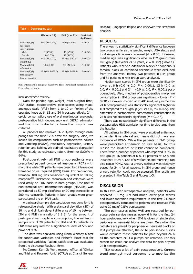
69 November 27, 2016|Volume 5|Issue 3|WJA|www.wjgnet.com
local anesthetic toxicity. Data for gender, age, weight, total surgical time,
ASA status, postoperative pain scores using visual analogue scale (VAS) from 1 to 10 on flexion of the operated knee at 6, 12 and 24 h postoperatively, 24 h opioid consumption, use of oral multimodal analgesia, postoperative high dependency unit (HDU) admission and the time to discharge from the hospital was collected.
All patients had received O2 2 lit/min through nasal prongs for the first 12-h after the surgery. Also, we looked for complications such as postoperative nausea and vomiting (PONV), respiratory depression, urinary retention and itching. We defined respiratory depression for this study as respiratory rate < 8/min or SaO2% < 94%.
Postoperatively, all FNB group patients were prescribed patient controlled analgesia (PCA) with morphine while ITM patients were prescribed intravenous tramadol on as required (PRN) basis. For calculations, tramadol 100 mg was considered equivalent to 10 mg morphine[3]. Diclofenac, eterocoxib and celecoxib were used orally on PRN basis in both groups. One unit of non-steroidal anti-inflammatory drugs (NSAIDs) was considered as 50 mg diclofenac or 90 mg eterocoxib or 200 mg celecoxib. Patients in both groups were given paracetamol 1 g on PRN basis.
A backward sample size calculation was done for this retrospective study: With a standard deviation (SD) of 10 mg and clinical relevant difference of 10 mg between ITM and FNB (in a ratio of 1:1.5) for the amount of post-operative morphine consumption, the minimum sample size of 20 patients for ITM and 30 patients for FNB were required for a significance level of 5% and power of 90%.
The data was analyzed using Mann-Whitney U test for continuous variables and Fisher’s Exact test for categorical variables. Patient satisfaction was evaluated from the discharge feedback form.
Ms Carmen Kam Jia Wen, research officer at “Clinical and Trial and Research Unit” (CTRU) at Changi General
Hospital, Singapore helped and reviewed this statistical analysis.
RESULTSThere was no statistically significant difference between two groups as far as the gender, weight, ASA status and total surgery time was concerned (P > 0.05). However, median age was significantly older in ITM group than FNB group (69 years vs 61 years, P = 0.002) (Table 1). Patients who received additional blocks or continuous femoral block or combined technique were excluded from the analysis. Twenty two patients in ITM group and 32 patients in FNB group were analyzed.
Median pain scores in ITM group were significantly lower at 6 h (0.0 vs 2.0, P < 0.001), 12 h (0.0 vs 2.0, P < 0.001) and 24 h (0.0 vs 2.0, P < 0.001) post-operatively. Also, median of postoperative morphine consumption in ITM group was significantly lower (P < 0.001). However, median of NSAID (unit) requirement in 24 h postoperatively was statistically significant higher in ITM compared to FNB group (2.0 vs 1.0, P = 0.025). The difference in postoperative paracetamol consumption in 24 h was not statistically significant (P = 0.147).
There was no statistically significant difference in the postoperative HDU admission or time to discharge from the hospital.
All patients in ITM group were prescribed antiemetic at regular time interval and hence did not have any incidence of PONV. While the patients in FNB group were prescribed antiemetic on PRN basis; for this reason the incidence of PONV cannot be compared. There were a number of patients who reported PONV in FNB group: 16 patients at 6 h, 11 patients at 12 h and 9 patients at 24 h. Use of sevoflurane and morphine can also cause PONV. Also, a urinary catheter was electively placed for 24 h for all patients in ITM group and hence urinary retention could not be assessed. The results are presented in the Table 2 and Figures 1-3.
DISCUSSIONIn this two-year retrospective analysis, patients who received 0.2 mg ITM had much lower pain scores and lower morphine requirement in the first 24 hour postoperatively compared to patients who received FNB using 20 mL of 0.5% bupivacaine.
The pain scores using VAS are recorded by our acute pain service nurses every 6 h for the first 24 hour postoperatively when ITM is given or single shot peripheral or neuraxial blocks are given. Only when the catheters are placed for peripheral or neuraxial blocks or PCA pumps are attached, the acute pain service nurses would evaluate these patients for pain beyond 24 h till the catheters or PCA pumps are removed. For this reason we could not analyze the data for pain beyond 24 h postoperatively.
TKA causes a lot of pain postoperatively. Current trend amongst most surgeons is to mobilize the
ITM (n = 22) FNB (n = 32) Statistical significance
Median (IQR) age: Years
69.0 (62.8-74.3) 61.0 (57.0-67) P = 0.002
Sex: Numbers Male 9 (37.5%) 15 (62.5%) P = 0.665 Female 13 (43.3%) 17 (56.7%) Median (IQR) weight: Kg
64.3 (53.2-77.2) 67.3 (61.2-80.2) P = 0.275
Median (IQR) ASA status
3.0 (2.0-3.0) 2.0 (2.0-3.0) P = 0.161
Median (IQR) total surgery time in minutes
117.5 (108.8-135.0) 107.5 (86.3-128.8) P = 0.163
Table 1 Demographic data
IQR: Interquantile range; n: Numbers; ITM: Intrathecal morphine; FNB: Femoral nerve block.
DeSousa K et al . ITM vs FNB
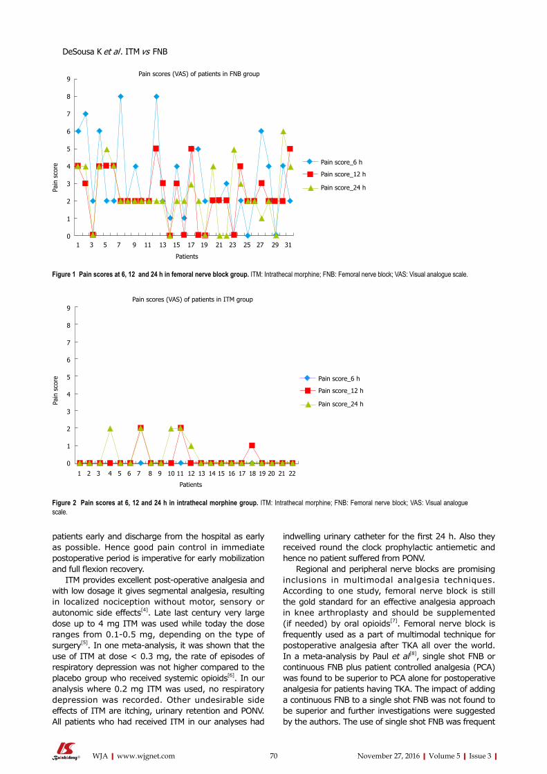
70 November 27, 2016|Volume 5|Issue 3|WJA|www.wjgnet.com
patients early and discharge from the hospital as early as possible. Hence good pain control in immediate postoperative period is imperative for early mobilization and full flexion recovery.
ITM provides excellent post-operative analgesia and with low dosage it gives segmental analgesia, resulting in localized nociception without motor, sensory or autonomic side effects[4]. Late last century very large dose up to 4 mg ITM was used while today the dose ranges from 0.1-0.5 mg, depending on the type of surgery[5]. In one meta-analysis, it was shown that the use of ITM at dose < 0.3 mg, the rate of episodes of respiratory depression was not higher compared to the placebo group who received systemic opioids[6]. In our analysis where 0.2 mg ITM was used, no respiratory depression was recorded. Other undesirable side effects of ITM are itching, urinary retention and PONV. All patients who had received ITM in our analyses had
indwelling urinary catheter for the first 24 h. Also they received round the clock prophylactic antiemetic and hence no patient suffered from PONV.
Regional and peripheral nerve blocks are promising inclusions in multimodal analgesia techniques. According to one study, femoral nerve block is still the gold standard for an effective analgesia approach in knee arthroplasty and should be supplemented (if needed) by oral opioids[7]. Femoral nerve block is frequently used as a part of multimodal technique for postoperative analgesia after TKA all over the world. In a meta-analysis by Paul et al[8], single shot FNB or continuous FNB plus patient controlled analgesia (PCA) was found to be superior to PCA alone for postoperative analgesia for patients having TKA. The impact of adding a continuous FNB to a single shot FNB was not found to be superior and further investigations were suggested by the authors. The use of single shot FNB was frequent
Pain scores (VAS) of patients in FNB group
Patients
Pain
sco
re
9
8
7
6
5
4
3
2
1
01 3 5 7 9 11 13 15 17 19 21 23 25 27 29 31
Pain scores (VAS) of patients in ITM group
Pain
sco
re
9
8
7
6
5
4
3
2
1
0
1 2 3 4 5 6 7 8 9 10 11 12 13 14 15 16 17 18 19 20 21 22
Patients
Pain score_6 h
Pain score_12 h
Pain score_24 h
Pain score_6 h
Pain score_12 h
Pain score_24 h
Figure 1 Pain scores at 6, 12 and 24 h in femoral nerve block group. ITM: Intrathecal morphine; FNB: Femoral nerve block; VAS: Visual analogue scale.
Figure 2 Pain scores at 6, 12 and 24 h in intrathecal morphine group. ITM: Intrathecal morphine; FNB: Femoral nerve block; VAS: Visual analogue scale.
DeSousa K et al . ITM vs FNB

71 November 27, 2016|Volume 5|Issue 3|WJA|www.wjgnet.com
at our hospital in 2013 and 2014. Also, single shot FNB can have analgesic effect from 12 to 24 h[9,10] and may extend up to 48 h[11]. Similarly analgesic effect of ITM also lasts for around 24 h and hence it is only fair to compare ITM and single shot FNB for their efficacy for postoperative analgesia.
In a meta-analysis by PROSPECT working group[1], for joint replacement surgery GA combined with an FNB for surgery and postoperative analgesia, or SA with local anesthetic plus ITM is recommended. The group also recommends cooling and compression techniques, paracetamol and conventional NSAIDs or COX-2-selective inhibitors, plus intravenous strong opioids (high-intensity pain) or weak opioids (moderate- to low-intensity pain) for postoperative analgesia. At our institute we follow these recommendations. In addition,
the surgical team also prefers to use local infiltration technique as described earlier.
FNB requires the use of ultrasound machine and a special needle, which add to the cost. Also, an expertise in performing the FNB is necessary for good outcome. Additional time to perform and to evaluate the block is also required. In addition, there is a possibility of motor weakness which can increase the risk of postoperative fall. ITM is very cost effective, time saving and a rela-tively simple technique. Having these considerations on our mind we decided to analyze the outcome for these two techniques and found that ITM group had lower pain scores in the first 24 hour after surgery though the number of patients that we could include was small.
We found three more clinical trials in the literature comparing ITM and FNB for primary unilateral TKA but in all three trials both groups had received SA. In the study published by Frassanito et al[2], where SA was used for both ITM and FNB group, the 24 h and 48 h morphine consumption was lower in ITM group though not statistically significant. In this prospective study, a very low dose of ITM of 0.1 mg was used. In another trial by Sites et al[12] there was no statistically significant difference in the morphine consumption or pain scores between ITM and FNB group where SA was used for both groups. This trial reports more adverse effects in ITM group compared to FNB group but a slightly higher dose of ITM 0.25 mg was used. Tarkkila et al[13] compared ITM with continuous 3 in 1 femoral block in primary unilateral TKA patients and reported lower pain scores in ITM group. Our study involved use of 0.2 mg ITM with statistically significant lower pain scores and morphine consumption without any respiratory depression. Perhaps a large prospective trial with standardized drug regime and multivariate analysis may
ITM (n = 22) FNB (n = 32)
Ease of performance Very easy, no special training Expertise and training required, special equipment required Added cost Only morphine Ultrasound machine, special needle and local anesthetic Median (IQR) pain score at: Low High 6 h 0.0 (0.0-0.0) 2.0 (2.0-4.8) 12 h 0.0 (0.0-0.0) 2.0 (2.0-3.8) 24 h 0.0 (0.0-0.3) 2.0 (2.0-4.0) Median (IQR) of morphine requirement (in mg) in 24 h Low 5.0 (3.8-10.0) High 16.5 (8.0-21.5) Median (IQR) of NSAID unit in 24 h 2.0 (1.0-2.0) 1.0 (0.0-1.3) Median (IQR) of Paracetamol (G) in 24 h 3.0 (2.0-3.0) 3.0 (2.0-4.0) Complications Low Low
No PONV since antiemetic prescribed in anticipation
Incidence of PONV was higher since no regular antiemetic given (16 at 6 h, 11 at 12 h and 9 at 24 h)
No urinary retention since all patients were catheterized
No urine retention
No respiratory depression No respiratory depressionNo itching recorded No itching recorded
Number of elective HDU admission status 3 (15%) 2 (6.5%) Median (IQR) days to discharge 4.5 (3.0-5.0) 4.0 (3.0-6.0) Patient satisfaction 18/20 (90.0%) rated good 4/31 (12.9%) rated good
Table 2 Results
IQR: Interquantile range; n: Numbers; ITM: Intrathecal morphine; FNB: Femoral nerve block; PONV: Post-operative nausea and vomiting; HDU: High dependency unit.
Comparison of median analgesic requirement
Morphineequivalent mg
NSAID units Paracetamol G
ITM
FNB
18
16
14
12
10
8
6
1
2
0
Figure 3 Comparison of median analgesic requirement in 24 h. ITM: Intrathecal morphine; FNB: Femoral nerve block: NSAID: Non-steroid anti-inflammatory drug.
DeSousa K et al . ITM vs FNB

72 November 27, 2016|Volume 5|Issue 3|WJA|www.wjgnet.com
be needed to prove superiority of ITM over FNB in this particular group of surgical patients.
From our analysis we conclude that 0.2 mg ITM with SA is more effective for first 24 hour postoperatively compared to single shot FNB with GA for primary unilateral TKA. Also, 0.2 mg ITM is safe, time saving and cost effective with better patient satisfaction compared to FNB.
ACKNOWLEDGMENTSWe thank Ms Carmen Kam Jia Wen, research officer at “Clinical and Trial and Research Unit” (CTRU) at Changi General Hospital, Singapore for her statistical help.
COMMENTSBackgroundTotal knee arthroplasty (TKA) leads to considerable post-operative pain, which requires optimal control for early mobilization. Till date, varieties of techniques and drugs have been used to achieve this optimal pain control. Use of intrathecal morphine (ITM) with spinal anesthesia (SA) is extremely easy and can be time saving and cost effective. Also, femoral nerve block (FNB) is one of the easiest peripheral nerve blocks. In this study the authors have compared these two easy techniques for postoperative pain management after TKA.
Research frontiersRegional blocks are commonly used for pain management after TKA. Most recent studies are comparing femoral nerve blocks with adductor canal block and local infiltration techniques. This study compares use of ITM with SA and FNB with GA. In the past only three other studies have compared ITM and FNB, albeit both groups had undergone surgery under SA.
Innovations and breakthroughsIn this study, 0.2 mg ITM with SA was more effective for first 24 hour post-operatively compared to single shot FNB with GA for primary unilateral TKA. In one previous study, which compared 0.1 mg ITM with SA and FNB with SA, pain scores and postoperative opioid consumption were lower in ITM group though statistically not significant. In another similar study, ITM group had more complications when a higher dose of 0.25 mg ITM was used. In their study using 0.2 mg ITM, lower pain scores and opioid consumption were statistically significant. This study also illustrates that use of adequate prophylaxis can prevent known complications of ITM.
ApplicationsThis retrospective analysis shows that 0.2 mg ITM with SA is more effective for first 24 h postoperatively compared to single shot FNB with GA for primary unilateral TKA. Perhaps a prospective trial with standardized drug regime and multivariate analysis in a large group of patients is required to prove their findings.
TerminologyITM: Intrathecal morphine; FNB: Femoral nerve block; SA: Spinal anesthesia; GA: General anesthesia; PONV: Postoperative nausea and vomiting; PCA: Patient controlled analgesia; NSAIDs: Non-steroidal anti-inflammatory drugs; VAS: Visual analogue scale.
Peer-reviewThis retrospective analysis is worthy of publishing as it provides value clinical
information for how pre-emptive analgesia could effectively prevent or mitigate postoperative pain.
REFERENCES1 Fischer HB, Simanski CJ, Sharp C, Bonnet F, Camu F, Neugebauer
EA, Rawal N, Joshi GP, Schug SA, Kehlet H. A procedure-specific systematic review and consensus recommendations for postoperative analgesia following total knee arthroplasty. Anaesthesia 2008; 63: 1105-1123 [PMID: 18627367 DOI: 10.1111/j.1365-2044.2008.05565.x]
2 Frassanito L, Vergari A, Zanghi F, Messina A, Bitondo M, Antonelli M. Post-operative analgesia following total knee arthroplasty: comparison of low-dose intrathecal morphine and single-shot ultrasound-guided femoral nerve block: a randomized, single blinded, controlled study. Eur Rev Med Pharmacol Sci 2010; 14: 589-596 [PMID: 20707248]
3 Vergnion M, Degesves S, Garcet L, Magotteaux V. Tramadol, an alternative to morphine for treating posttraumatic pain in the prehospital situation. Anesth Analg 2001; 92: 1543-1546 [PMID: 11375843 DOI: 10.1097/00000539-200106000-00039]
4 DeSousa K, Chandran R. Intrathecal Morphine for postoperative analgesia: Current trends. World J Anesthesiol 2014; 27: 191-202 [DOI: 10.5313/wja.v3.i3.191]
5 Mugabure Bujedo B. A clinical approach to neuraxial morphine for the treatment of postoperative pain. Pain Res Treat 2012; 2012: 612145 [PMID: 23002426]
6 Gehling M, Tryba M. Risks and side-effects of intrathecal morphine combined with spinal anaesthesia: a meta-analysis. Anaesthesia 2009; 64: 643-651 [PMID: 19462494 DOI: 10.1111/j.1365-2044.2008.05817.x]
7 Bauer MC, Pogatzki-Zahn EM, Zahn PK. Regional analgesia techniques for total knee replacement. Curr Opin Anaesthesiol 2014; 27: 501-506 [PMID: 25111605 DOI: 10.1097/ACO.000-0000000000115]
8 Paul JE, Arya A, Hurlburt L, Cheng J, Thabane L, Tidy A, Murthy Y. Femoral nerve block improves analgesia outcomes after total knee arthroplasty: a meta-analysis of randomized controlled trials. Anesthesiology 2010; 113: 1144-1162 [PMID: 20966667 DOI: 10.1097/ALN.0b013e3181f4b18]
9 Allen HW, Liu SS, Ware PD, Nairn CS, Owens BD. Peripheral nerve blocks improve analgesia after total knee replacement surgery. Anesth Analg 1998; 87: 93-97 [PMID: 9661553]
10 Szczukowski MJ, Hines JA, Snell JA, Sisca TS. Femoral nerve block for total knee arthroplasty patients: a method to control postoperative pain. J Arthroplasty 2004; 19: 720-725 [PMID: 15343531 DOI: 10.1016/j.arth.2004.02.043]
11 Ng HP, Cheong KF, Lim A, Lim J, Puhaindran ME. Intraoperative single-shot “3-in-1” femoral nerve block with ropivacaine 0.25%, ropivacaine 0.5% or bupivacaine 0.25% provides comparable 48-hr analgesia after unilateral total knee replacement. Can J Anaesth 2001; 48: 1102-1108 [PMID: 11744586 DOI: 10.1007/BF03020376]
12 Sites BD, Beach M, Gallagher JD, Jarrett RA, Sparks MB, Lundberg CJ. A single injection ultrasound-assisted femoral nerve block provides side effect-sparing analgesia when compared with intrathecal morphine in patients undergoing total knee arthroplasty. Anesth Analg 2004; 99: 1539-1543; table of contents [PMID: 15502061 DOI: 10.1213/01.ANE.0000136470.51029.52]
13 Tarkkila P, Tuominen M, Huhtala J, Lindgren L. Comparison of intrathecal morphine and continuous femoral 3-in-1 block for pain after major knee surgery under spinal anaesthesia. Eur J Anaesthesiol 1998; 15: 6-9 [PMID: 9522133 DOI: 10.1097/00003643-199801000-00002]
P- Reviewer: Chen CC, Ma SXL, Rikalainen-Salmi R S- Editor: Qiu S L- Editor: A E- Editor: Wu HL
COMMENTS
DeSousa K et al . ITM vs FNB

© 2016 Baishideng Publishing Group Inc. All rights reserved.
Published by Baishideng Publishing Group Inc8226 Regency Drive, Pleasanton, CA 94588, USA
Telephone: +1-925-223-8242Fax: +1-925-223-8243
E-mail: [email protected] Desk: http://www.wjgnet.com/esps/helpdesk.aspx
http://www.wjgnet.com