wounds tissues aspirates culture manual mt.sinai.pdf
DESCRIPTION
Wounds Tissues Aspirates Culture Manual Mt.SINAITRANSCRIPT
PROCEDURE MANUAL TORONTO MEDICAL LABORATORIES / MOUNT SINAI HOSPITAL MICROBIOLOGY DEPARTMENT
1
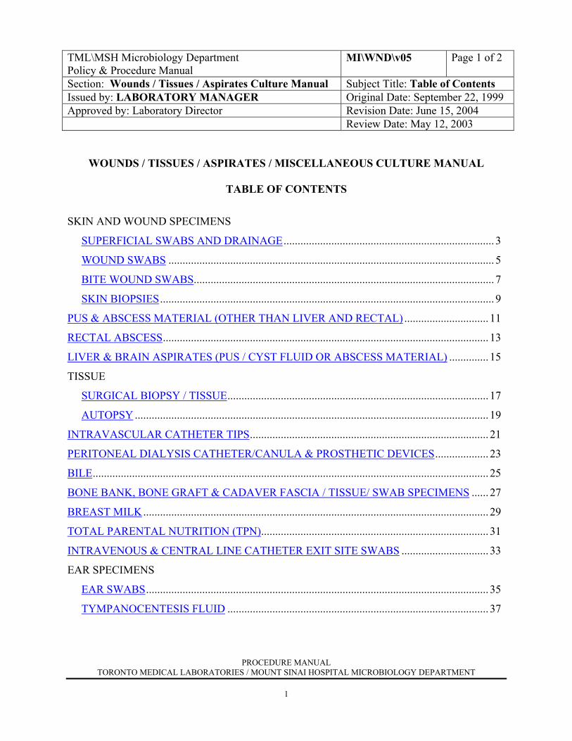
TML\MSH Microbiology Department Policy & Procedure Manual
MI\WND\v05 Page 1 of 2
Section: Wounds / Tissues / Aspirates Culture Manual Subject Title: Table of Contents Issued by: LABORATORY MANAGER Original Date: September 22, 1999 Approved by: Laboratory Director Revision Date: June 15, 2004
Review Date: May 12, 2003
WOUNDS / TISSUES / ASPIRATES / MISCELLANEOUS CULTURE MANUAL
TABLE OF CONTENTS
SKIN AND WOUND SPECIMENS
SUPERFICIAL SWABS AND DRAINAGE........................................................................... 3
WOUND SWABS .................................................................................................................... 5
BITE WOUND SWABS........................................................................................................... 7
SKIN BIOPSIES....................................................................................................................... 9
PUS & ABSCESS MATERIAL (OTHER THAN LIVER AND RECTAL) .............................. 11
RECTAL ABSCESS.................................................................................................................... 13
LIVER & BRAIN ASPIRATES (PUS / CYST FLUID OR ABSCESS MATERIAL) .............. 15
TISSUE
SURGICAL BIOPSY / TISSUE............................................................................................. 17
AUTOPSY .............................................................................................................................. 19
INTRAVASCULAR CATHETER TIPS..................................................................................... 21
PERITONEAL DIALYSIS CATHETER/CANULA & PROSTHETIC DEVICES................... 23
BILE............................................................................................................................................. 25 BONE BANK, BONE GRAFT & CADAVER FASCIA / TISSUE/ SWAB SPECIMENS ...... 27
BREAST MILK........................................................................................................................... 29
TOTAL PARENTAL NUTRITION (TPN)................................................................................. 31
INTRAVENOUS & CENTRAL LINE CATHETER EXIT SITE SWABS ............................... 33
EAR SPECIMENS
EAR SWABS.......................................................................................................................... 35
TYMPANOCENTESIS FLUID ............................................................................................. 37
PROCEDURE MANUAL TORONTO MEDICAL LABORATORIES / MOUNT SINAI HOSPITAL MICROBIOLOGY DEPARTMENT
2
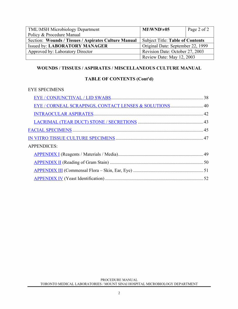
TML\MSH Microbiology Department Policy & Procedure Manual
MI\WND\v05 Page 2 of 2
Section: Wounds / Tissues / Aspirates Culture Manual Subject Title: Table of Contents Issued by: LABORATORY MANAGER Original Date: September 22, 1999 Approved by: Laboratory Director Revision Date: October 27, 2003
Review Date: May 12, 2003
WOUNDS / TISSUES / ASPIRATES / MISCELLANEOUS CULTURE MANUAL
TABLE OF CONTENTS (Cont'd) EYE SPECIMENS
EYE / CONJUNCTIVAL / LID SWABS............................................................................... 38
EYE / CORNEAL SCRAPINGS, CONTACT LENSES & SOLUTIONS............................ 40
INTRAOCULAR ASPIRATES.............................................................................................. 42
LACRIMAL (TEAR DUCT) STONE / SECRETIONS ........................................................ 43
FACIAL SPECIMENS ................................................................................................................ 45
IN VITRO TISSUE CULTURE SPECIMENS ........................................................................... 47
APPENDICES:
APPENDIX I (Reagents / Materials / Media)......................................................................... 49
APPENDIX II (Reading of Gram Stain) ................................................................................ 50
APPENDIX III (Commensal Flora – Skin, Ear, Eye) ............................................................ 51
APPENDIX IV (Yeast Identification) .................................................................................... 52
PROCEDURE MANUAL TORONTO MEDICAL LABORATORIES / MOUNT SINAI HOSPITAL MICROBIOLOGY DEPARTMENT
3
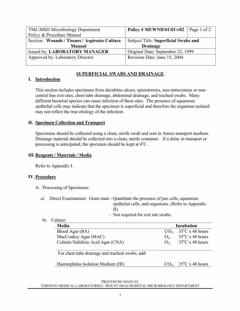
TML\MSH Microbiology Department Policy & Procedure Manual
Policy # MI\WND\01\01\v02 Page 1 of 2
Section: Wounds / Tissues / Aspirates Culture Manual
Subject Title: Superficial Swabs and Drainage
Issued by: LABORATORY MANAGER Original Date: September 22, 1999 Approved by: Laboratory Director
Revision Date: June 15, 2004
SUPERFICIAL SWABS AND DRAINAGE
I. Introduction
This section includes specimens from decubitus ulcers, episiotomies, non-intravenous or non-central line exit sites, chest tube drainage, abdominal drainage, and tracheal swabs. Many different bacterial species can cause infection of these sites. The presence of squamous epithelial cells may indicate that the specimen is superficial and therefore the organism isolated may not reflect the true etiology of the infection.
II. Specimen Collection and Transport Specimens should be collected using a clean, sterile swab and sent in Amies transport medium. Drainage material should be collected into a clean, sterile container. If a delay in transport or
processing is anticipated, the specimen should be kept at 4oC. III. Reagents / Materials / Media Refer to Appendix I. IV. Procedure
A. Processing of Specimens:
a) Direct Examination: Gram stain - Quantitate the presence of pus cells, squamous epithelial cells, and organisms. (Refer to Appendix II).
- Not required for exit site swabs. b) Culture:
Media Incubation Blood Agar (BA) MacConkey Agar (MAC) Colistin Nalidixic Acid Agar (CNA)
CO2, 35oC x 48 hours O2, 35oC x 48 hours O2, 35oC x 48 hours
For chest tube drainage and tracheal swabs, add: Haemophilus Isolation Medium (HI)
CO2, 35oC x 48 hours

PROCEDURE MANUAL TORONTO MEDICAL LABORATORIES / MOUNT SINAI HOSPITAL MICROBIOLOGY DEPARTMENT
4
TML\MSH Microbiology Department Policy & Procedure Manual
Policy # MI\WND\01\01\v02 Page 2 of 2
Wounds / Tissues / Aspirates Culture Manual
B. Interpretation of Cultures:
Examine the plates after 24 and 48 hours incubation. Any growth of S. aureus, group B streptococcus from neonates, beta-hemolytic
streptococcus groups A, C and G and Pseudomonas aeruginosa is significant. For chest tube drainage and tracheal swabs, any growth of H. influenzae and S. pneumoniae is also significant. A heavy, pure growth of other organisms that correlates with the predominant organism seen in the Gram stain is significant if there is >1+ pus cells (not for exit sites). If a specific organism is requested, then it will be looked for and its presence or absence reported. Growth of ≥ 3 types of coliforms or other Gram negative bacilli will be reported as a negative report stating commensal flora including mixed Gram negative bacilli.
C. Susceptibility Testing:
Refer to Susceptibility Testing Manual.
V. Reporting Results
a) Gram stain: Report with quantitation the presence of pus cells, squamous epithelial cells and organisms.
b) Culture: Negative report: "No growth" or "Commensal flora" "Commensal flora including mixed Gram negative bacilli".
Positive report: Quantitate all significant isolates with appropriate sensitivities. If commensal flora is also present, report with quantitation.
PROCEDURE MANUAL TORONTO MEDICAL LABORATORIES / MOUNT SINAI HOSPITAL MICROBIOLOGY DEPARTMENT
5
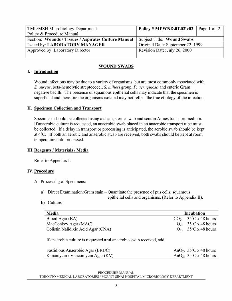
TML\MSH Microbiology Department Policy & Procedure Manual
Policy # MI\WND\01\02\v02 Page 1 of 2
Section: Wounds / Tissues / Aspirates Culture Manual Subject Title: Wound Swabs Issued by: LABORATORY MANAGER Original Date: September 22, 1999 Approved by: Laboratory Director
Revision Date: July 26, 2000
WOUND SWABS
I. Introduction
Wound infections may be due to a variety of organisms, but are most commonly associated with S. aureus, beta-hemolytic streptococci, S. milleri group, P. aeruginosa and enteric Gram negative bacilli. The presence of squamous epithelial cells may indicate that the specimen is superficial and therefore the organisms isolated may not reflect the true etiology of the infection.
II. Specimen Collection and Transport Specimens should be collected using a clean, sterile swab and sent in Amies transport medium. If anaerobic culture is requested, an anaerobic swab placed in an anaerobic transport tube must
be collected. If a delay in transport or processing is anticipated, the aerobic swab should be kept at 4oC. If both an aerobic and anaerobic swab are received, both swabs should be kept at room temperature until processed.
III. Reagents / Materials / Media
Refer to Appendix I. IV. Procedure A. Processing of Specimens:
a) Direct Examination:Gram stain – Quantitate the presence of pus cells, squamous epithelial cells and organisms. (Refer to Appendix II).
b) Culture:
Media Incubation Blood Agar (BA) MacConkey Agar (MAC) Colistin Nalidixic Acid Agar (CNA)
CO2, 35oC x 48 hours O2, 35oC x 48 hours O2, 35oC x 48 hours
If anaerobic culture is requested and anaerobic swab received, add: Fastidious Anaerobic Agar (BRUC) Kanamycin / Vancomycin Agar (KV)
AnO2, 350C x 48 hours AnO2, 350C x 48 hours

PROCEDURE MANUAL TORONTO MEDICAL LABORATORIES / MOUNT SINAI HOSPITAL MICROBIOLOGY DEPARTMENT
6
TML\MSH Microbiology Department Policy & Procedure Manual
Policy # MI\WND\01\02\v02 Page 2 of 2
Wounds / Tissues / Aspirates Culture Manual
B. Interpretation of Cultures:
Examine the aerobic plates after 24 and 48 hours incubation and the anaerobic plates after 48 hours incubation.
Any growth of S. aureus, beta-hemolytic streptococci groups A, B, C and G and Pseudomonas aeruginosa is significant. For sternal wound specimens, isolation of coagulase negative staphylococci is significant and appropriate antimicrobial susceptibility testing should be performed and reported. A growth of 1 or 2 types of organisms other than skin commensals is significant if there was >1+ pus cells seen on the original Gram stain. Growth of ≥ 3 types of coliforms or other Gram negative bacilli will be reported as a negative report stating "commensal flora including mixed Gram negative bacilli".
C. Susceptibility Testing:
Refer to Susceptibility Testing Manual.
V. Reporting Results
a) Gram stain: Report with quantitation the presence of pus cells, squamous epithelial cells and organisms.
b) Culture: Negative report: "No growth" or "Commensal flora"
"Commensal flora including mixed Gram negative bacilli".
Positive report: Quantitate all significant isolates with appropriate sensitivities. If commensal flora is also present, report with quantitation.
“The __________________ seen in the Gram stain failed to grow in aerobic culture”.
NB: If anaerobic culture requested and no anaerobic swab received, report the following phrase with both the negative and positive reports:
"No anaerobic swab received; anaerobic culture not done". Enter under test
field in LIS.
PROCEDURE MANUAL TORONTO MEDICAL LABORATORIES / MOUNT SINAI HOSPITAL MICROBIOLOGY DEPARTMENT
7
TML\MSH Microbiology Department Policy & Procedure Manual
Policy # MI\WND\01\03\v01 Page 1 of 2
Section: Wounds / Tissues / Aspirates Culture Manual Subject Title: Bite Wound Swabs Issued by: LABORATORY MANAGER Original Date: September 22, 1999 Approved by: Laboratory Director
Revision Date: July 26, 2000
BITE WOUND SWABS
I. Introduction
Bite wounds may become infected with many different organisms but most commonly include S. aureus, Pasteurella spp., S. milleri group and beta-hemolytic streptococci. The presence of squamous epithelial cells may indicate that the specimen is superficial and therefore the organisms isolated may not reflect the true etiology of the infection.
II. Specimen Collection and Transport Specimens should be collected using a clean, sterile swab and sent in Amies transport medium.
If anaerobic culture is requested, an anaerobic swab placed in an anaerobic transport tube must be collected. If a delay in transport or processing is anticipated, the aerobic swab should be kept at 4oC and the anaerobic swab at room temperature.
III. Reagents / Materials / Media
Refer to Appendix I. IV. Processing of Specimens
A. Processing of Specimens:
a) Direct Examination: Gram stain – Quantitate the presence of pus cells, squamous epithelial cells, and organisms. (Refer to Appendix II)
b) Culture:
Media Incubation Blood Agar (BA) MacConkey Agar (MAC) Chocolate Agar (CHOC)
CO2, 35oC x 48 hours O2, 35oC x 48 hours CO2, 35oC x 48 hours
If anaerobic culture is requested and anaerobic swab received, add: Fastidious Anaerobic Agar (BRUC) Kanamycin / Vancomycin Agar (KV)
AnO2, 35oC x 48 hours AnO2, 35oC x 48 hours
PROCEDURE MANUAL TORONTO MEDICAL LABORATORIES / MOUNT SINAI HOSPITAL MICROBIOLOGY DEPARTMENT
8
TML\MSH Microbiology Department Policy & Procedure Manual
Policy # MI\WND\01\03\v01 Page 2 of 2
Wounds / Tissues / Aspirates Culture Manual
B. Interpretation of Cultures:
Examine aerobic plates after 24 and 48 hours incubation and anaerobic plates after 48 hours incubation.
Any growth of S. aureus, Pasteurella spp., S. milleri group, beta-haemolytic streptococci
and Pseudomonas aeruginosa is significant. For other organisms such as Enterobacteriaceae and other Gram negative bacilli, a significant result is determined by the isolation of a moderate to heavy predominant growth, or if growth correlates with the predominent organism seen on Gram stain.
For suspected anaerobes, minimal identification is performed.
C. Susceptibility Testing: Refer to Susceptibility Testing Manual. V. Reporting Results
a) Gram stain: Report with quantitation the presence of pus cells, squamous epithelial cells and organisms.
b) Culture: Negative Report: "No growth" or "Commensal flora"
Positive Report: Quantitate all significant isolates with appropriate sensitivities. If commensal flora is also present, report with
quantitation. NB: If anaerobic culture requested and no anaerobic swab received, report the
following phrase with both the negative and positive reports: "No anaerobic swab received; anaerobic culture not done". Enter under test
field in LIS.
PROCEDURE MANUAL TORONTO MEDICAL LABORATORIES / MOUNT SINAI HOSPITAL MICROBIOLOGY DEPARTMENT
9
TML\MSH Microbiology Department Policy & Procedure Manual
Policy # MI\WND\01\04\v01 Page 1 of 2
Section: Wounds / Tissues / Aspirates Culture Manual Subject Title: Skin Biopsies Issued by: LABORATORY MANAGER Original Date: September 22, 1999 Approved by: Laboratory Director
Revision Date: July 26, 2000
SKIN BIOPSIES
I. Introduction
A variety of organisms may be associated with skin lesions and thus any growth of organisms other than skin commensals should be considered significant.
II. Specimen Collection and Transport Skin biopsy specimens should be placed in a sterile container with sterile saline. If a delay in
transport or processing is anticipated, the specimen should be kept at 4oC. III. Reagents / Materials /Media Refer to Appendix I. IV. Procedure
A. Processing of Specimen: 1. Macerate the entire specimen with a small volume of sterile saline from the original
container using a grinder (small tissue sample) or a scalpel and stomacher (large tissue sample).
2. Prepare 3 slides from the ground material: one for Gram stain, one for Calcofluor white stain and one unstained (stored in the “extra smear” slide box).
3. If TB has been requested, forward half the specimen to the Public Health Laboratory (PHL) for processing. If viral isolation is requested, a portion of the ground material should be given to the Virology section for processing. If parasitology is requested, forward a portion of the specimen (before macerating) to the Parasitology section for processing.
a) Direct Examination: Gram stain: Quantitate the presence of pus cells and
organisms. (Refer to Appendix II). Calcofluor white stain.

PROCEDURE MANUAL TORONTO MEDICAL LABORATORIES / MOUNT SINAI HOSPITAL MICROBIOLOGY DEPARTMENT
10
TML\MSH Microbiology Department Policy & Procedure Manual
Policy # MI\WND\01\04\v01 Page 2 of 2
Wounds / Tissues / Aspirates Culture Manual b) Culture:
Media Incubation Blood Agar (BA) MacConkey Agar (MAC) Chocolate Agar (CHOC) Inhibitory Mold Agar (IMA)* Esculin Base Medium (EBM)*
CO2, 35oC x 48 hours O2, 35oC x 48 hours CO2, 35oC x 48 hours O2, 30oC x 3 weeks O2, 30oC x 3 weeks
* Forward the fungus culture media to the Mycology section for incubation and work-up. B. Interpretation of Cultures:
Examine the culture plates after 24 and 48 hours incubation. Any growth of organisms other than skin commensals should be considered significant.
C. Susceptibility Testing: Refer to Susceptibility Testing Manual. V. Reporting Results a) Gram stain: Report with quantitation the presence of pus cells and organisms. b) Culture:
Negative Report: "No growth" or "Commensal flora"
Positive Report: Quantitate all significant isolates with appropriate sensitivities. If commensal flora is also present, report with quantitation.
PROCEDURE MANUAL TORONTO MEDICAL LABORATORIES / MOUNT SINAI HOSPITAL MICROBIOLOGY DEPARTMENT
11
TML\MSH Microbiology Department Policy & Procedure Manual
Policy # MI\WND\02\v01 Page 1 of 2
Section: Wounds / Tissues / Aspirates Culture Manual
Subject Title: Pus & Abscess Material (Other than liver and rectal)
Issued by: LABORATORY MANAGER Original Date: September 22, 1999 Approved by: Laboratory Director
Revision Date: July 26, 2000
PUS & ABSCESS MATERIAL (OTHER THAN LIVER AND RECTAL)
I. Introduction
Abscesses are usually due to a mixture of different aerobic and anaerobic bacteria depending on the location of the abscess.
II. Specimen Collection and Transport Pus from an abscess should be sent in a clean, sterile container and an anaerobic transport
container. An aspirate of pus or abscess material may be collected using a syringe and sent to the laboratory with the needle removed. If a delay in transport or processing is anticipated, keep the specimen at 4oC.
III. Reagents / Materials / Media Refer to Appendix I. IV. Procedure
A. Processing of Specimens: a) Direct Examination: Gram stain – Quantitate the presence of pus cells and organisms. (Refer to Appendix II).
Modified acid fast stain - If Actinomyces or Nocardia is requested or suggested on Gram stain.
Calcofluor white stain - If fungus is requested. (Refer to Mycology Manual).
b) Culture:
Media Incubation Blood Agar (BA)1
MacConkey Agar (MAC) Chocolate Agar (CHOC)1 Fastidious Anaerobic Agar (BRUC)2
Kanamycin/Vancomycin Agar (KV)2
CO2, 35oC x 48 hours O2, 35oC x 48 hours CO2, 35oC x 48 hours AnO2, 35oC x 48 hours AnO2, 35oC x 48 hours
PROCEDURE MANUAL TORONTO MEDICAL LABORATORIES / MOUNT SINAI HOSPITAL MICROBIOLOGY DEPARTMENT
12
TML\MSH Microbiology Department Policy & Procedure Manual
Policy # MI\WND\02\v01 Page 2 of 2
Wounds / Tissues / Aspirates Culture Manual
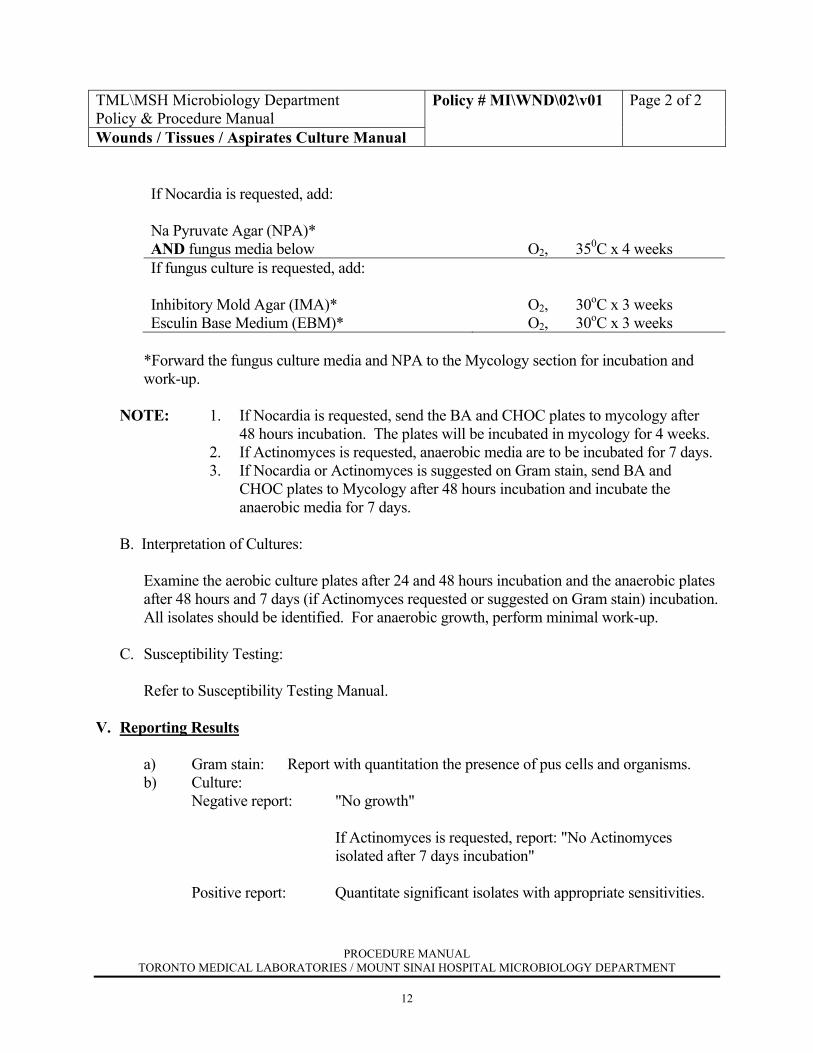
If Nocardia is requested, add: Na Pyruvate Agar (NPA)* AND fungus media below
O2, 350C x 4 weeks
If fungus culture is requested, add: Inhibitory Mold Agar (IMA)* Esculin Base Medium (EBM)*
O2, 30oC x 3 weeks O2, 30oC x 3 weeks
*Forward the fungus culture media and NPA to the Mycology section for incubation and
work-up.
NOTE: 1. If Nocardia is requested, send the BA and CHOC plates to mycology after 48 hours incubation. The plates will be incubated in mycology for 4 weeks.
2. If Actinomyces is requested, anaerobic media are to be incubated for 7 days. 3. If Nocardia or Actinomyces is suggested on Gram stain, send BA and
CHOC plates to Mycology after 48 hours incubation and incubate the anaerobic media for 7 days.
B. Interpretation of Cultures:
Examine the aerobic culture plates after 24 and 48 hours incubation and the anaerobic plates
after 48 hours and 7 days (if Actinomyces requested or suggested on Gram stain) incubation. All isolates should be identified. For anaerobic growth, perform minimal work-up.
C. Susceptibility Testing:
Refer to Susceptibility Testing Manual. V. Reporting Results a) Gram stain: Report with quantitation the presence of pus cells and organisms. b) Culture: Negative report: "No growth"
If Actinomyces is requested, report: "No Actinomyces
isolated after 7 days incubation"
Positive report: Quantitate significant isolates with appropriate sensitivities.
PROCEDURE MANUAL TORONTO MEDICAL LABORATORIES / MOUNT SINAI HOSPITAL MICROBIOLOGY DEPARTMENT
13
TML\MSH Microbiology Department Policy & Procedure Manual
Policy # MI\WND\03\v01 Page 1 of 2
Section: Wounds / Tissues / Aspirates Culture Manual Subject Title: Rectal Abscess Issued by: LABORATORY MANAGER Original Date: September 22, 1999 Approved by: Laboratory Director
Revision Date: July 26, 2000
RECTAL ABSCESS
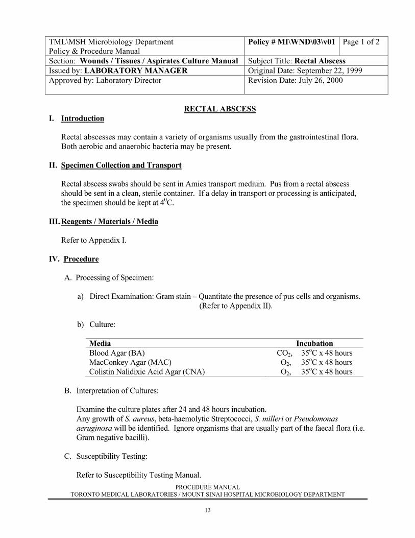
I. Introduction
Rectal abscesses may contain a variety of organisms usually from the gastrointestinal flora. Both aerobic and anaerobic bacteria may be present.
II. Specimen Collection and Transport Rectal abscess swabs should be sent in Amies transport medium. Pus from a rectal abscess
should be sent in a clean, sterile container. If a delay in transport or processing is anticipated, the specimen should be kept at 40C.
III. Reagents / Materials / Media
Refer to Appendix I. IV. Procedure A. Processing of Specimen:
a) Direct Examination: Gram stain – Quantitate the presence of pus cells and organisms. (Refer to Appendix II). b) Culture:
Media Incubation Blood Agar (BA) MacConkey Agar (MAC) Colistin Nalidixic Acid Agar (CNA)
CO2, 35oC x 48 hours O2, 35oC x 48 hours O2, 35oC x 48 hours
B. Interpretation of Cultures:
Examine the culture plates after 24 and 48 hours incubation. Any growth of S. aureus, beta-haemolytic Streptococci, S. milleri or Pseudomonas
aeruginosa will be identified. Ignore organisms that are usually part of the faecal flora (i.e. Gram negative bacilli).
C. Susceptibility Testing:
Refer to Susceptibility Testing Manual.
PROCEDURE MANUAL TORONTO MEDICAL LABORATORIES / MOUNT SINAI HOSPITAL MICROBIOLOGY DEPARTMENT
14
TML\MSH Microbiology Department Policy & Procedure Manual
Policy # MI\WND\03\v01 Page 2 of 2
Wounds / Tissues / Aspirates Culture Manual V. Reporting Results a) Gram stain: Report with quantitation the presence of pus cells and organisms. b) Culture: Negative Report: "No growth" or "Mixed faecal flora"
Positive Report: Quantitate all significant isolates with appropriate sensitivities. Report "Mixed faecal flora" if also present.
PROCEDURE MANUAL TORONTO MEDICAL LABORATORIES / MOUNT SINAI HOSPITAL MICROBIOLOGY DEPARTMENT
15
TML\MSH Microbiology Department Policy & Procedure Manual
Policy # MI\WND\04\v02 Page 1 of 2
Section: Wounds / Tissues / Aspirates Culture Manual
Subject Title: Liver and Brain Aspirates (Pus / Cyst Fluid or Abscess Material)
Issued by: LABORATORY MANAGER Original Date: September 22, 1999 Approved by: Laboratory Director
Revision Date: April 17, 2002
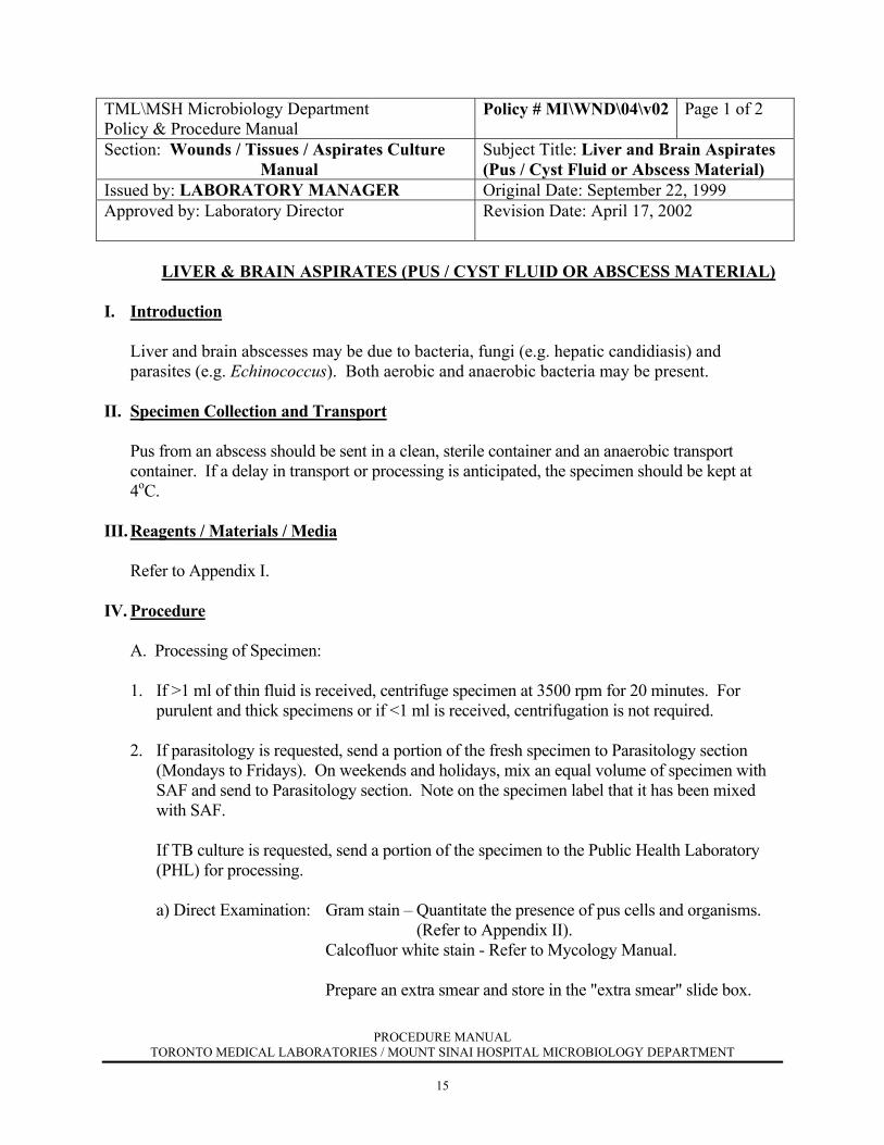
LIVER & BRAIN ASPIRATES (PUS / CYST FLUID OR ABSCESS MATERIAL)
I. Introduction
Liver and brain abscesses may be due to bacteria, fungi (e.g. hepatic candidiasis) and parasites (e.g. Echinococcus). Both aerobic and anaerobic bacteria may be present.
II. Specimen Collection and Transport Pus from an abscess should be sent in a clean, sterile container and an anaerobic transport
container. If a delay in transport or processing is anticipated, the specimen should be kept at 4oC.
III. Reagents / Materials / Media Refer to Appendix I. IV. Procedure A. Processing of Specimen:
1. If >1 ml of thin fluid is received, centrifuge specimen at 3500 rpm for 20 minutes. For purulent and thick specimens or if <1 ml is received, centrifugation is not required.
2. If parasitology is requested, send a portion of the fresh specimen to Parasitology section
(Mondays to Fridays). On weekends and holidays, mix an equal volume of specimen with SAF and send to Parasitology section. Note on the specimen label that it has been mixed with SAF.
If TB culture is requested, send a portion of the specimen to the Public Health Laboratory
(PHL) for processing. a) Direct Examination: Gram stain – Quantitate the presence of pus cells and organisms. (Refer to Appendix II).
Calcofluor white stain - Refer to Mycology Manual. Prepare an extra smear and store in the "extra smear" slide box.
PROCEDURE MANUAL TORONTO MEDICAL LABORATORIES / MOUNT SINAI HOSPITAL MICROBIOLOGY DEPARTMENT
16
TML\MSH Microbiology Department Policy & Procedure Manual
Policy # MI\WND\04\v02 Page 2 of 2
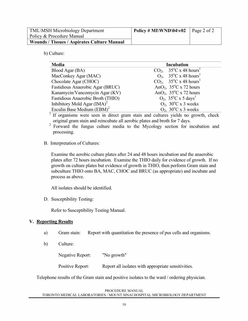
Wounds / Tissues / Aspirates Culture Manual b) Culture:
Media Incubation Blood Agar (BA) MacConkey Agar (MAC) Chocolate Agar (CHOC) Fastidious Anaerobic Agar (BRUC) Kanamycin/Vancomycin Agar (KV) Fastidious Anaerobic Broth (THIO) Inhibitory Mold Agar (IMA)2 Esculin Base Medium (EBM)2
CO2, 35oC x 48 hours1
O2, 35oC x 48 hours1
CO2, 35oC x 48 hours1
AnO2, 35oC x 72 hours AnO2, 35oC x 72 hours
O2, 35oC x 5 days1
O2, 30oC x 3 weeks O2, 30oC x 3 weeks
1 If organisms were seen in direct gram stain and cultures yields no growth, check original gram stain and reincubate all aerobic plates and broth for 7 days.
2 Forward the fungus culture media to the Mycology section for incubation and processing.
B. Interpretation of Cultures:
Examine the aerobic culture plates after 24 and 48 hours incubation and the anaerobic plates after 72 hours incubation. Examine the THIO daily for evidence of growth. If no growth on culture plates but evidence of growth in THIO, then perform Gram stain and subculture THIO onto BA, MAC, CHOC and BRUC (as appropriate) and incubate and process as above.
All isolates should be identified.
D. Susceptibility Testing: Refer to Susceptibility Testing Manual. V. Reporting Results a) Gram stain: Report with quantitation the presence of pus cells and organisms. b) Culture: Negative Report: "No growth"
Positive Report: Report all isolates with appropriate sensitivities.
Telephone results of the Gram stain and positive isolates to the ward / ordering physician.

PROCEDURE MANUAL TORONTO MEDICAL LABORATORIES / MOUNT SINAI HOSPITAL MICROBIOLOGY DEPARTMENT
17
TML\MSH Microbiology Department Policy & Procedure Manual
Policy # MI\WND\05\01\v02 Page 1 of 2
Section: Wounds / Tissues / Aspirates Culture Manual
Subject Title: Surgical Biopsy / Tissue
Issued by: LABORATORY MANAGER Original Date: September 22, 1999 Approved by: Laboratory Director
Revision Date: April 17, 2002
SURGICAL BIOPSY / TISSUE
I. Introduction
Surgical biopsies should be considered sterile specimens and therefore the isolation of any organism(s) should be considered significant.
II. Specimen Collection and Transport Tissue should be collected in a clean, sterile container with a small amount of sterile saline. If a
delay in transport or processing is anticipated, the specimen should be kept at 4oC. III. Reagents / Materials / Media
Refer to Appendix I. IV. Procedure
A. Processing of Specimen: 1. Macerate the tissue using a grinder (small tissue sample) or a scalpel and stomacher
(large tissue sample). Bone should be inoculated directly into Fastidious Anaerobic Broth and is not macerated.
2. Prepare 3 slides from the macerated material: one for Gram stain, one for Calcofluor white stain and one unstained (Stored in the “extra smear” slide box).
3. Fungus culture is NOT set up for wound debridement tissue, joint capsules, gas gangrene tissue and necrotizing fasciitis tissue unless specifically requested.
4. Send a portion of ALL macerated specimens or tissues to the Public Health Laboratory (PHL) for TB except for wound debridement tissue, joint capsules, gas gangrene tissue, and necrotizing fasciitis tissue unless specifically requested. If viral isolation is requested, send a portion of the specimen to the Virology section for processing.
5. Inoculate the following media with the remaining sample:
Media Incubation Blood Agar (BA) MacConkey Agar (MAC) Chocolate Agar (CHOC) Fastidious Anaerobic Agar (BRUC) Kanamycin/Vancomycin Agar (KV) Fastidious Anaerobic Broth (THIO)
CO2, 35oC x 48 hours1
O2, 35oC x 48 hours1
CO2, 35oC x 48 hours1 AnO2, 35oC x 48 hours AnO2, 35oC x 48 hours O2, 35oC x 48 hours1

PROCEDURE MANUAL TORONTO MEDICAL LABORATORIES / MOUNT SINAI HOSPITAL MICROBIOLOGY DEPARTMENT
18
TML\MSH Microbiology Department Policy & Procedure Manual
Policy # MI\WND\05\01\v02 Page 2 of 2
Wounds / Tissues / Aspirates Culture Manual
Inhibitory Mold Agar (IMA)2
Esculin Base Medium (EBM)2
Blood Egg Albumin Agar (BEAA)
O2, 30oC x 4 weeks O2, 30oC x 4 weeks O2, 30oC x 4 weeks
1 If organisms were seen on direct gram stain and cultures yields no growth, check original gram stain and reincubate all aerobic plates and broth for 7 days.
2 Forward the fungal culture media to the Mycology section for incubation and work-up.
B. Interpretation of Smears:
a) Gram stain – Quantitate the presence of pus cells and organisms. (Refer to Appendix II).
b) Calcofluor white stain – Refer to Mycology Manual. C. Interpretation of Cultures: Examine the aerobic culture plates after 24 and 48 hours incubation and the anaerobic plates
after 48 hours incubation. Examine the THIO daily for evidence of growth. If no growth on culture plates but evidence of growth in THIO, then perform Gram stain and subculture THIO onto BA, MAC, CHOC and BRUC (as appropriate) and incubate and process as above. After 48 hours incubation, keep the THIO at room temperature for a total of 5 days before discarding. All isolates are to be identified as appropriate.
D. Susceptibility Testing:
Refer to Susceptibility Testing Manual. V. Reporting Results a) Gram stain: Report with quantitation the presence of pus cells and organisms. b) Culture: Negative Report: "No growth"
Positive Report: Report all isolates with appropriate sensitivities.

PROCEDURE MANUAL TORONTO MEDICAL LABORATORIES / MOUNT SINAI HOSPITAL MICROBIOLOGY DEPARTMENT
19
TML\MSH Microbiology Department Policy & Procedure Manual
Policy # MI\WND\05\02\v01 Page1 of 2
Section: Wounds / Tissues / Aspirates Culture Manual Subject Title: Autopsy Issued by: LABORATORY MANAGER Original Date: September 22, 1999 Approved by: Laboratory Director
Revision Date: July 26, 2000
AUTOPSY
I. Introduction
Specimens collected at autopsy are often contaminated with faecal or skin flora. Interpretation of cultures must take into account the presence of commensal flora from different body sites.
II. Specimen Collection and Transport
This specimen should be received in a clean, sterile container. If a delay in transport or processing is anticipated, the specimen should be kept at 4oC.
III. Reagents / Materials / Media
Refer to Appendix I.
IV. Procedure
A. Processing of Specimens:
1. Macerate the tissue using a grinder (small tissue sample) or a scalpel and stomacher (large tissue sample). Bone should be inoculated into Fastidious Anaerobic Broth and not macerated.
2. Prepare 2 slides from the macerated material: one for a Gram stain and a second unstained. (Stored in the “extra smear” slide box).
3. A portion of all autopsy lung tissue (except newborns) is to be sent to the Public Health Lab (PHL) for Legionella detection and TB culture.
4. Inoculate the following media with the remaining sample:
Media Incubation Blood Agar (BA) MacConkey Agar (MAC) Chocolate Agar (CHOC) Colistin Nalidixic Acid Agar (CNA)
CO2, 35oC x 48 hours O2, 35C x 48 hours CO2, 35oC x 48 hours O2, 35oC x 48 hours

PROCEDURE MANUAL TORONTO MEDICAL LABORATORIES / MOUNT SINAI HOSPITAL MICROBIOLOGY DEPARTMENT
20
TML\MSH Microbiology Department Policy & Procedure Manual
Policy # MI\WND\05\02\v01 Page 2 of 2
Wounds / Tissues / Aspirates Culture Manual
For all lung tissue or if fungal culture is requested, add: Inhibitory Mold Agar (IMA)* Esculin Base Medium (EBM)*
O2, 30oC x 4 weeks O2, 30oC x 4 weeks
* Forward the fungus culture media to the Mycology section for incubation and work-
up.
B. Interpretation of Smears:
a) Gram stain – Quantitate the presence of pus cells and organisms. (Refer to Appendix II).
C. Interpretation of Cultures:
Examine plates after 24 and 48 hours incubation. Identify all pure growth Gram negatives and all significant pathogens.
D. Susceptibility Testing:
Not Required. V. Reporting Results a) Gram stain: Report with quantitation the presence of pus cells and organisms. b) Culture: Negative Report: "No growth" or "Mixed flora suggesting contamination"
Positive Report: Report all significant isolates without sensitivities.
PROCEDURE MANUAL TORONTO MEDICAL LABORATORIES / MOUNT SINAI HOSPITAL MICROBIOLOGY DEPARTMENT
21
TML\MSH Microbiology Department Policy & Procedure Manual
Policy # MI\WND\06\v02 Page 1 of 2
Section: Wounds / Tissues / Aspirates Culture Manual
Subject Title: Intravascular Catheter Tips
Issued by: LABORATORY MANAGER Original Date: September 22, 1999 Approved by: Laboratory Director
Revision Date: October 27, 2003
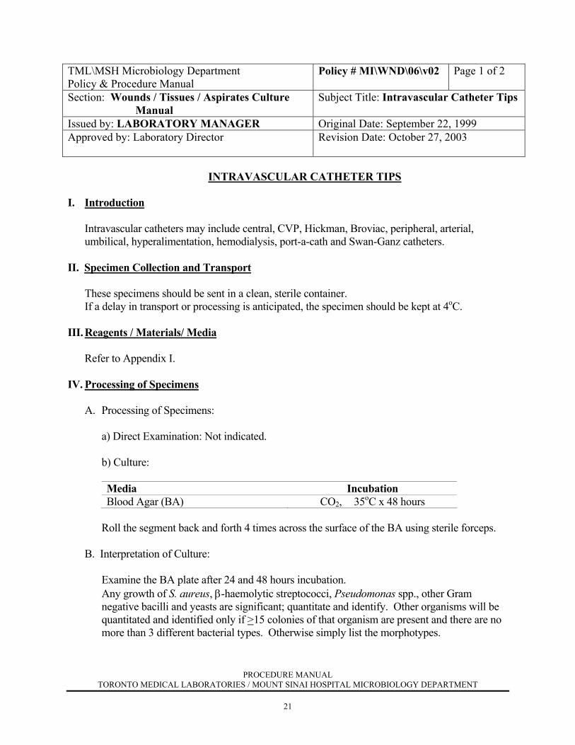
INTRAVASCULAR CATHETER TIPS
I. Introduction
Intravascular catheters may include central, CVP, Hickman, Broviac, peripheral, arterial, umbilical, hyperalimentation, hemodialysis, port-a-cath and Swan-Ganz catheters.
II. Specimen Collection and Transport These specimens should be sent in a clean, sterile container. If a delay in transport or processing is anticipated, the specimen should be kept at 4oC. III. Reagents / Materials/ Media
Refer to Appendix I.
IV. Processing of Specimens
A. Processing of Specimens: a) Direct Examination: Not indicated. b) Culture:
Media Incubation Blood Agar (BA) CO2, 35oC x 48 hours
Roll the segment back and forth 4 times across the surface of the BA using sterile forceps. B. Interpretation of Culture: Examine the BA plate after 24 and 48 hours incubation. Any growth of S. aureus, β-haemolytic streptococci, Pseudomonas spp., other Gram
negative bacilli and yeasts are significant; quantitate and identify. Other organisms will be quantitated and identified only if >15 colonies of that organism are present and there are no more than 3 different bacterial types. Otherwise simply list the morphotypes.
PROCEDURE MANUAL TORONTO MEDICAL LABORATORIES / MOUNT SINAI HOSPITAL MICROBIOLOGY DEPARTMENT
22
TML\MSH Microbiology Department Policy & Procedure Manual
Policy # MI\WND\06\v01 Page 2 of 2
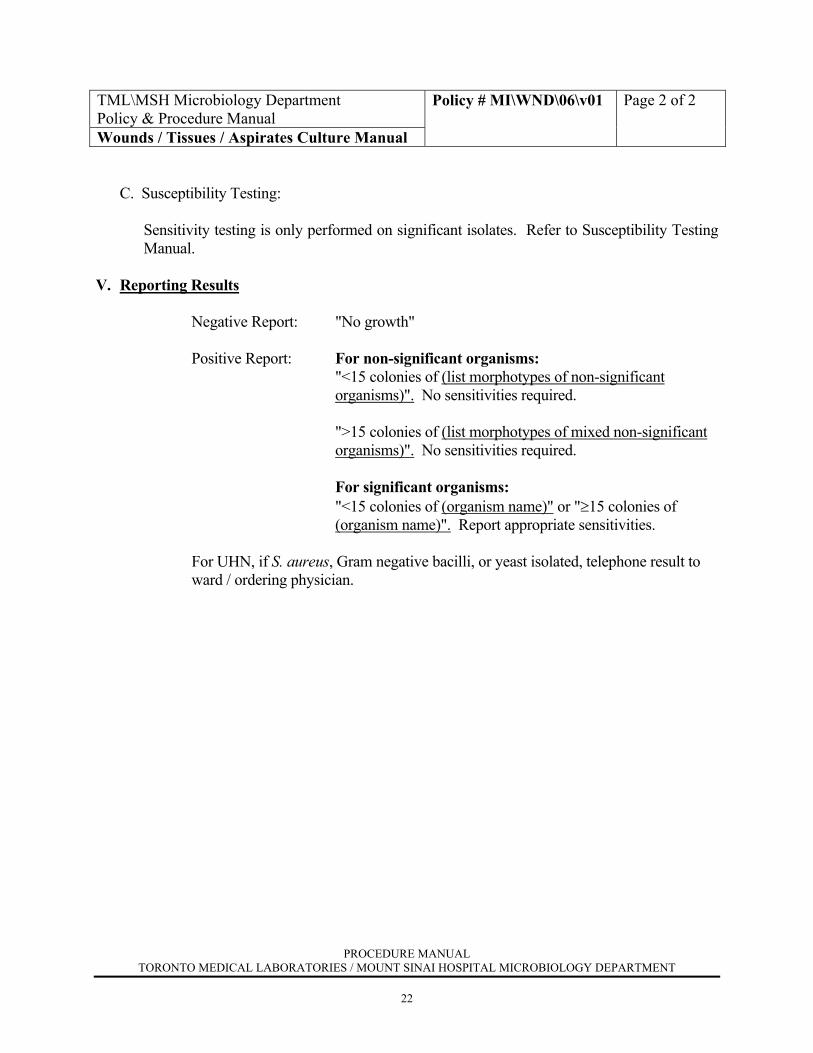
Wounds / Tissues / Aspirates Culture Manual C. Susceptibility Testing: Sensitivity testing is only performed on significant isolates. Refer to Susceptibility Testing
Manual. V. Reporting Results
Negative Report: "No growth"
Positive Report: For non-significant organisms: "<15 colonies of (list morphotypes of non-significant organisms)". No sensitivities required. ">15 colonies of (list morphotypes of mixed non-significant organisms)". No sensitivities required. For significant organisms: "<15 colonies of (organism name)" or "≥15 colonies of (organism name)". Report appropriate sensitivities.
For UHN, if S. aureus, Gram negative bacilli, or yeast isolated, telephone result to ward / ordering physician.
PROCEDURE MANUAL TORONTO MEDICAL LABORATORIES / MOUNT SINAI HOSPITAL MICROBIOLOGY DEPARTMENT
23
TML\MSH Microbiology Department Policy & Procedure Manual
Policy # MI\WND\07\v01 Page 1 of 2
Section: Wounds / Tissues / Aspirates Culture Manual
Subject Title: Peritoneal Dialysis Catheter/Canula & Prosthetic Devices
Issued by: LABORATORY MANAGER Original Date: September 22, 1999 Approved by: Laboratory Director
Revision Date:
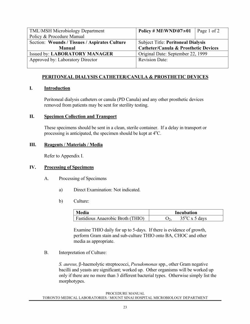
PERITONEAL DIALYSIS CATHETER/CANULA & PROSTHETIC DEVICES
I. Introduction
Peritoneal dialysis catheters or canula (PD Canula) and any other prosthetic devices removed from patients may be sent for sterility testing.
II. Specimen Collection and Transport
These specimens should be sent in a clean, sterile container. If a delay in transport or processing is anticipated, the specimen should be kept at 4oC.
III. Reagents / Materials / Media
Refer to Appendix I.
IV. Processing of Specimens
A. Processing of Specimens
a) Direct Examination: Not indicated.
b) Culture:
Media Incubation Fastidious Anaerobic Broth (THIO) O2, 35oC x 5 days
Examine THIO daily for up to 5-days. If there is evidence of growth, perform Gram stain and sub-culture THIO onto BA, CHOC and other media as appropriate.
B. Interpretation of Culture:
S. aureus, β-haemolytic streptococci, Pseudomonas spp., other Gram negative bacilli and yeasts are significant; worked up. Other organisms will be worked up only if there are no more than 3 different bacterial types. Otherwise simply list the morphotypes.
PROCEDURE MANUAL TORONTO MEDICAL LABORATORIES / MOUNT SINAI HOSPITAL MICROBIOLOGY DEPARTMENT
24
TML\MSH Microbiology Department Policy & Procedure Manual
Policy # MI\WND\07\v01 Page 2 of 2
Wounds / Tissues / Aspirates Culture Manual
C. Susceptibility Testing:
Sensitivity testing is only performed on significant isolates. Refer to Susceptibility Testing Manual.
V. Reporting Results
Negative Report: "No growth" or "No significant growth including (list of non- significant organisms)" Positive Report: Report all significant isolates with appropriate sensitivities.
PROCEDURE MANUAL TORONTO MEDICAL LABORATORIES / MOUNT SINAI HOSPITAL MICROBIOLOGY DEPARTMENT
25
TML\MSH Microbiology Department Policy & Procedure Manual
Policy # MI\WND\08\v01 Page 1 of 2
Section: Wounds / Tissues / Aspirates Culture Manual Subject Title: Bile Issued by: LABORATORY MANAGER Original Date: September 22, 1999 Approved by: Laboratory Director
Revision Date: July 26, 2000
BILE
I. Introduction
Bile is a normally sterile fluid. However, it may become contaminated when collected from a post-op drain. Bile may also be collected at the time of percutaneous cholangiography (PTC).
II. Specimen Collection and Transport Bile may be aspirated with a syringe during surgery or collected in a sterile container from a
post-op drain. If a delay in transport or processing is anticipated, the specimen should be kept at 4oC.
III. Reagents / Material / Media Refer to Appendix I. IV. Procedure
A. Processing of Specimens: a) Direct Examination: Gram stain – Quantitate the presence of pus cells and organisms. (Refer to Appendix II). b) Culture:
Media Incubation Blood Agar (BA) MacConkey Agar (MAC)
CO2, 35oC x 48 hours O2, 35oC x 48 hours
If anaerobic culture is requested or bile is collected by PTC, add: Fastidious Anaerobic Agar (BRUC) Kanamycin/Vancomycin Agar ( KV) Fastidious Anaerobic Broth (THIO)
AnO2, 35oC x 48 hours AnO2, 35oC x 48 hours O2, 35oC x 48 hours
PROCEDURE MANUAL TORONTO MEDICAL LABORATORIES / MOUNT SINAI HOSPITAL MICROBIOLOGY DEPARTMENT
26
TML\MSH Microbiology Department Policy & Procedure Manual
Policy # MI\WND\08\v01 Page 2 of 2
Wounds / Tissues / Aspirates Culture Manual
B. Interpretation of Cultures:
Examine the aerobic culture plates after 24 and 48 hours incubation and the anaerobic plates after 48 hours incubation. Any growth of Salmonella species, S. aureus, and Ps. aeruginosa are significant. For other organisms, a significant result is determined by the isolation of ≤3 organisms. For non-lactose fermenters (NLF), screen for Salmonella species.
C. Susceptibility Testing: Refer to Susceptibility Testing Manual. V. Reporting Results a) Gram stain: Report with quantitation the presence of pus cells and organisms. b) Culture: Negative Report: "No growth" or "Mixed faecal flora"
Positive Report: Quantitate all significant isolates with appropriate sensitivities. If faecal flora is also present, report without quantitation.

PROCEDURE MANUAL TORONTO MEDICAL LABORATORIES / MOUNT SINAI HOSPITAL MICROBIOLOGY DEPARTMENT
27
TML\MSH Microbiology Department Policy & Procedure Manual
Policy # MI\WND\09\v01 Page 1 of 2
Section: Wounds / Tissues / Aspirates Culture Manual Subject Title: Bone Bank, Bone Graft & Cadaver Fascia / Tissue/ Swab Specimens
Issued by: LABORATORY MANAGER Original Date: September 22, 1999 Approved by: Laboratory Director
Revision Date: July 26, 2000
BONE BANK, BONE GRAFT & CADAVER FASCIA / TISSUE/ SWAB SPECIMENS
I. Introduction
Bone specimens for the bone bank are collected for use in transplantation. Specimens are usually collected ante-mortem or just prior to transplantation and should normally be sterile. Occasionally, fascia may be used for transplantation in which case a swab or tissue sample may be collected for sterility testing.
II. Specimen Collection and Transport Swabs from the donor bones or fascia should be collected using a clean, sterile swab and sent in
Amies transport medium. If anaerobic culture is requested, an anaerobic swab sent in anaerobic transport medium should be collected. Bone or fascia tissue should be sent in a clean, sterile container. If a delay in transport or processing is anticipated, the aerobic swab and bone/fascia tissue should be kept at 4oC and the anaerobic swab at room temperature.
III. Reagents / Material / Media Refer to Appendix I. IV. Procedure
A. Processing of Specimen:
a) Direct Examination: Not indicated b) Culture:
Media Incubation Fastidious Anaerobic Broth (THIO)*
O2, 35oC x 7 days
* A separate THIO should be inoculated for each specimen / swab received.
PROCEDURE MANUAL TORONTO MEDICAL LABORATORIES / MOUNT SINAI HOSPITAL MICROBIOLOGY DEPARTMENT
28
TML\MSH Microbiology Department Policy & Procedure Manual
Policy # MI\WND\09\v01 Page 2 of 2
Wounds / Tissues / Aspirates Culture Manual
B. Interpretation of Culture:
Examine the THIO daily for 7 days. If evidence of growth, perform Gram stain and subculture THIO onto Blood agar (BA), MacConkey (MAC), Chocolate (CHOC) and (BRUC) as appropriate based on Gram stain.
All isolates are to be identified. Freeze all isolates at -70oC. C. Susceptibility Testing: Not required for isolates from bone collected ante-mortem.
For isolates from bone or fascia cultured just prior to transplantation, perform susceptibility testing on all isolates. Refer to Susceptibility Testing Manual.
V. Reporting Results Negative Report: "No growth after 7 days of incubation". Positive Report: Report all isolates with sensitivities as appropriate.
PROCEDURE MANUAL TORONTO MEDICAL LABORATORIES / MOUNT SINAI HOSPITAL MICROBIOLOGY DEPARTMENT
29
TML\MSH Microbiology Department Policy & Procedure Manual
Policy # MI\WND\10\v01 Page 1 of 2
Section: Wounds / Tissues / Aspirates Culture Manual Subject Title: Breast Milk Issued by: LABORATORY MANAGER Original Date: September 22, 1999 Approved by: Laboratory Director
Revision Date: July 26, 2000
BREAST MILK
I. Introduction
Breast milk may become infected with a variety of organisms and all species should be identified except skin commensals.
II. Specimen Collection and Transport Breast milk should be sent in a clean, sterile container. If a delay in transport or processing is
anticipated, the specimen should be kept at 4oC. III. Reagents / Materials/ Media
Refer to Appendix I.
IV. Procedure
A. Processing of Specimens: a) Direct Examination: Not required
b) Culture:
Media Incubation Blood Agar (BA) MacConkey Agar (MAC)
CO2, 35oC x 48 hours O2, 35oC x 48 hours
B. Interpretation of Cultures: Examine the culture plates after 24 and 48 incubation Any growth of organisms other than skin commensals should be considered significant.
C. Susceptibility Testing: Refer to Susceptibility Testing Manual.

PROCEDURE MANUAL TORONTO MEDICAL LABORATORIES / MOUNT SINAI HOSPITAL MICROBIOLOGY DEPARTMENT
30
TML\MSH Microbiology Department Policy & Procedure Manual
Policy # MI\WND\10\v01 Page 2 of 2
Wounds / Tissues / Aspirates Culture Manual V. Reporting Results Negative Report: "No growth" or "Commensal flora" Positive Report: Quantitate all significant isolates with appropriate sensitivities. If
commensal flora is also present, report with quantitation.
PROCEDURE MANUAL TORONTO MEDICAL LABORATORIES / MOUNT SINAI HOSPITAL MICROBIOLOGY DEPARTMENT
31
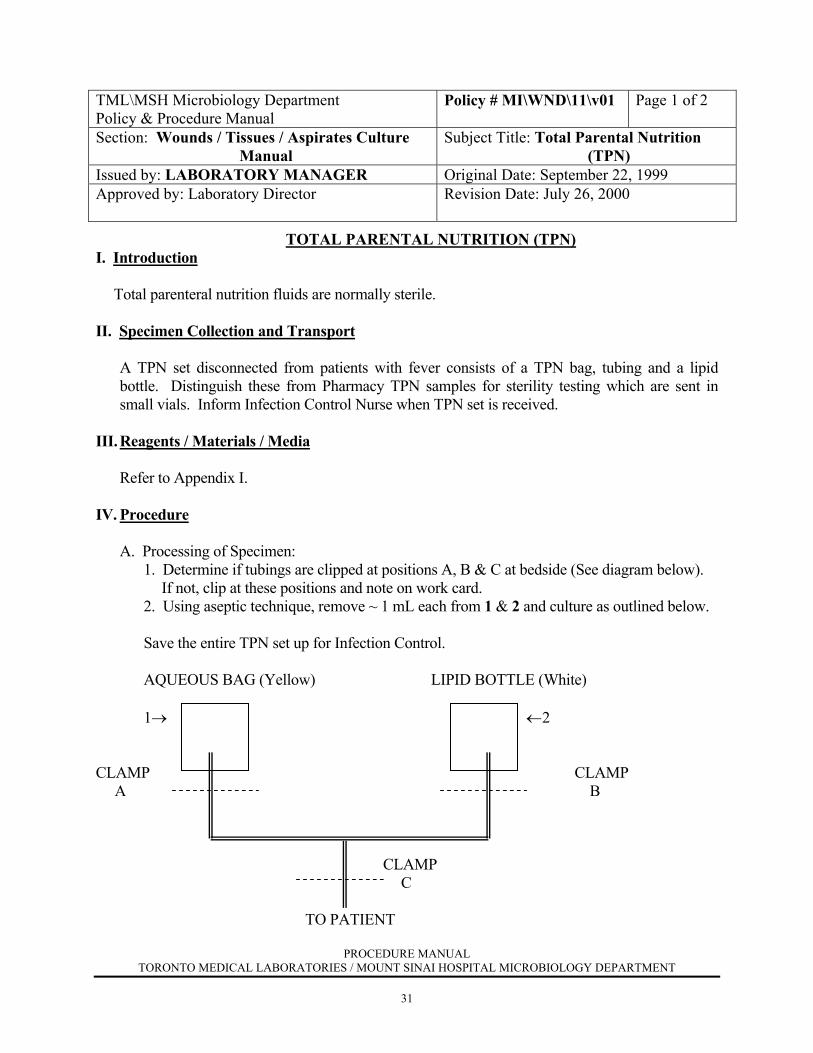
TML\MSH Microbiology Department Policy & Procedure Manual
Policy # MI\WND\11\v01 Page 1 of 2
Section: Wounds / Tissues / Aspirates Culture Manual
Subject Title: Total Parental Nutrition (TPN)
Issued by: LABORATORY MANAGER Original Date: September 22, 1999 Approved by: Laboratory Director
Revision Date: July 26, 2000
TOTAL PARENTAL NUTRITION (TPN) I. Introduction Total parenteral nutrition fluids are normally sterile. II. Specimen Collection and Transport A TPN set disconnected from patients with fever consists of a TPN bag, tubing and a lipid
bottle. Distinguish these from Pharmacy TPN samples for sterility testing which are sent in small vials. Inform Infection Control Nurse when TPN set is received.
III. Reagents / Materials / Media Refer to Appendix I. IV. Procedure
A. Processing of Specimen:
1. Determine if tubings are clipped at positions A, B & C at bedside (See diagram below). If not, clip at these positions and note on work card.
2. Using aseptic technique, remove ~ 1 mL each from 1 & 2 and culture as outlined below. Save the entire TPN set up for Infection Control. AQUEOUS BAG (Yellow) LIPID BOTTLE (White) 1→ ←2 CLAMP CLAMP A B CLAMP C TO PATIENT

PROCEDURE MANUAL TORONTO MEDICAL LABORATORIES / MOUNT SINAI HOSPITAL MICROBIOLOGY DEPARTMENT
32
TML\MSH Microbiology Department Policy & Procedure Manual
Policy # MI\WND\11\v01 Page 2 of 2
Wounds / Tissues / Aspirates Culture Manual b) Culture:
Media Incubation Blood Agar (BA) Fastidious Anaerobic Broth (THIO) Inhibitory Mold Agar (IMA)* IMA with sterile olive oil overlay (olive oil is stored in media room)*
CO2, 35oC x 2 days O2, 35oC x 5 days O2, 30oC x 3 weeks O2, 30oC x 1 week
*Forward these plates to the Mycology section for incubation and work-up.
B. Interpretation of Cultures: Examine the BA plate after 24 and 48 hours incubation and the THIO daily for up to 5 days. Any growth should be considered significant. Freeze all isolates at -70oC and put into "Sterile Sites" box.
C. Susceptibility Testing: Refer to Susceptibility Testing Manual. V. Reporting Result Culture: Negative Report: "No growth" Positive Report: Report all organisms with appropriate sensitivities. Do not
quantitate.

PROCEDURE MANUAL TORONTO MEDICAL LABORATORIES / MOUNT SINAI HOSPITAL MICROBIOLOGY DEPARTMENT
33
TML\MSH Microbiology Department Policy & Procedure Manual
Policy # MI\WND\12\v01 Page 1 of 2
Section: Wounds / Tissues / Aspirates Culture Manual
Subject Title: Intravenous and Central Line Catheter Exit Site Swabs
Issued by: LABORATORY MANAGER Original Date: September 22, 1999 Approved by: Laboratory Director
Revision Date: July 26, 2000
INTRAVENOUS & CENTRAL LINE CATHETER EXIT SITE SWABS
I. Introduction
The intravenous or central line catheter exit site may become infected with a variety of organisms which may lead to tunnel infections or bacteraemia.
II. Specimen Collection and Transport Specimens should be collected using a clean, sterile swab and sent in Amies transport medium.
If a delay in transport or processing is anticipated, keep the specimen at 40C. III. Reagents / Materials / Media
Refer to Appendix I. IV. Procedure
A. Processing of Specimen: a) Direct Examination: Not indicated. b) Culture:
Media Incubation Blood Agar (BA) MacConkey Agar (MAC)
CO2, 35oC x 48 hours O2, 35oC x 48 hours
B. Interpretation of Cultures:
Examine the culture plates after 24 and 48 hours incubation.
Quantitate and identify any growth of S. aureus, Pseudomonas species, yeast and beta-haemolytic streptococci. Quantitate and identify any pure or predominant growth of other Gram negative bacilli and enterococci. A heavy, pure growth of any other organism is significant.
C. Susceptibility Testing:
Refer to Susceptibility Testing Manual.
PROCEDURE MANUAL TORONTO MEDICAL LABORATORIES / MOUNT SINAI HOSPITAL MICROBIOLOGY DEPARTMENT
34
TML\MSH Microbiology Department Policy & Procedure Manual
Policy # MI\WND\12\v01 Page 2 of 2
Wounds / Tissues / Aspirates Culture Manual V. Reporting Results Negative report: "No growth" or "Commensal flora" Positive report: Quantitate all significant isolates with appropriate
sensitivities. If commensal flora is also present, report with quantitation.
PROCEDURE MANUAL TORONTO MEDICAL LABORATORIES / MOUNT SINAI HOSPITAL MICROBIOLOGY DEPARTMENT
35
TML\MSH Microbiology Department Policy & Procedure Manual
Policy # MI\WND\13\\01\v01 Page 1 of 2
Section: Wounds / Tissues / Aspirates Culture Manual
Subject Title: Ear Specimens–Ear Swabs
Issued by: LABORATORY MANAGER Original Date: September 22, 1999 Approved by: Laboratory Director
Revision Date: January 25, 2001
EAR SPECIMENS - EAR SWABS
I. Introduction
Ear swabs are collected for the diagnosis of otitis externa; they are not useful in the diagnosis of otitis media. Otitis externa is a bacterial infection of the external auditory canal usually caused by P. aeruginosa, S. aureus, S. pneumoniae, Group A streptococcus or fungus / yeast.
II. Specimen Collection and Transport The ear swab should be collected using a clean, sterile swab and sent in Amies transport
medium. If a delay in transport or processing is anticipated, the specimen should be kept at 40C. III. Reagents / Materials / Media Refer to Appendix I. IV. Procedure
A. Processing of Specimens: a) Direct Examination: Gram stain – Quantitate the presence of pus cells and organisms. (Refer to Appendix II). Calcofluor white stain (If fungus is requested). - Refer to
Mycology Manual. b) Culture:
Media Incubation Blood Agar (BA) MacConkey Agar (MAC) Colistin Nalidixic Acid Agar (CNA)
CO2, 35oC x 48 hours O2, 35oC x 48 hours O2, 35oC x 48 hours
If fungus culture is requested, add: Inhibitory Mold Agar (IMA)* Esculin Base Medium (EBM)*
O2, 30oC x 3 weeks O2, 30oC x 3 weeks
* Forward the fungal culture media to the Mycology section for incubation and work-
up.

PROCEDURE MANUAL TORONTO MEDICAL LABORATORIES / MOUNT SINAI HOSPITAL MICROBIOLOGY DEPARTMENT
36
TML\MSH Microbiology Department Policy & Procedure Manual
Policy # MI\WND\13\\01\v01 Page 2 of 2
Wounds / Tissues / Aspirates Culture Manual
B. Interpretation of Cultures: Examine the culture plates after 24 and 48 hours incubation.
Any growth of S. aureus, P. aeruginosa, S. pneumoniae, Group A streptococcus or yeast is significant. For specimens from neonates only, identify and report Group B streptococcus. For other organisms, a significant result is determined by the presence of a moderate to heavy growth of an organism which correlates with the predominant organism on the Gram stain. The Gram stain should also show >1+ pus cells. Full identification is required for all significant organisms except yeast.
C. Susceptibility Testing:
Refer to Susceptibility Testing Manual. V. Reporting Results a) Gram stain: Report with quantitation the presence of pus cells and organisms. b) Culture: Negative Report: "Commensal flora" or "No growth". Positive Report: Quantitate all significant isolates with appropriate
sensitivities. If commensal flora is also present, report with quantitation.

PROCEDURE MANUAL TORONTO MEDICAL LABORATORIES / MOUNT SINAI HOSPITAL MICROBIOLOGY DEPARTMENT
37
TML\MSH Microbiology Department Policy & Procedure Manual
Policy # MI\WND\13\\02\v01 Page 1 of 1
Section: Wounds / Tissues / Aspirates Culture Manual
Subject Title: Tympanocentesis Fluid
Issued by: LABORATORY MANAGER Original Date: September 22, 1999 Approved by: Laboratory Director
Revision Date: July 26, 2000
TYMPANOCENTESIS FLUID
I. Introduction
Tympanocentesis fluid is obtained for the diagnosis of otitis media. These specimens are handled as sterile fluids. (Refer to Sterile Fluids Culture Manual)

PROCEDURE MANUAL TORONTO MEDICAL LABORATORIES / MOUNT SINAI HOSPITAL MICROBIOLOGY DEPARTMENT
38
TML\MSH Microbiology Department Policy & Procedure Manual
Policy # MI\WND\14\01\v01 Page 1 of 2
Section: Wounds / Tissues / Aspirates Culture Manual
Subject Title: Eye / Conjunctival / Lid Swabs
Issued by: LABORATORY MANAGER Original Date: September 22, 1999 Approved by: Laboratory Director
Revision Date: July 26, 2000
EYE / CONJUNCTIVAL / LID SWABS
I. Introduction
Eye / conjunctival / lid swabs are collected for the diagnosis of conjunctivitis. II. Specimen Collection and Transport It is preferable that both eyes be swabbed, even if the infection is unilateral. Swabs should be
collected prior to the instillation of topical anaesthetics or antibiotics, and sent in Amies transport medium. Viral isolation requires special transport media. If a delay in transport or processing is anticipated, the specimen should be kept at 40C.
Occasionally, specimens collected by an ophthalmologist will be inoculated directly onto
culture plates at the bedside. The ophthalmologist will inoculate the plates in a short spiral line. If lid swabs are also collected, these will be inoculated onto the same culture plates next to the conjunctival inoculation. Lid swabs will be inoculated in the shape of an "L" or "R" indicating left or right, respectively. These plates should be kept in the incubator (350C) until processed.
III. Reagents / Materials / Media
A. Processing of Specimens: NB: If pre-inoculated culture plates are received, these should be incubated as listed below. No Gram stain will be performed. a) Direct Examination: Gram stain – Quantitate the presence of pus cells and organisms. (Refer to Appendix II). b) Culture:
Media Incubation Blood Agar (BA) Chocolate Agar (CHOC)
CO2, 35oC x 48 hours CO2, 35oC x 48 hours
l For all neonates < 1 week of age, or if N. gonorrhoeae is requested, add: Martin-Lewis Agar (ML)
CO2, 35oC x 72 hours
PROCEDURE MANUAL TORONTO MEDICAL LABORATORIES / MOUNT SINAI HOSPITAL MICROBIOLOGY DEPARTMENT
39
TML\MSH Microbiology Department Policy & Procedure Manual
Policy # MI\WND\14\01\v01 Page 2 of 2
Wounds / Tissues / Aspirates Culture Manual B. Interpretation of Cultures:
Examine the BA and CHOC plates after 24 and 48 hours incubation and the ML plate after 48 and 72 hours incubation. Any growth of S. aureus, H. influenzae, M. catarrhalis, N. gonorrheae, Gp.A Strep, S. pneumoniae, Moraxella species, and P. aeruginosa is potentially significant. For other organisms, a significant result is determined by the isolation of a moderate or heavy growth of a potential pathogen correlated with the predominant organism on the Gram stain. There should be >1+ pus cells on the Gram stain. Full identification is required for all significant organisms.
For work-up and identification of N. gonorrhaeae, refer to the Genital Tract Manual.
C. Susceptibility Testing: Refer to Susceptibility Testing Manual. V. Reporting Results a) Gram stain: Report with quantitation the presence of pus cells and organisms. b) Culture: Negative Report: "Commensal flora" or "No growth".
Positive Report: Quantitate all significant isolates with appropriate sensitivities. If commensal flora is also present, report with
quantitation.
PROCEDURE MANUAL TORONTO MEDICAL LABORATORIES / MOUNT SINAI HOSPITAL MICROBIOLOGY DEPARTMENT
40
TML\MSH Microbiology Department Policy & Procedure Manual
Policy # MI\WND\14\02\v01 Page 1 of 2
Section: Wounds / Tissues / Aspirates Culture Manual
Subject Title: Eye / Corneal Scrapings, Contact Lenses and Solutions
Issued by: LABORATORY MANAGER Original Date: September 22, 1999 Approved by: Laboratory Director
Revision Date: July 26, 2000
EYE / CORNEAL SCRAPINGS, CONTACT LENSES & SOLUTIONS
I. Introduction
Eye / corneal scrapings are collected for the diagnosis of keratitis. Contact lenses and solutions may be submitted to the microbiology laboratory for detection of contamination including the presence of acanthamoeba.
II. Specimen Collection and Transport The physician usually prepares two or three slides and inoculates the appropriate media at the
time of specimen collection. The following media is to be supplied to the physician for each eye: BA, CHOC, IMA and THIO. The physician will inoculate the plates in rows of "C" - shaped marks, with each row representing a separate sample. If a delay in transport or processing is anticipated, the specimen should be kept in the incubator (350C) in Specimen Management area. Virus and chlamydia detection require special transport media. (See Virology Manual). If acanthamoeba is requested, forward specimen to Parasitology section for processing. If E. coli overlay plate is received and parasitology section is closed, incubate plate at 350C in O2 until processed. All other specimens received for acanthamoeba should be kept at room temperature until processed.
III. Reagents / Materials / Media Refer to Appendix I. IV. Procedure
A. Processing of Specimens:
NB: If previously inoculated plates received and no specimen or swab received, then direct examination is not performed.
a) Direct Examination: Gram stain – Quantitate the presence of pus cells and organisms. (Refer to Appendix II). Calcofluor white stain. (If two smears are provided).
An extra smear is held in reserve for special stains (eg, Giemsa stain if requested).
PROCEDURE MANUAL TORONTO MEDICAL LABORATORIES / MOUNT SINAI HOSPITAL MICROBIOLOGY DEPARTMENT
41
TML\MSH Microbiology Department Policy & Procedure Manual
Policy # MI\WND\14\02\v01 Page 2 of 2
Wounds / Tissues / Aspirates Culture Manual
b) Culture:
Media Incubation Blood Agar (BA) Chocolate Agar (CHOC) Fastidious Anaerobic Broth (THIO) Inhibitory Mold Agar (IMA)*
CO2, 35oC x 4 days CO2, 35oC x 4 days O2, 35oC x 5 days
O2, 30oC x 3 weeks *Forward the fungal culture media to the Mycology section for incubation and workup.
B. Interpretation of Cultures:
Examine the culture plates daily. If no growth on culture plates but growth in THIO, perform Gram stain and sub-culture THIO onto BA, and CHOC and incubate x 48 hours.
For Conjunctival scrapings see Eye swabs. For Corneal scrapings all organisms should be identified.
C. Susceptibility Testing: Refer to Susceptibility Testing Manual. V. Reporting Results For conjunctival scrapings, see Eye swabs. For corneal scrapings: a) Gram stain: Report with quantitation the presence of pus cells and organisms. b) Culture: Negative report: "No growth." Positive report: Quantitate all isolates with appropriate sensitivities.
PROCEDURE MANUAL TORONTO MEDICAL LABORATORIES / MOUNT SINAI HOSPITAL MICROBIOLOGY DEPARTMENT
42
TML\MSH Microbiology Department Policy & Procedure Manual
Policy # MI\WND\14\03\v01 Page 1 of 1
Section: Wounds / Tissues / Aspirates Culture Manual
Subject Title: Intraocular Aspirates
Issued by: LABORATORY MANAGER Original Date: September 22, 1999 Approved by: Laboratory Director
Revision Date: July 26, 2000
INTRAOCULAR ASPIRATES
I. Introduction
Aspirates of intraocular fluids are submitted for the diagnosis of uveitis and endophthalmitis. These specimens are handled as sterile fluids. (Refer to the Sterile Fluids Culture Manual)
Any requests for specialized procedures should be discussed with a medical microbiologist or the chief technologist before proceeding.

PROCEDURE MANUAL TORONTO MEDICAL LABORATORIES / MOUNT SINAI HOSPITAL MICROBIOLOGY DEPARTMENT
43
TML\MSH Microbiology Department Policy & Procedure Manual
Policy # MI\WND\14\04\v01 Page 1 of 2
Section: Wounds / Tissues / Aspirates Culture Manual
Subject Title: Lacrimal (Tear Duct) Stone / Secretions
Issued by: LABORATORY MANAGER Original Date: September 22, 1999 Approved by: Laboratory Director
Revision Date: July 26, 2000
LACRIMAL (TEAR DUCT) STONE / SECRETIONS
I. Introduction
Stones may form in the lacrimal duct resulting in obstruction and secondary infection of the lacrimal gland.
II. Specimen Collection and Transport
Specimens are to be collected and transported in a clean, sterile container. If a delay in transport or processing is anticipated, the specimen should be kept at 40C.
III. Reagents / Materials / Media
Refer to Appendix I. IV. Procedure
A. Processing of Specimens: a) Direct examination: Crush specimen on glass slide to obtain a thin smear for Gram stain. Examine for pus cells and organisms especially branching gram
positive bacilli resembling Actinomyces species.
b) Culture: Crush specimen using a sterile wood applicator stick or urine loop before plating onto the following media:
Media Incubation Blood Agar (BA) Chocolate Agar (CHOC) Fastidious Anaerobic Broth (THIO)
CO2, 35oC x 48 hours CO2, 35oC x 48 hours
O2, 35oC x 5 days B. Interpretation of Cultures:
Examine the culture plates after 24 and 48 hours incubation. Examine the THIO daily for evidence of growth. If no growth on culture plates but evidence of growth in THIO, then perform Gram stain and subculture THIO onto BA, CHOC and BRUC (as appropriate) and incubate and process as above. Identify all significant isolates.
PROCEDURE MANUAL TORONTO MEDICAL LABORATORIES / MOUNT SINAI HOSPITAL MICROBIOLOGY DEPARTMENT
44
TML\MSH Microbiology Department Policy & Procedure Manual
Policy # MI\WND\14\04\v01 Page 2 of 2
Wounds / Tissues / Aspirates Culture Manual
C. Susceptibility Testing: Refer to Susceptibility Testing Manual.
V. Reporting Results a) Gram stain: Report presence of organisms. "Organisms resembling Actinomyces seen in Gram stain". b) Culture: Negative Report: "Commensal flora" or "No growth".
Positive Report: Quantitate all significant isolates with appropriate sensitivities. If commensal flora is also present, report with quantitation.
PROCEDURE MANUAL TORONTO MEDICAL LABORATORIES / MOUNT SINAI HOSPITAL MICROBIOLOGY DEPARTMENT
45
TML\MSH Microbiology Department Policy & Procedure Manual
Policy # MI\WND\15\v01 Page 1 of 2
Section: Wounds / Tissues / Aspirates Culture Manual Subject Title: Facial Specimens Issued by: LABORATORY MANAGER Original Date: September 22, 1999 Approved by: Laboratory Director
Revision Date: July 26, 2000
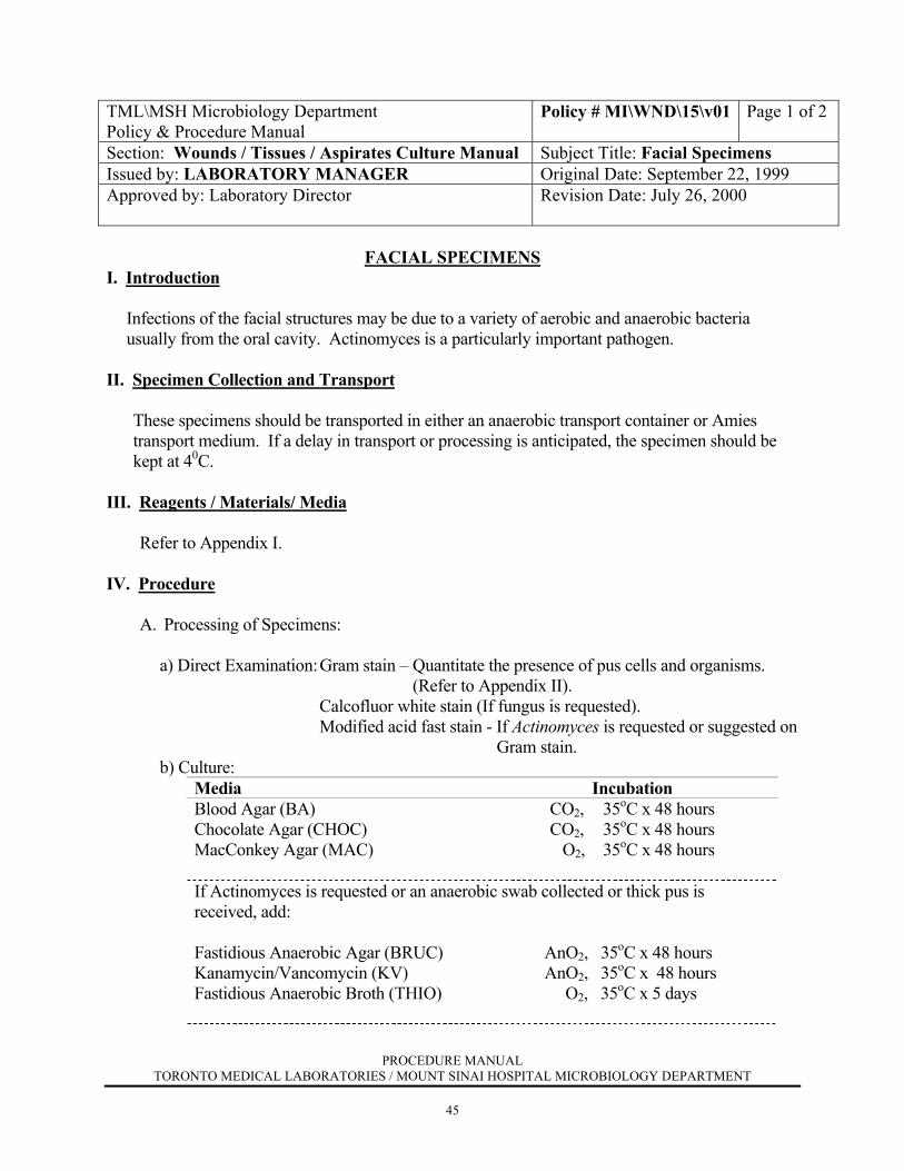
FACIAL SPECIMENS
I. Introduction
Infections of the facial structures may be due to a variety of aerobic and anaerobic bacteria usually from the oral cavity. Actinomyces is a particularly important pathogen.
II. Specimen Collection and Transport These specimens should be transported in either an anaerobic transport container or Amies
transport medium. If a delay in transport or processing is anticipated, the specimen should be kept at 40C.
III. Reagents / Materials/ Media Refer to Appendix I. IV. Procedure
A. Processing of Specimens: a) Direct Examination: Gram stain – Quantitate the presence of pus cells and organisms. (Refer to Appendix II). Calcofluor white stain (If fungus is requested). Modified acid fast stain - If Actinomyces is requested or suggested on Gram stain. b) Culture:
Media Incubation Blood Agar (BA) Chocolate Agar (CHOC) MacConkey Agar (MAC)
CO2, 35oC x 48 hours CO2, 35oC x 48 hours O2, 35oC x 48 hours
If Actinomyces is requested or an anaerobic swab collected or thick pus is received, add: Fastidious Anaerobic Agar (BRUC)
Kanamycin/Vancomycin (KV)
Fastidious Anaerobic Broth (THIO)
AnO2, 35oC x 48 hours AnO2, 35oC x 48 hours O2, 35oC x 5 days
PROCEDURE MANUAL TORONTO MEDICAL LABORATORIES / MOUNT SINAI HOSPITAL MICROBIOLOGY DEPARTMENT
46
TML\MSH Microbiology Department Policy & Procedure Manual
Policy # MI\WND\15\v01 Page 2 of 2
Wounds / Tissues / Aspirates Culture Manual
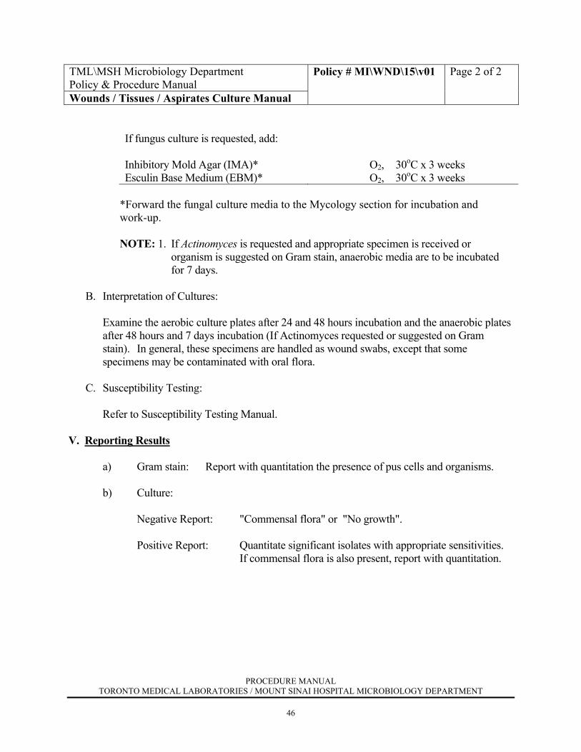
If fungus culture is requested, add: Inhibitory Mold Agar (IMA)* Esculin Base Medium (EBM)*
O2, 30oC x 3 weeks O2, 30oC x 3 weeks
*Forward the fungal culture media to the Mycology section for incubation and work-up.
NOTE: 1. If Actinomyces is requested and appropriate specimen is received or organism is suggested on Gram stain, anaerobic media are to be incubated for 7 days.
B. Interpretation of Cultures:
Examine the aerobic culture plates after 24 and 48 hours incubation and the anaerobic plates after 48 hours and 7 days incubation (If Actinomyces requested or suggested on Gram stain). In general, these specimens are handled as wound swabs, except that some specimens may be contaminated with oral flora.
C. Susceptibility Testing:
Refer to Susceptibility Testing Manual. V. Reporting Results a) Gram stain: Report with quantitation the presence of pus cells and organisms. b) Culture: Negative Report: "Commensal flora" or "No growth". Positive Report: Quantitate significant isolates with appropriate sensitivities. If commensal flora is also present, report with quantitation.

PROCEDURE MANUAL TORONTO MEDICAL LABORATORIES / MOUNT SINAI HOSPITAL MICROBIOLOGY DEPARTMENT
47
TML\MSH Microbiology Department Policy & Procedure Manual
Policy # MI\WND\16\v01 Page 1 of 2
Section: Wounds / Tissues / Aspirates Culture Manual Subject Title: In Vitro Tissue Culture Specimens
Issued by: LABORATORY MANAGER Original Date: September 22, 1999 Approved by: Laboratory Director
Revision Date: July 26, 2000
IN VITRO TISSUE CULTURE SPECIMENS
I. Introduction
Human cells may be collected from patients for in vitro culture to expand certain cell populations. This is usually done in the Cytogenetics Laboratory. As with any cell culture, these may become contaminated with bacteria or other organisms. Because this is a normally sterile procedure, the isolation of any organism should be considered significant.
II. Specimen Collection and Transport
A minimum of 1 ml of the cell culture media should be collected into a clean sterile container. If a delay in transport or processing is anticipated, the specimen should be kept at 40C.
III. Reagents / Material / Media Refer to Appendix I. IV. Procedure
A. Processing of Specimens:
a) Direct Examination: Gram stain: Note the presence of organisms. c) Culture:
Media Incubation Blood Agar (BA) Chocolate Agar (CHOC) MacConkey Agar (MAC) Fastidious Anaerobic Broth (THIO) If fungus culture is requested, add: Inhibitory Mold Agar (IMA)* Esculin Base Medium (EBM)*
CO2, 350C x 48 hours CO2, 350C x 48 hours O2, 350C x 48 hours O2, 350C x 5 days O2, 350C x 3 weeks O2, 350C x 3 weeks
*Forward the fungal culture media to the Mycology section for incubation and work-up.
PROCEDURE MANUAL TORONTO MEDICAL LABORATORIES / MOUNT SINAI HOSPITAL MICROBIOLOGY DEPARTMENT
48
TML\MSH Microbiology Department Policy & Procedure Manual
Policy # MI\WND\16\v01 Page 2 of 2
Wounds / Tissues / Aspirates Culture Manual
B. Interpretation of Cultures:
Examine the plates after 24 and 48 hours incubation. Identify all isolates.
C. Susceptibility Testing:
Refer to Susceptibility Testing Manual.
V. Reporting Results a) Gram Stain: Report the presence of organisms.
b) Culture: Negative Report: "No growth". Positive Report: Report all isolates with appropriate sensitivities. If requested STAT, telephone Gram stain and positive culture results to ordering laboratory.

PROCEDURE MANUAL TORONTO MEDICAL LABORATORIES / MOUNT SINAI HOSPITAL MICROBIOLOGY DEPARTMENT
49
TML\MSH Microbiology Department Policy & Procedure Manual
Policy # MI\WND\17\01\v01 Page 1of 1
Section: Wounds / Tissues / Aspirates Culture Manual Subject Title: Appendix I REAGENTS/MATERIALS/MEDIA
Issued by: LABORATORY MANAGER Original Date: September 22, 1999 Approved by: Laboratory Director
Revision Date: July 26, 2000
APPENDIX I
REAGENTS / MATERIALS / MEDIA
Acid-fast stain – VWR Calcofluor white – Difco Catalase (3% H2O2) – Ingram & Bell Gram Stain – Refer to Media Manual for preparation Strep. Latex agglutination – Pro Lab Diagnostics Staph. Latex agglutination – Sanofi Tube coagulase - VWR Amies transport median – NCS Diagnostics / Quelab Blood Agar (BA) – MedPrep Blood Egg Albumin Agar (BEAA) – Biomedia Chocolate Agar (CHOC) – Oxoid Unipath Colistin Nalidixic Acid Agar (CNA) – MedPrep Esculin Base Medium (EBM) Fastidious Anaerobic Agar (BRUC) - MedPrep Fastidious Anaerobic Broth (THIO) - MedPrep Haemophilus Isolation Agar (HI) - MedPrep Inhibitory Mold Agar (IMA) – MedPrep Kanamycin / Vancomycin Agar (KV) - MedPrep MacConkey Agar (MAC) – MedPrep Mannitol Salt Agar with Oxacillin (MSAOX) – MedPrep Martin-Lewis Agar (ML) – Quelab Oxidase Strip - API Vitek Cards - bioMerieux Antibiotic Disks - Oxoid Mueller Hinton Agar - MedPrep PYR Disks - Remel LAP Disks - Remel Rapid Yeast Plus Strips - Oxoid Rapid Ana II Strips - Oxoid MRSA Screen - Denka Seiken

PROCEDURE MANUAL TORONTO MEDICAL LABORATORIES / MOUNT SINAI HOSPITAL MICROBIOLOGY DEPARTMENT
50
TML\MSH Microbiology Department Policy & Procedure Manual
Policy # MI\WND\17\02\v01 Page 1 of 1
Section: Wounds / Tissues / Aspirates Culture Manual Subject Title: Appendix II READING OF GRAM STAIN
Issued by: LABORATORY MANAGER Original Date: September 22, 1999 Approved by: Laboratory Director
Revision Date: July 26, 2000
APPENDIX II
READING OF GRAM STAIN
1. Examine stained smear microscopically by first focusing under low power. 2. Pick the best field for white cells, squamous epithelial cells, bacteria, and other structures
and quantitate as below:
NB: PMN / WBC will be reported as Pus cells < 1 cell per 1000 x oil immersion field = ± 1 – 4 cells per 1000 x oil immersion field = + 5 – 10 cells per 1000 x oil immersion field = ++ >10 cells per 1000 x oil immersion field = +++
PROCEDURE MANUAL TORONTO MEDICAL LABORATORIES / MOUNT SINAI HOSPITAL MICROBIOLOGY DEPARTMENT
51
TML\MSH Microbiology Department Policy & Procedure Manual
Policy # MI\WND\17\03\v01 Page 1 of 1
Section: Wounds / Tissues / Aspirates Culture Manual Subject Title: Appendix III Commensal Flora – Skin, Ear, Eye
Issued by: LABORATORY MANAGER Original Date: September 22, 1999 Approved by: Laboratory Director
Revision Date: July 26, 2000
APPENDIX III
COMMENSAL FLORA – SKIN, EAR, EYE
Type Organisms Aerobic bacteria Corynebacterium, Coagulase negative
Staphylococcus, Micrococcus, nonpathogenic Neisseria, Acinetobacter, Aerococcus
Anaerobic bacteria Propionibacterium, Clostridium, Peptostreptococcus
Fungi Candida spp., Malassezia Murray P.A, et al. 1999. Manual of Clinical Microbiology, 7th Edition, ASM Press.

PROCEDURE MANUAL TORONTO MEDICAL LABORATORIES / MOUNT SINAI HOSPITAL MICROBIOLOGY DEPARTMENT
52
TML\MSH Microbiology Department Policy & Procedure Manual
Policy # MI\WND\17\04\v01 Page 1 of 2
Section: Wounds / Tissues / Aspirates Culture Manual Subject Title: Appendix IV Yeast Identification
Issued by: LABORATORY MANAGER Original Date: September 22, 1999 Approved by: Laboratory Director
Revision Date:
APPENDIX IV
YEAST IDENFIFICATION
All yeast isolates except from voided urines, sputums (if growth is ≤ 1+), superficial sites, wounds,and drainage fluid will be screened using a Germ tube (see Appendix VII). Depending on the result of the Germ tube, proceed as follows:
1) Sterile sites and biopsy speciemens:
a) Germ tube: Positive - Report as "Candida albicans". b) Germ tube: Negative - Perform a yeast strip and chlamydospore / corn-meal agar for full identification. If unable to identify using the yeast strip, and chlamydospore / corn-meal agar, consult charge technologist or medical microbiologist.
2) Sputum and bronchoscopy isolates: i) Growth ≤ 1+:
No Germ tube performed. Report as part of Commensal flora without specifically commenting on the presence of yeast.
ii) Growth ≥ 2+: a) Germ tube: Positive - Report as "Candida albicans" b) Germ tube : Negative - Rule out Cryptococcus using urease test.
If organism is not cryptococcus, then report as "Yeast, not Candida albicans or Cryptococcus".
3) Voided urines, superficial sites, wounds and drainage fluids:
No Germ tube performed. Report as "Yeast isolated".
PROCEDURE MANUAL TORONTO MEDICAL LABORATORIES / MOUNT SINAI HOSPITAL MICROBIOLOGY DEPARTMENT
53
TML\MSH Microbiology Department Policy & Procedure Manual
Policy # MI\WND\17\04\v01 Page 2 of 2
Section: Wounds / Tissues / Aspirates Culture Manual
Subject Title: Appendix IV Yeast Identification
4) All other isolates:
a) Germ tube: Positive - Report as "Candida albicans". b) Germ tube: Negative - Report as "Yeast, not Candida albicans".