x-ray microtomography and its use for non-destructive ... · x-ray microtomography and its use for...
TRANSCRIPT

MEASUREMENT 2011, Proceedings of the 8th International Conference, Smolenice, Slovakia
123
X-Ray Microtomography and Its Use for Non-destructive Characterisation of Materials
1M. Hain, 2M. Nosko, 2F. Simančík, 2T. Dvorák, 2R. Florek 1Institute of Measurement Science Slovak Academy of Sciences, Bratislava, Slovakia
2Institute of Materials and Machine Mechanics Slovak Academy of Sciences, Bratislava, Slovakia
Email: [email protected]
Abstract. The aim of the paper is to describe use of X-ray microtomography for non-destructive characterisation of materials. The microtomography method is used for visualisation of the internal structure of composite and porous materials, identifying of inhomogenities of porous materials after their pressure infiltration with metals, and quality control of porous structure of aluminium foam after the die casting. The paper will refer also to other potential uses of microtomography in material research.
Keywords: X-Ray, Microtomography, Non-Destructive Testing, Defect, Metal Foam
1. Introduction
X-ray radiation was discovered in r. 1895 by the German physicist W.C. Röntgen. It is electromagnetic radiation in the wavelength range from 10 nm to 0.01 nm and its outstanding feature is - similarly to the gamma radiation and cosmic rays - the high penetration ability through materials. This property of X-ray radiation can be effectively used for the non-destructive visualisation of internal structures of objects and crack detection in materials, allowing the expansion of X-ray imaging methods in the materials research, and also in industry applications. Since the discovery of X-ray radiation the two-dimensional imaging methods were intensively developed. These 2D methods often provide valuable information about the internal structure of objects under test, but really a new quality in the information obtained using X-rays appeared with the discovery of computed tomography CT [1]. This imaging method enables the visualisation of internal 3D structures and brings a new quality in non-destructive testing. Further progress in the development of X-ray tomographic imaging methods is based on the principle of X-ray shadow microscopy [2]. This imaging method is called X-ray microtomography and brings significant increase in CT resolution.
In this article we will address specific possibilities of using of X-ray microtomography, particularly quality control of ceramic plates after pressure infiltration with lead and observation of internal structure of foam materials. 3D microtomography can also be used to observe the distribution of particles or fibers in composites, or even to monitor abuses in mechanical stress [4]. X-ray tomography has been proven as a good tool for non-destructive quality control of welds and solders joints of all types.
2. Method of X-ray microtomography measurement
X-ray microtomography measurement is composed of two basic steps – acquisition of projections and volume reconstruction. The first step – acquisition of projections is schematically illustrated in Fig. 1. In this phase the object under test is stepwise turned around the rotation axis at a small selected angle (e.g. 15 ') and so called X-ray projections are measured. Projections are X-ray absorption images, which are created on two-dimensional detector after transition of beams through the object. The object is at standard conditions of

MEASUREMENT 2011, Proceedings of the 8th International Conference, Smolenice, Slovakia
124
measurement rotated totally at 360 °. During this one turn the selected number of projections is captured. In the case when the angle of rotation is 15 ', the number of projections is 1440.
Fig.1 Acquisition of projections – first step of microtomographic measurement
During the acquisition of projections it is extremely important to ensure the high mechanical stability of the object - both dimensional stability and position of the object in a coordinate system of the microtomograph are important. The rate of the stability during the measurement can be assessed by a comparison of the first and the last projections measured at angles 0 ° and 360°. In the case of small changes of a position or size of the object (most often due to temperature drift), they can be corrected using special software; whereas in the case of major changes it is necessary to repeat the acquisition of projections. After obtaining all necessary projections the second phase starts: volume image reconstruction. It is a set of mathematical operations that are based on the so called inverse Radon transform, when from the group of measured projections the 3D image of an object is reconstructed. This phase of evaluation is extremely challenging to computing power and can be in the case of large voxel volumes very time-consuming, even when using a powerful computer cluster.
Fig.2 Laboratory of X-ray microtomography with Nanotom 180
In the framework of the project CEKOMAT a microtomographic laboratory with a device Nanotom 180 [3] was established (Fig. 2). Nanotom is equipped with nanofocusing X-ray tube, maximal accelerating voltage 180 kV and output power 15 W. X-ray detector has a resolution of 2300x2300 pixels, and the dimension of one pixel is 50x50 micrometers. These parameters allow the voxel resolution in reconstructed images down to 0.5 m. The maximum transverse dimension of the measured object is limited to 120 mm and height up to 150 mm, then the voxel resolution is around 50 m.
MEASUREMENT 2011, Proceedings of the 8th International Conference, Smolenice, Slovakia
125
3. Results
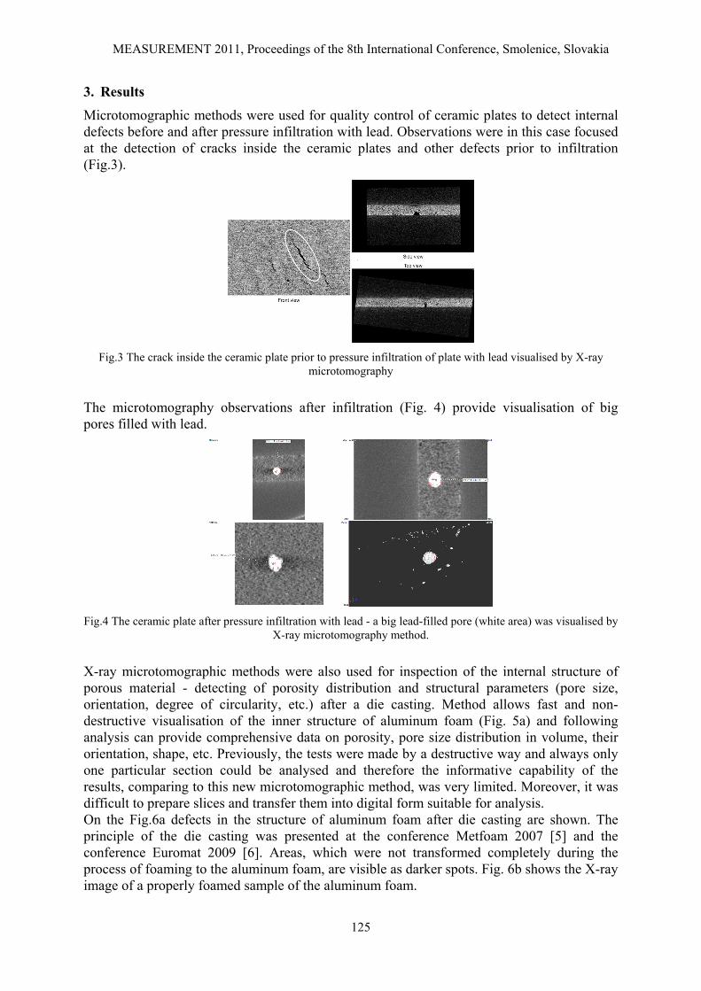
Microtomographic methods were used for quality control of ceramic plates to detect internal defects before and after pressure infiltration with lead. Observations were in this case focused at the detection of cracks inside the ceramic plates and other defects prior to infiltration (Fig.3).
Fig.3 The crack inside the ceramic plate prior to pressure infiltration of plate with lead visualised by X-ray
microtomography
The microtomography observations after infiltration (Fig. 4) provide visualisation of big pores filled with lead.
Fig.4 The ceramic plate after pressure infiltration with lead - a big lead-filled pore (white area) was visualised by
X-ray microtomography method.
X-ray microtomographic methods were also used for inspection of the internal structure of porous material - detecting of porosity distribution and structural parameters (pore size, orientation, degree of circularity, etc.) after a die casting. Method allows fast and non-destructive visualisation of the inner structure of aluminum foam (Fig. 5a) and following analysis can provide comprehensive data on porosity, pore size distribution in volume, their orientation, shape, etc. Previously, the tests were made by a destructive way and always only one particular section could be analysed and therefore the informative capability of the results, comparing to this new microtomographic method, was very limited. Moreover, it was difficult to prepare slices and transfer them into digital form suitable for analysis. On the Fig.6a defects in the structure of aluminum foam after die casting are shown. The principle of the die casting was presented at the conference Metfoam 2007 [5] and the conference Euromat 2009 [6]. Areas, which were not transformed completely during the process of foaming to the aluminum foam, are visible as darker spots. Fig. 6b shows the X-ray image of a properly foamed sample of the aluminum foam.

MEASUREMENT 2011, Proceedings of the 8th International Conference, Smolenice, Slovakia
126
a b
Fig.5 Reconstructed 3D image of an aluminum foam: (a) the internal structure (b) the outer surface
a b
Fig.6 X-ray image of an aluminum foam: (a) non-foamed areas, (b) properly foamed sample.
4. Discussion and Conclusions
The paper presents the basic principle of X-ray microtomography method and examples of its use. The utilisation of this method in the non-destructive testing of ceramic disc before and after pressure infiltration with lead has been reported. Moreover, it was presented as the microtomography can be used for non-destructive reconstruction and visualisation of aluminum foam structures aimed at the monitoring of the porosity distribution and detection of non-foamed places in the internal structure.
Acknowledgements
This paper was created by the realisation of the project “CEKOMAT”, ITMS No. 26240120006 based on the support of Operational Program Research and Development financially supported by European Regional Development Fund. This work was also supported by the Slovak Research and Development Agency under contract APVV No. 0736-07 "LOWCOSTFOAM" and by a grant from Scientific Grant Agency - VEGA 2/0201/10.
References
[1] Kak A C, Slaney M. Principles of Computerized Tomographic Imaging. IEEE Press., New York, 1988.
[2] Cosslet V E, Nixon W C. X-ray Shadow Microscope. Nature, 168: 24 -25, 1951. [3] http://www.ge-mcs.com/en/radiography-x-ray/ct-computed-tomography/nanotom-s.html [4] Maire E. Three-dimensional microstructural information from diffraction and
tomography of synchrotron X-rays, GEMPPM INSA, Lyon. [5] Simančík F, Florek R, Tobolka P, Nosko M. Rapid Prototyping for Complex 3-D Parts of
Aluminium Foams. In proceedings of Metfoam, 2009. [6] Nosko M, Simančík F, Florek R, Tobolka P. New manufacturing route for cheaper
aluminium foam. In proceedings of Euromat, 2009.