y chromosome microdeletion detection system v.4€¦ · the portion of the male-specific region of...
TRANSCRIPT

Y CHROMOSOME MICRODELETION
Detection System v.4.0
www.lifetechindia.comWeb:[email protected]:
Fax: +91-11-42208444 Mobile: +91-9810521400 Ph: +91-11-42208000, 42208111, 42208222 Delhi – 110034 (INDIA). 306, Aggarwal City Mall, Opposite M2K Pitampura, Life Technologies (India) Pvt. Ltd.
India Contact:

Y CHROMOSOME MICRODELETION DETECTION İ
Contents Section 1: Introduction .................................................................................... 1
Section 2: Product Overview .......................................................................... 2
Description of Test .......................................................................................... 2
Chromosomal regions of STS ........................................................................ 4
Performing the Test ........................................................................................ 5
Laboratory Design and Organization .............................................................. 5
Workflow Overview ......................................................................................... 6
Section 3: Materials, Safety Warnings and Precautions.............................. 7
Kit Contents .................................................................................................... 7
Safety Warnings and Precautions .................................................................. 8
Shipping Conditions ........................................................................................ 9
Storage and Handling Requirements ............................................................. 9
Section 4: Y Chromosome Microdeletion Detection Kit Procedure ......... 10
Preparing Genomic DNA Template .............................................................. 10
Preparing PCR ............................................................................................. 10
Section 5: Electrophoretic Detection and Data Analysis ........................... 11
Performing Capillary Electrophoresis with an ABI Genetic Analyzer ........... 11
Data Analysis with GeneMapper® Software v5.0 ........................................ 12
Preparing the Software for Analysis .......................................................... 12
External Control Patterns for All Panels ....................................................... 23
Section 6: Troubleshooting .......................................................................... 25
Assignment of Size Standard ....................................................................... 25
Validation of the Y Chromosome Microdeletion Detection Kit ...................... 28

1
Section 1: Introduction
Microdeletions of the Y chromosome are the second most frequent genetic
cause of spermatogenetic failure in infertile men after Klinefelter syndrome. The
molecular diagnosis of Y chromosomal microdeletions is routinely performed in
the workup of male infertility in men with azoospermia or severe
oligozoospermia (M. Simoni et al., 2004).
The portion of the male-specific region of the Y chromosome (MSY), comprising
95% of the Y chromosome and flanked by pseudoautosomal regions (Skaletsky
et al., 2003), which is affected by deletions, has been classically subdivided into
four regions called AZFa, AZFb, AZFc and proximal AZFc (AZFd), respectively.
Microdeletions are relatively frequent among infertile men, although the
incidence can vary considerably depending on the selection criteria of the
patients. Azoospermic men have a higher incidence of microdeletions than
oligozoospermic men and consequently the deletion frequency found in different
laboratories may vary from 2 to 55% (or even higher), reflecting the composition
of the study population.
The AZFa region is about 1100 kb long and contains the single copy genes
DFFRY (or USP9Y) and DBY. In any case the complete deletion of the AZFa
region removes about 792 kb including both USP9Y and DBY genes, the only
two genes in AZFa. Deletions involving only the USPY9 gene or the DBY gene
have been reported only by one group (Ferlin et al., 1999; Foresta et al., 2000).
The type and mechanism of deletions of the AZFb and AZFc region have been
recently clarified as well (Kuroda- Kawaguchi et al., 2001). Both regions together
comprise 24 genes, most of which present in multiple copies for a total of 46
copies.
The complete deletion of AZFb removes 6.2 Mb (including 32 copies of genes
and transcription units) and results from homologous recombination between
the palindromes P5/proximal P1 (Repping et al., 2002).
The AZFc region includes 12 genes and transcription units, each present in a
variable number of copies making a total of 32 copies (Repping et al., 2003).
The classical complete deletion of AZFc, the most frequent pattern among men
with deletions of the Y chromosome, removes 3.5 Mb, originates from the
homologous recombination between amplicons b2 and b4 in palindromes P3
and P1 respectively and removes 21 copies of genes and transcription units
(Kuroda-Kawaguchi et al., 2001).

Y CHROMOSOME MICRODELETION DETECTION 2
Section 2: Product Overview
Description of Test
This system consists of 14 primer pairs that are homologous to previously
identified and mapped sequence-tagged sites (STS). Y chromosome deletions
in the regions that are amplified by these primer sets have been associated with
male infertility. This system uses a five-dye fluorescent system for automated
DNA fragment analysis, which allows multiplex amplification and
electrophoresis of over 14 STS which include AMXY marker (Chr.X 104bp,
Chr.Y 109bp; Xp22.1, Yp11.2) simultaneously. The kit is intended for use on
Applied Biosystems ABI PRISM® genetic analysis instrumentation.
Fluorochromes include FAM, JOE, ATTO550 and ATTO565, to be used in
conjunction with GeneScan 500 LIZ™ size standard (Applied Biosystems PN
4322682).
The Y-Del Primer Mix includes a primer pair to amplify chromose-specific
sequences of the paralogous gene Amelogenin, which has a high degree of
sequence identity between chromosomes Chr.X and Chr.Y. However nucleotide
differences occur within each locus and can be used to generate chromose-
specific PCR product. The primer pair included in the Y-Del Primer Mix exploits
a 5bp deletion (Chr.X 104bp, Chr.Y 109bp; Xp22.1, Yp11.2) to generate one
Chr.X-specific product that is 5bp shorter than the corresponding product on Y
chromosome.
Although the Y Chromosome Microdeletion Detection Kit has been designed for
the detection of microdeletions occurring on the q-arm of the Y chromosome,
relative quantification between Chr.X and Chr.Y allows assessment of
Klinefelter syndrome, the most significant factor in male infertility. However,
comparative assessment of the AMXY marker cannot be used for the stand-
alone diagnosis of Klinefelter syndrome. Further investigations are necessary
for precise diagnosis.
Control primer pairs are internal controls for the amplification reaction and the
integrity of the genomic DNA sample. The Primer Mix includes a primer pair that
amplifies a region of the SRY gene. This is a control for the testis determining
factor on the short arm of the Y chromosome and allows detection of XX males
arising from Y to X translocations.
The Y Chromosome Microdeletion Detection Kit has been extensively tested for
reproducibility and robustness, and quality controlled to ensure optimum
performance. The system incorporates controls to ensure performance of the

Y CHROMOSOME MICRODELETION DETECTION 3
amplification reagents and to identify problems with genomic DNA samples. A
positive control Male Genomic DNA is included for use in evaluating the
amplification reactions. The presence of the appropriate amplification products
in the positive control reactions indicates that the reagents are performing as
expected.
With the GML Y Chromosome Microdeletion Detection Kit, 14 markers are
performed successfully. Of those, 6 markers (Sy84, Sy86, Sy127, Sy134,
Sy254, and Sy255) are determined by European Molecular Genetics Quality
Network guidelines. The remaining 6 are markers for all regions and so verify
the Detection Kit in addition to control markers (SRY and AMXY).
STS included in the Y Chromosome Microdeletion Detection Kit
Marker Region bp Dye
SRY(Y14) Control 464 JOE
Sy255 AZFc 123 JOE
Sy134 AZFb 303 JOE
AMXY Control Chr.X 104 Chr.Y 109 FAM
RBMY RBMY 238 FAM
AZFa AZFa(Prox2) 217 FAM
Sy133 AZFb 176 FAM
Sy153 AZFd 136 FAM
Sy152 AZFd 124 ATTO565
Sy157 AZFc 291 ATTO565
Sy84 AZFa 330 ATTO565
Sy254 AZFc 382 ATTO565
Sy86 AZFa 317 ATTO550
Sy127 AZFb 271 ATTO550
Fragment sizes may vary up to 3 bp depending on the instrument and
electrophoresis conditions employed. Sizes in this table have been obtained on
an ABI 3130xL Genetic Analyser using the 36cm capillary array, POP7 polymer,
FragmentAnalysis_36_POP7_1 default module and G5 Dye Set.

Y CHROMOSOME MICRODELETION DETECTION 4
Ch
rom
oso
mal
reg
ion
s o
f S
TS

Y CHROMOSOME MICRODELETION DETECTION 5
Performing the Test
The Y Chromosome Microdeletion Detection Kit contains all necessary reagents
for the amplification of human genomic DNA.
The reagents are designed for use with the following Applied Biosystems
instruments:
• ABI PRISM® 3100/3100-Avant Genetic Analyzer
• Applied Biosystems 3130/3130xl Genetic Analyzer
• Applied Biosystems 3500/3500xl Genetic Analyzer
• Applied Biosystems 310 Genetic Analyzer
• GeneAmp® PCR System 9600 or 9700
Laboratory Design and Organization
Special consideration should be given to the design and organization of the
laboratory. The laboratory must be organized so that the area in which amplified
DNA is handled is physically isolated from the work areas for DNA extraction
and PCR setup. Strict physical isolation must be maintained between the area
designated for handling amplified DNA and the other areas, to avoid transfer of
amplified DNA out of the designated work area.
Amplified DNA or equipment and supplies used to handle amplified DNA should
not be taken out of the designated work area. If the work area for amplified DNA
is in a separate but contiguous room, make sure that air flows toward the
amplified DNA area. In addition, it is helpful if there is a separate exit from the
amplified DNA work area that does not exit into the pre-PCR work areas.
The laboratory should have four designated Work Areas, each ideally
equipped with dedicated equipment and supplies to minimize the potential for
contamination:
1. Evidence Handling Work Area: Pieces of evidence are examined,
photographed, and divided for analysis.
2. DNA Extraction Work Area: This work space is used to perform the extraction
steps.
3. PCR Setup Work Area: PCR reagent and DNA sample additions are made
here.
4. Amplified DNA Work Area: This area is dedicated to PCR amplification and
detection, and other activities that require handling of amplified DNA.

Y CHROMOSOME MICRODELETION DETECTION 6
Workflow Overview
Extract DNA
Quantify DNA
Perform PCR
Perform Electrophoresis
Analyze Data

Y CHROMOSOME MICRODELETION DETECTION 7
Section 3: Materials, Safety Warnings and Precautions
Kit Contents
Component Description 25 X Volume Storage
Y-DEL PCR Mix Contains salts, dNTPs and glycerol.
300 µL -20 oC
GML Taq. Pol. Contains enzyme (5U/µL). 15 µL -20 oC
Y-DEL Primer Mix
Contains forward and reverse primers
to amplify human Y-Chromosome
targets.
300 µL -20 oC
Control DNA Contains 10 ng/µL human male DNA. 30 µL -20 oC

Y CHROMOSOME MICRODELETION DETECTION 8
Safety Warnings and Precautions
Please be sure to observe the following warnings.
Warning Caution
1. For research use only.
2. Not for diagnostic purpose.
3. Use appropriate laboratory
practices for sample handling.
4. Wear protective disposable
gloves and eye protection when
handling diluents and human
DNA.
5. Wash hands properly after
handling diluents and human
DNA.
6. Use recommended materials,
procedures, and equipment only.
7. Do not eat, drink or smoke in work
areas.
8. Use sterile disposable pipettes
and filtered pipette tips.
9. Do not mix reagents from different
lots.
10. Do not use more or less than the
required volumes of reagents.
11. Do not use any expired reagents.
12. To reduce the risk of
contamination, the area where
amplified DNA is handled must
be physically isolated from the
work areas for sample
preparation and PCR setup.
13. Store the fluorescent dye labeled
reagents in the dark <-15 oC.
14. Open and close all sample tubes
or wells carefully to avoid
splashing or spraying.
15. Minimize the exposure of Hi-Di
Formamide to the air and do not
contaminate Hi-Di Formamide
when sampling.
16. Chemical Hazard. Pop-7
polymer causes eye, skin, and
respiratory tract irritation. It is a
possible reproductive and birth
defect hazard. Please read the
Applied Biosystems MSDS and
follow the handling instructions.
Wear appropriate protective
eyewear, clothing, and gloves.
17. Chemical Hazard.
Formamide causes eye, skin, and
respiratory tract irritation. It is a
possible reproductive and birth
defect hazard. Please read the
Applied Biosystems MSDS and
follow the handling instructions.
Wear appropriate protective
eyewear, clothing, and gloves.
Y CHROMOSOME MICRODELETION DETECTION 9
Shipping Conditions
The GML Y CHROMOSOME MICRODELETION DETECTION KIT is shipped
on ice packs and should still be frozen on arrival.
Avoid unnecessary freeze-thawing of the contents of the kit.
Storage and Handling Requirements
Fluorescent primers should be stored away from light. The GML Y Chromosome
Microdeletion Detection Kit box is internally coated to increase light protection.
This kit is stable for up to one year if stored at -20 ºC. PCR and primer mixes
can be stored as ready to use aliquots in PCR tubes at -20 ºC; this will keep
freeze-thaw cycles to a minimum reducing the risk of contamination and
shortening PCR set-up times.
Y CHROMOSOME MICRODELETION DETECTION 10
Section 4: Y Chromosome Microdeletion Detection Kit
Procedure
Preparing Genomic DNA Template
1. Prepare genomic DNA according to standard protocols.
2. Determine the concentration by A260 or fluorescence.
3. The amount of DNA required is 10-50 ng/µl.
Preparing PCR
The Y Chromosome Microdeletion Detection Kit has been optimized for use with
the Perkin-Elmer GeneAmp PCR System 9600 or 9700.
1. Prepare 1 PCR tube for each sample and external control.
2. Thaw vials to be used and mix thoroughly by vortexing for a few minutes.
3. Aliquot PCR Mix into each PCR tube as shown in the table below.
Component Volume
PCR Mix 10.0 µl
Primer Mix 10.0 µl
Taq. Pol. 0.5 µl
Total 20.5 µl
4. Add 5.0 µl of DNA (10–50 ng/µl) to each PCR tube.
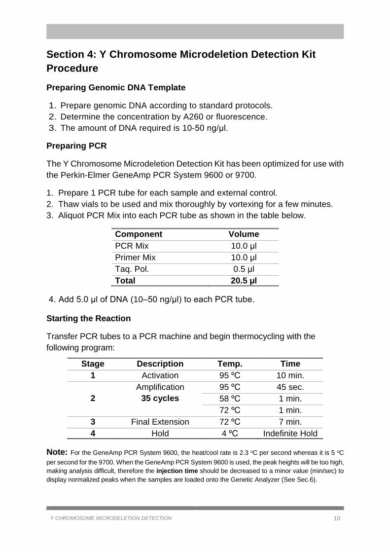
Starting the Reaction
Transfer PCR tubes to a PCR machine and begin thermocycling with the
following program:
Stage Description Temp. Time
1 Activation 95 ºC 10 min.
2
Amplification
35 cycles
95 ºC 45 sec.
58 ºC 1 min.
72 ºC 1 min.
3 Final Extension 72 ºC 7 min.
4 Hold 4 ºC Indefinite Hold
Note: For the GeneAmp PCR System 9600, the heat/cool rate is 2.3 oC per second whereas it is 5 oC
per second for the 9700. When the GeneAmp PCR System 9600 is used, the peak heights will be too high,
making analysis difficult, therefore the injection time should be decreased to a minor value (min/sec) to
display normalized peaks when the samples are loaded onto the Genetic Analyzer (See Sec.6).

Y CHROMOSOME MICRODELETION DETECTION 11
Section 5: Electrophoretic Detection and Data Analysis
Performing Capillary Electrophoresis with an ABI Genetic Analyzer
1. Prepare the necessary amount of size standard for all samples to be analysed
by combining:
To run samples on 3100, 3100 Avant, 3130xL, 3730xL, 3500xL Genetic
Analyzers:
10µl Hi-Di™ Formamide
0.3 µl GeneScan™-500 LIZ™
To run samples on the 310 Genetic Analyzer:
20 Hi-Di™ Formamide
0.5 µl GeneScan™-500 LIZ™
2. For 3100, 3100 Avant, 3130xL, 3730xL, 3500xL Genetic Analyzers:
Use 10 µl of this mix to inject 1 µl of PCR products collected in the same tube.
For the 310 Genetic Analyzer:
Use 20 µl of this mix to inject 1.5 µl of PCR products collected in the same tube.
3. Create Plate Sample Sheet using the Data Collection Software:
Select the appropriate Result Group.
Select the appropriate Instrument Protocol for 3100, 3130 and 3130xL,
3500xL instruments, or the appropriate Run Module for 310, using the
following specifications:
Instrument Type Run Modules and Conditions References
3130/3130xL
3500xL Analyzer FragmentAnalysis36_POP7_1,
Dye Set G5
ABI PRISM® 3130/
3130xl, 3500xL Genetic
Analyzer User’s Manual
3100 Analyzer FragmentAnalysis36_POP7_1,
Dye Set G5
ABI PRISM® 3100/3100-
Avant Genetic Analyzer
User’s Manual
310 Analyzer
GS STR POP4 (1mL) G5 v2.md5
Injection condition: 15 kV/5 sec
ABI PRISM® 310
Genetic Analyzer User’s
Manual

Y CHROMOSOME MICRODELETION DETECTION 12
Data Analysis with GeneMapper® Software v5.0
Preparing the Software for Analysis
Double click to open GeneMapper software on desktop.
Login with User Name and Password to access the software.
Y CHROMOSOME MICRODELETION DETECTION 13
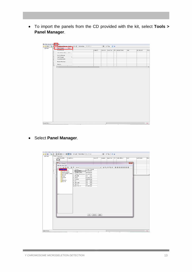
To import the panels from the CD provided with the kit, select Tools >
Panel Manager.
Select Panel Manager.

Y CHROMOSOME MICRODELETION DETECTION 14
Click File > Import Panels.
Select GML_YdEL_Panels and click Import.

Y CHROMOSOME MICRODELETION DETECTION 15
To import the binsets from the CD provided with the kit, click on
GML_YdEL.
Click File > Import Bin Set.

Y CHROMOSOME MICRODELETION DETECTION 16
Select GML_YdEL_bins and click Import.
Click Apply and OK.
To import the other parameters from the CD provided with the kit, select
Tools > GeneMapper Manager.

Y CHROMOSOME MICRODELETION DETECTION 17
On the Analysis Methods tab, click Import. Choose GML_Ydel_anl file
and click Import.
GML_ CVD-1 Panel_anlys file for Analysis Methods
GML_ CVD-1 Panel_rep file for Report Settings
GML_ CVD-1 Panel_size file for Size Standard
GML_ CVD-1 Panel_tbl file for Table Settings
GML_ CVD-1 Panel_plt file for Plot Settings
Import the other files below in the same manner:
On Table Settings tab: GML_YdEL_tbl
On Plot Settings tab: GML_YdEL_plt
On Report Settings tab: GML_YdEL_rep
On Size Standard tab: GML_YdEL_size
Click Done.
1
2
3

Y CHROMOSOME MICRODELETION DETECTION 18
To open a New Project, click on the icon and choose Microsatellite.
Click OK.
Click on the icon , to Add Samples to Project.
Choose the pathway for the run files, click Add to List and Add. More
samples can be selected with CTRL button on keyboard.
2 3
2
1
3
1

Y CHROMOSOME MICRODELETION DETECTION 19
To change the Table Settings, open Table Settings tab and select
GML_YdEL_tbl in the drop-down list.
Select GML_YdEL in the drop-down lists under Analysis Method,
Panel and Size Standard as indicated below.
Analysis Method: Select GML_YdEL_anl

Y CHROMOSOME MICRODELETION DETECTION 20
Panel: Select GML_YdEL
Size Standard: Select GML_YDEL_size
Select all of the boxes by holding left mouse button and pressing CTRL+D
buttons on keyboard.
Click on the Analyze icon.

Y CHROMOSOME MICRODELETION DETECTION 21
Define a Name for the project created.
Analysis begins.

Y CHROMOSOME MICRODELETION DETECTION 22
Before analyzing the samples, ensure that the size peaks are correctly
named as 75, 100, 139, 150, 160, 200, 300, 340, 350, 400, 450, 490 and
500 respectively (for more detailed definitions, see Sec. 6).
Select the Samples tab.
Select samples to view by pressing CTRL button and click to display
plots.

Y CHROMOSOME MICRODELETION DETECTION 23
External Control Patterns for All Panels
Positive male genomic DNA (10 ng/µl) is included in the external control and the
patterns for all markers are shown below. Before analyzing the samples, check
all markers and bins for external control, and then evaluate the samples.

Y CHROMOSOME MICRODELETION DETECTION 24

Y CHROMOSOME MICRODELETION DETECTION 25
Section 6: Troubleshooting
This section covers problems and difficulties which may be encountered while
using the kit, and their solutions.
Assignment of Size Standard
After analyzing the samples added to GeneMapper, the Size Match Editor
should be carefully checked to confirm the sizing data of the samples (even if
all of the samples have green sizing quality), because the correct assignment of
peaks is a prerequisite for accurate analysis.
If the Sizing Quality (SQ) flag is , it means that the samples pass the
peak quality value. Note that peak assignments should be visually
checked even if the SQ flag is green.
If the Sizing Quality (SQ) flag is or , select (Size Match Editor)
to view the size standard and peak assignments. If the peak assignments
are correct, override the size quality value by clicking the button.
If the peak assignments are incorrect for one or more
samples, define a new size standard for the affected sample(s) as
described above and reanalyze the sample(s).
Y CHROMOSOME MICRODELETION DETECTION 26
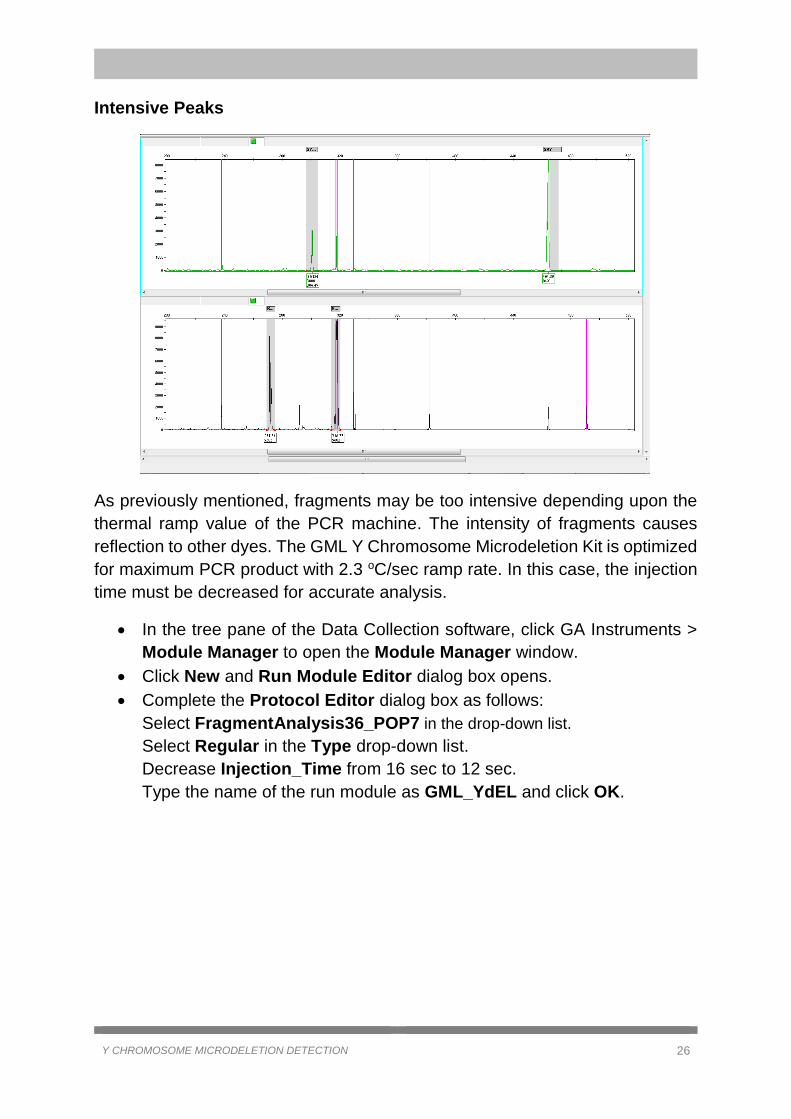
Intensive Peaks
As previously mentioned, fragments may be too intensive depending upon the
thermal ramp value of the PCR machine. The intensity of fragments causes
reflection to other dyes. The GML Y Chromosome Microdeletion Kit is optimized
for maximum PCR product with 2.3 oC/sec ramp rate. In this case, the injection
time must be decreased for accurate analysis.
In the tree pane of the Data Collection software, click GA Instruments >
Module Manager to open the Module Manager window.
Click New and Run Module Editor dialog box opens.
Complete the Protocol Editor dialog box as follows:
Select FragmentAnalysis36_POP7 in the drop-down list.
Select Regular in the Type drop-down list.
Decrease Injection_Time from 16 sec to 12 sec.
Type the name of the run module as GML_YdEL and click OK.

Y CHROMOSOME MICRODELETION DETECTION 27
In the tree pane of the Data Collection software, click GA Instruments >
Protocol Manager to open the Protocol Manager window.
In the Instrument Protocols pane, click New and the Protocol Editor
dialog box opens.
Complete the Protocol Editor dialog box as following:
Type the name as GML_YdeL.
Select Regular in the Type drop-down list.
Select GML_YdeL in the Run Module drop-down list.
Select GML-5DYE as Dye Set.
Click OK.

Y CHROMOSOME MICRODELETION DETECTION 28
Validation of the Y Chromosome Microdeletion Detection Kit
Different Concentrations of DNA
The GML Y Chromosome Microdeletion Detection Kit has been studied and
validated on numerous templates, indicating that the GML Y Chromosome
Microdeletion Detection Kit responds to various concentrations of DNA.
The GML Y Chromosome Microdeletion Detection Kit has also been validated
with amnion DNA extracted by Chelex.
20 ng/µl
5 ng/µl
10 ng/µl
*

Y CHROMOSOME MICRODELETION DETECTION 29
Excessive Concentration of DNA
If the concentration of DNA is decreased below 10 ng/µl, the peak heights are
lowered, which complicates analysis. However too much concentrated DNA
causes contamination due to the reflection of other dyes. Some extra peaks can
be seen and pink reflection images can be an obstacle to evaluating the usual
peaks. Furthermore, extensive peaks pick up other dyes of the same size.
The GML Y Chromosome Microdeletion Detection Kit is optimized for 10-50
ng/µl DNA.
100 ng/µl

Y CHROMOSOME MICRODELETION DETECTION 30
Marker Amplification of Amelogenin Gene
With female DNA only the single copy of amelogenin gene located on the X
chromosome can be amplified as indicated below. With male DNA, AMELX is
amplified along with AMELY due to the existence of the amelogenin gene on
both chromosomes.
AMELX’s intron 1 contains a 6 bp deletion relative to intron 1 of AMELY.
Male
Female

Y CHROMOSOME MICRODELETION DETECTION 31
Sample DNAs with Microdeletions on Y Chromosome
Sample DNA (40 ng/µl) with multiple deleted markers Y152, Y153, Y133, Y134,
RBMY, Y255, Y127, Y157, and Y254 is demonstrated in the table below.
*Applied Biosystems, GeneScan, Genetic Analyser, ABI, Hi-Di, GeneMapper are products of Thermo Fisher Scientific, Inc.

Y CHROMOSOME MICRODELETION DETECTION 32
Sample DNA (45 ng/µl) with multiple deleted markers Y153, Y255, Y152, Y157,
and Y254 is demonstrated in the table below.
Life Technologies (India) Pvt. Ltd. 306, Aggarwal City Mall, Road No. 44, Pitampura, Delhi – 110034, India
Mobile: +91-98105-21400, Tel: +91-11-42208000, 8111, 8222, Fax: +91-11-42208444
Email: [email protected], www.atzlabs.com ; www.lifetechindia.com