zach sutton pt, dpt - wild apricot annual meeting...zach sutton, pt, dpt, ... •pathophysiology:...
TRANSCRIPT

Zach Sutton PT, DPT Zach Sutton, PT, DPT, is a physical therapist for University of Florida Health Rehab Centers specializing in the treatment of patients with vestibular and concussion related disorders. Zach serves as serves multiple roles within the UF system, including the lead therapist at a multidisciplinary concussion clinic at the UF Center for Movement Disorders and Neurorestoration and the UF Student Health Sports Concussion Clinic. He completed his Doctor of Physical Therapy at the University of Florida in 2008 and currently serves as a Courtesy Clinical Instructor in the UF Department of Physical Therapy, where he provides lectures on vestibular and concussion rehabilitation. He has also presented at national and international conferences on concussion rehabilitation and acts as consultant to the University of Florida Athletic Association for treatment of athletes with concussions.
Objectives: 1. Describe basic vestibular anatomy and physiology. 2. Identify common vestibular pathologies common with sports related concussions. 3. Describe evaluation techniques to assess for presence of vestibular pathology in sports
related concussions. 4. Recognize the role of vestibular rehab in concussion management and describe treatment
strategies to address specific vestibular impairments in concussed athletes.
No conflicts of interest or disclosures reported.
University of Florida Health Rehabilitation Center
Gainesville, FL [email protected]
1
Vestibular RehabilitationZach Sutton, PT, DPT
UF Health Rehab Centers: Magnolia ParkeUF Student Health Care Center
Objectives
• Review normal vestibular anatomy and physiology
• Understand what happens when the vestibularsystem is damaged
• Become familiar with common vestibularpathologies
• Be able to conduct a basic vestibular exam
• Understand the role of vestibular rehab in the recovery process
• Understand treatment theories and progressions
Case 1: 12 yo basketball player
11 yo female basketball player referred to physical therapy for dizziness and balance impairment• Sustained a head-to-head injury playing basketball• Initial symptoms: blacked out, memory loss, dizzy, off-balance, headache• IE 7 weeks post-injury
• Has returned to basketball practice but not games• Told by MD to gradually return to sports under coach’s (mom)
supervision• Still has HA, dizziness, and poor balance• Mom reports she falls a lot at practice• Symptoms provoked by jumping, defensive drills, reading and eased by
rest
Case 1: 12 yo basketball player
Oculomotor:Smooth Pursuit: Normal Saccades (30sec): Horizontal: 42 Vertical: 40 (compensated, fast and slow, stuck for a while) (HA 5/10) Covergence: 20 cm (dizzy 6/10) Accomodation: 25 cm 25 cm Cover/Uncover: normal Alternate cover normal
Cervical Spine:Pain (yes/no): no Sharp's Purser: negative Alar Ligament test: negative
Gaze Stability:DVAT: Static: LogMar = 1.8 Dynamic: LogMar = 1.8
VORx1: Horizontal: 63/30 sec (off balance) Vertical: 70x30 sec, 2nd test x 20 sec increased HA to 7/10
VOR suppression: 30sec: 5/10 dizzy
BESS: 39/60
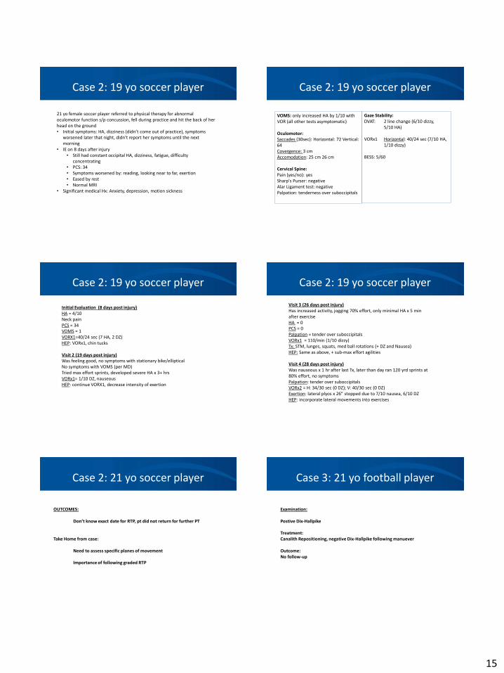
Case 2: 21yo soccer player
21 yo female soccer player referred to physical therapy for abnormal oculomotor function s/p concussion sustained when she fell during practice and hit the back of her head on the ground
• Initial symptoms: HA, dizziness (didn’t come out of practice), symptoms worsened later that night and the next morning reported her symptoms to ATC
• IE on 8 days post injury• Current symptoms: constant occipital HA, dizziness, fatigue, difficulty
concentrating• PCS: 34• Symptoms worsened by: reading, looking near to far, exertion• Eased by rest• Normal MRI
Case 2: 21 yo soccer player
VOMS: only increased HA by 1/10 withVOR (all other tests asymptomatic)
Oculomotor:Saccades (30sec): Horizontal: 72 Vertical: 64 Covergence: 3 cmAccomodation: 25 cm 26 cm
Cervical Spine:Pain (yes/no): yesSharp's Purser: negative Alar Ligament test: negative Palpation: tenderness over suboccipitals
Gaze Stability:DVAT: 2 line change (6/10 dizzy, 5/10 HA)
VORx1 Horizontal: 40/24 sec (7/10 HA, 1/10 dizzy)
BESS: 5/60
2
Case 3: 21 yo football player
21 yo male football player referred to physical therapy for sudden onset of dizziness that occurred when getting up from the floor after an ab routine
• Symptoms continued to occur episodically• Description: spinning• Provoked by lying down, rolling in bed, quick head movements• Presented to PT 2 weeks after onset• Other factors: reports he started taking fish oil pills around the time his
symptoms began• Had URI at the time of PT eval• No history of head trauma
Function
Gaze Stabilization
• Ability to maintain visual acuity while the body/head is in motion
Postural Stability
• Ability to maintain upright posture, balance and equilibrium inresponse to body movements
Motion Sensitivity
• information about the position and movement of the head in space
Is this Important to the Athlete?
• Head can accelerate up to 6,000o/sec during sports activities
• Achieve frequencies of up to 20 Hz
• Decreases in visual acuity can occur with as little a 1o of error
• Only vestibular system can detect movements this fast(oculomotor system < 60 deg/sec
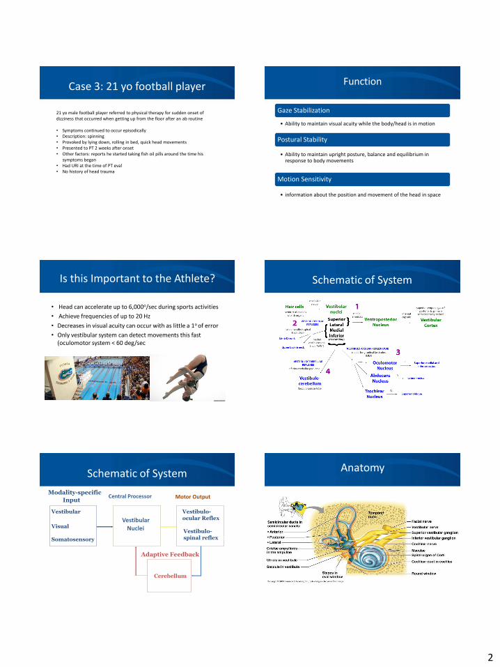
Schematic of System
Schematic of System
Vestibular
Visual
Somatosensory
Modality-specific
Input
VestibularNuclei
Central Processor
Vestibulo-ocular Reflex
Vestibulo-spinal reflexex
Motor Output
Cerebellum
Adaptive Feedback
Anatomy
3
Anterior (superior) semicircular canal
Posterior
semicircular canal
Common crus and duct
Lateral semicircularcanal and duct
Stapes in oval(vestibular) window
Incus
Malleus
Tympanic cavity
External acoustic meatus
Tympanic membrane
Round (cochlear)
window (closed by secondary tympanic membrane)
Auditory (Eustachian) tube
Vestibul
e
Cochlear aqueduct
Scala tympani
Cochlear duct
Scala vestibuli
Ductus reuniens
Helicotrema of cochlea
Saccule
Utricle
Endolymphatic sac
Endolymphatic duct in
vestibular aqueduct
Dura materAmpullae
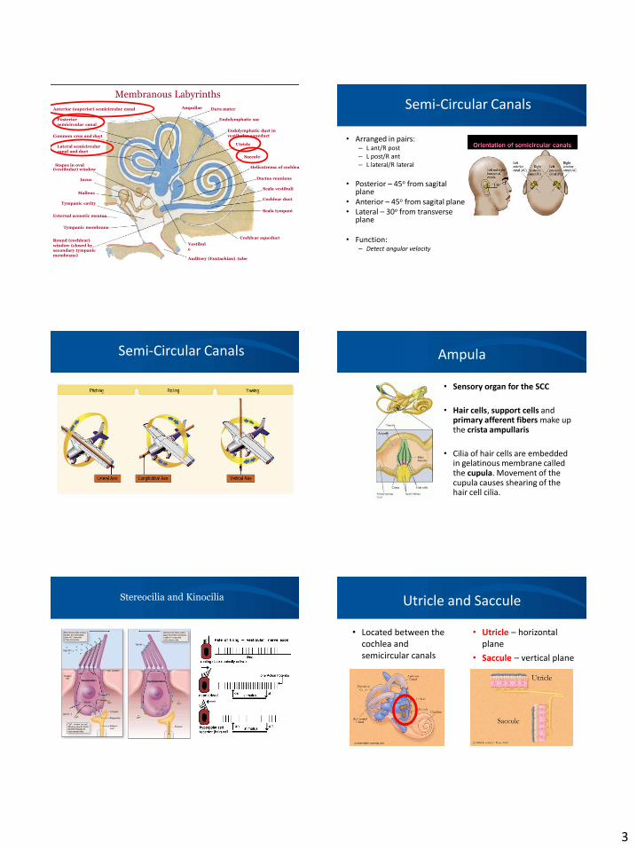
Membranous LabyrinthsSemi-Circular Canals
• Arranged in pairs:– L ant/R post– L post/R ant– L lateral/R lateral
• Posterior – 45o from sagitalplane
• Anterior – 45o from sagital plane• Lateral – 30o from transverse
plane
• Function:– Detect angular velocity
Semi-Circular Canals Ampula
• Sensory organ for the SCC
• Hair cells, support cells and primary afferent fibers make upthe crista ampullaris
• Cilia of hair cells are embedded in gelatinous membrane calledthe cupula. Movement of the cupula causes shearing of the hair cell cilia.
Stereocilia and Kinocilia Utricle and Saccule
• Located between the cochlea andsemicircular canals
• Utricle – horizontalplane
• Saccule – vertical plane
4
The Macula
Otolith organs
Gravity and Linear Acceleration
Vestibulocochlear Nerve (CN VIII) Central Processing
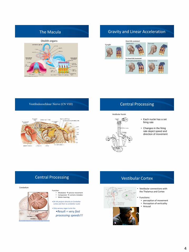
Vestibular Nuclei
• Each nuclei has a set firing rate
• Changes in the firing rate depict speed and direction of movement
Central Processing
CerebellumFunction:
• Modulator precise movement• Comparator corrects mistakes• Motor learning
•CN VIII projects directly to Cerebellarcortex and then to cerebellar nuclei
• Only sensory organ to do this
•Result = very fastprocessing speeds!!!
Vestibular Cortex
• Vestibular connections with the Thalamus and Cortex
• Functions:• perception of movement• Perception of verticality• Arousal

5
Motor Output
• Vestibulo-Ocular Reflex (VOR)
• Vestibulo-Spinal Reflex (VSR)
• Vestibulo-Collic Reflex (VCR)
VOR
• Provides visualstability duringactive headmovement
Nystagmus
• Involuntary rhythmic oscillation of the eyes
• Named by fast phase
• Will beat towards stronger side
– Horizontal
– Vertical
– Torsional
– Mixed
• Plane of Canal
VSR
VCR
• Acts on themuscles in theneck to stabilizethe head
VESTIBULAR PATHOLOGIES
Peripheral Pathologies• Benign Paroxysmal Positional
Vertigo• Vestibular Neuritis• Labyrinthitis
Cervogenic Dizziness
Central Pathologies• Migraines• Trauma (ie, Concussion)• Tumor• Vascular
Other• Drug Toxicity• Dehydration• Psychological
6
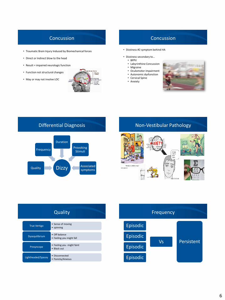
Concussion
• Traumatic Brain Injury Induced by Biomechanical forces
• Direct or Indirect blow to the head
• Result = impaired neurologic function
• Function not structural changes
• May or may not involve LOC
Concussion
• Dizziness #2 symptom behind HA
• Dizziness secondary to…• BPPV• Labyrinthine Concussion• Migraine• Oculomotor impairment• Autonomic dysfunction• Cervical Spine• Anxiety
Differential Diagnosis
DizzyQuality
Frequency
Duration
Provoking Stimuli
Associated symptoms
Non-Vestibular Pathology
Quality
• Sense of moving
• spinningTrue Vertigo
• Off balance
• Feeling you might fallDysequilibrium
• Feeling you might faint
• Black outPresyncope
• Disconnected
• Panicky/AnxiousLightheaded/Spacey
Frequency
Episodic
Episodic
Episodic
Episodic
Vs Persistent

7
Duration
• BPPV, Chronic Vestibular NeuritisSeconds:
• BPPVMinutes:
• MigraineHours
• Acute Vestibular Neuritis, MigraineDays
• Psychogenic (constant vertigo without improvement)Months
Provoking Stimuli
Changes in head position BPPV, Acute Labyrinthitis, Cervicogenic, cardiac*
Spontaneous Acute vestibular neuritis, CVA/TIA, migraine
Recent URI Acute vestibular neuritis
Stress Psychiatric/psychosocial, migraine
exertion Cardiac
Visual stimuli neuritis, migraine
Pearls
– Have patient describe their symptoms withoutusing the word dizzy
– Differentiate between duration of vertigo andduration of symptoms (dysequilibrium, nausea)
Examination
• Rule out more sinister pathology
– VBI, bleed, cervical injury
• Cervical Screen
• Oculomotor Exam
• Evaluation 3 functions of vestibular system
– Gaze stability
– Postural stability
– Motion sensitivity
Examination
• Oculomotor Examination• Need to assess patients eye movements without head
movement
– Smooth Pursuit
– Gaze
– Saccades
– Convergence
Examination
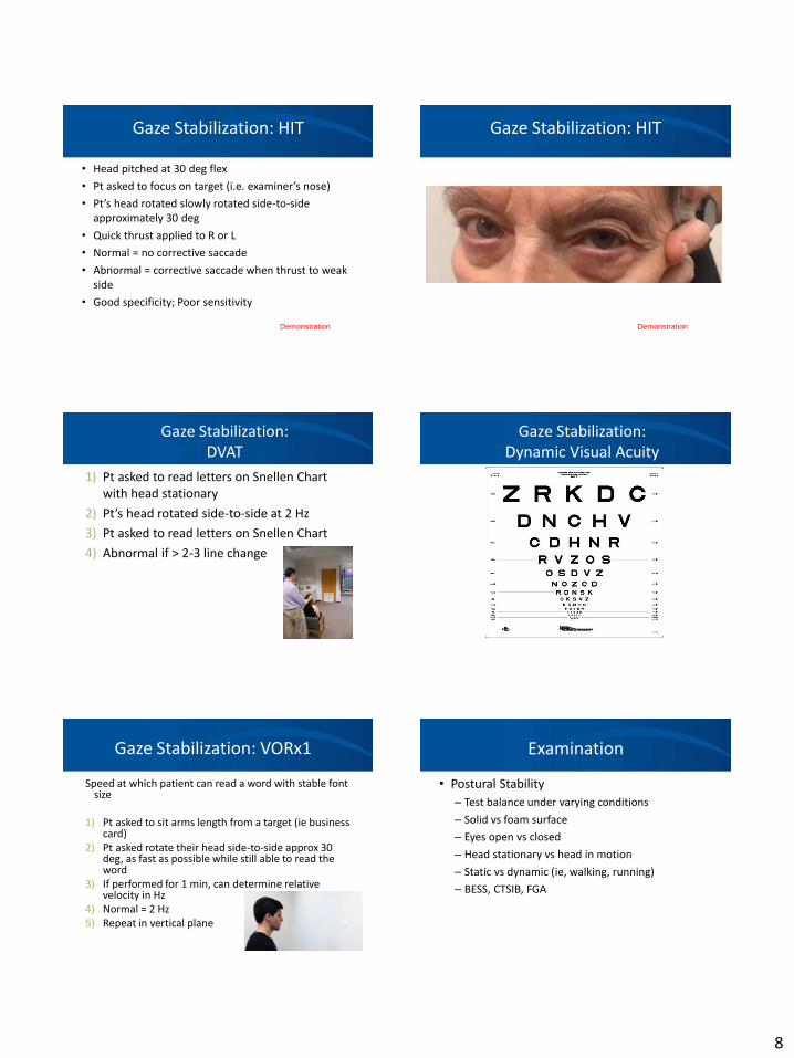
• Gaze stability
– Head Impulse Test (HIT)
• Evaluates function of the VOR
– Dynamic Visual Acuity Test (DVAT)
• Measures visual acuity at set speed (2 Hz)
– Gaze Stability
• Pt selects speed of movement while viewing set target
8
Gaze Stabilization: HIT
• Head pitched at 30 deg flex
• Pt asked to focus on target (i.e. examiner’s nose)
• Pt’s head rotated slowly rotated side-to-side approximately 30 deg
• Quick thrust applied to R or L
• Normal = no corrective saccade
• Abnormal = corrective saccade when thrust to weak side
• Good specificity; Poor sensitivity
Demonstration
Gaze Stabilization: HIT
Demonstration
Gaze Stabilization: DVAT
1) Pt asked to read letters on Snellen Chartwith head stationary
2) Pt’s head rotated side-to-side at 2 Hz
3) Pt asked to read letters on Snellen Chart
4) Abnormal if > 2-3 line change
Gaze Stabilization: Dynamic Visual Acuity
Gaze Stabilization: VORx1
Speed at which patient can read a word with stable font size
1) Pt asked to sit arms length from a target (ie business card)
2) Pt asked rotate their head side-to-side approx 30 deg, as fast as possible while still able to read the word
3) If performed for 1 min, can determine relative velocity in Hz
4) Normal = 2 Hz5) Repeat in vertical plane
Demonstration
Examination
• Postural Stability
– Test balance under varying conditions
– Solid vs foam surface
– Eyes open vs closed
– Head stationary vs head in motion
– Static vs dynamic (ie, walking, running)
– BESS, CTSIB, FGA
9
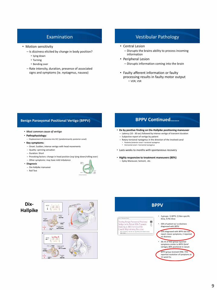
Examination
• Motion sensitivity
– Is dizziness elicited by change in body position?
• lying down
• Turning
• Bending over
– Rate intensity, duration, presence of associatedsigns and symptoms (ie. nystagmus, nausea)
Vestibular Pathology
• Central Lesion– Disrupts the brains ability to process incoming
information
• Peripheral Lesion– Disrupts information coming into the brain
• Faulty afferent information or faultyprocessing results in faulty motor output
• VOR, VSR
Benign Paroxysmal Positional Vertigo (BPPV)
• Most common cause of vertigo
• Pathophysiology: – Displacement of otoconia into SCC (predominantly posterior canal)
• Key symptoms:– Onset: Sudden, intense vertigo with head movements
– Quality: spinning sensation
– Duration: Short
– Provoking factors: change in head position (esp lying down/rolling over)
– Other symptoms: may have mild imbalance
• Diagnosis
– Dix-Hallpike manuever
– Roll Test
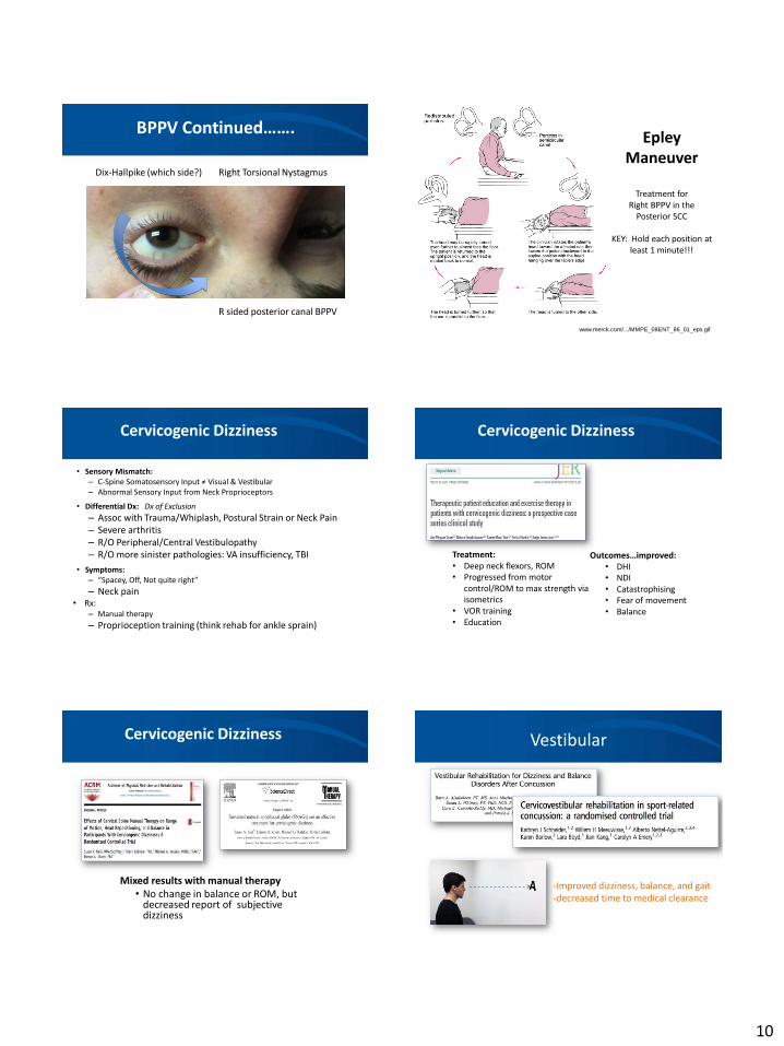
BPPV Continued…….
• Dx by positive finding on Dix-Hallpike positioning maneuver– Latency (10 - 30 sec) followed by intense vertigo of transient duration
– Subjective report of vertigo by patient
– Rotary-torsional nystagmus in the direction of the involved canal• Posterior/anterior canal = torsional nystagmus
• Horizontal canal = horizontal nystagmus
• Lasts weeks to months with spontaneous recovery
• Highly responsive to treatment maneuvers (80%)– Epley Maneuver, Semont, etc.
Dix-Hallpike
BPPV
• 3 groups: 1) BPPV, 2) Non-specificdizzy, 3) No dizzy
• 50% of patient w/ co dizziness diagnosed with BPPV
• 33% diagnosed with BPPV did notreport classic symptoms, 2 reported no dizziness
• 38.1% of NSD group reported symptoms similar to BPPV (brief vertigo), 80% positional in nature
• BPPV group received CRM, 75% reported resolution of symptoms at12 week f/u
10
BPPV Continued…….
R sided posterior canal BPPV
Dix-Hallpike (which side?) Right Torsional Nystagmus
Epley Maneuver
Treatment for Right BPPV in the
Posterior SCC
KEY: Hold each position at least 1 minute!!!
www.merck.com/.../MMPE_08ENT_86_01_eps.gif
Cervicogenic Dizziness
• Sensory Mismatch:– C-Spine Somatosensory Input ≠ Visual & Vestibular– Abnormal Sensory Input from Neck Proprioceptors
• Differential Dx: Dx of Exclusion
– Assoc with Trauma/Whiplash, Postural Strain or Neck Pain– Severe arthritis– R/O Peripheral/Central Vestibulopathy– R/O more sinister pathologies: VA insufficiency, TBI
• Symptoms:– “Spacey, Off, Not quite right”
– Neck pain• Rx:
– Manual therapy
– Proprioception training (think rehab for ankle sprain)
Cervicogenic Dizziness
Treatment:• Deep neck flexors, ROM• Progressed from motor
control/ROM to max strength via isometrics
• VOR training• Education
Outcomes…improved:• DHI• NDI• Catastrophising• Fear of movement• Balance
Cervicogenic Dizziness
Mixed results with manual therapy• No change in balance or ROM, but
decreased report of subjective dizziness
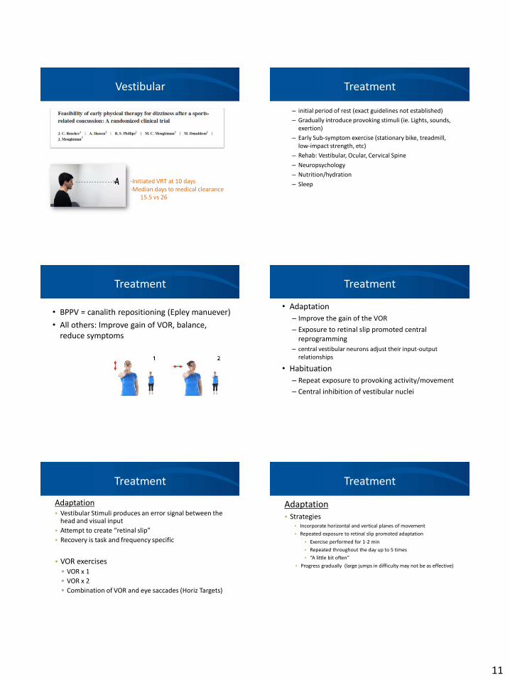
Vestibular
- - - - - - - - - - - - - - > -Improved dizziness, balance, and gait-decreased time to medical clearance
11
Vestibular
- - - - - - - - - - - - - - > -Initiated VRT at 10 days-Median days to medical clearance
15.5 vs 26
Treatment
– initial period of rest (exact guidelines not established)
– Gradually introduce provoking stimuli (ie. Lights, sounds, exertion)
– Early Sub-symptom exercise (stationary bike, treadmill,low-impact strength, etc)
– Rehab: Vestibular, Ocular, Cervical Spine
– Neuropsychology
– Nutrition/hydration
– Sleep
Treatment
• BPPV = canalith repositioning (Epley manuever)
• All others: Improve gain of VOR, balance,reduce symptoms
Treatment
• Adaptation
– Improve the gain of the VOR
– Exposure to retinal slip promoted centralreprogramming
– central vestibular neurons adjust their input-outputrelationships
• Habituation
– Repeat exposure to provoking activity/movement
– Central inhibition of vestibular nuclei
Treatment
Adaptation• Vestibular Stimuli produces an error signal between the
head and visual input
• Attempt to create “retinal slip”
• Recovery is task and frequency specific
• VOR exercises▫ VOR x 1
▫ VOR x 2
▫ Combination of VOR and eye saccades (Horiz Targets)
Treatment
Adaptation• Strategies
• Incorporate horizontal and vertical planes of movement
• Repeated exposure to retinal slip promoted adaptation
• Exercise performed for 1-2 min
• Repeated throughout the day up to 5 times
• “A little bit often”
• Progress gradually (large jumps in difficulty may not be as effective)
12
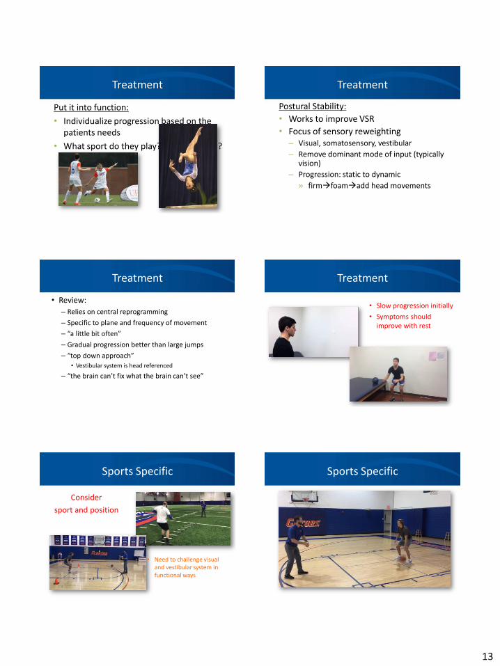
Treatment
• 1. PredictableRandom Moving
• 2. Small-amplitudelarge-amplitude movements.
• 3. SlowFast
• 4. Large targetsSmall targets
• 5. Solid/Simple backgroundComplex or Moving background
Treatment
Treatment Treatment
Other progressions• static to dynamic
• Sitting, standing, stand of foam, walking
• Include Hand-eye coordination (ie catch/throw)
• Cognitive tasks• Math problems, memory, decision making
Treatment
• Habituation– Determine the plane of movement that provokes the
patients symptoms– Symptoms will be most intense if movement occurs in
the plane of the SCCs or otolith organs– Speed of movement matters– Begin with simple movements, progress to more
dynamic as symptoms improve• Vertical movements:
– Sitting nose-to-kneestanding wood chopsburpees
• Horizontal:– Rolling in bedwall rolling (90o, 180o…)turn&catch
Treatment
Brandt Daroff
13
Treatment
Put it into function:
• Individualize progression based on thepatients needs
• What sport do they play? What position?
Treatment
Postural Stability:
• Works to improve VSR
• Focus of sensory reweighting– Visual, somatosensory, vestibular
– Remove dominant mode of input (typicallyvision)
– Progression: static to dynamic
» firmfoamadd head movements
Treatment
• Review:
– Relies on central reprogramming
– Specific to plane and frequency of movement
– “a little bit often”
– Gradual progression better than large jumps
– “top down approach”
• Vestibular system is head referenced
– “the brain can’t fix what the brain can’t see”
Treatment
• Slow progression initially
• Symptoms shouldimprove with rest
Sports Specific
Consider
sport and position
• Need to challenge visualand vestibular system in functional ways
Sports Specific
14
How do we relate this to the athlete?
• Recent AP survey found majority of NFL players were “not at all” or “less concerned” about the effects of concussions compared to other injuries
• Visual and Vestibular deficits Associated with
– Neurocognitive deficits
– Proprioception
– Processing speed
• Neurocognitive deficits are emerging as a risk factor for other injuries, including ACL tears
Case 1: 12 yo basketball player
11 yo female basketball player referred to physical therapy for dizziness and balance impairment• Sustained a concussion, head-to-head contact on 8/15/14• Initial symptoms: blacked out, memory loss, dizzy, off-balance, headache• IE on 10/2/15
• Has returned to basketball practice but not games• Told by MD to gradually return to sports under coach’s (mom)
supervision• Still has HA, dizziness, and poor balance• Mom reports she falls a lot at practice• Symptoms provoked by jumping, defensive drills, reading and eased by
rest
Case 1: 12 yo basketball player
Oculomotor:Smooth Pursuit: Normal Saccades (30sec): Horizontal: 42 Vertical: 40 (compensated, fast and slow, stuck for a while) (HA 5/10) Convergence: 20 cm (dizzy 6/10) Accomodation: 25 cm 25 cm Cover/Uncover: normal Alternate cover normal
Cervical Spine:Pain (yes/no): no Sharp's Purser: negative Alar Ligament test: negative
Gaze Stability:DVAT: Static: LogMar = 1.8 Dynamic: LogMar = 1.8
VORx1 Horizontal: 63/30 sec (off balance) Vertical: 70x30 sec, 2nd test x 20 sec increased HA to 7/10
VOR suppression: 30sec: 5/10 dizzy
BESS: 39/60
Case 1: 12 yo basketball player
Treatment:HEPVORx1Static balanceEye saccadesGraded aerobic training
Progressed from walking on treadmill to high intensity interval training (box jumps, lateral plyos, jogging, etc)
Gradual progression of sport specific exercises:SLDLbasketball passes with jump turnsDribbling around cones
Case 1: 12 yo basketball player
10/2 10/9 10/16 10/23 10/29 11/17
HA 5/10 4/10 0/10 0/10 0/10 0/10
Dizziness 0/10 3/10 0/10 0/10 0/10 0/10
BESS 39 31 38 27 16
BCTT 16 minHA6-7Dz 6
16 minHA 0-1Dz 3
Exertion BCTT BCTT Pitt
Symptoms #@ #@ @ &
King Devick 70.8/81.9
Convergence 20 cm <6cm
# = HA during Tx & = no symptoms at practice (took naproxen)@ = Dizziness during Tx
Case 1: 12 yo basketball player
Outcomes:• Normal balance?• No symptoms with high intensity exercise• Still HA in class (feels related to her glasses)• Still slow oculomotor function (mom states she had
reading problem pre-injury• No symptoms during basketball practice• Recommended referral to OT for reading assessment• Feb 2015, ankle sprain during gymnastics
15
Case 2: 19 yo soccer player
21 yo female soccer player referred to physical therapy for abnormal oculomotor function s/p concussion, fell during practice and hit the back of her head on the ground• Initial symptoms: HA, dizziness (didn’t come out of practice), symptoms
worsened later that night, didn’t report her symptoms until the nextmorning
• IE on 8 days after injury• Still had constant occipital HA, dizziness, fatigue, difficulty
concentrating• PCS: 34• Symptoms worsened by: reading, looking near to far, exertion• Eased by rest• Normal MRI
• Significant medical Hx: Anxiety, depression, motion sickness
Case 2: 19 yo soccer player
VOMS: only increased HA by 1/10 withVOR (all other tests asymptomatic)
Oculomotor:Saccades (30sec): Horizontal: 72 Vertical: 64 Covergence: 3 cmAccomodation: 25 cm 26 cm
Cervical Spine:Pain (yes/no): yesSharp's Purser: negative Alar Ligament test: negative Palpation: tenderness over suboccipitals
Gaze Stability:DVAT: 2 line change (6/10 dizzy,
5/10 HA)
VORx1 Horizontal: 40/24 sec (7/10 HA, 1/10 dizzy)
BESS: 5/60
Case 2: 19 yo soccer player
Initial Evaluation (8 days post injury)HA = 4/10Neck painPCS = 34VOMS = 1VORX1=40/24 sec (7 HA, 2 DZ)HEP: VORx1, chin tucks
Visit 2 (19 days post injury)Was feeling good, no symptoms with stationary bike/ellipticalNo symptoms with VOMS (per MD)Tried max effort sprints, developed severe HA x 3+ hrsVORx1= 1/10 DZ, nauseousHEP: continue VORX1, decrease intensity of exertion
Case 2: 19 yo soccer player
Visit 3 (26 days post injury)Has increased activity, jogging 70% effort, only minimal HA x 5 min after exerciseHA = 0PCS = 0Palpation = tender over suboccipitalsVORx1 = 110/min (1/10 dizzy)Tx: STM, lunges, squats, med ball rotations (+ DZ and Nausea)HEP: Same as above, + sub-max effort agilities
Visit 4 (28 days post injury)Was nauseous x 1 hr after last Tx, later than day ran 120 yrd sprints at 80% effort, no symptomsPalpation: tender over suboccipitalsVORx2 = H: 34/30 sec (0 DZ); V: 40/30 sec (0 DZ)Exertion: lateral plyos x 26” stopped due to 7/10 nausea, 6/10 DZHEP: incorporate lateral movements into exercises
Case 2: 21 yo soccer player
OUTCOMES:
Don’t know exact date for RTP, pt did not return for further PT
Take Home from case:
Need to assess specific planes of movement
Importance of following graded RTP
Case 3: 21 yo football player
Examination:
Postive Dix-Hallpike
Treatment:Canalith Repositioning, negative Dix-Hallpike following manuever
Outcome:No follow-up
16
Thank You
Resources
• Alhilali LM, Yaeger K, Collins M, Fakhran S. Detection of central white matter injury underlying vestibulopaty after mild traumatic brain injury. Radiology. Vol 272; No 1. July 2014.
• Balaban, Carey, et al. Top-down approach to vestibular compensation: Translational lessons fromvestibular rehabilitation. Brain Research 1482 (2012) 101-111
• Barlow M, Schlabach D, Peiffer J, Cook C. Differences in change scores and the predictive validity of three commonly used measures following concussion in the middle school and high school aged population. Int J Sports Phys Ther. 2011 Sep;6(3):150-7.
• Furman J, Whitney S. Central Causes of Dizziness. Physical Therapy 2000; 80; 179-187• Gans R. American Institute of Balance – Vestibular Rehabilitation continuing education course• Han, Byung. Vestibular Rehabilitation Therapy: Review of Indications, Mechanisms, and Key
exercises. Journal of Clin Neurology. 2011;7:184-196• Holmberg, J. North American Seminars – Spinning Beyond the Basics, An Advanced Vestibular
Rehabilitative Course 2011• Kentala E, Rauch S. A practical assessment algorithm for diagnosis of dizziness. Otolaryngology-
Head and Neck Surgery. January 2003. 54-59• Labuguen R. Initial Evaluation of Vertigo. American Family Physician. January 2006. Volume 73,
Number 2• Schubert, M. Vestibulo-ocular Physicology Underlying Vestbular Hypofunctions. Physical Therapy.
Volume 84 Number 4 April 2004