a putative glutathione-binding site in cdzn-metallothionein
TRANSCRIPT
Biochem. J. (1993) 294, 219-225 (Printed in Great Britain)
A putative glutathione-binding site in CdZn-metallothionein identified byequilibrium binding and molecular-modelling studiesMarius BROUWER,* Thea HOEXUM-BROUWER and Robert E. CASHONDuke University School of the Environment, Marine Laboratory/Marine Biomedical Center, Beaufort, NC 28516, U.S.A.
Glutathione (GSH) has been found to form a complex with bothvertebrate and invertebrate copper-metallothionein (CuMT)[Freedman, Ciriolo and Peisach (1989) J. Biol. Chem. 264,5598-5605; Brouwer and Brouwer-Hoexum (1991) Arch.Biochem. Biophys. 290, 207-213]. In this paper we report on theinteraction of GSH with CdZnMT-I and CdZnMT-II fromrabbit liver and with CdMT-I from Blue crab hepatopancreas.Ultrafiltration experiments showed that all three MTs combinedwith GSH. The measured binding data for the three MTs couldbe described by a single binding isotherm. The GSH/MTstoichiometry was 1.4+ 0.3 and KdlSS = 14+ 6 /sM. Partially Zn-depleted MT does not significantly bind GSH, indicating that theGSH-binding site is located on MT's Zn-containing N-terminaldomain. The putative GSH-binding site on rabbit liver MT was
investigated using molecular-graphics analysis. A cleft on theMT's N-terminal domain, which has the labile Zn-2 at its base,could easily accommodate GSH. Cysteine-ligand exchange be-tween the terminal (non-bridging) Cys-26, bound to Zn-2, andthe cysteine in GSH is stereochemically possible. Based on theseconsiderations a model of MT-GSH was built in which GSH's
cysteine replaces Cys-26 as a terminal Zn-2 ligand. This complexwas energy-minimized by molecular-mechanics calculations,taking into account computed partial electrostatic charges on allatoms, including Cd and Zn. These calculations showed that theMT-GSH complex was thermodynamically more stable thanMT, due to favourable non-bonded, electrostatic and van derWaals interactions. Six hydrogen bonds can form between GSHand MT. The average pairwise root-mean-square deviations(RMSD) of the metals in energy-minimized MT and MT-GSH,compared with the metals in the crystal structure, were
0.0087+0.0028nm (0.087+0.028 A) and 0.0168+0.0087 nm(0.168+0.087 A) respectively. The RMSD values for thepolypeptide-backbone a carbons were 0.0136+0.0060 nm(0.136+0.060 A) and 0.0491 +0.0380 nm (0.491 +0.380 A) re-
spectively. No other docking sites for GSH were found. Theenergy-minimized structure of an MT-2-mercaptoethanol com-
plex was somewhat less stable than the native MT domain,attesting to the specificity of the MT-GSH interaction. Thepossible physiological significance of the MT-GSH interactionis discussed.
INTRODUCTION
Metallothioneins (MTs) constitute a family of low-molecular-mass cysteine-rich, metal-binding proteins. MTs are expressed inmany different cell lines and tissues following exposure to heavymetals, glucocorticoid hormones, interferon, interleukin-1, bac-terial endotoxin, haem-haemopexin and u.v. radiation. MTsappear to be involved in metal detoxification, regulation of Znand Cu, and donation of metals to apometalloproteins, andpossibly in the scavenging offree radicals (Cousins, 1985; Hamer,1986; Engel and Brouwer, 1989; Waalkes and Goering, 1990;Alam and Smith, 1992). The expression ofMT genes is regulatedat the transcriptional level, through interactions between cis-acting metal-, glucocorticoid- and haem-responsive elements,and trans-acting DNA-binding, metal-regulatory (metallo)-proteins (Andersen et al., 1990; Seguin, 1991; Butler and Thiele,1991; Alam and Smith, 1992; Thiele, 1992). N.m.r. spectroscopy(Otvos and Armitage, 1980; Messerle et al., 1990) and X-raydiffraction studies (Robbins et al., 1991) have shown that theCd/Zn form of mammalian MT is a dumbbell-shaped molecule,composed of an N-terminal, nine-cysteine/three-metal cluster,and a C-terminal, 11 -cysteine/four-metal cluster. Each metal istetrahedrally co-ordinated by four sulphur atoms. There are no
contacts between the two metal-binding domains. The CdMT ofmarine crustacea is composed of two nine-cysteine/three-metalclusters (Otvos et al., 1982). The Cd-thiolate clusters, especially
the three-metal cluster in mammalian MT, possess a high degreeof dynamic freedom. The continual breaking and re-forming ofco-ordination bonds allow for intramolecular metal exchange,primarily within the N-terminal cluster, and intermolecular metalexchange between clusters of different MT molecules (Vasak,1986; Otvos et al., 1989). Differences in exchange rates stronglysuggest that the two domains of mammalian MTs have evolvedto perform different functional roles. The kinetically labile N-terminal domain may function in metal-exchange processes,whereas the kinetically stable C-terminal domain is important indetoxification. Studies on mutant MTs in which cysteine residuesin either domain have been replaced corroborate this conclusion(Cismowski and Huang, 1991; Cismowski et al., 1991). Whetherintracellular metal exchange occurs between MTs or betweenMT and (apo)metalloproteins is unknown.
Recent studies have demonstrated the existence of intra-cellular Cu(I)MT-GSH and Cu(I)-GSH complexes in mam-
malian cell lines (Freedman et al., 1989) and in the hepato-pancreas (digestive gland) of marine crustacea (Brouwer andBrouwer-Hoexum, 1991, 1992). Cu(I)-GSH is proposed as an
intracellular source of Cu for the biosynthesis of Cu-dependentproteins, and Cu(I)-GSH may be derived from the Cu(I)-MTpool under Cu-deficient conditions. We have found that GSHforms a stable (KdiB8 value of 1 ,uM) 1: 1 complex with CuMT-IIIfrom the American lobster, Homarus americanus. CuMT iso-forms I and II form a transient MT-GSH complex which may
Abbreviations used: MT, metallothionein; MSH, 2-mercaptoethanol; DTT, dithiothreitol; DTNB, 5,5'-dithio-bis(2-nitrobenzoic acid); RMSD, root-mean-square deviation.
* To whom correspondence should be addressed.
Biochem. J. (1993) 294, 219-225 (Printed in Great Britain) 219
220 M. Brouwer, T. Hoexum-Brouwer and R. E. Cashon
release copper as a Cu(I)-GSH complex. The latter can restorethe oxygen-binding capacity of Cu-free haemocyanin, whereasCuMT cannot (Brouwer and Brouwer-Hoexum, 1991, 1992).Mammalian CuMT contains 12 Cu atoms (Nielson et al.,
1985; Kille et al., 1992), which are trigonally co-ordinated tocysteine residues ofMT (Abrahams et al., 1986). Lobster CuMTbinds 10 Cu atoms (Brouwer and Brouwer-Hoexum, 1991). Theseobservations raise the question whether the interaction betweenGSH and MT is specific for the Cu-containing MTs, or whetherthis is a general phenomenon that also occurs with the Cd/Zn-containing MTs. In view of this we have examined/characterizedthe structural and physical chemical properties of the interactionbetween GSH and CdMT from the Blue crab, Callinectes sapidus,and CdZnMT from the rabbit, Oryctolagus cuniculus. Both theinvertebrate and mammalian CdMTs were found to complexwith GSH. A putative GSH-binding site on the N-terminaldomain ofMT has been identified by molecular-graphics analysis.
EXPERIMENTAL
Preparation of CdZnMTCd6MT-I was isolated from the hepatopancreas of Blue crabs(Brouwer et al., 1992). CdZnMT-I/II isolated from rabbit liverwas a gift from Dr. F. A. Liberatore, E. I. Dupont, MedicalProducts Department, Billerica, MA, U.S.A. The protein hadbeen purified by ethanol precipitation, gel-permeation chromat-ography and batch-wise elution from a DEAE-cellulose column.The protein was thus a mixture of CdZnMT-I and CdZnMT-II.The two isoforms were separated by anion-exchange chromat-ography as described before (Brouwer et al., 1992).
Repair of partialiy oxidized MTCdZnMT from rabbit liver appeared to have lost some of its Zn(see the Results section). To restore its full metal-bindingcomplement, a solution of 10 mg of CdZnMT in 2 ml of N2-saturated 10 mM Hepes/50 mM NaCl, pH 7.4, was incubatedwith 5 mM dithiothreitol (DTT) and 1.5 mM ZnC12 at room
temperature for 10 min. The mixture was then chromatographedon a Sephadex G-50 column (1.6 cm x 58 cm) in 10 mM Tris/HCl(pH 7.0) at a flow rate of 15 ml/h. Fractions containing mono-
meric MT were pooled and separated into CdZnMT-I andCdZnMT-II by anion-exchange chromatography on a DEAE-cellulose column (2.5 cm x 20 cm) using a 1 litre gradient of10-200 mM Tris/HCl (pH 7) at a flow rate of 35 ml/h. TheCdZnMT isoforms were dialysed against 10 mM Hepes/50 mMNaCl (pH 7.4) before GSH-binding experiments were performed.All procedures were carried out in N2-saturated buffers at 4 'C.The Cd/Zn ratio in the Zn-reconstituted MT was measured by
atomic absorption spectroscopy. The amount ofZn bound to thecysteine residues of MT was determined by u.v. spectroscopy.CdCl2, giving a final concentration of 50 ,uM Cd, was added toa degassed solution of M CdZnMT in 10 mM Hepes(pH 7.4), and the u.v. absorbance spectrum from 220 to 300 nmwas recorded. The amount of thiolate-bound Zn displaced by Cdwas calculated from the increase of the absorbance value at250 nm using a molar absorptivity for the Cd-(thiolate)4 complexat 250 nm of 14500 (Vasak et al., 1981).
GSH-binding studiesSolutions containing 3-8,M rabbit liver CdZnMT-I andCdZnMT-II and Blue crab CdMT-I in 10 mM Hepes/50 mM
capacity Amicon cell with a 2.5-cm-diam. YM-10 membrane asdescribed previously (Brouwer and Brouwer-Hoexum, 1991).MT concentrations were calculated assuming seven metal atoms/rabbit MT and six metal atoms/crab MT. Retentate and filtratewere analysed for GSH using the enzymic recycling assay in thepresence of 5,5'-dithiobis(2-nitrobenzoic acid) (DTNB) (Ander-son, 1985). It was found that the assay of the retentate wasinhibited, possibly by the reaction of DTNB with the cysteineresidues of MT. To correct for this apparent loss of GSH, GSHwas assayed in the retentate after the addition of a knownamount of GSH as an internal standard. If we assume that theGSH concentration in the retentate is z ,M, and that theefficiency of the assay is a%, then az = b, where b is the measuredconcentration of GSH. After the addition of c ,uM GSH to theretentate we have a(z + c) = m, where m is the GSH concentrationmeasured in the presence of the internal standard. From thesetwo equations it follows that the efficiency is a = (m - b)/c. TheGSH concentration in the retentate is thus z= b/a. GSH wasfound to be freely permeable through the YM-10 membrane, andno loss of GSH was observed when GSH was subjected toultrafiltration in the absence of CdMT. The YM-10 membraneswere virtually impermeable to CdZnMT. The percentage of totalCd in the filtrates was 4.28 + 1.13 % (n = 13). It is possible tocorrect for the effect of this slight permeability of CdZnMT onthe calculated amounts of free and bound GSH (Brouwer andBrouwer-Hoexum, 1991). However, the correction did not signifi-cantly alter the outcome of the data analysis, and was thereforenot necessary.
In principle, to determine the amount of GSH bound to MT,it is not necessary to measure the GSH concentration in theretentate. We know the amount of GSH, and MT, present in theequilibrium mixture before the ultrafiltration step: GSHdded andMTadded' During ultrafiltration, the ratio of GSH-MT to MTdoes not change since both are concentrated to the same extentduring the concentration step. This implies that the free GSHconcentration does not change during the ultrafiltration step.Therefore, the GSH concentration in the filtrate is equal to theconcentration of free GSH in the mixture before ultraffiltration.From this it follows that [GSH]bound = [GSH]added- [GSH]filtrateand GSH/MT = [GSH]bound/[MT]a4ded.Data were analysed according to r = nK[GSH],ree/
(1 + K[GSH],ree), using standard non-linear least-squares minim-ization, where r is [GSH]bound/MT, [GSH],ree is the concentrationof free GSH, n is the number of GSH-binding sites/MT, and Kis the binding constant for the MT-GSH complex.
Molecular-graphics analysis and energy minimizationGraphics modelling was performed interactively on an Evansand Sutherland ESV Workstation, using the Graphics programSYBYL (version 5.5, Tripos Associates, St. Louis, MO, U.S.A.).Energy minimization by molecular-mechanics calculations wasperformed using SYBYL's MAXIMIN2 function. To evaluatethe effect of the electrostatic charges of the metals on theminimized structures, three different energy-minimization cal-culations were performed. In the first one, electrostatic chargeswere completely ignored. In the second one, the metals were eachassigned a point charge of + 2 and the distribution ofelectrostaticcharges on all other atoms was calculated using the method ofGasteiger and Marsili (1980). In the third one, the metals wereincluded in the calculation of the charge distribution. Furtherdetails ofthese procedures are provided in the Discussion section.Minimizations, starting with the crystal structure, were carriedout in the TRIPOS force-field, using the conjugate gradientmethod (Kini and Evans, 1991). The bridging and terminal
NaCl (pH 7.4) in the presence of different concentrations ofGSH were subjected to ultrafiltration, in the cold, in a 10-ml-

Interaction between glutathione and metallothionein 221
sulphur atoms were assigned a tetrahedral environment with one
and two lone electron pairs respectively (Arseniev et al., 1988;Messerle et al., 1990). The S-Me2+-S angles (where Me2+ is themetal ion) were constrained to the values obtained from thecrystal structure (Robbins et al., 1991). The Cd-S and Zn-Sequilibrium bond lengths were set to 0.2511 nm (2.511 A) and0.2378 nm (2.378 A), which are the average Cd-S and Zn-Sbond lengths in the crystal structure. All of the calculationsemployed a dielectric constant of 1 for the evaluation of theelectrostatic energy. All other parameters for minimization were
the default parameters in SYBYL. At present it is quite difficultto make calculations in the presence of explicit solvent molecules.Therefore calculations were carried out on the structure of MTand the MT-GSH complex in the absence of water. The presentcalculated structure should be viewed with this limitation inmind. Structures were refined until the final energy change was
less than 0.0001 kcal/mol. The protein structures obtained afterenergy minimization were compared with the experimentalstructure by superposition of the minimized structure on to thecrystal structure using a least-squares fit for all a carbons. Theroot-mean-square deviation (RMSD) is used as an estimate of fit.
RESULTS
Restoration of oxidized CdZnMTThe Cd/Zn ratio of the purified rabbit MT was approx. 6, whichis much higher than the 5/2 stoichiometry generally found inCdZnMT in solution (Winge and Miklossy, 1982a) and in thecrystal structure (Robbins et al., 1991). This suggested that thelabile Zn (Li et al., 1980) had been lost from the protein. Toevaluate this possibility, and to restore the metal-binding capacityof MT, 25 4uM CdZnMT was reduced with 1 mM 2-mercaptoethanol (MSH) at pH 8 for 3 h at 37 °C and sub-sequently incubated for 5 h with different concentrations ofZnCl2at room temperature, followed by size-exclusion h.p.l.c. toseparate MT from Zn and MSH. The Cd/Zn ratio decreasedfrom 7 to 4 (at 400 ,uM Zn). The decrease in the Cd/Zn ratio wasfound to be associated with an increase in Zn, whereas theamount of Cd bound to the protein remained unchanged. The
._
0
QCD
0
Icn
O
0
In
cn
m
[GSH]fra, (M)2.39x1le
Figure 1 GSH binding to CdZnMT and CdMT
Binding of GSH to rabbit liver CdZnMT (A) and Blue crab CdMT (U) was evaluated byultrafiltration experiments. The line was calculated with a Kdiss value of 14 /uM and a GSH/MTvalue of 1.4, determined from the data by non-linear least-squares minimization.
amount of Zn was measured by atomic absorption spectroscopyand calculated from the change in u.v. absorption that accom-panies displacement of Zn by Cd. The measured and calculatedvalues were identical, indicating that all the Zn was bound as zinctetrathiolate.When CdZnMT was reduced, followed by size-exclusion
h.p.l.c. and subsequent incubation with Zn, no restoration ofZn-binding capacity was observed. These experiments suggest thatMSH reduces disulphide bridges in oxidized MT. Once the MSHhas been removed the reduced thiol residues (or thiol and mixeddisulphide) rapidly re-oxidize. Reduction of protein disulphidesis often unfavourable, due to the high value for kintra, i.e. the rateconstant for the reaction of the protein thiol group with theprotein S-S-M, mixed disulphide group (Creighton, 1986).The re-oxidation of protein thiol groups can be prevented by
carrying out the reduction with DTT, which has two thiol groupson the same molecule. In this case the protein mixed disulphidereacts to give protein-SH and oxidized DTT. Using DTT and theconditions as described in the Experimental section we obtainedCdZnMT-I and CdZnMT-II preparations with Cd/Zn ratios of2.50 and 2.42 respectively. The amount of Cd bound to the MTsremained unchanged. U.v. titration with Cd showed that all ofthe Zn was bound as zinc tetrathiolate.
GSH-binding experimentsGSH-binding studies, by means of ultrafiltration experiments,were carried out with restored CdZnMT-I and CdZnMT-II fromrabbit liver and with CdMT from the Blue crab Callinectessapidus. The assays for GSH in the retentates, which contain MTand GSH, were inhibited, whereas those in the filtrates were not.Such inhibition was not observed during studies ofthe binding ofGSH to CuMT (Brouwer and Brouwer-Hoexum, 1991). Thereason for the inhibition is not clear, but is probably, at least inpart, due to the reaction of DTNB with the thiol groups ofCdZnMT in the presence ofEDTA. The latter is a component ofthe assay mixture and is known to remove metals from CdZnMT(Li et al., 1980; Winge and Miklossy, 1982a). CuMT does notreact with DTNB to a noticeable extent under the same con-ditions. When the amount ofGSH bound to MT was calculatedwith the eqns. [GSH]bO,,,d = [GSH].dE,d,-[GSH]filtrte and GSH/MT = [GSH],.und/[MT]dLe (see the Experimental section), nu-merical analysis of the binding data by non-linear least-squaresminimization gave a K... value of 14+6,uM, and a GSH/MTratio of 1.41 + 0.32 (Figure 1). Analysis of the results with theassumption of two classes of independent binding sites for GSHdid not improve the fit between measured and calculated data.When [GSH]bOund was measured, using an internal standard tocorrect for inhibition of the GSH assay, analysis of the data gavethe same, but less accurate, results: Kdi88 = 32+ 26 uM, andGSH/MT = 1.52+0.32. The GSH-binding data obtained withC. sapidus CdMT and rabbit liver CdZnMT-I and CdZnMT-IIare described by the same binding isotherm, indicating that thethree different MTs interact to the same extent with GSH.
Molecular-graphics analysisGSH-binding studies with partially oxidized, and partially Zn-depleted CdZnMT, showed that the maximum amount of GSHbound/MT did not exceed 0.2 mol of GSH/mol of MT. Re-duction of MT and re-introduction of Zn into the moleculeincreased the GSH-binding capacity to 1.4 + 0.3 GSH/MT. Thisstrongly suggests that the labile Zn site needs to be occupied forGSH binding to occur. Zn is bound to MT's N-terminal domain,and interactions between the N- and C-terminal metal-bindingdomains do not occur (Messerle et al., 1990; Robbins et al.,
222 M. Brouwer, T. Hoexum-Brouwer and R. E. Cashon
(h)
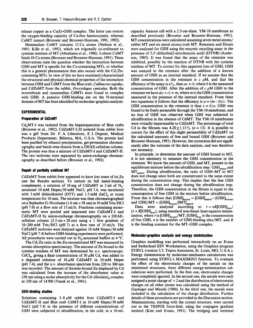
Figure 3 Space-filling model of the energy-minimized MT-GSH complex
Dark spheres represent the GSH molecule. This view is derived from the structure shown inFigure 2(a) by rotation of the structure around the x-axis by 900. The Gly-Cys-Glu residues ofGSH run from top to bottom.
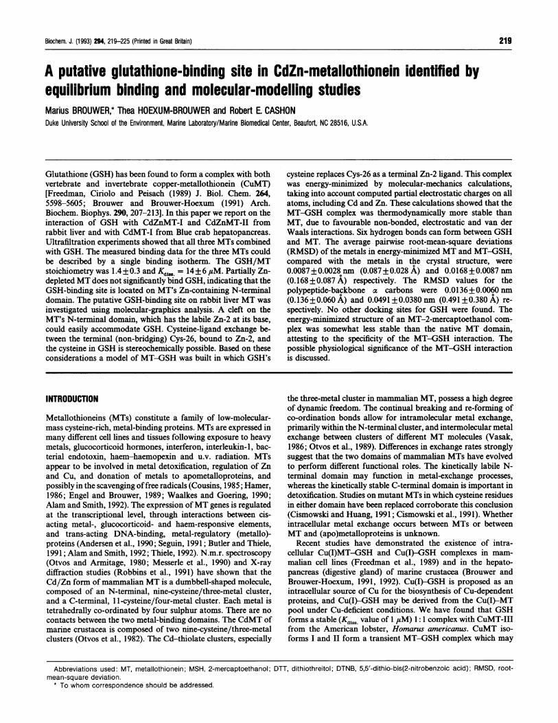
Table 1 Hydrogen bonds between GSH and MT
Number of bond refers to the structure shown in Figure 2.
Amino acid/atom
Bond GSH MT
23456
GluGluGlyGluCysGlu
C=OC=ON-HN-HC=ON-H
oa-Carboxyly-CarbonylAmidecx-AminoCarbonylo-Amino
Thr-27Thr-27Lys-25Cys-1 3Gly-1 1Gly-1 1
H-0H-NO=CO=CH-NO=C
HydroxylAmideCarbonylCarbonylAmideCarbonyl
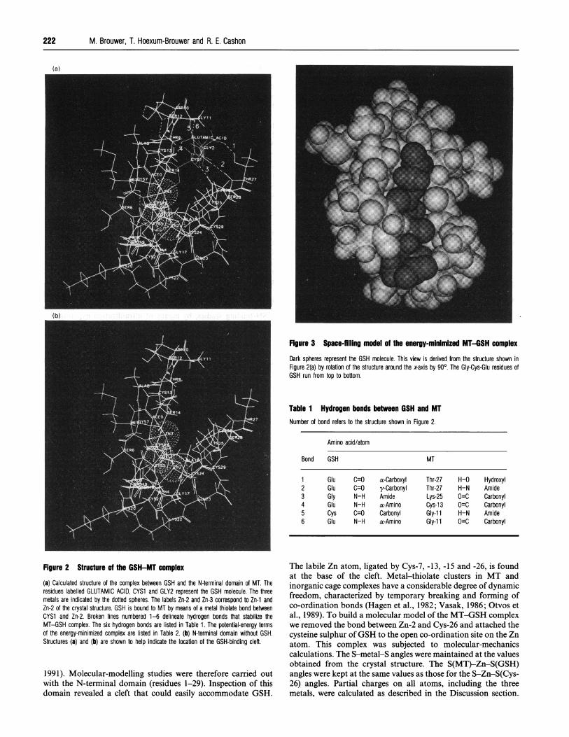
Figure 2 Structure of the GSH-MT complex
(a) Calculated structure of the complex between GSH and the N-terminal domain of MT. Theresidues labelled GLUTAMIC ACID, CYS1 and GLY2 represent the GSH molecule. The threemetals are indicated by the dotted spheres. The labels Zn-2 and Zn-3 correspond to Zn-1 andZn-2 of the crystal structure. GSH is bound to MT by means of a metal thiolate bond betweenCYS1 and Zn-2. Broken lines numbered 1-6 delineate hydrogen bonds that stabilize theMT-GSH complex. The six hydrogen bonds are listed in Table 1. The potential-energy termsof the energy-minimized complex are listed in Table 2. (b) N-terminal domain without GSH.Structures (a) and (b) are shown to help indicate the location of the GSH-binding cleft.
1991). Molecular-modelling studies were therefore carried outwith the N-terminal domain (residues 1-29). Inspection of thisdomain revealed a cleft that could easily accommodate GSH.
The labile Zn atom, ligated by Cys-7, -13, -15 and -26, is foundat the base of the cleft. Metal-thiolate clusters in MT andinorganic cage complexes have a considerable degree of dynamicfreedom, characterized by temporary breaking and forming ofco-ordination bonds (Hagen et al., 1982; Vasak, 1986; Otvos etal., 1989). To build a molecular model of the MT-GSH complexwe removed the bond between Zn-2 and Cys-26 and attached thecysteine sulphur ofGSH to the open co-ordination site on the Znatom. This complex was subjected to molecular-mechanicscalculations. The S-metal-S angles were maintained at the valuesobtained from the crystal structure. The S(MT)-Zn-S(GSH)angles were kept at the same values as those for the S-Zn-S(Cys-26) angles. Partial charges on all atoms, including the threemetals, were calculated as described in the Discussion section.
Interaction between glutathione and metallothionein 223
Table 2 Potential energy terms of energy-minimized MT and MT-GSHcomplex (kcal/mol)Both structures were minimized in the TRIPOS force-field. Charges on all atoms, includingmetals, were calculated as described in the text. 1 kcal/mol = 4.2 kJ/mol.
Potential energy (kcal/mol)
Energy term MT* MT-GSHt
Bond-stretching energyAngle-bending energyTorsional energyOut-of-plane-bending energy1-4 van der Waals energyvan der Waals energy1-4 Electrostatic energyElectrostatic energyFixed-angle energy
Total potential energy
9.70454.55872.1151.322
25.689-90.848136.723
- 298.9940.005
10.356
68.595
71.523
1.736
28.425
-105.508
134.660
-418.916
0.003
-89.726 -209.122
* N-terminal domain (residues 1-29).t The bond between Cys-26 and Zn-2 was clipped. The cysteine sulphur of GSH was
attached to the open co-ordination site on the Zn.
The structure of the energy-minimized complex is shown inFigures 2 and 3. The complex is stabilized by six hydrogen bondsbetween GSH and MT (Figure 2 and Table 1). The calculatedpotential energy terms of the two energy-minimized structuresare listed in Table 2. The MT-GSH complex is energeticallyfavoured over the free domain due to non-bonded van der Waalsand electrostatic interactions.
DISCUSSION
GSH has been implicated in protection against Cd- and Cu-induced toxicity (Singhal et al., 1987; Freedman et al., 1989) andin tumour-cell resistance to metal-based anti-cancer drugs(Godwin et al., 1992). The existence of intracellular Cu(I)-GSHand CuMT-GSH has been reported for both mammals andinvertebrates (Freedman et al., 1989; Brouwer and Brouwer-Hoexum, 1991, 1992). The Cu(I)-GSH complex may act as asource of Cu for the biosynthesis of CuMT (Freedman et al.,1989), haemocyanin (Brouwer and Brouwer-Hoexum, 1991,1992) and superoxide dismutase (Steinkuhler et al., 1991).Whether or not GSH binds to CdZnMT was unknown. The datareported in this study demonstrate that GSH can bind to bothinvertebrate (Blue crab) and vertebrate (rabbit) CdZnMT. A fullcomplement of metals bound to the N-terminal domain of MTwas prerequisite for binding to occur. The stoichiometry andbinding constant for the CdZnMT-GSH interaction (GSH/MT = 1.4+ 0.3 and KdiSS = 14+ 6 #uM) showed rather large S.D.values. This may, at least in part, be due to the difficulty ofassaying GSH in the presence of CdZnMT and to the non-rigid,dynamic organization of the metal-thiolate clusters in the N-terminal domain of MT. Several interchangeable structural MTisoforms may co-exist (Vasak, 1986), which may interact to adifferent extent with GSH. The results obtained from ourultrafiltration experiments have been confirmed by size-exclusionh.p.l.c. of GSH, and mixtures of GSH and CdZnMT-I orCdZnMT-II. The amount of free GSH, which can be determinedreliably, decreases considerably when GSH and MT are co-
chromatographed. No measurable release of Cd or Zn wasobserved during our experiments.
Part of the Zn in the N-terminal domain of CdZnMT isextremely reactive with EDTA (Li et al., 1980; Winge and
Miklossy, 1982a,b). The solvent accessibilities of the sulphuratoms bonded to Zn-I and Zn-2, which may be a useful indicatorof preferred sites of metal-exchange reactions, are 0.00505 nm2(5.05 A2) for Zn-I and 0.3583 nm2 (35.83 A2) for Zn-2 (Robbinset al., 1991). This strongly suggests that Zn-2 represents thereactive Zn atom. The fact that partially Zn-depleted MT doesnot combine to a significant degree with GSH led us to postulatethat Zn-2 is part of the GSH-binding site. Molecular-graphicsanalysis revealed that this Zn atom was at the bottom of a cleftthat could easily accommodate GSH. It was found to beimpossible to dock GSH at either Zn-I or Cd. Based on theknown dynamic nature of the metal-thiolate cluster in the N-terminal domain of MT, which involves breaking and reformingof metal-to-cysteine bonds, we hypothesized that the cysteinesulphur of GSH might compete with one of the terminal cysteine(Cys-13 or Cys-26) sulphurs for a co-ordination site on Zn-2. Wefound that such cysteine-ligand exchange was stereochemicallypossible between GSH and Cys-26, but not between GSH andCys-13. Based on these considerations the Zn-2-Cys-26 bond inMT was broken and replaced with a Zn-2-Cys(GSH) thiolatebond. This complex was then energy-minimized by molecular-mechanics calculations using the TRIPOS force-field.The TRIPOS valence force-field expresses the total potential
energy of a molecule as the sum of the bond-stretching, angle-bending, out-of-plane-bending, torsional and non-bonded (vander Waals, and electrostatic) terms. The application of empiricalforce-field computations to metal co-ordination compounds isproblematic because ofthe large variation ofligand-metal-ligandbond angles observed at transition-metal centres (Lauher, 1986;Allured et al., 1991). Energy minimization of the CdZnMT wastherefore carried out keeping the sulphur-metal-sulphur anglesconstrained to the values found in the crystal structure. Thefixed-angle constraint introduced in this way was almost neg-ligible (Table 1).
It is unsuitable in molecular-dynamics and -mechanics calcula-tions of simple metal complexes and metalloproteins to assignthe formal charges +2 to Cd and Zn. Quantum-mechanicalcalculations on a series of metal complexes, including zincthiolate compounds, indicate that the computed charges on themetals are generally much less than the formal charges (Fischer-Hjalmars and Henriksson-Enflo, 1982). Partial charges for theactive site ofmetalloproteins are generally obtained from calcula-tions ab initio for a simplified but representative model thatmimics the metal-ligand structure of the protein. These chargescan then be used in molecular-mechanics and -dynamics calcula-tions (Shen et al., 1990; Hoops et al., 1991; Banci et al., 1992).To evaluate the importance of charge in the energy minim-
ization of MT, the structure of MT was calculated in threedifferent ways. In the first procedure electrostatic charges wereignored. In the second procedure the three metals were assigneda formal point charge of +2, and were not included in thecomputation of the charge distribution. The atomic charges of allother atoms were calculated according to the method of partialequalization of orbital electronegativity (Gasteiger and Marsili,1980), as implemented by SYBYL. In the third procedure themetals were included in the calculation ofthe charge distribution,which requires knowledge ofthe electronegativities ofthe neutral,anionic and cationic forms of Cd and Zn in their tetrahedralvalence state. Since these values are not known, we chose to usethe corresponding values for the ground state as a first, empirical,approximation. The electronegativity values for the unchargedatoms were obtained from Lange's Handbook of Chemistry(1973). Electronegativities ofthe cation and anion were calculatedusing Mulliken's definition: X = 1/2(I+ E), where I and E arethe ionization potential and electron affinity respectively. The
224 M. Brouwer, T. Hoexum-Brouwer and R. E. Cashon
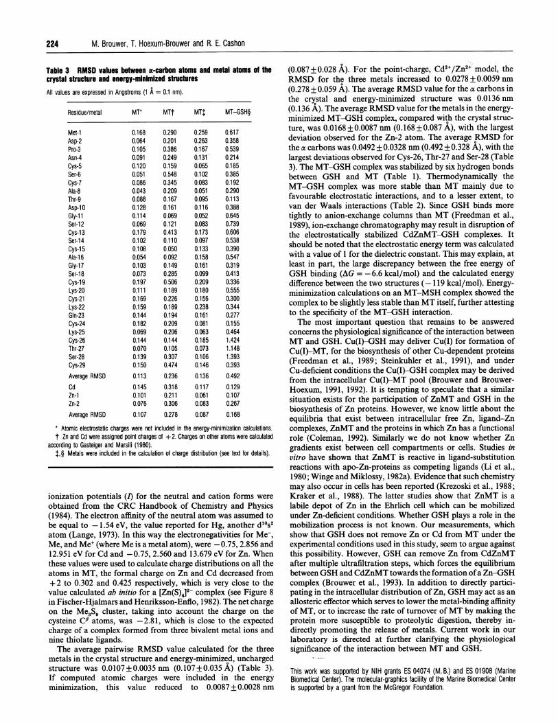
Table 3 RMSD values between a-carbon atoms and metal atoms of thecrystal structure and energy-minimized structuresAll values are expressed in Angstroms (1 A = 0.1 nm).
Residue/metal MT* MTt MT$ MT-GSH§
Met-iAsp-2Pro-3Asn-4Cys-5Ser-6Cys-7Ala-8Thr-9Asp-1 0Gly-l1Ser-12Cys-13Ser-14Cys-1 5Ala-16Gly-17Ser-1 8Cys-19Lys-20Cys-21Lys-22Gln-23Cys-24Lys-25Cys-26Thr-27Ser-28Cys-29Average RMSDCdZn-iZn-2Average RMSD
0.1680.0640.1050.0910.1200.0510.0860.0430.0880.1280.1140.0690.1790.1020.1080.0540.1030.0730.1970.1110.1690.1590.1440.1820.0690.1440.0700.1390.150
0.113
0.1450.1010.076
0.107
0.2900.2010.3860.2490.1590.5480.3450.2090.1670.1610.0690.1210.4130.1100.0500.0920.1490.2850.5060.1890.2260.1890.1940.2090.2060.1440.1050.3070.4740.2360.3180.2110.3060.278
0.2590.2630.1670.1310.0650.1020.0830.0510.0950.1160.0520.0830.1730.0970.1330.1580.1610.0990.2090.1800.1560.2380.1610.0810.0630.1850.0730.1060.1460.1360.1170.0610.0830.087
0.6170.3580.5390.2140.1850.3850.1920.2900.1130.3880.6450.7390.6060.5380.3900.5470.3190.4130.3360.5550.3000.3440.2770.1550.4641.4241.1481.3930.393
0.4920.1290.1070.267
0.168
* Atomic electrostatic charges were not included in the energy-minimization calculations.t Zn and Cd were assigned point charges of + 2. Charges on other atoms were calculated
according to Gasteiger and Marsili (1980).J,§ Metals were included in the calculation of charge distribution (see text for details).
ionization potentials (I) for the neutral and cation forms were
obtained from the CRC Handbook of Chemistry and Physics(1984). The electron affinity of the neutral atom was assumed to
be equal to -1.54 eV, the value reported for Hg, another d10s2atom (Lange, 1973). In this way the electronegativities for Me-,Me, and Me' (where Me is a metal atom), were -0.75, 2.856 and12.951 eV for Cd and -0.75, 2.560 and 13.679 eV for Zn. Whenthese values were used to calculate charge distributions on all theatoms in MT, the formal charge on Zn and Cd decreased from+ 2 to 0.302 and 0.425 respectively, which is very close to thevalue calculated ab initio for a [Zn(S)4]2- complex (see Figure 8in Fischer-Hjalmars and Henriksson-Enflo, 1982). The net chargeon the Me3S5 cluster, taking into account the charge on thecysteine C0 atoms, was -2.81, which is close to the expectedcharge of a complex formed from three bivalent metal ions andnine thiolate ligands.The average pairwise RMSD value calculated for the three
metals in the crystal structure and energy-minimized, unchargedstructure was 0.0107+0.0035nm (0.107+0.035 A) (Table 3).If computed atomic charges were included in the energy
minimization, this value reduced to 0.0087 + 0.0028 nm
(0.087+ 0.028 A). For the point-charge, Cd2+/Zn2+ model, theRMSD for the three metals increased to 0.0278 + 0.0059 nm(0.278 + 0.059 A). The average RMSD value for the a carbons inthe crystal and energy-minimized structure was 0.0136 nm(0.136 A). The average RMSD value for the metals in the energy-minimized MT-GSH complex, compared with the crystal struc-ture, was 0.0168 + 0.0087 nm (0.168+ 0.087 A), with the largestdeviation observed for the Zn-2 atom. The average RMSD forthe a carbons was 0.0492 + 0.0328 nm (0.492 + 0.328 A), with thelargest deviations observed for Cys-26, Thr-27 and Ser-28 (Table3). The MT-GSH complex was stabilized by six hydrogen bondsbetween GSH and MT (Table 1). Thermodynamically theMT-GSH complex was more stable than MT mainly due tofavourable electrostatic interactions, and to a lesser extent, tovan der Waals interactions (Table 2). Since GSH binds moretightly to anion-exchange columns than MT (Freedman et al.,1989), ion-exchange chromatography may result in disruption ofthe electrostatically stabilized CdZnMT-GSH complexes. Itshould be noted that the electrostatic energy term was calculatedwith a value of 1 for the dielectric constant. This may explain, atleast in part, the large discrepancy between the free energy ofGSH binding (AG = -6.6 kcal/mol) and the calculated energydifference between the two structures (- 119 kcal/mol). Energy-minimization calculations on an MT-MSH complex showed thecomplex to be slightly less stable than MT itself, further attestingto the specificity of the MT-GSH interaction.The most important question that remains to be answered
concerns the physiological significance of the interaction betweenMT and GSH. Cu(I)-GSH may deliver Cu(I) for formation ofCu(I)-MT, for the biosynthesis of other Cu-dependent proteins(Freedman et al., 1989; Steinkuhler et al., 1991), and underCu-deficient conditions the Cu(I)-GSH complex may be derivedfrom the intracellular Cu(I)-MT pool (Brouwer and Brouwer-Hoexum, 1991, 1992). It is tempting to speculate that a similarsituation exists for the participation of ZnMT and GSH in thebiosynthesis of Zn proteins. However, we know little about theequilibria that exist between intracellular free Zn, ligand-Zncomplexes, ZnMT and the proteins in which Zn has a functionalrole (Coleman, 1992). Similarly we do not know whether Zngradients exist between cell compartments or cells. Studies invitro have shown that ZnMT is reactive in ligand-substitutionreactions with apo-Zn-proteins as competing ligands (Li et al.,1980; Winge and Miklossy, 1982a). Evidence that such chemistrymay also occur in cells has been reported (Krezoski et al., 1988;Kraker et al., 1988). The latter studies show that ZnMT is alabile depot of Zn in the Ehrlich cell which can be mobilizedunder Zn-deficient conditions. Whether GSH plays a role in themobilization process is not known. Our measurements, whichshow that GSH does not remove Zn or Cd from MT under theexperimental conditions used in this study, seem to argue againstthis possibility. However, GSH can remove Zn from CdZnMTafter multiple ultrafiltration steps, which forces the equilibriumbetween GSH and CdZnMT towards the formation ofa Zn-GSHcomplex (Brouwer et al., 1993). In addition to directly partici-pating in the intracellular distribution of Zn, GSH may act as anallosteric effector which serves to lower the metal-binding affinityof MT, or to increase the rate of turnover of MT by making theprotein more susceptible to proteolytic digestion, thereby in-directly promoting the release of metals. Current work in ourlaboratory is directed at further clarifying the physiologicalsignificance of the interaction between MT and GSH.
This work was supported by NIH grants ES 04074 (M. B.) and ES 01908 (MarineBiomedical Center). The molecular-graphics facility of the Marine Biomedical Centeris supported by a grant from the McGregor Foundation.
Interaction between glutathione and metallothionein
REFERENCESAbrahams, L., Bremner, I., Diakun, G. P., Garner, C. D., Hasnain, S. S., Ross, I. and Vasak,
M. (1986) Biochem. J. 236, 585-589Alam, J. and Smith, A. (1992) J. Biol. Chem. 267,16379-16384Allured, V. S., Kelly, C. M. and Landis, C. R. (1991) J. Am. Chem. Soc. 113, 1-12Andersen, R. D., Taplitz, S. J., Oberbauer, A. M., Calame, K. L. and Herschman, H. R.
(1990) Nucleic Acids Res. 20, 6049-6055Anderson, M. E. (1985) Methods Enzymol. 113, 548-555Arseniev, A., Schultze, P., Worgotter, E., Braun, W., Wagner, G., Vasak, M., Kagi, J. H. R.
and Wutrich, K. (1988) J. Mol. Biol. 201, 637-657Banci, L., Schroder, S. and Kollman, P. A. (1992) Proteins: Struct. Funct. Genet. 13,
288-305Brouwer, M. and Brouwer-Hoexum, T. (1991) Arch. Biochem. Biophys. 290, 207-213Brouwer, M. and Brouwer-Hoexum, T. (1992) Biochemistry 31, 4096-4102Brouwer, M., Schlenk, D., Ringwood, A. H. and Brouwer-Hoexum, T. M. (1992) Arch.
Biochem. Biophys. 294, 461-468Brouwer, M., Brouwer-Hoexum, T. and Cashon, R. (1993) Mar. Environ. Res. 35,13-17Butler, G. and Thiele, D. J. (1991) Mol. Cell. Biol. 11, 476-485Cismowski, M. J. and Huang, P. C. (1991) Biochemistry 30, 6626-6632Cismowski, M. J., Narula, S. S., Armitage, I. M., Chernaik, M. L. and Huang, P. C. (1991)
J. Biol. Chem. 36, 24390-24397Coleman, J. (1992) Annu. Rev. Biochem. 61, 897-946Cousins, R. J. (1985) Physiol. Rev. 65, 230-309Creighton, T. E. (1986) Methods Enzymol. 131, 83-106Engel, D. W. and Brouwer, M. (1989) Adv. Comp. Environ. Physiol. 5, 53-75Fischer-Hjalmars, I. and Henriksson-Enflo, A. (1982) Adv. Quantum Chem. 16,1-42Freedman, J. H., Ciriolo, M. R. and Peisach, J. (1989) J. Biol. Chem. 264, 5598-5605Gasteiger, J. and Marsili, M. (1980) Tetrahedron 36, 3219-3228Godwin, A. K., Meister, A., O'Dwyer, P. J., Huang, C. S., Hamilton, T. C. and Anderson,
M. E. (1992) Proc. Natl. Acad. Sci. U.S.A. 89, 3070-3074Hagen, K. S., Stephan, D. W. and Holm, R. H. (1982) lnorg. Chem. 21, 3928-3936Hamer, D. H. (1986) Annu. Rev. Biochem. 55, 913-951Handbook of Chemistry and Physics (1984) (Weast, R. C., Astle, M. J. and Beyer, W. H.,
eds.), 65th edn., CRC Press, Boca Raton, Florida
Received 18 January 1993/8 March 1993; accepted 12 March 1993
Hoops, S. C., Anderson, K. W. and Merz, K. M., Jr. (1991) J. Am. Chem. Soc. 113,8262-8270
Kille, P., Lees, W. E., Darke, B. M., Winge, D. R., Dameron, C. T., Stephens, P. E. andKay, J. (1992) J. Biol. Chem. 267, 8042-8049
Kini, R. M. and Evans, H. J. (1991) J. Biomol. Struct. Dynamics 9, 475-487Kraker, A. J., Krakower, G., Shaw, C. F., Ill, Petering, D. H. and Garvey, J. (1988) Cancer
Res. 48, 3381-3388Krezoski, S. K., Villalobos, J., Shaw, C. F. and Petering, D. H. (1988) Biochem. J. 255,
483-491Lange's Handbook of Chemistry (1973), (Dean, J. A., ed.) 11th edn., McGraw-Hill Book
Company, New YorkLauher, J. W. (1986) J. Am. Chem. Soc. 108, 1521-1531Li, T. Y., Kraker, A. J., Shaw, C. F. and Petering, D. H. (1980) Proc. Natl. Acad. Sci. U.S.A.
77, 6334-6338Messerle, B. A., Schaffer, A., Vasak, M., Kagi, J. H. R. and Wuthrich, K. (1990) J. Mol.
Biol. 214, 765-779Nielson, K. B., Atkin, C. L. and Winge, D. R. (1985) J. Biol. Chem. 260, 5342-5350Otvos, J. D. and Armitage, I. M. (1980) Proc. Natl. Acad. Sci. U.S.A. 77, 7094-7098Otvos, J. D., Olafson, R. W. and Armitage, I. M. (1982) J. Biol. Chem. 257, 2427-2431Otvos, J. D., Chen, S. and Liu, X. (1989) in Metal Ion Homeostasis, Molecular Biology and
Chemistry (Winge, D. and Hamer, D., eds.), pp. 197-206, Alan R. Liss, New YorkRobbins, A. H., McRee, D. E., Williamson, M., Collet, S. A., Xuong, N. H., Furey, W. F.,
Wang, B. C. and Stout, C. D. (1991) J. Mol. Biol. 221, 1269-1293Seguin, C. (1991) Gene 97, 295-300Shen, J., Wong, C. F., Subramaniam, S., Albright, T. A. and McCammon, J. A. (1990)
J. Comp. Chem. 11, 346-350Singhal, R. K., Anderson, M. E. and Meister, A. (1987) FASEB J. 1, 220-223Steinkuhler, C., Sapora, O., Carri, M. T., Nagel, W., Marcocci, L., Ciriolo, M. R., Weser, U.
and Rotilio, G. (1991) J. Biol. Chem. 266, 24580-24587Thiele, D. (1992) Nucleic Acids Res. 20, 1183-1191Vasak, M. (1986) Environ. Health Persp. 65, 193-197Vasak, M., Kagi, J. H. R. and Hill, H. A. 0. (1981) Biochemistry 20, 2852-2856Waalkes, M. P. and Goering, P. L. (1990) Chem. Res. Toxicol. 3, 281-288Winge, D. R. and Miklossy, K.-A. (1982a) Arch. Biochem. Biophys. 214, 80-88Winge, D. R. and Miklossy, K.-A. (1982b) J. Biol. Chem. 257, 3741-3746
225