al cerebellum 1
TRANSCRIPT
8/6/2019 Al Cerebellum 1
http://slidepdf.com/reader/full/al-cerebellum-1 1/7
Drug and Chemical Toxicology , 2009; 32(3): 215–221
R E S E A R C H A R T I C L E
Eect o concurrent chronic exposure o fuoride andaluminum on rat brain
anzeer Kaur1, Rakesh K. Bijarnia2, and Bimla Nehru1
1Department of Biophysics, Panjab University, Chandigarh, India, and 2Department of Biotechnology, Shaheed
Udham Singh College of Engineering and Technology, Tangori, Mohali, India
Address for Correspondence: anzeer Kaur (present address), Department o Biotechnology and Bioinormatics, Jaypee University o Inormationechnology, Waknaghat, Solan 173215, H.P, India. Fax: 91-1792245362; E-mail: [email protected]
(Received 19 October 2008; revised 13 December 2008; accepted 30 January 2009)
Introduction
Te uoridation o public water is known to be oneo the most successul public health measures o the20th century (Padilla and Davis, 2001). Over the lastthree decades, the condition o the teeth o childrenhas improved tremendously. Tis has generally beenattributed to the increased use o uoride toothpasteand as added to drinking water (Van Loveren andDuggal, 2001). Sodium uoride is currently addedto the majority o municipal water systems in many developed countries to prevent cavities in children.Te studies done by Mullenix et al. (1995) discussedthe controversy between the benets and risks o uoride. Teir research suggested that uoride hasaccess to the brain and could possibly be neurotoxic.In addition, it has been shown that uoride levels at3–11 ppm aected the nervous system without any apparent physical malormation and a dose level o 100 ppm caused symptoms o attention decit (Xianget al., 2003)
Te exposure o aluminum to humans may play anactive role in Alzheimer’s disease (Perl and Moalem,
2006). It was ound that the dietary supplement o alu-minum results in an increased brain oxidative stressthat could promote Alzheimer’s disease, such as amy-loidosis (Pratico et al., 2002). Aluminum exposure tothe brain has been linked with the onset o oxidativestress (Nehru and Anand, 2005) and alters the redox status o glutathione (Nehru and Bhalla, 2006). Teassociation o aluminum and uoride is o particularimportance, as it relates to Alzheimer’s disease. It ispossible that long-term use o drinking water with ahigh aluminum concentration and with low uorideconcentration is associated with the increased relativerisk o Alzheimer’s disease (Belojevic and Jakovljevic,1998). Some o the pathologic changes are not raisedby aluminum alone, but by aluminum uoride com-plexes (Strunecká, 1999).
Te present experiment was an attempt to comparethe toxicity caused by subchronic exposure o uoridealone and uoride aluminum together in rat brain.
ISSN 0148-0545 print/ISSN 1525-6014 online © 2009 Inorma UK Ltd
DOI: 10.1080/01480540902862251
Abstract The present in vivo study was designed to investigate the toxic potential o uoride alone and in conjuga-tion with aluminum on the rat brain. The region-specic response o both elements was studied in diferent
regions o brain, namely the cerebrum, cerebellum, and medulla oblongata. Following uoride exposure,oxidative stress increased signicantly, estimated by increased lipid peroxidation and a decrease in theactivity o the antioxidant enzyme, superoxide dismutase. The neurotransmitter (e.g., dopamine, norepine-phrine, and serotonin) content was also altered. However, these aspects were more pronounced in animalsgiven uoride and aluminum together. Histological evidence showed deprival o neuronal integrity withhigher magnitude in concurrent uoride and aluminum exposure, as compared to uoride alone. Thus, itcan be concluded that aluminum appears to enhance the neurotoxic hazards caused by uoride.
Keywords: Fluoride; aluminum; oxidative stress; neurotransmitters
http://www.inormapharmascience.com/dct

8/6/2019 Al Cerebellum 1
http://slidepdf.com/reader/full/al-cerebellum-1 2/7
216 Tanzeer Kaur et al.
Te uoride toxicity was created by exposing the ratsto sodium uoride (125 ppm) supplemented in thedrinking water o rats or 8 weeks. o see the cumula-tive eect o uoride and aluminum, the same doseo uoride was given with aluminum as aluminumchloride (40 mg/kg body weight/day), administeredby oral gavage, or the same 8 weeks.
Methods and materials
Animals and treatments
Healthy emale rats o the Spargue-Dawley (SD)strain were procured rom the central animal houseo Panjab University (Chandigarh, India) and wereacclimatized in the departmental animal house or 2 weeks in plastic cages under hygienic conditions and
were provided eed and water ad libitum. A total o 24 SD emale rats were used to orm threegroups (8 rats each). Te rats were healthy, mature,and initially weighed 175–200 g. Te animals o group1 were ed standard animal chow and water providedad libitum. Animals o group 2 were administereduoride as sodium uoride (125 ppm) in their drink-ing water or 8 weeks. Group 3 animals were exposedto aluminum (as AlCl
340 mg/kg body weight/day)
orally simultaneous to uoride (as sodium uoride,125 ppm) in drinking water. Te animals in variousgroups were monitored or their physical health anddietary intake.
At the end o 8 weeks, the animals were sacricedby decapitation. Teir brains were removed andimmediately divided into three regions viz. cerebrum,cerebellum, and medulla oblongata. All processes were carried out under cold conditions. Te threebrain regions were homogenized (10% w/v) in 10 mMo phosphate-buered saline (PBS) (pH 7.4). Tehomogenate was centriuged at 1,000 g or 10 minutesat 4°C, and the supernatant was used or biochemicalassays.
Lipid peroxidation
Te quantitative measurement o lipid peroxida-tion was perormed according to the method o Buege and Aust (1978). Te malondialdehyde(MDA) ormed ater a series o peroxidation reac-tion as an end-product was measured by the reac-tion with thiobarbituric acid at 532 nm. Te results were expressed as micromoles ( moles) o MDAper milligram o protein, using the molar extinctioncoefcient o MDA-thiobarbituric chromophore(1.56 × 10⁵ M− 1cm−1).
Superoxide dismutase (SOD)
Te assay was perormed according to the method o Kono (1978). Te extent o inhibition o reduction o nitro blue tetrazolium to blue ormazon by the addi-tion o the enzyme was measured at 560 nm. Te activ-ity o the enzyme was expressed as units per milligramo protein, where one unit o enzyme was dened asthe amount inhibiting the rate o reaction by 50%.
Total lipid extraction and estimation
Lipids were extracted by the method o Folch et al.(1957). Te content o total lipids was estimated by the method o Frings et al. (1972).
Phospholipid content
Te content o phospholipids was estimated by themethod o Bartlett (1959).
Cholesterol content
Cholesterol was estimated in the lipid raction by themethod o Zlatkis et al. (1953).
Estimations of neurotransmitters
Estimations o norepinephrine, dopamine, and sero-tonin were done by the method o Ansell and Beeson(1968).
Histopathological studies
Cerebrum and cerebellum were xed in ormaldehyde(10%). Te tissues were then dehydrated and embed-ded in parafn wax (MP, 68°C). Te parafn sections were than cut and stained in Delaeld’s hematoxylinand eosin (H&E) staining.
Statistical analysis
Te data are expressed as mean ± standard deviation(SD) and were analyzed by the Student’s t -test.
Results
As shown in Figure 1, the level o MDA indicating lipidperoxidation was ound to increase in all the three

8/6/2019 Al Cerebellum 1
http://slidepdf.com/reader/full/al-cerebellum-1 3/7
Fluoride and aluminum on rat brain 217
regions o brain ater uoride exposure (group 2).Exposure with both uoride and aluminum showedadditive eects on MDA level (group 3). Among theentire three regions, the cerebellum showed maxi-mum decrease in MDA content, pertaining to be themost sensitive among all regions. SOD activity was
decreased ater uoride treatment (Figure 2).Cotreatment with uoride and aluminum urtherdecreased the SOD activity and the activity was againleast in the cerebellum, as shown in Figure 2. Tedecrease in its activity, in comparison to uoride expo-sure, is also highly signicant (P < 0.001) in all regions.
Both treatments caused degeneration o lipids inall parts o the brain (able 1). Te cerebrum showedmaximum degeneration, ollowed by cerebellum andmedulla oblongata. One o the main constituents o membrane lipid (i.e., phospholipids level) declinedater uoride exposure, and a more pronounced
decrease in phospholipids in all regions o brainater cotreatment with both elements was observed. All the three brain regions showed signicantly increased (P < 0.001) ratio o cholesterol to phos-pholipids (CHL/PHL) in uoride exposed animals.Moreover, the decline in CHL/PHL ratio ollowingconcurrent treatment o uoride and aluminum wasmore pronounced and the all was highly signi-cant (P < 0.001), in comparison to uoride exposurealone.
Te catecholamine neurotransmitters (e.g., nore-pinephrine and dopamine) o all the three regionsare also ound to be altered with these exposures
(able 2). Te level o norepinephrine and dopaminedecreased with uoride exposure and reduction intheir content ater uoride and aluminum coadmin-istration was more pronounced. Te same trend wasobserved in all three brain regions, with the cerebrumregion most aected. Monoamine neurotransmit-ter serotonin also showed a drastic and signicant(P < 0.001) decrease in its level ater uoride exposure
2
1.8
1.6
1.4
1.2
1
0.8
0.6
0.4
0.2
0 n m o l e s o f M D
A / m g o f p r o t e i n
Group 1 Group 2 Group 3
**
***
***
***
***
##
###
***###
Cerebrum
Cerebellum
Medulla
oblongata
Figure 1. Estimation o MDA content in all three regions o brain(viz. cerebrum, cerebellum, and medulla oblongata) o control(group 1) animals, uoride-exposed (group 2) animals, and uo-ride and aluminum–exposed (group 3) animals. [Values are mean± standard deviation o six to seven determinations: * P < 0.05;**P < 0.01; ***P < 0.001; indicates signifcant change, in compari-son to control group 1; #P < 0.05; ##P < 0.01; ###P < 0.001; indicates
signifcant change between groups 2 and 3].
1
0.9
0.8
0.7
0.6
0.5
0.4
0.3
0.2
0.1
0
U n i t s / m g o f p r o t e i n
Group 1 Group 2 Group 3
*** *** ******
***
##### ***
###Cerebrum
Cerebellum
Medulla Oblongata
Figure 2. Estimation o superoxide dismutase activity in all threeregions o brain (viz. cerebrum, cerebellum, and medulla oblon-gata) o control (group 1) animals, uoride-exposed (group 2)animals, and uoride and aluminum–exposed (group 3) animals.[Values are mean ± standard deviation o six to seven determina-tions: *P < 0.05; **P < 0.01; ***P < 0.001; indicates signifcant change,in comparison to control group 1; #P < 0.05; ##P < 0.01; ###P < 0.001;indicates signifcant change between groups 2 and 3].
Table 1. otal lipids, phospholipids, cholesterol, and cholesterol/phospholipids ratio in three dierent regions o brain.
Brain region Group otal lipids Phospholipids Cholesterol Cholesterol/phospholipids
Cerebrum 1 117.00 ± 16.6 87.58 ± 8.4 6.63 ± 0.84 0.075 ± 0.007
2 70.71 ± 13.8** 60.87 ± 11.1** 7.92 ± 6.64* 0.130 ± 0.011**3 56.77 ± 5.8**,# 45.97 ± 10.68***,# 8.90 ± 7.80** 0.193 ± 0.026***,###
Cerebellum 1 155.45 ± 11.6 107.67 ± 11.4 2.66 ± 1.00 0.025 ± 0.015
2 98.11 ± 10.9*** 73.75 ± 8.16** 5.25 ± 1.47* 0.074 ± 0.021**
3 72.55 ± 9.3***,# 54.52 ± 9.55***,## 6.82 ± 1.36** 0.125 ± 0.027***,###
Medulla oblongta 1 201.59 ± 12.9 161.61 ± 2.70 16.60 ± 1.75 0.102 ± 0.016
2 143.19 ± 16.34** 108.51 ± 3.84*** 15.84 ± 1.26 0.145 ± 0.021**
3 113.19 ± 5.9***,### 73.93 ± 4.86***,### 18.94 ± 1.32* 0.256 ± 0.035***,###
Group 1 (control); group 2 (F exposed); group 3 (F + Al exposed). Values are mean ± standard deviation o six to seven determinations.*P < 0.05; **P < 0.01; ***P < 0.001; indicates signifcant change, in comparison to control group 1.#P < 0.05; ## P < 0.01; ###P < 0.001; indicates signifcant change between groups 2 and 3.

8/6/2019 Al Cerebellum 1
http://slidepdf.com/reader/full/al-cerebellum-1 4/7
218 Tanzeer Kaur et al.
in cerebrum and cerebellum. Concurrent exposureo uoride and aluminum signicantly (P < 0.001)reduced its level, in comparison to exposure to uo-ride alone.
Histological analysis o cerebrum and cer-ebellum ater both doses are depicted inFigures 3 and 4, respectively. Control animalsshowed normal morphology with intact neuron andcytoplasm (Figure 3A). It also showed normal oli-godendrocytes and ew astrocytes. Fluoride exposurein Figure 3B showed necrosis, which is depicted by shrunken nuclei, hyperchromasia, and disintegratedcytoplasm. Cytoplasm also showed edema depictedby vacuoles, and the number o astrocytes haveincreased. Another eature o necrosis, eosinophilia(red neurons), is also visible at many instances. Teanimals given both uoride and aluminum showedsimilar, but more severe, necrotic signs (Figure 3C).
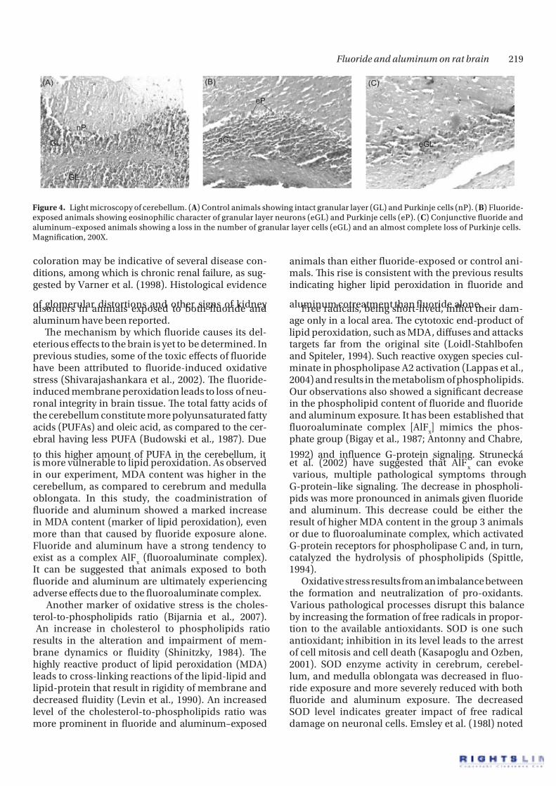
Figure 4A showed normal morphology o cerebel-lum o control animals. Tese animals showed intactgranular layer, normal count, and morphology o Purkinje cells. Fluoride exposure (Figure 4B) hasresulted in a decrease in the number o granular cells,
and Purkinje cells have shown eosinophilic character(red neurons). Exposure o both uoride and alumi-num (Figure 4C) showed more distortion in cerebel-lum morphology with a decrease in granular layercells and an almost complete loss o Purkinje cells inthese animals.
Discussion
In the present study, we have attempted to measurethe inuence o aluminum exposure on uoride neu-rotoxicity in rat brain. Te animals were examineddaily or their general health and physical activity.Generally, the dose o both uoride and uoride with aluminum was well tolerated. However, towardthe end o the treatment period, 1 animal o 8 died ingroup 3, suggesting the deleterious eect o uoride
and aluminum given together. Further, in this group,the animals were lethargic and remained in the cor-ners o the cage. Te animals in group 3 presented anunusual appearance with sparse hair and discoloredunderlying skin. Te hair loss and presence o skin
Table 2. Level o neurotransmitters: norepinephrine, dopamine, and serotonin in cerebrum, cerebellum, and medulla oblongata o ratbrain.
Brain region Group Norepinephrine (ng/g) Dopamine (ng/g) Serotonin (ng/g)
Cerebrum 1 413.49 ± 21.66 891.03 ± 65.18 1833.06 ± 13.9
2 361.42 ± 19.24** 662.97 ± 8.83** 1345.50 ± 43.95***
3 282.75 ± 18.66***,# 599.12 ± 24.49***,## 1254.43 ± 7.17***,##
Cerebellum 1 689.93 ± 43 952.58 ± 5.39 1593.28 ± 15.9
2 407.18 ± 20.06*** 631.79 ± 15.05*** 1107.23 ± 50.6***3 397 ± 13.5***,# 530.63 ± 8.53***,## 1065.12 ± 48.96***,##
Medulla oblongata 1 540.46 ± 36.32 1034.76 ± 50.54 1622.51 ± 9.82
2 419.62 ± 7.46** 785.16 ± 7.7*** 1197.38 ± 23.24**
3 362.09 ± 6.59***,## 741.45 ± 28.69***,## 1038.5 ± 40.70***,###
Group 1 (control); group 2 (F exposed); group 3 (F + Al exposed). Values are mean ± standard deviation o six to seven determinations.*P < 0.05; **P < 0.01; ***P < 0.001; indicates signifcant change, in comparison to control group 1,#P < 0.05; ##P < 0.01; ###P < 0.001; indicates signifcant change between groups 2 and 3.
(A)
nN
nN
nN Od
At
(B)
Od
At
At
Hc
At
Es
V
(C) At
At
Hc
HcHc
Es
EsV
V
Figure 3. Light microscopy o cerebral cortex. ( A) Control animals showing normal morphology o neurons (nN), intact oligodendrocytes(Od), and ew astrocytes (At). (B) Fluoride-exposed animals showing hyperchromasia (Hc) with shrunken nucleus, vacuolated cells (V),and eosinophilia (Es). (C) Conjunctive uoride and aluminum–exposed animals showing increased number o astrocytes (At), vacuolatedoligodendrocytes (V), increased hyperchromasia (Hc), and eosinophilia (Es). Magnifcation, 200X.
8/6/2019 Al Cerebellum 1
http://slidepdf.com/reader/full/al-cerebellum-1 5/7
Fluoride and aluminum on rat brain 219
coloration may be indicative o several disease con-ditions, among which is chronic renal ailure, as sug-gested by Varner et al. (1998). Histological evidence
o glomerular distortions and other signs o kidney disorders in animals exposed to both uoride andaluminum have been reported.
Te mechanism by which uoride causes its del-eterious eects to the brain is yet to be determined. Inprevious studies, some o the toxic eects o uoridehave been attributed to uoride-induced oxidativestress (Shivarajashankara et al., 2002). Te uoride-induced membrane peroxidation leads to loss o neu-ronal integrity in brain tissue. Te total atty acids o the cerebellum constitute more polyunsaturated atty acids (PUFAs) and oleic acid, as compared to the cer-ebral having less PUFA (Budowski et al., 1987). Due
to this higher amount o PUFA in the cerebellum, itis more vulnerable to lipid peroxidation. As observedin our experiment, MDA content was higher in thecerebellum, as compared to cerebrum and medullaoblongata. In this study, the coadministration o uoride and aluminum showed a marked increasein MDA content (marker o lipid peroxidation), evenmore than that caused by uoride exposure alone.Fluoride and aluminum have a strong tendency toexist as a complex AlF
x (uoroaluminate complex).
It can be suggested that animals exposed to bothuoride and aluminum are ultimately experiencingadverse eects due to the uoroaluminate complex.
Another marker o oxidative stress is the choles-terol-to-phospholipids ratio (Bijarnia et al., 2007). An increase in cholesterol to phospholipids ratioresults in the alteration and impairment o mem-brane dynamics or uidity (Shinitzky, 1984). Tehighly reactive product o lipid peroxidation (MDA)leads to cross-linking reactions o the lipid-lipid andlipid-protein that result in rigidity o membrane anddecreased uidity (Levin et al., 1990). An increasedlevel o the cholesterol-to-phospholipids ratio wasmore prominent in uoride and aluminum–exposed
animals than either uoride-exposed or control ani-mals. Tis rise is consistent with the previous resultsindicating higher lipid peroxidation in uoride and
aluminum cotreatment than uoride alone.Free radicals, being short-lived, inict their dam-age only in a local area. Te cytotoxic end-product o lipid peroxidation, such as MDA, diuses and attackstargets ar rom the original site (Loidl-Stahlboenand Spiteler, 1994). Such reactive oxygen species cul-minate in phospholipase A2 activation (Lappas et al.,2004) and results in the metabolism o phospholipids.Our observations also showed a signicant decreasein the phospholipid content o uoride and uorideand aluminum exposure. It has been established thatuoroaluminate complex [AlF
x ] mimics the phos-
phate group (Bigay et al., 1987; Antonny and Chabre,
1992) and inuence G-protein signaling. Struneckáet al. (2002) have suggested that AlF
x can evoke
various, multiple pathological symptoms throughG-protein–like signaling. Te decrease in phospholi-pids was more pronounced in animals given uorideand aluminum. Tis decrease could be either theresult o higher MDA content in the group 3 animalsor due to uoroaluminate complex, which activatedG-protein receptors or phospholipase C and, in turn,catalyzed the hydrolysis o phospholipids (Spittle,1994).
Oxidative stress results rom an imbalance betweenthe ormation and neutralization o pro-oxidants. Various pathological processes disrupt this balanceby increasing the ormation o ree radicals in propor-tion to the available antioxidants. SOD is one suchantioxidant; inhibition in its level leads to the arresto cell mitosis and cell death (Kasapoglu and Ozben,2001). SOD enzyme activity in cerebrum, cerebel-lum, and medulla oblongata was decreased in uo-ride exposure and more severely reduced with bothuoride and aluminum exposure. Te decreasedSOD level indicates greater impact o ree radicaldamage on neuronal cells. Emsley et al. (198l) noted
nP
GL
GL
(A)
eP
eGL
(B)
eGL
(C)
Figure 4. Light microscopy o cerebellum. ( A) Control animals showing intact granular layer (GL) and Purkinje cells (nP). (B) Fluoride-exposed animals showing eosinophilic character o granular layer neurons (eGL) and Purkinje cells (eP). (C) Conjunctive uoride andaluminum–exposed animals showing a loss in the number o granular layer cells (eGL) and an almost complete loss o Purkinje cells.Magnifcation, 200X.

8/6/2019 Al Cerebellum 1
http://slidepdf.com/reader/full/al-cerebellum-1 6/7
220 Tanzeer Kaur et al.
that the amide uoride hydrogen bond was the sec-ond-strongest hydrogen bond known, and it seemedcertain that the uoride ion was able to competesuccessully or the amide bond in proteins. SOD asa proteinaceous enzyme might have been altered by uoride ion, thus showing its decreased content aterits chronic exposure. As suggested by Spittle (1994),that uoroaluminate complex interere in proteinsynthesis, the drastic reduction in SOD enzyme activ-ity ater uoride and aluminum exposure could havebeen due to the intererence o uoroaluminate com-plex in its synthesis.
Catecholamine neurotransmitters, such asnorepinephrine and dopamine, on oxidation withmonoamine oxidase, result in the production o toxicaldehyde metabolites, a precursor o oxidative stress(Grima et al., 2003). A decrease in catecholamineneurotransmitters ollowing uoride and uoride
and aluminum exposure predicts a higher produc-tion o such toxic metabolites in the brain. Temonoamine oxidase metabolites o catecholamine,such as catecholamine aldehydes, are also reportedto be accumulated in the Alzheimer’s and Parkinson’sdisease individual’s cerebrum (Burke et al., 2004). Inthe present experiment, the coadministration o uo-ride and aluminum led to higher production o suchtoxic metabolites and, consequently, higher oxidativestress in the brain.
Serotonin, a monoamine neurotransmitter, alsoshowed a similar decrease in both treatments.Serotonin is known to convert to melatonin by induc-
ing a rate-limiting enzyme, N-acetyltranserase, viaa cAMP-dependent pathway (Grad and Rozencwaig,1993). Melatonin is a very powerul antioxidant and itsconcentration was high, particularly in mitochondriaand the cell nucleus (Reiter et al., 2005). We observedthe production o ree radicals (increased MDAlevel) due to uoride and conjunctive uoride andaluminum exposure, and the decrease o serotoninmight have occurred due to conversion o serotoninto melatonin (which is also considered as a powerulantioxidant) to combat the oxidative stress causedby both exposures. Serotonin and melatonin existin proportion to each other; as melatonin increases,serotonin decreases in the brain.
Further evidence o higher neurotoxic eects o uoride and aluminum exposure, as compared to u-oride exposure together, are shown by the histologi-cal analysis o cerebrum and cerebellum o rat brain. Astrocytes have been implicated in both a deensiveand acilitatory capacity or many toxic injuries. But,activated astrocytes dier rom nonactivated astro-cytes with respect to their ability to protect neuronsrom excitotoxicity (Ahlemeyer et al., 2002). Activatedastrocytes secrete nitric oxide, which is a ree radical
that implicates its neurotoxic nature (Chao et al.,1996; Aschner, 1998). Tus, an increase in astrocytesnumber ater both doses suggests that neurotoxicity can be caused by both elements.
Purkinje cells are vital or the expression o behav-ior and other motor unctions (Goodlett et al., 1992).Purkinje cells undergo necrosis or apoptosis, ollowedby oxidative stress (Klein and Ackerman, 2003). Tepresent results showed a loss o Purkinje cells, indicat-ing necrosis ollowed by ree radical production. Teloss o Purkinje cells in these animals suggests deprivalo motor unctions. Since the deprivation o Purkinjecells was greater in uoride and aluminum–exposedanimals, it indicates a higher degree o motor unc-tion loss in these animals. Te destruction o granularlayer o the cerebellum ater uoride exposure wasalso observed by rabelsi et al. (2001) in 2-week-oldmice. Tey suggested that the destruction o granular
layer could be a consequence o uoride intereringin the mitosis o these cells. Tey have also postulatedthat, due to a lack o proper contact between granularcells and Purkinje cells, the loss o granular cells inthese animals might have resulted. Almost the com-plete destruction o Purkinje cells and an abrupt losso granular cells were observed in animals treated with both uoride and aluminum, predicting theintererence o AlF
x complex in their mitosis and sub-
sequent prolieration.
Conclusion
In conclusion, it was ound that, although uoridepossesses neurotoxic characters, when given with alu-minum, the neurotoxic nature o uoride increases.Te neurotoxicity is elicited by ree radical generationand alteration o neurotransmitters. Te morphology o both cerebrum and cerebellum is also aected.Fluoride and aluminum have a strong tendency toorm a complex, and even i their ingestion is romdierent sources, they have a tendency to orm thecomplex in vivo.
Acknowledgements
Declaration of interest : Te authors report nonancial conicts o interest. Te authors alone areresponsible or the content and writing o this paper.
References
Ahlemeyer, B., Kolker, S., Zhu, Y., Homann, G.F., Krieglstein,J. (2002). Increase in glutamate-induced neurotoxicity by
8/6/2019 Al Cerebellum 1
http://slidepdf.com/reader/full/al-cerebellum-1 7/7
Fluoride and aluminum on rat brain 221
activated astrocytes involves stimulation o protein kinaseC. J Neurochem 82:504–515.
Ansell, G.B., Beeson, M.F. (1968). A rapid and sensitive proce-dure or the combined assay o noradrenaline, dopamine,and serotonin in a single brain sample. Anal Biochem2:196–206.
Antonny, B., Chabre, M. (1992). Characterization o the alumi-
num and beryllium uoride species which activate trans-ducin: analysis o the binding and dissociation kinetics. JBiol Chem 267:6710–6718.
Aschner, M. (1998). Astrocytes as mediators o immune andinammatory responses in the CNS. Neurotoxicology 19:269–281.
Bartlett, G.R. (1959). Phosphorous assay in column chromatogra-phy. J Biol Chem 234:466–468.
Belojevic, G., Jakovljevic, B. (1998). Aluminum and Alzheimer’sdisease. Srp Arh Celok Lek 126:283–289.
Bigay, J., Deterre, P., Pfster, C., Chabre, M. (1987). Fluoridecomplexes o aluminum or beryllium act on G-proteins asreversibly bound analogues o the gamma phosphate o GP. EMBO J 6:2907–2913.
Bijarnia, R.K., Kaur, ., Singla, S.K., andon, C. (2007). Reversal o
hyperoxaluria-induced alteration in rat liver by administra-tion o N-acetylcysteine. Drug Chem oxicol 30:229–240.
Budowski, P., Leighfeld, M.J., Craword, M.A. (1987). Nutritionalencephalomalacia in the chick: an exposure o the vulnera-ble period or cerebellar development and the possible needor both6- and 3-atty acids. Br J Nutr 58:511–520.
Buege, J.A., Aust, D. (1978). Microsomal lipid peroxidationmethod. Enzymology 52:302–310.
Burke, W.J., Li, S.W., Chung, H.D., Ruggiero, D.A., Kristal, B.S.,Johnson, E.M., et al. (2004). Neurotoxicity o MAO metabo-lites o catecholamine neurotransmitters: role in neurode-generative diseases. Neurotoxicology 25:101–115.
Chao, C.C., Hu, S., Sheng, W.S., Bu, D., Bukrinsky, M.I., Peterson,P.K. (1996). Cytokine stimulated astrocytes damage humanneurons via a nitric oxide mechanism. Glia 16:276–284.
Emsley, J., Jones, D.J., Miller, J.M., Overill, R.E., Waddilove, R.A.(1981). An unexpectedly strong hydrogen bond: ab initiocalculations and spectroscopic studies o amide-uoridesystems. J Am Chem Soc l03:24–28.
Folch, S.J., Less, M., Stavley, GHS. (1957). A simple method orthe isolation and purifcation o total lipids rom animal tis-sues. J Biol Chem 226:497–509.
Frings, C.S., Fendley, .W., Dunn, R.., Queen, C.A. (1972).Improved determination o total serum lipids by thesulo-phospho-vanillin reaction. Clin Chem 18:673–674.
Goodlett, C.R., Hamre, K.M., West, J.R. (1992). Dissociationo spatial navigation and visual guidance perormance inPurkinje cell degeneration (PCD) mutant mice. Behav BrainRes 47:129–141.
Grad, B.R., Rozencwaig, R. (1993). Te role o melatonin andserotonin in aging: update. Psychoneuroendocrinology 18:283–295.
Grima, G., Benz, B., Parpura, V., Cuénod, M., Do, K.Q. (2003).Dopamine-induced oxidative stress in neurons with glu-tathione defcit: implication or schizophrenia. SchizophrRes 62:213–224.
Kasapoglu, M., Ozben, . (2001). Alterations o antioxidantenzymes and oxidative stress markers in aging. Expt Gerontol36:209–230.
Klein, J.A., Ackerman, S.L. (2003). Oxidative stress, cell cycle, andneurodegeneration. J Clin Invest 111:785–793.
Kono, Y. (1978). Generation o superoxide radical during auto-oxidation o hydroxylamine and assay o SOD. Arch BiochemBiophys 186:189–215.
Lappas, M., Permezel, M., Georgiou, H.M., Rice, G.E. (2004).Regulation o phospholipase isozymes by nuclear actor-Bin human gestational tissues in vitro. J Clin EndocrinolMetab 89:2365–2372.
Levin, G., Cogan, U., Levy, Y., Mokady, S. (1990). Riboavin def-ciency and the unction and uidity o rat erythrocyte mem-branes. J Nutr 120:857–861.
Loidl-Stahlboen, A., Spiteler, G. (1994). Alpha-hydroxyaldehydes,products o lipid peroxidation. Biochim Biophys Acta1211:156–160.
Mullenix, P.J., Denbesten, P.K., Schunior, A., Kernan, W.J. (1995).Neurotoxicity o sodium uoride in rats. Neurotoxicoleratol 17:169–172.
Nehru, B., Anand, P. (2005). Oxidative damage ollowing chronicaluminum exposure in adult and pup rat brains. J raceElem Med Biol 19:203–208.
Nehru, B., Bhalla, P. (2006). Reversal o an aluminum-inducedalteration in redox status in dierent regions o rat brainby administration o centrophenoxine. Mol Cell Biochem
290:185–191.Padilla, O., Davis, M.J. (2001). Fluorides in the new millennium.
NY State Dent J 67:34–38.Perl, D.P., Moalem, S. (2006). Aluminum and Alzheimer’s dis-
ease, a personal perspective ater 25 years. J Alzheimers Dis9:291–300.
Pratico, D., Uryu, K., Sung, S., ang, S., rojanowski, J.Q., Lee, V.M. (2002). Aluminum modulates brain amyloidosisthrough oxidative stress in APP transgenic mice. FASEB J16:1138–1140.
Reiter, R.J., an, D., Leon, J., Kilic, U., Kilic, E. (2005). When mela-tonin gets on your nerves: its benefcial actions in experi-mental models o stroke. Exp Biol Med 230:104–117.
Shinitzky, M. (1984). Physiology o Membrane Fluidity (pp 1–51).Boca Raton, Florida, USA: CRC Press.
Shivarajashankara, Y.M., Shivashankara, A.R., GopalakrishnaBhat, P., Hanumanth, Rao S. (2002). Brain lipid peroxidationand antioxidant systems o young rats in chronic uorideintoxication. Fluoride 35:197–203.
Spittle, B. (1994). Psychopharmacology o uoride: a review. IntClin Psychopharmacol 9:79 – 82.
Strunecká, A. (1999). Aluminum plus uoride: a new, deadly duoin AD. News Dement 1:2–3.
Strunecká, A. Strunecky, O., Patocka, J. (2002). Fluoride plus alu-minum: useul tools in laboratory investigations, but mes-sengers o alse inormation. Physiol Res 51:557–564.
rabelsi, M., Guermazi, F., Zeghal, N. (2001). Eect o uorideon thyroid unction and cerebellar development in mice.Fluoride 34:165–173.
Van Loveren, C., Duggal, M.S. (2001). Role o diet in caries pre- vention. Int Dent J 51:399–406.
Varner, A., Jensen, K.F., Horvath, W., Isaacson, R.L. (1998).Chronic administration o aluminum-uoride or sodium-uoride to rats in drinking water: alterations to neuronaland cerebrovascular integrity. Brain Res 16:284–298.
Xiang, Q., Liang, Y., Chen, L., Wang, C., Chen, B., Chen, X., et al.(2003). Eect o uoride in drinking water on children’sintelligence. Fluoride 36:84–94.
Zlatkis, A., Zak, B., Boyle, A.J. (1953). A new method or thedirect determination o serum cholesterol. J Lab Clin Med41:486–492.