anatomical position body erect feet slightly apart palms facing forward thumbs point away from body...
TRANSCRIPT

Anatomical Position
• Body erect
• Feet slightly apart
• Palms facing forward
• Thumbs point away from body
Figure 1.7a
Directional Terms
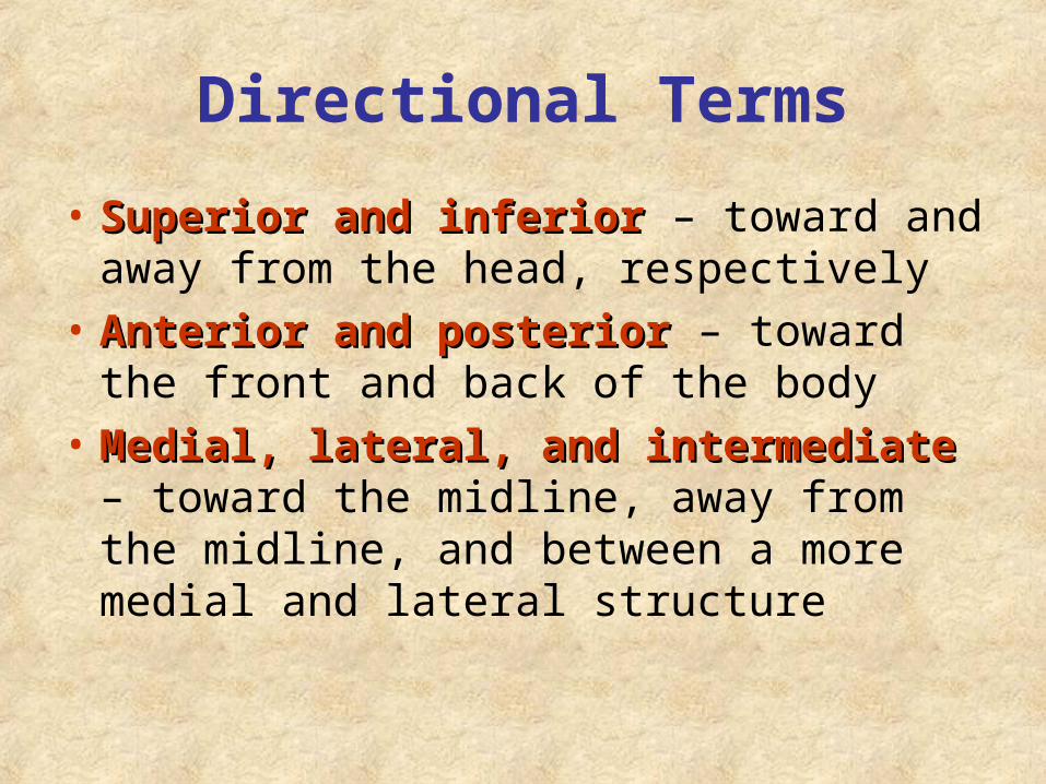
• Superior and inferiorSuperior and inferior – toward and away from the head, respectively
• Anterior and posteriorAnterior and posterior – toward the front and back of the body
• Medial, lateral, and intermediateMedial, lateral, and intermediate – toward the midline, away from the midline, and between a more medial and lateral structure

Directional Terms
• Proximal and distalProximal and distal – closer to and farther from the origin of the body
• Superficial and deepSuperficial and deep – toward and away from the body surface

Directional Terms Table 1.1

Directional Terms Table 1.1

Regional Terms: Anterior View
• AxialAxial – head, neck, and trunk
• AppendicularAppendicular – appendages or limbs
• Specific Specific regional regional terminologyterminology
Figure 1.7a

Regional Terms: Posterior View
Figure 1.7b

Body Planes• SagittalSagittal – divides the body into right and
left parts
• Midsagittal or medialMidsagittal or medial – sagittal plane that lies on the midline
• Frontal or coronalFrontal or coronal – divides the body into anterior and posterior parts
• Transverse or horizontalTransverse or horizontal (cross section) – divides the body into superior and inferior parts
• Oblique sectionOblique section – cuts made diagonally

Body Planes Figure 1.8

Anatomical Variability
• Humans vary slightly in both external Humans vary slightly in both external and internal anatomyand internal anatomy
• Over 90% of all anatomical structures match textbook descriptions, but:
– Nerves or blood vessels may be somewhat out of place
– Small muscles may be missing
• Extreme anatomical variations are Extreme anatomical variations are seldom seenseldom seen

Body Cavities
Figure 1.9a

Body Cavities• Dorsal cavityDorsal cavity protects the nervous system,
and is divided into two subdivisions
– Cranial cavity is within the skull and encases the brain
– Vertebral cavity runs within the vertebral column and encases the spinal cord
• Ventral cavityVentral cavity houses the internal organs (viscera), and is divided into two subdivisions: - Thoracic and Abdominopelvic cavities

Body CavitiesFigure 1.9b

Body Cavities
• Thoracic cavityThoracic cavity is subdivided into pleural cavities, the mediastinum, and the pericardial cavity
– Pleural cavities – each houses a lung
– Mediastinum – contains the pericardial cavity, and surrounds the remaining thoracic organs
– Pericardial cavity – encloses the heart


Body Cavities• The abdominopelvic cavity is separated The abdominopelvic cavity is separated
from the superior thoracic cavity by the from the superior thoracic cavity by the dome-shaped diaphragmdome-shaped diaphragm
• It is composed of two subdivisions
– Abdominal cavity – contains the stomach, intestines, spleen, liver, and other organs
– Pelvic cavity – lies within the pelvis and contains the bladder, reproductive organs, and rectum

Ventral Body Cavity Membranes
• Parietal serosaParietal serosa lines internal body walls
• Visceral serosaVisceral serosa covers the internal organs
• Serous fluid separates the serosae

Ventral Body Cavity Membranes
Figure 1.10a

Ventral Body Cavity Membranes
Figure 1.10b

Other Body Cavities
• Oral and digestiveOral and digestive – mouth and cavities of the digestive organs
• NasalNasal –located within and posterior to the nose
• OrbitalOrbital – house the eyes
• Middle earMiddle ear – contain bones (ossicles) that transmit sound vibrations
• SynovialSynovial – joint cavities

Abdominopelvic Regions
• Umbilical
• Epigastric
• Hypogastric
• Right and left iliac or inguinal
• Right and left lumbar
• Right and left hypochondriac
Figure 1.11a
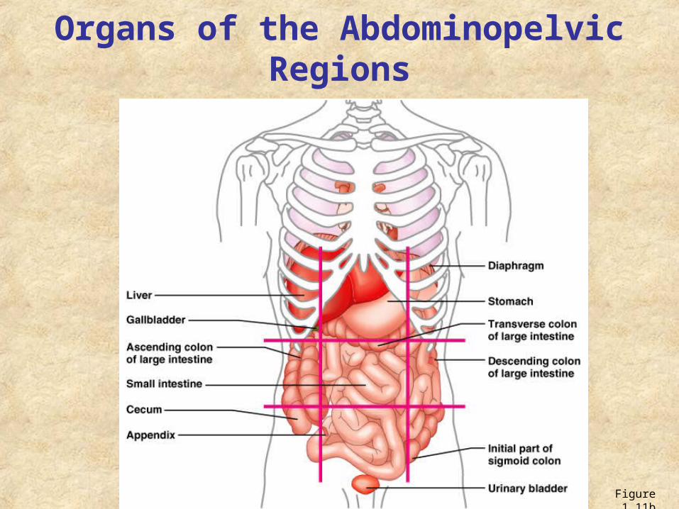
Organs of the Abdominopelvic Regions
Figure 1.11b

Abdominopelvic Quadrants
• Right upper (RUQ)
• Left upper (LUQ)
• Right lower (RLQ)
• Left lower (LLQ)
Figure 1.12