anatomy morphometric analysis of growing tenocytes in the
TRANSCRIPT
1960
Correspondence to Tangkawattana P prasarnkkuacthcopy2017 The Japanese Society of Veterinary Science
This is an open-access article distributed under the terms of the Creative Commons Attribution Non-Commercial No Derivatives (by-nc-nd) License (CC-BY-NC-ND 40 httpscreativecommonsorglicensesby-nc-nd40)
FULL PAPERAnatomy
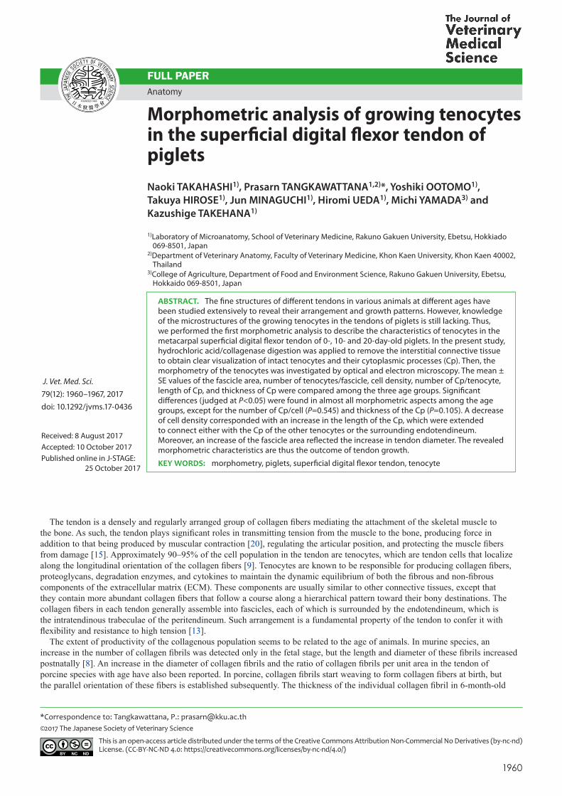
Morphometric analysis of growing tenocytes in the superficial digital flexor tendon of pigletsNaoki TAKAHASHI1) Prasarn TANGKAWATTANA12) Yoshiki OOTOMO1) Takuya HIROSE1) Jun MINAGUCHI1) Hiromi UEDA1) Michi YAMADA3) and Kazushige TAKEHANA1)
1)Laboratory of Microanatomy School of Veterinary Medicine Rakuno Gakuen University Ebetsu Hokkiado 069-8501 Japan
2)Department of Veterinary Anatomy Faculty of Veterinary Medicine Khon Kaen University Khon Kaen 40002 Thailand
3)College of Agriculture Department of Food and Environment Science Rakuno Gakuen University Ebetsu Hokkaido 069-8501 Japan
ABSTRACT The fine structures of different tendons in various animals at different ages have been studied extensively to reveal their arrangement and growth patterns However knowledge of the microstructures of the growing tenocytes in the tendons of piglets is still lacking Thus we performed the first morphometric analysis to describe the characteristics of tenocytes in the metacarpal superficial digital flexor tendon of 0- 10- and 20-day-old piglets In the present study hydrochloric acidcollagenase digestion was applied to remove the interstitial connective tissue to obtain clear visualization of intact tenocytes and their cytoplasmic processes (Cp) Then the morphometry of the tenocytes was investigated by optical and electron microscopy The mean plusmn SE values of the fascicle area number of tenocytesfascicle cell density number of Cptenocyte length of Cp and thickness of Cp were compared among the three age groups Significant differences (judged at Plt005) were found in almost all morphometric aspects among the age groups except for the number of Cpcell (P=0545) and thickness of the Cp (P=0105) A decrease of cell density corresponded with an increase in the length of the Cp which were extended to connect either with the Cp of the other tenocytes or the surrounding endotendineum Moreover an increase of the fascicle area reflected the increase in tendon diameter The revealed morphometric characteristics are thus the outcome of tendon growth
KEY WORDS morphometry piglets superficial digital flexor tendon tenocyte
The tendon is a densely and regularly arranged group of collagen fibers mediating the attachment of the skeletal muscle to the bone As such the tendon plays significant roles in transmitting tension from the muscle to the bone producing force in addition to that being produced by muscular contraction [20] regulating the articular position and protecting the muscle fibers from damage [15] Approximately 90ndash95 of the cell population in the tendon are tenocytes which are tendon cells that localize along the longitudinal orientation of the collagen fibers [9] Tenocytes are known to be responsible for producing collagen fibers proteoglycans degradation enzymes and cytokines to maintain the dynamic equilibrium of both the fibrous and non-fibrous components of the extracellular matrix (ECM) These components are usually similar to other connective tissues except that they contain more abundant collagen fibers that follow a course along a hierarchical pattern toward their bony destinations The collagen fibers in each tendon generally assemble into fascicles each of which is surrounded by the endotendineum which is the intratendinous trabeculae of the peritendineum Such arrangement is a fundamental property of the tendon to confer it with flexibility and resistance to high tension [13]
The extent of productivity of the collagenous population seems to be related to the age of animals In murine species an increase in the number of collagen fibrils was detected only in the fetal stage but the length and diameter of these fibrils increased postnatally [8] An increase in the diameter of collagen fibrils and the ratio of collagen fibrils per unit area in the tendon of porcine species with age have also been reported In porcine collagen fibrils start weaving to form collagen fibers at birth but the parallel orientation of these fibers is established subsequently The thickness of the individual collagen fibril in 6-month-old
Received 8 August 2017Accepted 10 October 2017Published online in J-STAGE 25 October 2017
J Vet Med Sci 79(12) 1960ndash1967 2017doi 101292jvms17-0436
GROWING TENOCYTES IN THE PIGLET SDFT
1961doi 101292jvms17-0436
pigs was reported to be twice (52ndash102 microm) that of newborn piglets This increase of collagenous components would result in a corresponding increase of the total length and cross-sectional area of the tendon Thus we speculated that age might be one of the major factors contributing to the observed differences in tendinous generation Since the growth of the tendon is surely related to the activity of tenocytes investigating the morphometry of the tenocytic population in the tendon of growing animals should yield a better understanding of the growth pattern of tenocytes
It is well established that Cp of tenocytes are extended in all directions to connect with either the collagen bundle of the endotendineum or Cp of the vicinal tenocytes [2 21] Variation and different amounts of collagenous components would affect the morphometry of the tenocytes including the length and thickness of their Cp in the tendons of each animal species [7] The tenocytes in the tendons of the adult rat tail were found to vary in size with a range of 4ndash7 microm in width and 15ndash25 microm in length moreover the Cp were approximately 3 microm or less in length [22] However it is difficult to conduct microscopic observations of tenocytes and their processes in an intact tendon since the interstitial connective tissues can interfere with the visual information Therefore ample elimination of these fibrous components should alleviate the challenges associated with the morphometric investigation of tenocytes A connective tissue digestion technique using hydrochloric acid (HCl) and collagenase has been verified to effectively remove these extracellular components without damaging the cellular components of any chemically fixed tissues [15] These two chemicals were first used for the investigation of collagen fibers and the basement membrane in biopsied samples The technique was then modified for removing extracellular substances to yield better information of the tenocytes [6] Thus far one study has applied this technique to investigating the Achilles tendon of the rat [15] Therefore we sought to conduct the first study using a similar technique to facilitate the morphometric analysis of tenocytes and their Cp in the superficial digital flexor tendon (SDFT) in the forelimb of piglets aged 0 10 and 20 days
MATERIALS AND METHODS
AnimalsFifteen (five 0-day-old five 10-day-old and five 20-day-old) crossbred (Land Race times Large Yorkshire times Duroc) piglets of
the Rakuno Gakuen University farms were used in this study Animal experiments strictly conformed to the Laboratory Animal Guidelines of the Experimental Animal Committee of Rakuno Gakuen University (approval number VH14C4)
Tendon collectionAnesthesia was performed by the intraperitoneal administration of 20 mgkg pentobarbital (Somnopentylreg Kyoritsu
Pharmaceutical Tokyo Japan) The animals were euthanized by exsanguination and then the SDFT coursing behind the metacarpus was collected
Removal of extracellular connective tissueFive 5-mm-thick sections from each tendon were transversely resected with a sharp razor blade The blade was wiped with
absolute ethanol prior to resecting so as to remove any moisture grease dust and rust that might contaminate the tendons The resected samples of each tendon were placed in separate test tubes and fixed with 30 glutaraldehyde with 01 M phosphate-buffered saline (PBS) pH 74 overnight at room temperature The sections were then washed three times consecutively for 10 min each in 01 M PBS with mild shaking The PBS was then replaced by 6 N HCl and digestion was conducted for 15 min in a 60degC water bath with periodical shaking Thereafter three more consecutive 10-min washings with PBS were carried out in the water bath at 60degC for the first two washings and then at 30degC for the third wash The PBS was then replaced by a collagenase solution prepared by dissolving 1 mgml collagenase (Brightase-C Nippi Co Tokyo Japan) in a buffer solution containing 50 mM Tris-HCl 200 mM NaCl and 5 mM CaCl2 The digestion was performed in the 30degC water bath for 12 hr followed by three consecutive 10-min washes with PBS in the 30degC water bath These samples were used for further investigations by optical microscopy and electron microscopy
Scanning electron microscopyThe digested samples were post-fixed for 1 hr with 10 osmium tetroxide followed by three consecutive 10-min PBS washes
Thereafter a series of treatments and washes were performed to obtain conductive dyeing 1 tannic acid for 30 min three consecutive 10-min PBS washes 1 osmium tetroxide for 1 hr and three consecutive 10-min PBS washes Dehydration with an ethanol series was carried out for 30 min at each concentration followed by three consecutive 30-min dehydrations in 100 ethanol The samples were further treated with a mixture of 100 ethanol and t-butyl alcohol (11) for 30 min and then with only t-butyl alcohol for 30 min three times After freezing the samples were freeze-dried in a freeze dryer (JFD-300 JEOL Ltd Tokyo Japan) The samples were ion-coated with platinum using a magnetron sputtering apparatus (JUC-5000 JEOL Ltd) A scanning electron microscope (JSM-5200 JEOL Ltd) was used at an acceleration voltage of 20 kV to confirm the successful removal of the interstitial connective tissue and number of Cp
Optical and transmission electron microscopyThe digested samples were post-fixed with a 10 osmium tetroxide solution dehydrated with an ethanol series and embedded
in Quetol 812 (Nisshin EM Tokyo Japan) An ultramicrotome (Reichert Supernova Leica Microsystem Tokyo Japan) was used for preparing semi-thin (10ndash15 microm) and ultra-thin (80 nm) sections The semi-thin sections were mounted on glass slides stained
N TAKAHASHI ET AL
1962doi 101292jvms17-0436
with toluidine blue and observed with an optical microscope for determination of the number of tenocytesfascicle and the fascicle area The ultra-thin sections were mounted on a 200-mesh copper grid dried and counter-stained with 1 uranyl acetate for 1 min and then with 2 lead citrate for 5 min A transmission electron microscope (JEM-1220 JEOL Ltd) was applied at an accelerating voltage of 80 kV to investigate the length thickness and adhering termini of the Cp
Morphometric analysis of tenocytesImage J analysis software (version 148v National Institutes of Health Bethesda MD USA) was used for the morphometric
analysis of tenocytes observed by optical and electron microscopy On the optical micrographs five fascicles in each semi-thin section were randomly selected for measuring the area of the fascicles (microm2) and to count the number of tenocytes per fascicle Then the cell density (number of tenocyte104 microm2) was analyzed The scanning electron micrographs were used to count the number of Cp per tenocyte Measurement of the length and thickness of Cp was performed on the transmission electron micrographs using only the Cp for which the whole length from the cell body to the terminal adherence was visible The thickness of each Cp was measured at its proximal middle and distal positions
Statistical analysisThe Kruskal-Wallis rank sum test was employed for examining the number of Cp Variation in the other morphometric aspects
among the three age groups was tested by one-way analysis of variance followed by the Tukey post-hoc test for multiple comparisons of means Statistical significance was determined at Plt005 in all cases
RESULTS
Structural organization of the digested tendonsTendons of the 0- 10- and 20-day-old piglets digested with 6 N HCl for 15 min were processed for scanning electron
microscopy Clear observation of the tenocytes Cp and intercellular spaces strongly proved that the digestion with HCl and collagenase could effectively remove the interstitial connective tissue in the tendon of each age group without damaging the tenocytes (Fig 1AndashC) The intercellular spaces between each Cp varied in size In addition the peritendineum the thick connective tissue sheath enclosing the whole tendon still existed in situ (Fig 1B) This sheath sent its intratendinous trabeculae or endotendineum into the tendon (Fig 1AndashF) Each primary endotendineum also gave off secondary and tertiary branches to encircle each group of tenocytes to form fascicles of different sizes The Cp of each tenocyte were found to randomly connect either with the endotendineum or with the Cp of the neighboring cells This pattern was highly consistent with that observed by optical microscopy (Fig 1DndashF)
Tenocyte populationThe digested tendons of all age groups were processed for optical microscopy to analyze the fascicle area number of cells per
fascicle and cell density (Fig 1CndashF) Although the fascicle areas and cell number per fascicle increased the cell density decreased with increasing age (Fig 3 Table 1) Significant differences (Plt005) in each of these three morphometric aspects were found among the three age groups
Cp of tenocytesAfter digestion the tendons of all age groups were processed for electron microscopy In general the tenocytes displayed an
elongated shape and the elongated nucleus usually occupied almost the entire perikaryon Cp of different lengths and thicknesses were found extending from all sides of the cell (Fig 2A and 2B) Two different adhering destinations of Cp were observed either to the collagen fibrils of the vicinal endotendineum (Cp-to-endotendineum pattern Fig 2A) or to the Cp of the adjacent tenocyte (Cp-to-Cp pattern Fig 2B) In the Cp-to-endotendineum pattern numerous finger-like projections were seen emanating from the terminal Cp to adhere to each collagen fibril of the endotendineum (Fig 2A inset) Adherences were detected not only with the superficial fibrils but also with the collagen fibrils residing inside These projections spanned approximately 600ndash700 nm along the longitudinal axis of the collagen fibrils Adherence in the Cp-to-Cp pattern could appear either in a side-to-side (data not shown) or end-to-end fashion (Fig 2B inset) Slight expansion at the terminal ends of both Cp was evident The two Cp adhered to each other by an intercellular junction
The number of Cp per cell varied from 2 to 6 in the 0-day-old group and from 2 to 7 in the other two groups Although the distribution of the number of Cp per cell showed slight variation among groups it did not appear to increase with age (Fig 4) Indeed there was no significant difference in the number of Cp per cell among groups (P=0545 Table 1)
The thickness of each Cp was measured perpendicularly at its proximal middle and distal portions and the average values of the three sites are presented The thickness of the Cp ranged from 1725 to 8093 nm (with the majority found to be in the range of 30ndash70 nm) in the 0-day-old piglets from 3191 to 8381 nm (with the majority being 30ndash60 nm) in the 10-day-old piglets and from 3441 to 9355 nm (with the majority being 30ndash80 nm) in the 20-day-old piglets (Fig 4) Although the distribution of the thickness of Cp was slightly shifted to the right indicating greater thickness with increased age there was no significant difference in the thickness of Cp among groups (P=0105 Table 1)
The length of each Cp was measured from the initial cytoplasmic protrusion at the perikaryon to its end at the adhering terminal This length varied from 165 to 1020 270 to 1535 and 378 to 1939 microm in the 0- 10- and 20-day-old piglets respectively (Fig 4)
GROWING TENOCYTES IN THE PIGLET SDFT
1963doi 101292jvms17-0436
The distribution of Cp length was markedly shifted to the right in the older piglets indicating that the length of Cp increased with increasing age Indeed significant differences in the length of the Cp were found among groups (Plt005 Table 1)
DISCUSSION
The superficial digital flexor muscle of the forelimb in piglets originates from the medial epicondyle at the distal end of the humerus It is composed of two heads of different sizes The SDFT originates from the smaller head that descends along the caudomedial side to the ulna and along the caudal side to the metacarpus At the distal end of the metacarpus the SDFT divides into medial and lateral branches which run to insert onto the palmar surface of the middle phalangeal bones In the present study only the metacarpal portion of the SDFT of piglets was used for morphometric analysis In general the whole tendon is ensheathed by the peritendineum a thick connective tissue sheath that sends its endotendineum or intratendinous trabeculae into the tendon The strands of this primary connective tissue send their secondary and perhaps tertiary branches throughout the tendon in a random fashion to form loops surrounding the group of tenocytes Each loop the so-called fascicles contain numerous tenocytes dense connective tissue and amorphous ground substances The aim of this study was to investigate only the tenocytes thus connective tissue digestion was applied The results of the digestion were similar to those reported for the rabbit calcaneal tendon and rat tail tendon [3 6 7 15] Moreover HCl KOH and NaOH either used alone or in combination with collagenase have also been reported to have the capability to digest the intercellular connective tissue in the tubular organs and ventricle of rats and humans [11 26] Papain was also used for the isolation of collagen and proteoglycans in the human rotator cuff tendon [19] These acid or basic treatments split the triple helical strands to break down the collagen fibrils into short dialyzable peptides which are
Fig 1 Scanning electron (AndashC) and optical (DndashF) microscopy images of the superficial digital flexor tendon of 0- (A D) 10- (B E) and 20-day-old (C F) piglets after removal of the intercellular connective tissue Random ramifications of cytoplasmic processes with intercellular spaces of different sizes were clearly observed Tenocytes (arrowheads) are enclosed in groups of fasciles of different sizes extending from the peritendineum (P) endotendineum (En) and their secondary to tertiary branches The intercellular distances varied among different areas but were generally widened with increasing age [Bar=50 microm (A) 10 microm (B C) 01 mm (DndashF)]
N TAKAHASHI ET AL
1964doi 101292jvms17-0436
then further degraded into hydrolysable fragments by relevant enzymes [14] Collagenase does not affect the tenocytes since the membrane of the cell does not contain collagen Moreover the dense and regular arrangement of the endotendineum and peritendineum should play a protective role against the digestion by collagenase Thus the intact tenocytes and collagenous trabeculae observed in the whole tendon indicated that the samples were well-prepared for the morphometric analysis (Fig 1AndashC)
Comparison between the digested tendons of 0- 10- and 20-day-old piglets using optical microscopy clearly revealed that the tenocyte number per fascicle intercellular spans between each tenocyte and fascicle area were significantly increased with increasing age (Table 1 Fig 1CndashF) In younger animals an increased number of tenocytes and amount of the ECM of the growing tendon truly reflect the active mitotic activity of the tenocytic precursor cells which subsequently transform to tenocytes to produce the ECM in order to cope with the increasing area of each fascicle [5 8 16 25] In the present study the total numbers of tenocytes changed significantly with age but still dispersed within the growing fascicles The increase in the amount of intercellular connective tissue and amorphous ground substances could certainly widen the intercellular space thereby resulting in the observed increase of the fascicle area Although the number of tenocytes shows a marked increase in young animals the rate of increase might not be constant throughout the animalrsquos life Accordingly Lavagnino et al [10] demonstrated a significant decrease in this number in older rats Moreover the non-proportional increase between cell number and fascicle area would result in a diminishing outcome with respect to the cell density (Table 1) Although similar results were reported by Ippolito et al [7] Lavagnino et al [10] Nakagawa et al [17] and Stanley et al [24] different results were demonstrated in other studies including an increase [18 24] and no change in cell density [4 27] The likely sources of these conflicting findings are different quantification methods ages of the animals and treatments in the different studies
Our ultrastructural investigation did not reveal a substantial difference in the morphology of tenocytes and their Cp in the piglet tendons The shape and growth of the tenocyte followed the direction of the running course of the intercellular connective tissue as found in other studies [8 15] Although the number of Cp per tenocyte did not differ significantly among the age groups in the present study Murata et al [15] reported a reduction of the secondary branches of the Cp in the calcaneal tendon of older rats These branches frequently appeared with fragmentations or perforations especially at their ends However similar to Murata et al [15] we observed elongation of the primary Cp of the tenocyte The increase in Cp length appeared to correspond with the increase of fascicle area Since the tenocytes in young animals actively produce the ECM the distance between each cell would likely increase as a result of the increased amount of ECM [8 25] which would in turn increase the fascicle area Thus we suggest that the length of Cp increased to conform to the increased fascicle area because the tenocyte had to increase its Cp length to be able to span across the enlarged space [8] Moreover the increased fibrous and non-fibrous components [8] should be able to yield a certain compressive effect against the thickening process of the lengthened Cp Accordingly there was no significant difference in the thickness of Cp among the tendons of piglets of different ages
Adherence to the terminal end of each Cp appeared in two different patterns Cp-to-Cp and Cp-to-endotendineum The Cp-to-Cp pattern is the adherence of a Cp of one tenocyte to a Cp of the adjacent tenocyte where intercellular communication takes place The gap junction located in this intercellular structure should be necessary for the cellular coordinate for proliferation especially in the growing tendon [1 12 23 24] In the Cp-to-endotendineum pattern the functional tenocyte extends its Cp across the intercellular space to anchor or hold onto the vicinal intratendinous trabeculae From a morphological standpoint the adherence of the Cp-to-endotendineum pattern seems to be more secure than that of the Cp-to-Cp pattern The finger-like projections allow one Cp to adhere to many superficial and deep collagen fibrils In addition the span of the adherence of a Cp covers a larger area of the
Fig 2 Transmission electron micrographs of the superficial digi-tal flexor tendon of 0-day-old piglets demonstrating two different adhering patterns of the cytoplasmic processes (Cp) of tenocytes (Te) the Cp-to-endotendineum (A) and Cp-to-Cp pattern (B) Cp of different lengths extended from all sides of the tenocytes Insets in A and B showed enlargement of the rectangles in their corresponding micrographs At the endotendineum (A inset) the Cp expanded and sent its finger-like projections to adhere with the collagen fibrils Slight expansion was detected at the terminal ends of both Cp in the Cp-to-Cp pattern The intercellular junc-tion was also evident (B inset arrow) [Bar=1 microm (A) 100 nm (A inset) 2 microm (B) and 02 microm (B inset)]
GROWING TENOCYTES IN THE PIGLET SDFT
1965doi 101292jvms17-0436
endotendineum Therefore this adherence should function to conserve the cellular stabilization and integrity of the tendonIn summary an increase in length and diameter of the SDFT in growing piglets likely results from the increase in the tenocyte
number per fascicle and fascicle area The decreased cell density results from the disproportional increase between the tenocyte number and fascicle area The length of the Cp was increased in correspondence with the expanding intercellular space together with the increasing fascicle area However no difference was found in the number of Cp per cell and the thickness of Cp among age groups These morphometric characteristics correlate with the growth of the tendon to some extent Further study on these particular aspects for example using serial block face-scanning electron microscopy [25] would help to gain a deeper understanding of the growing pattern and provide valuable information for three-dimensional reconstruction of the tenocytes intercellular tissues and tendon as a whole
Fig 3 Distribution of the fascicle area number of cells per fascicle and cell density in the superficial digital flexor tendon of 0- 10- and 20-day-old piglets The fascicle area and cell number increased whereas the cell density decreased with age However there was only a slight increase in the number of cells per fascicle
Table 1 Morphometric analyses of the tendons of 0- (0 d) 10- (10 d) and 20-day-old (20 d) piglets
Morphometry (Mean plusmn SE) 0 d 10 d 20 dArea of fascicle (microm2) 24289 plusmn 15842a 40575 plusmn 26436b 98979 plusmn 77529c
Number of cellsfascicle 231 plusmn 162a 337 plusmn 236b 401 plusmn 335c
Cell density (104 microm2) 967 plusmn 242a 816 plusmn 154b 409 plusmn 089c
Number of Cpcell 43 plusmn 012a 42 plusmn 016a 44 plusmn 016a
Thickness of Cp (nm) 481 plusmn 199a 534 plusmn 206a 530 plusmn 175a
Length of Cp (microm) 45 plusmn 018a 66 plusmn 028b 95 plusmn 038c
Different superscripts in the same row indicate statistical significance at Plt005 Cp cytoplasmic processes
N TAKAHASHI ET AL
1966doi 101292jvms17-0436
REFERENCES
1 Becker D L and Mobbs P 1999 Connexin alpha1 and cell proliferation in the developing chick retina Exp Neurol 156 326ndash332 [Medline] [CrossRef]
2 Benjamin M and Ralphs J R 1997 Tendons and ligaments--an overview Histol Histopathol 12 1135ndash1144 [Medline] 3 Coffey J W Fiedler-Nagy C Georgiadis A G and Salvador R A 1976 Digestion of native collagen denatured collagen and collagen
fragments by extracts of rat liver lysosomes J Biol Chem 251 5280ndash5282 [Medline] 4 Couppeacute C Svensson R B Heinemeier K M Thomsen E W Bayer M L Christensen L Kjaeligr M Magnusson S P and Schjerling P
2017 Quantification of cell density in rat Achilles tendon development and application of a new method Histochem Cell Biol 147 97ndash102 [Medline] [CrossRef]
5 Dowling B A Dart A J Hodgson D R and Smith R K 2000 Superficial digital flexor tendonitis in the horse Equine Vet J 32 369ndash378 [Medline] [CrossRef]
6 Evan A P Dail W G Dammrose D and Palmer C 1976 Scanning electron microscopy of cell surfaces following removal of extracellular material Anat Rec 185 433ndash445 [Medline] [CrossRef]
7 Ippolito E Natali P G Postacchini F Accinni L and De Martino C 1980 Morphological immunochemical and biochemical study of rabbit achilles tendon at various ages J Bone Joint Surg Am 62 583ndash598 [Medline] [CrossRef]
8 Kalson N S Lu Y Taylor S H Starborg T Holmes D F and Kadler K E 2015 A structure-based extracellular matrix expansion mechanism of fibrous tissue growth eLife 4 e05958 [Medline] [CrossRef]
9 Kannus P 2000 Structure of the tendon connective tissue Scand J Med Sci Sports 10 312ndash320 [Medline] [CrossRef] 10 Lavagnino M Gardner K and Arnoczky S P 2013 Age-related changes in the cellular mechanical and contractile properties of rat tail tendons
Connect Tissue Res 54 70ndash75 [Medline] [CrossRef] 11 Macchiarelli G and Ohtani O 2001 Endomysium in left ventricle Heart 86 416 [Medline] [CrossRef] 12 Maeda E Ye S Wang W Bader D L Knight M M and Lee D A 2011 Gap junction permeability between tenocytes within tendon fascicles
is suppressed by tensile loading Biomech Model Mechanobiol 11 439ndash447 [Medline] [CrossRef] 13 Magnusson S P Hansen P and Kjaer M 2003 Tendon properties in relation to muscular activity and physical training Scand J Med Sci Sports
13 211ndash223 [Medline] [CrossRef] 14 Mookhtiar K A and Van Wart H E 1992 Clostridium histolyticum collagenases a new look at some old enzymes Matrix Suppl 1 116ndash126
[Medline] 15 Murata H Nishizono H and Miyoshi M 2000 Postnatal development of structure and arrangement of tendon cells A scanning and transmission
electron microscope study in the rat calcaneal tendon Okajimas Folia Anat Jpn 77 77ndash86 [Medline] [CrossRef] 16 Nagy I Z Von Hahn H P and Verzaacuter F 1969 Age-related alterations in the cell nuclei and the DNA content of rat tail tendon Gerontologia 15
Fig 4 Number of cytoplasmic processes (Cp) per cell thickness of Cp and length of Cp in the superficial digital flexor tendon of 0- 10- and 20-day-old piglets
GROWING TENOCYTES IN THE PIGLET SDFT
1967doi 101292jvms17-0436
258ndash264 [Medline] [CrossRef] 17 Nakagawa Y Majima T and Nagashima K 1994 Effect of ageing on ultrastructure of slow and fast skeletal muscle tendon in rabbit Achilles
tendons Acta Physiol Scand 152 307ndash313 [Medline] [CrossRef] 18 Pingel J Lu Y Starborg T Fredberg U Langberg H Nedergaard A Weis M Eyre D Kjaer M and Kadler K E 2014 3-D ultrastructure
and collagen composition of healthy and overloaded human tendon evidence of tenocyte and matrix buckling J Anat 224 548ndash555 [Medline] [CrossRef]
19 Riley G P Harrall R L Constant C R Chard M D Cawston T E and Hazleman B L 1994 Tendon degeneration and chronic shoulder pain changes in the collagen composition of the human rotator cuff tendons in rotator cuff tendinitis Ann Rheum Dis 53 359ndash366 [Medline] [CrossRef]
20 Roberts T J Marsh R L Weyand P G and Taylor C R 1997 Muscular force in running turkeys the economy of minimizing work Science 275 1113ndash1115 [Medline] [CrossRef]
21 Squier C A and Bausch W H 1984 Three-dimensional organization of fibroblasts and collagen fibrils in rat tail tendon Cell Tissue Res 238 319ndash327 [Medline] [CrossRef]
22 Squier C A and Magnes C 1983 Spatial relationships between fibroblasts during the growth of rat-tail tendon Cell Tissue Res 234 17ndash29 [Medline] [CrossRef]
23 Stanley R L Fleck R A Patterson-Kane J C Goodship A E and Ralphs J R 2006 Confocal microscopy and image analysis of connexin plaques in equine tendon Microsc Analys 3 5ndash6
24 Stanley R L Fleck R A Becker D L Goodship A E Ralphs J R and Patterson-Kane J C 2007 Gap junction protein expression and cellularity comparison of immature and adult equine digital tendons J Anat 211 325ndash334 [Medline] [CrossRef]
25 Starborg T Kalson N S Lu Y Mironov A Cootes T F Holmes D F and Kadler K E 2013 Using transmission electron microscopy and 3View to determine collagen fibril size and three-dimensional organization Nat Protoc 8 1433ndash1448 [Medline] [CrossRef]
26 Ushiki T and Murakumo M 1991 Scanning electron microscopic studies of tissue elastin components exposed by a KOH-collagenase or simple KOH digestion method Arch Histol Cytol 54 427ndash436 [Medline] [CrossRef]
27 Wood L K and Brooks S V 2016 Ten weeks of treadmill running decreases stiffness and increases collagen turnover in tendons of old mice J Orthop Res 34 346ndash353 [Medline] [CrossRef]
GROWING TENOCYTES IN THE PIGLET SDFT
1961doi 101292jvms17-0436
pigs was reported to be twice (52ndash102 microm) that of newborn piglets This increase of collagenous components would result in a corresponding increase of the total length and cross-sectional area of the tendon Thus we speculated that age might be one of the major factors contributing to the observed differences in tendinous generation Since the growth of the tendon is surely related to the activity of tenocytes investigating the morphometry of the tenocytic population in the tendon of growing animals should yield a better understanding of the growth pattern of tenocytes
It is well established that Cp of tenocytes are extended in all directions to connect with either the collagen bundle of the endotendineum or Cp of the vicinal tenocytes [2 21] Variation and different amounts of collagenous components would affect the morphometry of the tenocytes including the length and thickness of their Cp in the tendons of each animal species [7] The tenocytes in the tendons of the adult rat tail were found to vary in size with a range of 4ndash7 microm in width and 15ndash25 microm in length moreover the Cp were approximately 3 microm or less in length [22] However it is difficult to conduct microscopic observations of tenocytes and their processes in an intact tendon since the interstitial connective tissues can interfere with the visual information Therefore ample elimination of these fibrous components should alleviate the challenges associated with the morphometric investigation of tenocytes A connective tissue digestion technique using hydrochloric acid (HCl) and collagenase has been verified to effectively remove these extracellular components without damaging the cellular components of any chemically fixed tissues [15] These two chemicals were first used for the investigation of collagen fibers and the basement membrane in biopsied samples The technique was then modified for removing extracellular substances to yield better information of the tenocytes [6] Thus far one study has applied this technique to investigating the Achilles tendon of the rat [15] Therefore we sought to conduct the first study using a similar technique to facilitate the morphometric analysis of tenocytes and their Cp in the superficial digital flexor tendon (SDFT) in the forelimb of piglets aged 0 10 and 20 days
MATERIALS AND METHODS
AnimalsFifteen (five 0-day-old five 10-day-old and five 20-day-old) crossbred (Land Race times Large Yorkshire times Duroc) piglets of
the Rakuno Gakuen University farms were used in this study Animal experiments strictly conformed to the Laboratory Animal Guidelines of the Experimental Animal Committee of Rakuno Gakuen University (approval number VH14C4)
Tendon collectionAnesthesia was performed by the intraperitoneal administration of 20 mgkg pentobarbital (Somnopentylreg Kyoritsu
Pharmaceutical Tokyo Japan) The animals were euthanized by exsanguination and then the SDFT coursing behind the metacarpus was collected
Removal of extracellular connective tissueFive 5-mm-thick sections from each tendon were transversely resected with a sharp razor blade The blade was wiped with
absolute ethanol prior to resecting so as to remove any moisture grease dust and rust that might contaminate the tendons The resected samples of each tendon were placed in separate test tubes and fixed with 30 glutaraldehyde with 01 M phosphate-buffered saline (PBS) pH 74 overnight at room temperature The sections were then washed three times consecutively for 10 min each in 01 M PBS with mild shaking The PBS was then replaced by 6 N HCl and digestion was conducted for 15 min in a 60degC water bath with periodical shaking Thereafter three more consecutive 10-min washings with PBS were carried out in the water bath at 60degC for the first two washings and then at 30degC for the third wash The PBS was then replaced by a collagenase solution prepared by dissolving 1 mgml collagenase (Brightase-C Nippi Co Tokyo Japan) in a buffer solution containing 50 mM Tris-HCl 200 mM NaCl and 5 mM CaCl2 The digestion was performed in the 30degC water bath for 12 hr followed by three consecutive 10-min washes with PBS in the 30degC water bath These samples were used for further investigations by optical microscopy and electron microscopy
Scanning electron microscopyThe digested samples were post-fixed for 1 hr with 10 osmium tetroxide followed by three consecutive 10-min PBS washes
Thereafter a series of treatments and washes were performed to obtain conductive dyeing 1 tannic acid for 30 min three consecutive 10-min PBS washes 1 osmium tetroxide for 1 hr and three consecutive 10-min PBS washes Dehydration with an ethanol series was carried out for 30 min at each concentration followed by three consecutive 30-min dehydrations in 100 ethanol The samples were further treated with a mixture of 100 ethanol and t-butyl alcohol (11) for 30 min and then with only t-butyl alcohol for 30 min three times After freezing the samples were freeze-dried in a freeze dryer (JFD-300 JEOL Ltd Tokyo Japan) The samples were ion-coated with platinum using a magnetron sputtering apparatus (JUC-5000 JEOL Ltd) A scanning electron microscope (JSM-5200 JEOL Ltd) was used at an acceleration voltage of 20 kV to confirm the successful removal of the interstitial connective tissue and number of Cp
Optical and transmission electron microscopyThe digested samples were post-fixed with a 10 osmium tetroxide solution dehydrated with an ethanol series and embedded
in Quetol 812 (Nisshin EM Tokyo Japan) An ultramicrotome (Reichert Supernova Leica Microsystem Tokyo Japan) was used for preparing semi-thin (10ndash15 microm) and ultra-thin (80 nm) sections The semi-thin sections were mounted on glass slides stained
N TAKAHASHI ET AL
1962doi 101292jvms17-0436
with toluidine blue and observed with an optical microscope for determination of the number of tenocytesfascicle and the fascicle area The ultra-thin sections were mounted on a 200-mesh copper grid dried and counter-stained with 1 uranyl acetate for 1 min and then with 2 lead citrate for 5 min A transmission electron microscope (JEM-1220 JEOL Ltd) was applied at an accelerating voltage of 80 kV to investigate the length thickness and adhering termini of the Cp
Morphometric analysis of tenocytesImage J analysis software (version 148v National Institutes of Health Bethesda MD USA) was used for the morphometric
analysis of tenocytes observed by optical and electron microscopy On the optical micrographs five fascicles in each semi-thin section were randomly selected for measuring the area of the fascicles (microm2) and to count the number of tenocytes per fascicle Then the cell density (number of tenocyte104 microm2) was analyzed The scanning electron micrographs were used to count the number of Cp per tenocyte Measurement of the length and thickness of Cp was performed on the transmission electron micrographs using only the Cp for which the whole length from the cell body to the terminal adherence was visible The thickness of each Cp was measured at its proximal middle and distal positions
Statistical analysisThe Kruskal-Wallis rank sum test was employed for examining the number of Cp Variation in the other morphometric aspects
among the three age groups was tested by one-way analysis of variance followed by the Tukey post-hoc test for multiple comparisons of means Statistical significance was determined at Plt005 in all cases
RESULTS
Structural organization of the digested tendonsTendons of the 0- 10- and 20-day-old piglets digested with 6 N HCl for 15 min were processed for scanning electron
microscopy Clear observation of the tenocytes Cp and intercellular spaces strongly proved that the digestion with HCl and collagenase could effectively remove the interstitial connective tissue in the tendon of each age group without damaging the tenocytes (Fig 1AndashC) The intercellular spaces between each Cp varied in size In addition the peritendineum the thick connective tissue sheath enclosing the whole tendon still existed in situ (Fig 1B) This sheath sent its intratendinous trabeculae or endotendineum into the tendon (Fig 1AndashF) Each primary endotendineum also gave off secondary and tertiary branches to encircle each group of tenocytes to form fascicles of different sizes The Cp of each tenocyte were found to randomly connect either with the endotendineum or with the Cp of the neighboring cells This pattern was highly consistent with that observed by optical microscopy (Fig 1DndashF)
Tenocyte populationThe digested tendons of all age groups were processed for optical microscopy to analyze the fascicle area number of cells per
fascicle and cell density (Fig 1CndashF) Although the fascicle areas and cell number per fascicle increased the cell density decreased with increasing age (Fig 3 Table 1) Significant differences (Plt005) in each of these three morphometric aspects were found among the three age groups
Cp of tenocytesAfter digestion the tendons of all age groups were processed for electron microscopy In general the tenocytes displayed an
elongated shape and the elongated nucleus usually occupied almost the entire perikaryon Cp of different lengths and thicknesses were found extending from all sides of the cell (Fig 2A and 2B) Two different adhering destinations of Cp were observed either to the collagen fibrils of the vicinal endotendineum (Cp-to-endotendineum pattern Fig 2A) or to the Cp of the adjacent tenocyte (Cp-to-Cp pattern Fig 2B) In the Cp-to-endotendineum pattern numerous finger-like projections were seen emanating from the terminal Cp to adhere to each collagen fibril of the endotendineum (Fig 2A inset) Adherences were detected not only with the superficial fibrils but also with the collagen fibrils residing inside These projections spanned approximately 600ndash700 nm along the longitudinal axis of the collagen fibrils Adherence in the Cp-to-Cp pattern could appear either in a side-to-side (data not shown) or end-to-end fashion (Fig 2B inset) Slight expansion at the terminal ends of both Cp was evident The two Cp adhered to each other by an intercellular junction
The number of Cp per cell varied from 2 to 6 in the 0-day-old group and from 2 to 7 in the other two groups Although the distribution of the number of Cp per cell showed slight variation among groups it did not appear to increase with age (Fig 4) Indeed there was no significant difference in the number of Cp per cell among groups (P=0545 Table 1)
The thickness of each Cp was measured perpendicularly at its proximal middle and distal portions and the average values of the three sites are presented The thickness of the Cp ranged from 1725 to 8093 nm (with the majority found to be in the range of 30ndash70 nm) in the 0-day-old piglets from 3191 to 8381 nm (with the majority being 30ndash60 nm) in the 10-day-old piglets and from 3441 to 9355 nm (with the majority being 30ndash80 nm) in the 20-day-old piglets (Fig 4) Although the distribution of the thickness of Cp was slightly shifted to the right indicating greater thickness with increased age there was no significant difference in the thickness of Cp among groups (P=0105 Table 1)
The length of each Cp was measured from the initial cytoplasmic protrusion at the perikaryon to its end at the adhering terminal This length varied from 165 to 1020 270 to 1535 and 378 to 1939 microm in the 0- 10- and 20-day-old piglets respectively (Fig 4)
GROWING TENOCYTES IN THE PIGLET SDFT
1963doi 101292jvms17-0436
The distribution of Cp length was markedly shifted to the right in the older piglets indicating that the length of Cp increased with increasing age Indeed significant differences in the length of the Cp were found among groups (Plt005 Table 1)
DISCUSSION
The superficial digital flexor muscle of the forelimb in piglets originates from the medial epicondyle at the distal end of the humerus It is composed of two heads of different sizes The SDFT originates from the smaller head that descends along the caudomedial side to the ulna and along the caudal side to the metacarpus At the distal end of the metacarpus the SDFT divides into medial and lateral branches which run to insert onto the palmar surface of the middle phalangeal bones In the present study only the metacarpal portion of the SDFT of piglets was used for morphometric analysis In general the whole tendon is ensheathed by the peritendineum a thick connective tissue sheath that sends its endotendineum or intratendinous trabeculae into the tendon The strands of this primary connective tissue send their secondary and perhaps tertiary branches throughout the tendon in a random fashion to form loops surrounding the group of tenocytes Each loop the so-called fascicles contain numerous tenocytes dense connective tissue and amorphous ground substances The aim of this study was to investigate only the tenocytes thus connective tissue digestion was applied The results of the digestion were similar to those reported for the rabbit calcaneal tendon and rat tail tendon [3 6 7 15] Moreover HCl KOH and NaOH either used alone or in combination with collagenase have also been reported to have the capability to digest the intercellular connective tissue in the tubular organs and ventricle of rats and humans [11 26] Papain was also used for the isolation of collagen and proteoglycans in the human rotator cuff tendon [19] These acid or basic treatments split the triple helical strands to break down the collagen fibrils into short dialyzable peptides which are
Fig 1 Scanning electron (AndashC) and optical (DndashF) microscopy images of the superficial digital flexor tendon of 0- (A D) 10- (B E) and 20-day-old (C F) piglets after removal of the intercellular connective tissue Random ramifications of cytoplasmic processes with intercellular spaces of different sizes were clearly observed Tenocytes (arrowheads) are enclosed in groups of fasciles of different sizes extending from the peritendineum (P) endotendineum (En) and their secondary to tertiary branches The intercellular distances varied among different areas but were generally widened with increasing age [Bar=50 microm (A) 10 microm (B C) 01 mm (DndashF)]
N TAKAHASHI ET AL
1964doi 101292jvms17-0436
then further degraded into hydrolysable fragments by relevant enzymes [14] Collagenase does not affect the tenocytes since the membrane of the cell does not contain collagen Moreover the dense and regular arrangement of the endotendineum and peritendineum should play a protective role against the digestion by collagenase Thus the intact tenocytes and collagenous trabeculae observed in the whole tendon indicated that the samples were well-prepared for the morphometric analysis (Fig 1AndashC)
Comparison between the digested tendons of 0- 10- and 20-day-old piglets using optical microscopy clearly revealed that the tenocyte number per fascicle intercellular spans between each tenocyte and fascicle area were significantly increased with increasing age (Table 1 Fig 1CndashF) In younger animals an increased number of tenocytes and amount of the ECM of the growing tendon truly reflect the active mitotic activity of the tenocytic precursor cells which subsequently transform to tenocytes to produce the ECM in order to cope with the increasing area of each fascicle [5 8 16 25] In the present study the total numbers of tenocytes changed significantly with age but still dispersed within the growing fascicles The increase in the amount of intercellular connective tissue and amorphous ground substances could certainly widen the intercellular space thereby resulting in the observed increase of the fascicle area Although the number of tenocytes shows a marked increase in young animals the rate of increase might not be constant throughout the animalrsquos life Accordingly Lavagnino et al [10] demonstrated a significant decrease in this number in older rats Moreover the non-proportional increase between cell number and fascicle area would result in a diminishing outcome with respect to the cell density (Table 1) Although similar results were reported by Ippolito et al [7] Lavagnino et al [10] Nakagawa et al [17] and Stanley et al [24] different results were demonstrated in other studies including an increase [18 24] and no change in cell density [4 27] The likely sources of these conflicting findings are different quantification methods ages of the animals and treatments in the different studies
Our ultrastructural investigation did not reveal a substantial difference in the morphology of tenocytes and their Cp in the piglet tendons The shape and growth of the tenocyte followed the direction of the running course of the intercellular connective tissue as found in other studies [8 15] Although the number of Cp per tenocyte did not differ significantly among the age groups in the present study Murata et al [15] reported a reduction of the secondary branches of the Cp in the calcaneal tendon of older rats These branches frequently appeared with fragmentations or perforations especially at their ends However similar to Murata et al [15] we observed elongation of the primary Cp of the tenocyte The increase in Cp length appeared to correspond with the increase of fascicle area Since the tenocytes in young animals actively produce the ECM the distance between each cell would likely increase as a result of the increased amount of ECM [8 25] which would in turn increase the fascicle area Thus we suggest that the length of Cp increased to conform to the increased fascicle area because the tenocyte had to increase its Cp length to be able to span across the enlarged space [8] Moreover the increased fibrous and non-fibrous components [8] should be able to yield a certain compressive effect against the thickening process of the lengthened Cp Accordingly there was no significant difference in the thickness of Cp among the tendons of piglets of different ages
Adherence to the terminal end of each Cp appeared in two different patterns Cp-to-Cp and Cp-to-endotendineum The Cp-to-Cp pattern is the adherence of a Cp of one tenocyte to a Cp of the adjacent tenocyte where intercellular communication takes place The gap junction located in this intercellular structure should be necessary for the cellular coordinate for proliferation especially in the growing tendon [1 12 23 24] In the Cp-to-endotendineum pattern the functional tenocyte extends its Cp across the intercellular space to anchor or hold onto the vicinal intratendinous trabeculae From a morphological standpoint the adherence of the Cp-to-endotendineum pattern seems to be more secure than that of the Cp-to-Cp pattern The finger-like projections allow one Cp to adhere to many superficial and deep collagen fibrils In addition the span of the adherence of a Cp covers a larger area of the
Fig 2 Transmission electron micrographs of the superficial digi-tal flexor tendon of 0-day-old piglets demonstrating two different adhering patterns of the cytoplasmic processes (Cp) of tenocytes (Te) the Cp-to-endotendineum (A) and Cp-to-Cp pattern (B) Cp of different lengths extended from all sides of the tenocytes Insets in A and B showed enlargement of the rectangles in their corresponding micrographs At the endotendineum (A inset) the Cp expanded and sent its finger-like projections to adhere with the collagen fibrils Slight expansion was detected at the terminal ends of both Cp in the Cp-to-Cp pattern The intercellular junc-tion was also evident (B inset arrow) [Bar=1 microm (A) 100 nm (A inset) 2 microm (B) and 02 microm (B inset)]
GROWING TENOCYTES IN THE PIGLET SDFT
1965doi 101292jvms17-0436
endotendineum Therefore this adherence should function to conserve the cellular stabilization and integrity of the tendonIn summary an increase in length and diameter of the SDFT in growing piglets likely results from the increase in the tenocyte
number per fascicle and fascicle area The decreased cell density results from the disproportional increase between the tenocyte number and fascicle area The length of the Cp was increased in correspondence with the expanding intercellular space together with the increasing fascicle area However no difference was found in the number of Cp per cell and the thickness of Cp among age groups These morphometric characteristics correlate with the growth of the tendon to some extent Further study on these particular aspects for example using serial block face-scanning electron microscopy [25] would help to gain a deeper understanding of the growing pattern and provide valuable information for three-dimensional reconstruction of the tenocytes intercellular tissues and tendon as a whole
Fig 3 Distribution of the fascicle area number of cells per fascicle and cell density in the superficial digital flexor tendon of 0- 10- and 20-day-old piglets The fascicle area and cell number increased whereas the cell density decreased with age However there was only a slight increase in the number of cells per fascicle
Table 1 Morphometric analyses of the tendons of 0- (0 d) 10- (10 d) and 20-day-old (20 d) piglets
Morphometry (Mean plusmn SE) 0 d 10 d 20 dArea of fascicle (microm2) 24289 plusmn 15842a 40575 plusmn 26436b 98979 plusmn 77529c
Number of cellsfascicle 231 plusmn 162a 337 plusmn 236b 401 plusmn 335c
Cell density (104 microm2) 967 plusmn 242a 816 plusmn 154b 409 plusmn 089c
Number of Cpcell 43 plusmn 012a 42 plusmn 016a 44 plusmn 016a
Thickness of Cp (nm) 481 plusmn 199a 534 plusmn 206a 530 plusmn 175a
Length of Cp (microm) 45 plusmn 018a 66 plusmn 028b 95 plusmn 038c
Different superscripts in the same row indicate statistical significance at Plt005 Cp cytoplasmic processes
N TAKAHASHI ET AL
1966doi 101292jvms17-0436
REFERENCES
1 Becker D L and Mobbs P 1999 Connexin alpha1 and cell proliferation in the developing chick retina Exp Neurol 156 326ndash332 [Medline] [CrossRef]
2 Benjamin M and Ralphs J R 1997 Tendons and ligaments--an overview Histol Histopathol 12 1135ndash1144 [Medline] 3 Coffey J W Fiedler-Nagy C Georgiadis A G and Salvador R A 1976 Digestion of native collagen denatured collagen and collagen
fragments by extracts of rat liver lysosomes J Biol Chem 251 5280ndash5282 [Medline] 4 Couppeacute C Svensson R B Heinemeier K M Thomsen E W Bayer M L Christensen L Kjaeligr M Magnusson S P and Schjerling P
2017 Quantification of cell density in rat Achilles tendon development and application of a new method Histochem Cell Biol 147 97ndash102 [Medline] [CrossRef]
5 Dowling B A Dart A J Hodgson D R and Smith R K 2000 Superficial digital flexor tendonitis in the horse Equine Vet J 32 369ndash378 [Medline] [CrossRef]
6 Evan A P Dail W G Dammrose D and Palmer C 1976 Scanning electron microscopy of cell surfaces following removal of extracellular material Anat Rec 185 433ndash445 [Medline] [CrossRef]
7 Ippolito E Natali P G Postacchini F Accinni L and De Martino C 1980 Morphological immunochemical and biochemical study of rabbit achilles tendon at various ages J Bone Joint Surg Am 62 583ndash598 [Medline] [CrossRef]
8 Kalson N S Lu Y Taylor S H Starborg T Holmes D F and Kadler K E 2015 A structure-based extracellular matrix expansion mechanism of fibrous tissue growth eLife 4 e05958 [Medline] [CrossRef]
9 Kannus P 2000 Structure of the tendon connective tissue Scand J Med Sci Sports 10 312ndash320 [Medline] [CrossRef] 10 Lavagnino M Gardner K and Arnoczky S P 2013 Age-related changes in the cellular mechanical and contractile properties of rat tail tendons
Connect Tissue Res 54 70ndash75 [Medline] [CrossRef] 11 Macchiarelli G and Ohtani O 2001 Endomysium in left ventricle Heart 86 416 [Medline] [CrossRef] 12 Maeda E Ye S Wang W Bader D L Knight M M and Lee D A 2011 Gap junction permeability between tenocytes within tendon fascicles
is suppressed by tensile loading Biomech Model Mechanobiol 11 439ndash447 [Medline] [CrossRef] 13 Magnusson S P Hansen P and Kjaer M 2003 Tendon properties in relation to muscular activity and physical training Scand J Med Sci Sports
13 211ndash223 [Medline] [CrossRef] 14 Mookhtiar K A and Van Wart H E 1992 Clostridium histolyticum collagenases a new look at some old enzymes Matrix Suppl 1 116ndash126
[Medline] 15 Murata H Nishizono H and Miyoshi M 2000 Postnatal development of structure and arrangement of tendon cells A scanning and transmission
electron microscope study in the rat calcaneal tendon Okajimas Folia Anat Jpn 77 77ndash86 [Medline] [CrossRef] 16 Nagy I Z Von Hahn H P and Verzaacuter F 1969 Age-related alterations in the cell nuclei and the DNA content of rat tail tendon Gerontologia 15
Fig 4 Number of cytoplasmic processes (Cp) per cell thickness of Cp and length of Cp in the superficial digital flexor tendon of 0- 10- and 20-day-old piglets
GROWING TENOCYTES IN THE PIGLET SDFT
1967doi 101292jvms17-0436
258ndash264 [Medline] [CrossRef] 17 Nakagawa Y Majima T and Nagashima K 1994 Effect of ageing on ultrastructure of slow and fast skeletal muscle tendon in rabbit Achilles
tendons Acta Physiol Scand 152 307ndash313 [Medline] [CrossRef] 18 Pingel J Lu Y Starborg T Fredberg U Langberg H Nedergaard A Weis M Eyre D Kjaer M and Kadler K E 2014 3-D ultrastructure
and collagen composition of healthy and overloaded human tendon evidence of tenocyte and matrix buckling J Anat 224 548ndash555 [Medline] [CrossRef]
19 Riley G P Harrall R L Constant C R Chard M D Cawston T E and Hazleman B L 1994 Tendon degeneration and chronic shoulder pain changes in the collagen composition of the human rotator cuff tendons in rotator cuff tendinitis Ann Rheum Dis 53 359ndash366 [Medline] [CrossRef]
20 Roberts T J Marsh R L Weyand P G and Taylor C R 1997 Muscular force in running turkeys the economy of minimizing work Science 275 1113ndash1115 [Medline] [CrossRef]
21 Squier C A and Bausch W H 1984 Three-dimensional organization of fibroblasts and collagen fibrils in rat tail tendon Cell Tissue Res 238 319ndash327 [Medline] [CrossRef]
22 Squier C A and Magnes C 1983 Spatial relationships between fibroblasts during the growth of rat-tail tendon Cell Tissue Res 234 17ndash29 [Medline] [CrossRef]
23 Stanley R L Fleck R A Patterson-Kane J C Goodship A E and Ralphs J R 2006 Confocal microscopy and image analysis of connexin plaques in equine tendon Microsc Analys 3 5ndash6
24 Stanley R L Fleck R A Becker D L Goodship A E Ralphs J R and Patterson-Kane J C 2007 Gap junction protein expression and cellularity comparison of immature and adult equine digital tendons J Anat 211 325ndash334 [Medline] [CrossRef]
25 Starborg T Kalson N S Lu Y Mironov A Cootes T F Holmes D F and Kadler K E 2013 Using transmission electron microscopy and 3View to determine collagen fibril size and three-dimensional organization Nat Protoc 8 1433ndash1448 [Medline] [CrossRef]
26 Ushiki T and Murakumo M 1991 Scanning electron microscopic studies of tissue elastin components exposed by a KOH-collagenase or simple KOH digestion method Arch Histol Cytol 54 427ndash436 [Medline] [CrossRef]
27 Wood L K and Brooks S V 2016 Ten weeks of treadmill running decreases stiffness and increases collagen turnover in tendons of old mice J Orthop Res 34 346ndash353 [Medline] [CrossRef]
N TAKAHASHI ET AL
1962doi 101292jvms17-0436
with toluidine blue and observed with an optical microscope for determination of the number of tenocytesfascicle and the fascicle area The ultra-thin sections were mounted on a 200-mesh copper grid dried and counter-stained with 1 uranyl acetate for 1 min and then with 2 lead citrate for 5 min A transmission electron microscope (JEM-1220 JEOL Ltd) was applied at an accelerating voltage of 80 kV to investigate the length thickness and adhering termini of the Cp
Morphometric analysis of tenocytesImage J analysis software (version 148v National Institutes of Health Bethesda MD USA) was used for the morphometric
analysis of tenocytes observed by optical and electron microscopy On the optical micrographs five fascicles in each semi-thin section were randomly selected for measuring the area of the fascicles (microm2) and to count the number of tenocytes per fascicle Then the cell density (number of tenocyte104 microm2) was analyzed The scanning electron micrographs were used to count the number of Cp per tenocyte Measurement of the length and thickness of Cp was performed on the transmission electron micrographs using only the Cp for which the whole length from the cell body to the terminal adherence was visible The thickness of each Cp was measured at its proximal middle and distal positions
Statistical analysisThe Kruskal-Wallis rank sum test was employed for examining the number of Cp Variation in the other morphometric aspects
among the three age groups was tested by one-way analysis of variance followed by the Tukey post-hoc test for multiple comparisons of means Statistical significance was determined at Plt005 in all cases
RESULTS
Structural organization of the digested tendonsTendons of the 0- 10- and 20-day-old piglets digested with 6 N HCl for 15 min were processed for scanning electron
microscopy Clear observation of the tenocytes Cp and intercellular spaces strongly proved that the digestion with HCl and collagenase could effectively remove the interstitial connective tissue in the tendon of each age group without damaging the tenocytes (Fig 1AndashC) The intercellular spaces between each Cp varied in size In addition the peritendineum the thick connective tissue sheath enclosing the whole tendon still existed in situ (Fig 1B) This sheath sent its intratendinous trabeculae or endotendineum into the tendon (Fig 1AndashF) Each primary endotendineum also gave off secondary and tertiary branches to encircle each group of tenocytes to form fascicles of different sizes The Cp of each tenocyte were found to randomly connect either with the endotendineum or with the Cp of the neighboring cells This pattern was highly consistent with that observed by optical microscopy (Fig 1DndashF)
Tenocyte populationThe digested tendons of all age groups were processed for optical microscopy to analyze the fascicle area number of cells per
fascicle and cell density (Fig 1CndashF) Although the fascicle areas and cell number per fascicle increased the cell density decreased with increasing age (Fig 3 Table 1) Significant differences (Plt005) in each of these three morphometric aspects were found among the three age groups
Cp of tenocytesAfter digestion the tendons of all age groups were processed for electron microscopy In general the tenocytes displayed an
elongated shape and the elongated nucleus usually occupied almost the entire perikaryon Cp of different lengths and thicknesses were found extending from all sides of the cell (Fig 2A and 2B) Two different adhering destinations of Cp were observed either to the collagen fibrils of the vicinal endotendineum (Cp-to-endotendineum pattern Fig 2A) or to the Cp of the adjacent tenocyte (Cp-to-Cp pattern Fig 2B) In the Cp-to-endotendineum pattern numerous finger-like projections were seen emanating from the terminal Cp to adhere to each collagen fibril of the endotendineum (Fig 2A inset) Adherences were detected not only with the superficial fibrils but also with the collagen fibrils residing inside These projections spanned approximately 600ndash700 nm along the longitudinal axis of the collagen fibrils Adherence in the Cp-to-Cp pattern could appear either in a side-to-side (data not shown) or end-to-end fashion (Fig 2B inset) Slight expansion at the terminal ends of both Cp was evident The two Cp adhered to each other by an intercellular junction
The number of Cp per cell varied from 2 to 6 in the 0-day-old group and from 2 to 7 in the other two groups Although the distribution of the number of Cp per cell showed slight variation among groups it did not appear to increase with age (Fig 4) Indeed there was no significant difference in the number of Cp per cell among groups (P=0545 Table 1)
The thickness of each Cp was measured perpendicularly at its proximal middle and distal portions and the average values of the three sites are presented The thickness of the Cp ranged from 1725 to 8093 nm (with the majority found to be in the range of 30ndash70 nm) in the 0-day-old piglets from 3191 to 8381 nm (with the majority being 30ndash60 nm) in the 10-day-old piglets and from 3441 to 9355 nm (with the majority being 30ndash80 nm) in the 20-day-old piglets (Fig 4) Although the distribution of the thickness of Cp was slightly shifted to the right indicating greater thickness with increased age there was no significant difference in the thickness of Cp among groups (P=0105 Table 1)
The length of each Cp was measured from the initial cytoplasmic protrusion at the perikaryon to its end at the adhering terminal This length varied from 165 to 1020 270 to 1535 and 378 to 1939 microm in the 0- 10- and 20-day-old piglets respectively (Fig 4)
GROWING TENOCYTES IN THE PIGLET SDFT
1963doi 101292jvms17-0436
The distribution of Cp length was markedly shifted to the right in the older piglets indicating that the length of Cp increased with increasing age Indeed significant differences in the length of the Cp were found among groups (Plt005 Table 1)
DISCUSSION
The superficial digital flexor muscle of the forelimb in piglets originates from the medial epicondyle at the distal end of the humerus It is composed of two heads of different sizes The SDFT originates from the smaller head that descends along the caudomedial side to the ulna and along the caudal side to the metacarpus At the distal end of the metacarpus the SDFT divides into medial and lateral branches which run to insert onto the palmar surface of the middle phalangeal bones In the present study only the metacarpal portion of the SDFT of piglets was used for morphometric analysis In general the whole tendon is ensheathed by the peritendineum a thick connective tissue sheath that sends its endotendineum or intratendinous trabeculae into the tendon The strands of this primary connective tissue send their secondary and perhaps tertiary branches throughout the tendon in a random fashion to form loops surrounding the group of tenocytes Each loop the so-called fascicles contain numerous tenocytes dense connective tissue and amorphous ground substances The aim of this study was to investigate only the tenocytes thus connective tissue digestion was applied The results of the digestion were similar to those reported for the rabbit calcaneal tendon and rat tail tendon [3 6 7 15] Moreover HCl KOH and NaOH either used alone or in combination with collagenase have also been reported to have the capability to digest the intercellular connective tissue in the tubular organs and ventricle of rats and humans [11 26] Papain was also used for the isolation of collagen and proteoglycans in the human rotator cuff tendon [19] These acid or basic treatments split the triple helical strands to break down the collagen fibrils into short dialyzable peptides which are
Fig 1 Scanning electron (AndashC) and optical (DndashF) microscopy images of the superficial digital flexor tendon of 0- (A D) 10- (B E) and 20-day-old (C F) piglets after removal of the intercellular connective tissue Random ramifications of cytoplasmic processes with intercellular spaces of different sizes were clearly observed Tenocytes (arrowheads) are enclosed in groups of fasciles of different sizes extending from the peritendineum (P) endotendineum (En) and their secondary to tertiary branches The intercellular distances varied among different areas but were generally widened with increasing age [Bar=50 microm (A) 10 microm (B C) 01 mm (DndashF)]
N TAKAHASHI ET AL
1964doi 101292jvms17-0436
then further degraded into hydrolysable fragments by relevant enzymes [14] Collagenase does not affect the tenocytes since the membrane of the cell does not contain collagen Moreover the dense and regular arrangement of the endotendineum and peritendineum should play a protective role against the digestion by collagenase Thus the intact tenocytes and collagenous trabeculae observed in the whole tendon indicated that the samples were well-prepared for the morphometric analysis (Fig 1AndashC)
Comparison between the digested tendons of 0- 10- and 20-day-old piglets using optical microscopy clearly revealed that the tenocyte number per fascicle intercellular spans between each tenocyte and fascicle area were significantly increased with increasing age (Table 1 Fig 1CndashF) In younger animals an increased number of tenocytes and amount of the ECM of the growing tendon truly reflect the active mitotic activity of the tenocytic precursor cells which subsequently transform to tenocytes to produce the ECM in order to cope with the increasing area of each fascicle [5 8 16 25] In the present study the total numbers of tenocytes changed significantly with age but still dispersed within the growing fascicles The increase in the amount of intercellular connective tissue and amorphous ground substances could certainly widen the intercellular space thereby resulting in the observed increase of the fascicle area Although the number of tenocytes shows a marked increase in young animals the rate of increase might not be constant throughout the animalrsquos life Accordingly Lavagnino et al [10] demonstrated a significant decrease in this number in older rats Moreover the non-proportional increase between cell number and fascicle area would result in a diminishing outcome with respect to the cell density (Table 1) Although similar results were reported by Ippolito et al [7] Lavagnino et al [10] Nakagawa et al [17] and Stanley et al [24] different results were demonstrated in other studies including an increase [18 24] and no change in cell density [4 27] The likely sources of these conflicting findings are different quantification methods ages of the animals and treatments in the different studies
Our ultrastructural investigation did not reveal a substantial difference in the morphology of tenocytes and their Cp in the piglet tendons The shape and growth of the tenocyte followed the direction of the running course of the intercellular connective tissue as found in other studies [8 15] Although the number of Cp per tenocyte did not differ significantly among the age groups in the present study Murata et al [15] reported a reduction of the secondary branches of the Cp in the calcaneal tendon of older rats These branches frequently appeared with fragmentations or perforations especially at their ends However similar to Murata et al [15] we observed elongation of the primary Cp of the tenocyte The increase in Cp length appeared to correspond with the increase of fascicle area Since the tenocytes in young animals actively produce the ECM the distance between each cell would likely increase as a result of the increased amount of ECM [8 25] which would in turn increase the fascicle area Thus we suggest that the length of Cp increased to conform to the increased fascicle area because the tenocyte had to increase its Cp length to be able to span across the enlarged space [8] Moreover the increased fibrous and non-fibrous components [8] should be able to yield a certain compressive effect against the thickening process of the lengthened Cp Accordingly there was no significant difference in the thickness of Cp among the tendons of piglets of different ages
Adherence to the terminal end of each Cp appeared in two different patterns Cp-to-Cp and Cp-to-endotendineum The Cp-to-Cp pattern is the adherence of a Cp of one tenocyte to a Cp of the adjacent tenocyte where intercellular communication takes place The gap junction located in this intercellular structure should be necessary for the cellular coordinate for proliferation especially in the growing tendon [1 12 23 24] In the Cp-to-endotendineum pattern the functional tenocyte extends its Cp across the intercellular space to anchor or hold onto the vicinal intratendinous trabeculae From a morphological standpoint the adherence of the Cp-to-endotendineum pattern seems to be more secure than that of the Cp-to-Cp pattern The finger-like projections allow one Cp to adhere to many superficial and deep collagen fibrils In addition the span of the adherence of a Cp covers a larger area of the
Fig 2 Transmission electron micrographs of the superficial digi-tal flexor tendon of 0-day-old piglets demonstrating two different adhering patterns of the cytoplasmic processes (Cp) of tenocytes (Te) the Cp-to-endotendineum (A) and Cp-to-Cp pattern (B) Cp of different lengths extended from all sides of the tenocytes Insets in A and B showed enlargement of the rectangles in their corresponding micrographs At the endotendineum (A inset) the Cp expanded and sent its finger-like projections to adhere with the collagen fibrils Slight expansion was detected at the terminal ends of both Cp in the Cp-to-Cp pattern The intercellular junc-tion was also evident (B inset arrow) [Bar=1 microm (A) 100 nm (A inset) 2 microm (B) and 02 microm (B inset)]
GROWING TENOCYTES IN THE PIGLET SDFT
1965doi 101292jvms17-0436
endotendineum Therefore this adherence should function to conserve the cellular stabilization and integrity of the tendonIn summary an increase in length and diameter of the SDFT in growing piglets likely results from the increase in the tenocyte
number per fascicle and fascicle area The decreased cell density results from the disproportional increase between the tenocyte number and fascicle area The length of the Cp was increased in correspondence with the expanding intercellular space together with the increasing fascicle area However no difference was found in the number of Cp per cell and the thickness of Cp among age groups These morphometric characteristics correlate with the growth of the tendon to some extent Further study on these particular aspects for example using serial block face-scanning electron microscopy [25] would help to gain a deeper understanding of the growing pattern and provide valuable information for three-dimensional reconstruction of the tenocytes intercellular tissues and tendon as a whole
Fig 3 Distribution of the fascicle area number of cells per fascicle and cell density in the superficial digital flexor tendon of 0- 10- and 20-day-old piglets The fascicle area and cell number increased whereas the cell density decreased with age However there was only a slight increase in the number of cells per fascicle
Table 1 Morphometric analyses of the tendons of 0- (0 d) 10- (10 d) and 20-day-old (20 d) piglets
Morphometry (Mean plusmn SE) 0 d 10 d 20 dArea of fascicle (microm2) 24289 plusmn 15842a 40575 plusmn 26436b 98979 plusmn 77529c
Number of cellsfascicle 231 plusmn 162a 337 plusmn 236b 401 plusmn 335c
Cell density (104 microm2) 967 plusmn 242a 816 plusmn 154b 409 plusmn 089c
Number of Cpcell 43 plusmn 012a 42 plusmn 016a 44 plusmn 016a
Thickness of Cp (nm) 481 plusmn 199a 534 plusmn 206a 530 plusmn 175a
Length of Cp (microm) 45 plusmn 018a 66 plusmn 028b 95 plusmn 038c
Different superscripts in the same row indicate statistical significance at Plt005 Cp cytoplasmic processes
N TAKAHASHI ET AL
1966doi 101292jvms17-0436
REFERENCES
1 Becker D L and Mobbs P 1999 Connexin alpha1 and cell proliferation in the developing chick retina Exp Neurol 156 326ndash332 [Medline] [CrossRef]
2 Benjamin M and Ralphs J R 1997 Tendons and ligaments--an overview Histol Histopathol 12 1135ndash1144 [Medline] 3 Coffey J W Fiedler-Nagy C Georgiadis A G and Salvador R A 1976 Digestion of native collagen denatured collagen and collagen
fragments by extracts of rat liver lysosomes J Biol Chem 251 5280ndash5282 [Medline] 4 Couppeacute C Svensson R B Heinemeier K M Thomsen E W Bayer M L Christensen L Kjaeligr M Magnusson S P and Schjerling P
2017 Quantification of cell density in rat Achilles tendon development and application of a new method Histochem Cell Biol 147 97ndash102 [Medline] [CrossRef]
5 Dowling B A Dart A J Hodgson D R and Smith R K 2000 Superficial digital flexor tendonitis in the horse Equine Vet J 32 369ndash378 [Medline] [CrossRef]
6 Evan A P Dail W G Dammrose D and Palmer C 1976 Scanning electron microscopy of cell surfaces following removal of extracellular material Anat Rec 185 433ndash445 [Medline] [CrossRef]
7 Ippolito E Natali P G Postacchini F Accinni L and De Martino C 1980 Morphological immunochemical and biochemical study of rabbit achilles tendon at various ages J Bone Joint Surg Am 62 583ndash598 [Medline] [CrossRef]
8 Kalson N S Lu Y Taylor S H Starborg T Holmes D F and Kadler K E 2015 A structure-based extracellular matrix expansion mechanism of fibrous tissue growth eLife 4 e05958 [Medline] [CrossRef]
9 Kannus P 2000 Structure of the tendon connective tissue Scand J Med Sci Sports 10 312ndash320 [Medline] [CrossRef] 10 Lavagnino M Gardner K and Arnoczky S P 2013 Age-related changes in the cellular mechanical and contractile properties of rat tail tendons
Connect Tissue Res 54 70ndash75 [Medline] [CrossRef] 11 Macchiarelli G and Ohtani O 2001 Endomysium in left ventricle Heart 86 416 [Medline] [CrossRef] 12 Maeda E Ye S Wang W Bader D L Knight M M and Lee D A 2011 Gap junction permeability between tenocytes within tendon fascicles
is suppressed by tensile loading Biomech Model Mechanobiol 11 439ndash447 [Medline] [CrossRef] 13 Magnusson S P Hansen P and Kjaer M 2003 Tendon properties in relation to muscular activity and physical training Scand J Med Sci Sports
13 211ndash223 [Medline] [CrossRef] 14 Mookhtiar K A and Van Wart H E 1992 Clostridium histolyticum collagenases a new look at some old enzymes Matrix Suppl 1 116ndash126
[Medline] 15 Murata H Nishizono H and Miyoshi M 2000 Postnatal development of structure and arrangement of tendon cells A scanning and transmission
electron microscope study in the rat calcaneal tendon Okajimas Folia Anat Jpn 77 77ndash86 [Medline] [CrossRef] 16 Nagy I Z Von Hahn H P and Verzaacuter F 1969 Age-related alterations in the cell nuclei and the DNA content of rat tail tendon Gerontologia 15
Fig 4 Number of cytoplasmic processes (Cp) per cell thickness of Cp and length of Cp in the superficial digital flexor tendon of 0- 10- and 20-day-old piglets
GROWING TENOCYTES IN THE PIGLET SDFT
1967doi 101292jvms17-0436
258ndash264 [Medline] [CrossRef] 17 Nakagawa Y Majima T and Nagashima K 1994 Effect of ageing on ultrastructure of slow and fast skeletal muscle tendon in rabbit Achilles
tendons Acta Physiol Scand 152 307ndash313 [Medline] [CrossRef] 18 Pingel J Lu Y Starborg T Fredberg U Langberg H Nedergaard A Weis M Eyre D Kjaer M and Kadler K E 2014 3-D ultrastructure
and collagen composition of healthy and overloaded human tendon evidence of tenocyte and matrix buckling J Anat 224 548ndash555 [Medline] [CrossRef]
19 Riley G P Harrall R L Constant C R Chard M D Cawston T E and Hazleman B L 1994 Tendon degeneration and chronic shoulder pain changes in the collagen composition of the human rotator cuff tendons in rotator cuff tendinitis Ann Rheum Dis 53 359ndash366 [Medline] [CrossRef]
20 Roberts T J Marsh R L Weyand P G and Taylor C R 1997 Muscular force in running turkeys the economy of minimizing work Science 275 1113ndash1115 [Medline] [CrossRef]
21 Squier C A and Bausch W H 1984 Three-dimensional organization of fibroblasts and collagen fibrils in rat tail tendon Cell Tissue Res 238 319ndash327 [Medline] [CrossRef]
22 Squier C A and Magnes C 1983 Spatial relationships between fibroblasts during the growth of rat-tail tendon Cell Tissue Res 234 17ndash29 [Medline] [CrossRef]
23 Stanley R L Fleck R A Patterson-Kane J C Goodship A E and Ralphs J R 2006 Confocal microscopy and image analysis of connexin plaques in equine tendon Microsc Analys 3 5ndash6
24 Stanley R L Fleck R A Becker D L Goodship A E Ralphs J R and Patterson-Kane J C 2007 Gap junction protein expression and cellularity comparison of immature and adult equine digital tendons J Anat 211 325ndash334 [Medline] [CrossRef]
25 Starborg T Kalson N S Lu Y Mironov A Cootes T F Holmes D F and Kadler K E 2013 Using transmission electron microscopy and 3View to determine collagen fibril size and three-dimensional organization Nat Protoc 8 1433ndash1448 [Medline] [CrossRef]
26 Ushiki T and Murakumo M 1991 Scanning electron microscopic studies of tissue elastin components exposed by a KOH-collagenase or simple KOH digestion method Arch Histol Cytol 54 427ndash436 [Medline] [CrossRef]
27 Wood L K and Brooks S V 2016 Ten weeks of treadmill running decreases stiffness and increases collagen turnover in tendons of old mice J Orthop Res 34 346ndash353 [Medline] [CrossRef]
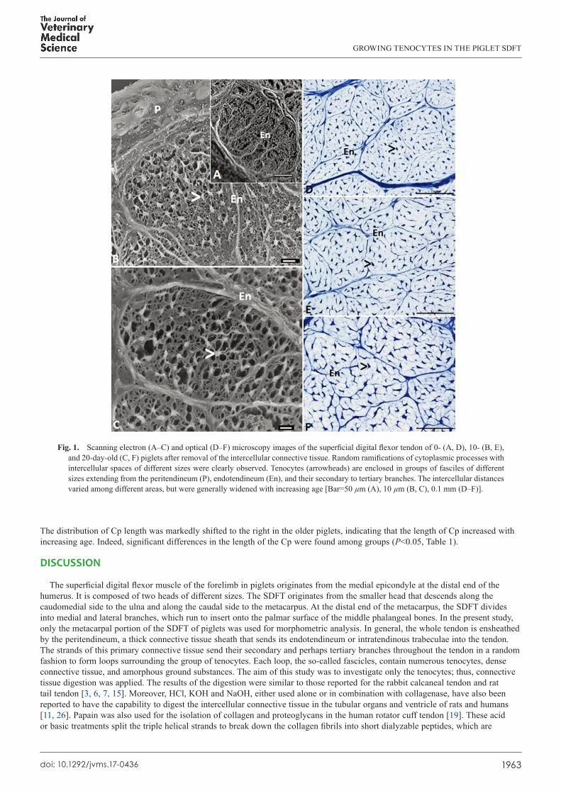
GROWING TENOCYTES IN THE PIGLET SDFT
1963doi 101292jvms17-0436
The distribution of Cp length was markedly shifted to the right in the older piglets indicating that the length of Cp increased with increasing age Indeed significant differences in the length of the Cp were found among groups (Plt005 Table 1)
DISCUSSION
The superficial digital flexor muscle of the forelimb in piglets originates from the medial epicondyle at the distal end of the humerus It is composed of two heads of different sizes The SDFT originates from the smaller head that descends along the caudomedial side to the ulna and along the caudal side to the metacarpus At the distal end of the metacarpus the SDFT divides into medial and lateral branches which run to insert onto the palmar surface of the middle phalangeal bones In the present study only the metacarpal portion of the SDFT of piglets was used for morphometric analysis In general the whole tendon is ensheathed by the peritendineum a thick connective tissue sheath that sends its endotendineum or intratendinous trabeculae into the tendon The strands of this primary connective tissue send their secondary and perhaps tertiary branches throughout the tendon in a random fashion to form loops surrounding the group of tenocytes Each loop the so-called fascicles contain numerous tenocytes dense connective tissue and amorphous ground substances The aim of this study was to investigate only the tenocytes thus connective tissue digestion was applied The results of the digestion were similar to those reported for the rabbit calcaneal tendon and rat tail tendon [3 6 7 15] Moreover HCl KOH and NaOH either used alone or in combination with collagenase have also been reported to have the capability to digest the intercellular connective tissue in the tubular organs and ventricle of rats and humans [11 26] Papain was also used for the isolation of collagen and proteoglycans in the human rotator cuff tendon [19] These acid or basic treatments split the triple helical strands to break down the collagen fibrils into short dialyzable peptides which are
Fig 1 Scanning electron (AndashC) and optical (DndashF) microscopy images of the superficial digital flexor tendon of 0- (A D) 10- (B E) and 20-day-old (C F) piglets after removal of the intercellular connective tissue Random ramifications of cytoplasmic processes with intercellular spaces of different sizes were clearly observed Tenocytes (arrowheads) are enclosed in groups of fasciles of different sizes extending from the peritendineum (P) endotendineum (En) and their secondary to tertiary branches The intercellular distances varied among different areas but were generally widened with increasing age [Bar=50 microm (A) 10 microm (B C) 01 mm (DndashF)]
N TAKAHASHI ET AL
1964doi 101292jvms17-0436
then further degraded into hydrolysable fragments by relevant enzymes [14] Collagenase does not affect the tenocytes since the membrane of the cell does not contain collagen Moreover the dense and regular arrangement of the endotendineum and peritendineum should play a protective role against the digestion by collagenase Thus the intact tenocytes and collagenous trabeculae observed in the whole tendon indicated that the samples were well-prepared for the morphometric analysis (Fig 1AndashC)
Comparison between the digested tendons of 0- 10- and 20-day-old piglets using optical microscopy clearly revealed that the tenocyte number per fascicle intercellular spans between each tenocyte and fascicle area were significantly increased with increasing age (Table 1 Fig 1CndashF) In younger animals an increased number of tenocytes and amount of the ECM of the growing tendon truly reflect the active mitotic activity of the tenocytic precursor cells which subsequently transform to tenocytes to produce the ECM in order to cope with the increasing area of each fascicle [5 8 16 25] In the present study the total numbers of tenocytes changed significantly with age but still dispersed within the growing fascicles The increase in the amount of intercellular connective tissue and amorphous ground substances could certainly widen the intercellular space thereby resulting in the observed increase of the fascicle area Although the number of tenocytes shows a marked increase in young animals the rate of increase might not be constant throughout the animalrsquos life Accordingly Lavagnino et al [10] demonstrated a significant decrease in this number in older rats Moreover the non-proportional increase between cell number and fascicle area would result in a diminishing outcome with respect to the cell density (Table 1) Although similar results were reported by Ippolito et al [7] Lavagnino et al [10] Nakagawa et al [17] and Stanley et al [24] different results were demonstrated in other studies including an increase [18 24] and no change in cell density [4 27] The likely sources of these conflicting findings are different quantification methods ages of the animals and treatments in the different studies
Our ultrastructural investigation did not reveal a substantial difference in the morphology of tenocytes and their Cp in the piglet tendons The shape and growth of the tenocyte followed the direction of the running course of the intercellular connective tissue as found in other studies [8 15] Although the number of Cp per tenocyte did not differ significantly among the age groups in the present study Murata et al [15] reported a reduction of the secondary branches of the Cp in the calcaneal tendon of older rats These branches frequently appeared with fragmentations or perforations especially at their ends However similar to Murata et al [15] we observed elongation of the primary Cp of the tenocyte The increase in Cp length appeared to correspond with the increase of fascicle area Since the tenocytes in young animals actively produce the ECM the distance between each cell would likely increase as a result of the increased amount of ECM [8 25] which would in turn increase the fascicle area Thus we suggest that the length of Cp increased to conform to the increased fascicle area because the tenocyte had to increase its Cp length to be able to span across the enlarged space [8] Moreover the increased fibrous and non-fibrous components [8] should be able to yield a certain compressive effect against the thickening process of the lengthened Cp Accordingly there was no significant difference in the thickness of Cp among the tendons of piglets of different ages
Adherence to the terminal end of each Cp appeared in two different patterns Cp-to-Cp and Cp-to-endotendineum The Cp-to-Cp pattern is the adherence of a Cp of one tenocyte to a Cp of the adjacent tenocyte where intercellular communication takes place The gap junction located in this intercellular structure should be necessary for the cellular coordinate for proliferation especially in the growing tendon [1 12 23 24] In the Cp-to-endotendineum pattern the functional tenocyte extends its Cp across the intercellular space to anchor or hold onto the vicinal intratendinous trabeculae From a morphological standpoint the adherence of the Cp-to-endotendineum pattern seems to be more secure than that of the Cp-to-Cp pattern The finger-like projections allow one Cp to adhere to many superficial and deep collagen fibrils In addition the span of the adherence of a Cp covers a larger area of the
Fig 2 Transmission electron micrographs of the superficial digi-tal flexor tendon of 0-day-old piglets demonstrating two different adhering patterns of the cytoplasmic processes (Cp) of tenocytes (Te) the Cp-to-endotendineum (A) and Cp-to-Cp pattern (B) Cp of different lengths extended from all sides of the tenocytes Insets in A and B showed enlargement of the rectangles in their corresponding micrographs At the endotendineum (A inset) the Cp expanded and sent its finger-like projections to adhere with the collagen fibrils Slight expansion was detected at the terminal ends of both Cp in the Cp-to-Cp pattern The intercellular junc-tion was also evident (B inset arrow) [Bar=1 microm (A) 100 nm (A inset) 2 microm (B) and 02 microm (B inset)]
GROWING TENOCYTES IN THE PIGLET SDFT
1965doi 101292jvms17-0436
endotendineum Therefore this adherence should function to conserve the cellular stabilization and integrity of the tendonIn summary an increase in length and diameter of the SDFT in growing piglets likely results from the increase in the tenocyte
number per fascicle and fascicle area The decreased cell density results from the disproportional increase between the tenocyte number and fascicle area The length of the Cp was increased in correspondence with the expanding intercellular space together with the increasing fascicle area However no difference was found in the number of Cp per cell and the thickness of Cp among age groups These morphometric characteristics correlate with the growth of the tendon to some extent Further study on these particular aspects for example using serial block face-scanning electron microscopy [25] would help to gain a deeper understanding of the growing pattern and provide valuable information for three-dimensional reconstruction of the tenocytes intercellular tissues and tendon as a whole
Fig 3 Distribution of the fascicle area number of cells per fascicle and cell density in the superficial digital flexor tendon of 0- 10- and 20-day-old piglets The fascicle area and cell number increased whereas the cell density decreased with age However there was only a slight increase in the number of cells per fascicle
Table 1 Morphometric analyses of the tendons of 0- (0 d) 10- (10 d) and 20-day-old (20 d) piglets
Morphometry (Mean plusmn SE) 0 d 10 d 20 dArea of fascicle (microm2) 24289 plusmn 15842a 40575 plusmn 26436b 98979 plusmn 77529c
Number of cellsfascicle 231 plusmn 162a 337 plusmn 236b 401 plusmn 335c
Cell density (104 microm2) 967 plusmn 242a 816 plusmn 154b 409 plusmn 089c
Number of Cpcell 43 plusmn 012a 42 plusmn 016a 44 plusmn 016a
Thickness of Cp (nm) 481 plusmn 199a 534 plusmn 206a 530 plusmn 175a
Length of Cp (microm) 45 plusmn 018a 66 plusmn 028b 95 plusmn 038c
Different superscripts in the same row indicate statistical significance at Plt005 Cp cytoplasmic processes
N TAKAHASHI ET AL
1966doi 101292jvms17-0436
REFERENCES
1 Becker D L and Mobbs P 1999 Connexin alpha1 and cell proliferation in the developing chick retina Exp Neurol 156 326ndash332 [Medline] [CrossRef]
2 Benjamin M and Ralphs J R 1997 Tendons and ligaments--an overview Histol Histopathol 12 1135ndash1144 [Medline] 3 Coffey J W Fiedler-Nagy C Georgiadis A G and Salvador R A 1976 Digestion of native collagen denatured collagen and collagen
fragments by extracts of rat liver lysosomes J Biol Chem 251 5280ndash5282 [Medline] 4 Couppeacute C Svensson R B Heinemeier K M Thomsen E W Bayer M L Christensen L Kjaeligr M Magnusson S P and Schjerling P
2017 Quantification of cell density in rat Achilles tendon development and application of a new method Histochem Cell Biol 147 97ndash102 [Medline] [CrossRef]
5 Dowling B A Dart A J Hodgson D R and Smith R K 2000 Superficial digital flexor tendonitis in the horse Equine Vet J 32 369ndash378 [Medline] [CrossRef]
6 Evan A P Dail W G Dammrose D and Palmer C 1976 Scanning electron microscopy of cell surfaces following removal of extracellular material Anat Rec 185 433ndash445 [Medline] [CrossRef]
7 Ippolito E Natali P G Postacchini F Accinni L and De Martino C 1980 Morphological immunochemical and biochemical study of rabbit achilles tendon at various ages J Bone Joint Surg Am 62 583ndash598 [Medline] [CrossRef]
8 Kalson N S Lu Y Taylor S H Starborg T Holmes D F and Kadler K E 2015 A structure-based extracellular matrix expansion mechanism of fibrous tissue growth eLife 4 e05958 [Medline] [CrossRef]
9 Kannus P 2000 Structure of the tendon connective tissue Scand J Med Sci Sports 10 312ndash320 [Medline] [CrossRef] 10 Lavagnino M Gardner K and Arnoczky S P 2013 Age-related changes in the cellular mechanical and contractile properties of rat tail tendons
Connect Tissue Res 54 70ndash75 [Medline] [CrossRef] 11 Macchiarelli G and Ohtani O 2001 Endomysium in left ventricle Heart 86 416 [Medline] [CrossRef] 12 Maeda E Ye S Wang W Bader D L Knight M M and Lee D A 2011 Gap junction permeability between tenocytes within tendon fascicles
is suppressed by tensile loading Biomech Model Mechanobiol 11 439ndash447 [Medline] [CrossRef] 13 Magnusson S P Hansen P and Kjaer M 2003 Tendon properties in relation to muscular activity and physical training Scand J Med Sci Sports
13 211ndash223 [Medline] [CrossRef] 14 Mookhtiar K A and Van Wart H E 1992 Clostridium histolyticum collagenases a new look at some old enzymes Matrix Suppl 1 116ndash126
[Medline] 15 Murata H Nishizono H and Miyoshi M 2000 Postnatal development of structure and arrangement of tendon cells A scanning and transmission
electron microscope study in the rat calcaneal tendon Okajimas Folia Anat Jpn 77 77ndash86 [Medline] [CrossRef] 16 Nagy I Z Von Hahn H P and Verzaacuter F 1969 Age-related alterations in the cell nuclei and the DNA content of rat tail tendon Gerontologia 15
Fig 4 Number of cytoplasmic processes (Cp) per cell thickness of Cp and length of Cp in the superficial digital flexor tendon of 0- 10- and 20-day-old piglets
GROWING TENOCYTES IN THE PIGLET SDFT
1967doi 101292jvms17-0436
258ndash264 [Medline] [CrossRef] 17 Nakagawa Y Majima T and Nagashima K 1994 Effect of ageing on ultrastructure of slow and fast skeletal muscle tendon in rabbit Achilles
tendons Acta Physiol Scand 152 307ndash313 [Medline] [CrossRef] 18 Pingel J Lu Y Starborg T Fredberg U Langberg H Nedergaard A Weis M Eyre D Kjaer M and Kadler K E 2014 3-D ultrastructure
and collagen composition of healthy and overloaded human tendon evidence of tenocyte and matrix buckling J Anat 224 548ndash555 [Medline] [CrossRef]
19 Riley G P Harrall R L Constant C R Chard M D Cawston T E and Hazleman B L 1994 Tendon degeneration and chronic shoulder pain changes in the collagen composition of the human rotator cuff tendons in rotator cuff tendinitis Ann Rheum Dis 53 359ndash366 [Medline] [CrossRef]
20 Roberts T J Marsh R L Weyand P G and Taylor C R 1997 Muscular force in running turkeys the economy of minimizing work Science 275 1113ndash1115 [Medline] [CrossRef]
21 Squier C A and Bausch W H 1984 Three-dimensional organization of fibroblasts and collagen fibrils in rat tail tendon Cell Tissue Res 238 319ndash327 [Medline] [CrossRef]
22 Squier C A and Magnes C 1983 Spatial relationships between fibroblasts during the growth of rat-tail tendon Cell Tissue Res 234 17ndash29 [Medline] [CrossRef]
23 Stanley R L Fleck R A Patterson-Kane J C Goodship A E and Ralphs J R 2006 Confocal microscopy and image analysis of connexin plaques in equine tendon Microsc Analys 3 5ndash6
24 Stanley R L Fleck R A Becker D L Goodship A E Ralphs J R and Patterson-Kane J C 2007 Gap junction protein expression and cellularity comparison of immature and adult equine digital tendons J Anat 211 325ndash334 [Medline] [CrossRef]
25 Starborg T Kalson N S Lu Y Mironov A Cootes T F Holmes D F and Kadler K E 2013 Using transmission electron microscopy and 3View to determine collagen fibril size and three-dimensional organization Nat Protoc 8 1433ndash1448 [Medline] [CrossRef]
26 Ushiki T and Murakumo M 1991 Scanning electron microscopic studies of tissue elastin components exposed by a KOH-collagenase or simple KOH digestion method Arch Histol Cytol 54 427ndash436 [Medline] [CrossRef]
27 Wood L K and Brooks S V 2016 Ten weeks of treadmill running decreases stiffness and increases collagen turnover in tendons of old mice J Orthop Res 34 346ndash353 [Medline] [CrossRef]

N TAKAHASHI ET AL
1964doi 101292jvms17-0436
then further degraded into hydrolysable fragments by relevant enzymes [14] Collagenase does not affect the tenocytes since the membrane of the cell does not contain collagen Moreover the dense and regular arrangement of the endotendineum and peritendineum should play a protective role against the digestion by collagenase Thus the intact tenocytes and collagenous trabeculae observed in the whole tendon indicated that the samples were well-prepared for the morphometric analysis (Fig 1AndashC)
Comparison between the digested tendons of 0- 10- and 20-day-old piglets using optical microscopy clearly revealed that the tenocyte number per fascicle intercellular spans between each tenocyte and fascicle area were significantly increased with increasing age (Table 1 Fig 1CndashF) In younger animals an increased number of tenocytes and amount of the ECM of the growing tendon truly reflect the active mitotic activity of the tenocytic precursor cells which subsequently transform to tenocytes to produce the ECM in order to cope with the increasing area of each fascicle [5 8 16 25] In the present study the total numbers of tenocytes changed significantly with age but still dispersed within the growing fascicles The increase in the amount of intercellular connective tissue and amorphous ground substances could certainly widen the intercellular space thereby resulting in the observed increase of the fascicle area Although the number of tenocytes shows a marked increase in young animals the rate of increase might not be constant throughout the animalrsquos life Accordingly Lavagnino et al [10] demonstrated a significant decrease in this number in older rats Moreover the non-proportional increase between cell number and fascicle area would result in a diminishing outcome with respect to the cell density (Table 1) Although similar results were reported by Ippolito et al [7] Lavagnino et al [10] Nakagawa et al [17] and Stanley et al [24] different results were demonstrated in other studies including an increase [18 24] and no change in cell density [4 27] The likely sources of these conflicting findings are different quantification methods ages of the animals and treatments in the different studies
Our ultrastructural investigation did not reveal a substantial difference in the morphology of tenocytes and their Cp in the piglet tendons The shape and growth of the tenocyte followed the direction of the running course of the intercellular connective tissue as found in other studies [8 15] Although the number of Cp per tenocyte did not differ significantly among the age groups in the present study Murata et al [15] reported a reduction of the secondary branches of the Cp in the calcaneal tendon of older rats These branches frequently appeared with fragmentations or perforations especially at their ends However similar to Murata et al [15] we observed elongation of the primary Cp of the tenocyte The increase in Cp length appeared to correspond with the increase of fascicle area Since the tenocytes in young animals actively produce the ECM the distance between each cell would likely increase as a result of the increased amount of ECM [8 25] which would in turn increase the fascicle area Thus we suggest that the length of Cp increased to conform to the increased fascicle area because the tenocyte had to increase its Cp length to be able to span across the enlarged space [8] Moreover the increased fibrous and non-fibrous components [8] should be able to yield a certain compressive effect against the thickening process of the lengthened Cp Accordingly there was no significant difference in the thickness of Cp among the tendons of piglets of different ages
Adherence to the terminal end of each Cp appeared in two different patterns Cp-to-Cp and Cp-to-endotendineum The Cp-to-Cp pattern is the adherence of a Cp of one tenocyte to a Cp of the adjacent tenocyte where intercellular communication takes place The gap junction located in this intercellular structure should be necessary for the cellular coordinate for proliferation especially in the growing tendon [1 12 23 24] In the Cp-to-endotendineum pattern the functional tenocyte extends its Cp across the intercellular space to anchor or hold onto the vicinal intratendinous trabeculae From a morphological standpoint the adherence of the Cp-to-endotendineum pattern seems to be more secure than that of the Cp-to-Cp pattern The finger-like projections allow one Cp to adhere to many superficial and deep collagen fibrils In addition the span of the adherence of a Cp covers a larger area of the
Fig 2 Transmission electron micrographs of the superficial digi-tal flexor tendon of 0-day-old piglets demonstrating two different adhering patterns of the cytoplasmic processes (Cp) of tenocytes (Te) the Cp-to-endotendineum (A) and Cp-to-Cp pattern (B) Cp of different lengths extended from all sides of the tenocytes Insets in A and B showed enlargement of the rectangles in their corresponding micrographs At the endotendineum (A inset) the Cp expanded and sent its finger-like projections to adhere with the collagen fibrils Slight expansion was detected at the terminal ends of both Cp in the Cp-to-Cp pattern The intercellular junc-tion was also evident (B inset arrow) [Bar=1 microm (A) 100 nm (A inset) 2 microm (B) and 02 microm (B inset)]
GROWING TENOCYTES IN THE PIGLET SDFT
1965doi 101292jvms17-0436
endotendineum Therefore this adherence should function to conserve the cellular stabilization and integrity of the tendonIn summary an increase in length and diameter of the SDFT in growing piglets likely results from the increase in the tenocyte
number per fascicle and fascicle area The decreased cell density results from the disproportional increase between the tenocyte number and fascicle area The length of the Cp was increased in correspondence with the expanding intercellular space together with the increasing fascicle area However no difference was found in the number of Cp per cell and the thickness of Cp among age groups These morphometric characteristics correlate with the growth of the tendon to some extent Further study on these particular aspects for example using serial block face-scanning electron microscopy [25] would help to gain a deeper understanding of the growing pattern and provide valuable information for three-dimensional reconstruction of the tenocytes intercellular tissues and tendon as a whole
Fig 3 Distribution of the fascicle area number of cells per fascicle and cell density in the superficial digital flexor tendon of 0- 10- and 20-day-old piglets The fascicle area and cell number increased whereas the cell density decreased with age However there was only a slight increase in the number of cells per fascicle
Table 1 Morphometric analyses of the tendons of 0- (0 d) 10- (10 d) and 20-day-old (20 d) piglets
Morphometry (Mean plusmn SE) 0 d 10 d 20 dArea of fascicle (microm2) 24289 plusmn 15842a 40575 plusmn 26436b 98979 plusmn 77529c
Number of cellsfascicle 231 plusmn 162a 337 plusmn 236b 401 plusmn 335c
Cell density (104 microm2) 967 plusmn 242a 816 plusmn 154b 409 plusmn 089c
Number of Cpcell 43 plusmn 012a 42 plusmn 016a 44 plusmn 016a
Thickness of Cp (nm) 481 plusmn 199a 534 plusmn 206a 530 plusmn 175a
Length of Cp (microm) 45 plusmn 018a 66 plusmn 028b 95 plusmn 038c
Different superscripts in the same row indicate statistical significance at Plt005 Cp cytoplasmic processes
N TAKAHASHI ET AL
1966doi 101292jvms17-0436
REFERENCES
1 Becker D L and Mobbs P 1999 Connexin alpha1 and cell proliferation in the developing chick retina Exp Neurol 156 326ndash332 [Medline] [CrossRef]
2 Benjamin M and Ralphs J R 1997 Tendons and ligaments--an overview Histol Histopathol 12 1135ndash1144 [Medline] 3 Coffey J W Fiedler-Nagy C Georgiadis A G and Salvador R A 1976 Digestion of native collagen denatured collagen and collagen
fragments by extracts of rat liver lysosomes J Biol Chem 251 5280ndash5282 [Medline] 4 Couppeacute C Svensson R B Heinemeier K M Thomsen E W Bayer M L Christensen L Kjaeligr M Magnusson S P and Schjerling P
2017 Quantification of cell density in rat Achilles tendon development and application of a new method Histochem Cell Biol 147 97ndash102 [Medline] [CrossRef]
5 Dowling B A Dart A J Hodgson D R and Smith R K 2000 Superficial digital flexor tendonitis in the horse Equine Vet J 32 369ndash378 [Medline] [CrossRef]
6 Evan A P Dail W G Dammrose D and Palmer C 1976 Scanning electron microscopy of cell surfaces following removal of extracellular material Anat Rec 185 433ndash445 [Medline] [CrossRef]
7 Ippolito E Natali P G Postacchini F Accinni L and De Martino C 1980 Morphological immunochemical and biochemical study of rabbit achilles tendon at various ages J Bone Joint Surg Am 62 583ndash598 [Medline] [CrossRef]
8 Kalson N S Lu Y Taylor S H Starborg T Holmes D F and Kadler K E 2015 A structure-based extracellular matrix expansion mechanism of fibrous tissue growth eLife 4 e05958 [Medline] [CrossRef]
9 Kannus P 2000 Structure of the tendon connective tissue Scand J Med Sci Sports 10 312ndash320 [Medline] [CrossRef] 10 Lavagnino M Gardner K and Arnoczky S P 2013 Age-related changes in the cellular mechanical and contractile properties of rat tail tendons
Connect Tissue Res 54 70ndash75 [Medline] [CrossRef] 11 Macchiarelli G and Ohtani O 2001 Endomysium in left ventricle Heart 86 416 [Medline] [CrossRef] 12 Maeda E Ye S Wang W Bader D L Knight M M and Lee D A 2011 Gap junction permeability between tenocytes within tendon fascicles
is suppressed by tensile loading Biomech Model Mechanobiol 11 439ndash447 [Medline] [CrossRef] 13 Magnusson S P Hansen P and Kjaer M 2003 Tendon properties in relation to muscular activity and physical training Scand J Med Sci Sports
13 211ndash223 [Medline] [CrossRef] 14 Mookhtiar K A and Van Wart H E 1992 Clostridium histolyticum collagenases a new look at some old enzymes Matrix Suppl 1 116ndash126
[Medline] 15 Murata H Nishizono H and Miyoshi M 2000 Postnatal development of structure and arrangement of tendon cells A scanning and transmission
electron microscope study in the rat calcaneal tendon Okajimas Folia Anat Jpn 77 77ndash86 [Medline] [CrossRef] 16 Nagy I Z Von Hahn H P and Verzaacuter F 1969 Age-related alterations in the cell nuclei and the DNA content of rat tail tendon Gerontologia 15
Fig 4 Number of cytoplasmic processes (Cp) per cell thickness of Cp and length of Cp in the superficial digital flexor tendon of 0- 10- and 20-day-old piglets
GROWING TENOCYTES IN THE PIGLET SDFT
1967doi 101292jvms17-0436
258ndash264 [Medline] [CrossRef] 17 Nakagawa Y Majima T and Nagashima K 1994 Effect of ageing on ultrastructure of slow and fast skeletal muscle tendon in rabbit Achilles
tendons Acta Physiol Scand 152 307ndash313 [Medline] [CrossRef] 18 Pingel J Lu Y Starborg T Fredberg U Langberg H Nedergaard A Weis M Eyre D Kjaer M and Kadler K E 2014 3-D ultrastructure
and collagen composition of healthy and overloaded human tendon evidence of tenocyte and matrix buckling J Anat 224 548ndash555 [Medline] [CrossRef]
19 Riley G P Harrall R L Constant C R Chard M D Cawston T E and Hazleman B L 1994 Tendon degeneration and chronic shoulder pain changes in the collagen composition of the human rotator cuff tendons in rotator cuff tendinitis Ann Rheum Dis 53 359ndash366 [Medline] [CrossRef]
20 Roberts T J Marsh R L Weyand P G and Taylor C R 1997 Muscular force in running turkeys the economy of minimizing work Science 275 1113ndash1115 [Medline] [CrossRef]
21 Squier C A and Bausch W H 1984 Three-dimensional organization of fibroblasts and collagen fibrils in rat tail tendon Cell Tissue Res 238 319ndash327 [Medline] [CrossRef]
22 Squier C A and Magnes C 1983 Spatial relationships between fibroblasts during the growth of rat-tail tendon Cell Tissue Res 234 17ndash29 [Medline] [CrossRef]
23 Stanley R L Fleck R A Patterson-Kane J C Goodship A E and Ralphs J R 2006 Confocal microscopy and image analysis of connexin plaques in equine tendon Microsc Analys 3 5ndash6
24 Stanley R L Fleck R A Becker D L Goodship A E Ralphs J R and Patterson-Kane J C 2007 Gap junction protein expression and cellularity comparison of immature and adult equine digital tendons J Anat 211 325ndash334 [Medline] [CrossRef]
25 Starborg T Kalson N S Lu Y Mironov A Cootes T F Holmes D F and Kadler K E 2013 Using transmission electron microscopy and 3View to determine collagen fibril size and three-dimensional organization Nat Protoc 8 1433ndash1448 [Medline] [CrossRef]
26 Ushiki T and Murakumo M 1991 Scanning electron microscopic studies of tissue elastin components exposed by a KOH-collagenase or simple KOH digestion method Arch Histol Cytol 54 427ndash436 [Medline] [CrossRef]
27 Wood L K and Brooks S V 2016 Ten weeks of treadmill running decreases stiffness and increases collagen turnover in tendons of old mice J Orthop Res 34 346ndash353 [Medline] [CrossRef]
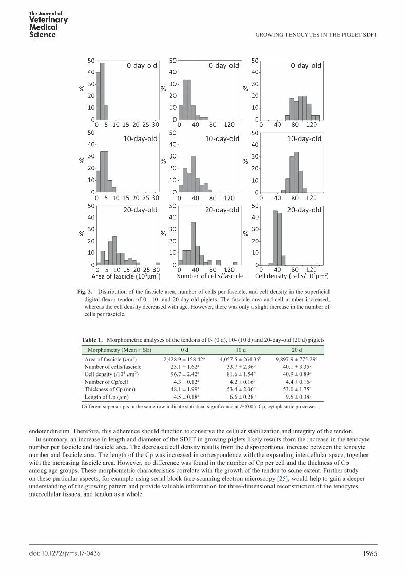
GROWING TENOCYTES IN THE PIGLET SDFT
1965doi 101292jvms17-0436
endotendineum Therefore this adherence should function to conserve the cellular stabilization and integrity of the tendonIn summary an increase in length and diameter of the SDFT in growing piglets likely results from the increase in the tenocyte
number per fascicle and fascicle area The decreased cell density results from the disproportional increase between the tenocyte number and fascicle area The length of the Cp was increased in correspondence with the expanding intercellular space together with the increasing fascicle area However no difference was found in the number of Cp per cell and the thickness of Cp among age groups These morphometric characteristics correlate with the growth of the tendon to some extent Further study on these particular aspects for example using serial block face-scanning electron microscopy [25] would help to gain a deeper understanding of the growing pattern and provide valuable information for three-dimensional reconstruction of the tenocytes intercellular tissues and tendon as a whole
Fig 3 Distribution of the fascicle area number of cells per fascicle and cell density in the superficial digital flexor tendon of 0- 10- and 20-day-old piglets The fascicle area and cell number increased whereas the cell density decreased with age However there was only a slight increase in the number of cells per fascicle
Table 1 Morphometric analyses of the tendons of 0- (0 d) 10- (10 d) and 20-day-old (20 d) piglets
Morphometry (Mean plusmn SE) 0 d 10 d 20 dArea of fascicle (microm2) 24289 plusmn 15842a 40575 plusmn 26436b 98979 plusmn 77529c
Number of cellsfascicle 231 plusmn 162a 337 plusmn 236b 401 plusmn 335c
Cell density (104 microm2) 967 plusmn 242a 816 plusmn 154b 409 plusmn 089c
Number of Cpcell 43 plusmn 012a 42 plusmn 016a 44 plusmn 016a
Thickness of Cp (nm) 481 plusmn 199a 534 plusmn 206a 530 plusmn 175a
Length of Cp (microm) 45 plusmn 018a 66 plusmn 028b 95 plusmn 038c
Different superscripts in the same row indicate statistical significance at Plt005 Cp cytoplasmic processes
N TAKAHASHI ET AL
1966doi 101292jvms17-0436
REFERENCES
1 Becker D L and Mobbs P 1999 Connexin alpha1 and cell proliferation in the developing chick retina Exp Neurol 156 326ndash332 [Medline] [CrossRef]
2 Benjamin M and Ralphs J R 1997 Tendons and ligaments--an overview Histol Histopathol 12 1135ndash1144 [Medline] 3 Coffey J W Fiedler-Nagy C Georgiadis A G and Salvador R A 1976 Digestion of native collagen denatured collagen and collagen
fragments by extracts of rat liver lysosomes J Biol Chem 251 5280ndash5282 [Medline] 4 Couppeacute C Svensson R B Heinemeier K M Thomsen E W Bayer M L Christensen L Kjaeligr M Magnusson S P and Schjerling P
2017 Quantification of cell density in rat Achilles tendon development and application of a new method Histochem Cell Biol 147 97ndash102 [Medline] [CrossRef]
5 Dowling B A Dart A J Hodgson D R and Smith R K 2000 Superficial digital flexor tendonitis in the horse Equine Vet J 32 369ndash378 [Medline] [CrossRef]
6 Evan A P Dail W G Dammrose D and Palmer C 1976 Scanning electron microscopy of cell surfaces following removal of extracellular material Anat Rec 185 433ndash445 [Medline] [CrossRef]
7 Ippolito E Natali P G Postacchini F Accinni L and De Martino C 1980 Morphological immunochemical and biochemical study of rabbit achilles tendon at various ages J Bone Joint Surg Am 62 583ndash598 [Medline] [CrossRef]
8 Kalson N S Lu Y Taylor S H Starborg T Holmes D F and Kadler K E 2015 A structure-based extracellular matrix expansion mechanism of fibrous tissue growth eLife 4 e05958 [Medline] [CrossRef]
9 Kannus P 2000 Structure of the tendon connective tissue Scand J Med Sci Sports 10 312ndash320 [Medline] [CrossRef] 10 Lavagnino M Gardner K and Arnoczky S P 2013 Age-related changes in the cellular mechanical and contractile properties of rat tail tendons
Connect Tissue Res 54 70ndash75 [Medline] [CrossRef] 11 Macchiarelli G and Ohtani O 2001 Endomysium in left ventricle Heart 86 416 [Medline] [CrossRef] 12 Maeda E Ye S Wang W Bader D L Knight M M and Lee D A 2011 Gap junction permeability between tenocytes within tendon fascicles
is suppressed by tensile loading Biomech Model Mechanobiol 11 439ndash447 [Medline] [CrossRef] 13 Magnusson S P Hansen P and Kjaer M 2003 Tendon properties in relation to muscular activity and physical training Scand J Med Sci Sports
13 211ndash223 [Medline] [CrossRef] 14 Mookhtiar K A and Van Wart H E 1992 Clostridium histolyticum collagenases a new look at some old enzymes Matrix Suppl 1 116ndash126
[Medline] 15 Murata H Nishizono H and Miyoshi M 2000 Postnatal development of structure and arrangement of tendon cells A scanning and transmission
electron microscope study in the rat calcaneal tendon Okajimas Folia Anat Jpn 77 77ndash86 [Medline] [CrossRef] 16 Nagy I Z Von Hahn H P and Verzaacuter F 1969 Age-related alterations in the cell nuclei and the DNA content of rat tail tendon Gerontologia 15
Fig 4 Number of cytoplasmic processes (Cp) per cell thickness of Cp and length of Cp in the superficial digital flexor tendon of 0- 10- and 20-day-old piglets
GROWING TENOCYTES IN THE PIGLET SDFT
1967doi 101292jvms17-0436
258ndash264 [Medline] [CrossRef] 17 Nakagawa Y Majima T and Nagashima K 1994 Effect of ageing on ultrastructure of slow and fast skeletal muscle tendon in rabbit Achilles
tendons Acta Physiol Scand 152 307ndash313 [Medline] [CrossRef] 18 Pingel J Lu Y Starborg T Fredberg U Langberg H Nedergaard A Weis M Eyre D Kjaer M and Kadler K E 2014 3-D ultrastructure
and collagen composition of healthy and overloaded human tendon evidence of tenocyte and matrix buckling J Anat 224 548ndash555 [Medline] [CrossRef]
19 Riley G P Harrall R L Constant C R Chard M D Cawston T E and Hazleman B L 1994 Tendon degeneration and chronic shoulder pain changes in the collagen composition of the human rotator cuff tendons in rotator cuff tendinitis Ann Rheum Dis 53 359ndash366 [Medline] [CrossRef]
20 Roberts T J Marsh R L Weyand P G and Taylor C R 1997 Muscular force in running turkeys the economy of minimizing work Science 275 1113ndash1115 [Medline] [CrossRef]
21 Squier C A and Bausch W H 1984 Three-dimensional organization of fibroblasts and collagen fibrils in rat tail tendon Cell Tissue Res 238 319ndash327 [Medline] [CrossRef]
22 Squier C A and Magnes C 1983 Spatial relationships between fibroblasts during the growth of rat-tail tendon Cell Tissue Res 234 17ndash29 [Medline] [CrossRef]
23 Stanley R L Fleck R A Patterson-Kane J C Goodship A E and Ralphs J R 2006 Confocal microscopy and image analysis of connexin plaques in equine tendon Microsc Analys 3 5ndash6
24 Stanley R L Fleck R A Becker D L Goodship A E Ralphs J R and Patterson-Kane J C 2007 Gap junction protein expression and cellularity comparison of immature and adult equine digital tendons J Anat 211 325ndash334 [Medline] [CrossRef]
25 Starborg T Kalson N S Lu Y Mironov A Cootes T F Holmes D F and Kadler K E 2013 Using transmission electron microscopy and 3View to determine collagen fibril size and three-dimensional organization Nat Protoc 8 1433ndash1448 [Medline] [CrossRef]
26 Ushiki T and Murakumo M 1991 Scanning electron microscopic studies of tissue elastin components exposed by a KOH-collagenase or simple KOH digestion method Arch Histol Cytol 54 427ndash436 [Medline] [CrossRef]
27 Wood L K and Brooks S V 2016 Ten weeks of treadmill running decreases stiffness and increases collagen turnover in tendons of old mice J Orthop Res 34 346ndash353 [Medline] [CrossRef]
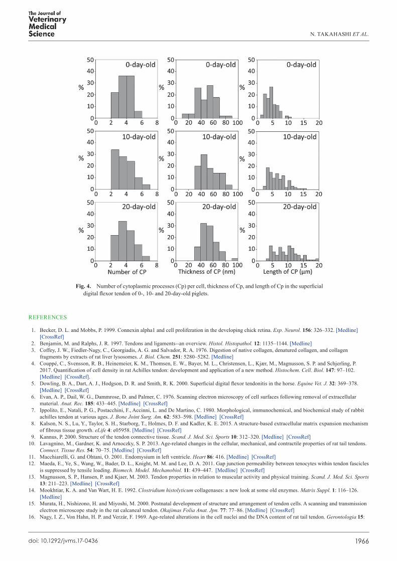