approach to arti cular and musculoskeletal di … · complaints secondary to tendinitis or carpal...
TRANSCRIPT

Print Close Window
Note: Large im ages and tables on t his page may necessitate print ing in landscape m ode.
Copyright © The McGraw - Hill Com panies. All r ights reserved.
Harr ison 's I nt ernal Medicine > Chapter 325. Approach t o Art icular and Musculoskelet al Disorders >
APPROACH TO ARTI CULAR AND MUSCULOSKELETAL DI SORDERS: I NTRODUCTI ONMusculoskeletal complaints account for > 315 m illion outpatient visits per year. Recent surveys by the Centers for Disease Cont rol and Prevent ion suggest that 33% (69.9 m illion) of the U.S. populat ion is affected by arthrit is or joint disorders. Many of these are self- limited condit ions requir ing minim al evaluat ion and only symptom at ic therapy and reassurance. However, in som e pat ients, specific musculoskeletal symptom s or their persistence may herald a m ore serious condit ion that requires further evaluat ion or laboratory test ing to establish a diagnosis or docum ent the extent and nature of the pathologic process. The goal of the musculoskeletal evaluat ion is to form ulate a different ial diagnosis that leads to an accurate diagnosis and t imely therapy, while avoiding excessive diagnost ic test ing and unnecessary t reatment (Table 325-1) . There are several urgent condit ions that m ust be diagnosed promptly to avoid signif icant morbid or mortal sequelae. These "red flag" diagnoses include sept ic arthrit is, acute crystal- induced arthrit is (e.g., gout ) , and fracture. Each may be suspected by its acute onset and monart icular or focal presentat ion ( see below) .
I ndividuals with musculoskeletal complaints should be evaluated with a thorough history, a com prehensive physical exam inat ion, and, if appropriate, laboratory test ing. The init ial encounter should determine whether the m usculoskeletal com plaint is (1) art icular or nonart icular in origin, (2) inflamm atory or noninf lam matory in nature, (3) acute or chronic in durat ion, and (4) localized or widespread (system ic) in dist r ibut ion.
With such an approach and an understanding of the pathophysiologic processes that underlie musculoskeletal complaints, a diagnosis can be made in the vast majority of individuals. However, som e patients will not f it im mediately into an established diagnost ic category. Many musculoskeletal disorders resemble each other at the outset , and som e m ay take weeks or months to evolve into a readily
Table 3 2 5 - 1 Eva luat ion of Pat ients w ith Musculoskeletal Com plaints
Goals
Accurate diagnosis
Timely provision of therapy
Avoidance of unnecessary diagnost ic test ing
Approach
Anatomic localizat ion of com plaint (art icular vs. nonart icular)
Determinat ion of the nature of the pathologic process ( inflam matory vs. noninflam matory)
Determinat ion of the extent of involvement (monart icular, polyart icular, focal, widespread)
Determinat ion of chronology (acute vs. chronic)
Consider the most comm on disorders first
Formulat ion of a different ial diagnosis
Page 1 of 22Print: Chapter 325. Approach to Articular and M usculoskeletal Disorders
1/3/2013mk:@M SITStore:C:\Users\Lesea\Downloads\Harrison's%20Principles%20of%20Internal%...
recognizable diagnost ic entity . This considerat ion should temper the desire to establish a definit ive diagnosis at the first encounter.
ARTI CULAR VERSUS NONARTI CULARThe m usculoskeletal evaluat ion must discriminate the anatom ic origin(s) of the pat ient 's com plaint . For example, ankle pain can result from a variety of pathologic condit ions involving disparate anatomic st ructures, including gonococcal arthrit is, calcaneal fracture, Achilles tendinit is, cellulit is, and peripheral neuropathy. Dist inguishing between art icular and nonart icular condit ions requires a careful and detailed examinat ion. Art icular structures include the synovium , synovial fluid, art icular cart ilage, int raart icular ligam ents, joint capsule, and juxtaart icular bone. Nonart icular (or periart icular) st ructures, such as support ive ext raart icular ligam ents, tendons, bursae, muscle, fascia, bone, nerve, and overly ing skin, may be involved in the pathologic process. Although m usculoskeletal com plaints are often ascribed to the joints, nonart icular disorders ( rather than art icular) m ore frequent ly underlie such complaints. Dist inguishing between these potent ial sources of pain m ay be challenging to the unskilled exam iner. Art icular disorders m ay be characterized by deep or diffuse pain, pain or limited range of m otion on act ive and passive m ovem ent, and swelling (caused by synovial proliferat ion, effusion, or bony enlargem ent ) , crepitat ion, instabilit y, " lock ing," or deformity. By cont rast , nonart icular disorders tend to be painful on act ive, but not passive (or assisted) , range of mot ion, demonst rate point or focal tenderness in regions adjacent to art icular st ructures, and have physical findings remote from the joint capsule. Moreover, nonart icular disorders seldom demonst rate swelling, crepitus, instabilit y, or deform ity.
I NFLAMMATORY VERSUS NONI NFLAMMATORY DI SORDERSI n the course of a m usculoskeletal evaluation, the examiner should determ ine the nature of the underlying pathologic process and whether inf lam matory or noninflamm atory f indings exist . I nflam matory disorders may be infect ious ( infect ion with Neisseria gonorrhoea or Mycobacterium tuberculosis ) , crystal- induced (gout , pseudogout) , im mune-related [ rheumatoid arthrit is (RA) , system ic lupus erythematosus ( SLE) ] , react ive ( rheumatic fever, Reiter 's syndrome) , or idiopathic. I nf lam matory disorders m ay be ident ified by any of the four cardinal signs of inflam mation (erythema, warmth, pain, or swelling) , system ic symptom s ( fat igue, fever, rash, weight loss) , or laboratory evidence of inflamm at ion [ elevated erythrocyte sedimentat ion rate (ESR) or C-react ive protein (CRP) , thrombocytosis, anemia of chronic disease, or hypoalbuminemia] . Art icular st iffness com monly accom panies chronic m usculoskeletal disorders. However, the severity and durat ion of st iffness may be diagnost ically important . Morning st iffness related to inf lam matory disorders (such as RA or polymyalgia rheumatica) is precipitated by prolonged rest , is described as severe, lasts for hours, and may improve with act ivity and ant i- inf lam matory medicat ions. By cont rast , intermittent st iffness (also known as gel phenomenon) , associated with noninflamm atory condit ions [ such as osteoarthrit is (OA) ] , is precipitated by brief periods of rest, usually lasts < 60 m in, and is exacerbated by act ivity. Fat igue may accom pany inflamm at ion (as seen in RA and polymyalgia rheumatica) but may also be prominent in f ibromyalgia (a noninflamm atory disorder) , anem ia, cardiac failure, endocrinopathy, poor nut r it ion, poor sleep, or depression. Noninf lam matory disorders m ay be related to t rauma ( rotator cuff tear) , repet it ive use (bursit is, tendonit is) , degeneration or ineffect ive repair (OA) , neoplasm (pigmented villonodular synovit is) , or pain am plificat ion ( fibromyalgia) . Noninflamm atory disorders are often characterized by pain without synovial swelling or warm th, absence of inflamm atory or systemic features, dayt im e gel phenomena rather than m orning st iffness, and norm al ( for age) or negat ive laboratory invest igat ions.
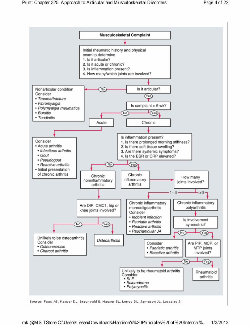
I dent ificat ion of the nature of the underly ing process and the site of the complaint will enable the examiner to narrow the diagnost ic considerat ions and to assess the need for im mediate diagnostic or therapeutic intervent ion, or for cont inued observat ion. Figure 325-1 presents a logical approach to the
Page 2 of 22Print: Chapter 325. Approach to Articular and M usculoskeletal Disorders
1/3/2013mk:@M SITStore:C:\Users\Lesea\Downloads\Harrison's%20Principles%20of%20Internal%...
evaluat ion of pat ients with m usculoskeletal com plaints.
Figure 3 2 5 - 1
Page 3 of 22Print: Chapter 325. Approach to Articular and M usculoskeletal Disorders
1/3/2013mk:@M SITStore:C:\Users\Lesea\Downloads\Harrison's%20Principles%20of%20Internal%...
Page 4 of 22Print: Chapter 325. Approach to Articular and M usculoskeletal Disorders
1/3/2013mk:@M SITStore:C:\Users\Lesea\Downloads\Harrison's%20Principles%20of%20Internal%...
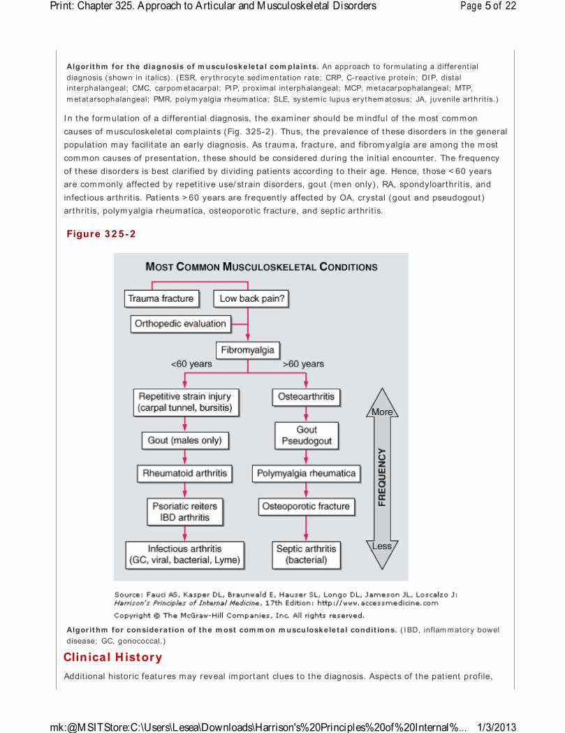
I n the form ulat ion of a different ial diagnosis, the examiner should be m indful of the most comm on causes of m usculoskeletal com plaints (Fig. 325-2) . Thus, the prevalence of these disorders in the general populat ion may facilitate an early diagnosis. As t raum a, fracture, and fibrom yalgia are among the m ost com mon causes of presentat ion, these should be considered during the init ial encounter. The frequency of these disorders is best clarified by dividing pat ients according to their age. Hence, those < 60 years are com monly affected by repet it ive use/ st rain disorders, gout (men only) , RA, spondyloarthrit is, and infect ious arthrit is. Pat ients > 60 years are frequently affected by OA, crystal (gout and pseudogout ) arthrit is, polym yalgia rheumat ica, osteoporot ic fracture, and septic arthrit is.
Clinical H istoryAddit ional historic features may reveal im portant clues to the diagnosis. Aspects of the pat ient profile,
Algorithm for the diagnosis of m usculoskeleta l com pla in ts. An approach to formulat ing a different ial diagnosis ( shown in italics) . (ESR, ery throcy te sedimentat ion rate; CRP, C-react ive protein; DI P, distal interphalangeal; CMC, carpom etacarpal; PI P, prox imal interphalangeal; MCP, metacarpophalangeal; MTP, m etatarsophalangeal; PMR, polym yalgia rheum at ica; SLE, system ic lupus erythem atosus; JA, juvenile art hrit is.)
Figure 3 2 5 - 2
Algorithm for considerat ion of the m ost com m on m usculoskeleta l condit ions. ( IBD, inflam matory bowel disease; GC, gonococcal.)
Page 5 of 22Print: Chapter 325. Approach to Articular and M usculoskeletal Disorders
1/3/2013mk:@M SITStore:C:\Users\Lesea\Downloads\Harrison's%20Principles%20of%20Internal%...
com plaint chronology, extent of joint involvement , and precipitat ing factors can provide important information. Certain diagnoses are more frequent in different age groups (Fig. 325-2) . SLE and react ive arthrit is occur more frequent ly in the young, whereas fibrom yalgia and RA are frequent in middle age and OA and polym yalgia rheumat ica are m ore prevalent am ong the elderly . Diagnostic clustering is also evident when sex and race are considered. Gout and the spondyloarthropathies (e.g., ankylosing spondylit is) are m ore com mon in men, whereas RA, fibromyalgia, and lupus are m ore frequent in wom en. Racial predilect ions may be influent ial. Thus, polymyalgia rheum at ica, giant cell arterit is, and Wegener's granulom atosis commonly affect whites, whereas sarcoidosis and SLE m ore com monly affect Afr ican Am ericans. Familial aggregat ion m ay be seen in disorders such as ankylosing spondylit is, gout , and Heberden's nodes of OA.
The chronology of the complaint is an im portant diagnost ic feature and can be divided into the onset , evolut ion, and durat ion. The onset of disorders such as septic arthrit is or gout tends to be abrupt , whereas OA, RA, and f ibromyalgia may have m ore indolent presentat ions. The pat ients' com plaints m ay evolve different ly and be classif ied as chronic (OA) , intermittent (crystal or Lym e arthrit is) , m igratory ( rheumatic fever, gonococcal or viral arthrit is) , or addit ive (RA, psoriat ic arthrit is) . Musculoskeletal disorders are typically classified as acute or chronic based upon a sym ptom durat ion that is either less than or greater than 6 weeks, respect ively. Acute arthropathies tend to be infect ious, crystal- induced, or react ive. Chronic condit ions include noninflamm atory or im munologic arthrit ides (e.g., OA, RA) and nonart icular disorders (e.g., fibromyalgia) .
Th e extent of art icular involvem ent is often diagnost ic. Art icular disorders are classified based on the num ber of joints involved, as either monart icular (one joint ) , oligoart icular or pauciart icular ( two or three joints) , or polyart icular (more than three joints) . Although crystal and infect ious arthrit is are often m ono-or oligoart icular, OA and RA are polyart icular disorders. Nonart icular disorders may be classified as either focal or widespread. Complaints secondary to tendinit is or carpal tunnel syndrome are typically focal, whereas weakness and myalgia, due to polym yosit is or f ibromyalgia, are more diffuse in their presentat ion. Joint involvem ent in RA tends to be sym metric, whereas the spondyloarthropathies and gout are often asymm etric and oligoart icular. The upper extremit ies are frequent ly involved in RA and OA, whereas lower ext remity arthrit is is characterist ic of react ive arthrit is and gout at their onset . I nvolvem ent of the axial skeleton is com mon in OA and ankylosing spondylit is but is infrequent in RA, with the notable except ion of the cervical spine.
The clinical history should also ident ify precipitat ing events, such as t rauma, drug adm inistrat ion (Table 325-2) , or antecedent or intercurrent illnesses, that may have cont ributed to the patient 's complaint . Certain com orbidit ies may predispose to musculoskeletal consequences. This is especially so for diabetes mellitus (carpal tunnel syndrom e) , renal insufficiency (gout ) , psoriasis (psoriat ic arthrit is) , myeloma ( low back pain) , cancer (m yosit is) , and osteoporosis ( fracture) or when using certain drugs such as glucocort icoids (osteonecrosis, sept ic arthrit is) and diuret ics or chemotherapy (gout ) . Last ly, a thorough rheumatic review of systems m ay disclose useful diagnost ic information. A variety of m usculoskeletal disorders m ay be associated with systemic features such as fever (SLE, infect ion) , rash (SLE, psoriat ic arthrit is) , nail abnormalit ies (psoriat ic or react ive arthrit is) , myalgias ( fibromyalgia, m yopathy) , or weakness (polym yosit is, neuropathy) . I n addit ion, som e condit ions are associated with involvement of other organ system s including the eyes (Behçet 's disease, sarcoidosis, spondyloarthrit is) , gast rointest inal t ract (scleroderma, inflamm atory bowel disease) , genitourinary t ract ( reactive arthrit is, gonococcem ia) , or the nervous system (Lym e disease, vasculit is) .
Table 3 2 5 - 2 Drug- I nduced Musculoskeletal Condit ions
Page 6 of 22Print: Chapter 325. Approach to Articular and M usculoskeletal Disorders
1/3/2013mk:@M SITStore:C:\Users\Lesea\Downloads\Harrison's%20Principles%20of%20Internal%...
Lastly, the examiner should assess the level of pain and physical lim itat ion that accompanies the com plaint. The intensity of the patient 's pain, st iffness, or weakness can be quant ified (0–10) verbally or with the use of a 10-cm visual analogue scale (0 = no pain and 10 = the worst possible pain) . Funct ional lim itat ion and disabilit y should be ident ified and recorded for future comparisons. There are several validated funct ional m easures that are easily incorporated into the musculoskeletal evaluat ion, such as the modified Health Assessm ent Quest ionnaire (Fig. 325-3) .
Arthralgias
Quinidine, cim et idine, quinolones, chronic acyclov ir, interferon, I L-2, nicardipine, vaccines, r ifabut in, arom atase and HI V protease inhibitors
Myalgias/ m yopathy
Glucocort icoids, penicillamine, hydroxychloroquine, AZT, lovastat in, simvastat in, pravastat in, clofibrate, interferon, I L-2, alcohol, cocaine, taxol, docetaxel, colchicine, quinolones, cyclosporine
Tendon rupture
Quinolones, glucocort icoids
Gout
Diuret ics, aspir in, cytotoxics, cyclosporine, alcohol, moonshine, ethambutol
Drug- induced lupus
Hydralazine, procainamide, quinidine, phenytoin, carbemazepine, m ethyldopa, isoniazid, chlorpromazine, lithium, penicillamine, tet racyclines, TNF inhibitors, ACE inhibitors, t iclopidine
Osteonecrosis
Glucocort icoids, alcohol, radiat ion, bisphosphonates
Osteopenia
Glucocort icoids, chronic heparin, phenytoin, methotrexate
Scleroderma
Vinyl chloride, bleom ycin, pentazocine, organic solvents, carbidopa, t ryptophan, rapeseed oil
Vasculit is
Allopurinol, amphetamines, cocaine, thiazides, penicillamine, propylthiouracil, montelukast , TNF inhibitors, hepatit is B vaccine, t r im ethoprim / sulfamethoxazole
Note: I L-2, interleukin 2; TNF, tumor necrosis factor; ACE, angiotensin-convert ing enzym e.
Figure 3 2 5 - 3
Page 7 of 22Print: Chapter 325. Approach to Articular and M usculoskeletal Disorders
1/3/2013mk:@M SITStore:C:\Users\Lesea\Downloads\Harrison's%20Principles%20of%20Internal%...
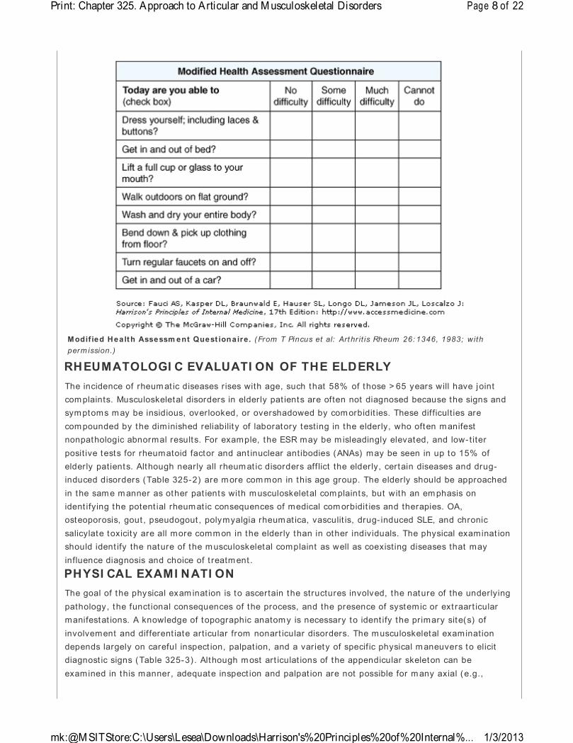
Modif ied Health Assessm ent Quest ionaire. (From T Pincus et al: Art hr it is Rheum 26: 1346, 1983; with perm ission.)
RHEUMATOLOGI C EVALUATI ON OF THE ELDERLYThe incidence of rheum at ic diseases r ises with age, such that 58% of those > 65 years will have joint com plaints. Musculoskeletal disorders in elderly pat ients are often not diagnosed because the signs and sym ptoms m ay be insidious, overlooked, or overshadowed by com orbidit ies. These dif ficult ies are com pounded by the diminished reliabilit y of laboratory test ing in the elderly, who often m anifest nonpathologic abnormal results. For example, the ESR may be m isleadingly elevated, and low- t iter posit ive tests for rheumatoid factor and ant inuclear antibodies (ANAs) may be seen in up to 15% of elderly patients. Although nearly all rheum at ic disorders aff lict the elderly, certain diseases and drug-induced disorders (Table 325-2) are m ore com mon in this age group. The elderly should be approached in the sam e m anner as other pat ients with m usculoskeletal com plaints, but with an em phasis on ident ifying the potent ial rheum at ic consequences of medical com orbidit ies and therapies. OA, osteoporosis, gout , pseudogout , polymyalgia rheum at ica, vasculit is, drug- induced SLE, and chronic salicylate toxicity are all more comm on in the elderly than in other individuals. The physical examinat ion should ident ify the nature of the m usculoskeletal complaint as well as coexist ing diseases that may influence diagnosis and choice of t reatm ent .
PHYSI CAL EXAMI NATI ONThe goal of the physical exam inat ion is to ascertain the structures involved, the nature of the underlying pathology, the functional consequences of the process, and the presence of systemic or ext raart icular manifestat ions. A knowledge of topographic anatom y is necessary to ident ify the primary site(s) of involvement and dif ferent iate art icular from nonart icular disorders. The m usculoskeletal examination depends largely on careful inspect ion, palpat ion, and a variety of specif ic physical m aneuvers to elicit diagnost ic signs (Table 325-3) . Although m ost art iculat ions of the appendicular skeleton can be examined in this manner, adequate inspect ion and palpat ion are not possible for m any axial (e.g.,
Page 8 of 22Print: Chapter 325. Approach to Articular and M usculoskeletal Disorders
1/3/2013mk:@M SITStore:C:\Users\Lesea\Downloads\Harrison's%20Principles%20of%20Internal%...
zygapophyseal) and inaccessible (e.g., sacroiliac or hip) joints. For such joints, there is a greater reliance upon specific maneuvers and imaging for assessment.
Exam inat ion of involved and uninvolved joints will determine whether pain , warmth , erythema, or swelling is present . The locale and level of pain elicited by palpat ion or m ovement should be quant if ied. One example would be to count the num ber of tender joints on palpat ion of 28 easily exam ined joints [ proxim al interphalangeals (PI Ps) , metacarpophalangeals (MCPs) , wrists, elbows, shoulders, and knees] (with a range of 0–28) . Similarly , the number of swollen joints (0–28) can be counted and recorded. Careful exam inat ion should dist inguish between t rue art icular swelling (caused by synovial effusion or synovial proliferat ion) and nonart icular (or periart icular ) involvement , which usually extends beyond the norm al joint m argins. Synovial effusion can be dist inguished from synovial hypert rophy or bony hypert rophy by palpat ion or specific m aneuvers. For exam ple, sm all to moderate knee effusions may be ident ified by the "bulge sign" or "ballot tement of the patellae." Bursal effusions (e.g., effusions of the olecranon or prepatellar bursa) are often focal, periart icular, overlie bony prom inences, and are fluctuant with sharply defined borders. Joint stabilit y can be assessed by palpat ion and by the applicat ion of manual st ress. Subluxat ion or dislocat ion, which may be secondary to t raumat ic, m echanical, or inflamm atory causes, can be assessed by inspect ion and palpat ion. Joint swelling orvolume can be assessed by palpat ion. Distent ion of the art icular capsule usually causes pain and evident swelling. The patient will at tempt to m inim ize the pain by m aintaining the joint in the posit ion of least int raart icular pressure and greatest volum e, usually part ial f lexion. For this reason, inflamm atory effusions may give r ise to flexion cont ractures. Clinically, this may be detected as f luctuant or "squishy" swelling, voluntary or fixed flexion deformit ies, or diminished range of m ot ion— especially on extension, when joint volumes
Table 3 2 5 - 3 Glossary of Musculoskeletal Term s
Crepitus
A palpable ( less com monly audible) vibratory or crackling sensat ion elicited with joint mot ion; fine joint crepitus is com mon and often insignificant in large joints; coarse joint crepitus indicates advanced cart ilaginous and degenerat ive changes (as in osteoarthrit is)
Subluxat ion
Alterat ion of joint alignm ent such that art iculat ing surfaces incompletely approxim ate each other
Dislocat ion
Abnormal displacem ent of art iculat ing surfaces such that the surfaces are not in contact
Range of mot ion
For diarthrodial joints, the arc of measurable m ovement through which the joint moves in a single plane
Contracture
Loss of full movement result ing from a fixed resistance caused either by tonic spasm of muscle ( reversible) or to fibrosis of periart icular st ructures (permanent)
Deform ity
Abnormal shape or size of a st ructure; m ay result from bony hypert rophy, malalignm ent of art iculat ing structures, or damage to periart icular support ive st ructures
Ent hesit is
I nflamm at ion of the entheses ( tendinous or ligam entous insert ions on bone)
Epicondylit is
I nfect ion or inf lam mat ion involving an epicondyle
Page 9 of 22Print: Chapter 325. Approach to Articular and M usculoskeletal Disorders
1/3/2013mk:@M SITStore:C:\Users\Lesea\Downloads\Harrison's%20Principles%20of%20Internal%...
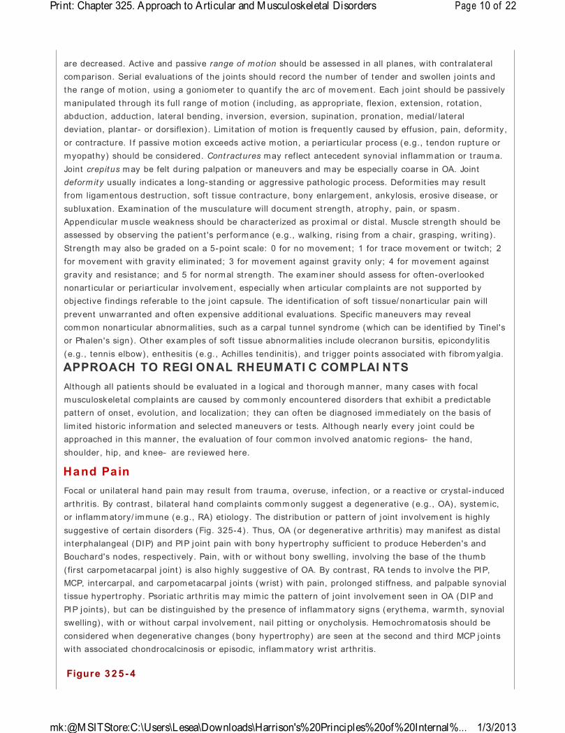
are decreased. Act ive and passive range of mot ion should be assessed in all planes, with cont ralateral com parison. Serial evaluat ions of the joints should record the num ber of tender and swollen joints and the range of m ot ion, using a goniom eter to quant ify the arc of m ovem ent. Each joint should be passively manipulated through its full range of m otion ( including, as appropriate, flexion, extension, rotat ion, abduct ion, adduct ion, lateral bending, inversion, eversion, supinat ion, pronat ion, medial/ lateral deviat ion, plantar- or dorsiflexion) . Limitat ion of mot ion is frequently caused by effusion, pain, deformity , or contracture. I f passive m otion exceeds act ive mot ion, a periart icular process (e.g., tendon rupture or myopathy) should be considered. Cont ractures may ref lect antecedent synovial inflamm at ion or t raum a. Joint crepitus m ay be felt during palpat ion or maneuvers and may be especially coarse in OA. Joint deform ity usually indicates a long-standing or aggressive pathologic process. Deformit ies may result from ligamentous destruct ion, soft t issue cont racture, bony enlargem ent , ankylosis, erosive disease, or subluxat ion. Examinat ion of the musculature will document st rength, at rophy, pain, or spasm . Appendicular m uscle weakness should be characterized as proxim al or distal. Muscle st rength should be assessed by observ ing the pat ient 's perform ance (e.g., walking, r ising from a chair, grasping, writ ing) . St rength m ay also be graded on a 5-point scale: 0 for no movem ent ; 1 for t race m ovem ent or twitch; 2 for movement with gravity elim inated; 3 for m ovement against gravity only; 4 for m ovement against gravity and resistance; and 5 for norm al st rength. The exam iner should assess for often-overlooked nonart icular or periart icular involvem ent , especially when art icular com plaints are not supported by object ive f indings referable to the joint capsule. The ident if icat ion of soft t issue/ nonart icular pain will prevent unwarranted and often expensive addit ional evaluat ions. Specif ic maneuvers may reveal com mon nonart icular abnorm alit ies, such as a carpal tunnel syndrome (which can be identified by Tinel's or Phalen's sign) . Other exam ples of soft t issue abnormalit ies include olecranon bursit is, epicondylit is (e.g., tennis elbow) , enthesit is (e.g., Achilles tendinit is) , and t r igger points associated with f ibrom yalgia.
APPROACH TO REGI ONAL RHEUMATI C COMPLAI NTSAlthough all pat ients should be evaluated in a logical and thorough m anner, m any cases with focal musculoskeletal complaints are caused by com monly encountered disorders that exhibit a predictable pattern of onset , evolut ion, and localizat ion; they can often be diagnosed immediately on the basis of lim ited historic informat ion and selected m aneuvers or tests. Although nearly every joint could be approached in this m anner, the evaluat ion of four com mon involved anatomic regions— the hand, shoulder , hip, and knee— are reviewed here.
Hand PainFocal or unilateral hand pain may result from trauma, overuse, infect ion, or a react ive or crystal- induced arthrit is. By contrast , bilateral hand com plaints comm only suggest a degenerative (e.g., OA) , systemic, or inflamm atory/ im mune (e.g., RA) et iology. The dist r ibut ion or pat tern of joint involvem ent is highly suggestive of certain disorders (Fig. 325-4) . Thus, OA (or degenerat ive arthrit is) may m anifest as distal interphalangeal (DIP) and PIP joint pain with bony hypertrophy sufficient to produce Heberden's and Bouchard's nodes, respectively. Pain, with or without bony swelling, involving the base of the thumb ( first carpometacarpal joint ) is also highly suggest ive of OA. By cont rast , RA tends to involve the PIP, MCP, intercarpal, and carpom etacarpal joints (wrist ) with pain, prolonged st iffness, and palpable synovial t issue hypert rophy. Psoriat ic arthrit is m ay m im ic the pattern of joint involvement seen in OA (DI P and PI P joints) , but can be dist inguished by the presence of inflammatory signs (erythema, warmth, synovial swelling) , with or without carpal involvement , nail pit t ing or onycholysis. Hemochromatosis should be considered when degenerat ive changes (bony hypert rophy) are seen at the second and third MCP joints with associated chondrocalcinosis or episodic, inflam matory wrist arthrit is.
Figure 3 2 5 - 4
Page 10 of 22Print: Chapter 325. Approach to Articular and M usculoskeletal Disorders
1/3/2013mk:@M SITStore:C:\Users\Lesea\Downloads\Harrison's%20Principles%20of%20Internal%...
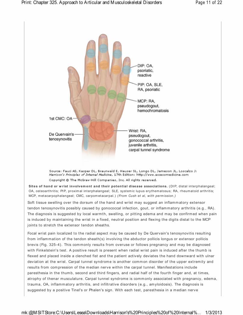
Soft t issue swelling over the dorsum of the hand and wrist may suggest an inflamm atory extensor tendon tenosynovit is possibly caused by gonococcal infect ion, gout, or inflam matory arthrit is (e.g., RA) . The diagnosis is suggested by local warmth, swelling, or pit t ing edema and may be confirm ed when pain is induced by m aintaining the wrist in a f ixed, neut ral posit ion and flex ing the digits distal to the MCP joints to st retch the extensor tendon sheaths.
Focal wrist pain localized to the radial aspect may be caused by De Quervain's tenosynovit is result ing from inf lam mat ion of the tendon sheath(s) involving the abductor pollicis longus or extensor pollicis brevis (Fig. 325-4) . This com monly results from overuse or follows pregnancy and may be diagnosed with Finkelstein's test . A posit ive result is present when radial wrist pain is induced after the thum b is flexed and placed inside a clenched fist and the pat ient act ively deviates the hand downward with ulnar deviat ion at the wrist . Carpal tunnel syndrom e is another comm on disorder of the upper ext remity and results from com pression of the m edian nerve within the carpal tunnel. Manifestat ions include paresthesia in the thum b, second and third fingers, and radial half of the fourth finger and, at t im es, at rophy of thenar musculature. Carpal tunnel syndrome is comm only associated with pregnancy, edema, t rauma, OA, inflamm atory arthrit is, and infilt rat ive disorders (e.g., am yloidosis) . The diagnosis is suggested by a posit ive Tinel's or Phalen's sign. With each test , paresthesia in a m edian nerve
Sites of hand or w rist involvem ent and the ir potent ia l disease associat ions. (DI P, distal interphalangeal; OA, osteoarthrit is; PIP, proxim al interphalangeal; SLE, system ic lupus ery thematosus; RA, rheumatoid ar thr it is; MCP, metacarpophalangeal; CMC, carpom etacarpal. ) (From Cush et al, with perm ission.)
Page 11 of 22Print: Chapter 325. Approach to Articular and M usculoskeletal Disorders
1/3/2013mk:@M SITStore:C:\Users\Lesea\Downloads\Harrison's%20Principles%20of%20Internal%...
dist r ibut ion is induced or increased by either "thum ping" the volar aspect of the wrist (Tinel's sign) or pressing the extensor surfaces of both flexed wrists against each other (Phalen's sign) .
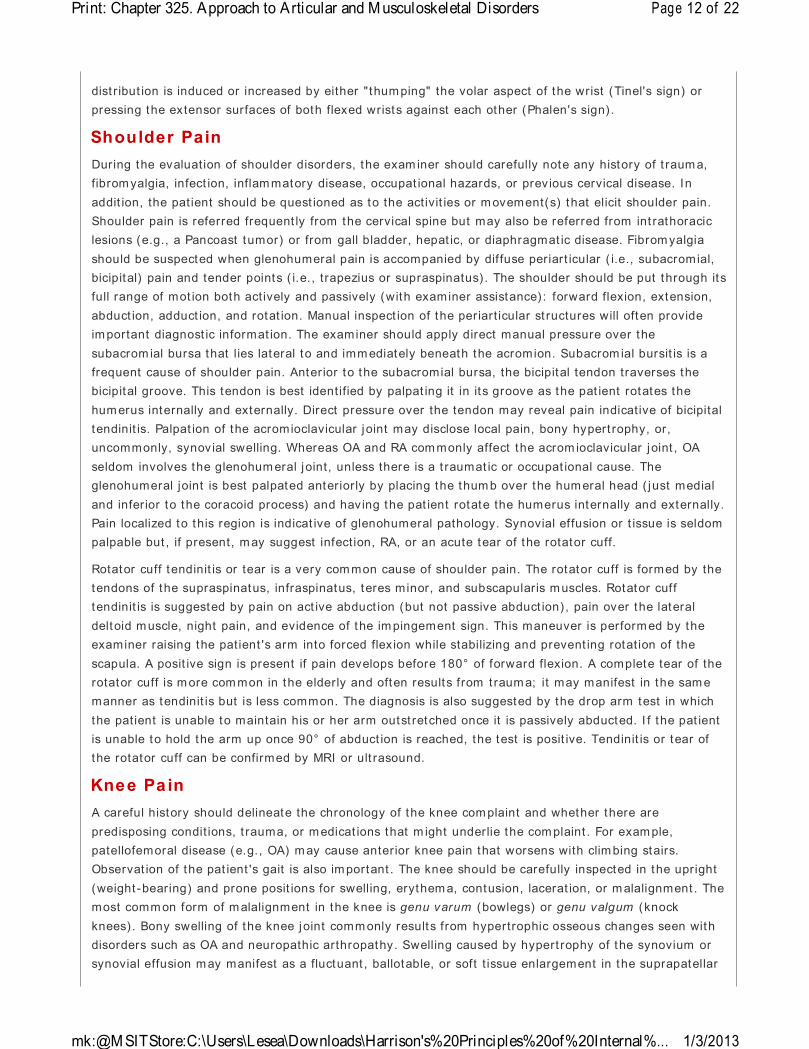
Shoulder PainDuring the evaluation of shoulder disorders, the exam iner should carefully note any history of t raum a, fibrom yalgia, infect ion, inflam matory disease, occupat ional hazards, or previous cervical disease. In addit ion, the patient should be quest ioned as to the act ivit ies or m ovement(s) that elicit shoulder pain. Shoulder pain is referred frequent ly from the cervical spine but may also be referred from int rathoracic lesions (e.g., a Pancoast tumor) or from gall bladder, hepat ic, or diaphragmat ic disease. Fibrom yalgia should be suspected when glenohumeral pain is accompanied by dif fuse periart icular ( i.e., subacromial, bicipital) pain and tender points ( i.e., trapezius or supraspinatus) . The shoulder should be put through its full range of mot ion both act ively and passively (with examiner assistance) : forward flexion, extension, abduct ion, adduct ion, and rotat ion. Manual inspect ion of the periart icular st ructures will often provide im portant diagnost ic informat ion. The examiner should apply direct manual pressure over the subacrom ial bursa that lies lateral to and imm ediately beneath the acromion. Subacrom ial bursit is is a frequent cause of shoulder pain. Anterior to the subacromial bursa, the bicipital tendon traverses the bicipital groove. This tendon is best identified by palpat ing it in it s groove as the pat ient rotates the hum erus internally and externally. Direct pressure over the tendon may reveal pain indicat ive of bicipital tendinit is. Palpat ion of the acromioclavicular joint may disclose local pain, bony hypert rophy, or, uncommonly, synovial swelling. Whereas OA and RA com monly affect the acrom ioclavicular joint , OA seldom involves the glenohum eral joint, unless there is a t raumat ic or occupat ional cause. The glenohumeral joint is best palpated anteriorly by placing the thum b over the hum eral head ( just medial and inferior to the coracoid process) and having the pat ient rotate the humerus internally and externally. Pain localized to this region is indicat ive of glenohumeral pathology. Synovial effusion or t issue is seldom palpable but , if present, m ay suggest infect ion, RA, or an acute tear of the rotator cuff.
Rotator cuff tendinit is or tear is a very com mon cause of shoulder pain. The rotator cuff is formed by the tendons of the supraspinatus, infraspinatus, teres minor, and subscapularis m uscles. Rotator cuff tendinit is is suggested by pain on act ive abduct ion (but not passive abduct ion) , pain over the lateral deltoid m uscle, night pain, and evidence of the im pingement sign. This maneuver is perform ed by the examiner raising the patient 's arm into forced flex ion while stabilizing and prevent ing rotat ion of the scapula. A posit ive sign is present if pain develops before 180° of forward flexion. A complete tear of the rotator cuff is m ore com mon in the elderly and often results from t raum a; it may manifest in the sam e manner as tendinit is but is less common. The diagnosis is also suggested by the drop arm test in which the patient is unable to maintain his or her arm outst retched once it is passively abducted. I f the pat ient is unable to hold the arm up once 90° of abduct ion is reached, the test is posit ive. Tendinit is or tear of the rotator cuff can be confirmed by MRI or ult rasound.
Knee PainA careful history should delineate the chronology of the knee com plaint and whether there are predisposing condit ions, t rauma, or m edicat ions that m ight underlie the com plaint . For exam ple, patellofemoral disease (e.g., OA) m ay cause anterior knee pain that worsens with clim bing stairs. Observat ion of the pat ient 's gait is also im portant . The knee should be carefully inspected in the upright (weight -bearing) and prone posit ions for swelling, erythem a, contusion, lacerat ion, or m alalignment . The most comm on form of m alalignment in the knee is genu varum (bowlegs) or genu valgum (knock knees) . Bony swelling of the knee joint comm only results from hypertrophic osseous changes seen with disorders such as OA and neuropathic arthropathy. Swelling caused by hypert rophy of the synovium or synovial effusion m ay manifest as a fluctuant , ballotable, or soft t issue enlargement in the suprapatellar
Page 12 of 22Print: Chapter 325. Approach to Articular and M usculoskeletal Disorders
1/3/2013mk:@M SITStore:C:\Users\Lesea\Downloads\Harrison's%20Principles%20of%20Internal%...
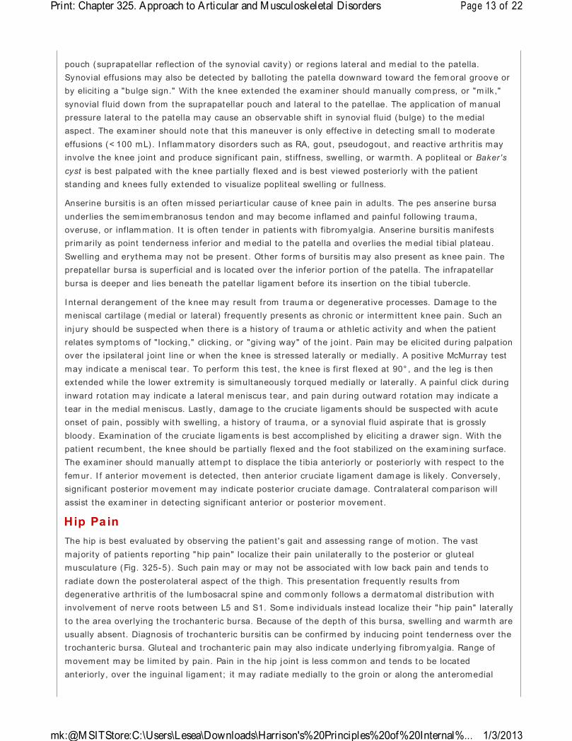
pouch (suprapatellar reflect ion of the synovial cavity) or regions lateral and m edial to the patella. Synovial effusions may also be detected by ballot ing the patella downward toward the fem oral groove or by elicit ing a "bulge sign." With the knee extended the exam iner should manually com press, or "m ilk ," synovial f luid down from the suprapatellar pouch and lateral to the patellae. The applicat ion of m anual pressure lateral to the patella may cause an observable shift in synovial fluid (bulge) to the m edial aspect . The exam iner should note that this maneuver is only effect ive in detect ing sm all to moderate effusions (< 100 mL) . I nf lam matory disorders such as RA, gout , pseudogout , and react ive arthrit is may involve the knee joint and produce signif icant pain, st iffness, swelling, or warmth. A popliteal or Baker 's cyst is best palpated with the knee part ially flexed and is best viewed posteriorly with the pat ient standing and knees fully extended to visualize popliteal swelling or fullness.
Anserine bursit is is an often missed periart icular cause of knee pain in adults. The pes anserine bursa underlies the sem im em branosus tendon and may become inflamed and painful following t rauma, overuse, or inf lam mat ion. I t is often tender in pat ients with fibromyalgia. Anserine bursit is manifests prim arily as point tenderness inferior and m edial to the patella and overlies the m edial t ibial plateau. Swelling and erythema may not be present . Other form s of bursit is m ay also present as knee pain. The prepatellar bursa is superficial and is located over the inferior port ion of the patella. The infrapatellar bursa is deeper and lies beneath the patellar ligam ent before its insert ion on the t ibial tubercle.
I nternal derangement of the knee m ay result from t raum a or degenerat ive processes. Dam age to the meniscal cart ilage ( medial or lateral) frequently presents as chronic or intermittent knee pain. Such an injury should be suspected when there is a history of t raum a or athlet ic act ivity and when the pat ient relates sym ptoms of " locking," clicking, or "giv ing way" of the joint . Pain may be elicited during palpat ion over the ipsilateral joint line or when the knee is st ressed laterally or medially. A posit ive McMurray test may indicate a meniscal tear. To perform this test , the knee is first flexed at 90° , and the leg is then extended while the lower extremity is simultaneously torqued medially or laterally. A painful click during inward rotat ion m ay indicate a lateral m eniscus tear, and pain during outward rotat ion may indicate a tear in the medial m eniscus. Last ly, dam age to the cruciate ligam ents should be suspected with acute onset of pain, possibly with swelling, a history of t rauma, or a synovial fluid aspirate that is grossly bloody. Examinat ion of the cruciate ligam ents is best accomplished by elicit ing a drawer sign. With the patient recum bent , the knee should be part ially flexed and the foot stabilized on the exam ining surface. The exam iner should manually at tem pt to displace the t ibia anteriorly or posteriorly with respect to the fem ur. I f anterior m ovement is detected, then anterior cruciate ligament dam age is likely . Conversely, significant posterior m ovement m ay indicate posterior cruciate damage. Cont ralateral com parison will assist the exam iner in detect ing signif icant anterior or posterior m ovement .
Hip PainThe hip is best evaluated by observing the patient 's gait and assessing range of m ot ion. The vast majority of pat ients report ing "hip pain" localize their pain unilaterally to the posterior or gluteal musculature (Fig. 325-5) . Such pain may or m ay not be associated with low back pain and tends to radiate down the posterolateral aspect of the thigh. This presentat ion frequent ly results from degenerat ive arthrit is of the lumbosacral spine and comm only follows a dermatom al dist r ibut ion with involvement of nerve roots between L5 and S1. Som e individuals instead localize their "hip pain" laterally to the area overlying the t rochanteric bursa. Because of the depth of this bursa, swelling and warmth are usually absent. Diagnosis of trochanteric bursit is can be confirmed by inducing point tenderness over the t rochanteric bursa. Gluteal and t rochanteric pain m ay also indicate underly ing fibromyalgia. Range of movement may be limited by pain. Pain in the hip joint is less comm on and tends to be located anteriorly , over the inguinal ligament ; it m ay radiate medially to the groin or along the anteromedial
Page 13 of 22Print: Chapter 325. Approach to Articular and M usculoskeletal Disorders
1/3/2013mk:@M SITStore:C:\Users\Lesea\Downloads\Harrison's%20Principles%20of%20Internal%...

thigh. Uncomm only, iliopsoas bursit is may m im ic true hip joint pain. Diagnosis of iliopsoas bursit is m ay be suggested by a history of t raum a or inf lam matory arthrit is. Pain associated with iliopsoas bursit is is localized to the groin or anterior thigh and tends to worsen with hyperextension of the hip; m any patients prefer to f lex and externally rotate the hip to reduce the pain from a distended bursa.
Figure 3 2 5 - 5
Or igins of hip pain and dysesthesias. (From Cush et al, with perm ission.)
LABORATORY I NVESTI GATI ONSThe vast m ajority of musculoskeletal disorders can be easily diagnosed by a complete history and physical exam inat ion. An addit ional object ive of the init ial encounter is to determ ine whether addit ional invest igat ions or im mediate therapy are required. A number of features indicate the need for addit ional evaluat ion. Monart icular condit ions require addit ional evaluation, as do traumatic or inflam matory condit ions and condit ions accompanied by neurologic changes or system ic manifestat ions of serious disease. Finally, individuals with chronic sym ptoms (> 6 weeks) , especially when there has been a lack of response to symptom at ic m easures, are candidates for addit ional evaluat ion. The extent and nature of the addit ional invest igat ion should be dictated by the clinical features and suspected pathologic process. Laboratory tests should be used to confirm a specific clinical diagnosis and not be used to screen or evaluate patients with vague rheumat ic com plaints. I ndiscrim inate use of broad batteries of diagnostic tests and radiographic procedures is rarely a useful or cost -effect ive means to establish a diagnosis.
Besides a com plete blood count , including a white blood cell ( WBC) and different ial count , the rout ine evaluat ion should include a determination of an acute-phase reactant such as the ESR or CRP, which can be useful in discriminat ing inflam matory from noninf lam matory disorders. Both are inexpensive and
Page 14 of 22Print: Chapter 325. Approach to Articular and M usculoskeletal Disorders
1/3/2013mk:@M SITStore:C:\Users\Lesea\Downloads\Harrison's%20Principles%20of%20Internal%...
easily obtained and m ay be elevated with infect ion, inflamm at ion, autoimm une disorders, neoplasia, pregnancy, renal insufficiency, and advanced age.
Serum uric acid determinations are useful only when gout has been diagnosed and therapy contemplated. Uric acid, the end product of purine metabolism , is prim arily excreted in the urine. Serum values range from 238–516 m ol/ L (4.0–8.6 m g/ dL) in men; the lower values [ 178–351 mol/ L (3.0–5.9 mg/ dL) ] seen in women are caused by the uricosuric effects of est rogen. Urinary uric acid levels are norm ally < 750 mg per 24 h. Although hyperuricemia [ especially levels > 535 m ol/ L (9 mg/ dL) ] is associated with an increased incidence of gout and nephrolithiasis, levels do not correlate with the severity of disease. Uric acid levels (and the r isk of gout ) m ay be increased by inborn errors of metabolism (Lesch-Nyhan syndrome) , disease states ( renal insufficiency, myeloproliferat ive disease, psoriasis) , or drugs (alcohol, cytotoxic therapy, thiazides) . Although nearly all pat ients with gout will dem onst rate hyperuricem ia at som e t im e during their illness, up to 40% of pat ients with an acute gouty at tack will have norm al serum uric acid levels. Monitoring serum uric acid m ay be useful in assessing the response to hypouricem ic therapy or chem otherapy.
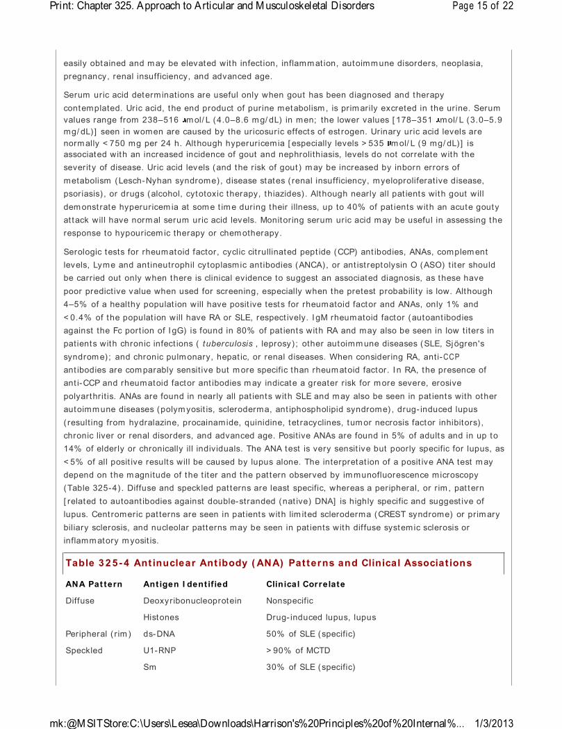
Serologic tests for rheumatoid factor, cyclic cit rullinated pept ide (CCP) ant ibodies, ANAs, complem ent levels, Lyme and antineutrophil cytoplasmic antibodies (ANCA), or ant ist reptolysin O (ASO) t iter should be carried out only when there is clinical evidence to suggest an associated diagnosis, as these have poor predict ive value when used for screening, especially when the pretest probability is low. Although 4–5% of a healthy populat ion will have posit ive tests for rheumatoid factor and ANAs, only 1% and < 0.4% of the populat ion will have RA or SLE, respect ively. I gM rheumatoid factor (autoant ibodies against the Fc port ion of I gG) is found in 80% of pat ients with RA and may also be seen in low t iters in patients with chronic infect ions ( tuberculosis , leprosy) ; other autoimm une diseases (SLE, Sjögren's syndrom e) ; and chronic pulm onary, hepat ic, or renal diseases. When considering RA, anti-CCP ant ibodies are com parably sensit ive but m ore specific than rheum atoid factor. I n RA, the presence of ant i-CCP and rheumatoid factor ant ibodies m ay indicate a greater r isk for m ore severe, erosive polyarthrit is. ANAs are found in nearly all pat ients with SLE and m ay also be seen in pat ients with other autoimm une diseases (polym yosit is, scleroderma, ant iphospholipid syndrome) , drug- induced lupus ( result ing from hydralazine, procainamide, quinidine, tet racyclines, tum or necrosis factor inhibitors) , chronic liver or renal disorders, and advanced age. Posit ive ANAs are found in 5% of adults and in up to 14% of elderly or chronically ill individuals. The ANA test is very sensit ive but poorly specific for lupus, as < 5% of all posit ive results will be caused by lupus alone. The interpretat ion of a posit ive ANA test m ay depend on the m agnitude of the t iter and the pat tern observed by im munofluorescence microscopy (Table 325-4) . Diffuse and speckled pat terns are least specific, whereas a peripheral, or r im , pat tern [ related to autoant ibodies against double-stranded (nat ive) DNA] is highly specific and suggest ive of lupus. Cent romeric pat terns are seen in pat ients with lim ited scleroderma (CREST syndrome) or prim ary biliary sclerosis, and nucleolar patterns may be seen in pat ients with dif fuse systemic sclerosis or inflamm atory m yosit is.
Table 3 2 5 - 4 Ant inuclear Ant ibody ( ANA) Pat terns and Clinical Associat ions
ANA Pat tern Ant igen I dent ified Clinical Correlate
Diffuse Deoxyribonucleoprotein Nonspecific
Histones Drug- induced lupus, lupus
Peripheral ( r im ) ds-DNA 50% of SLE (specific)
Speckled U1-RNP > 90% of MCTD
Sm 30% of SLE (specific)
Page 15 of 22Print: Chapter 325. Approach to Articular and M usculoskeletal Disorders
1/3/2013mk:@M SITStore:C:\Users\Lesea\Downloads\Harrison's%20Principles%20of%20Internal%...
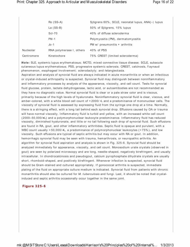
Aspirat ion and analysis of synovial fluid are always indicated in acute monarthrit is or when an infect ious or crystal- induced arthropathy is suspected. Synovial fluid may dist inguish between noninflam matory and inflamm atory processes by analysis of the appearance, viscosity, and cell count . Tests for synovial fluid glucose, protein, lactate dehydrogenase, lact ic acid, or autoant ibodies are not recom mended as they have no diagnost ic value. Normal synovial fluid is clear or a pale straw color and is viscous, prim arily because of the high levels of hyaluronate. Noninflamm atory synovial fluid is clear, viscous, and am ber-colored, with a white blood cell count of < 2000/ L and a predom inance of m ononuclear cells. The viscosity of synovial fluid is assessed by expressing f luid from the syringe one drop at a t ime. Norm ally, there is a st r inging effect, with a long tail behind each synovial drop. Effusions caused by OA or t rauma will have normal v iscosity . I nflamm atory fluid is turbid and yellow, with an increased white cell count (2000–50,000/ L) and a polymorphonuclear leukocyte predom inance. I nf lam matory fluid has reduced viscosity, dim inished hyaluronate, and lit t le or no tail following each drop of synovial f luid. Such effusions are found in RA, gout , and other inflammatory arthrit ides. Sept ic fluid is opaque and purulent , with a WBC count usually > 50,000/ L, a predominance of polym orphonuclear leukocytes ( > 75% ), and low viscosity. Such effusions are typical of septic arthrit is but may occur with RA or gout . In addit ion, hemorrhagic synovial f luid m ay be seen with t rauma, hem arthrosis, or neuropathic arthrit is. An algorithm for synovial fluid aspirat ion and analysis is shown in Fig. 325-6. Synovial f luid should be analyzed imm ediately for appearance, v iscosity, and cell count . Monosodium urate crystals (observed in gout) are seen by polarized m icroscopy and are long, needle-shaped, negat ively birefr ingent , and usually int racellular. I n chondrocalcinosis and pseudogout , calcium pyrophosphate dihydrate crystals are usually short , rhomboid-shaped, and posit ively birefr ingent . Whenever infect ion is suspected, synovial fluid should be Gram-stained and cultured appropriately . I f gonococcal arthrit is is suspected, im mediate plat ing of the f luid on appropriate culture m edium is indicated. Synovial fluid from pat ients with chronic monarthrit is should also be cultured for M. tuberculosis and fungi. Last , it should be noted that crystal-induced and sept ic arthrit is occasionally occur together in the same joint .
Ro (SS- A) Sjögrens 60% , SCLE, neonatal lupus, ANA(–) lupus
La (SS-B) 50% of Sjögrens, 15% lupus
Scl-70 40% of dif fuse scleroderma
PM-1 Polym yosit is (PM), derm atomyosit is
Jo-1 PM w/ pneumonit is + arthrit is
Nucleolar RNA polymerase I , others 40% of PSS
Cent romere Kinetochore 75% CREST ( lim ited scleroderma)
Note: SLE, systemic lupus erythem atosus; MCTD, mixed connect ive t issue disease; SCLE, subacute cutaneous lupus erythematosus; PSS, progressive system ic sclerosis; CREST, calcinosis, R aynaud phenomenon, esophageal involvem ent; sclerodacty ly; and telangiectasia.
Figure 3 2 5 - 6
Page 16 of 22Print: Chapter 325. Approach to Articular and M usculoskeletal Disorders
1/3/2013mk:@M SITStore:C:\Users\Lesea\Downloads\Harrison's%20Principles%20of%20Internal%...
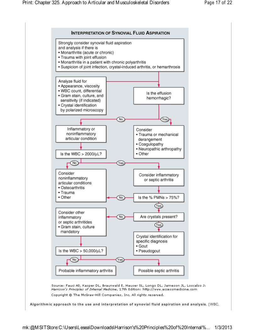
Algorithm ic approach to the use and interpretat ion of synovia l fluid aspirat ion and analysis. [ WBC,
Page 17 of 22Print: Chapter 325. Approach to Articular and M usculoskeletal Disorders
1/3/2013mk:@M SITStore:C:\Users\Lesea\Downloads\Harrison's%20Principles%20of%20Internal%...
white blood cell ( count) ; PMNs, polymorphonuclear ( leukocytes) . ]
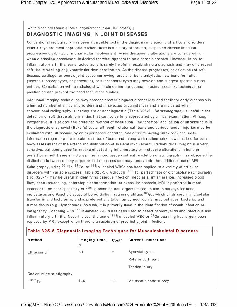
DI AGNOSTI C I MAGI NG I N JOI NT DI SEASESConventional radiography has been a valuable tool in the diagnosis and staging of art icular disorders. Plain x- rays are m ost appropriate when there is a history of trauma, suspected chronic infect ion, progressive disability , or m onart icular involvement; when therapeut ic alterat ions are considered; or when a baseline assessment is desired for what appears to be a chronic process. However, in acute inflamm atory arthrit is, early radiography is rarely helpful in establishing a diagnosis and may only reveal soft t issue swelling or juxtaart icular demineralizat ion. As the disease progresses, calcificat ion (of soft t issues, cart ilage, or bone) , joint space narrowing, erosions, bony ankylosis, new bone format ion (sclerosis, osteophytes, or periost it is) , or subchondral cysts may develop and suggest specific clinical ent it ies. Consultat ion with a radiologist will help define the optimal imaging m odality, technique, or posit ioning and prevent the need for further studies.
Addit ional im aging techniques may possess greater diagnost ic sensit ivity and facilitate early diagnosis in a lim ited number of art icular disorders and in selected circum stances and are indicated when convent ional radiography is inadequate or nondiagnost ic (Table 325-5) . Ult rasonography is useful in the detect ion of soft t issue abnorm alit ies that cannot be fully appreciated by clinical examinat ion. Although inexpensive, it is seldom the preferred m ethod of evaluation. The foremost applicat ion of ult rasound is in the diagnosis of synovial (Baker's) cysts, although rotator cuff tears and various tendon injuries m ay be evaluated with ultrasound by an experienced operator. Radionuclide scint igraphy provides useful information regarding the metabolic status of bone and, along with radiography, is well suited for total-body assessment of the extent and distr ibut ion of skeletal involvement . Radionuclide imaging is a very sensit ive, but poorly specific, m eans of detect ing inflamm atory or m etabolic alterat ions in bone or periart icular soft t issue st ructures. The lim ited t issue cont rast resolut ion of scint igraphy may obscure the dist inct ion between a bony or periart icular process and may necessitate the addit ional use of MRI .
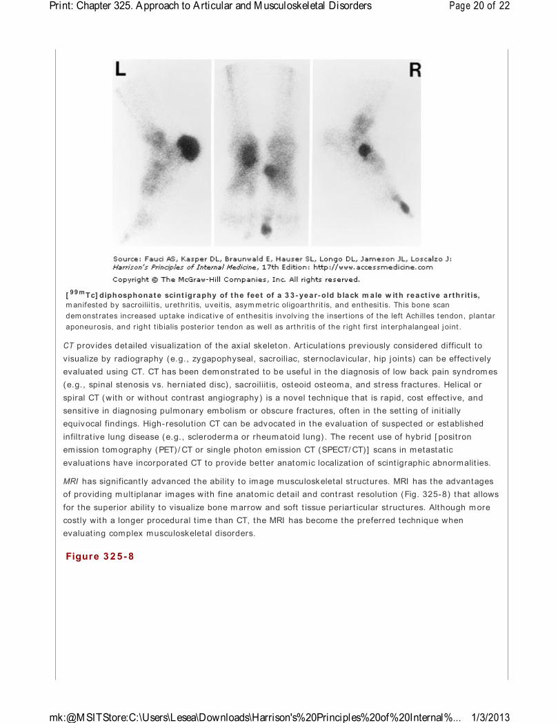
Scint igraphy, using 99mTc, 67Ga, or 111I n- labeled WBCs has been applied to a variety of art icular disorders with variable success (Table 325-5) . Although [ 99m Tc] pertechnate or diphosphate scint igraphy (Fig. 325-7) m ay be useful in identifying osseous infect ion, neoplasia, inflamm at ion, increased blood flow, bone rem odeling, heterotopic bone format ion, or avascular necrosis, MRI is preferred in most
instances. The poor specificity of 99m Tc scanning has largely limited its use to surveys for bone metastases and Paget 's disease of bone. Gallium scanning ut ilizes 67Ga, which binds serum and cellular t ransferrin and lactoferrin, and is preferent ially taken up by neut rophils, macrophages, bacteria, and tum or t issue (e.g., lym phoma) . As such, it is prim arily used in the ident if icat ion of occult infect ion or
malignancy. Scanning with 111I n- labeled WBCs has been used to detect osteom yelit is and infect ious and inflamm atory arthrit is. Nevertheless, the use of 111I n- labeled WBC or 67Ga scanning has largely been replaced by MRI , except when there is a suspicion of prosthet ic joint infect ions.
Table 3 2 5 - 5 Diagnost ic I m aging Techniques for Musculoskeletal Disorders
Method I m aging Tim e, h
Costa Current I ndicat ions
Ult rasoundb < 1 + Synovial cysts
Rotator cuff tears
Tendon injury
Radionuclide scint igraphy
99m Tc 1–4 + + Metastat ic bone survey
Page 18 of 22Print: Chapter 325. Approach to Articular and M usculoskeletal Disorders
1/3/2013mk:@M SITStore:C:\Users\Lesea\Downloads\Harrison's%20Principles%20of%20Internal%...
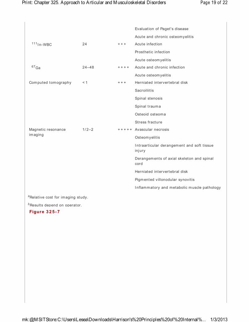
Evaluat ion of Paget 's disease
Acute and chronic osteomyelit is
111I n-WBC 24 + + + Acute infect ion
Prosthet ic infect ion
Acute osteom yelit is
67Ga 24–48 + + + + Acute and chronic infect ion
Acute osteom yelit is
Com puted tomography < 1 + + + Herniated intervertebral disk
Sacroiliit is
Spinal stenosis
Spinal t raum a
Osteoid osteoma
Stress fracture
Magnetic resonance im aging
1/ 2–2 + + + + + Avascular necrosis
Osteomyelit is
I ntraart icular derangem ent and soft t issue injury
Derangem ents of axial skeleton and spinal cord
Herniated intervertebral disk
Pigm ented v illonodular synovit is
I nflammatory and metabolic m uscle pathology
aRelat ive cost for imaging study.
bResults depend on operator.
Figure 3 2 5 - 7
Page 19 of 22Print: Chapter 325. Approach to Articular and M usculoskeletal Disorders
1/3/2013mk:@M SITStore:C:\Users\Lesea\Downloads\Harrison's%20Principles%20of%20Internal%...
CT provides detailed visualizat ion of the axial skeleton. Art iculat ions previously considered difficult to visualize by radiography (e.g., zygapophyseal, sacroiliac, sternoclavicular, hip joints) can be effect ively evaluated using CT. CT has been dem onstrated to be useful in the diagnosis of low back pain syndrom es (e.g., spinal stenosis vs. herniated disc) , sacroiliit is, osteoid osteoma, and st ress fractures. Helical or spiral CT (with or without cont rast angiography) is a novel technique that is rapid, cost effect ive, and sensit ive in diagnosing pulmonary embolism or obscure fractures, often in the set t ing of init ially equivocal findings. High- resolut ion CT can be advocated in the evaluat ion of suspected or established infilt rat ive lung disease (e.g., scleroderm a or rheumatoid lung) . The recent use of hybrid [ positron em ission tom ography (PET) / CT or single photon em ission CT (SPECT/ CT) ] scans in metastat ic evaluat ions have incorporated CT to provide better anatomic localizat ion of scint igraphic abnormalit ies.
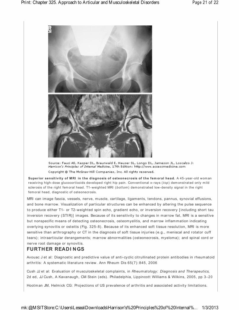
MRI has significant ly advanced the abilit y to image musculoskeletal st ructures. MRI has the advantages of providing m ult iplanar images with fine anatomic detail and cont rast resolut ion (Fig. 325-8) that allows for the superior abilit y to visualize bone m arrow and soft t issue periart icular st ructures. Although m ore costly with a longer procedural t im e than CT, the MRI has become the preferred technique when evaluat ing complex musculoskeletal disorders.
[ 99 mTc] diphosphonate scint igraphy of the feet of a 3 3 - year-old b lack m ale w ith react ive arthr it is, m anifested by sacroiliit is, urethrit is, uveit is, asym metr ic oligoar thr it is, and enthesit is. This bone scan dem onst rates increased uptake indicat ive of enthesit is involv ing t he insert ions of t he left Achilles t endon, plantar aponeurosis, and r ight t ibialis posterior t endon as well as art hrit is of the r ight first interphalangeal j oint .
Figure 3 2 5 - 8
Page 20 of 22Print: Chapter 325. Approach to Articular and M usculoskeletal Disorders
1/3/2013mk:@M SITStore:C:\Users\Lesea\Downloads\Harrison's%20Principles%20of%20Internal%...
MRI can image fascia, vessels, nerve, muscle, cart ilage, ligaments, tendons, pannus, synovial effusions, and bone marrow. Visualizat ion of part icular st ructures can be enhanced by altering the pulse sequence to produce either T1- or T2-weighted spin echo, gradient echo, or inversion recovery [ including short tau inversion recovery (STI R) ] images. Because of it s sensit ivity to changes in m arrow fat , MRI is a sensit ive but nonspecific m eans of detect ing osteonecrosis, osteomyelit is, and marrow inflam mat ion indicat ing overly ing synovit is or osteit is (Fig. 325-8) . Because of its enhanced soft t issue resolut ion, MRI is m ore sensit ive than arthrography or CT in the diagnosis of soft t issue injuries (e.g., m eniscal and rotator cuff tears) ; int raart icular derangements; m arrow abnorm alit ies (osteonecrosis, myeloma) ; and spinal cord or nerve root damage or synovit is.
Superior sensit ivity of MRI in the diagnosis of osteonecrosis of the fem or al head. A 45-year-old wom an receiv ing high-dose glucocor t icoids developed r ight hip pain. Convent ional x- rays ( top ) dem onst rated only m ild sclerosis of the right fem oral head. T1-weighted MRI (bot tom ) demonstrated low-density signal in the right femoral head, diagnost ic of osteonecrosis.
FURTHER READI NGSAvouac J et al: Diagnost ic and predict ive value of ant i-cyclic cit rullinated protein ant ibodies in rheum atoid arthrit is: A systemat ic literature rev iew. Ann Rheum Dis 65(7) : 845, 2006
Cush JJ et al: Evaluation of m usculoskeletal com plaints, in Rheum atology: Diagnosis and Therapeut ics, 2d ed, JJ Cush, A Kavanaugh, CM Stein (eds) . Philadelphia, Lippincot t Williams & Wilkins, 2005, pp 3–20
Hootm an JM, Helm ick CG: Project ions of US prevalence of arthrit is and associated act ivity limitat ions.
Page 21 of 22Print: Chapter 325. Approach to Articular and M usculoskeletal Disorders
1/3/2013mk:@M SITStore:C:\Users\Lesea\Downloads\Harrison's%20Principles%20of%20Internal%...
Arthrit is Rheum 54(1) : 226, 2006
Kavanaugh A: The ut ility of im munologic laboratory tests in pat ients with rheumatic diseases. Arthrit is Rheum 44: 2221, 2001 [ PMI D: 11665961]
Ory PA: Radiography in the assessment of m usculoskeletal condit ions. Best Pract Res Clin Rheum atol 17(3) : 495, 2003
Rudwaleit M et al: How to diagnose axial spondyloarthrit is early. Ann Rheum Dis 63(5) : 535, 2004
Shmerling RH et al: Synovial fluid tests: What should be ordered? JAMA 264: 1009, 1990 [ PMID: 2198352]
Siva C et al: Diagnosing acute monoarthrit is in adults: A pract ical approach for the fam ily physician. Am Fam Physician 68(1) : 83, 2003
Copyr ight © The McGraw-Hill Com panies. All r ights reserved.Pr ivacy Not ice. Any use is subject t o the Term s of Use and Not ice. Addit ional Credits and Copyright I nformat ion.
Page 22 of 22Print: Chapter 325. Approach to Articular and M usculoskeletal Disorders
1/3/2013mk:@M SITStore:C:\Users\Lesea\Downloads\Harrison's%20Principles%20of%20Internal%...