ascl1 is required for the development of specific … · development/plasticity/repair...
TRANSCRIPT

Development/Plasticity/Repair
Ascl1 Is Required for the Development of Specific NeuronalSubtypes in the Enteric Nervous System
X Fatima Memic,1 Viktoria Knoflach,1 X Rebecca Sadler,1 Gunilla Tegerstedt,2 Erik Sundstrom,3,4 X Francois Guillemot,5
Vassilis Pachnis,5 and X Ulrika Marklund1
1Division of Molecular Neurobiology, Department of Medical Biochemistry and Biophysics, Karolinska Institutet, S-17177 Stockholm, Sweden,2Department of Clinical Science, Intervention and Technology, Karolinska University Hospital, Huddinge, Karolinska Institutet, S-17177 Stockholm,Sweden, 3Division of Neurodegeneration, Department of Neurobiology, Care Sciences and Society, Novum, Karolinska Institutet, S-14186 Stockholm,Sweden, 4Stockholms Sjukhem Foundation, R& D Unit, S-10226 Stockholm, Sweden, and 5Francis Crick Institute, Mill Hill Laboratory, The Ridgeway,Mill Hill, London NW7 1AA, United Kingdom
The enteric nervous system (ENS) is organized into neural circuits within the gastrointestinal wall where it controls the peristalticmovements, secretion, and blood flow. Although proper gut function relies on the complex neuronal composition of the ENS, little isknown about the transcriptional networks that regulate the diversification into different classes of enteric neurons and glia duringdevelopment. Here we redefine the role of Ascl1 (Mash1), one of the few regulatory transcription factors described during ENS develop-ment. We show that enteric glia and all enteric neuronal subtypes appear to be derived from Ascl1-expressing progenitor cells. In the gutof Ascl1 �/� mutant mice, neurogenesis is delayed and reduced, and posterior gliogenesis impaired. The ratio of neurons expressingCalbindin, TH, and VIP is selectively decreased while, for instance, 5-HT � neurons, which previously were believed to be Ascl1-dependent, are formed in normal numbers. Essentially the same differentiation defects are observed in Ascl1KINgn2 transgenic mutants,where the proneural activity of Ngn2 replaces Ascl1, demonstrating that Ascl1 is required for the acquisition of specific enteric neuronalsubtype features independent of its role in neurogenesis. In this study, we provide novel insights into the expression and function of Ascl1in the differentiation process of specific neuronal subtypes during ENS development.
Key words: differentiation; enteric nervous system; gliogenesis; mutant mice; neurogenesis; transcription factor
IntroductionThe enteric nervous system (ENS) is the largest, most phenotyp-ically complex part of the PNS, and regulates the peristaltic move-
ments, blood flow, and secretion within the gut (Furness, 2006;Sasselli et al., 2012; Obermayr et al., 2013a). The ENS is mainlyderived from neural crest stem cells (NCSCs), which upon enter-ing the foregut colonize the full extent of the bowel wall at earlydevelopmental stages. While undertaking this extensive migra-tion, the enteric neural stem cells (ENSCs) proliferate immenselyand differentiate into a multitude of distinct neuronal subtypes
Received Jan. 19, 2016; revised Feb. 18, 2016; accepted Feb. 25, 2016.Author contributions: F.M., V.K., R.S., and U.M. designed research; F.M., V.K., R.S., G.T., E.S., and U.M. performed
research; G.T., E.S., F.G., and V.P. contributed unpublished reagents/analytic tools; F.M., V.K., R.S., V.P., and U.M.analyzed data; F.M., V.K., R.S., V.P., and U.M. wrote the paper.
This work was supported by the Knut and Alice Wallenberg Foundation KAW2008.0123, Swedish ResearchCouncil 521-2012-1676, EMBO, Swedish Society for Medical Research, Swedish Medical Society, Ruth and RichardJulin Foundation, Magnus Bergvall Foundation, and Åke Wiberg Foundation. We thank Reena Lasrado, TiffanyHeanue, Daniel Gyllborg, Eva Hedlund, and Moritz Lubke for constructive comments on the manuscript.
The authors declare no competing financial interests.
Correspondence should be addressed to Dr. Ulrika Marklund, Karolinska Institutet, Department of MedicalBiochemistry and Biophysics, Unit of Molecular Neurobiology, Scheeles vag 1, S-17177 Stockholm, Sweden.E-mail: [email protected].
DOI:10.1523/JNEUROSCI.0202-16.2016Copyright © 2016 the authors 0270-6474/16/364339-12$15.00/0
Significance Statement
The molecular mechanisms underlying the generation of different neuronal subtypes during development of the enteric nervoussystem are poorly understood despite its pivotal function in gut motility and involvement in gastrointestinal pathology. Thisreport identifies novel roles for the transcription factor Ascl1 in enteric gliogenesis and neurogenesis. Moreover, independent ofits proneurogenic activity, Ascl1 is required for the normal expression of specific enteric neuronal subtype characteristics. Distinctenteric neuronal subtypes are formed in a temporally defined order, and we observe that the early-born 5-HT � neurons aregenerated in Ascl1�/� mutants, despite the delayed neurogenesis. Enteric nervous system progenitor cells may therefore possessstrong intrinsic control over their specification at the initial waves of neurogenesis.
The Journal of Neuroscience, April 13, 2016 • 36(15):4339 – 4350 • 4339

and enteric glia in a nonsynchronized manner (Sasselli et al.,2012). Developmental failure to form neurons in the distal part ofthe gut results in the most well-characterized ENS disorder,Hirschsprung disease. In other ENS-linked diseases (e.g., achala-sia), specific subtypes of neurons are selectively affected (Furness,2006, 2012). Despite the critical role of distinct neuronal subtypesfor gut function and a significant progress in understanding themolecular basis to ENSC migration and proliferation, today thereis little knowledge of the regulatory mechanisms and networks oftranscription factors controlling the diversification of ENSCsinto different classes of enteric neurons during development.
Considerable recent advances have been made in identifyinggene regulatory networks that control the development of clini-cally relevant neuronal subtypes in other parts of the PNS and inthe CNS (Kiyasova and Gaspar, 2011; Lallemend and Ernfors,2012; Hegarty et al., 2013). One transcription factor that has beenimplicated in cell fate specification is Ascl1 (Mash1), which be-longs to the proneural family of neural bHLH genes [also includ-ing Neurogenin (Ngn) 1–3 and Math1]. Through loss-of-function(LOF) analyses, Ascl1 was initially identified as a key regulator ofneurogenesis in both PNS and CNS, but gain-of-function (GOF)experiments suggested additional roles in regulating subtype-specific aspects of neuronal differentiation (Guillemot et al.,1993; Hirsch et al., 1998; Lo et al., 1998; Fode et al., 2000). Toinvestigate these functions separately, Parras et al. (2002) gener-ated transgenic mice (Ascl1KINgn2), where the coding region ofAscl1 was replaced by Ngn2. In Ascl1KINgn2 mutants, neurogenesisis rescued in many regions of the CNS, revealing the critical re-quirement for Ascl1 in subtype differentiation, for instance, ofhindbrain 5-HT neurons and spinal V2a interneurons (Parras etal., 2002; Pattyn et al., 2004).
Ascl1 is expressed in ENSCs, and previous studies have indicateda role in both enteric neurogenesis (Lo et al., 1991; Guillemot et al.,1993; Lo and Anderson, 1995; Sang et al., 1999) and the selectiveformation of an enteric neuronal subtype (5-HT�) (Blaugrund etal., 1996). However, it is unclear whether Ascl1 expression is con-fined to specific ENSC populations and what possible roles it has inthe generation of the various enteric neurons and glia. Consideringthe importance of Ascl1 in the formation of accurate types and num-bers of neurons during CNS development, we sought to make acomprehensive study of the functions of Ascl1 in the developingENS. Our data indicate that progenitors of all enteric neuronal sub-types and enteric glia express Ascl1 at early stages of development. Inthe gut of Ascl1�/� mutant mice, gliogenesis is locally impaired,whereas neurogenesis is notably delayed and reduced. We identifiedselective reductions in the percentage of neurons expressing Calbin-din (stomach, small intestine), VIP (stomach), and TH (stomach).These abnormal ratios of selective neuronal subgroups were not dueto the delayed onset of neurogenesis as a similar phenotype wasobserved in Ascl1KINgn2 mutants, where Ngn2 rescues enteric neuro-genesis. Together, these data demonstrate novel roles for Ascl1 in theacquisition of specific neuronal subtype characteristics during ENSdevelopment, redefining the prevailing model of Ascl1 function inthe diversification of enteric neurons.
Materials and MethodsMouse and human embryos. The generation of Ascl1 CreERT2 (The JacksonLaboratory) (Kim et al., 2011), Ascl1KINgn2 (Parras et al., 2002), Wnt1-Cre(Danielian et al., 1998), and R26ReYFP (Srinivas et al., 2001) mouse lineshas previously been described. Ascl1CreERT2/ CreERT2 mutant embryos aredenoted Ascl1�/� throughout this paper. Remains of human embryosand fetuses (5.5 and 8 weeks after conception) were collected after elec-tive routine abortions with written consent given by the pregnant
woman. Collection of tissue for research was approved by the RegionalHuman Ethics Committee, Stockholm.
Tissue preparation. E10.5–13.75 mouse embryos were collected andfixed in 4% PFA in PBS, pH 7.4, at 4°C for 1.5 h. E15.5 and E18.5 gutswere dissected out from the embryos and fixed for 2 h. Human gut tissuewas fixed for 2 h. Samples were subsequently washed in PBS at 4°C for 1 hand cryoprotected by incubating at 4°C overnight in 30% sucrose in PBS.The tissue was embedded in OCT (Histolab) and stored at �80°C. Sam-ples were cryosectioned at 14 �m and stored at �20°C after drying atroom temperature for 1 h.
In situ hybridization. In situ hybridization on cryosections was per-formed as previously described (Briscoe et al., 2000). The isolation of RetcDNA was described by Pachnis et al. (1993).
Immunohistochemistry. Fluorescent immunohistochemical stainingswere performed as described previously (Briscoe et al., 2000) using thefollowing primary antibodies: mouse HuC/D (1:300; Molecular Probes),Ascl1 (1:300; BD Biosciences Pharmingen), Math1 (1:20; DSHB); goatChat (1:500; Millipore), Sox10 (1:200; Santa Cruz Biotechnology), Neu-roD1 (1:100; Santa Cruz Biotechnology), Ngn1 (1:500; Santa Cruz Bio-technology), GFP (488-conjugated, 1:1000; Abcam); rabbit Ki67 (1:500;Thermo Scientific), NPY (1:3000; DiaSorin), VIP (1:2500; Abcam),cleaved caspase-3 (1:1000; Cell Signaling Technology), CGRP (1:1000;Immunostar), NOS1 (1:500; Santa Cruz Biotechnology), substance P(1:1000; Millipore Bioscience Research Reagents), Calbindin (1:1000;Millipore Bioscience Research Reagents), TH (1:500; Pelfreeze), 5-HT(1:2000; Sigma), S100 (1:1000; DAKO), Blbp (1:300; Millipore), Ngn2(1:500; Millipore), Sox2 (1:4000; Seven Hill Bioreagents), and guinea pigSox10 (1:1000, kind gift from M. Wegner). Primary antibodies weredetected with secondary antibody conjugated with AlexaFluor-488, -555,and -647 (Invitrogen) or CY5 (Jackson ImmunoResearch Laboratories).
5-Ethynyl-2�-deoxyuridine (EdU) and tamoxifen experiments. Timeplug-mated Ascl1 CreERT2 and Ascl1KINgn2 mice received a single intraperi-toneal injection of EdU (0.1 mg/g animal; Invitrogen) at E12.5. Micewere killed 90 min after injection. Embryos were prepared for immuno-histochemistry as described above. Tissue sections were first immuno-stained for HuC/D and Sox10 and then reacted for EdU using the Click iTEdU AlexaFluor-488 Imaging kit (Invitrogen). After 20 min, slides werewashed in PBS and mounted. Time plug-mated Ascl1CreERT2 �R26ReYFP mice received intraperitoneal injection of tamoxifen dissolvedin peanut oil (0.1 mg/g animal, Sigma) at E10.5 and/or E13.5. E18.5embryos were collected and prepared as described.
Statistical analysis and imaging. All cells were counted on a minimum of5 sections (up to 15 sections) per investigated region of the gut in mutant andlittermate wt embryos. Paired Student’s t test comparisons were performedfor all statistical examinations. Bars indicate mean � SD; n � 3–6. Theimages were taken using a Zeiss LSM700 confocal microscope and processedin Adobe Photoshop CS6 or ImageJ (National Institutes of Health). Count-ing of fluorescent cells was performed using the “Cell Counter” plugin inImageJ or directly under a Zeiss fluorescent microscope.
ResultsAscl1 is transiently expressed in progenitors and precursorsof all perinatal enteric neuronal subtypesTo investigate the expression pattern of Ascl1 in the developingmouse gut, we performed fluorescent immunohistochemistry oncryosections from embryonic day 9.5 (E9.5) to E18.5. Onset ofAscl1 expression correlated with the NCSC entry into the foregutand was present in the majority of these cells as assessed by coex-pression with YFP in Wnt1-Cre � R26ReYFP E9.5 embryos (Fig.1A) (Lo et al., 1991). Throughout development, Ascl1 was pri-marily confined to the Sox10� population, which corresponds tothe ENSCs, both in the stomach and small intestine (�55%; Fig.1E–G,K). In addition, a subset of cells expressing the neuronalmarker HuC/D, colabeled with Ascl1 at early stages (Fig. 1B,L).Most Ascl1�HuC/D� cells coexpressed Sox10 and the cell cyclemarker Ki67 (Fig. 1C,D), indicating that they were immatureneurons. Between E15.5 and E18.5, Ascl1 expression was only
4340 • J. Neurosci., April 13, 2016 • 36(15):4339 – 4350 Memic et al. • Ascl1 Function during ENS Development
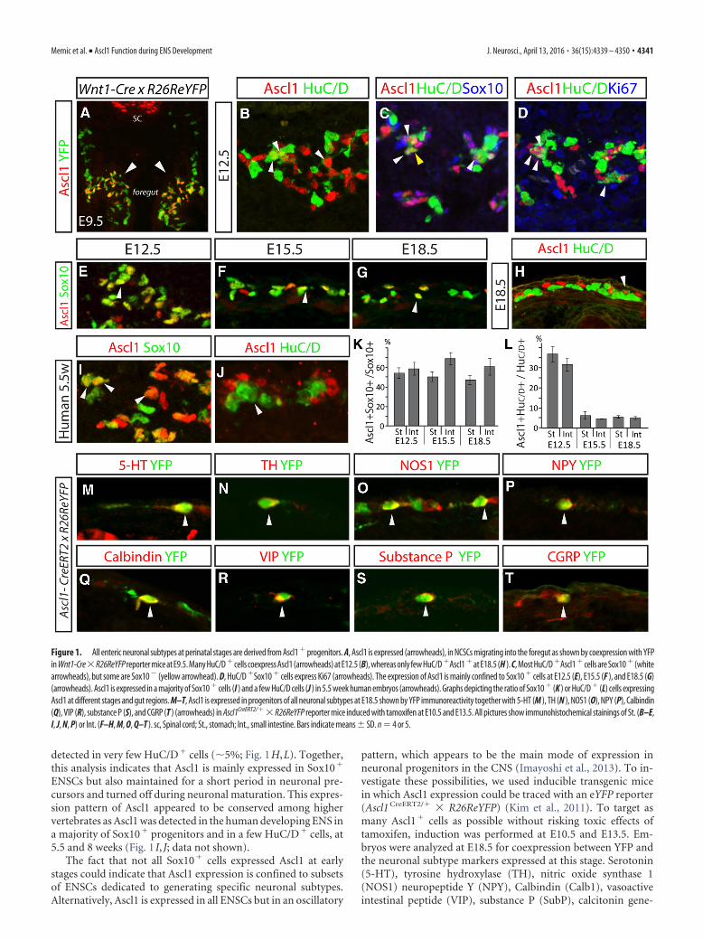
detected in very few HuC/D� cells (�5%; Fig. 1H,L). Together,this analysis indicates that Ascl1 is mainly expressed in Sox10�
ENSCs but also maintained for a short period in neuronal pre-cursors and turned off during neuronal maturation. This expres-sion pattern of Ascl1 appeared to be conserved among highervertebrates as Ascl1 was detected in the human developing ENS ina majority of Sox10� progenitors and in a few HuC/D� cells, at5.5 and 8 weeks (Fig. 1 I, J; data not shown).
The fact that not all Sox10� cells expressed Ascl1 at earlystages could indicate that Ascl1 expression is confined to subsetsof ENSCs dedicated to generating specific neuronal subtypes.Alternatively, Ascl1 is expressed in all ENSCs but in an oscillatory
pattern, which appears to be the main mode of expression inneuronal progenitors in the CNS (Imayoshi et al., 2013). To in-vestigate these possibilities, we used inducible transgenic micein which Ascl1 expression could be traced with an eYFP reporter(Ascl1 CreERT2/� � R26ReYFP) (Kim et al., 2011). To target asmany Ascl1� cells as possible without risking toxic effects oftamoxifen, induction was performed at E10.5 and E13.5. Em-bryos were analyzed at E18.5 for coexpression between YFP andthe neuronal subtype markers expressed at this stage. Serotonin(5-HT), tyrosine hydroxylase (TH), nitric oxide synthase 1(NOS1) neuropeptide Y (NPY), Calbindin (Calb1), vasoactiveintestinal peptide (VIP), substance P (SubP), calcitonin gene-
Figure 1. All enteric neuronal subtypes at perinatal stages are derived from Ascl1 � progenitors. A, Ascl1 is expressed (arrowheads), in NCSCs migrating into the foregut as shown by coexpression with YFPin Wnt1-Cre�R26ReYFP reporter mice at E9.5. Many HuC/D �cells coexpress Ascl1 (arrowheads) at E12.5 (B), whereas only few HuC/D �Ascl1 �at E18.5 (H ). C, Most HuC/D �Ascl1 �cells are Sox10 � (whitearrowheads), but some are Sox10 � (yellow arrowhead). D, HuC/D �Sox10 � cells express Ki67 (arrowheads). The expression of Ascl1 is mainly confined to Sox10 � cells at E12.5 (E), E15.5 (F ), and E18.5 (G)(arrowheads). Ascl1 is expressed in a majority of Sox10 � cells (I ) and a few HuC/D cells (J ) in 5.5 week human embryos (arrowheads). Graphs depicting the ratio of Sox10 � (K ) or HuC/D � (L) cells expressingAscl1 at different stages and gut regions. M–T, Ascl1 is expressed in progenitors of all neuronal subtypes at E18.5 shown by YFP immunoreactivity together with 5-HT (M ), TH (N ), NOS1 (O), NPY (P), Calbindin(Q), VIP (R), substance P (S), and CGRP (T ) (arrowheads) in Ascl1CreERT2/�� R26ReYFP reporter mice induced with tamoxifen at E10.5 and E13.5. All pictures show immunohistochemical stainings of St. (B–E,I, J, N, P) or Int. (F–H, M, O, Q–T ). sc, Spinal cord; St., stomach; Int., small intestine. Bars indicate means � SD. n � 4 or 5.
Memic et al. • Ascl1 Function during ENS Development J. Neurosci., April 13, 2016 • 36(15):4339 – 4350 • 4341

related peptide (CGRP), and choline acetyltransferase (ChAT)were all detected in YFP� cells at all regions investigated (stom-ach and/or small intestine and/or large intestine) (Fig. 1M–T;data not shown). These data demonstrate that Ascl1� progeni-tors are capable of generating all phenotypically different neu-rons present at perinatal stages, suggesting that Ascl1 is expressedin the progenitors of all neuronal subtypes.
Loss of Ascl1 does not affect ENSC migration but results indelayed and reduced neurogenesisTo evaluate the role of Ascl1 in ENS development, we nextanalyzed the gut of Ascl1 mutant mice in which a CreER T2
cassette had replaced the entire coding region of Ascl1
(Ascl1 CreERT2/CreERT2) (Kim et al., 2011). For simplicity, we here-after denote homozygote embryos from this mouse line Ascl1�/�.Previous studies of an Ascl1 LOF mouse line (Guillemot et al.,1993) have shown that deletion of this locus delays the entericneurogenesis resulting in essential loss of neurons in the esopha-gus and reduced numbers of neurons in the stomach and intes-tine (Guillemot et al., 1993; Blaugrund et al., 1996; Sang et al.,1999). We compared wild-type (wt) and Ascl1�/� embryosthroughout the development and found that at stages E10.5-E12.5, when HuC/D expression is initiated in the ENS of wt em-bryos, HuC/D immunoreactivity was virtually undetectable inthe gut of Ascl1�/� embryos (Fig. 2A,B; data not shown). Be-tween E13.25 and 13.75, many strongly expressing HuC/D� neu-
Figure 2. The enteric neurogenesis is delayed and reduced in Ascl1�/� embryos. A, B, At E12.5, many cells express HuC/D in wt controls, but no or only weak HuC/D � cells can be detected inAscl1�/� embryos. The number and distribution of Sox10 (C, D) and Ret (E, F ) expressing cells at E12.5 are similar in wt and Ascl1�/� embryos. G–J, Between E13.25 and E13.75, expression ofHuC/D changes from being faint in a few cells to strong and defined in many cells in the stomach. K, L, At E18.5, the number of HuC/D-expressing cells is decreased in the stomach of Ascl1�/� mutantembryos. M, N, The number of neurons expressing TH (arrowheads) is diminished in Ascl1�/� mutants compared with control embryos at E13.75. O, P, There is no increased active caspase-3expression in Ascl1�/� embryos compared with wt embryos. Q–S, Ngn1, Ngn2, and Math1 were detected at E12.5 in the dorsal spinal cord (insets) but not in the ENS of Ascl1�/� mutant embryosat E15.5. Sox2 � cells are ENSCs (Heanue and Pachnis, 2011). T, Graph showing the ratio of numbers of neurons in Ascl1�/� mutant embryos compared with wt controls at different parts of the gutat E18.5. U, Graph showing that the ratio of Sox10 � cells that incorporated EdU after a 90 min pulse injection is slightly reduced in Ascl1�/� mutants compared with wt controls at E12.5. V, Graphshowing the percentage of HuC/D � neurons that express TH in Ascl�/� mutants compared with in wt control embryos at E13.75. The control was set to 1 in T, U. All pictures show immunohisto-chemical staining, except E, F, where in situ hybridization was applied. St.: A–D, G–L, O–S; Int: M, N. S.Int., Small intestine; n � 3–5. Bars indicate means � SD. *p 0.05, ***p 0.001.
4342 • J. Neurosci., April 13, 2016 • 36(15):4339 – 4350 Memic et al. • Ascl1 Function during ENS Development

ronal precursors appeared in the gut of Ascl1�/� mutants, thuswith a 3 d delay (Fig. 2G–J). A fraction of immature neurons aretransiently catecholaminergic (TH�) at �E10-E14 (Baetge andGershon, 1989), and these cells have been reported lost in Ascl1LOF mutants (Blaugrund et al., 1996). In our analysis, TH wasdetected at E13.75 concurrently with the first HuC/D� neuronsalbeit in reduced proportions (��80%) compared with controlembryos (Fig. 2M,N,V). Because Ascl1 LOF mutant mice dieshortly after birth (Guillemot et al., 1993), the latest possibletime-point for analysis was at E18.5-E19. At this stage, manyHuC/D� enteric neurons were observed in the guts of Ascl1�/�
mutants, but with a clear reduction compared with wt guts. Thedecrease was most pronounced in the anterior portions of thegastrointestinal tract, and there was a gradual normalization ofneuronal numbers toward the posterior end (esophagus (�93 �2.2%); stomach (�58 � 2.0%); small intestine (�34 � 16%);and colon (�20 � 15%, not significant) (Fig. 2K,L,T). Thatneurons eventually are generated in Ascl1�/� mutants could in-dicate an upregulation of partially compensating alternative pro-neural genes. However, we did not detect expression of Ngn1,Ngn2, or Math1 in the gut of either wt or Ascl1�/� mutant em-bryos, although immunoreactivity was readily observed in thespinal cord (Fig. 2Q–S; data not shown). In summary, these datashow a delayed neurogenesis and diminished, but not abolished,transient expression of TH in the gut of Ascl1�/� mutants.
The deficient neurogenesis in Ascl1�/� mutant embryosprompted us to investigate whether other basic cellular processes,such as migration, proliferation, and survival, were impaired inthe absence of Ascl1. At E12.5, we observed similar numbers ofcells expressing the early ENSC marker Ret throughout the gas-trointestinal tract in Ascl1�/� and wt control embryos, indicatingthat mutant ENSCs migrated normally (Fig. 2E,F). The numberof Sox10� cells was also not altered in the mutant compared withcontrol guts at E12.5 (Fig. 2C,D). The ratio of Sox10� cells in theS-phase of the cell cycle, incorporating the nucleoside analogEdU after a 2 h pulse were slightly decreased in the mutant versuscontrol embryos (Fig. 2U). This could indicate that progenitorsat this stage are only slowly dividing or temporarily stalled in thecell cycle in the absence of Ascl1. We also did not observe anyincreased cell death in the ENS of Ascl1�/� mutants comparedwith wt as assessed by cleaved caspase-3 expression at E13.75 (Fig.2O,P). Together, this analysis suggests that Ascl1 function is notcrucial for ENSCs migration and survival but required for thetimely initiation of neuronal differentiation.
The expression of specific enteric neuronal subtype markersis reduced in the gut of Ascl1�/� mutantsWe next addressed whether the generation of specific entericneuronal subtypes was affected in the Ascl1�/� mutants. BecauseAscl1 mutants die shortly after birth, we focused our analysis onrepresentative segments of the stomach (St.) and small intestine(S.Int.) at E18.5-E19. At this stage, the following subtype markersare expressed in wt embryos: (St.) 5-HT, VIP, NOS1, Calbindin,TH, NPY; (S.Int.) 5-HT, VIP, NOS1, Calbindin, and CGRP. SubPand ChAT are also detectable, but the staining is punctate andtherefore difficult to quantify. Robust expression of Calretininand GABA has not yet commenced at E18.5. To unequivocallycompare the expression of subtype marker genes in Ascl1�/� em-bryos to littermate wt controls (in which the number of HuC/D�
neurons were higher), we compared the percentage of neuronsexpressing a given subtype marker. In the stomach, the percent-age of neurons expressing Calbindin, VIP, or TH was substan-tially reduced (�60% to �70% compared with control) (Fig.
3A,B,E,F,Q,R,W,X). The Calbindin-expressing proportion ofneurons was equally affected in the S.Int as in the stomach(�60%) (Fig. 3C,D,W,X). VIP, however, was not significantlyreduced in the S.Int (Fig. 3G,H,W,X). The number of neuronsexpressing TH was too low to quantify in the S.Int; but althoughscattered TH� cells were observed in the wt control, hardly anywere found in the mutant (data not shown). Surprisingly, 5-HT�
neurons, which are thought to require Ascl1 for their differenti-ation (Blaugrund et al., 1996), were generated in normal toslightly increased (however nonsignificant) ratios in Ascl1�/�
embryos compared with wt controls (Fig. 3M–P,W,X). The twomarkers, which had the most similar neuronal ratios in mutantand control, were CGRP (S.Int.) and NPY (St.) (Fig. 3S–V).NOS1 expression was slightly increased in the S.Int (30%) but notsignificantly different from control in the St. (Fig. 3I–L,W,X).Together, in the gut of Ascl1�/� mutants, the ratios of neuronsexpressing Calbindin, VIP, and TH in the stomach, and Calbin-din in the small intestine were drastically reduced. In contrast, thepercentage of neurons expressing CGRP, NPY, 5-HT, NOS1(St.), and VIP (St.) was not significantly different to control,whereas NOS1 neurons (S.Int.) were generated in an increasedproportion compared with wt control embryos (Fig. 3W,X).
The neurogenic deficiency in the absence of Ascl1 can berescued by Ngn2 in AsclKINgn2 mutantsThe decreased expression of selective enteric subtype markers inthe gut of Ascl1�/� embryos indicates that Ascl1 is not merelyinvolved in enteric neurogenesis but also plays a role in neuronalsubtype specification. However, as neurogenesis was perturbedin the absence of Ascl1, the reduction of neurons expressing spe-cific subtype markers could be due to a delay in marker expres-sion. To distinguish between the neurogenic function of Ascl1and its potential role in neuronal subtype specification, we ana-lyzed a transgenic knock-in mouse (Ascl1KINgn2), in which thecoding region of Ascl1 was replaced with the proneural geneNgn2, a gene that is capable of rescuing the neurogenic deficits ofAscl1�/� mice in the CNS (Parras et al., 2002; Pattyn et al., 2004).At E12.5, when the ENS of Ascl1�/� mutant had not yet started toexpress HuC/D (Fig. 2B), we observed normal or slightly in-creased numbers of HuC/D� cells in Ascl1KINgn2 mutants (Fig.4A,B), demonstrating that Ngn2 can rescue the neurogenesisdefect in the ENS of Ascl1�/� mutants. The neurogenesis ap-peared to be induced at the normal stage as only very fewHuC/D� cells were present at E10.5 in the gut of both Ascl1KINgn2
and wt embryos (data not shown). Analysis of the ENS at E18.5showed no difference in the final number of HuC/D� neuronsgenerated in Ascl1KINgn2 embryos compared with wt controls(104.3 � 9.2% in the small intestine; Fig. 4C,D, J). However,similar to in Ascl1�/� embryos, there was a reduced ratio of neu-rons transiently expressing TH at E12.5 (�80%; Fig. 4E,F,L).Proliferation appeared to be unaffected in Ascl1KINgn2 mutants asindicated by similar numbers of Sox10� cells compared withcontrol and the same ratio of EdU�Sox10�/Sox10� cells after a2 h pulse in the gut of mutant and wt embryos at E12.5 (Fig. 4K;data not shown). To verify that Ngn2 did not confer propertiesnormally associated with tissues regulated by Ngns, we examinedexpression of NeuroD1, which is a transcriptional target of Ngns(Ma et al., 1996; Sommer et al., 1996; Seo et al., 2007). At E12.5,when Ngn2-expressing tissues, such as the DRGs and intermedi-ate spinal cord, showed high expression of NeuroD1 (Fig. 4G), wedid not detect expression in the ENS of either Ascl1KINgn2 or wtcontrol embryos (Fig. 4H, I). Together, our analysis shows that
Memic et al. • Ascl1 Function during ENS Development J. Neurosci., April 13, 2016 • 36(15):4339 – 4350 • 4343

Figure 3. The ratio of neurons expressing specific subtype markers is reduced in the gut of Ascl1�/� mutant mice. A–V, Representative pictures of the stomach (St.) and small intestine (S.Int.)at E18.5 in wt and Ascl1�/� embryos showing drastically reduced ratios of neurons (marker �/HuC/D �; arrowhead) expressing Calbindin (A–D), VIP (E, F ), and TH (Q, R), a slight increased ratioof NOS1 (K, L) and no effect on 5-HT (M–P), VIP (G, H ), NOS1 (I, J ), NPY (S, T ), and CGRP (U, V ) neuronal ratios. W, The percentage of HuC/D � neurons expressing either of the markers (mean �SD) was calculated in the gut of Ascl1�/� mutants and wt controls at E18.5-E19. The table shows the percentages in Ascl1�/� embryos compared with wt controls (set to 100%). Red boxesrepresent a relative decrease. Blue box represents an increase compared with control. X, Graph showing the ratio of the percentage of neurons in the Ascl1�/� embryos expressing the varioussubmarkers (A–V ) compared with the percentage in the wt controls (set to 1). All pictures show immunohistochemical stainings. n � 4 – 6. Bars indicate means � SD. *p 0.05, **p 0.01,***p 0.001.
4344 • J. Neurosci., April 13, 2016 • 36(15):4339 – 4350 Memic et al. • Ascl1 Function during ENS Development

the proneural activity of Ngn2 can compensate for Ascl1 inAscl1KINgn2 embryos and induce normal neurogenesis in the ENSwithout respecifying the ENSCs.
Ascl1 is required for the acquisition of selective neuronalsubtype characteristics during ENS developmentAs enteric neurogenesis is restored in Ascl1KINgn2 mutants, wenext determined subtype marker expression in the gut of theseanimals. For this, we performed a similar analysis to that per-formed in Ascl1�/� mutants, counting the percentage of neuronsexpressing the various subtype markers in the stomach and smallintestine at E18.5-E19. We observed that the ratio of neuronsexpressing Calbindin (St., S.Int), VIP (St.), and TH (St.) wasdrastically reduced in Ascl1KINgn2 mutants (�60% to �80%; Fig.5A–D–F,Q,R,W,X), whereas VIP (S.Int.), 5-HT (St., S.Int), NPY(St.), and CGRP (S.Int.) were unaffected compared with controlembryos (Fig. 5G,H,M–P,S–X). NOS1 expression was normal inthe St. and marginally decreased in the S.Int compared with wtembryos (Fig. 5I–L,W,X). Thus, despite rescued neurogenesis inthe gut of Ascl1KINgn2 mutants, the proportions of neurons ex-pressing specific neuronal subtype markers were similar to thatobserved in Ascl1�/� mutants, indicating that Ascl1 function isrequired for the normal differentiation of specific neuronal sub-types (Fig. 5W,X).
Enteric glial marker expression is reduced in the posterior gutof Ascl1�/� and Ascl1KINgn2 mutantsConsidering that Sox10 is expressed in enteric glia in addition toENSCs and that the majority of the Sox10� cells throughoutdevelopment were Ascl1� (Fig. 1E–G,K), we next addressed
whether Ascl1 is expressed in progenitors and/or precursors ofthe glial lineage. To test whether glial-producing ENSCs expressAscl1, we lineage-traced Ascl1� cells using Ascl1 CreERT2/� �R26ReYFP animals, induced at E10.5 with tamoxifen. Analysis atE18.5 showed coexpression between YFP and the glial markersS100 and Blbp (Fig. 6A,B). Furthermore, we found coexpressionbetween Ascl1 and S100 or Blbp at both E15.5 and E18.5 in wtmice (Fig. 6C,D; data not shown). Together, these experimentsshow that Ascl1 is expressed both in glial (or bipotential) progen-itors and glial precursors during ENS development.
Next, to assess the function Ascl1 might have in gliogenesis, weanalyzed the expression of glial markers at the latest possible stage(E18.5) in the gut of both Ascl1�/� and Ascl1KINgn2 mutant mice. Thenumber of Sox10� cells appeared slightly reduced in both mutantsbut was only significantly lower in the distal part of the Ascl1KINgn2
(��20%) mutants compared with wt controls (Fig. 6H,I). To spe-cifically address the glial proportion of the Sox10� cells, we calcu-lated the ratio of Sox10 cells expressing the more mature markerS100 and found a reduction (��50%) in the ileum and colon but anormal ratio in the jejunum in Ascl1�/� mutants compared withcontrol embryos (Fig. 6E,F,H). In Ascl1KINgn2 mutants, theS100�Sox10�/Sox10� ratio was close to normal in the jejunum andileum, but a 40% reduction was observed in the colon comparedwith control embryos (Fig. 6E,G,I). S100 immunostaining in theanterior regions (esophagus and stomach) was indistinct in cell bod-ies at E18.5 and therefore hard to quantify, although no obviousdifferences between wt and the Ascl1 mutants were observed. AtE15.5, S100 staining was well defined also anteriorly and theS100�Sox10�/Sox10� ratio was not different in the stomach ofAscl1�/� and Ascl1KINgn2 mutants compared with controls (�5 �
Figure 4. The neurogenic impairment in the absence of Ascl1 is rescued in Ascl1KINgn2 embryos. A–D, At E12.5 and E18.5, Ascl1KINgn2 mutant embryos have similar numbers of HuC/D � cellscompared with wt controls. E, F, The number of neurons expressing TH (arrowheads) is reduced in Ascl1KINgn2 mutants compared with wt controls at E12.5. G–I, NeuroD1 expression is present atE12.5 in the SC and DRG, but not in the ENS of wt and Ascl1KINgn2 mutants. J, Graph showing the ratio of the number of neurons in Ascl1KINgn2 compared with control embryos in various parts of thegut. K, Graph showing that the ratio of Sox10 � cells that incorporated EdU after a 90 min pulse injection is similar in wt and Ascl1KINgn2 mutant embryos at E12.5. L, Graph showing the percentageof HuC/D � cells expressing TH in Ascl1KINgn2 and control embryos. Control was set to 1 in J, K. All pictures show immunohistochemical stainings of St. (A–D, H, I ) or Int. (E, F ). DRG, Dorsal rootganglia; sc, spinal cord; S.Int., small intestine. n � 4 or 5. Bars indicate means � SD. **p 0.01, ***p 0.001.
Memic et al. • Ascl1 Function during ENS Development J. Neurosci., April 13, 2016 • 36(15):4339 – 4350 • 4345

Figure 5. The ratio of neurons expressing specific subtype markers is reduced in the gut of Ascl1KINgn2 mutant mice. A–V, Representative pictures of the stomach (St.) and small intestine (s.Int)at E18.5 in wt and Ascl1KINgn2 embryos showing drastically reduced ratios (marker �/HuC/D �; arrowhead) of neurons expressing Calbindin (A–D), VIP (E, F ), and TH (Q, R), a slight reduction of theratio of NOS1 (K, L) and no effect on 5-HT (M–P), VIP (G, H ), NOS1 (I, J ), NPY (S, T ), and CGRP (U, V ) neuronal ratios. W, The percentage of HuC/D � neurons expressing either of the markers(mean � SD) was calculated in the gut of Ascl1KINgn2 mutants and wt controls at E18.5-E19. The table shows the percentages in Ascl1KINgn2 mutants compared with wt controls (set to 100%). Redboxes represent a relative decrease compared with control. X, Graph showing the ratio of the percentage of neurons in the Ascl1KINgn2 mutants expressing the various submarkers (A–V ) comparedwith the percentage in the wt controls (set to 1). All pictures show immunohistochemical stainings. n � 4 – 6. Bars indicate means � SD. **p 0.01, ***p 0.001.
4346 • J. Neurosci., April 13, 2016 • 36(15):4339 – 4350 Memic et al. • Ascl1 Function during ENS Development

14%; 2 � 24%), whereas statistically significant reductions were ob-served in the colon (�32 � 17%; �38 � 12%) similar to at E18.5.Thus, in the absence of Ascl1, gliogenesis appears to be compromisedin the posterior part of the bowel and cannot be fully compensatedfor by Ngn2 expression.
DiscussionThe mechanisms and transcriptionalnetworks, which regulate the sequentialgeneration of distinct types of enteric neu-rons in the developing gut, are largely un-known. This study demonstrates novel rolesfor Ascl1 in the acquisition of specific en-teric neuronal subtype traits, amending theprevailing model of Ascl1 expression andfunction during ENS development (Fig. 7).Our data suggest that all neuronal subtypesand glia arise from Ascl1� progenitors (Fig.7A), in which Ascl1 is required for a prop-erly timed neurogenesis and normalgliogenesis (Fig. 7A–C). By analyzing andcomparing Ascl1�/� (Fig. 7B) andAscl1KINgn2 (Fig. 7C) mutant embryos, wereveal that normal expression of Calbindin,VIP, and TH (but not 5-HT) requires Ascl1,independent of its function in neurogenesis.
The role of Ascl1 in gliogenesis andneurogenesis during ENS developmentIn the absence of Ascl1, sympatheticneurons are atrophic and delayed intheir formation (Guillemot et al., 1993).In contrast, partial ENS ganglia doform, although their generation is de-layed, similar to what has been observedin regions of the CNS (e.g., nucleus ofthe solitary tract) (Guillemot et al.,1993; Blaugrund et al., 1996; Pattyn etal., 2006). This indicates the presence orupregulation of proteins with addition-al/compensatory proneural activity inthe ENS. As we did not detect Ngn1,Ngn2, or Math1 in Ascl1�/� embryos,plausible compensatory candidates
could instead be Phox2b and Dlx proteins, which have genericproneural properties and are expressed in the developing ENS(Anderson et al., 1997; Dubreuil et al., 2002; Elworthy et al.,2005; Heanue and Pachnis, 2006).
Figure 6. Glial precursors express Ascl1, and glial numbers are reduced in the posterior ENS of Ascl1�/� and Ascl1KINgn2 mutants. A, B, Ascl1 is expressed in progenitors of enteric glia as shownby YFP expression (arrowheads) together with S100 and Blbp in Ascl1CreERT2/� � R26ReYFP reporter mice induced with tamoxifen at E10.5 and harvested at E18.5. C, D, At E15.5, subsets of S100 �
and Blbp � cells coexpress Ascl1 (arrowheads). E–G, The ratio of Sox10 � cells expressing S100 is reduced in the posterior gut of Ascl1�/� and Ascl1 KINgn2 embryos compared with wt controls. H,I, Graphs showing the percentage of Sox10 cells or S100 �Sox10 �/Sox10 � in Ascl1�/� (H ) and Ascl1KINgn2 (I ) embryos compared with wt controls (set to 100%) at E18.5. All pictures showimmunohistochemical stainings. n � 4 or 5. Bars indicate means � SD. *p 0.05, **p 0.01.
Figure 7. Schematic drawing summarizing the role of Ascl1 in enteric neurogenesis, neuronal subtype differentiation, andgliogenesis. A, Ascl1 (magenta) is expressed in the majority of progenitor cells at a given time, is transiently maintained in glial andneuronal precursors, and turned off in mature glia and neurons. B, In Ascl1�/� mutant mice, glial marker expression is reducedposteriorly and the neurogenesis is delayed and reduced. The percentages of neurons expressing Calbindin, VIP, and TH arereduced, while the ratio of NOS1 � neurons is slightly increased. C, In Ascl1KINgn2 mutant mice where the coding region of Ascl1 isreplaced with the proneural gene Ngn2 (blue), the neurogenesis is rescued but a mild defect in glial marker expression remains. Theratios of neurons expressing Calbindin, VIP, and TH are similar as in Ascl1�/� mutants, whereas the ratio of NOS1 � neurons isslightly decreased. In summary, we show that Ascl1 is required for the appropriate timing of neurogenesis and for the acquisitionof specific neuronal subtype characteristics during ENS development. n/a, Not applicable.
Memic et al. • Ascl1 Function during ENS Development J. Neurosci., April 13, 2016 • 36(15):4339 – 4350 • 4347

At perinatal stages, very few neurons are present in the esoph-agus, whereas the numbers increase posteriorly reaching normalamounts in the colon of Ascl1�/� embryos. This could reflect thatnormal gangliogenesis of the bowel depends on differentiation(i.e., differentiated cells are more likely to stay and form gangliathan are proliferating cells). Thus, the neurogenic delay inAscl1�/� mutants may have enabled ENSCs to migrate throughthe anterior parts leaving slightly fewer numbers of cells behind.Moreover, as normal neurogenesis occurs in an anterior to pos-terior gradient, the relative neurogenic delay would be shorter atposterior regions, enabling a close to normal neurogenesis in thecolon. Also, the ENS of the colon is partially derived from sacralNCSCs, which potentially could be less sensitive than vagalENSCs to loss of Ascl1.
We observed decreased numbers of Sox10� cells expressing theglial marker S100 at E18-E19 in the absence of Ascl1 at posteriorregions of the gut. As Ascl1KINgn2 mutants display a similar pheno-type (albeit milder), it is probably not only a secondary consequenceof the abnormal neurogenesis. Rather, it could be that Ascl1 partic-ipates in the process of gliogenesis, a role that has been attributed toAscl1 in the CNS. For example, studies in the spinal cord have shownthat Ascl1 regulates the maturation of oligodendrocytes and gener-ation of appropriate numbers of astrocytes (Battiste et al., 2007;Sugimori et al., 2008; Vue et al., 2014).
The function of Ascl1 in enteric neuronal subtypedifferentiationAscl1 regulates neuronal subtype differentiation of distinct neu-rons in the CNS (Parras et al., 2002; Pattyn et al., 2004) and directtranscriptional targets of Ascl1 have been identified comparingLOF and GOF genome-wide expression profiling with ChIP-chippromoter analysis (Castro et al., 2011). It was discovered thatAscl1 controls the acquisition of specific neuronal subtype fea-tures both by direct transcriptional regulation (e.g., TH) andsecondary downstream transcriptional cascades. Using Gene-Friends software (www.GeneFriends.org) (van Dam et al., 2015)for coexpressed genes, we found that VIP and TH have a highassociation with Ascl1 (fifth highest transcription factor, p �7.12e-04), whereas NOS1, CGRP, and NPY were not linked toAscl1. These findings are in agreement with our data that Ascl1regulates VIP and TH (either directly or indirectly) but notNOS1, CGRP, or NPY.
The specific reductions of Calb1�, VIP�, and TH� neurons inthe stomach and Calb1� neurons in the intestine of Ascl1 LOF mu-tants (Ascl1�/� and Ascl1KINgn2) indicate that the differentiation ofcertain subgroups of enteric neurons had been compromised. Theneuronal subtypes of the stomach are poorly characterized, in con-trast to the ileum (small intestine) where 16 subgroups have beendefined (Sang and Young, 1996; Sang et al., 1997; Qu et al., 2008;Mongardi Fantaguzzi et al., 2009). Calbindin is expressed in CGRP�
intrinsic primary afferent neurons, but also in an uncharacterizedCGRP� neuronal subpopulation (Qu et al., 2008). We believe thatthe lost Calb1� cells are likely to correspond to the Calb1�CGRP�
rather than intrinsic primary afferent neurons because CGRP ex-pression was not altered in the Ascl1 LOF mutants. In further sup-port of this, Ascl1 LOF mutants displayed a marked reduction oftransient catecholaminergic (TH�) cells, which recently have beenfate-mapped to generate Calb1� neurons but only a small propor-tion of intrinsic primary afferent neurons (Obermayr et al., 2013b).The combined loss of multiple markers (TH and Calb1) at sequen-tial stages during Calb1�CGRP� neuron differentiation in the ab-sence of Ascl1 indicates an early requirement for Ascl1 during thespecification process of this neuronal subgroup. Thus, our data sup-
port that Calb1�CGRP� neurons are reduced or have incompleteexpression profiles in the absence of Ascl1, but we are unable todetermine the identity of the lost TH� and VIP� neurons. However,of interest is that gut biopsies from patients with Parkinson’s diseaseshow pathology, affecting primarily TH� and VIP� neurons.(Wakabayashi et al., 2010). Although given less attention than thehallmark motor symptoms, Parkinson’s disease commonly involvessignificant gastrointestinal dysfunction that may be associated to theeffect seen on these neuronal subtypes (Fasano et al., 2015).
The ratio of neurons expressing NOS1 in the small intestinewas slightly increased in Ascl1�/� mutants. One possible expla-nation could be that the lost Calb1� cells had been undergoing afate switch to NOS1� neurons. Alternatively, NOS1� neuronscould be generated in normal absolute numbers because they areborn relatively late (peaking at E15.5) (Bergner et al., 2014). InAscl1KINgn2 mice, the percentage of NOS1� neurons was slightlydecreased in the small intestine, making it the only marker thatwas differentially affected in the two mutants. In these embryos,the neurogenesis appeared to be slightly enhanced at early stages,which perhaps could explain the negative impact on the genera-tion of late born NOS1� neurons.
The Ascl1-independent generation of 5-HT� neurons in the cur-rent study contradicts a previous analysis of an Ascl1 LOF mutant inwhich substantial loss of 5-HT neurons was reported (Blaugrund etal., 1996). One important difference between the present and previ-ous study is the detection methodology of 5-HT neurons. We usedantibodies with high affinity and selectivity to 5-HT, providingquantifiable detection of 5-HT� neurons. The previous study ap-plied radioautography to measure retrograde uptake of 3H-5-HT,which relies on a functional 5-HT-transporter (SERT). However,SERT has been shown to be expressed in nonserotonergic cells inother parts of the developing PNS and in the CNS (Narboux-Nemeet al., 2008; Chen et al., 2015). To our knowledge, the specificity ofSERT expression to 5-HT cells has not been addressed in the devel-oping ENS, making the validity of the 5-HT reuptake assay uncer-tain. Nevertheless, a combined account of our two studies of Ascl1LOF mutants could point to that 5-HT producing cells are made, buthave compromised functionality.
Birth-dating studies have demonstrated that the differententeric neurons are born in a temporally defined pattern; andin this, 5-HT � neurons are generated at early developmentaltime points, peaking at E11.5 (Pham et al., 1991; Bergner et al.,2014). In Ascl1�/� mutants, neurogenesis is delayed 3 d, com-mencing at E13.5. Despite this, we detected normal to slightlyincreased percentages of neurons expressing 5-HT at E18-E19.This could indicate that, at least in the first phase of the neu-rogenic period, ENSCs possess a strong intrinsic sequentialorder of differentiation occurring accurately even when de-layed, seemingly unaffected by possible changes occurring inthe external microenvironment.
The relevance of Ascl1 in ENS disorders and regenerativemedicineWe demonstrate that Ascl1 is expressed in the developing humanENS. Considering the important role for Ascl1 in enteric devel-opment in the mouse, it is possible that allelic Ascl1 variants couldpredispose to human gastrointestinal deficiencies. Mutations inAscl1 have been identified in patients with Ondine’s curse, a syn-drome including Hirschsprung disease (de Pontual et al., 2003),lending support for this idea.
The prospect of developing novel regenerative strategies totreat ENS disorders (e.g., Hirschsprung disease) emphasizes therequirement of understanding the differentiation cascades of dis-
4348 • J. Neurosci., April 13, 2016 • 36(15):4339 – 4350 Memic et al. • Ascl1 Function during ENS Development

tinct enteric neuronal subtypes. In other contexts, Ascl1 is beingexplored in its utility of converting various cell types to neurons.For instance, Liu et al. (2015) showed that forced Ascl1 expres-sion could convert midbrain astrocytes into functional neurons.Enteric glia are similar to CNS astrocytes; and following injury tothe ENS, there is a spontaneous conversion of enteric glia toneurons during which Ascl1 is transiently expressed (Laranjeiraet al., 2011). Therefore, one could speculate whether overexpres-sion of Ascl1 in enteric glia could be used as a strategy to makenew enteric neurons (perhaps specific types) in vivo in futuretreatments of enteric disorders.
ReferencesAnderson SA, Qiu M, Bulfone A, Eisenstat DD, Meneses J, Pedersen R,
Rubenstein JL (1997) Mutations of the homeobox genes Dlx-1 andDlx-2 disrupt the striatal subventricular zone and differentiation of lateborn striatal neurons. Neuron 19:27–37. CrossRef Medline
Baetge G, Gershon MD (1989) Transient catecholaminergic (TC) cells inthe vagus nerves and bowel of fetal mice: relationship to the developmentof enteric neurons. Dev Biol 132:189 –211. CrossRef Medline
Battiste J, Helms AW, Kim EJ, Savage TK, Lagace DC, Mandyam CD, EischAJ, Miyoshi G, Johnson JE (2007) Ascl1 defines sequentially generatedlineage-restricted neuronal and oligodendrocyte precursor cells in thespinal cord. Development 134:285–293. CrossRef Medline
Bergner AJ, Stamp LA, Gonsalvez DG, Allison MB, Olson DP, Myers MG Jr,Anderson CR, Young HM (2014) Birthdating of myenteric neuron sub-types in the small intestine of the mouse. J Comp Neurol 522:514 –527.CrossRef Medline
Blaugrund E, Pham TD, Tennyson VM, Lo L, Sommer L, Anderson DJ,Gershon MD (1996) Distinct subpopulations of enteric neuronal pro-genitors defined by time of development, sympathoadrenal lineage mark-ers and Mash-1-dependence. Development 122:309 –320. Medline
Briscoe J, Pierani A, Jessell TM, Ericson J (2000) A homeodomain proteincode specifies progenitor cell identity and neuronal fate in the ventralneural tube. Cell 101:435– 445. CrossRef Medline
Castro DS, Martynoga B, Parras C, Ramesh V, Pacary E, Johnston C, DrechselD, Lebel-Potter M, Garcia LG, Hunt C, Dolle D, Bithell A, Ettwiller L,Buckley N, Guillemot F (2011) A novel function of the proneural factorAscl1 in progenitor proliferation identified by genome-wide characteriza-tion of its targets. Genes Dev 25:930 –945. CrossRef Medline
Chen X, Ye R, Gargus JJ, Blakely RD, Dobrenis K, Sze JY (2015) Disruptionof transient serotonin accumulation by non-serotonin-producing neu-rons impairs cortical map development. Cell Rep 10:346 –358. CrossRefMedline
Danielian PS, Muccino D, Rowitch DH, Michael SK, McMahon AP (1998)Modification of gene activity in mouse embryos in utero by a tamoxifen-inducible form of Cre recombinase. Curr Biol 8:1323–1326. CrossRefMedline
de Pontual L, Nepote V, Attie-Bitach T, Al Halabiah H, Trang H, Elghouzzi V,Levacher B, Benihoud K, Auge J, Faure C, Laudier B, Vekemans M, Mun-nich A, Perricaudet M, Guillemot F, Gaultier C, Lyonnet S, Simonneau M,Amiel J (2003) Noradrenergic neuronal development is impaired bymutation of the proneural HASH-1 gene in congenital central hypoven-tilation syndrome (Ondine’s curse). Hum Mol Genet 12:3173–3180.CrossRef Medline
Dubreuil V, Hirsch MR, Jouve C, Brunet JF, Goridis C (2002) The role ofPhox2b in synchronizing pan-neuronal and type-specific aspects of neu-rogenesis. Development 129:5241–5253. Medline
Elworthy S, Pinto JP, Pettifer A, Cancela ML, Kelsh RN (2005) Phox2b func-tion in the enteric nervous system is conserved in zebrafish and is sox10-dependent. Mech Dev 122:659 – 669. CrossRef Medline
Fasano A, Visanji NP, Liu LW, Lang AE, Pfeiffer RF (2015) Gastrointestinaldysfunction in Parkinson’s disease. Lancet Neurol 14:625– 639. CrossRefMedline
Fode C, Ma Q, Casarosa S, Ang SL, Anderson DJ, Guillemot F (2000) A rolefor neural determination genes in specifying the dorsoventral identity oftelencephalic neurons. Genes Dev 14:67– 80. CrossRef Medline
Furness JB (2006) The enteric nervous system. Hoboken, NJ: Blackwell.Furness JB (2012) The enteric nervous system and neurogastroenterology.
Nat Rev Gastroenterol Hepatol 9:286 –294. CrossRef MedlineGuillemot F, Lo LC, Johnson JE, Auerbach A, Anderson DJ, Joyner AL
(1993) Mammalian achaete-scute homolog 1 is required for the earlydevelopment of olfactory and autonomic neurons. Cell 75:463– 476.CrossRef Medline
Heanue TA, Pachnis V (2006) Expression profiling the developing mamma-lian enteric nervous system identifies marker and candidate Hirschsprungdisease genes. Proc Natl Acad Sci U S A 103:6919 – 6924. CrossRefMedline
Heanue TA, Pachnis V (2011) Prospective identification and isolation ofenteric nervous system progenitors using Sox2. Stem Cells 29:128 –140.CrossRef Medline
Hegarty SV, Sullivan AM, O’Keeffe GW (2013) Midbrain dopaminergicneurons: a review of the molecular circuitry that regulates their develop-ment. Dev Biol 379:123–138. CrossRef Medline
Hirsch MR, Tiveron MC, Guillemot F, Brunet JF, Goridis C (1998) Controlof noradrenergic differentiation and Phox2a expression by MASH1 in thecentral and peripheral nervous system. Development 125:599 – 608.Medline
Imayoshi I, Isomura A, Harima Y, Kawaguchi K, Kori H, Miyachi H, FujiwaraT, Ishidate F, Kageyama R (2013) Oscillatory control of factors deter-mining multipotency and fate in mouse neural progenitors. Science 342:1203–1208. CrossRef Medline
Kim EJ, Ables JL, Dickel LK, Eisch AJ, Johnson JE (2011) Ascl1 (Mash1)defines cells with long-term neurogenic potential in subgranular and sub-ventricular zones in adult mouse brain. PLoS One 6:e18472. CrossRefMedline
Kiyasova V, Gaspar P (2011) Development of raphe serotonin neuronsfrom specification to guidance. Eur J Neurosci 34:1553–1562. CrossRefMedline
Lallemend F, Ernfors P (2012) Molecular interactions underlying the spec-ification of sensory neurons. Trends Neurosci 35:373–381. CrossRefMedline
Laranjeira C, Sandgren K, Kessaris N, Richardson W, Potocnik A, VandenBerghe P, Pachnis V (2011) Glial cells in the mouse enteric nervoussystem can undergo neurogenesis in response to injury. J Clin Invest121:3412–3424. CrossRef Medline
Liu Y, Miao Q, Yuan J, Han S, Zhang P, Li S, Rao Z, Zhao W, Ye Q, Geng J,Zhang X, Cheng L (2015) Ascl1 converts dorsal midbrain astrocytes intofunctional neurons in vivo. J Neurosci 35:9336 –9355. CrossRef Medline
Lo L, Anderson DJ (1995) Postmigratory neural crest cells expressing c-RETdisplay restricted developmental and proliferative capacities. Neuron 15:527–539. CrossRef Medline
Lo LC, Johnson JE, Wuenschell CW, Saito T, Anderson DJ (1991) Mamma-lian achaete-scute homolog 1 is transiently expressed by spatially re-stricted subsets of early neuroepithelial and neural crest cells. Genes Dev5:1524 –1537. CrossRef Medline
Lo L, Tiveron MC, Anderson DJ (1998) MASH1 activates expression of thepaired homeodomain transcription factor Phox2a, and couples pan-neuronal and subtype-specific components of autonomic neuronal iden-tity. Development 125:609 – 620. Medline
Ma Q, Kintner C, Anderson DJ (1996) Identification of neurogenin, a ver-tebrate neuronal determination gene. Cell 87:43–52. CrossRef Medline
Mongardi Fantaguzzi C, Thacker M, Chiocchetti R, Furness JB (2009) Iden-tification of neurin types in the submucosal ganglia of the mouse ileum.Cell Tissue Res 336:179 –189. CrossRef Medline
Narboux-Neme N, Pavone LM, Avallone L, Zhuang X, Gaspar P (2008)Serotonin transporter transgenic (SERTcre) mouse line reveals develop-mental targets of serotonin specific reuptake inhibitors (SSRIs). Neuro-pharmacology 55:994 –1005. CrossRef Medline
Obermayr F, Hotta R, Enomoto H, Young HM (2013a) Development anddevelopmental disorders of the enteric nervous system. Nat Rev Gastro-enterol Hepatol 10:43–57. CrossRef Medline
Obermayr F, Stamp LA, Anderson CR, Young HM (2013b) Genetic fate-mapping of tyrosine hydroxylase expressing cells in the enteric nervoussystem. Neurogastroenterol Motil 25:283–291. CrossRef
Pachnis V, Mankoo B, Costantini F (1993) Expression of the c-ret proto-oncogene during mouse embryogenesis. Development 119:1005–1017.Medline
Parras CM, Schuurmans C, Scardigli R, Kim J, Anderson DJ, Guillemot F(2002) Divergent functions of the proneural genes Mash1 and Ngn2 inthe specification of neuronal subtype identity. Genes Dev 16:324 –338.CrossRef Medline
Pattyn A, Simplicio N, van Doorninck JH, Goridis C, Guillemot F, Brunet JF
Memic et al. • Ascl1 Function during ENS Development J. Neurosci., April 13, 2016 • 36(15):4339 – 4350 • 4349

(2004) Ascl1/Mash1 is required for the development of central seroto-nergic neurons. Nat Neurosci 7:589 –595. CrossRef Medline
Pattyn A, Guillemot F, Brunet JF (2006) Delays in neuronal differentiationin Mash1/Ascl1 mutants. Dev Biol 295:67–75. CrossRef Medline
Pham TD, Gershon MD, Rothman TP (1991) Time of origin of neurons inthe murine enteric nervous system: sequence in relation to phenotype.J Comp Neurol 314:789 –798. CrossRef Medline
Qu ZD, Thacker M, Castelucci P, Bagyanszki M, Epstein ML, Furness JB(2008) Immunohistochemical analysis of neuron types in the mousesmall intestine. Cell Tissue Res 334:147–161. CrossRef Medline
Sang Q, Young HM (1996) Chemical coding of neurons in the myentericplexus and external muscle of the small and large intestine of the mouse.Cell Tissue Res 284:39 –53. CrossRef Medline
Sang Q, Williamson S, Young HM (1997) Projections of chemically identi-fied myenteric neurons of the small and large intestine of the mouse.J Anat 190:209 –222. CrossRef Medline
Sang Q, Ciampoli D, Greferath U, Sommer L, Young HM (1999) Innerva-tion of the esophagus in mice that lack MASH1. J Comp Neurol 408:1–10.CrossRef Medline
Sasselli V, Pachnis V, Burns AJ (2012) The enteric nervous system. Dev Biol366:64 –73. CrossRef Medline
Seo S, Lim JW, Yellajoshyula D, Chang LW, Kroll KL (2007) Neurogeninand NeuroD direct transcriptional targets and their regulatory enhancers.EMBO J 26:5093–5108. CrossRef Medline
Sommer L, Ma Q, Anderson DJ (1996) neurogenins, a novel family ofatonal-related bHLH transcription factors, are putative mammalian neu-ronal determination genes that reveal progenitor cell heterogeneity in thedeveloping CNS and PNS. Mol Cell Neurosci 8:221–241. CrossRefMedline
Srinivas S, Watanabe T, Lin CS, William CM, Tanabe Y, Jessell TM, Costan-tini F (2001) Cre reporter strains produced by targeted insertion ofEYFP and ECFP into the ROSA26 locus. BMC Dev Biol 1:4. CrossRefMedline
Sugimori M, Nagao M, Parras CM, Nakatani H, Lebel M, Guillemot F, Na-kafuku M (2008) Ascl1 is required for oligodendrocyte development inthe spinal cord. Development 135:1271–1281. CrossRef Medline
van Dam S, Craig T, de Magalhaes JP (2015) GeneFriends: a human RNA-seq-based gene and transcript co-expression database. Nucleic Acids Res43:D1124 –D1132. CrossRef Medline
Vue TY, Kim EJ, Parras CM, Guillemot F, Johnson JE (2014) Ascl1 controlsthe number and distribution of astrocytes and oligodendrocytes in thegray matter and white matter of the spinal cord. Development 141:3721–3731. CrossRef Medline
Wakabayashi K, Mori F, Tanji K, Orimo S, Takahashi H (2010) Involve-ment of the peripheral nervous system in synucleinopathies, tauopathiesand other neurodegenerative proteinopathies of the brain. Acta Neuro-pathol 120:1–12. CrossRef Medline
4350 • J. Neurosci., April 13, 2016 • 36(15):4339 – 4350 Memic et al. • Ascl1 Function during ENS Development