bacteriological and clinical studies of genito-urinary
TRANSCRIPT

Henry Ford Hospital Medical Journal
Manuscript 1316
Bacteriological And Clinical Studies Of Genito-urinary TuberculosisJ. P. Truant
J. M. Hodson
Follow this and additional works at: https://scholarlycommons.henryford.com/hfhmedjournal
Part of the Life Sciences Commons, Medical Specialties Commons, and the Public HealthCommons
BACTERIOLOGICAL AND CLINICAL STUDIES OF
GENITO-URINARY TUBERCULOSIS I . p. TRUANT, Ph.D* AND J. M. HODSON, M.D.**
Clincal urinary tract tuberculosis is generally described as taking one of two forms: 1) acute miliary and 2) chronic renal tuberculosis. In acute miliary tuberculosis the renal involvement is merely part of the generalized process. In this instance the infection usually involves both kidneys (95% of cases of Medlar et al\) is rapidly progressive and frequently fatal. The second type is often unilateral clinically and usually secondary to a focus of infection elsewhere in the body. Chronic renal tuberculosis may frequently be present in the absence of the original extra-renal source of the infection which has healed completely'.
Since it is generally accepted that the tubercle bacilli reach the kidney by the hematogenous route', it would follow that the tubercle bacilli would lodge initially in the glomerular apparatus. Thus, the first lesion of renal tuberculosis is generally found in the cortical zone of the kidney. The infection may spread from the original tubercle to the medullary zone and subsequently to the lower urinary tract. Tuberculose cystitis of varying severity complicates the majority of cases and various investigators have described a high incidence of concomitant genital infection'''''^
The symptoms of urinary tuberculosis are varied. I f the kidney alone is involved there may be a complete absence of symptoms. The most common symptoms arise from lower urinary tract involvement. Progressive day time frequency and nocturia with bladder pain and dysuria are the most common manifestations of this complication. If renal symptoms are present they are generally secondary to ureteral or bladder involvement. Constitutional symptoms are not often present and when found may reflect extra urinary disease rather than genito-urinary involvement.
It is frequently said that renal tuberculosis is more common than has formeriy been assumed. The reports of many investigators to this effect have been confirmed by the 5424 necropsy studies reported by Medlar et aP. Therefore, the bacteriologic diagnosis of genito-urinary tuberculosis is all the more important since the isolation and identification of Mycobacterium tuberculosis is frequently the only criteria for the recognition and/or confirmation of this disease. As a result, it is imperative that the most suitable bacteriologic methods should be adopted for the routine isolation and identification of tubercle bacilli from urine specimens.
In routine medical practice, urine specimens are submitted irregularly and often at long intervals, making it difficult to define the accuracy of the methods of culturing M . tuberculosis. Since extensive bacteriologic studies have shown that cultures positive for M . tuberculosis are not always found in all of the specimens, the intermittent nature of the discharge of these organisms into the urinary tract is emphasized. These facts stress the need for more frequent culturing of urine when tuberculosis of the genit-urinary tract is suspected. The bacteriological and clinical
* Department of Laboratories. ** Division of Urology.
330
Genito-Urinary Tuberculosis
data presented in this report will emphasize the necessity for culturing the urine
frequently in cases of suspected urogenital tuberculosis.
MATERIALS AND METHODS
The majority of urine specimens were 24-hour specimens which were collected into a wide-mouth, 2-litre screw cap bottle without the use of any chemical preservative. Sedimentation of urine was improved by the addition of 2 or 3 ml. of 30 per cent acetic acid followed by the addition of 2 ml. of 5 per cent tannic acid (per litre) and the bottle allowed to stand for a few hours in the refrigerator. The sediment is subsequently removed and digested with an equal volume of tri-sodium phosphate (23 per cent Na3P04. 12 H j O ) ' " at 37 degree C for an 18-hour period. The specimens are treated with IN HCL and adjusted to a Ph range of 6.5 — 6.9 to maintain optimal conditions.
The specimens are then centrifugalized at approximately 3000 RPM and the supernatant fluid decanted. Air-dried smears of the sediment are prepared on a slide which has been covered lightly with egg albumin. The air-dried smears are fixed by passing them through a low flame several times or by exposing them to a heating tray. The preparations are then stained by the Ziehl-Neelsen procedure'. Methylene blue is subsequently used as the counterstain.
Each specimen of sediment was inoculated into each of the three following media: 1) Petragnani 2) Lowenstein-Jensen 3) ATS-American Trudeau Society medium'. The cultures were incubated at 37 degree C until positive or for an eight-week period if negative. The specimens whose smears showed acid-fast bacilli were incubated for a 12-week period, but are presently being observed during a much longer time interval since it has been shown that some tubercle baciUi occasionally grow beyond the periods stated above.
Pathogenicity studies using guinea pigs were performed at the request of the clinician and both typical and atypical Mycobacteria were tested. Atypical acid-fast organisms have been studied more extensively during the past few years and have been identified by cultural, morphological and virulence tests with both guinea pigs and white mice. Differential methods which include tests of the biochemical activities of both M . tuberculosis var. hominis and the atypical acid-fast strains are in progress in our laboratory.
Susceptibility tests were performed because it is important from a therapeutic standpoint to know the degree of bacterial resistance. The technique for preparing suspensions of tubercle bacilli and procedures for the incorporation of chemotherapeutic agents are modifications of the methods described by Willis and Martin.'
RESULTS A total of 707 patients submitted 880 specimens during a two-year period from
1959 through 1960. One-hundred and nineteen of these patients (17%) submitted 2 or more specimens (Table I ) . Among the group who submitted multiple specimens only 6 per cent of the patients were evaluated on the basis of three or more bacteriolo-
331
Truant and Hodson
gical examinations. The importance of multiple specimens and their significance will be discussed later in this report.
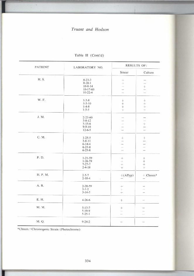
Of the 707 patients, 21 or 2.96 per cent had urine specimens whose smears or cultures contained acid-fast bacilli (Table I I and I I I ) . The data shown in Table I I clearly demonstrates the value of examining both smears and cultures. It can be seen that 10 of 21 patients or 48 per cent had specimens which yielded a positive culture in the absence of observable tubercle bacilli on the smears. Four patients (19%) submitted specimens whose smears were positive and the cultures were negative. In seven patients (33%) both smear and culture were positive.
Table I I I is a brief clinical summary of patients who during the period of this survey demonstrated positive bacteriologic findings. The group was about equally divided as to sex and the ages ranged from 18 to 83 years. The highest incidence was found in those over fifty years of age. No patients in the pediatric age group were included in this study.
Of the seventeen patients with positive cultures, four gave a prior history of tuberculosis. Two of these had undergone nephrectomy, fourteen and sixteen years previous, for "unilateral" renal tuberculosis and another had been treated in the past for involvement of the hip. The fourth (WF) underwent enucleation of the left eye for tuberculosis at 2 years of age. This was some 17 years before the discovery of his urinary tract disease. None of these 4 patients showed evidence of active extra renal disease at the time of this study.
The most common presenting symptoms were frequency, nocturia and dysuria. Flank pain was present in two cases and one patient gave a history of gross hematuria. Ten patients were entirely asymptomatic and urologic investigation was prompted by the discovery of an abnormal urinalysis. Three of the four patients who presented with systemic complaints (fever, malaise, night sweats, etc.) demonstrated concomitant extra renal disease.
As is shown on Table I I I , all of the patients who had cultures which grew acid-fast bacilli showed an abnormal urinalysis. Al l but one had pyuria with or without microscopic hematuria. The patient (ES) without pyuria had microscopic hematuria alone. Eleven patients with positive cultures demonstrated albuminuria on routine urinalysis.
Table I Number of Urine Specimens Obtained Per Patient.
No. of urine specimens No. of patients
1 588 2 77 3 27 4 12 5 1 6 1 7 1
" Note 119 of 707 patients submitted 2 or more specimens. Total 707*
332

r Genito-Urinary Tuberculosis
In seven of the seventeen patients with positive acid-fast cultures intravenous or retrograde pyelograms revealed no abnormality, and in only one were there changes pathognomonic for tuberculosis. Pyelographic changes in the remainder of the series could be ascribed with certainty to tuberculosis only in the light of the history, the clinical findings and the demonstration of M . tuberculosis in the urine.
Table I I
Summary of Smears and Cultures Isolated from Urine Specimens of "Positive" Patients
P A T I E N T L A B O R A T O R Y NO. RESULTS OF:
P A T I E N T L A B O R A T O R Y NO.
Smear Culture
C. M . 10-14-2 12-30-1
- +
E. S. 11-2-5 11-17-3
+ ( A T Y P . ) *
R. H . 1- 9-14 2- 4-2
+
A. W. 9-15-2 9-18-6
• -1-
-1-
W. K. 2- 24-60 3- 18-6 3-24-4
E. C. 1-6-10 1-7-7 1-10-17 1-11-7
-1--1--1-
+ + -1-
W. O. 12-19-59 12-22-3
--h
M . B. 1-14-5 3-29-9 —
+
J. G. 10-25-4 1-28-8 —
-t-
J. H . 2-5-17 4-22-3 4-23-5 4-25-11 4- 30-2 5- 2-5 5-13-19
-+
J. K. 7-19-3 7-19-7
+
*ATyp.=A Typical.
333
r Truant and Hodson
Table I I (Cont'd)
*Chrom.=Chromogenic Strain (Photochrome)
P A T I E N T LABORATORY NO. RESUt -TS OF:
Smear Culture
H. S. 8-23-3 -1-8-28-1 -1- — 10-8-14 — -1-10-17-60 — _ 10-22-4 - -
W. F. 1-3-8 + -1-1-3-10 -1- -1-1-4-8 -1- -1-1-5-5 -h
J. M . 2-23-60 3-8-12 — — 5-15-6 — - I -9-9-10 — — 12-6-5 - —
C. M . 1-25-5 -1-3-8-11 — — 6-14-4 — — 6-23-8 — — 6-25-8 - -
P. D. 1-21-59 -1- + 1-26-59 -1- + 7-27-7 -1-2-4-18 + -h
H. P. M . 2-5-7 + ( A T y p ) -|- Chrom* 2-18-4 — —
A. R. 2-28-59 -1-3-1-2 + — 3-14-5 + -
E. H . 4-26-6 + —
M . M . 5-13-7 + 5-19-9 _ — 5-25-1 - -
M . Q. 9-24-2 -1- -
334
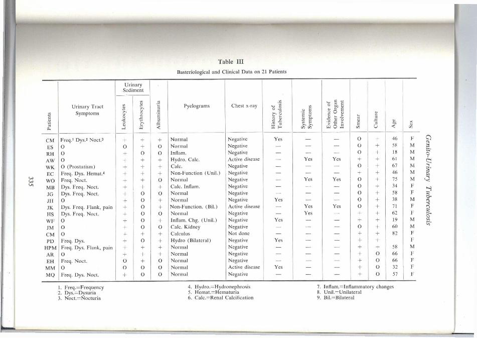
Table I I I Basteriological and Clinical Data on 21 Patients
Urinary Sediment
Urinary Tract u l/i V
uria
Pyelograms Chest x-ray
of
losis
Sy
stem
ic
Sy
mp
tom
s
e of
rean
nen
t
Pat
ien
ts Symptoms
Leu
ko
cy
Ery
thro
t
Alb
um
in
His
tory
Tu
ber
cu
Sy
stem
ic
Sy
mp
tom
s
Ev
iden
c
Oth
er 0
Inv
olv
ei
Sm
ear
Cu
ltu
re
Ag
e
Sex
C M Freq.' Dys.' Noet.^ - f -1- + Normal Negative Yes — — O + 46 F
ES O O -h O Normal Negative — — — O -b 58 M
RH O + 0 o Inflam. Negative — — — O + 18 M
AW o + -h -t- Hydro. Calc. Active disease — Yes Yes -(- -b 61 M
W K O (Prostatism) + + + Calc. Negative — — — o + 67 M
EC Freq. Dys. Hemat.< + + -h Non-Function (Unil . ) Negative — — — -b + 46 M
WO Freq. Noct. + -H o Normal Negative — Yes Yes o -b 75 M
M B Dys. Freq. Noct. -h -1- -h Calc. Inflam. Negative - — — o -b 34 F
JG Dys. Freq. Noct. + O o Normal Negative — — — o -b 58 F
JH 0 •f 0 -1- Normal Negative Yes — — o -b 38 M
JK Dys. Freq. Flank, pain + o -1- Non-Function. (Bil . ) Active disease — Yes Yes o + 71 F
HS Dys. Freq. Noct. + o o Normal Negative — Yes — + -b 62 F
W F O 0 -1- Inflam. Chg. (Uni l . ) Negative Yes — — + -b 19 M
JM O + 0 o Calc. Kidney Negative — — — o -b 60 M
C M O + -1- Calculus Not done — — — -b -b 82 F
PD Freq. Dys. + o -1- Hydro (Bilateral) Negative Yes — — -b -b F
H P M Freq. Dys. Flank, pain + -1- -h Normal Negative — — — -b -b 58 M
AR 0 + + 4- Normal Negative — — — •f O 66 F
EH Freq. Noct. O -h o Normal Negative — — — + O 66 F
M M O O o o Normal Active disease Yes - — -b O 32 F
MQ Freq. Dys. Noct. + 0 o Normal Negative — — — + o 57 F
=:
S'
1. Freq.=Frequency 2. Dys.=Dysuria 3. Noct.=Nocturia
4. Hydro.=Hydronephrosis 5. Hemat.=Hematuria 6. Calc.=Renal Calcification
7. Inflam.=Inflammatory changes 8. Unil.=Unilateral 9. Bil.=Bilateral
Truant and Hodson
DISCUSSION
The literature on genito-urinary tuberculosis deals primarily with the clinical problems of diagnosis and treatment with littie emphasis placed upon the bacteriological aspects. Delay in the diagnosis of renal tuberculosis results from a delay in establishing a bacteriologic diagnosis. One of the reasons for this is the acceptance of negative results, based on a single urine collection, as ruling out genito-urinary tuberculosis.
Since M . tuberculosis is difficult to culture from specimens even under favorable conditions it is especially necessary to accept the advice of the "Research Unit for Genito-Urinary Tuberculosis"" that the routine procedure should consist of at least three 24-hour urine specimens collected from each patient within a two-week period. This method, provides an excellent opportunity to conduct a reliable bacteriological study on each patient and will give a reasonably accurate bacteriologic status of the discharge of tubercle bacilli from urologic foci. I f the patient is being treated with antituberculous drugs, urine cultures cannot be relied on entirely. For example, Kenney et al.'"' have shown that during isoniazid therapy sterile urine cultures do not necessarily reflect permanent arrest of renal tuberculosis. These investigators have shown that a waiting period of ten days after the cessation of therapy is necessary before cultures are reliable in determining the true bacteriologic status of patients being treated for urogenital tuberculosis.
Another common oversight in the evaluation of genito-urinary tuberculosis is the assumption that a negative smear for acid-fast bacilli is sufficient evidence to "rule out" the disease. It can be readily seen from our results (Table I I and I I I ) that 48 per cent of the patients' specimens had positive cultures in the presence of 'negative smear reports'. The data of other investigators' is in agreement with these results.
It has been generally accepted that 24-hour urine specimens should be used for the isolation of M . tuberculosis. This procedure is based on the supposition that tubercle bacilli may be present in such small numbers as to render isolation from small samples of urine very difficult. However, the collection of a 24-hour volume has certain inherent disadvantages:
(a) refrigeration of bulky 24-hour specimens during collection and handhng is often cumbersome.
(b) contamination may present significant problems during the collection and processing.
(c) the collection may be impractical for ambulatory patients.
(d) these specimens necessitate an additional time period of 4-24 hours for sedimentation.
(e) not all the sediment can be processed and thus only part of bacillary population is cultured, thereby frequently defeating the primary purpose of the 24-hour collection.
336
Genito-Urinary Tuberculosis
Because of these disadvantages we have given some consideration to the reports of Kenney et a/." who have recently suggested the use of morning specimens instead of 24-hour urine collections on the basis of their experiences. A comparative study of these two methods by these investigators showed a greater percentage of recoveries of M . tuberculosis from morning urine specimens. Therefore we plan to evaluate the efficacy of this procedure in the very near future.
The symptoms of urinary tuberculosis are not specific and similar symptoms are present in many common urologic disorders. This fact along with the recent tendency to focus less attention on tuberculosis in general probably accounts for the frequent delay in diagnosis or its being missed altogether. These same considerations apply to the pyelographic changes and urine findings in genito-urinary tuberculosis. Although the results of pyelography or urinalysis may strongly suggest the presence of the disease the ultimate diagnosis rests on the isolation and identification of the organism in the urine. For this reason it is extremely important that the physician maintain a high index of suspicion in order to initiate proper diagnostic procedures and avoid serious delay in diagnosis.
Urinary tract tuberculosis should be suspected in any patient with recurrent or persistent urinary infection, prostatitis or epididymitis. A contracted bladder, ureteral stricture, or renal calcification should suggest its possibility as should any unexplained hematuria. Any patient in whom the disease is suspected should have a complete urologic evaluation. It is imperative that the physician be aware of the status of both the involved and uninvolved structures in the genito-urinary tract if future therapy is to succeed.
CONCLUSION
Eight-hundred and eighty urine specimens were obtained from 707 patients for microscopic and cultural examination. Twenty-one patients (2.96%) submitted one or more specimens which contained acid-fast bacilli. Of these, seven (33%) were positive for acid-fast bacilli by both smear and culture whereas the others were diagnosed by either microscopic or cultural methods. The report stresses the need for examining multiple urine specimens of patients who may have urinary tract tuberculosis.
The extreme variability of the clinical signs and symptoms of genito-urinary tuberculosis emphasizes the need for awareness on the part of the physician. This disease should be considered in any patient with recurrent and persistent urinary tract infection, prostatitis or epididymitis. A contracted bladder, ureteral stricture or renal calcification should suggest its possibility, as should unexplained hematuria.
REFERENCES
1. Medlar, E. M., Spain, D. M., and Holhday, R. W.: Post-mortem compared with clinical diagnosis of genito-urinary tuberculosis in adult males, J. Urol. 61:1078, 1949.
2. Reisner, D.: Relations between extrapulmonary and pulmonary tuberculosis, Am. Rev. Tuberc. 30:375, 1934.
3. Nesbit, R. M., Lapides, J., and Baum, W. C : Fundamentals of Urology, ed. 4, Ann Arbor, Michigan, Edwards, 1953, pp. 56-60.
337
1
Truant and Hodson
c " ' 'S"} '^^ ' ,^ ' Tuberculous infections and inflammations of the urinary tract, In. Campbeh M. F., ed.: Urology, Philadelphia, Saunders, 1954, v.l, pp. 525-538.
5. Wechsler, H., Westfall, M.. and Laltimer, J. K..: The earliest signs and symptoms in n7 male patients with genito-urinary tuberculosis, J. Urol. 83:801, I960.
6 Corper, H. J., and Stoner, R. E.: An improved procedure for the diagnoshc culture of mammalian tubercle bacilh, J. Lab. & Clin. Med. 31:1364, 1946.
I • ' ' ' T ' ^ ^ f f',?--;,,^"'' Cummings, M. M.: Diagnostic and Experimental Methods in Tuberculosis, ed. 2, Springfield, III., C. C. Thomas, 1952.
8. Cummings, M. M. : Laboratory diagnosis of tuberculosis, Am. J. Pub. Health 39:361, 1949.
9 Steeiiken, W., Jr.: Medium recommended on evaluation of laboratory procedures of the Amerrcal Trudeau Society for the culture of tubercle bacilli, Trudeau Sanatorium, Trudeau, N. Y.
10. Kenny M., Lattimer, J. K., and Goldman, M. : Urine cultures for M. tuberculosis during chemotherapy, Am. Rev. Tuberc. 70:149, 1954. *
11. Peizer, L. R., Chaves, A. D., and Widelock, D.: A trisodium phosphate transport-digestion method of processing sputum and gastic specimens for the detection of Mycobacterit^m ttiberculo.'<is. Am. Rev. Tuberc. 70:363, 1954.
T K ^^«-^^^7',n'^A' A- and Lovelock, F. J.: Urine cultures in tuberculosis. Am. Rev. tuberc. 82:564, 1960.
338