spring elective 2018 spring genito-urinary systemmousepheno.ucsd.edu/pdfs/gu.pdf · 2018-05-07 ·...
TRANSCRIPT

Spring Elective
2018 Spring
Genito-Urinary system

Human Uterus, fallopian tubes, Ovaries
Mouse bi-cornuate uterus, tubes and ovaires

Human Male Reproductive system
Mouse
The mouse seminal vesicles are very prominent

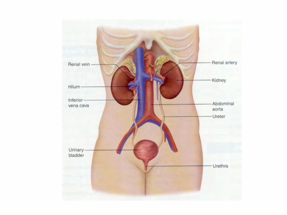
Human kidneys, ureters, bladder, prostate

Mouse kidneys, adrenals
Alan J. Davidson et al., 2008. Mouse kidney development


Filtration: Glomerulus
Secretion & Reabsorption:
Proximal tubule, loop of Henle, and Distal tubule
The Nephron

Bowman’s capsule
Loop of Henle
Nephron segments
Proximal tubule
Distal tubule
Collecting duct
Peritubular capillaries and vasa recta
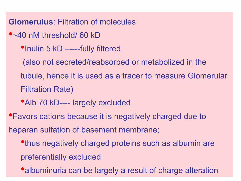
Glomerulus: Filtration of molecules
• ~40 nM threshold/ 60 kD
• Inulin 5 kD –----fully filtered
(also not secreted/reabsorbed or metabolized in the
tubule, hence it is used as a tracer to measure Glomerular
Filtration Rate)
• Alb 70 kD---- largely excluded
• Favors cations because it is negatively charged due to
heparan sulfation of basement membrane;
• thus negatively charged proteins such as albumin are
preferentially excluded
• albuminuria can be largely a result of charge alteration


Continued growth and transformation of mesenchymal cell clusters results in epithelialization and tubulogenesis, ultimately forming the portion of the nephron comprising the glomerulus, proximal tubule, loop of Henle, and distal tubule.


H&E of human Kidney x40 magnification!
H&E of mouse Kidney x40 magnification!
H&E of mouse Kidney x200 magnification!

PAS stain (Periodic Acid Schiff) highlights basement membranes (carbohydrate structures)
Proximal
Distal
glomerulus


PAS (mouse kidneys sectioned at 3 micron thickness)
Wild type aged mouse x400 Siglec-E null aged mouse
H&E of same mouse kidney x400
Human Kidney with Expanded nodular formations in glomerular capillaries -- positive staining with PAS-- in diabetic glomerulosclerosis

Capillaries of the glomerulus highlighted with the UEA lectin

Epithelial cells!
Endothelial cells!
Basement membrane!
Mesangial cells!

H&E of Embryo d16.5 !Mouse Kidney cortex with developing glomeruli!

H&E of Embryo d 16.5 Mouse Kidney cortex x400 magnification!

Thin section of glomerulus

Primer on Kidney Diseases; NKF


Pathology of kidney:!Inflammation:! !---glomerular = glomerulonephritis,-autoimmune!
!---tubular=pyelonephritis!
hyperplasia, accumulations:!fibrosis/scarring, cysts!
Infarct!Cancer: primary adenocarcinomas or metastatic!

http://www-medlib.med.utah.edu
As mice age, the kidneys develop similar cysts due to chronic pyelonephritis(Tubules inflammation, scarring and repair) with consequent stasis
Simple cysts in human kidney

Resected renal cell carcinoma, at upper pole of the kidney

Compared to a normal kidney (right), those from people with polycystic kidney disease (left) are enlarged and ravaged with cysts. Defects in the primary cilia on kidney tubule epithelial cells (inset) lead to the devastation. Science Oct 14, 2005

H&E of mouse Kidney Pelvis x40 magnification!
To ureter

Transitional epithelium of bladder and ureter

Transitional epithelium of bladder
Histologic highlights within the Female and Male
Reproductive systems

Uterus, Fallopian Tubes, ovaries
Human Mouse

Stages of ovarian follicular development

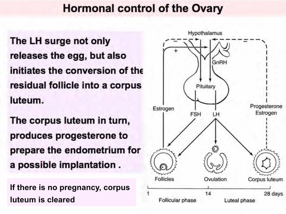
If there is no pregnancy, corpus luteum is cleared


Fluid in antrum of secondary follicle
Stages of development of the ovarian follicle


Zona pellucida

Bulges from the ovarian surface
The Primary oocyte resumes the First Meiotic division, forming the Secondary oocyte and the first polar body
Just as the Secondary oocyte enters the Second meiotic division, Ovulation occurs
“mittelschmerz”
Mature or Graafian follicle and ovulation:

OVARIES
Corpus luteum (yellow body --after ovulation)
http://library.med.utah.edu/WebPath/FEMHTML/FEM045.html


Mouse ovary, fallopian tube, uterus
Many follicles develop at the same time unlike in human

Abnormal maturation of mouse ovarian follicles
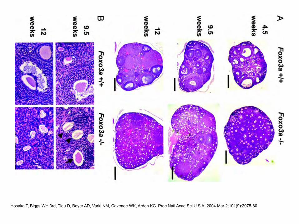
Hosaka T, Biggs WH 3rd, Tieu D, Boyer AD, Varki NM, Cavenee WK, Arden KC. Proc Natl Acad Sci U S A. 2004 Mar 2;101(9):2975-80


Fallopian tube at different magnifications


Normal human fallopian tube with highly folded mucosa at the ampulla which is the usual site of fertilization
Abnormal human fallopian tube which are both blocked and dilated after infectious events


Squamo-columnar junction of cervix
Human ecto and endo cervix Mouse endo and ecto cervix

Differences from Human Menstrual Cycle One of the main differences between the rodent and the human cycle, other that the overall time it takes for a full cycle, is that the peaks of estrogens and progesterone are typically separated in humans, whereas these overlap in rodent. In the mouse, the estrous cycle is divided into 4 stages (proestrus, estrus, metestrus, and diestrus) and repeats every 4 to 5 days unless interrupted by pregnancy, pseudopregnancy, or anestrus.

http://www.biobserve.com/downloads/maria-gulinello/Rodent-Estrous-Cycle.pdf


Human

Uterine endometrium changes during the human menstrual cycle


A section through a portion of the human uterus
endometrium
myometrium

Mouse Uterus, fallopian tube, Ovary

Mouse Uterus

Mouse Uterus
Endometrium and stroma

Mouse Uterus
Endometrium and stroma


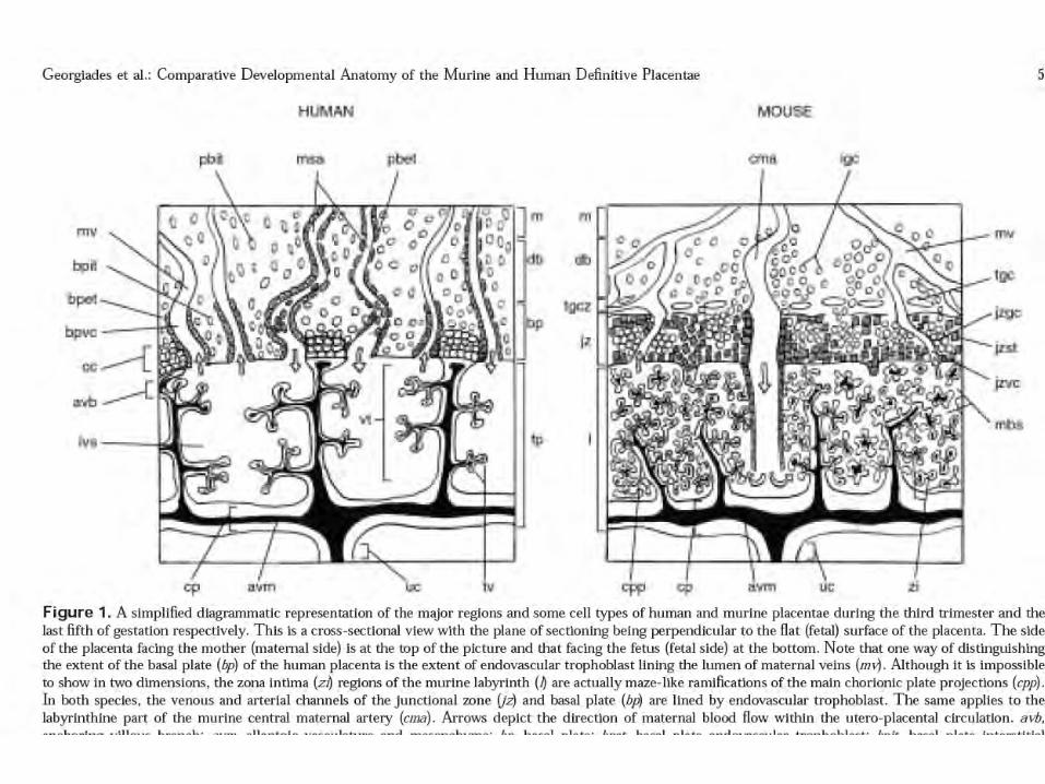
Human Placenta : diagram
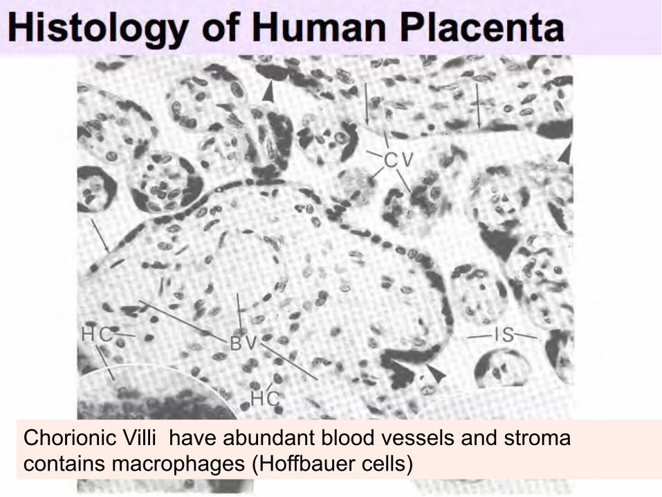
Chorionic Villi have abundant blood vessels and stroma contains macrophages (Hoffbauer cells)

Chorionic Villi sections show trophoblast which is composed of an inner Cytotrophoblast and an outer multi-nucleated Syncytiotrophoblast.

FIGURE 2 | Comparative anatomy of the mouse and human placenta. Placental development: Lessons from mouse mutants Janet Rossant & James C. Cross Nature Reviews Genetics 2, 538-548 July 2001


H&E of placenta from a day12.5 embryo

H&E of placenta from a day12.5 embryo

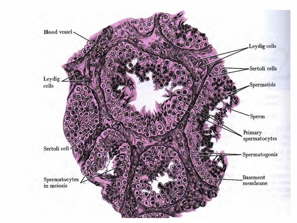

TESTIS: site of the development of spermatozoa and the production of testosterone. Spermatogenesis and testosterone production are controlled by FSH and LH (ICSH --
Interstitial Cell Stimulating Hormone).
Seminiferous tubule - site of spermatogenesis
Interstitial cells (Leydig cells) are in the stroma, between the seminiferous tubules, and are
the site of testosterone production

H&E of a section of mouse testis!
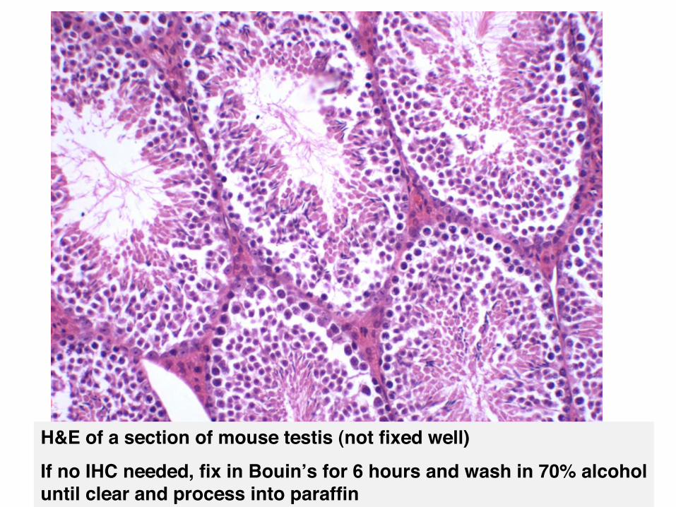
H&E of a section of mouse testis (not fixed well)!If no IHC needed, fix in Bouinʼs for 6 hours and wash in 70% alcohol until clear and process into paraffin!

Sections of testes from --null animal showing development arrest
Seminiferous tubules with maturing sperm

Abnormal degeneration of cells lining seminiferous tubules of mouse testis

H&E of testes from littermate controls and from ---null mice, showing degenerated follicles!
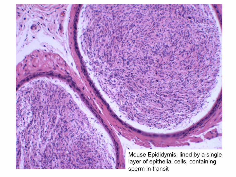
Mouse Epididymis, lined by a single layer of epithelial cells, containing sperm in transit


Human prostate: Tubulo-alveolar glands embedded in fibro-muscular stroma

Histology of Normal Human prostate

Normal Human Prostate Single layer of epithelial cells marked with anti-keratin

Markers of Differentiation
Normal Hyperplasia Tumor
Smooth Muscle Actin (SMA)

Prostate carcinoma around a normal prostate duct, with IHC (?prostate specific acid phosphatase PSA) marker

http://tvmouse.ucdavis.edu/prostate/mouse/prostateindex2.htm

Mouse prostate

Normal mouse prostate mouse prostate carcinoma in situ

Mouse prostate early carcinoma

Human breast lobules and ducts Normal with one layer of lining epithelial cells and an outer layer of myoepithelial cells
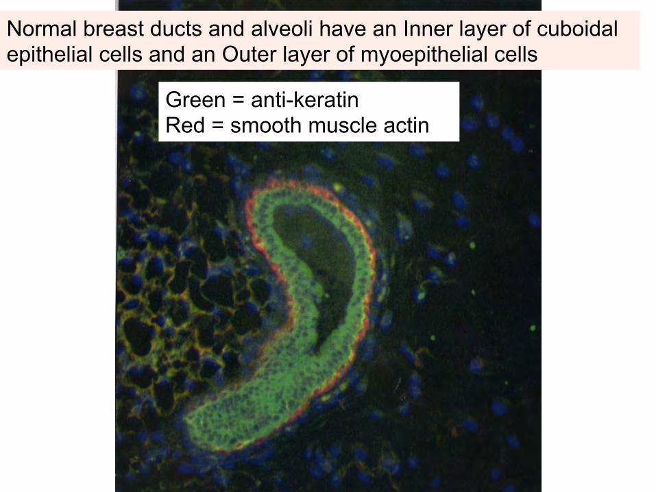
Normal breast ducts and alveoli have an Inner layer of cuboidal epithelial cells and an Outer layer of myoepithelial cells
Green = anti-keratin Red = smooth muscle actin

Normal mammary gland and carcinoma in-situ
