biologia funzionale dei sistemi cellulari e molecolari a novel
TRANSCRIPT

1
Alma Mater Studiorum Alma Mater Studiorum –– Univers ità di Bologna Univers ità di Bologna
DOTTORATO DI RICERCA IN
Biologia Funzionale dei Sistemi Cellulari e Molecolari
Ciclo XXI°
Settore scientifico-disciplinare: BIO/11
A novel phase variation mechanism in the meningococcus driven by a ligand-responsive repressor
and differential spacing of distal promoter elements
Presentata da: Matteo Maria Emiliano Metruccio Coordinatore Dottorato Relatori
Chiar.mo Prof. Chiar.mo Prof. Vincenzo Scarlato Vincenzo Scarlato Dott.Ssa Isabel Delany
Bologna 2010

2

3
Durante il Dottorato di ricerca mi sono occupato dello studio della regolazione
dell´espressione genica in Neisseria meningitidis. In particolare ho studiato la
regolazione trascrizionale in seguito a stess ossidativo, ho caratterizzato un circuito di
regolazione che coinvolge un piccolo RNA non codificante (sRNA) e lo chaperone Hfq,
e piú in dettaglio ho approfondito la complessa regolazione trascrizionale di NadA,
argomento quest’ultimo del lavoro di tesi di seguito presentato.
Nel periodo del Dottorato di Ricerca sono stato co-autore dei seguenti lavori
scientifici:
Ieva R, Roncarati D, Metruccio MM, Seib KL, Scarlato V, Delany I. “OxyR tightly
regulates catalase expression in Neisseria meningitidis through both repressive and
activation mechanisms.” Mol Microbiol. 2008 Dec;70(5):1152-65.
Metruccio MM, Fantappiè L, Serruto D, Muzzi A, Roncarati D, Donati C, Scarlato V,
Delany I. “The Hfq-dependent small noncoding RNA NrrF directly mediates Fur-
dependent positive regulation of succinate dehydrogenase in Neisseria meningitidis.” J
Bacteriol. 2009 Feb;191(4):1330-42. Epub 2008 Dec 5.
Fantappiè L, Metruccio MM, Seib KL, Oriente F, Cartocci E, Ferlicca F, Giuliani MM,
Scarlato V, Delany I. “The RNA chaperone Hfq is involved in the stress response and
virulence in Neisseria meningitidis and is a pleiotropic regulator of protein expression.”
Infect Immun. 2009 May;77(5):1842-53. Epub 2009 Feb 17.
Metruccio MM, Pigozzi E, Roncarati D, Berlanda-Scorza F, Norais N, Hill S, Scarlato
V, Delany I. “A novel phase variation mechanism in meningococcus driven by a ligand
responsive repressor and differential spacing of distal promoter elements.” PLoS
Pathog. 2009 Dec;5(12): e1000710. Epub 2009 Dec 24.

4

5
Table of Contents
TitleTitle ............................................................................................................................ 1Table of Contents ........................................................................................................ 5Riassunto ..................................................................................................................... 7Abstract ....................................................................................................................... 9Introduction .............................................................................................................. 11
1.1 Meningococcal disease.................................................................................. 111.2 The pathogen................................................................................................. 111.3 Genomes ....................................................................................................... 131.4 Colonization and Invasion ............................................................................. 141.5 Virulence factors ........................................................................................... 161.6 Phase Variation ............................................................................................. 171.7 Vaccines available......................................................................................... 201.8 NadA ............................................................................................................ 211.9 Alpha subunit of RNA polymerase ................................................................ 231.10 MarR family transcriptional regulators .......................................................... 241.11 IHF ............................................................................................................... 26
Results ....................................................................................................................... 291.12 Phase variation in expression and transcript level of NadA............................ 291.13 RNA polymerase interaction over the nadA promoter.................................... 321.14 IHF interacts with the nadA promoter............................................................ 351.15 Identification of a cis acting element responsible for growth phase regulation381.16 A protein in meningcoccus cell extract binds GPR region.............................. 401.17 Identification of the GPR binding protein ...................................................... 421.18 NMB 1843 interacts with the nadA promoter................................................. 441.19 NadR represses NadA expression .................................................................. 461.20 NadR represses to a different extent different phase variant promoters .......... 481.21 4-hydroxyphenylacetic acid (4HPA) interacts with NadR and derepresses
NadA expression ........................................................................................... 50Discussion .................................................................................................................. 53
1.22 Three-dimensional MODEL.......................................................................... 561.23 Regulation MODEL ...................................................................................... 58
Materials and Methods ............................................................................................. 631.24 Bacterial strains and culture conditions.......................................................... 631.25 DNA techniques. ........................................................................................... 641.26 Construction of nadA promoter fusions ......................................................... 641.27 Construction of knockouts............................................................................. 661.28 Expression and purification of the E.coli RNA polymerase α-subunit and N.
meningitidis NMB1843 protein. .................................................................... 661.29 Western blot analysis..................................................................................... 671.30 Co-over expression of alpha subunit in E. coli ............................................... 681.31 Crude extract preparation, DNA affinity purification of GPR-binding protein
and Electrophoretic mobility shift assay (EMSA) .......................................... 691.32 RNA preparation and Primer extension ......................................................... 701.33 DNase I footprinting (NMB1843, Neisseria gonorrhoeae IHF, Escherichia
coli RNApol and Alpha-sub) ......................................................................... 711.34 MALDI TOF mass spectrometry ................................................................... 72
References ................................................................................................................. 79

6

7
Riassunto
Il rapido adattamento all´ambiente circostante costituisce uno dei requisiti fondamentali
per i batteri patogeni. La capacitá di alcuni batteri di esprimere determinati geni in
maniera casuale (variazione di fase), grazie al cambiamento nella lunghezza di sequenze
semplici ripetute (SSR) durante la replicazione, conferisce alla popolazione batterica
una varietá che le permette un piú rapido adattamento ai cambiamenti dell´ambiente
circostante. In questo lavoro si mostra un nuovo meccanismo alla base della “variazione
di fase” di NadA, un invasina e adesina di Neisseria meningitidis. Il repressore NadR
contatta due operatori sul DNA posizionati alle estremitá del tratto di sequenze ripetute
sul promotore di nadA, e contribuisce al diverso livello di espressione in promotori con
diverso numero di ripetizioni probabilmente a causa della variazione della distanza
relativa tra i due operatori. Qui si dimostra inoltre che IHF lega tra questi operatori e,
probabilmente tramite la formazione di una piega del DNA, favorirebbe l´interazione di
NadR legato all´operatore distale con quello sovrapposto al promotore. L´acido 4-
idrossifenilacetico, un metabolita del catabolismo degli aminoacidi aromatici che viene
secreto nella saliva, induce l´espressione di NadA inibendo il legame del repressore al
DNA. Tra i promotori con diverso numero di ripetizioni sono osservabili solo minime
differenze nei livelli di trascrizione di nadA in assenza di NadR, queste differenze sono
probabilmente dovute ad un diverso legame della RNA polimerasi sui diversi
promotori, che porta ad un´alterata attivitá trascrizionale. Questi risultati suggeriscono
che l´espressione di NadA si trova sotto il controllo di eventi sia casuali che finemente
regolati in base all´ambiente, entrambi mediati dal repressore NadR, e che questa
importante adesina potrebbe essere indotta durante la colonizzazione dell´orofaringe,
dove esplica un ruolo importante nell´adesione e invasione della mucosa. In
conclusione, la presenza di semplici sequenze ripetute nei promotori, puó essere una

8
strategia sfruttata da alcuni batteri patogeni per modificare casualmente il livello di
espressione di alcuni geni, pur mantenendo la possibilitá di indurli in presenza del
segnale specifico.

9
Abstract
Phase variable expression, mediated by high frequency reversible changes in the length
of simple sequence repeats, facilitates adaptation of bacterial populations to changing
environments and is frequently important in bacterial virulence. Here we elucidate a
novel phase variable mechanism for NadA expression, an adhesin and invasin of
Neisseria meningitidis. The NadR repressor protein binds to operators flanking the
phase variable tract of the nadA promoter gene and contributes to the differential
expression levels of phase variant promoters with different numbers of repeats, likely
due to different spacing between operators. It is shown that IHF binds between these
operators, and may permit looping of the promoter, allowing interaction of NadR at
operators located distally or overlapping the promoter. The 4-hydroxyphenylacetic acid,
a metabolite of aromatic amino acid catabolism that is secreted in saliva, induces nadA
expression by inhibiting the DNA binding activity of the NadR repressor. When
induced, only minor differences are evident between NadR-independent transcription
levels of promoter phase variants, which are likely due to differential RNA polymerase
contacts leading to altered promoter activity. These results suggest that NadA
expression is under both stochastic and tight environmental-sensing regulatory control,
and both regulations are mediated by the NadR repressor that and may be induced
during colonization of the oropharynx where it plays a major role in the successful
adhesion and invasion of the mucosa. Hence, simple sequence repeats in promoter
regions may be a strategy used by host-adapted bacterial pathogens to randomly switch
between expression states that may nonetheless still be induced by appropriate niche-
specific signals.

10

11
Introduction
1.1 Meningococcal disease
Neisseria meningitidis is a strictly human pathogen responsible for meningitis and
sepsis, two devastating diseases that can kill children and young adults within hours,
despite the availability of effective antibiotics. The reported annual incidence of
meningococcal disease varies from 0.5 to 10 per 100,000 persons; however, during
epidemics the incidence can rise above 1 per 1,000 (Stephens 2009). The case fatality
rate ranges from 5 to 15%, and up to 25% of survivors are left with neurological
sequelae (Comanducci, Bambini et al. 2002).
N. meningitidis colonises the upper respiratory tract in about 25% of the human
population where it can live as commensal. This carrier state not only provides a
reservoir for meningococcal infection but can also contribute to establish host immunity
(Stephens 2009). Furthermore, the human host is the only known reservoir for this
human-adapted bacterium. For largely unknown reasons, dependent from both host and
pathogen, in a small subset of carriers, meningococcus can invade the pharyngeal
mucosal epithelium and, in the absence of bactericidal serum activity, disseminate into
the bloodstream, causing septicaemia. In a subset of cases, the bacteria can also cross
the blood-brain barrier and infect the cerebrospinal fluid, causing meningitis.
1.2 The pathogen
N. meningitidis is a Gram-negative, spherical or kidney-shaped bacillus commonly seen
in pairs (diplococcus). It is aerobic, non-motile, non-sporulating, usually encapsulated
and piliated (Figure 1). Traditionally, different strains of N. meningitidis are classified
on the bases of the composition and antigenic property of their capsule. With this

12
method, 13 serogroups can be distinguish, five of which (A, B, C, W-135 and Y) are
responsible for virtually all meningococcal disease. Meningococci are further classified
into serotype and serosubtype, based on antigenic differences in their major outer
membrane proteins (OMPs), PorA and PorB. However, since this classification is based
on variation of few genes that are probably under selective pressure, serotyping is not
suitable for modern epidemiology. A genetic typing system based upon polymorphisms
in multiple housekeeping genes called Multilocus Sequence Typing (MLST) (Maiden,
Bygraves et al. 1998), is now the gold standard for molecular typing and epidemiologic
studies, although does not correlate with antigenic composition of the bacteria which
remains largely variable among lineages. MLST technique has shown that the majority
of disease associated isolates cluster into a minority of sequence type (ST) called hyper
invasive lineage (Maiden 2008). Why hyper virulent meningococcal lineages are more
pathogenic than others remain still unknown.
Figure 1: ImmunoGold labelling and transmission electron microscopy of Neisseria meningitidis strain.
Analysis of the strain was performed with antisera raised against NadA adhesin (Scale bars: 200 nm.)
(Pizza, Scarlato et al. 2000).

13
1.3 Genomes
Up to now seven N. meningitidis genomes have been sequenced, four disease associated
and three carriage strains These data show that the meningococcal chromosome is
between 2.0 and 2.1 mega bases in size and contains about 2000 genes (Parkhill,
Achtman et al. 2000; Tettelin, Saunders et al. 2000; Bentley, Vernikos et al. 2007;
Schoen, Tettelin et al. 2009). Mobile genetic elements, including IS elements and
prophage sequences, make up ∼10% of the genome (Parkhill, Achtman et al. 2000). The
GC percentage is widely variable along the chromosome, with defined regions of low
GC content that likely have been acquired by a relatively recent horizontal gene
transfer. These events are common in N. meningitidis due to its natural transformation
competence (Maiden 1993). The acquisition of the capsule locus by horizontal gene
transfer, possibly from Pasteurella multocida or P. hemolytica (Schoen, Blom et al.
2008), appears to be a major event in the evolution of the pathogenicity of the
meningococcus (Stephens 2009). Other than the genes encoding for the capsule, no core
pathogenome has been identified suggesting that virulence may be dependent on
multiple redundant genes. The most evident characteristic of neisserial genome is the
high abundance of repetitive DNA sequences which leads to genetic instability,
facilitating duplication or deletion of regions in the genome, as well as recombination
(Davidsen and Tonjum 2006). These events characterize the non-clonal behaviour of N.
meningitidis.

14
1.4 Colonization and Invasion
The first step in meningococcal colonization is the initial contact with nasopharyngeal
epithelial cells mediated by Type IV pili, then bacteria proceed to proliferate on the
surface of human non-ciliated epithelial cells, forming small microcolonies at the site of
initial attachment (Stephens 2009). After the initial colonization, there is a loss or down
regulation of the capsule, which sterically masks the outer membrane proteins. This
event can occur both via regulatory system upon cell contact (Deghmane, Giorgini et al.
2002), and by selection of low or no-capsule expressing bacteria due to phase variation
(Hammerschmidt, Muller et al. 1996). Close adherence of meningococci to the host
epithelial cells is mediated by a variety of possible redundant adhesins, previously
masked by the capsule. This results in the appearance of cortical plaques and the
recruitment of factors leading to the formation and extension of epithelial cell
pseudopodia that internalize the bacteria (Stephens 2009). This intracellular lifestyle can
give the bacteria the opportunity to evade host immune response, find more available
nutrients and is also a way to further cross the epithelium and enter the blood stream
(Stephens 2009). In this new environment meningococcus has to express again the
capsule, which can prevent antibody and complement deposition (Achtman 1995), is
anti-opsonic and anti-phagocytic and therefore aids survival in blood (Virji 2009).
These later steps in invasion of the blood stream and the possible subsequent crossing of
the blood-brain barrier are still poorly understood.

15
(Virji 2009)
Figure 2: Stages in the pathogenesis of N. meningitidis. N. meningitidis may be acquired through the
inhalation of respiratory droplets. The organism establishes intimate contact with non-ciliated mucosal
epithelial cells of the upper respiratory tract, where it may enter the cells briefly before migrating back to
the apical surfaces of the cells for transmission to a new host. Asymptomatic carriage is common in
healthy adults in which bacteria that enter the body by crossing the epithelial barrier are eliminated.
Besides transcytosis, N. meningitidis can cross the epithelium either directly following damage to the
monolayer integrity or through phagocytes in a ‘Trojan horse’ manner. In susceptible individuals, once
inside the blood, N. meningitidis may survive, multiply rapidly and disseminate throughout the body and
the brain. Meningococcal passage across the brain vascular endothelium (or the epithelium of the choroid
plexus) may then occur, resulting in infection of the meninges and the cerebrospinal fluid (Nassif 1999).

16
1.5 Virulence factors
The only known virulence factors that characterize pathogenic strains are mainly two:
the polysaccharide capsule, which is thought to protect meningococci during airborne
transmission between hosts (Virji 2009), allow survival in the blood as mentioned
before and may shield bacterial surface from the host immune effectors mechanisms.
The second principal virulence factors are the long surface proteins that protrude from
the capsule known as pili. These facilitate first adhesion to host tissues apart from being
the mediators of DNA uptake (Virji 2009). Pilin of Neisseria is expressed from the pilE
locus, but homologous recombination between the pilE gene and a number of non-
expressed ‘silent’ pilS genes results in a change in the pilE sequence. These variants
differ in their transformability, adherence and immunogenicity (Virji, Alexandrescu et
al. 1992). Additionally, Neisseria spp. possess host specific iron acquisition
mechanisms and numerous immune evasion mechanisms such as factor H binding
protein that is able to downregulate complement deposition (Virji 2009). Close adhesion
and invasion is mainly mediated by an array of proteins such as opacity proteins (Opc,
Opa) and other adhesins. These proteins are believed to be responsible for the host
specificity as well as for tissues within the host (Virji 2009). Host specificity poses a
problem for developing animal models of the disease, and as a result most of our
knowledge of the pathogenic mechanisms of Neisseria spp. comes from in vitro
investigations. Numerous additional apparently minor adhesins (several of which were
identified by homology searching of the available genomes) are generally expressed at
low levels during in vitro growth but may be important in in-vivo infections. For
example, in restricted iron environments, such as might be encountered in vivo, the
transcriptome of N. meningitidis is considerably altered (Grifantini, Sebastian et al.
2003) and as a result the minor adhesins may become expressed. Furthermore, several
adhesins are subject to antigenic variation and/or phase variation, which allow bacteria

17
to generate a broad and variable repertoire of surface structure that facilitates evasion of
immune effectors mechanisms and adaptation to different niche (Virji 2009). This
requires a multiplicity of adhesins (redundancy) to maintain colonization (Martin, van
de Ven et al. 2003; Martin, Sun et al. 2004). Several adhesins may also operate
simultaneously to increase the avidity of bacterial binding to the cell surface. This is
often a prelude to internalization into epithelial cells (Griffiths, Bradley et al. 2007),
which can be another immune evasion strategy.
1.6 Phase Variation
Phase variation is a process that results in differential expression of one or more genes
and results in two subpopulations within a clonal population: one lacking or having a
decreased level of expression of the phase variable gene(s) and the other subpopulation
expressing the gene fully. In this way the bacterial population can alter its antigenic
properties. This process is distinct from antigenic variation, however, in which a
bacterial structure, proteinaceous or otherwise, is consistently produced, but in different
antigenic forms. In specific cases, phase variation can lead to antigenic variation, for
example if phase variation affects expression of a lipopolysaccharide (LPS) modifying
enzyme such as SiaD (Hammerschmidt, Hilse et al. 1996). A key feature of phase
variation is that the ‘On’ and ‘Off’ phenotypes are interchangeable. Thus, a cell with
gene expression in the ‘Off’ phase, that is lacking expression, retains its ability to
switch to ‘On’ and vice versa. The frequency with which the switching occurs can vary
widely in different systems, ranging from as frequently as one cell in ten per generation
to as infrequent as one in ten thousand. The occurrence of phase variation thus results in
a heterogenic and dynamically changing phenotype of a bacterial population. A change
in the ratio of the two subpopulations could in principle be achieved either through

18
selection for or against a subpopulation, or through regulation of gene expression or of
the switch frequency (van der Woude 2006).
Phase variable genes in N. meningitidis are typically associated with the presence of
repetitive DNA motifs (oligonucleotide repeats and microsatellites) that exhibit high
mutation rates by a slipped-strand mispairing (SSM) mechanism. The presence of repeat
units cause a slippage of the synthesis strand over the template strand during replication
that leads to the addition or the deletion of units in the new born filament (Figure 3)
(Davidsen and Tonjum 2006). When repeats occur in the coding sequence, the promoter
region or close to the promoter region, they can change the transcriptional and
translational state of the gene, leading to an on/off switching of the gene product. In N.
meningitidis a considerably high quantity of phase variable genes exist, affecting all
kind of virulence factors or host-pathogen interacting structure such as capsule
biosynthesis (Hammerschmidt, Muller et al. 1996), pili and pilus modification
(Rytkonen, Albiger et al. 2004), several surface proteins including Opa, Opc, PorA and
iron binding proteins (van der Ende, Hopman et al. 1995) (Lewis, Gipson et al. 1999)
(Martin, van de Ven et al. 2003) (Virji 2009). It has been proposed that in N.
meningitidis over 100 genes are potentially phase variable (Snyder, Butcher et al. 2001)
(Martin, van de Ven et al. 2003), whereas for example H. influenzae or E. coli contains
less than 20 and 10 putative phase variable genes, respectively (Hood, Deadman et al.
1996) (Blatter et al. 1997).

19
Figure 3 | Mechanisms of meningococcal
phase variation. The molecular mechanism that
most often mediates meningococcal phase
variation is RecA-independent slipped strand
mispairing of repeats during DNA replication
or repair. a | Tandem repeats are found either in
the promoter region of the gene, affecting the
binding of the RNA polymerase and therefore
transcription, or in the open reading frame
(ORF), affecting translation. These repeats can
be homopolymeric, dipolymeric or can be
composed of tetranucleotides or greater. The
different meningococcal loci altered by phase
variation and the corresponding positions of
tandem repeats are listed. b | The mutational
mechanism responsible for phase variation of
the meningococcal outer-membrane protein
and adhesin NadA is shown.
(1) When 5 TAAA repeats are present upstream of the promoter region, the expression level of NadA is
high (Feil, Holmes et al. 2001). (2) The AT-rich sequence is prone to strand separation. (3) Subsequent
mispairing of the TAAA repeats can occur during re-annealing due to slippage of the DNA strands.
Slippage in the 5′ to 3′ direction as shown leaves an unpaired copy of TAAA (indicated by the asterisks).
(4) The unpaired TAAA is deleted, and the single-stranded loop is the target of excision-repair processes.
(5) With a reduction in the TAAA repeat number to four, the expression level of NadA is low (Feil,
Holmes et al. 2001) (Davidsen and Tonjum 2006).

20
1.7 Vaccines available
Effective capsular polysaccharide-based vaccines are today available for four of the five
disease-associated serogroups. For serogroup B that is predominant in many countries,
especially Europe and North America, no broad effective vaccine is currently available.
This is due to the identity of the serogroup B capsular polysaccharide with a widely
distributed human carbohydrate ([2→8]N-acetyl neuraminic acid or polysialic acid),
which is a self antigen, and therefore, is poorly immunogenic in humans. Furthermore,
the use of this polysaccharide in a vaccine may elicit autoantibodies (Hayrinen,
Jennings et al. 1995) (Finne, Bitter-Suermann et al. 1987).
To overcome these issues, an in-silico genome-based approach, called Reverse
Vaccinology has been used, allowing the identification of approximately 600 potentially
surface exposed antigens in N. meningitidis serogroup B (Pizza, Scarlato et al. 2000).
Among these, candidates that could be expressed in E. coli, shown to be surface
exposed, capable of inducing bactericidal antibodies in mice, conserved and with low
heterogeneity in a wide range of meningococcal strains, have been selected for further
studies (Comanducci, Bambini et al. 2002) (Masignani, Balducci et al. 2003; Welsch,
Moe et al. 2003). This led to the development of the 5-component vaccine against
MenB, 5CVMB, which combines five antigens: GNA 2132 (Welsch, Moe et al. 2003),
factor H-binding protein (FHBP) (Masignani, Comanducci et al. 2003) (Fletcher,
Bernfield et al. 2004) (Madico, Welsch et al. 2006), the invasin NadA (Capecchi, Adu-
Bobie et al. 2005) (Comanducci, Bambini et al. 2002), GNA 1030 and GNA 2091
(Rinaudo, Telford et al. 2009).
Study of regulation of expression of these genes could allow us not only in the better
understanding of the pathogenesis of this bacterium, but has also fundamental

21
implications in the Meningococcal B vaccine coverage. Among the five antigens that
compose the MenB vaccine, this thesis focuses on the regulation of NadA expression.
1.8 NadA
Bioinformatic analysis of the genome of a virulent N. meningitidis B strain (Tettelin,
Saunders et al. 2000) allowed the identification of previously unknown surface proteins
(Pizza, Scarlato et al. 2000), among which is the 45-kDa N. meningitidis Adhesin A
(NadA). Structure prediction and homology comparison suggests that NadA belongs to
the group of oligomeric coiled-coil adhesins (OCA) such as YadA of Yersinia
enterocolitica and UspA2 of Moraxella catarrhalis (Comanducci, Bambini et al. 2002).
Within these homotrimeric outer-membrane proteins, three structural regions are
present: a conserved –COOH terminal membrane anchor, having a β structure; an
intermediate coiled-coil stalk comprising a leucine zipper; and a –NH2 terminal region,
forming the binding site(s) for target cell receptors (Hoiczyk, Roggenkamp et al. 2000).
NadA is a risk factor for the development of meningococcal disease, as it was found in
50% of N. meningitidis strains isolated from patients and in only 5% of strains from
healthy individuals (Comanducci, Bambini et al. 2004). NadA has been implicated in
the mucosal colonization by N. meningitidis, as its expression enhances bacterial
adhesion to and invasion of mucosal cells (Capecchi, Adu-Bobie et al. 2005). NadA has
also been implicated in interaction and stimulation of monocyte, macrophage and
dendritic cells during N. meningitidis infection (Franzoso, Mazzon et al. 2008).
Little is known about the factors or mechanisms regulating expression of the NadA
protein. Its expression was shown to exhibit growth-phase dependent behaviour with
levels reported to be maximal in the stationary growth phase of all strains tested
(Comanducci, Bambini et al. 2002). Furthermore, the expression of NadA is phase
22
variable and a tetranucleotide tract (TAAA) upstream of the nadA gene promoter has
been demonstrated to control this phenomenon (Martin, van de Ven et al. 2003).
The phase variable tract of nadA is distally located upstream of the nadA promoter,
unlike the phase variable repeat tracts found in the porA, fetA, and opc genes where the
unstable homopolymeric stretches are found between the -10 and the -35 promoter
elements and are thought to result in altered sigma-factor binding (Sarkari, Pandit et al.
1994) (van der Ende, Hopman et al. 1995) (Carson, Stone et al. 2000). The phase
variation mechanism for the nadA gene is unique in that all phase variable promoter
variants result in the expression of full length protein, which is unlike the case when
repeats are located in coding regions (frame-shift mutation leading to generation of stop
codons) or when suboptimal -10 and -35 promoter spacing precludes or affects
promoter recognition or activation. The frequency of phase variation of nadA has been
experimentally estimated as ca. 4.4 x 10⁻⁴ (Martin, van de Ven et al. 2003), creating
variants where changes in the repeat number result in promoters with low, medium or
high activity. The transcriptional regulators Fur and IHF were implicated in the control
of nadA promoter activity from the specific binding of both proteins to the nadA
promoter and from the analysis of mutants deleted for multiple IHF- and Fur-binding
sites (Martin, Makepeace et al. 2005). Moreover, it has been reported that loss or gain of
a tetranucleotide repeat affects the binding of the IHF regulatory protein to the nadA
promoter in vitro, and this was proposed to be responsible for the modulation of
transcription of nadA in vivo (Martin, Makepeace et al. 2005). Nonetheless, the precise
mechanism governing transcriptional regulation of nadA remains unclear and the
inferred role of IHF or Fur and their involvement in phase variation of nadA expression
remain to be elucidated. However, a novel regulator of NadA expression has recently
been identified which was shown to repress NadA expression (Schielke, Huebner et al.
2009).

23
In this work we will show results on the interactions on nadA promoter region, and the
involvement in its transcriptional regulation, of three different regulatory factors: the α-
subunit of RNA polymerase, a MarR family transcriptional regulator named NadR
(NMB1843) and the architectural protein IHF.
1.9 Alpha subunit of RNA polymerase
In recent years, it has become clear that promoter recognition by bacterial RNA
polymerase (RNAP) involves interactions not only between core promoter elements and
the sigma subunit, but also between a DNA tract, called UP element, upstream of the
−35 sequence and the RNAP α subunit (Hawley and McClure 1983) (Ross, Gosink et
al. 1993) (Blatter, Ross et al. 1994). The best-characterized UP element is in the rrnB P1
promoter, in which the sequence determinants are located between positions −40 and
−60 with respect to the transcription start site (Rao, Ross et al. 1994), and UP element-
α-subunit interactions facilitate initial binding of RNAP and subsequent step(s) in
transcription initiation (Rao, Ross et al. 1994) (Strainic, Sullivan et al. 1998). A
consensus UP element sequence derived from binding-site selection experiments,
consists almost exclusively of A and T residues and increases promoter activity >300-
fold (Estrem, Gaal et al. 1998). Each RNAP α subunit consists of two domains
connected by an unstructured and flexible linker (Blatter, Ross et al. 1994) (Jeon,
Yamazaki et al. 1997). The 28-kD aminoterminal domain (αNTD) is responsible for
dimerization of α and for interaction with the remainder of RNAP (Igarashi and
Ishihama 1991; Busby and Ebright 1994). The 8-kD carboxy-terminal domain (αCTD)
is responsible for interaction with the UP element (Blatter, Ross et al. 1994) and with a
number of transcriptional activators (Igarashi and Ishihama 1991) (Busby and Ebright
1999) (Savery, Lloyd et al. 1998). The αCTD residues most crucial for DNA interaction
are nearly invariant in bacteria (Gaal, Ross et al. 1996) (Murakami, Fujita et al. 1996),

24
and therefore the DNA sequences recognized by α are also likely to be highly
conserved. The interdomain linker presumably accounts for the ability of αCTD to
interact with DNA and/or activator molecules at different locations upstream of the −35
element (Newlands, Josaitis et al. 1992) (Blatter, Ross et al. 1994) (Murakami, Owens et
al. 1997) (Belyaeva, Rhodius et al. 1998) (Hochschild and Dove 1998) (Law, Savery et
al. 1999).
1.10 MarR family transcriptional regulators
The MarR (multiple antibiotic resistance regulator) family of prokaryotic transcriptional
regulators includes proteins critical for control of virulence factor production, bacterial
response to antibiotic and oxidative stresses and catabolism of environmental aromatic
compounds. MarR proteins exist as homodimers in both free and DNA-bound states.
Sequence specific DNA-binding to palindromic or pseudopalindromic sites is mediated
by a conserved winged helix fold and, for numerous homologs, this association is
attenuated by specific anionic lipophilic ligands. The mechanism of ligand-mediated
allosteric control of DNA binding is unique amongst prokaryotic transcriptional
regulators in that the DNA- and ligand binding domains almost completely overlap in
the residues involved. Proteins belonging to this family of transcriptional regulators
serve physiological roles as sensors of changing environments, a capacity particularly
critical for pathogenic bacteria. Homologs of MarR are distributed throughout the
bacterial and archaeal domains and it has been suggested that the MarR family is one of
nine families of transcription factors to have evolved before the divergence of these
domains over 3 billion years ago (Perez-Rueda and Collado-Vides 2001) (Perez-Rueda,
Collado-Vides et al. 2004). Transcriptional regulation by MarR proteins is modulated
by specific anionic lipophilic (usually phenolic) compounds, which attenuate the ability
of MarR homodimers to bind their cognate DNA sequences. The gene encoding each
25
MarR homolog is generally part of a gene cluster containing the gene(s) under its
regulation (with a couple of notable exceptions) and in some cases, the MarR homolog
is encoded in its regulated operon. The locations of the MarR binding sites often overlap
the –35 and/or –10 promoter elements of their target genes, suggesting that repression is
achieved by steric inhibition of RNA polymerase binding to the promoter. However, the
homolog HpaR from E. coli likely represses transcription by blocking promoter escape
by RNA polymerase, while SlyA from Salmonella typhimurium has been suggested to
prevent open complex formation and the binding sites of other homologs suggest that
they impede transcriptional elongation (Stapleton, Norte et al. 2002) (Galan, Kolb et al.
2003). Cooperative binding to closely spaced recognition sequences has been
demonstrated for OhrR from Bacillus subtilis and may occur for other MarR homologs,
as well (Evans, Adewoye et al. 2001) (Fuangthong and Helmann 2002) (Stapleton and
Taylor 2002). The physiological roles of MarR proteins can be classified into three
general categories with some proteins serving multiple regulatory roles: 1) regulation in
response to environmental stress, 2) regulation of virulence factors, and 3) regulation of
aromatic catabolic pathways (Wilkinson and Grove 2006). A MarR homolog has been
characterized from N. gonorrhoeae that likely mediates the resistance of this organism
to antimicrobial hydrophobic agents. FarR represses its own transcription and that of the
distally located farAB operon of N. gonorrhoeae, which encodes an efflux pump that
exports host-derived antimicrobial agents such as long-chain fatty acids (Lee,
Rouquette-Loughlin et al. 2003).
Similarly, HpaR from E. coli represses transcription of the hpa-meta operon which
encodes genes for the catabolism of 4-hydroxyphenylacetic acid and this repression is
relieved by 4-hydroxyphenylacetic acid and structurally similar compounds (Galan,
Kolb et al. 2003).

26
1.11 IHF
For macromolecular complexes built on a DNA template, the active components are
often insufficient to generate the proper architecture, and accessory factors are needed.
Integration host factor (IHF) is a small heterodimeric protein that specifically binds to
DNA and functions as an architectural factor in many cellular processes in prokaryotes
(Rice, Yang et al. 1996).
IHF is composed of two subunits, which share about 25% homology. The alpha-subunit
is approximately 11kDa and the beta-subunit about 9.5kDa in size. The structure of the
hetero-dimeric protein has been resolved and can be described as a body with
protruding flexible arms that can be inserted into the minor groove of DNA (Rice, Yang
et al. 1996) (Swinger and Rice 2004). A typical specific IHF binding site is
approximately 30 bp. The 3’ region is conserved (matching the consensus
(A/T)ATCAANNNNTT(A/G) (where N=any nucleotide)), whereas the 5’ region is not
conserved, although commonly A/T rich (Goodrich, Schwartz et al. 1990) (Luijsterburg,
Noom et al. 2006).
Although first discovered as a host factor for bacteriophage lambda integration, IHF
assists in many processes that involve higher order protein–DNA complexes: e.g., in
replication, where it binds to oriC; in transcriptional regulation, where it binds upstream
of many σ54-dependent promoters; and in a variety of site specific recombination
systems (Rice, Yang et al. 1996) (Goosen and van de Putte 1995). IHF’s primary
function appears to be architectural, i.e., introducing a sharp bend in the DNA that
facilitates the interaction of other components in a nucleoprotein array (Figure 4).
Where tested, IHF can be at least partially replaced by heterologous DNA-bending
proteins or by intrinsically bent DNA (Goodman, Nicholson et al. 1992) (Molina-Lopez,
Govantes et al. 1994) (Perez-Martin, Timmis et al. 1994) (Segall, Goodman et al. 1994)
(Parekh and Hatfield 1996) (Rice, Yang et al. 1996).

27
Interestingly, IHF has been shown to enhance transcription levels from pilEp1 promoter
in N. gonorrhoeae with the possible involvement of two UP elements (Fyfe and Davies
1998). Moreover, in Pseudomonas putida, IHF binding mediate a topological switch
that governs the positioning of RNA polymerase alpha subunit over 2 distinct UP
elements in Pu promoter (Macchi, Montesissa et al. 2003). Finally it has been shown
that IHF can bind over nadA promoter in N. meningitidis (Martin, Makepeace et al.
2005).
(Rice, Yang et al. 1996)
Figure 4. Complex of IHF with Site H91N. (A) Front view. The alpha subunit is shown in white; beta,
pink. The consensus sequence is highlighted in green and interacts mainly with the arm of alpha and the
body of beta. The yellow proline at the tip of each arm (P65a/P64b) is intercalated between bp 28 and 29
on the left side and 37 and 38 on the right.

28

29
Results
1.12 Phase variation in expression and transcript level of NadA
Previous analysis of NadA expression in several meningococcal isolates indicated that
its expression is controlled by variation in the number of tetranucleotide repeats
(TAAA) upstream of the core promoter (Martin, van de Ven et al. 2003) and that the
protein is maximally expressed in stationary growth phase (Comanducci, Bambini et al.
2002).
Figure 5A shows key elements of the nadA promoter sequence, identified in this work
through footprinting analysis (shaded boxes), as well as positions of different deletions
in the promoter (ΔP from 2 to 5).
First of all, to confirm this data, we analyse NadA expression level in different N.
meningitidis strain harbouring different numbers of repeats in the nadA promoter. As
shown in the western blot of Figure 5B, from overnight bacteria grown on plates, NadA
is expressed at low level in MC58 strain (carrying 9 TAAA repeats upstream of the
promoter), intermediate level in 961-5945 and ISS838 (having 12 and 6 repeats,
respectively) and high level in strains BZ83 and 5/99 (5 and 8 repeats, respectively).
In order to rule out any possible effect in NadA expression due to strain differences we
decided to study transcriptional regulation of the nadA promoter generating isogenic N.
meningitidis MC58 strains, carrying a complete range of nadA phase variant promoter
fusions with a different number of repeats and determined the relative level of the
transcripts from these promoters. Steady state levels of nadA transcript were measured
by quantitative primer extension analyses in cells grown to the mid log and the
stationary growth phases.

30
As is evident from Figure 5C, the transcript level from the nadA promoter gives a quasi
periodic pattern with 4, 9 and 12 repeats resulting in the lowest transcription, 7, 8 and
10 has instead the highest transcript level, and 5, 6, 11, 13 and a promoter mutant
lacking TAAA repeats (pΔ) giving varying intermediate levels. These results mirror the
NadA expression levels seen in the five strains of panel A, strongly suggesting that
different expression levels could be due only to differences in TAAA repeats number.
Furthermore, each phase variant promoter exhibits a certain degree of growth-phase
dependent transcription, with a maximum level of transcription in stationary growth
phase from promoters harbouring 7, 8 and 10 repeats.

31
Figure 5: The PnadA promoter and transcript level. A) Schematic diagram of the PnadA elements. DR, direct
repeat (border of region of horizontal transfer); GPR, growth phase regulatory region; ΔP2-ΔP5 indicates
the nucleotide positions of the 5’ deletion mutants (Figure 8C). The nucleotide sequence of the promoter
is shown with the regions bound and protected in DNase I footprinting experiments shaded according to
the regulatory proteins tested in vitro: light grey, RNAP α-subunit; white, IHF; dark grey, NadR. (B)
Western Blot analysis of the level of expression of NadA in wild type strains 5/99, BZ83, ISS838, 961-
5945 and MC58 carrying nadA promoters with 8, 5, 6, 12, and 9 repeats, respectively. Cells were
recovered from overnight culture on plates and 5 µg of total protein were loaded on SDS-PAGE, blotted
and stained with anti-NadA polyclonal antiserum. (C) Transcription of each phase variant promoter is
growth phase responsive. Cultures of MC-PΔ, MC-P2(x4), MC-P2(x5), MC-P2(x6), MC-P2(x7), MC-
P2(x8), MC-P2(x9), MC-P2(x10), MC-P2(x11), MC-P2(x12), MC-P2(x13) strains, carrying single copy
transcriptional fusions of the phase variant nadA promoter with a defined number of copies of the
tetranucleotide repeat (TAAAxN) and the repeated tract deleted (Δ) (Table II), were grown to mid-log or
stationary growth phase and total RNA was prepared. Quantitative primer extension was performed as
described in materials and methods. Autoradiographs of a representative experiment are shown as well as
the quantification of transcript levels as determined by phosphorimaging.

32
1.13 RNA polymerase interaction over the nadA promoter
The repeated tract in the nadA promoter, due to its high content in AT and its
localization upstream the -35 sequence, can be a perfect candidate as an UP-element,
thus influencing transcription through interaction with the α−subunit of the RNA
polymerase (RNAP). In order to gain insight into the interaction of the polymerase with
nadA promoter region we performed footprinting experiments using the holoenzyme
and three radioactively labelled phase variant promoters, corresponding to low (9
repeats), medium (6 repeats), and high (7 repeats) transcript level.
As expected, addition of RNAP to the nadA promoter probe resulted in a characteristic
footprint over the core promoter spanning from -37 to +17, as well as protecting two
other regions, one directly upstream of the core promoter spanning positions -43 to -76,
partially overlapping the TAAA tract, and the second distally upstream spanning from -
116 to -154 with respect to the probe with 9 repeats (Figure 6A). Notably, at least three
hypersensitive bands appear between the two protected regions upstream of the
promoter, suggesting that there is a distortion in the DNA of this region upon binding of
the RNAP. Interestingly, no relevant differences in the protected regions are visible
between the phase variant promoters tested, with the exception of the PΔ promoter
variant, deleted of the TAAA tract, in which the region just upstream the core promoter
is not protected anymore (Figure 6B).
As both upstream protected areas are AT-rich regions, a typical feature of UP-like
elements bound by the C-terminal region of the α-subunit of RNAP to enhance
transcription (Ross, Aiyar et al. 1998) (Estrem, Gaal et al. 1998), we decided to verify
such a hypothesis in vitro by DNase I footprinting using the purified α-subunit of the
RNAP.

33
Results showed a specific binding of the α protein over the TAAA repeats at low protein
concentration (Figure 6A). Upon addition of increasing amounts of the α protein, this
protected area extended both to downstream and upstream regions, including regions
spanning positions -43 to -76 and -116 to -154 protected by the holoenzyme (Figure
6A).
Furthermore, because the nadA promoter is recognised and transcribed from the same
+1 in E. coli (data not shown), we decided to test whether the α-subunit of RNAP could
play a role in the transcription of PnadA in this system. We measured promoter activity
of a PnadA-gfp fusion (on plasmid pGX-nad- gfp) in an E. coli strain over-expressing
either a wild type α-subunit (RpoA) or a C-terminally truncated α–subunit (RpoAΔ256)
of E. coli. Expression of the PnadA-gfp fusion in the strain over-expressing the wild
type α-subunit gave 6393 ± 254 Units (fluorescence normalized with OD600), while in
the strain over-expressing the α truncated version, the activity was reduced by over 50%
giving 2867 ± 63 Units. No reduction in promoter activity was apparent when the PΔ
nadA fusion was co-expressed with the α or truncated α subunit (Figure 6C).
These data suggest that the incorporation of a complete α-subunit into the RNAP allows
maximum transcriptional activity at PnadA, possibly through contacts of the C-terminal
region of the α-subunit to upstream DNA regions containing AT-rich sequences sharing
similarities to an UP element. However, the roles of the distal region of interaction with
the α-subunit, as well as possible effects of different DNA topology removing the
TAAA tract, remain to be elucidated.

34
Figure 6: RNA polymerase interaction over NadA promoter. (A) DNase I footprinting of RNAP or the α-
subunit of RNAP to the indicated nadA promoter probe. The probe was incubated with 0, 0.25, 0.5, 1, 2,
4, and 5 U of RNAP (lanes 1-7) or 0, 0.17, 0.68, 2.7, 5.5, 11 µM of purified α-subunit (lanes 8-13). (B)
DNase I footprinting of RNAP to four different nadA phase variant promoters, the probes were incubated
with 0, 2, 4 and 6 U of RNAP (lanes 1-4). Black bars indicate RNAP core enzyme protection, whereas
grey bars indicate α-subunit of RNAP protection. (C) Fluorescence measurements of E. coli expressing
PnadA-GFP fusions and over-expressing either wild type α-subunit or a C-terminally truncated α-subunit
(plaw2 and plaw2D respectively). Bacteria were grown at mid log growth phase and then fluorescence
was measured normalized with OD600. Numbers are given as percentage of fluorescence with respect to
strains over-expressing wild type α-subunit.
35
1.14 IHF interacts with the nadA promoter
In vitro DNA binding assays suggested that regulation of nadA expression is under the
control of the Fur and IHF regulatory proteins and that loss or gain of TAAA repeats
could affect IHF binding, thus accounting for the different promoter activity of the
phase variants (Martin, Makepeace et al. 2005) Moreover, IHF seems to be involved in
allowing interaction of distal UP-elements with the RNAP in pilEp1 promoter of N.
gonorrhoeae and in pu promoter of P. putida (Fyfe and Davies 1998) (Bertoni, Fujita et
al. 1998) respectively. An involvement of IHF in nadA regulation, could also explain
how a DNA region distally upstream (from -116 to -154) can interact with the α-subunit
of the RNAP bound to the core promoter.
We mapped the precise location where Fur and IHF bind to the nadA promoter. DNase
I footprinting was performed with the purified proteins and the same phase variant
promoters used for RNAP experiments shown in Figure 6.
Addition of increasing amounts of a recombinant Fur protein (0.013-3.2 µM) showed a
region of protection at 3.2 µM Fur concentration (data not shown). This protection
overlaps the translational start site (+79) spanning from +61 to +96. Moreover, no
differences in nadA transcription were detected in a Fur null mutant background when
compared with the wild type strain, nor in response to changing iron concentrations
(data not shown). Therefore, the observed in vitro binding of Fur to the nadA promoter
appears to have no correlation with in vivo regulation of transcription by Fur in response
to iron.
On addition of 43 or 172 nM of the IHF heterodimer to the binding reactions resulted in
a similar region of protection in all three phase variant probes (Figure 7). IHF binds

36
upstream of the distal border of the TAAA tract and the protection spans the first 5
repeats, from -103 to -65 with respect to the promoter with 9 repeats (Figure 7).
Accordingly, no binding could be detected in PΔ promoter variant in which the TAAA
tract was deleted (Figure 7). Notably, variations of the number of repeats resulted in no
differential binding of IHF in these experimental conditions.
IHF is well known for its ability to bend DNA by up to 180° (Rice, Yang et al. 1996)
and this property may permit looping of the DNA and the interaction of regulators at
distal operators and the transcriptional machinery over the promoter.

37
Figure 7: IHF interaction over NadA promoter. DNase I footprinting of IHF protein to three different
phase variant nadA promoters with 9, 6 and 7 repeats corresponding to low, medium and high transcript
level in vivo, respectively, and the PΔ mutant PnadA variant with a deletion of the TAAA repeated tract.
To 20 fmoles of each radioactively labelled probe, 0, 43 and 172 nM (lanes 1-3) of IHF heterodimer were
added. Relevant regions are marked and numbers correspond to nucleotide positions with respect to the
transcriptional start site of a promoter with 9 repeats.

38
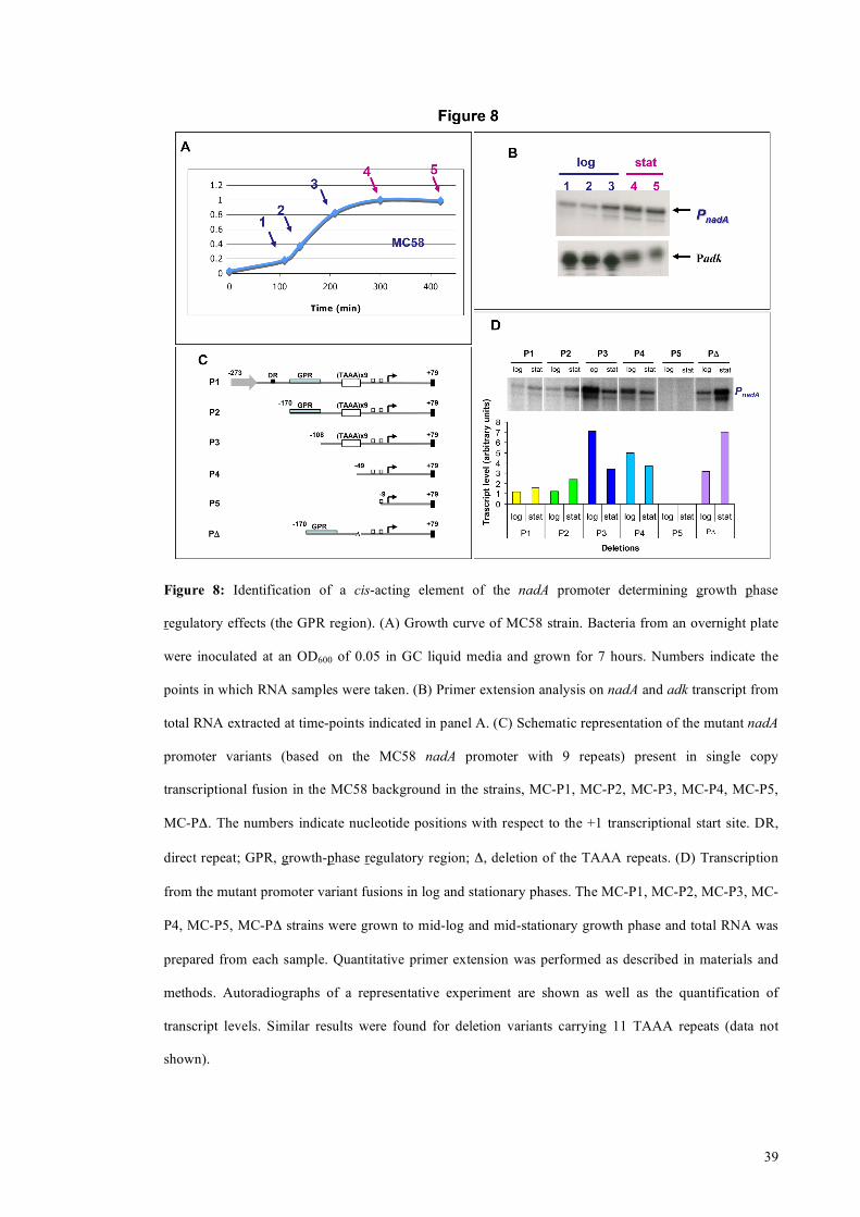
1.15 Identification of a cis acting element responsible for growth
phase regulation
Transcription from the nadA promoter is regulated during growth, as shown in the
primer extension experiment shown in Figure 8B, which was performed on total RNA
extracted at different time points from MC58 growing cells. As shown, nadA is
transcribed maximally in late logarithmic and stationary phase. Unlike adenylate kinase
gene that, as many other housekeeping genes, is less transcribed entering in stationary
phase due to a general slowdown in the bacterial metabolism.
In order to identify regulatory regions within the PnadA promoter we created a range of
deletion mutants in N. meningitidis MC58 strain, and measured the transcript level from
cells grown to the mid-log and stationary growth phases (Figure 8C and 8D). While
deletion of nucleotide sequences upstream of -170 with respect to the +1 transcriptional
start site had little or no effect on the level of transcript (promoter P2 versus P1),
promoter mutants lacking the region between -170 and -108, (P3 or P4) resulted in a
significant increase in transcription during log phase. The same results have been
obtained for promoters harbouring 11 repeats (data not shown). This finding suggests
that the growth-phase dependent regulation is due to a repression of expression in log
phase, more than an induction in stationary phase. Accordingly, removal of the TAAA
tract did not alter the growth-phase regulation of the resultant mutants (P4 versus P3, or
PΔ versus P2). Therefore, we have identified a distal upstream cis-acting region that we
call the GPR region (for growth phase regulatory), which is responsible for repression
of transcription from PnadA in log phase, possibly upon binding of a repressor protein.
39
Figure 8: Identification of a cis-acting element of the nadA promoter determining growth phase
regulatory effects (the GPR region). (A) Growth curve of MC58 strain. Bacteria from an overnight plate
were inoculated at an OD600 of 0.05 in GC liquid media and grown for 7 hours. Numbers indicate the
points in which RNA samples were taken. (B) Primer extension analysis on nadA and adk transcript from
total RNA extracted at time-points indicated in panel A. (C) Schematic representation of the mutant nadA
promoter variants (based on the MC58 nadA promoter with 9 repeats) present in single copy
transcriptional fusion in the MC58 background in the strains, MC-P1, MC-P2, MC-P3, MC-P4, MC-P5,
MC-PΔ. The numbers indicate nucleotide positions with respect to the +1 transcriptional start site. DR,
direct repeat; GPR, growth-phase regulatory region; Δ, deletion of the TAAA repeats. (D) Transcription
from the mutant promoter variant fusions in log and stationary phases. The MC-P1, MC-P2, MC-P3, MC-
P4, MC-P5, MC-PΔ strains were grown to mid-log and mid-stationary growth phase and total RNA was
prepared from each sample. Quantitative primer extension was performed as described in materials and
methods. Autoradiographs of a representative experiment are shown as well as the quantification of
transcript levels. Similar results were found for deletion variants carrying 11 TAAA repeats (data not
shown).

40
1.16 A protein in meningococcus cell extract binds GPR region
To assess whether a repressor factor could bind the GPR region we analysed crude cell
extracts of the MC58 strain for the ability to retard a radioactively labelled GPR probe
in Electrophoretic Mobility Shift Assays (EMSA). Addition of 15 µg of MC58 extracts
resulted in a complete shift of the GPR probe, which could be outcompeted with cold
GPR DNA but not with non-specific competitor (Figure 9A). We also found that the P5
promoter probe spanning from -9 to +79 of the PnadA promoter was specifically retarded
(lane 11, Figure 9A) by MC58 extracts but not an unrelated intergenic region (Pcon)
used as negative control.
We further characterize the nature of this binding factor performing EMSA after
incubation of the cell extract with different proteases or at different temperatures. Upon
treatment with 5 µg of Trypsin or ProteinaseK for 2 hours we were able to completely
abolish the binding activity of the cell extract (Figure 9B, lanes 4 and 6). Furthermore,
incubating the cell extract for 30 minutes at 60 or 100 °C has the same effect of protease
treatment, probably due to protein denaturation, whereas 30 minutes at 40 °C does not
seem to alter the binding activity. Then, we fractionate cell extract using sequential
ammonium sulphate precipitation, with increasing salt concentration. The binding
activity is retained only in the 70% precipitated fraction, in which there is an enrichment
of small proteins. These data taken together suggest that the GPR binding factor could
be a small protein relatively heat-resistant.

41
Figure 9: A protein in meningococcus cell extracts specifically binds the GPR region. (A) Binding
activity towards the nadA promoter in cell extracts of MC58. Cell extracts were prepared from mid-log
cultures of MC58 and increasing quantities were incubated with a radioactively labelled DNA probe
consisting of the GPR region (-170 to -108) or P5 (- 9 to +79) or an unrelated intergenic region Pcon as
negative control and submitted to EMSA analysis. To ca. 80 fmoles of radioactively labelled probe, 0,
0.2, 0.6, 1.8, 5.0, 15 µg of cell extract in lanes 1-6 were added, respectively; 0 µg in lanes 10 and 12; and
15 µg in lanes 7-9, 11 and 13, were added; and 130, 400, and 1000 fmoles of cold GPR probe in lanes 7,
8, and 9 were added as specific competitor. (B) Different treatments of the cell extract in order to
characterize the nature of the binding activity. Ten µg of cell extracts were incubated with 5 µg of
Trypsin or Proteinase K either for 30 minutes or 2 hours (lanes 3-6), or for 30 minutes at 40, 60 and 100
°C (lanes 9-11 respectively) and then subjected to EMSA assay. To fractionate cell extract we
sequentially precipitated it with ammonium sulphate salts at 20, 40, 60 and 70% (lanes 15-22,
respectively) and then perform EMSA assay using 2 µg (+) or 4 µg (++) of each fraction.

42
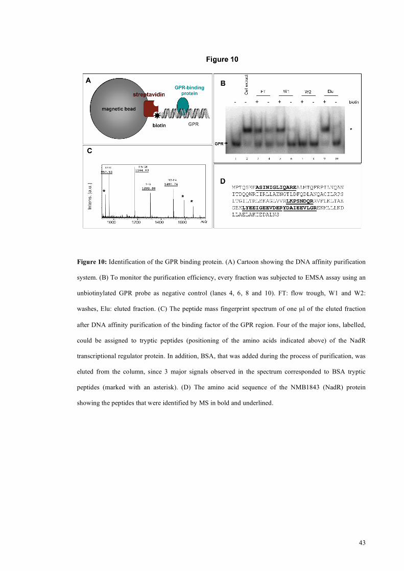
1.17 Identification of the GPR binding protein
To identify the GPR binding factor we performed DNA affinity purification using the
biotinylated GPR region as ‘bait’ and streptavidin coated magnetic beads (Figure 10A).
After incubation of the biotinylated GPR probe with the extract and the addition of
streptavidin coated magnetic beads, the sample was washed and the GPR binding factor
eluted with high salt. Each step of the purification was then analyzed for GPR binding
activity through EMSA. Figure 10B shows as the binding activity is successfully eluted
from the beads only in presence of biotinylated GPR DNA and not in the negative
control (same purification performed with a non biotinylated GPR DNA). The eluted
fraction was digested with trypsin, and the resulting peptides were analyzed by MALDI-
TOF mass spectrometry. Four of the seven major ions could be assigned to tryptic
peptides derived from the NMB1843 protein (Figure 10C and 10D). The other three
peaks could be assigned to bovine serum albumin which was added in the purification
step to block non-specific interaction. To confirm the interpretation, the major parental
ions were fragmented. Spectra of fragmentation were consistent with the expected
NMB1843 amino acid sequence (data not shown). We call this protein that binds the
GPR region of the nadA promoter NadR. The nadR gene encodes a transcriptional
regulator of the MarR family of repressors, is a homologue of FarR, the repressor of the
fatty acid resistance efflux pump of N. gonorrhoeae (Lee and Shafer 1999) (Lee,
Rouquette-Loughlin et al. 2003) and was recently implicated as a repressor of nadA
(Schielke, Huebner et al. 2009). We rename the meningococcal homologue NadR as,
unlike the FarR protein, it does not regulate the fatty acid efflux pump in
meningococcus (Pigozzi E, personal communication) and, therefore, is not involved in
fatty acid resistance.
43
Figure 10: Identification of the GPR binding protein. (A) Cartoon showing the DNA affinity purification
system. (B) To monitor the purification efficiency, every fraction was subjected to EMSA assay using an
unbiotinylated GPR probe as negative control (lanes 4, 6, 8 and 10). FT: flow trough, W1 and W2:
washes, Elu: eluted fraction. (C) The peptide mass fingerprint spectrum of one µl of the eluted fraction
after DNA affinity purification of the binding factor of the GPR region. Four of the major ions, labelled,
could be assigned to tryptic peptides (positioning of the amino acids indicated above) of the NadR
transcriptional regulator protein. In addition, BSA, that was added during the process of purification, was
eluted from the column, since 3 major signals observed in the spectrum corresponded to BSA tryptic
peptides (marked with an asterisk). (D) The amino acid sequence of the NMB1843 (NadR) protein
showing the peptides that were identified by MS in bold and underlined.

44
1.18 NMB 1843 interacts with the nadA promoter
We amplified and cloned the nadR gene (NMB1843) from the MC58 genome into an
expression plasmid and expressed and purified a recombinant form of the protein with
an N-terminal Histidine tag. We performed DNase I footprinting analysis with the NadR
protein and a radioactively labelled probe consisting of the entire nadA promoter. Figure
11A shows the autoradiogram of the results. On addition of increasing amounts of
NadR recombinant protein, three regions of protection of the nadA promoter are visible.
Two appear on addition of 30 nM of NadR protein: the first (OpI) spanning from -139
to -119 and the second (OpII) spanning from -15 to +7 and, therefore, within regions of
the GPR and P5 probes that were previously shown to be bound by the MC58 extracts
as well as a third region (OpIII) spanning the TAAA tract from -55 to -85. EMSA
analysis confirmed that NadR exhibits high affinity for the GPR and P5 operator regions
and exhibits a lower affinity for the TAAA tract. These observations were supported by
EMSA analysis with a probe spanning the entire PnadA promoter as three differential
protein-DNA complexes were formed, most likely following sequential binding of the
protein to the operators located within the PnadA probe (Figure 11B).
To confirm that NadR is the GPR-binding factor, we generated a deletion nadR mutant
by substituting the gene with an antibiotic resistance marker. Cell extracts derived from
the N. meningitidis Δ1843 mutant no longer possessed binding activity towards the GPR
probe (Figure 12A).
From this analysis we conclude that NadR encodes the GPR-binding factor that binds to
three operators; two high affinity operators OpI and OpII within the distal GPR region
and overlapping the nadA promoter, respectively, and a lower affinity operator OpIII
which spans the TAAA repeat tract.

45
Figure 11: The NadR repressor binds specifically to three operators in the nadA promoter. (A) DNase I
footprinting analysis with purified NadR on the nadA promoter with 9 repeats. The NadR protected
regions are indicated (OpI-III) and numbers represent the nucleotide positions with respect to the
transcriptional start site. The size of protected regions ranges from 20 bp (OpI and OpII), and 30 bp
(OpIII), a size compatible with the binding of a protein dimer. Binding reactions contained 40 fmoles of
probe radioactively labelled at one extremity and 0, 7.5, 15, 30, 60, 120 nM of NadR purified dimer
(lanes 1-6, respectively). (B) EMSA with radioactively labelled GPR, TAAA and P5 probes containing
the individual OpI, OpIII and OpII operators, respectively, or the entire P2 nadA promoter spanning from
-170 to +79 with increasing concentrations of recombinant NadR protein as indicated. The retarded
migration of protein DNA complexes are indicated with asterisks.

46
1.19 NadR represses NadA expression
To further study the role of NadR in regulating NadA expression, and its possible
involvement in mediating differential expression from phase variant promoters, we
generated isogenic knockouts in five representative strains bearing different numbers of
tetranucleotide repeat in their nadA promoter which correlate to high (8 repeats, 5/99)
and low (9 repeats, MC58), as well as three intermediary (5 repeats, BZ83; 6 repeats,
ISS838, and 12 repeats, 961-5945) levels of NadA expression. We evaluated by
Western Blot the NadA and NadR expression level in the wild type and Δ1843
meningococcal strains. The wild type strains showed, as expected, levels of NadA
expression that can be associated with transcript levels of the nadA phase variant
promoter they bear, and NadR was constitutively expressed in each strain (Figure 12B,
WT). Each of the knockout strains exhibits higher levels of NadA expression than their
respective wild type strain indicating that NadR represses nadA expression in each
strain (Figure 12B WT vs Δ1843). Surprisingly, the mutation of NadR results in almost
equivalent levels of NadA between the knockout strains, although the 5/99-Δ1843 and
BZ-Δ1843 still exhibit slightly higher NadA expression. This suggests that NadR,
although expressed to the same level, has a different repressive activity on the nadA
gene transcription in each strain and this may depend on the number of repeats in the
different phase variant promoters i.e. NadR does not efficiently repress the 8x promoter
of 5/99 but very efficiently represses the 9x promoter of MC58.

47
Figure 12: NadR represses NadA expression. (A) EMSA assay performed with MC58 and Δ1843 cell
extract on GPR probe showing the lack of binding activity in absence of NadR. (B) Western Blot analysis
of the level of expression of NadA and NadR in wild type strains 5/99, BZ83, ISS838, 961-5945 and
MC58 carrying nadA promoters with 8, 5, 6, 12, and 9 repeats respectively, and their NadR null mutant
derivatives. Cells were recovered from overnight culture on plates and 5 µg of total protein were loaded
on SDS-PAGE, blotted and stained with anti-NadA or anti-NadR polyclonal antiserum.

48
1.20 NadR represses to a different extent different phase variant
promoters
To further test the latter hypothesis and to rule out effects due to strain differences, we
deleted the nadR gene in the isogenic MC58 strains carrying high (x8), medium (x6)
and low (x9) promoter variants and measured the steady state levels of transcription
from the promoters at log and stationary growth phase in the presence or absence of the
NadR regulator. The results in Figure 13 confirm that in the mutant (Δ1843) all three
promoters are derepressed and, interestingly, little or no variation in transcript levels
between the phase variants is observed, suggesting that in the absence of NadR the
mechanism of transcriptional control exerted by variable number of repeats is alleviated
or negligible. It is worth noting, however, that the maximum level of transcription in
exponentially growing cells is observed from the promoter variant with 8 repeats, in
agreement with higher NadA expression in 5/99-Δ1843, suggesting that NadR is not the
sole modulator of phase variable promoter activity and that there is another factor which
may establish differential RNAP contacts to modulate transcription.
Furthermore, we also measured the transcript level of the PΔ promoter, which lacks the
TAAA tract and also no longer binds IHF, in the wild type and Δ1843 backgrounds and
results indicate that NadR does not efficiently repress this mutant promoter (lanes 9 and
10 versus 1 and 2) and implicates a major role for IHF in efficient NadR-mediated
repression of the nadA promoter.

49
Figure 13: The NadR repressor contributes to phase variable expression. Transcription of phase variant
promoters with 0, 9, 6, and 8 repeats, in the MC58 and NadR null mutant backgrounds. Total RNA was
prepared from cultures of strains MC-PΔ, MC-P2(x9), MC-P2(x6), MC-P2(x8), Δ1843-PΔ, Δ1843-
P2(x9), Δ1843-P2(x6), and Δ1843-P2(x8), grown to mid-log and stationary growth phase. Quantitative
primer extension was performed as described in materials and methods.

50
1.21 4-hydroxyphenylacetic acid (4HPA) interacts with NadR and
derepresses NadA expression
The MarR family of proteins regulates a wide variety of biological processes including
resistance to antibiotics and antimicrobial agents, virulence and environmental sensing
of aromatic compounds (Wilkinson and Grove 2006) (Ellison and Miller 2006). They
respond to small inducer molecules which attenuate the ability of MarR homodimers to
bind their cognate DNA sequences (Wilkinson and Grove 2006), and are often the
molecular substrates for the efflux pumps or metabolic pathways that are repressed by
this family of regulators. We set about identifying a small molecule inducer, which may
regulate NadR-mediated repression of NadA expression in meningococcus. We
assessed broad-specificity inducers such as salicylic acid, which have been shown to be
active against many members of this family, and also functionally relevant molecules
such as long-chain fatty acids, which are the substrate for the regulated efflux pump of
the gonococcal NadR homologue FarR (Lee, Rouquette-Loughlin et al. 2003) with no
success. However, we noticed that immediately downstream of the nadR gene is an
ORF which encodes a putative flavoprotein oxidoreductase with 42% amino acid
identity to the small subunit of 4-hydroxyphenylacetic acid 3-hydroxylase. In addition,
the closest BLAST neighbour of NadR in the MarR family of repressors is the HpaR
protein (50% identity), which represses the 4-hydroxyphenylacetic acid (4HPA)
catabolic pathway in E. coli. Moreover, it is responsive to the 4HPA substrate of the
pathway, which binds to the repressor and induces expression of the catabolic genes
(Galan, Kolb et al. 2003). We therefore, assessed whether the 4HPA molecule could act
as putative inducer of NadA expression in vivo. Addition of 1 mM or 5 mM 4HPA
(Figure 14A) to cultures of MC58 significantly induced NadA expression. No induction
could be detected in cultures of the Δ1843 mutant, indicating that the 4HPA molecule

51
induced a NadR-mediated derepression of NadA expression. Then we further analyse
transcript level of nadA after treatment of MC58 cultures with increasing concentration
of 4HPA. As shown in figure 14 B nadA transcript increases upon addition of 200µM of
the molecule and reaches the maximum at 1 mM after only 10 minutes of treatment
(Figure 14 B lanes 3, 4), suggesting that the induction is directly at the level of promoter
derepression. To confirm that the observed increases in NadA expression could
represent a direct interaction of the inducer with NadR, the ability of the compound to
dissociate purified recombinant NadR from the high affinity operator OpI was assessed
by EMSA. The 4HPA compound was found to attenuate the binding activity of the
NadR regulator to the GPR probe in vitro (Figure 14C). Furthermore, addition of 1 mM
4HPA to crude cell extracts containing the native NadR meningococcal protein resulted
in complete inhibition of retardation of the GPR probe in EMSA (data not shown),
suggesting that the recombinant and native NadR proteins respond in vitro similarly to
the compound. These data suggest that the 4HPA could be a ligand of the NadR
repressor and interaction of the ligand with the protein attenuates the DNA-binding
activity of the molecule for its specific operators and results in derepression or induction
in vivo of the nadA promoter.

52
Figure 14 (A) Induction of expression of NadA by addition of a small molecule ligand 4-hidroxy phenyl
acetic acid (4HPA). Broth cultures of MC58 or Δ1843 were grown to OD600 of 0.24 without (lane 1) or
with 1mM or 5mM (lane 2 and 3) 4HPA; or to OD600 of 0.24 and then incubated with 0, 1 or 5mM 4HPA
(lanes 4-6) added for 1 h. Cells were harvested and 5 µg of total protein from each culture was subjected
to SDS-PAGE and Western Blot analysis with anti-NadA or anti-NMB2091 antibodies as negative
control. (B) Primer extension showing nadA transcript level from total RNA of MC58 strain grown at
OD600 of 0.5 and treated for 10 minutes with 0, 0.04, 0.2, 1 and 5 mM of 4HPA (lanes 1-5 respectively).
(C) EMSA assays demonstrating dissociation of NadR from OpI operator in the GPR probe in vitro
following the addition of 4HPA (lanes 3-5) but not the broadly acting salicylic acid ligand (lanes 6-8).

53
Discussion
Host-pathogen interaction is a dynamic process that can lead to different outcomes such
as the clearance of the pathogen, the establishment of a disease or a sort of equilibrium
leading to commensalism. The factors that lead N. meningitidis to establish a productive
infection, switching from commensal to pathogenic, thought to be dependent on both
the host and the pathogen are still poorly understood. For these reasons, a better
understanding of the causes and mechanism that mediate the expression of proteins
involved in the interaction with host tissues is needed, both for predicting the
effectiveness of a vaccine which contains these proteins, as well as for characterizing at
the molecular level, novel strategies in the ever changing fight between pathogens and
their host.
In this scenario N. meningitidis has to arrange in concert the expression of a variety of
different genes in order to adapt and survive in the different tissues during an infection
of the human host. At the same time the bacteria must also escape the host immune
response, which targets mostly the same structures used by the meningococcus to
interact with the host. This issue is addressed by the high redundancy and variability in
the expression of surface structure and by the ability to acquire genes from other
bacteria species.
The low GC content, with respect to the average of the genome, and the presence of a
repeated sequence (TCAGAC) flanking the nadA gene, suggest it was acquired by
horizontal transfer (Comanducci, Bambini et al. 2002). Another repeated tract present in
the nadA locus (TAAA tetranucleotide repeated tract upstream of the promoter), has
been found to be responsible for its phase variable expression (Martin, Makepeace et al.
2005).
Phase variation is the adaptive process by which bacteria undergo frequent and
reversible phenotypic changes resulting from genetic alterations in specific loci of their

54
genomes and this process is crucial for the survival of pathogens and commensals in
hostile and ever-changing host environments. N. meningitidis has an unprecedented
potential for generating genetic diversity through slipped strand mispairing of simple
sequence repeats, as its genome contains over 100 repeat associated genes (Saunders,
Jeffries et al. 2000; Snyder, Butcher et al. 2001). The way in which genes are affected
by variation in the number of repeats is largely thought to occur through biphasic on/off
translational control due to frameshifting within the ORFs of coding regions. Recently
the on/off switching of methyltransferase genes has been shown to co-ordinate
expression of a phase-variable regulon of genes or "phasevarions" via differential
methylation of the genome (Srikhanta, Maguire et al. 2005) (Srikhanta, Dowideit et al.
2009). The role of simple sequence repeats in intergenic regions in modulating phase
variable expression, although frequently found, are less easy to predict. However,
differential spacing due to repeated tracts between the core promoter elements
modulating multi-phasic expression by affecting RNAP sigma factor binding has been
frequently reported (Sarkari, Pandit et al. 1994) (Carson, Stone et al. 2000) (van Ham,
van Alphen et al. 1993) (van der Ende, Hopman et al. 2000), as well as some
documented examples where repeats in 5’UTR (Dawid, Barenkamp et al. 1999)
(Lafontaine, Wagner et al. 2001) and distally upstream (Martin, van de Ven et al. 2003)
(Dawid, Barenkamp et al. 1999) (Puopolo and Madoff 2003) (Liu, Panangala et al.
2002) of promoters have been shown to affect expression through unknown
mechanisms.
In this study, we dissect the cis- and trans-acting elements as well as environmental
factors that control transcriptional regulation of the nadA promoter in order to elucidate
the mechanism by which sequence repeats distally upstream of the PnadA promoter
controls its activity. We describe a complex promoter architecture in which spontaneous
changes in the number of simple sequence repeats in a tract between the most distal

55
regulatory regions and the core promoter can alter the promoter activity and lead to
phase variable expression. We have shown that the NadR repressor is the major
contributor to the phase variable expression levels of the promoter as it binds to two
high affinity operators flanking the repeats. One operator overlaps the -10 region of the
promoter and the transcriptional start site, and therefore, binding of NadR is consistent
with its function as a repressor through sterically hindering RNAP access to the
promoter. The other high affinity operator is on the distal upstream side of the phase
variable repeat in a cis-acting region that we call the GPR, which is functionally active
in repressing the promoter despite its distal location (Figure 8). We have identified a
single IHF binding site that is located between these two high affinity operators and we
show that IHF binding to this promoter is necessary for efficient NadR-mediated
repression of PnadA. The IHF binding site comprises some sequence upstream of the
TAAA as well as part of the TAAA tract itself. If the TAAA tract is removed, the
protein no longer binds the mutant promoter (Figure 7). However, we found that IHF
binding is unaffected by the number of TAAA repeats, but rather the number of repeats
may change the spacing of the DNA on the upstream and downstream flanking regions
of the tract and, therefore, may influence the localisation, and possibly the orientation of
proteins that bind to the operators. The ability of IHF to bend DNA may facilitate the
looping of the DNA of the nadA promoter and bring the GPR element proximal to the
core promoter elements. A looping mechanism (Figure 15) would explain the function
of such a distal operator in repression of transcription, possibly through interactions of
dimers present on spatially proximal operators which lock the promoter to RNAP
similar to the mechanism described for the lac operon (Oehler, Eismann et al. 1990)
(Lewis, Chang et al. 1996).

56
1.22 Three-dimensional MODEL
Figure 15: Three-dimensional structure of nadA promoter region. The spatial conformation of the DNA
filament is predicted on the basis of its sequence composition (Predictor and Swiss-pdbViewer). Forward
strands of two nadA phase variants promoters (x9 and x8 TAAA repeats) are shown, superimposed from
their 3’ ends. The -35 and -10 examers are indicated and two nucleotides corresponding to the centre of
the binding sites for IHF (green) and NMB1843 (purple), identified by footprinting, are shown in Space-
Fill mode and depicted with either white or red arrows (for phase variant promoters with 9 and 8 repeats
respectively).

57
However, in the completely derepressed form there are still significant, albeit marginal
differences in the promoter strength of variants with different numbers of repeats. The
alpha-subunit of RNAP binds to the distal GPR regulatory region and also immediately
upstream of the core promoter overlapping the TAAA tract which may function as UP-
like elements. We propose a model in which differential distancing between the nadR
operators and the contact points of RNAP result in optimal or suboptimal configuration
of the protein complexes and, therefore, result in more or less efficient repression
mediated by NadR and/or more or less cis-enhancement of RNAP activity on the basal
promoter strength (Figure 16).

58
1.23 Regulation MODEL
Figure 16: Model of regulation of NadA promoter: Two promoter variants with 9 and 8 repeats
representing low activity and high activity promoter phase variants, respectively, highlighting the ability
of NadR to efficiently or less efficiently repress the promoters (top panels) and NadR-independent effects
on the derepressed promoter basal levels possibly due to differential contacts with the α-subunit of RNAP
(bottom panels) due to different spatial organization of the NadR and RNAP contact points resulting from
the different number of repeats.

59
Interestingly, in Pu promoter of P. putida, two distinct UP elements, located close to
the core promoter (-79) and distally upstream (-104), interact with RNAP α-subunits
enhancing gene expression (Macchi, Montesissa et al. 2003). This interaction is
modulated by IHF that allow the interchangeable positioning of the two α-subunits over
the two UP-elements (Macchi, Montesissa et al. 2003) (Bertoni, Fujita et al. 1998). This
scenario resembles in part what we observe with α-subunit interaction over nadA
promoter.
The gonococcal homologue of NadR is FarR, which has been characterised in
gonococcus as a repressor of the FarAB fatty acid resistance efflux pump (Lee,
Rouquette-Loughlin et al. 2003). FarR binds two distal operators on the farAB promoter
(located similarly to OpI and OpII in the nadA promoter) and represses transcription in
an IHF dependent way. It would appear from deletion analysis of the nadA promoter
that all regulatory elements necessary for control of nadA expression were horizontally
transferred together with the nadA gene, as the direct repeat delineating the border of
the transferred DNA is at -193. The intriguing question is then how the nadA locus,
which is present in only a fraction of strains, through a horizontal transfer event,
adopted such a complex regulatory mechanism pre-existing in Neisseria. The nadR
gene is well conserved in other Neisseria spp such as N .mucosa, N. cinerea, N.
lactamica, N. subflavia and N. flavescens and, therefore, must respond to signals in the
ecological niches of all these species.
The NadR protein is a member of the MarR ligand-responsive transcriptional regulators
and the majority of MarR family members are regulated by the non-covalent binding of
low molecular weight ligands. These signalling molecules regulate the activity of the
regulators. In this study, we have identified a putative ligand, 4-hydroxyphenylacetic
acid, which is able to relieve the DNA binding activity of NadR, thus derepressing or

60
inducing NadA expression. This molecule is a catabolite of aromatic amino acids and it
is secreted in human saliva (Takahama, Hirota et al. 2003) (and also urine), suggesting
that the inducer is present in the oropharynx and NadA may be induced in the mucosal
niche, which is bathed in saliva.
The 4HPA molecule is a catabolite of the aromatic amino acids, tyrosine and
phenylalanine. Two groups of bacteria, the soil inhabitants such as P. putida and the
enteric bacteria such as E. coli contain pathways for the breakdown of these amino acids
to succinate and pyruvate (Diaz, Ferrandez et al. 2001). However, such a pathway is not
present in the meningococcal genome (Tettelin, Saunders et al. 2000). Nevertheless,
nadR is present in a operon-like organization with 2 downstream genes one of which
shows significant homology to the HpaC small subunit of a hydroxylase involved in the
conversion of 4HPA to a less toxic form (3,4-dihydroxyphenylacetic acid). It is unclear
whether this operon may be involved in the utilisation of 4HPA inducer in some way in
meningococcus, or whether it is the remnant of a partial catabolic pathway that was
acquired horizontally and the 4HPA molecule simply acts as a signal inducing the
expression of the adhesin, which is necessary for colonization and invasion of the
mucosa.
Phase variation functions as an adaptive strategy generating spontaneous diverse sub-
populations of the bacterium which may be beneficial in adapting to different
microenvironments within the human host during the course of a natural infection.
However, in the case of nadA gene regulation, this type of mechanism is bi-functional,
in that the major mediation of phase variable expression levels of NadA is through
repression by NadR protein in the absence of the correct inducer signal. Subpopulations
expressing low levels of NadA through promoter phase variations still have the
potential to respond to the correct niche signal, such as 4HPA, and express high levels
under certain environmental or niche-specific conditions. Such variation will have an

61
impact on the interaction with the host tissues, as well as escaping immune responses.
Simple sequence repeats have been identified in distal promoter regions of known or
potential virulence factors in other pathogens including Helicobacter pylori (Saunders,
Peden et al. 1998), H. Influenza (Dawid, Barenkamp et al. 1999) (Parkhill, Achtman et
al. 2000), Moraxella catarrhalis (Linton, Karlyshev et al. 2001), Group B Streptococcus
(Puopolo and Madoff 2003) and pathogenic Mycoplasma (Liu, Panangala et al. 2002)
some of which have been shown to control phase variable expression through unknown
mechanisms. This suggests that complex regulatory mechanisms such as what we have
elucidated for NadA involving stochastic variations and environmentally-responsive
transcription factors may be widely used by pathogens. Elucidating these mechanisms is
important for our understanding of the intimate and complex relationship between the
host and disease-causing organisms.

62

63
Materials and Methods
1.24 Bacterial strains and culture conditions
The N. meningitidis strains used in this study are all listed in Table II. N. meningitidis
strains were routinely cultured in GC-based (Difco) agar medium supplemented with
Kellogg’s supplement I (Kellogg et al., 1963) at 37°C in a 5% CO2-95% air atmosphere
at 95% humidity. Strains were stocked in 10% skim milk and stored at -80°C. For liquid
cultures, N. meningitidis strains were grown overnight on solid medium, re-suspended
in GC broth to an optical density at 600 nm (OD600) of 1, and inoculated with a 1:20
dilution into GC broth supplemented with Kellogg’s supplement I, 12.5 µM Fe(NO3)3
or Mueller Hinton (MH) (Sigma, St. Louis, MO) containing 0,25% glucose and, when
required, erythromycin and/or chloramphenicol added to achieve final concentrations of
5 µg ml-1. For transformation by naturally competent N. meningitidis, four to five single
colonies of a freshly grown overnight culture were re-suspended in 20 µl of PBS,
spotted onto GC plates, and 5–10 µg of linearized plasmid DNA was added, allowed to
dry and incubated for 6–8 h at 37°C. Transformants were then selected on plates
containing erythromycin (5 µg ml-1) and/or chloramphenicol (5 µg ml-1), and single
colonies were re-streaked on selective media for further analysis. Transformants were
re-suspended in 50 µl of distilled water, placed in a boiling water bath for 5 min and
centrifuged in a bench top centrifuge for 5 min at 8000 g. One microlitre of the sample
was used as template for PCR analysis for correct insertion by a double homologous
recombination event, and nadA promoter regions were amplified and sequenced to
verify that no phase variation had occurred during manipulations. E. coli DH5-α
(Hanahan 1983) and BL21(DE3) (Studier and Moffatt 1986) cultures were grown in

64
Luria–Bertani medium, and when required, ampicillin chloramphenicol and/or IPTG
were added to achieve final concentrations of 100, 20 µg ml-1 and 1mM respectively.
1.25 DNA techniques.
DNA manipulations were carried out routinely as described by Sambrook, Fritsch et al.
1989. Small- and large-scale plasmid DNA preparations were carried out with a
QIAprep Spin Mini kit and Plasmid Midi kit (QIAGEN) according to the
manufacturer’s instructions. DNA fragments or PCR-amplified products were purified
from agarose gels with QiaEX DNA purification kit (QIAGEN). PCR was performed in
a Perkin-Elmer 2400 thermal cycler with Platinum Taq polymerase (Invitrogen). One
microlitre of each reaction mixture contained 10–50 ng of chromosomal DNA or 1 µl
bacterial sample (see above), 100 pmols of the required primers and 200 µM
concentration of each deoxynucleotide in a volume of 100 µl of 1 x PCR buffer
containing MgCl2 (Invitrogen). After the initial denaturing step at 95°C for 5 min, 30
cycles of denaturing at 95°C, annealing at the appropriate temperatures for the specific
primers and elongation at 72°C were carried out. DNA fragments were routinely
sequenced according to the dideoxy-chain termination method (Sanger, Air et al. 1977)
by using [α-32P]-dATP (NEN, Perkin Elmer) and a T7 sequencing kit (Pharmacia).
1.26 Construction of nadA promoter fusions
In order to analyze cis-elements involved in regulation of nadA expression a series of
plasmids carrying nadA promoters fused to the gfp gene were constructed, which could
be assayed in E. coli and also used for allelic exchange in N. meningitidis strains at a
chromosomal location between two converging open reading frames (ORFs),
NMB1074 and NMB1075. A plasmid consisting of a promoterless gfp gene and the
ermAM erythromycin-resistance genes flanked by upstream and downstream regions for

65
allelic replacement was generated (pGFP, (Ieva, Roncarati et al. 2008)). A series of 5’
deletion variants of nadA promoter were generated by PCR amplification from the
genome of MC58 using primers Nad-N1, Nad-N2, Nad-N3, Nad-N4 and Nad-N5 as
forwards and Nad-Sp as reverse, generating P1, P2, P3, P4 and P5 promoter deletion
fragments respectively, spanning from -273, -170, -108, -49, and -9, respectively, to +79
with respect to the transcriptional start site. The 5 promoter fragments were cloned
using NsiI and SphI sites, introduced by primers design into the pGFP plasmid and used
for transformation of MC58 generating the MC-P1, MC-P2, MC-P3, MC-P4, amd MC-
P5 strains respectively. (Table II and Figure 8). Phase variant nadA promoters with
different repeat numbers, from 4 to 13, were PCR amplified with the Nad-N2/Nad-Sp
primer pair using as a template genomic DNA from different clinical isolates with the
appropriate number of repeats as major clone kindly donated by Maurizio Comanducci
and Stefania Bambini. All promoters fragments generated, (spanning from -170 to +79
with respect to the +1 in MC58) were then cloned as NsiI- SphI fragments into the
pGFP plasmid and used for transformation of MC58 generating the MC-P2x4, MC-
P2x5, MC-P2x6, MC-P2x7, MC-P2x8, MC-P2x9, MC-P2x10, MC-P2x11, MC-P2x12,
MC-P2x13 strains respectively. (Table II and Figure 5). The PΔ nadA promoter variant,
lacking the TAAA tetranucleotide repeat, was generated by PCR amplificaion of
regions upstream and downstream of the TAAA repeated tract, using primer pairs Nad-
N2/n85-50R and n85-50F/Nad-Sp. Then, in a second round of PCR, the respective
upstream and downstream fragments, which contain regions of overlap due to the
primer design, were used in a self-priming PCR amplification for 5 cycles, and then the
corresponding united fragments were amplified using the external primers Nad-N2/Nad-
Sp, and cloned into pGFP. The pGFP-PΔ plasmid was used for the transformation of
MC58 generating MC-PΔ, containing a nadA promoter fusion in which the TAAA
repeated tract was substituted with an EcoRI site (Table II and Figure 8).

66
1.27 Construction of knockouts
In order to knockout the NMB1843 gene in the Neisseria background, the
pΔ1843ko::Cm plasmids was constructed. Upstream and downstream flanking regions
of the NMB1843 gene were amplified by PCR with the 1843-1/1843-2b and 1843-
3b/1843-4 primers respectively. Then in a second round of PCR the respective upstream
and downstream fragments, which contain regions of overlap due to the design of the
primers, were used in a self-priming PCR amplification for 5 cycles, and then the
corresponding united fragment was amplified using the external 1843-1/1843-4 primers.
This product was cloned into the pGEM-T (Promega) vector and a chloramphenicol
cassette from pDT2548 (Wang and Taylor 1990) was inserted into the unique BamHI
site, between the flanking regions, generating pΔ1843ko::Cm. The plasmid was then
linearised and used for transformation of the meningococcal strains to make the
respective NMB1843 knockout mutants (Table II). The correct double homologous
recombination event resulting on the knockout of the genes was verified by PCR.
1.28 Expression and purification of the E.coli RNA polymerase α-
subunit and N. meningitidis NMB1843 protein.
The NMB1843 gene was amplified from the MC58 genome with the 1843-F/1843-R
primer pair and cloned as a 448 bp NdeI-BamHI fragment into the pET15b expression
plasmid (Invitrogen) generating pET15-1843 which was subsequently transformed into
the E. coli strain BL21(DE3) for protein expression. For alpha subunit expression,
plasmid pHTT7f1-NHα (Tang, Severinov et al. 1995) was transformed into E. coli
strain BL21(DE3). From an overnight culture of the BL21(DE3)(pET15-1843) and
BL21(DE3)(pHTT7f1-NHa) strains, 200 ml of Luria-Bertani medium was inoculated

67
and grown to an OD600 of 0.5, and expression of the recombinant NMB1843 or E.coli
RNApol α-subunit proteins containing an N-terminal histidine tag was induced by the
addition of 1 mM IPTG (isopropyl-D-thiogalactopyranoside) and further incubation for
3 h. The proteins were purified from the harvested cells by Ni-nitrilotriacetic acid
(QIAGEN) affinity chromatography under nondenaturing conditions according to the
manufacturer’s instructions. The purified protein preparation was then diluted to 1
mg/ml and dialyzed overnight in 1 x Binding Buffer (20 mM Tris-HCl pH 8, 50 mM
KCL, 10 mM MgCl2, 01% NP40) containing 10% glycerol and then again overnight in
1x Binding buffer containing 50% glycerol. The purity of the proteins was estimated to
be greater than 98% by sodium dodecyl sulphate-polyacrylamide gel electrophoresis
(SDS-PAGE). The concentration of the proteins in this preparation was determined by
using the Bradford colorimetric assay (Bio-Rad), and the protein was aliquoted and
stored at -80°C. To generate anti-NMB1843 antibodies, 6-week-old female CD1 mice
(Charles River Laboratories) were immunized with 20 µg of NadR protein given
intraperitoneally, together with complete Freund's adjuvant in three doses (day 1, 21 and
35). Bleed-out samples were taken on day 49 and used for Western blot analysis.
1.29 Western blot analysis
N. meningitidis colonies from overnight plate cultures were either resuspended in PBS
until OD600 of 1, or grown to logarithmic phase (OD of 0.5, ca. 2 hours incubation) and
stationary phase (OD ≥ 0.9 ca. 5 hours incubation) from a starter inoculum of OD 0.05.
Samples of 1-2 ml were harvested and normalised in 1xSDS-PAGE loading buffer (50
mM Tris Cl pH 6.8, 2.5 % SDS, 0.1 % Bromophenol Blue, 10% glycerol, 5% B-
mercapto-Ethanol, 50 mM DTT) to a relative OD of 5. For Western blot analysis, 10 µg
of each total protein sample in 1 x SDS-PAGE loading buffer was separated by SDS-
PAGE, and transferred onto nitrocellulose membrane using an iBlot Dry Blotting

68
System (Invitrogen). Membranes were blocked overnight at 4C° by agitation in
blocking solution (10% skimmed milk, 0.05% Tween-20, in PBS) and incubated for 90
min at 37°C with anti-NadA or anti-NMB1843 polyclonal sera) in blocking solution.
After washing, the membranes were incubated in a 1:2000 dilution of peroxidase-
conjugated anti-rabbit immunoglobulin (Biorad) or anti-mouse immunoglobulin (Dako)
respectively, in blocking solution for 1 hour and the resulting signal was detected using
the Supersignal West Pico chemiluminescent substrate (Pierce).
1.30 Co-over expression of alpha subunit in E. coli
In order to allow co-expression in E. coli of nadA promoter fusions with pLAW2
(overexpressing RNAP α-subunit) and pLAW2Δ256 (overexpressing the N-terminus of
α-subunit), pXG-1 plasmid (Urban and Vogel 2007), harbouring pSC101 origin of
replication and a cloramphenicol resistance cassette, was used simply substituting the
181 bp AatII/NheI fragment containing the PLtetO-1 promoter with different nadA
promoter phase variants amplified with nad-Aa2/nadNh primer pairs (Table I),
generating nadA-gfp translational fusions.
To evaluate the role of RNAP alpha-subunit in regulation of transcription from the nadA
promoter, we co-transform pLAW2 or pLAW2Δ256 with pXG harbouring nadA-gfp
fusions in E. coli. After liquid growth to an OD600 of 0.5 in presence of 1 mM IPTG for
induction of α expression, GFP fluorescence was measured in 48 well plates using
TECAN Infinite 200 with excitation wavelength of 460 nm and an emission of 510 nm.
Experiments were performed in triplicate.

69
1.31 Crude extract preparation, DNA affinity purification of GPR-
binding protein and Electrophoretic mobility shift assay
(EMSA)
N. meningitidis strains were growth until OD600 of 0.5 as described above, then 100 ml
were harvested and resuspended in 10 ml of PBS, sonicated at maximum power (20
impulse of 0,8 second each) for 5 times at +4 °C and the cell debris was removed by
centrifugation at 8,000xg for 15 min. The soluble fraction was then filtered using a 0.2
µm membrane and stored at 4 °C. Proteins concentration in the sample was determined
using the Bradford colorimetric assay.
For the characterization of the binding activity in the cell extract, it was precipitated,
where indicated, with ammonium sulphate salts by adding, from a saturated solution,
the amount to reach 20, 40, 60 and 70% concentration. Cell extracts were also incubated
at different temperatures (40, 60 and 100°C) for 30 minutes or with 5 µg of Trypsin
(Promega) or Proteinase K (Gibco BRL) for 30 minutes or 2 hours as indicated.
For purification of GPR-binding protein from MC58 crude extract a probe spanning
from -170 to -116 with respect to the MC58 nadA promoter was amplified using a 5’
biotinylated forward primer Bio-nadN2 (Invitrogen custom primers) and gpr-R reverse
primer. 25 pmoles of the fragment were incubated with 1.3 ml of MC58 crude extract
(0.6µg/µl) in presence of 100µg of salmon sperm DNA and 500µg bovine serum
albumin (BSA) to block non specific interactions for 20 minutes at room temperature
with gentle rotation. The mixture was then added to 2,5 mg of Dynabeads M-280
streptavidin (Invitrogen), previously washed 4 times with 250 µl PBS, and incubated for
20 minutes at room temperature with gentle rotation. The tube was then placed on a
magnet for 2 minutes for magnetic separation of the beads and after 3 washes with PBS,
proteins bound to biotinylated GPR were eluted in 400µl of 1M NaCl. The sample was

70
then dialyzed over night against H2O at 4 C°. All fractions were then analysed for
binding activity using EMSA assay, and eluted fraction was further analyzed using
MALDI TOF.
For gel shift experiments, a probe corresponding to the GPR element and spanning from
-170 to -116 with respect to the MC58 nadA promoter was amplified using nad-N2/gpr-
R, a probe corresponding to the P5 promoter fragment spanning -9 to +81 was amplified
with NadN5/Nad-Sp, a probe corresponding to P2 promoter fragment spanning -170 to
+81 was amplified with nad-N2/ Nad-Sp, a probe corresponding to 6 TAAA repeats
using primers IHF-Lex and IHF-Rex2 annealed and a probe corresponding to an
unrelated intergenic region Pcon (157 bp between NMB2073 and NMB2074), was
amplified using SR-F and SR-R. Two pmoles of each fragment were then radioactively
labeled at their 5’ ends with 30 µCi of (γ-32P)-ATP (6000 Ci/mmol; NEN) using 10 U of
T4 polynucleotide kinase (New England Biolabs). The unincorporated radioactive
nucleotides were removed using the TE-10 chromaspin columns (Clontech). For each
binding reaction, 40 fmoles of labeled probes was incubated with increasing amounts of
crude extract or recombinant NMB1843 in 25 µl final volume of Gelshift Binding
Buffer (25 mM Tris-HCl pH 7.5, 1mM MgCl2, 10% glycerol) with 2µg salmon sperm
DNA as non-specific competitor, for 15 min at room temperature, and run on 6% native
polyacrilamide gels buffered with 0.5x TBE at 100 Volts for 80 min at 4 °C. Gels were
dried and exposed to autoradiographic films at –80°C and radioactivity was quantified
using a phosphorimager and the Image Quant software (Molecular Dynamics).
1.32 RNA preparation and Primer extension
N. meningitidis or E. coli strains were grown in liquid culture to logarithmic or stationary
phase and then 20 ml of cultures were added to an equal volume of equivalent frozen

71
medium to bring the temperature immediately to 4 °C and then centrifuged at 3,000 rpm
in a benchtop centrifuge at 4º C. RNA was extracted from the pelleted cells as
previously described (Spohn, Beier et al. 1997). Primer extension was performed as
previously reported (Delany, Ieva et al. 2003). To ensure correct mapping of the
promoter sequencing reaction was carried out with a T7 sequencing kit (USB
Corporation) using the same primer as in the primer extension reactions and the plasmid
consisting of the relevant cloned promoter. Quantification of the signals from the primer
elongated product was performed using Phosphoimager and ImageQuant software
(Molecular dynamics). For quantitative experiments, experiments were performed from
at least 2 independent biological replicates and figures show results from one
representative experiment. Internal negative controls were performed on each RNA set
quantifying the specific transcript of a gene whose expression is not altered, usually adk
or nmb1870.
1.33 DNase I footprinting (NMB1843, Neisseria gonorrhoeae IHF,
Escherichia coli RNApol and Alpha-sub)
The nadA promoter region was amplified from genomic DNA from different clinical
isolates with the appropriate number of repeats as major clone and from pGFP-PΔ
plasmid for 0 repeat, using primers nad-N1 and nad-B1 and cloned as 320, 342, 346 and
354 bp (for 0, 6, 7, 9 repeats respectively) NsiI-BamHI fragments into pGemT
(Promega). A radioactive probe for DNA footprinting of nadA promoters were prepared
as follows: approximately 2 pmol of the different plasmids were linearized with BamHI,
subjected to dephosphorylation using CIP (New England Biolabs), and then 5’ end
labelled using 5 pmol of [γ-32P]-ATP with T4 polynucleotide Kinase (New England

72
Biolabs). The plasmid was then digested with NsiI, and nadA promoter fragments,
labelled at one extremity, were purified by preparative polyacrylamide gel
electrophoresis (PAGE). Briefly, the probe was extracted from a 6% polyacrylamide gel
and eluted in 3 ml of elution buffer (10 mM Tris-HCl [pH 8.0], 1 mM EDTA, 300 mM
Na acetate [pH 5.2], 0.2% sodium dodecyl sulfate [SDS]) at 37°C overnight. The eluted
probe was then extracted once with an equal volume of phenol-chloroform (1:1) and
ethanol precipitated. Protein-DNA complexes were formed in 50 µl of footprinting
buffer (20 mM Tris-HCl, pH 7.9, 50 mM KCl, 10 mM MgCl2, 0.01% NP-40, 10%
glycerol) containing approximately 20 fmol (10 000 c.p.m) of the labelled probe and
200 ng of sonicated salmon sperm DNA as the non-specific competitor and recombinant
NMB1843 protein, Neisseria gonorreae purified IHF (kindly donated by Hill SA,
Belland RJ et al. 1997), E. coli RNA polymerase holoenzyme (USB) or alpha subunit
(Tang, Murakami et al. 1996) in final concentrations as indicated, for 15 min at room
temperature. Following incubation for 1 min at room temperature with 0.03 U of DNase
I (Roche) and 5 mM CaCl2, the reaction was stopped by addition of 140 µl of stop
buffer (192 mM Na acetate, 32 mM Na2EDTA, 0.14% SDS, 64 mg ml-1 of sonicated
salmon sperm DNA). Samples were phenol-extracted, ethanol-precipitated, re-
suspended in denaturing sample buffer and fractionated on urea-6% polyacrylamide
gels. As a molecular weight marker, a G+A sequence reaction (Maxam and Gilbert
1977) was performed for each DNA probe and run in parallel to the corresponding
footprinting reactions.
1.34 MALDI TOF mass spectrometry
Proteins eluted from Dynabeads M-280 streptavidin column and dialyzed against H2O
were dried with a Speed Vac. (Labconco) and solubilized with 20 µl of 5 mM
ammonium bicarbonate containing 0.1 % (wt/vol) of RapiGest™ SF surfactant

73
(Waters), incubated 5 min at 95°C and digested with 2 µg of trypsin (Sequencing grade
Promega). The reaction was allowed to proceed for 15 hours at 37°C. An aliquot of the
reaction was diluted 10 times with 0,1% (vol/vol) of trifluoroacetic acid, and 0.7 µl was
directly spotted on a matrix PAC target (Prespotted AnchorChip 96, set for Proteomics,
Bruker Daltonics). Air-dried spot was washed with 0.6 µl of a solution of 70% (vol/vol)
ethanol, 0.1% (vol/vol) TFA. Peptide mass fingerprint spectra were recorded with a
MALDI-TOF/TOF mass spectrometer UltraFlex (Bruker Daltonics). Ions generated by
laser desorption at 337 nm (N2 laser) were recorded at an acceleration of 25 kV in the
reflector mode. In general, about 200 single spectra were accumulated for improving the
signal/noise ratio and analyzed by FlexAnalysis (version 2.4, Bruker Daltonics).
External calibration was performed using standard peptides pre-spotted on the target.
The data of MS were further analyzed through an in-house licensed MASCOT, version
2.2.1 (Matrixscience Ltd), running on a local server containing the protein sequence
data downloaded from NCBI. The following parameters were used for database
searches: monoisotopic mass accuracy, 75 pm, missed cleavages, 1, oxidation of
methionine as variable modifications.

74
Table 1. Oligonucleotides used in this study
Name Sequence Site
Nad-N1 attcagatgcatGACGTCGACGTCCTCGATTACGAAGGC NsiI
Nad-N2 attcagatgcatTAAGACACGACACCGGCAGAATTG NsiI
Nad-N3 attcagatgcatCCGAACTACCTAACTGCAAG NsiI
Nad-N4 attcagatgcatTTGCGACAATGTATTGTATATATG NsiI
Nad-N5 attcagatgcatCTTTAATATGTAAACAAACTTGGTGG NsiI
Nad-Sp attcagcatgctacGCTCATTACCTTTGTGAGTGG SphI
Nad-B1 attcaggatcctacGCTCATTACCTTTGTGAGTGG BamHI
n-85/50F CTACCTAACTGCAAGAATTcTTGCGACAATGTATTG EcoRI
n-85/50R CAATACATTGTCGCAAgAATTCTTGCAGTTAGGTAG EcoRI
Bio-nad-N2 attcagatgcatTAAGACACGACACCGGCAGAATTG NsiI
nad-Aa2 attcaggacgtcTAAGACACGACACCGGCAGAATTG AatII
nad-Nh attcagctagcCATGCTCATTACCTTTGTGAGTGG NheI
1843-1 TACGTTCCGGCAGTTCGGCGG
1843-2b cgcatcctcgggatccGGGTAGGCATTGTTTAAGTCTCC BamHI
1843-3b caaatgcctacccggatccCGAGGATGCGTTGAACTCGTAATAC
GCCG
BamHI
1843-4 ACCGCTCTTCGGGCGACAGGCCGG
1843-F attcacatATGCCTACCCAATCAAAACATGCG NdeI
1843-R attcaggatcCGGCGTATTACGAGTTCAACGCATCCTCG BamHI
IHF-Lex ctagaACTGCAAGAATTAAATAAATAAATAAATAAATA
AATTGCGAC
XbaI
IHF-Rex2 ctagaGTCGCAATTTATTTATTTATTTATTTATTTAATTC
TTGCAGT
XbaI

75
Gpe-3 GAATTGGGACAACTCCAGTG
1870-pe gaatcagggcagtggtcagag
Adk-PE CGCGCCTAAAAGTAATGC
gpr-R gattagcatgcCGGCATTAATATCTGTTAATATGTGC SphI
SR-F TCGGAAGCCGTCCGTTCCGAACC
SR-R attatggatccATAAACGCCAAACCCACCGCGAAGGTGG BamHI
a Capital letters indicate N. meningitidis derived sequences, small letters indicate
sequences added for cloning purposes, and underlined letters indicate restriction enzyme
recognition sites.
Table 2. Strains and Plasmids used in this study.
Name Relevant characteristics Reference or
source
Neisseria meningitidis
MC58 Clinical isolate, sequenced strain containing 9
TAAA tetranucleotide repeats in the nadA
promoter
(Tettelin,
Saunders et al.
2000)
5/99 Clinical isolate containing 8 TAAA tetranucleotide
repeats in the nadA promoter
Norwegian
clinical isolate
BZ83 Clinical isolate containing 5 TAAA tetranucleotide
repeats in the nadA promoter
(Comanducci,
Bambini et al.
2002)
ISS838 Clinical isolate containing 6 TAAA tetranucleotide
repeats in the nadA promoter
(Comanducci,
Bambini et al.
2002)
961-5945 Clinical isolate containing 12 TAAA
tetranucleotide repeats in the nadA promoter
(Comanducci,
Bambini et al.
2002)
MC-Δ1843 1843 null mutant in MC58 strain, CmR This study
5/99-Δ1843 1843 null mutant in 5/99 strain, CmR This study

76
BZ83-Δ1843 1843 null mutant in BZ83 strain, CmR This study
ISS-Δ1843 1843 null mutant in ISS838 strain, CmR This study
961-Δ1843 1843 null mutant in 961-5945 strain, CmR This study
MC-P(1-5) Series of 5 derivatives of MC58, containing single
copy transcriptional fusion of 5’ deletions of the
nadA promoter fused to the gfp gene, EryR
This study
MC-PΔ Derivative of MC58, containing single copy
transcriptional fusion of a mutant variant of the
nadA promoter, with the tetranucleotide repeats
deleted, fused to the gfp gene, EryR
This study
MC-P2(x4-
x13)
Series of 10 derivatives of MC58, containing single
copy transcriptional fusion of the nadA P2
promoter variants, containing from 4 to 13
tetranucleotide repeats, fused to the gfp gene, EryR
This study
Δ1843-PΔ, -
P2(x6, x8, x9)
4 Derivatives of MC-Δ1843, containing single
copy transcriptional fusion of the nadA P2
promoter with either 0, 6, 8 or 9 tetra nucleotide
repeats, fused to the gfp gene, EryR
This study
MC-Fko Fur null mutant of MC58 (Delany, Ieva et
al. 2003)
Fko-P2(x9) Derivative of MC-Fko containing single copy
transcriptional fusion of the nadA P2 promoter with
9 repeats, fused to the gfp gene, EryR
This study
Plasmids
pGEMT Cloning vector, AmpR Promega
pΔ1843ko::Cm Construct for generating knockout of the
NMB1843 gene, CmR
This study
pGFP Construct for insertion of nadA promoter variants
and mutants fused to gfp in single copy between
ORF nmb1074 and NMB1075 in the N.
meningitidis chromosome, AmpR, EryR
(Ieva, Roncarati
et al. 2008)
pGX-1 Derivative pSC101 containing the gfp gene, CmR (Urban and Vogel
2007)
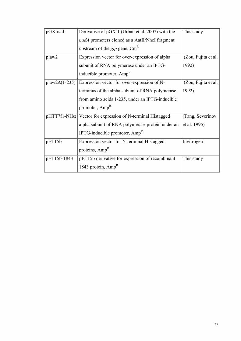
77
pGX-nad Derivative of pGX-1 (Urban et al. 2007) with the
nadA promoters cloned as a AatII/NheI fragment
upstream of the gfp gene, CmR
This study
plaw2 Expression vector for over-expression of alpha
subunit of RNA polymerase under an IPTG-
inducible promoter, AmpR
(Zou, Fujita et al.
1992)
plaw2Δ(1-235) Expression vector for over-expression of N-
terminus of the alpha subunit of RNA polymerase
from amino acids 1-235, under an IPTG-inducible
promoter, AmpR
(Zou, Fujita et al.
1992)
pHTT7f1-NHα Vector for expression of N-terminal Histagged
alpha subunit of RNA polymerase protein under an
IPTG-inducible promoter, AmpR
(Tang, Severinov
et al. 1995)
pET15b Expression vector for N-terminal Histagged
proteins, AmpR
Invitrogen
pET15b-1843 pET15b derivative for expression of recombinant
1843 protein, AmpR
This study

78

79
References
Achtman, M. (1995). "Epidemic spread and antigenic variability of Neisseria meningitidis."
Trends Microbiol 3(5): 186-92. Belyaeva, T. A., V. A. Rhodius, et al. (1998). "Transcription activation at promoters carrying
tandem DNA sites for the Escherichia coli cyclic AMP receptor protein: organisation of the RNA polymerase alpha subunits." J Mol Biol 277(4): 789-804.
Bentley, S. D., G. S. Vernikos, et al. (2007). "Meningococcal genetic variation mechanisms viewed through comparative analysis of serogroup C strain FAM18." PLoS Genet 3(2): e23.
Bertoni, G., N. Fujita, et al. (1998). "Active recruitment of sigma54-RNA polymerase to the Pu promoter of Pseudomonas putida: role of IHF and alphaCTD." Embo J 17(17): 5120-8.
Blatter, E. E., W. Ross, et al. (1994). "Domain organization of RNA polymerase alpha subunit: C-terminal 85 amino acids constitute a domain capable of dimerization and DNA binding." Cell 78(5): 889-96.
Busby, S. and R. H. Ebright (1999). "Transcription activation by catabolite activator protein (CAP)." J Mol Biol 293(2): 199-213.
Capecchi, B., J. Adu-Bobie, et al. (2005). "Neisseria meningitidis NadA is a new invasin which promotes bacterial adhesion to and penetration into human epithelial cells." Mol Microbiol 55(3): 687-98.
Carson, S. D., B. Stone, et al. (2000). "Phase variation of the gonococcal siderophore receptor FetA." Mol Microbiol 36(3): 585-93.
Comanducci, M., S. Bambini, et al. (2002). "NadA, a novel vaccine candidate of Neisseria meningitidis." J Exp Med 195(11): 1445-54.
Comanducci, M., S. Bambini, et al. (2004). "NadA diversity and carriage in Neisseria meningitidis." Infect Immun 72(7): 4217-23.
Davidsen, T. and T. Tonjum (2006). "Meningococcal genome dynamics." Nat Rev Microbiol 4(1): 11-22.
Dawid, S., S. J. Barenkamp, et al. (1999). "Variation in expression of the Haemophilus influenzae HMW adhesins: a prokaryotic system reminiscent of eukaryotes." Proc Natl Acad Sci U S A 96(3): 1077-82.
Deghmane, A. E., D. Giorgini, et al. (2002). "Down-regulation of pili and capsule of Neisseria meningitidis upon contact with epithelial cells is mediated by CrgA regulatory protein." Mol Microbiol 43(6): 1555-64.
Delany, I., R. Ieva, et al. (2003). "The iron-responsive regulator fur is transcriptionally autoregulated and not essential in Neisseria meningitidis." J Bacteriol 185(20): 6032-41.
Diaz, E., A. Ferrandez, et al. (2001). "Biodegradation of aromatic compounds by Escherichia coli." Microbiol Mol Biol Rev 65(4): 523-69, table of contents.
Ellison, D. W. and V. L. Miller (2006). "Regulation of virulence by members of the MarR/SlyA family." Curr Opin Microbiol 9(2): 153-9.
Estrem, S. T., T. Gaal, et al. (1998). "Identification of an UP element consensus sequence for bacterial promoters." Proc Natl Acad Sci U S A 95(17): 9761-6.
Evans, K., L. Adewoye, et al. (2001). "MexR repressor of the mexAB-oprM multidrug efflux operon of Pseudomonas aeruginosa: identification of MexR binding sites in the mexA-mexR intergenic region." J Bacteriol 183(3): 807-12.
Feil, E. J., E. C. Holmes, et al. (2001). "Recombination within natural populations of pathogenic bacteria: short-term empirical estimates and long-term phylogenetic consequences." Proc Natl Acad Sci U S A 98(1): 182-7.
Finne, J., D. Bitter-Suermann, et al. (1987). "An IgG monoclonal antibody to group B meningococci cross-reacts with developmentally regulated polysialic acid units of glycoproteins in neural and extraneural tissues." J Immunol 138(12): 4402-7.

80
Fletcher, L. D., L. Bernfield, et al. (2004). "Vaccine potential of the Neisseria meningitidis 2086 lipoprotein." Infect Immun 72(4): 2088-100.
Franzoso, S., C. Mazzon, et al. (2008). "Human monocytes/macrophages are a target of Neisseria meningitidis Adhesin A (NadA)." J Leukoc Biol 83(5): 1100-10.
Fuangthong, M. and J. D. Helmann (2002). "The OhrR repressor senses organic hydroperoxides by reversible formation of a cysteine-sulfenic acid derivative." Proc Natl Acad Sci U S A 99(10): 6690-5.
Fyfe, J. A. and J. K. Davies (1998). "An AT-rich tract containing an integration host factor-binding domain and two UP-like elements enhances transcription from the pilEp1 promoter of Neisseria gonorrhoeae." J Bacteriol 180(8): 2152-9.
Gaal, T., W. Ross, et al. (1996). "DNA-binding determinants of the alpha subunit of RNA polymerase: novel DNA-binding domain architecture." Genes Dev 10(1): 16-26.
Galan, B., A. Kolb, et al. (2003). "Molecular determinants of the hpa regulatory system of Escherichia coli: the HpaR repressor." Nucleic Acids Res 31(22): 6598-609.
Goodman, S. D., S. C. Nicholson, et al. (1992). "Deformation of DNA during site-specific recombination of bacteriophage lambda: replacement of IHF protein by HU protein or sequence-directed bends." Proc Natl Acad Sci U S A 89(24): 11910-4.
Goodrich, J. A., M. L. Schwartz, et al. (1990). "Searching for and predicting the activity of sites for DNA binding proteins: compilation and analysis of the binding sites for Escherichia coli integration host factor (IHF)." Nucleic Acids Res 18(17): 4993-5000.
Goosen, N. and P. van de Putte (1995). "The regulation of transcription initiation by integration host factor." Mol Microbiol 16(1): 1-7.
Grifantini, R., S. Sebastian, et al. (2003). "Identification of iron-activated and -repressed Fur-dependent genes by transcriptome analysis of Neisseria meningitidis group B." Proc Natl Acad Sci U S A 100(16): 9542-7.
Griffiths, N. J., C. J. Bradley, et al. (2007). "IFN-gamma amplifies NFkappaB-dependent Neisseria meningitidis invasion of epithelial cells via specific upregulation of CEA-related cell adhesion molecule 1." Cell Microbiol.
Hammerschmidt, S., R. Hilse, et al. (1996). "Modulation of cell surface sialic acid expression in Neisseria meningitidis via a transposable genetic element." Embo J 15(1): 192-8.
Hammerschmidt, S., A. Muller, et al. (1996). "Capsule phase variation in Neisseria meningitidis serogroup B by slipped-strand mispairing in the polysialyltransferase gene (siaD): correlation with bacterial invasion and the outbreak of meningococcal disease." Mol Microbiol 20(6): 1211-20.
Hanahan, D. (1983). "Studies on transformation of Escherichia coli with plasmids." J Mol Biol 166(4): 557-80.
Hawley, D. K. and W. R. McClure (1983). "The effect of a lambda repressor mutation on the activation of transcription initiation from the lambda PRM promoter." Cell 32(2): 327-33.
Hayrinen, J., H. Jennings, et al. (1995). "Antibodies to polysialic acid and its N-propyl derivative: binding properties and interaction with human embryonal brain glycopeptides." J Infect Dis 171(6): 1481-90.
Hochschild, A. and S. L. Dove (1998). "Protein-protein contacts that activate and repress prokaryotic transcription." Cell 92(5): 597-600.
Hoiczyk, E., A. Roggenkamp, et al. (2000). "Structure and sequence analysis of Yersinia YadA and Moraxella UspAs reveal a novel class of adhesins." Embo J 19(22): 5989-99.
Hood, D. W., M. E. Deadman, et al. (1996). "DNA repeats identify novel virulence genes in Haemophilus influenzae." Proc Natl Acad Sci U S A 93(20): 11121-5.
Ieva, R., D. Roncarati, et al. (2008). "OxyR tightly regulates catalase expression in Neisseria meningitidis through both repression and activation mechanisms." Mol Microbiol 70(5): 1152-65.
Igarashi, K. and A. Ishihama (1991). "Bipartite functional map of the E. coli RNA polymerase alpha subunit: involvement of the C-terminal region in transcription activation by cAMP-CRP." Cell 65(6): 1015-22.

81
Jeon, Y. H., T. Yamazaki, et al. (1997). "Flexible linker in the RNA polymerase alpha subunit facilitates the independent motion of the C-terminal activator contact domain." J Mol Biol 267(4): 953-62.
Lafontaine, E. R., N. J. Wagner, et al. (2001). "Expression of the Moraxella catarrhalis UspA1 protein undergoes phase variation and is regulated at the transcriptional level." J Bacteriol 183(5): 1540-51.
Law, E. C., N. J. Savery, et al. (1999). "Interactions between the Escherichia coli cAMP receptor protein and the C-terminal domain of the alpha subunit of RNA polymerase at class I promoters." Biochem J 337 ( Pt 3): 415-23.
Lee, E. H., C. Rouquette-Loughlin, et al. (2003). "FarR regulates the farAB-encoded efflux pump of Neisseria gonorrhoeae via an MtrR regulatory mechanism." J Bacteriol 185(24): 7145-52.
Lee, E. H. and W. M. Shafer (1999). "The farAB-encoded efflux pump mediates resistance of gonococci to long-chained antibacterial fatty acids." Mol Microbiol 33(4): 839-45.
Lewis, L. A., M. Gipson, et al. (1999). "Phase variation of HpuAB and HmbR, two distinct haemoglobin receptors of Neisseria meningitidis DNM2." Mol Microbiol 32(5): 977-89.
Lewis, M., G. Chang, et al. (1996). "Crystal structure of the lactose operon repressor and its complexes with DNA and inducer." Science 271(5253): 1247-54.
Linton, D., A. V. Karlyshev, et al. (2001). "Deciphering Campylobacter jejuni cell surface interactions from the genome sequence." Curr Opin Microbiol 4(1): 35-40.
Liu, L., V. S. Panangala, et al. (2002). "Trinucleotide GAA repeats dictate pMGA gene expression in Mycoplasma gallisepticum by affecting spacing between flanking regions." J Bacteriol 184(5): 1335-9.
Luijsterburg, M. S., M. C. Noom, et al. (2006). "The architectural role of nucleoid-associated proteins in the organization of bacterial chromatin: a molecular perspective." J Struct Biol 156(2): 262-72.
Macchi, R., L. Montesissa, et al. (2003). "Recruitment of sigma54-RNA polymerase to the Pu promoter of Pseudomonas putida through integration host factor-mediated positioning switch of alpha subunit carboxyl-terminal domain on an UP-like element." J Biol Chem 278(30): 27695-702.
Madico, G., J. A. Welsch, et al. (2006). "The meningococcal vaccine candidate GNA1870 binds the complement regulatory protein factor H and enhances serum resistance." J Immunol 177(1): 501-10.
Maiden, M. C. (1993). "Population genetics of a transformable bacterium: the influence of horizontal genetic exchange on the biology of Neisseria meningitidis." FEMS Microbiol Lett 112(3): 243-50.
Maiden, M. C. (2008). "Population genomics: diversity and virulence in the Neisseria." Curr Opin Microbiol 11(5): 467-71.
Maiden, M. C., J. A. Bygraves, et al. (1998). "Multilocus sequence typing: a portable approach to the identification of clones within populations of pathogenic microorganisms." Proc Natl Acad Sci U S A 95(6): 3140-5.
Martin, P., K. Makepeace, et al. (2005). "Microsatellite instability regulates transcription factor binding and gene expression." Proc Natl Acad Sci U S A 102(10): 3800-4.
Martin, P., L. Sun, et al. (2004). "Involvement of genes of genome maintenance in the regulation of phase variation frequencies in Neisseria meningitidis." Microbiology 150(Pt 9): 3001-12.
Martin, P., T. van de Ven, et al. (2003). "Experimentally revised repertoire of putative contingency loci in Neisseria meningitidis strain MC58: evidence for a novel mechanism of phase variation." Mol Microbiol 50(1): 245-57.
Masignani, V., E. Balducci, et al. (2003). "NarE: a novel ADP-ribosyltransferase from Neisseria meningitidis." Mol Microbiol 50(3): 1055-67.
Masignani, V., M. Comanducci, et al. (2003). "Vaccination against Neisseria meningitidis using three variants of the lipoprotein GNA1870." J Exp Med 197(6): 789-99.
Maxam, A. M. and W. Gilbert (1977). "A new method for sequencing DNA." Proc Natl Acad Sci U S A 74(2): 560-4.

82
Molina-Lopez, J. A., F. Govantes, et al. (1994). "Geometry of the process of transcription activation at the sigma 54-dependent nifH promoter of Klebsiella pneumoniae." J Biol Chem 269(41): 25419-25.
Murakami, K., N. Fujita, et al. (1996). "Transcription factor recognition surface on the RNA polymerase alpha subunit is involved in contact with the DNA enhancer element." Embo J 15(16): 4358-67.
Murakami, K., J. T. Owens, et al. (1997). "Positioning of two alpha subunit carboxy-terminal domains of RNA polymerase at promoters by two transcription factors." Proc Natl Acad Sci U S A 94(21): 11274-8.
Nassif, X. (1999). "Interactions between encapsulated Neisseria meningitidis and host cells." Int Microbiol 2(3): 133-6.
Newlands, J. T., C. A. Josaitis, et al. (1992). "Both fis-dependent and factor-independent upstream activation of the rrnB P1 promoter are face of the helix dependent." Nucleic Acids Res 20(4): 719-26.
Oehler, S., E. R. Eismann, et al. (1990). "The three operators of the lac operon cooperate in repression." Embo J 9(4): 973-9.
Parekh, B. S. and G. W. Hatfield (1996). "Transcriptional activation by protein-induced DNA bending: evidence for a DNA structural transmission model." Proc Natl Acad Sci U S A 93(3): 1173-7.
Parkhill, J., M. Achtman, et al. (2000). "Complete DNA sequence of a serogroup A strain of Neisseria meningitidis Z2491." Nature 404(6777): 502-6.
Perez-Martin, J., K. N. Timmis, et al. (1994). "Co-regulation by bent DNA. Functional substitutions of the integration host factor site at sigma 54-dependent promoter Pu of the upper-TOL operon by intrinsically curved sequences." J Biol Chem 269(36): 22657-62.
Perez-Rueda, E. and J. Collado-Vides (2001). "Common history at the origin of the position-function correlation in transcriptional regulators in archaea and bacteria." J Mol Evol 53(3): 172-9.
Perez-Rueda, E., J. Collado-Vides, et al. (2004). "Phylogenetic distribution of DNA-binding transcription factors in bacteria and archaea." Comput Biol Chem 28(5-6): 341-50.
Pizza, M., V. Scarlato, et al. (2000). "Identification of vaccine candidates against serogroup B meningococcus by whole-genome sequencing." Science 287(5459): 1816-20.
Puopolo, K. M. and L. C. Madoff (2003). "Upstream short sequence repeats regulate expression of the alpha C protein of group B Streptococcus." Mol Microbiol 50(3): 977-91.
Rao, L., W. Ross, et al. (1994). "Factor independent activation of rrnB P1. An "extended" promoter with an upstream element that dramatically increases promoter strength." J Mol Biol 235(5): 1421-35.
Rice, P. A., S. Yang, et al. (1996). "Crystal structure of an IHF-DNA complex: a protein-induced DNA U-turn." Cell 87(7): 1295-306.
Rinaudo, C. D., J. L. Telford, et al. (2009). "Vaccinology in the genome era." J Clin Invest: In press.
Ross, W., S. E. Aiyar, et al. (1998). "Escherichia coli promoters with UP elements of different strengths: modular structure of bacterial promoters." J Bacteriol 180(20): 5375-83.
Ross, W., K. K. Gosink, et al. (1993). "A third recognition element in bacterial promoters: DNA binding by the alpha subunit of RNA polymerase." Science 262(5138): 1407-13.
Rytkonen, A., B. Albiger, et al. (2004). "Neisseria meningitidis undergoes PilC phase variation and PilE sequence variation during invasive disease." J Infect Dis 189(3): 402-9.
Sambrook, J., E. F. Fritsch, et al. (1989). Molecular cloning: A laboratory manual, Cold Spring Harbor, NY: Cold Spring Harbor Laboratory.
Sanger, F., G. M. Air, et al. (1977). "Nucleotide sequence of bacteriophage phi X174 DNA." Nature 265(5596): 687-95.
Sarkari, J., N. Pandit, et al. (1994). "Variable expression of the Opc outer membrane protein in Neisseria meningitidis is caused by size variation of a promoter containing poly-cytidine." Mol Microbiol 13(2): 207-17.

83
Saunders, N. J., A. C. Jeffries, et al. (2000). "Repeat-associated phase variable genes in the complete genome sequence of Neisseria meningitidis strain MC58." Mol Microbiol 37(1): 207-15.
Saunders, N. J., J. F. Peden, et al. (1998). "Simple sequence repeats in the Helicobacter pylori genome." Mol Microbiol 27(6): 1091-8.
Savery, N. J., G. S. Lloyd, et al. (1998). "Transcription activation at Class II CRP-dependent promoters: identification of determinants in the C-terminal domain of the RNA polymerase alpha subunit." Embo J 17(12): 3439-47.
Schielke, S., C. Huebner, et al. (2009). "Expression of the meningococcal adhesin NadA is controlled by a transcriptional regulator of the MarR family." Mol Microbiol 72(4): 1054-67.
Schoen, C., J. Blom, et al. (2008). "Whole-genome comparison of disease and carriage strains provides insights into virulence evolution in Neisseria meningitidis." Proc Natl Acad Sci U S A 105(9): 3473-8.
Schoen, C., H. Tettelin, et al. (2009). "Genome flexibility in Neisseria meningitidis." Vaccine 27(Supplement 2): B103-B111.
Segall, A. M., S. D. Goodman, et al. (1994). "Architectural elements in nucleoprotein complexes: interchangeability of specific and non-specific DNA binding proteins." Embo J 13(19): 4536-48.
Snyder, L. A., S. A. Butcher, et al. (2001). "Comparative whole-genome analyses reveal over 100 putative phase-variable genes in the pathogenic Neisseria spp." Microbiology 147(Pt 8): 2321-32.
Spohn, G., D. Beier, et al. (1997). "Transcriptional analysis of the divergent cagAB genes encoded by the pathogenicity island of Helicobacter pylori." Mol Microbiol 26(2): 361-72.
Srikhanta, Y. N., S. J. Dowideit, et al. (2009). "Phasevarions mediate random switching of gene expression in pathogenic Neisseria." PLoS Pathog 5(4): e1000400.
Srikhanta, Y. N., T. L. Maguire, et al. (2005). "The phasevarion: a genetic system controlling coordinated, random switching of expression of multiple genes." Proc Natl Acad Sci U S A 102(15): 5547-51.
Stapleton, M. R., V. A. Norte, et al. (2002). "Interaction of the Salmonella typhimurium transcription and virulence factor SlyA with target DNA and identification of members of the SlyA regulon." J Biol Chem 277(20): 17630-7.
Stapleton, P. D. and P. W. Taylor (2002). "Methicillin resistance in Staphylococcus aureus: mechanisms and modulation." Sci Prog 85(Pt 1): 57-72.
Stephens, D. S. (2009). "Biology and pathogenesis of the evolutionarily successful, obligate human bacterium Neisseria meningitidis." Vaccine 27 Suppl 2: B71-7.
Strainic, M. G., Jr., J. J. Sullivan, et al. (1998). "Promoter recognition by Escherichia coli RNA polymerase: effects of the UP element on open complex formation and promoter clearance." Biochemistry 37(51): 18074-80.
Studier, F. W. and B. A. Moffatt (1986). "Use of bacteriophage T7 RNA polymerase to direct selective high-level expression of cloned genes." J Mol Biol 189(1): 113-30.
Swinger, K. K. and P. A. Rice (2004). "IHF and HU: flexible architects of bent DNA." Curr Opin Struct Biol 14(1): 28-35.
Takahama, U., S. Hirota, et al. (2003). "Human salivary peroxidase-catalyzed oxidation of nitrite and nitration of salivary components 4-hydroxyphenylacetic acid and proteins." Arch Oral Biol 48(10): 679-90.
Tang, H., K. Severinov, et al. (1995). "Rapid RNA polymerase genetics: one-day, no-column preparation of reconstituted recombinant Escherichia coli RNA polymerase." Proc Natl Acad Sci U S A 92(11): 4902-6.
Tang, Y., K. Murakami, et al. (1996). "Upstream interactions at the lambda pRM promoter are sequence nonspecific and activate the promoter to a lesser extent than an introduced UP element of an rRNA promoter." J Bacteriol 178(23): 6945-51.
Tettelin, H., N. J. Saunders, et al. (2000). "Complete genome sequence of Neisseria meningitidis serogroup B strain MC58." Science 287(5459): 1809-15.

84
Urban, J. H. and J. Vogel (2007). "Translational control and target recognition by Escherichia coli small RNAs in vivo." Nucleic Acids Res 35(3): 1018-37.
van der Ende, A., C. T. Hopman, et al. (2000). "Multiple mechanisms of phase variation of PorA in Neisseria meningitidis." Infect Immun 68(12): 6685-90.
van der Ende, A., C. T. Hopman, et al. (1995). "Variable expression of class 1 outer membrane protein in Neisseria meningitidis is caused by variation in the spacing between the -10 and -35 regions of the promoter." J Bacteriol 177(9): 2475-80.
van der Woude, M. W. (2006). "Re-examining the role and random nature of phase variation." FEMS Microbiol Lett 254(2): 190-7.
van Ham, S. M., L. van Alphen, et al. (1993). "Phase variation of H. influenzae fimbriae: transcriptional control of two divergent genes through a variable combined promoter region." Cell 73(6): 1187-96.
Virji, M. (2009). "Pathogenic neisseriae: surface modulation, pathogenesis and infection control." Nat Rev Microbiol 7(4): 274-86.
Virji, M., C. Alexandrescu, et al. (1992). "Variations in the expression of pili: the effect on adherence of Neisseria meningitidis to human epithelial and endothelial cells." Mol Microbiol 6(10): 1271-9.
Wang, Y. and D. E. Taylor (1990). "Chloramphenicol resistance in Campylobacter coli: nucleotide sequence, expression, and cloning vector construction." Gene 94(1): 23-8.
Welsch, J. A., G. R. Moe, et al. (2003). "Antibody to genome-derived neisserial antigen 2132, a Neisseria meningitidis candidate vaccine, confers protection against bacteremia in the absence of complement-mediated bactericidal activity." J Infect Dis 188(11): 1730-40.
Wilkinson, S. P. and A. Grove (2006). "Ligand-responsive transcriptional regulation by members of the MarR family of winged helix proteins." Curr Issues Mol Biol 8(1): 51-62.
Zou, C., N. Fujita, et al. (1992). "Mapping the cAMP receptor protein contact site on the alpha subunit of Escherichia coli RNA polymerase." Mol Microbiol 6(18): 2599-605.