biology - learner · pdf filerediscovering biology constitute only a small fraction of cells...
TRANSCRIPT

Molecular to Global
Perspectives
REDISCOVERING
BIOLOGY
The human nervous system is probably the most intricatelyorganized aggregate of matter on Earth. A single cubiccentimeter of the human brain may contain well over 50 million nerve cells, each of which may communicatewith thousands of other neurons in information-processingnetworks that make the most elaborate computer lookprimitive. These neural pathways control our everyperception and movement and enable us to learn, think,and be conscious of ourselves and our surroundings.”CAMPBELL AND REECE1
The most striking differences between humans and other animals arein the size and the complexity of our brains. With our big brains wehave acquired a rich culture, which far exceeds that of any otherspecies in scope and complexity. We have developed science tounderstand how and why an immensity of things and processes work,including those of our own brain. At the start of the twenty-firstcentury neuroscientists are increasingly able to explain the functions ofbrain in molecular terms.
To understand how the brain works we first must consider what thebrain does. This can be broken down into three basic functions: (1)take in sensory information, (2) process information between neurons,and (3) make outputs. The neurons that take in information from theenvironment are called sensory neurons. These are specialized torespond to a particular stimulus, such as light, heat, chemicals, orvibration — anything you might encounter from outside, or eveninside, the body. The processing within the brain can range from aknee-jerk reaction — which takes place entirely in the spinal cord — tothe strategy adopted by a master chess player. In humans, we usuallycall this “thinking.” The output is most often a body movement, whichresults from the action of motor neurons. The brain is the link betweenthe outside world and behavior, and is thus crucial for survival. Thesethree basic functions are shared by organisms from humans down toinvertebrates like Caenorhabditis elegans, a nematode that doesn’teven have a true “brain” but a collection of about three hundredneurons. (See the Genes and Development unit.)
But how does the individual neuron work to carry out these tasks?Neurons’ unique systems capabilities arise from their cellular ability tocommunicate with one another very rapidly, using both electrical andchemical communication. Keep in mind, however, that the neuron isnot the only type of cell in the brain. The neuron may be the star ofthe show but there are other supporting players. Indeed, neurons
Neurobiology
“
R E D I S C O V E R I N G B I O L O GY
constitute only a small fraction of cells in the brain. For every neuronthere are about ten to fifty supporting cells, called glial cells, in thebrain. The word “glial” means glue, and these cells are the “glue” ofthe nervous system. They perform many vital tasks, including removingdead neurons and debris, releasing critical growth factors to neurons,and acting as insulating material for the neurons.
The incredibly complex ways in which brains function exemplify theimportance of cell-cell interactions. Below we discuss the chemical andelectrical means by which neurons communicate, and describe howvarious therapeutic and recreational drugs alter these processes at themolecular level. We then turn to the molecular nature of memory andlearning. Finally, we describe recent studies that demonstrate that newneurons are being produced continuously in us.
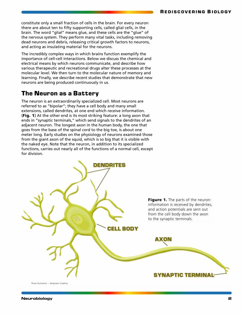
The Neuron as a BatteryThe neuron is an extraordinarily specialized cell. Most neurons arereferred to as “bipolar”; they have a cell body and many smallextensions, called dendrites, at one end which receive information.(Fig. 1) At the other end is its most striking feature: a long axon thatends in “synaptic terminals,” which send signals to the dendrites of anadjacent neuron. The longest axon in the human body, the one thatgoes from the base of the spinal cord to the big toe, is about onemeter long. Early studies on the physiology of neurons examined thosefrom the giant axon of the squid, which is so big that it is visible withthe naked eye. Note that the neuron, in addition to its specializedfunctions, carries out nearly all of the functions of a normal cell, exceptfor division.
2Neurobiology
DENDRITES
AXON
CELL BODY
SYNAPTIC TERMINAL
Figure 1. The parts of the neuron:information is received by dendrites,and action potentials are sent outfrom the cell body down the axon to the synaptic terminals.
Photo-illustration — Bergmann Graphics

R E D I S C O V E R I N G B I O L O GY
The neuron is an electric battery and works by changes in its voltage.Compared with its surroundings, the inside of a “resting neuron” has alower concentration of sodium ions and a higher concentration ofpotassium ions. Because of this imbalance of positively charged ionsacross the membrane, the inside of the resting neuron is negativerelative to the outside. This difference in voltage is called themembrane potential. A typical membrane potential for a neuron atrest, the resting potential, is -0.07 volts, or -70 mV. Although this is arather modest voltage (about five percent of that of an AA battery),consider that this voltage occurs across a miniscule length — that ofthe cell membrane. If this were an electric field, the charge separationwould be about 100,000 volts per centimeter.
Note that the term “resting neuron” refers only to its electrical state.The cell is really not at rest because, in addition to carrying out all ofthe normal functions of the cell, the neuron has to maintain this ionicimbalance. This is achieved by the sodium-potassium pump, whichactively transports potassium in and sodium out. The pump maintains anegative voltage because it actually pumps three sodium ions out forevery two potassium ions it pumps in. The membrane potential of aneuron at any given time is the product of many variables, includingthe imbalance of ions across the membrane and the membrane’spermeability to each ion. In addition to sodium and potassium,chloride is an important ion in “setting” a neuron’s rest potentialbecause negatively charged chloride ions can pass through open “leakchannels” at rest. Another ion crucial for neural communication iscalcium, which acts as a powerful intracellular signaling molecule onceit enters through its ion channels.
Voltage-Gated ChannelsThe neuron, like all cells, possesses a cell membrane that is mostly lipid.Ions like sodium and potassium cannot cross the lipid membrane ontheir own. In all cells transport of ions, as well as some small molecules,is carried out by channels, which are very tiny openings in themembrane formed by protein pores. These channels are often gated —that is, opened or closed — depending on the conditions of the cell.When open, the ions can enter and pass through channels by diffusion.Ions will always travel down their electrochemical gradient. Forexample, sodium is much more plentiful outside the cell than inside. Itis also positively charged, while the inside of the cell is typicallynegatively charged relative to outside. Thus, both the chemical andelectrical components of the gradient will drive sodium ions into thecell when sodium channels open. Voltage-gated channels are thosein which the membrane potential of the cell determines whether theyare opened or closed. Other channels can be opened or closed byvarious chemicals, such as neurotransmitters.
Channel proteins that span the cell membrane form the ion channels.To determine the structure of proteins, scientists have often used X-ray crystallography. (See the Proteins and Proteomics unit.) In2003 Roderick MacKinnon and his colleagues used this technique toexamine the structure of a voltage-gated potassium channel from aunicellular archaea. Previous studies have shown that ion channelshave a central ion-conduction pore. Like all proteins, ion channelproteins are made up of amino acids, some of which are charged.When voltage changes occur, these charged components of the protein
3Neurobiology
R E D I S C O V E R I N G B I O L O GY
make very small movements. This can result in more dramaticconformational changes, causing the channels to open and close.MacKinnon’s group found that “voltage-sensor paddles” surround thispore. It appears that with voltage changes in the membrane, thesepaddles will move and thus permit potassium ions across themembrane.2 Further study of the structure of the different classes ofion channels from other species will help elucidate the mechanisms bywhich they allow ion transport.
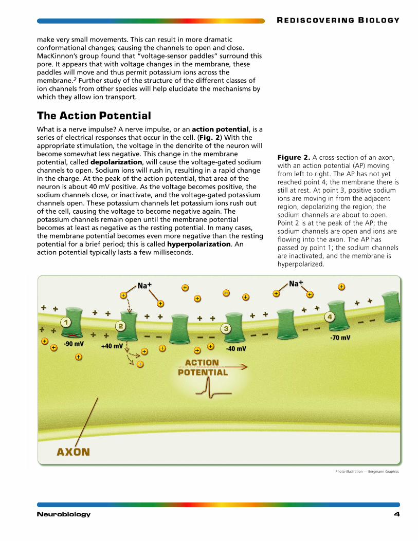
The Action PotentialWhat is a nerve impulse? A nerve impulse, or an action potential, is aseries of electrical responses that occur in the cell. (Fig. 2) With theappropriate stimulation, the voltage in the dendrite of the neuron willbecome somewhat less negative. This change in the membranepotential, called depolarization, will cause the voltage-gated sodiumchannels to open. Sodium ions will rush in, resulting in a rapid changein the charge. At the peak of the action potential, that area of theneuron is about 40 mV positive. As the voltage becomes positive, thesodium channels close, or inactivate, and the voltage-gated potassiumchannels open. These potassium channels let potassium ions rush outof the cell, causing the voltage to become negative again. Thepotassium channels remain open until the membrane potentialbecomes at least as negative as the resting potential. In many cases,the membrane potential becomes even more negative than the restingpotential for a brief period; this is called hyperpolarization. Anaction potential typically lasts a few milliseconds.
4Neurobiology
AXON
ACTION POTENTIAL
1 2 3
4
Na+ Na+
-40 mV-90 mV
-70 mV+40 mV
Figure 2. A cross-section of an axon,with an action potential (AP) movingfrom left to right. The AP has not yetreached point 4; the membrane there isstill at rest. At point 3, positive sodiumions are moving in from the adjacentregion, depolarizing the region; thesodium channels are about to open.Point 2 is at the peak of the AP; thesodium channels are open and ions areflowing into the axon. The AP haspassed by point 1; the sodium channelsare inactivated, and the membrane ishyperpolarized.
Photo-illustration — Bergmann Graphics
R E D I S C O V E R I N G B I O L O GY
How can this action potential be propagated along the neuron?When the sodium channels are opened, sodium ions rush in; onceinside they cause nearby regions of the neuron to becomedepolarized by moving laterally through the axon. This, in turn, causesthe opening of more voltage-gated sodium channels in those regions.Thus, the sodium channel activation moves in a wave-like fashion: theaction potential is propagated down the length of the neuron, fromits input source at the dendrites, to the cell body, and then down theaxon to the synaptic terminals. How does the action potentialmaintain this directional flow that is key to information processing?The sodium channels have a mechanism that avoids “backpropagation” of the action potential, which would result in aconfused signal. After opening, the sodium channels becomeinactivated as the potential becomes more positive, and they cannotopen again until they are “reset” by hyperpolarization at the end ofan action potential. This brief period of sodium channel inactivation,called a refractory period, prevents bidirectional propagation of theaction potential, constraining it to go in only one direction.
Myelin Speeds Up ThoughtMost neurons have a fatty outer layer called myelin, which insulatesand protects the axons of neurons. In this way, myelin is like the plasticthat surrounds electric wires. Myelin is actually made up of two specialclasses of glial cells, called the oligodendroglia and Schwann cells,which wrap themselves around the axon much like a jellyroll. Betweenthese cells there are small gaps in the myelin sheath called the Nodesof Ranvier. Action potentials are able to jump from one node to thenext one down the neuron incredibly rapidly. For this reason, impulseswill travel down a myelinated neuron faster than they will across anunmyelinated neuron. In myelinated neurons, action potentials usuallytravel at over 100 meters per second, which is about half the speed ofsound. In about one-hundredth of a second, an action potential cantravel from the brain to the base of the spinal cord of an adult.Though seemingly instantaneous, this rate is still on the order of amillion times slower than electricity.
Several degenerative diseases are due to the loss of myelin in certainneurons. The loss of muscle coordination that people with multiplesclerosis face is due to the degeneration of the myelin sheath in classesof neurons that are involved in the movement of muscles. The diseaseis suspected to be an autoimmune disorder — the immune systemattacks the myelin sheaths. While MS is usually strikes first in earlyadulthood, many other diseases that are due to myelin degenerationoccur in infancy or early childhood.
Across the SynapseHow is information transferred from one neuron to the next?Neurons communicate at their meeting points, called synapses; thesmall gaps separating the neurons are referred to as the synapticspace. These synapses are not merely gaps but are functional linksbetween the two neurons. Signals are transferred in only onedirection across the synapse. The neuron that transmits informationwhen it fires is called the presynaptic neuron. The synapticterminals of the presynaptic neuron are on one side of the synapse;the dendrites of the other neuron, the postsynaptic neuron, are on
5Neurobiology

R E D I S C O V E R I N G B I O L O GY
the other side. Presynaptic and postsynaptic are relative adjectives; apostsynaptic neuron at one synaptic connection can be a presynapticneuron at another synapse.
Synapses can be either chemical or electrical. An electrical synapse iswhat is often called a “gap junction,” in which the membranes of twoneurons are continuous at tiny spots, making the cells electricallycontiguous. Gap junctions, which are not unique to neurons, allow foreven more rapid communication. No chemical intermediary is involvedin an electrical synapse. In the case of chemical synapses, however,chemicals called neurotransmitters are released from a presynapticneuron, and dock with receptor proteins on the postsynaptic neuron.Such binding causes the shape of the protein to change and ionchannels to open, much like the voltage-gated channels open inresponse to membrane potential changes (Fig. 3). We will discussneurotransmitters in more detail below. Neurons are typically separatedby about twenty to thirty nanometers in chemical synapses. Electricalsynapses are more rapid than chemical ones but chemical synapses areeasier to modulate. In vertebrates and many invertebrates, chemicalsynapses are more common than are electrical ones.
The action of the presynaptic neuron is referred to as an “all or none”response. A neuron can only fire or not fire; there is no “slightlyactivated” signal from a neuron. Whether or not a neuron will fire anaction potential — that is, send a signal down its axon to be receivedby other neurons — depends on how many inputs it is receiving. It alsodepends on the nature of each input signal — excitatory or inhibitory— at each synapse. The sort of “net total” result of those signalsdetermines whether the neuron will become excited, or depolarized,enough to fire an action potential and release neurotransmitter fromits axon terminals.
Also recall that a signal traveling through the brain often involvesmany neurons, each making so many connections. Thisinterconnectedness gives rise to the extraordinary complexity of thebrain. The activation of a single sensory neuron could quickly lead tothe activation or inhibition of thousands of neurons.
Neurotransmitters and ReceptorsNeurotransmitters are usually small molecules, such as amino acids(e.g., glutamate and aspartate) and amines (e.g., dopamine, serotonin,and histamine). Some neurotransmitters stimulate neurons to fire,while others inhibit firing. The effect of the neurotransmitter comesabout by its binding with receptor proteins on the membrane of thepostsynaptic neuron. Each neurotransmitter binds specifically in a lock-and-key mechanism to its type of receptor. Neurons in differentpathways will often have different types of receptors in a given family.For example, dopamine binds to dopamine receptors, but there areabout a dozen subtly different dopamine receptors. Neurobiologiststhink that the human nervous system uses at least fiftyneurotransmitters, but about ten carry out most neurotransmission.Many of these neurotransmitters are highly conserved in otherorganisms. Most neurons release only one type of neurotransmitter.
Neurotransmitters are released in a process called exocytosis. Whenthe action potential reaches the end of an axon the depolarizationcauses calcium channels to open. The calcium causes synaptic vesicles
6Neurobiology
PRESYNAPTIC MEMBRANE
NEUROTRANSMITTER
SYNAPTIC VESICLES
NEUROTRANSMITTER RECEPTOR
POSTSYNAPTIC MEMBRANE
Figure 3. Synaptic vesicles fuse withthe presynaptic membrane to releaseneurotransmitter into the synapticspace. Here, they bind withneurotransmitter receptors in thepostsynaptic membrane.
Photo-illustration — Bergmann Graphics

R E D I S C O V E R I N G B I O L O GY
that carry the neurotransmitter to fuse with the cell membrane. Thisfusion allows the neurotransmitter to be released into the synapse.Although exocytosis occurs in many cell types, neurons use aspecialized form in which calcium causes a chain of events thatculminates in fusion of the vesicles.
There are two general categories of receptor proteins: ionotropic andmetabotropic. Activation of ionotropic receptors causes membrane ionchannels to open or close. In contrast, activation of metabotropicreceptors involves an intracellular biochemical cascade. Such a cascademay end with the opening or closing of ion channels or otherintracellular effects.
As long as the neurotransmitter remains in the synapse, it will continueto bind its receptors and stimulate the postsynaptic neuron. At somepoint the signal is no longer needed. Moreover, continual stimulation caninjure some neurons. So, halting the stimulus is just as important as theappropriate starting of the stimulus. How does the neurotransmitterleave the synapse? There are several ways, such as diffusion away fromthe synapse or breakdown of the neurotransmitter by specific enzymes.Another common mode, called reuptake, involves specializedmolecules present on the membrane of the presynaptic neuron. Thesemolecules, called neurotransmitter transporters, have receptor sitesthat will bind to the neurotransmitter and actively transport it out ofthe synapse, back to the presynaptic neuron. That neuron can thenreuse the neurotransmitter. The action of several drugs takes place atthe reuptake stage.
Neurotransmitters, Psychoactive Drugs,and the Reward PathwayDrugs that have effects on the central nervous system are known aspsychoactive drugs. The mode of actions of both therapeutic drugs(e.g., Ritalin, Prozac, and Paxil) and recreational drugs (e.g., alcohol,cannabis, cocaine, and nicotine) affect the firing of certain neurons bychanges in various neurotransmitters or receptors. Not all drugs havespecific modes of action; alcohol, for example, has many and variedeffects. We will focus, however, on a few examples of those drugs thathave specific effects.
Humans and many other animals engage in many activities from whichthey derive pleasure. Researchers working with various animals haveshown that there are regions of the brain, such as the ventraltegmental area, that are more active when animals engage inpleasurable acts. When researchers stimulate these areasexperimentally, the animals will perform various tasks in order toreceive further stimulation. Hence, the neural pathway comprises thoseregions has been called the reward pathway.
Like many drugs, nicotine from tobacco products acts on the rewardpathway. This drug, however, is unusual in that it directly affects thedopamine receptor in the reward pathway’s neurons. Unlike the actionof most drugs, no intermediary steps are involved: nicotine binds tothe receptor and stimulates the postsynaptic neuron. Theoverstimulation of the postsynaptic cell, however, also has effects atthe cellular level. Over time, it leads to a decrease in the number ofdopamine receptors being expressed and inserted to the membrane, aswell as a change in the shape of the cell. The reduction of receptors is
7Neurobiology
R E D I S C O V E R I N G B I O L O GY
referred to as desensitization. When the nicotine is removed, becausethere are fewer receptors on the postsynaptic cell, more dopaminethan normal is required for proper stimulation of postsynaptic neuron.Addiction can result because nicotine becomes needed just to maintainthe normal stimulation of the postsynaptic cells.
Allelic variation at the dopamine receptor gene appears to affect one’slikelihood of becoming addicted to nicotine. Individuals who have theA1 allele have fewer dopamine receptors than those that do not havethe allele. These individuals also have more difficulty in quittingsmoking and are more likely to exhibit other addictive and compulsivebehaviors. The genetic components of many types of addiction are thetopic of intensive research — and often heated debate.
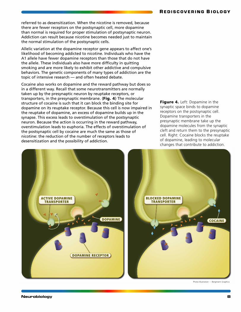
Cocaine also works on dopamine and the reward pathway but does soin a different way. Recall that some neurotransmitters are normallytaken up by the presynaptic neuron by reuptake receptors, ortransporters, in the presynaptic membrane. (Fig. 4) The molecularstructure of cocaine is such that it can block the binding site fordopamine on its reuptake receptor. Because this cell is now impaired inthe reuptake of dopamine, an excess of dopamine builds up in thesynapse. This excess leads to overstimulation of the postsynapticneuron. Because the action is occurring in the reward pathway,overstimulation leads to euphoria. The effects of overstimulation ofthe postsynaptic cell by cocaine are much the same as those ofnicotine: the reduction of the number of receptors leads todesensitization and the possibility of addiction.
8Neurobiology
DOPAMINE
DOPAMINE RECEPTOR
ACTIVE DOPAMINETRANSPORTER
COCAINE
BLOCKED DOPAMINETRANSPORTER
Photo-illustration — Bergmann Graphics
Figure 4. Left: Dopamine in thesynaptic space binds to dopaminereceptors on the postsynaptic cell.Dopamine transporters in thepresynaptic membrane take up thedopamine molecules from the synapticcleft and return them to the presynapticcell. Right: Cocaine blocks the reuptakeof dopamine, leading to molecularchanges that contribute to addiction.
R E D I S C O V E R I N G B I O L O GY
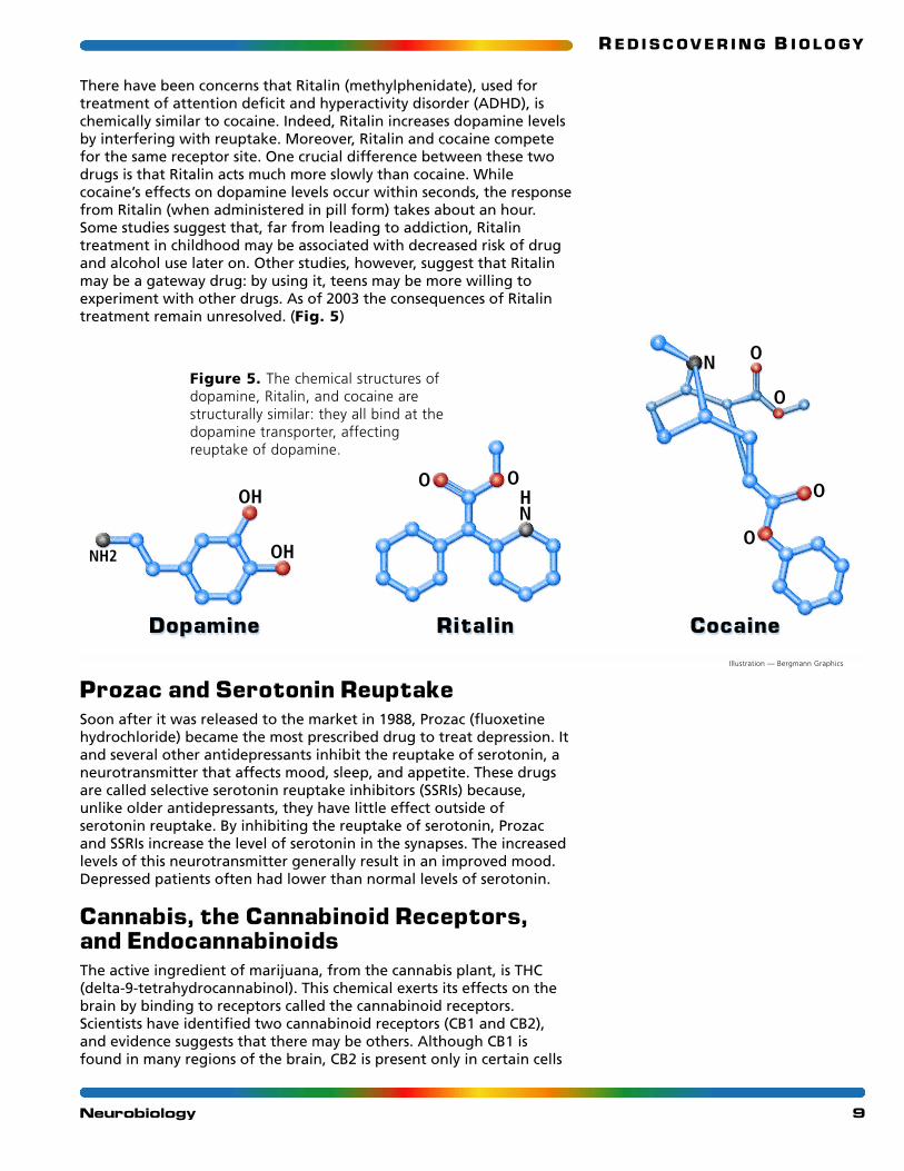
There have been concerns that Ritalin (methylphenidate), used fortreatment of attention deficit and hyperactivity disorder (ADHD), ischemically similar to cocaine. Indeed, Ritalin increases dopamine levelsby interfering with reuptake. Moreover, Ritalin and cocaine competefor the same receptor site. One crucial difference between these twodrugs is that Ritalin acts much more slowly than cocaine. Whilecocaine’s effects on dopamine levels occur within seconds, the responsefrom Ritalin (when administered in pill form) takes about an hour.Some studies suggest that, far from leading to addiction, Ritalintreatment in childhood may be associated with decreased risk of drugand alcohol use later on. Other studies, however, suggest that Ritalinmay be a gateway drug: by using it, teens may be more willing toexperiment with other drugs. As of 2003 the consequences of Ritalintreatment remain unresolved. (Fig. 5)
Prozac and Serotonin ReuptakeSoon after it was released to the market in 1988, Prozac (fluoxetinehydrochloride) became the most prescribed drug to treat depression. Itand several other antidepressants inhibit the reuptake of serotonin, aneurotransmitter that affects mood, sleep, and appetite. These drugsare called selective serotonin reuptake inhibitors (SSRIs) because,unlike older antidepressants, they have little effect outside ofserotonin reuptake. By inhibiting the reuptake of serotonin, Prozacand SSRIs increase the level of serotonin in the synapses. The increasedlevels of this neurotransmitter generally result in an improved mood.Depressed patients often had lower than normal levels of serotonin.
Cannabis, the Cannabinoid Receptors,and EndocannabinoidsThe active ingredient of marijuana, from the cannabis plant, is THC(delta-9-tetrahydrocannabinol). This chemical exerts its effects on thebrain by binding to receptors called the cannabinoid receptors.Scientists have identified two cannabinoid receptors (CB1 and CB2),and evidence suggests that there may be others. Although CB1 isfound in many regions of the brain, CB2 is present only in certain cells
9Neurobiology
OH
OHNH2
NH
OO
N
O
O
O
O
CocaineRitalinDopamine
Figure 5. The chemical structures ofdopamine, Ritalin, and cocaine arestructurally similar: they all bind at thedopamine transporter, affectingreuptake of dopamine.
Illustration — Bergmann Graphics

R E D I S C O V E R I N G B I O L O GY
of the immune system. Because the receptor is present in several brainregions, THC can have manifold effects. For instance, THC may affectmemory formation. CB1 is prevalent in the hippocampus, a region ofthe brain strongly associated with memory. By binding to andactivating CB1, THC decreases activity of neurons in the hippocampusand interferes with the proper function of that region, which maytranslate to an interference with memory formation.
The human body does not produce THC, so why would there bereceptors that can bind it? During the 1990s researchers discoveredthat the body makes chemicals, such as anandamide, that can bind tothe cannabinoid receptors. The function of these chemicals, calledendocannabinoids, and their receptors is still unknown. To investigatethe role of the CB1 receptor, scientists have studied mutant mice thatlack the receptor. Compared with normal mice, these mice have adecreased appetite, are less active, and have a reduced lifespan;however, the mice have an enhanced memory.
The CB receptors have recently been associated with some beneficialactions, such as pain relief and extinguishing some fear behaviors. THChas even been prescribed as medication in some states for pain relieffor various diseases, including glaucoma, AIDS, and cancer.3
The Molecular Basis of Learning and MemoryIt is clear that an understanding of mechanisms at the level of thesynapse explains changes in our behaviors, like movements. Butwhat about longer-term changes associated with learning andmemory? Can they be understood in molecular terms, too? Memory,and thus learning, involves molecular changes in the brain. Duringthe last few decades, researchers have started to map the molecularprocesses involved in memory formation. They have beenincreasingly able to link the ability to remember with physicalchanges in the structure of neurons.
One important change that occurs in memory formation is long-termpotentiation (LTP). This phenomenon involves the long-termmodification of the synaptic communication. Under normalcircumstances the rate at which a postsynaptic neuron fires depends onhow much stimulation it receives from presynaptic neurons. Once theincreased stimulation has stopped, the postsynaptic neuron will returnto its normal rate of firing. In LTP, however, the postsynaptic neuronwill continue to fire at an elevated rate, even after the increasedstimulation has subsided. It seems to become more sensitive — or givesa bigger reaction by firing more action potentials — to a givenstimulus. How does this happen?
Glutamate is the neurotransmitter involved in LTP. Glutamate can bindto several different types of ionotropic receptors, including the NMDA-(N-methyl-D-aspartate) and AMPA- (amino-3-hydroxy-5-methyl-4-isoxazolepropionate) type glutamate receptors, each of which opens aspecific type of channel within the receptor proteins. Both channels areinvolved in memory formation. The NMDA channel requires bothglutamate and depolarization from another source to open. Why? Themolecular mechanism is as follows. Normally, at negative potentials,positively-charged magnesium ions plug the pore of the NMDAchannel. While glutamate may “open” the pore, the ions cannot travel
10Neurobiology
R E D I S C O V E R I N G B I O L O GY
through the channel due to the magnesium block. When themembrane is depolarized, however, the inside of the cell becomesmore positive, and the magnesium ions are no longer driven into thechannel. Thus, the block is relieved, allowing sodium and calcium ionsto flow in.
So, this mechanism allows the NMDA-type glutamate receptor to act asa “coincidence detector.” When the neuron receives input from onlyone source — another neuron — glutamate binds to and opens bothNMDA- and AMPA-type receptors. (Fig. 6) Because theneurotransmitter arrives at a resting, negatively charged, postsynapticmembrane, magnesium ions prevent flow through NMDA channels.When, however, stimulation of a neuron occurs simultaneously frommore than one source — say several other neurons — some glutamatewill bind NMDA receptors in parts of the neuron that are alreadydepolarized, or less negatively charged.
Where does this voltage change come from? Recall that once an actionpotential has started, it spreads from its source throughout the entiremembrane of the neuron in a wave-like fashion; thus, other dendritesmay be “pre-depolarized” before glutamate binds. In this cas, theblock by magnesium is relieved and the NMDA channel also passesions. While AMPA channels can pass only sodium ions in, NMDAchannels also pass calcium. This calcium permeability gives the NMDAchannel its ability to trigger LTP.
Now that we have examined the requirements for LTP, what is theeffect? When calcium ions rush in, they set off an intracellularsignaling cascade that can involve dozens of molecules. Speculationabout the identity and functions of these molecules has been thesubject of intense scientific inquiry since the early 1990s — it wasperhaps the most studied aspect of neuroscience during that “decadeof the brain.”
So how could this intricate electrical mechanism act to form newmemories? LTP, like learning, is not just dependent on increasedstimulation from one particular neuron but on a repeated stimulus fromseveral sources. It is thought that when a particular stimulus isrepeatedly presented, so is a particular circuit of neurons. Withrepetition the activation of that circuit results in learning. Recall that thebrain is intricately complicated. Rather than a one-to-one line ofstimulating neurons, it involves a very complex web of interactingneurons. But it is the molecular changes occurring between theseneurons that appear to have global effects. LTP can lead to strengthenedsynapses in a variety of ways. One such way, as discussed in the video, isby the phosphorylation of glutamate receptor channels, which isaccomplished by a calcium-triggered signaling cascade. This results inthose channels passing more ions with subsequent stimulation,strengthening the signal to and from the neuron.
But more permanent changes — long-term memory — require thesynthesis of new proteins. In a variety of organisms, including flies(Drosophila) and humans, one enzyme, CREB (cyclic-AMP responseelement binding protein), seems to be involved in the steps thatfacilitate this new protein expression. When calcium flows in throughNMDA channels, one of the molecules it activates is CREB. In turn,activated CREB acts as a transcription factor (see the Genes andDevelopment unit) that activates the expression of other genes. This
11Neurobiology
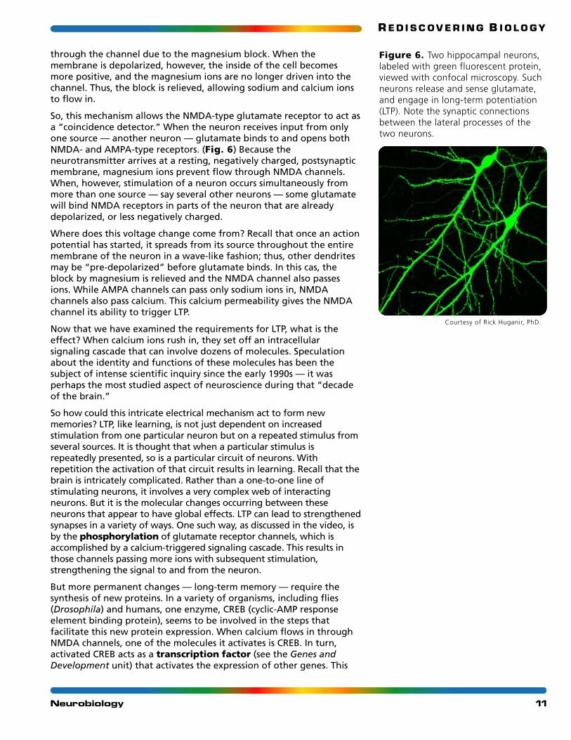
Courtesy of Rick Huganir, PhD.
Figure 6. Two hippocampal neurons,labeled with green fluorescent protein,viewed with confocal microscopy. Suchneurons release and sense glutamate,and engage in long-term potentiation(LTP). Note the synaptic connectionsbetween the lateral processes of thetwo neurons.
R E D I S C O V E R I N G B I O L O GY
gene expression can lead to the production of more ion channelreceptors, as well as structural proteins like actin, which cement thesynaptic connection between two repeatedly communicating neurons.
Mutant mice lacking the NMDA receptors show severe deficiencies inmemory tasks. On the other hand, researchers have geneticallyengineered (see the GMOS unit) mice that have more of the NMDAreceptors. These mice, dubbed “smart mice” by the popular press, aresubstantially better at several memory tasks than are normal mice.
Memory and the HippocampusPsychologists have long argued that there are many different types ofmemory. These can be classified by many criteria, based on decades ofexperimental research and the different memory defects seen in peoplewho have suffered brain damage. Scientists have agreed that memorycan be viewed in temporal terms; that is, there is a short-term memory,with a limited capacity for about a dozen items, and a long-termmemory, to which these items are presumably transferred for“storage.” Short-term memory seems to be much more vulnerable toloss due to trauma than does long-term memory: people may even losethe ability to form new memories, while their ability to remember theirentire lives before an accident remains intact. This memory defect isexemplified in the movie Memento (2000), in which a widower avengeshis wife’s murder — during which he suffered brain damage — overand over again. Such individuals with this condition of “anterogradeamnesia” usually have severe damage to their hippocampus. AsKempermann points out, the hippocampus is not the equivalent of thebrain’s hard drive but rather a gateway, “a structure, through which allinformation must pass, before it can be memorized.”4
It is widely agreed that while the hippocampus is undeniablyimportant for memory, the “recording” of information into long-termmemory involves plasticity, or physical changes, in multiple regionsthroughout the entire nervous system. Another interesting distinctionthat scientists have made in types of memory is between declarativememory, which allows you to remember facts and is extremelycomplex, and reflexive memory, which usually consists of learning byrepetition and often involves motor learning. While declarativememory can be reported, reflexive memory is exhibited byperformance of a task and cannot be expressed verbally. It is nowthought that the two types of memory may involve two entirelydifferent neuronal circuits.
The hippocampus plays a major role in spatial learning and memory ina number of animals. Research with black-capped chickadees and otherspecies of birds has shown that when the hippocampus is removed, thebirds still store food but cannot recall where they stored it. Moreover,bird species that rely heavily on stored food as a winter resource ingeneral have larger hippocampi than those species that don’t.
Studies of cab drivers in London have provided fascinating informationabout the role that the hippocampus plays in spatial memory. Londoncab drivers are known for their navigational skills and knowledge ofthe streets of London. To learn how to navigate the streets of the city,would-be cab drivers undergo “the Knowledge,” a rigorous trainingthat can take two years to complete. Recent studies using magneticresonance imaging (MRI) demonstrate that the hippocampi of the
12Neurobiology
R E D I S C O V E R I N G B I O L O GY
London cab drivers are somewhat different. Specifically, the posteriorregion is significantly larger and the anterior region is significantlysmaller in the cabbies when compared with control subjects. Otherstudies have found that the posterior region is active during tasksinvolving spatial memory. It is possible that the cabbies comedisproportionately from those individuals with excellent spatialmemories and corresponding larger posterior regions of thehippocampus. There is further evidence, however, that suggests thatthe memory work of the cabbies has altered their hippocampi. Thosecab drivers that have been working the longest tend to have largerposterior hippocampi than more recently hired cabbies. Furthermore,other imaging studies show that the right hippocampus is activated inthe cab drivers when they are asked to remember complex travelroutes but not when they are asked to provide information aboutfamous landmarks.5
Neuronal Stem CellsWhat neuronal processes have led to the changes in the hippocampi ofLondon taxi drivers? Perhaps this is achieved by neurons migrating fromone region to the posterior hippocampus? Another intriguing possibilityis that the changes are the result of new neurons going to the region.
New neurons? Don’t we have our complete store of neurons by earlychildhood? That previous dominant paradigm had been foundincorrect. In the past two decades, researchers have shown thatneurons are continually produced in a variety of animals, includinghumans. It isn’t that neurons divide. They don’t. Instead, the brainmaintains a reservoir of stem cells that are capable of generating newneurons (neurogenesis). One area of the brain where stem cells havebeen found is the hippocampus.
The discovery of stem cells and neurogenesis began with basic researchwith songbirds. During each breeding season male songbirds need torecall their mating song. Starting in the 1980s researchers noted thatthe number of neurons in certain areas of the brain (especially thehippocampus) would increase in male birds around the start of thebreeding season. The number of neurons in these areas woulddecrease after the mating season. This striking evidence led otherresearchers to look for neurogenesis in the brains of mammals. Studieson rats found substantial neurogenesis. In one part of thehippocampus alone nearly 10,000 new neurons are generated each dayin adult rats. Starting in the 1990s Elizabeth Gould of PrincetonUniversity found that the adult brains of several species of monkeysalso undergo considerable neurogenesis.
Following these animal studies researchers examined whether humanshave the capacity for neurogenesis. They studied postmortem braintissue from humans, using various stains to determine whether newneurons were being generated from dividing progenitor cells. Theywere able to find such new neurons in the hippocampus, showing thatneurogenesis proceeds throughout life in at least some regions of thehuman brain.
Engaging in mental and physical activity is one important way elderlypeople can maintain their mental acuity. This aspect of conventionalwisdom has been vindicated by medical research. Mental and physicalactivity reduces the risk of neurodegenerative disorders and improves
13Neurobiology
R E D I S C O V E R I N G B I O L O GY
the prognosis of stroke patients. Yet, we know little about themolecular mechanisms behind this effect. Studies in mice ofneurogenesis in the hippocampus, however, point to one possiblereason for why activity keeps the mind sharp. Mice who were exposedto an enriched environment for the second half of their lives showed adramatic increase in neurogenesis in the hippocampus as comparedwith control subjects. The hippocampi from the mice that received theenriched treatment also appeared like those of younger animals. Theseresults strongly suggest that activity maintains the proper function ofthe brain by increasing neurogenesis in the hippocampus.
Elizabeth Gould and other researchers studying neurogenesis thinkthat the new neurons generated in the hippocampus are involved inmodulation of the stress response as well as learning. There are somecomplications, however. Learning enhances neurogenesis but onlyunder certain conditions. Moreover, experimental blockage ofneurogenesis interferes with some types of learning but not others.
Our understanding of neurogenesis remains far from complete. Yet,tremendous progress has been made during the last two decades andfurther progress is expected. In addition to what these studies tell usabout how the brain works, they may also pave the way towardtreatment of degenerative diseases like Alzheimer’s and Parkinson’s aswell as brain trauma.
References1) Campbell, N. A., and J. B. Reece. 2002. Biology. 6th ed., 1002 SanFrancisco: Addison-Wesley Longman, Inc.
2) Jiang, Y., A. Lee, Y. Chen, V. Ruta, M. Cadene, B. T. Chait, and R,MacKinnon. 2003. X-ray structure of a voltage-dependent K+ channel.Nature. 423:33–41.
3) Sullivan, J. M. 2002. Cannabinoid receptors. Curr. Biol. 12:R681
4) Kempermann, G. 2002. Why new neurons? Possible functions foradult hippocampal neurogenesis. J. Neuroscience. 22:635–38.
5) Maguire, E., R. Frackowiak, and C. Firth. 1997. Recalling routesaround London: Activation of the right hippocampus in taxi drivers.J. Neuroscience. 17:7103–10.
14Neurobiology

R E D I S C O V E R I N G B I O L O GY
Further ReadingBooks
Calvin, W. H., and G. A. Ojemann.1995. Conversations with Neil’s brain:The neural nature of thought and language. Perseus Publishing.
Building from case examples, a neurobiologist and aneurosurgeon describe the workings of the brain.
Drickamer, L. C., S. H. Vessy, and E. M. Jakob. 2002. Animal behavior:Mechanisms, ecology, and evolution. 5th ed. McGraw-Hill.
A university-level textbook on animal behavior that has anexcellent section on the neurobiology of behavior.
Timmons, C. R., and L. W. Hamilton. Drugs, brains & behavior.www.rci.rutgers.edu/~lwh/drugs/.
A short e-book detailing the neuropharmalogical effects of drugs.
Article
Sullivan, J. M. 2002. Cannabinoid receptors. Curr. Biol. 12:R681.A short guide to recent research on cannabinoids and their receptors.
15Neurobiology
R E D I S C O V E R I N G B I O L O GY
16Neurobiology
GlossaryAction potential. The nerveimpulse, or “firing,” of a neuron.A traveling wave of depolarizedvoltage that is propagated along aneuron. Results in the release ofneurotransmitter and themovement of information toanother neuron.
Depolarization. The state inwhich the inside of a neuronbecomes more positive in voltagethan it is at rest.
Hippocampus. A region of thebrain associated with memoryformation.
Hyperpolarization. A state inwhich the membrane potential ismore negative than is the restingpotential; occurs transiently at theend of an action potential.
Ionotropic receptors. Receptorsfor which neurotransmitterbinding results directly in an ionchannel opening or closing.
Long-term potentiation. Anenduring increase in the strengthof the connection between twoneurons, which results fromrepeated stimulation of a giveninput pathway.
Membrane potential. Thedifference in voltage between theinside and the outside of aneuron; the outside is always zero.
Neurogenesis. The formation of new neurons from precursorstem cells.
Neurotransmitter. A moleculethat travels across the synapse and binds to its receptor on thepostsynaptic neuron, influencingits probability of firing.
Phosphorylation. The additionof a phosphate group to amolecule, such as a protein.
Postsynaptic neuron. At a givensynapse, the postsynaptic neuronis the receiving neuron at itsdendritic end.
Presynaptic neuron. At a givensynapse, the presynaptic neuron is the transmitting neuron, itsaxonal synaptic terminal forms the synapse.
Resting potential. The restingmembrane potential of a neuron;it is about -70 mV.
Reuptake. The recapture ofneurotransmitters from thesynapse back into the presynapticneuron; accomplished bytransporters.
Reward pathway. A pathway in the brain that is stimulatedwhen an animal is engaged inpleasurable activities.
Synapse. A functionalconnection between two neurons where information canbe exchanged in the form ofelectrical or chemical energy.
Transcription factor. A proteinthat influences transcription ofanother gene by binding to DNA.
Voltage-gated channels. Ion channels in the cell membranethat open or close in response tochanges in the membrane voltage.
X-ray crystallography. A method for determining thestructure of a molecule, such as a protein, based on the diffractionpattern resulting from focused X-ray radiation onto pure crystalsof the molecule.