bronchial anatomy - international society of · pdf filebronchial anatomy an anatomical ......
TRANSCRIPT
BRONCHIAL ANATOMY
AN ANATOMICAL REMINDER ABOUT THE DIFFERENT LOBES AND
SEGMENTS
Dr Etienne Leroy-Terquem Centre hospitalier de Meulan les Mureaux. France
French-cambodian association for pneumology (OFCP)
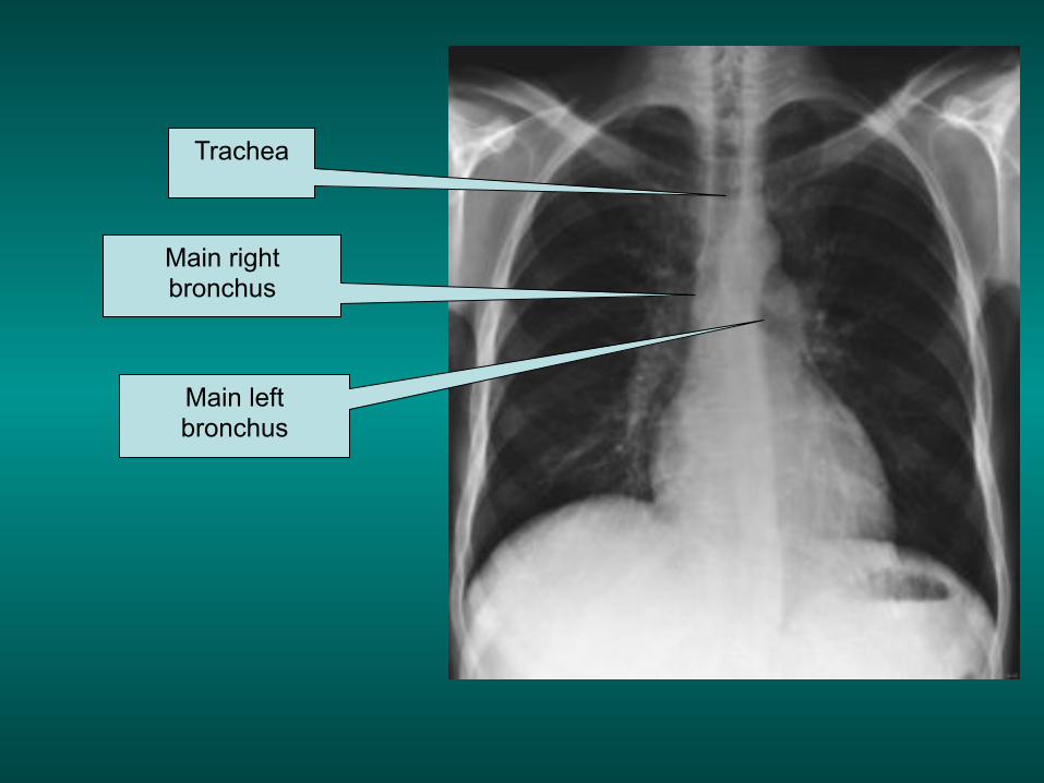
Trachea
Main right bronchus
Main left bronchus
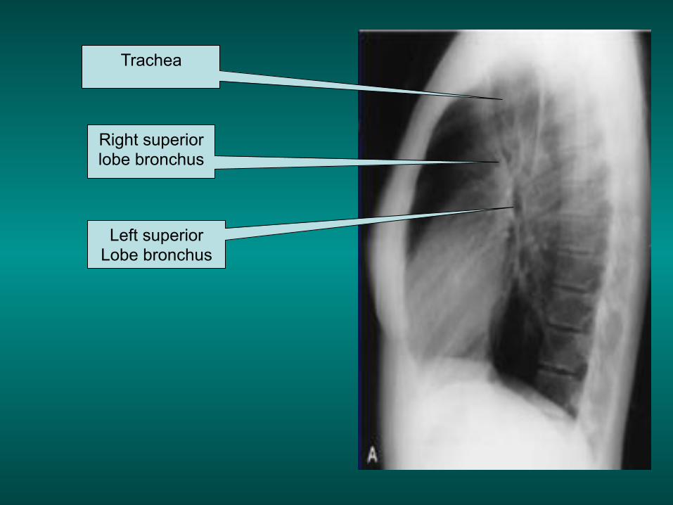
Trachea
Right superior lobe bronchus
Left superior Lobe bronchus
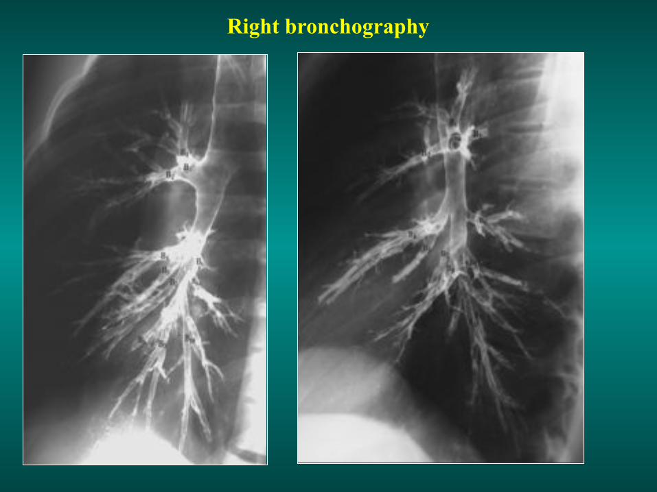
Right bronchography

Left bronchography
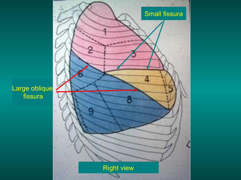
Right view
Small fissura
Large oblique fissura

Left view
Left fissura
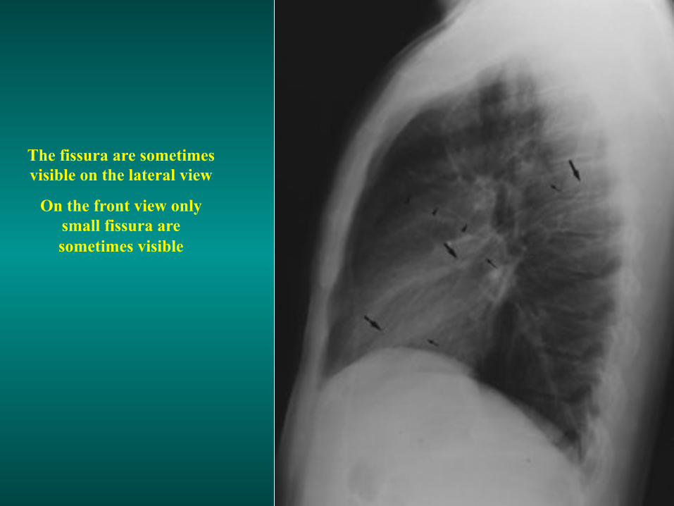
The fissura are sometimes visible on the lateral view
On the front view only small fissura are sometimes visible
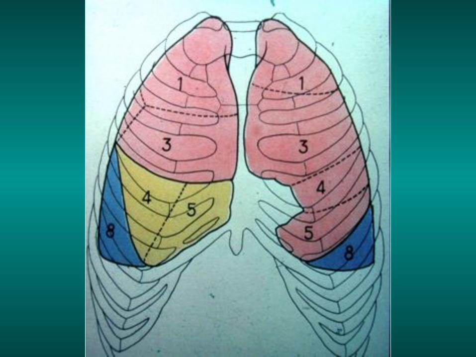
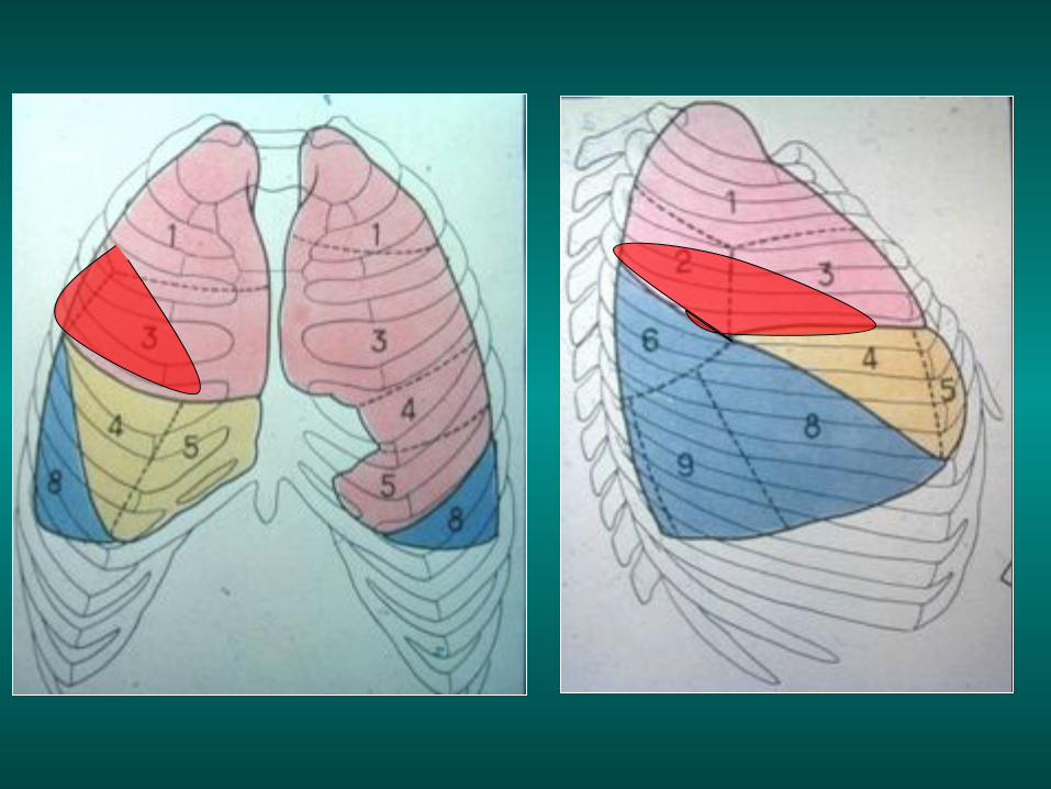
Ventilation Sectors
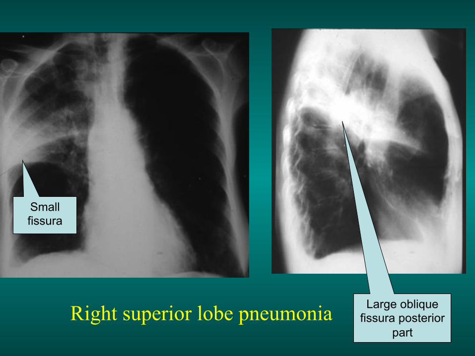
Right superior lobe pneumonia
Small fissura
Large oblique fissura posterior
part
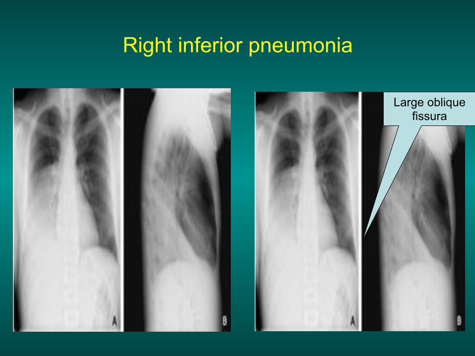
Right inferior pneumonia
Large oblique fissura

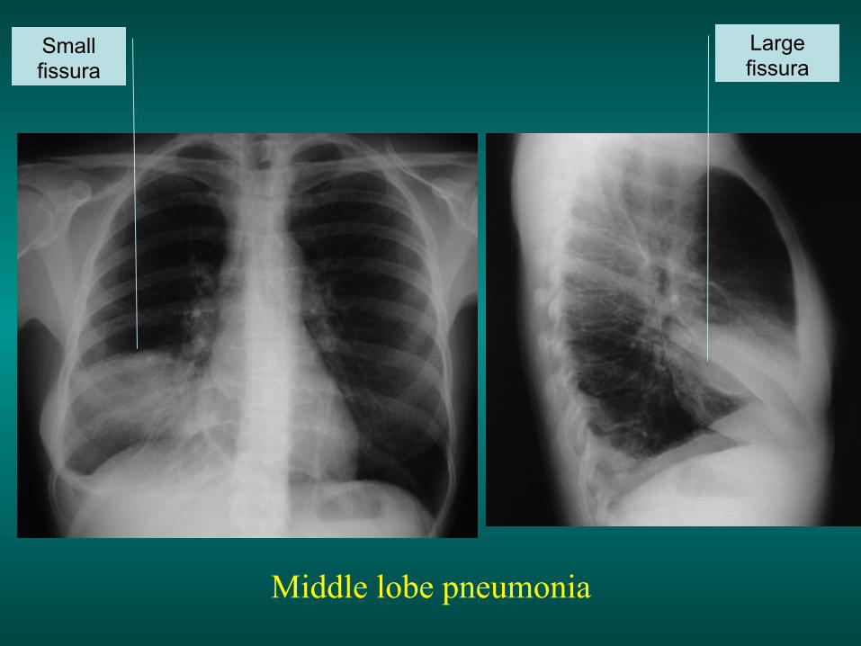
Middle lobe pneumonia
Large fissura
Small fissura

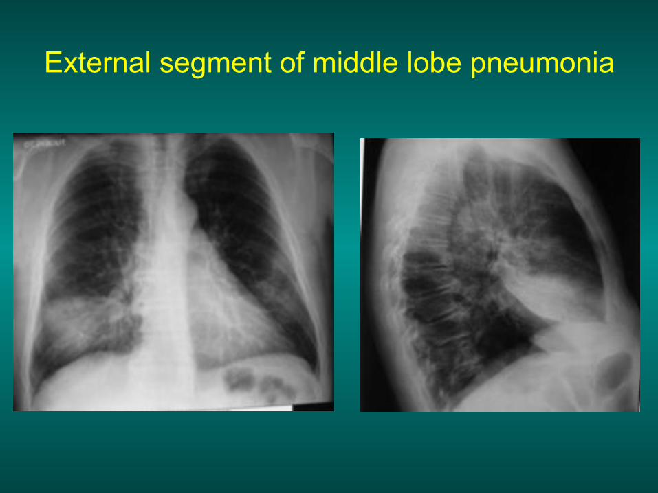
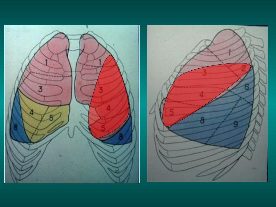
External segment of middle lobe pneumonia

External segment of middle lobe pneumonia
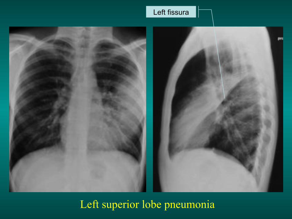
Left superior lobe pneumonia
Left fissura

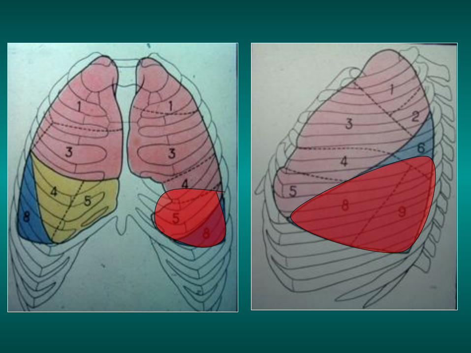
Left inferior pneumonia

Left inferior pneumonia
Left scissura

Bronchial syndrome
Atelectasis
Draining bronchus Bronchiectasis
Atelectasis
These are the consequence of an obstruction
of the bronchus by an intrinsic or extrinsic element (foreign body, benign or malignant tumor, acute or chronic inflammatory stenosis compression by adenopathy or tumor…) The alveolar air progressively disappears and the lung tissue retracts. This retraction can involve the segment, the lobe or the entire lung.
Main etiologies of atelectasis
• Bronchial cancer • Tuberculosis • Extrinsic compression by adenopathy or malignant tumor • Foreign body (+++ young children ) • Asthma • Chronic bronchitis • Viral or bacterial pneumonia • Atelectasis after thoracic or abdominal surgery, after traumatism • Many other rare etiologies: benign tumor, lymphoma, bronchus
metastasis, acute bronchiolitis, inflammatory granuloma regardless of the etiology, broncholithiasis, bronchiolitis obliterans, mucoviscidosis…

The radiologic image is a consolidation which is:
Systematised (close to a fissura) Retractile (loss of volume) Homogeneous Without aeric bronchogram With a varied size: segment, lobe, entire
lung
ATELECTASIS
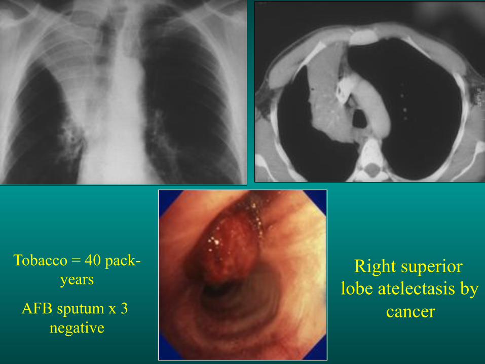
Right superior lobe atelectasis by
cancer
Tobacco = 40 pack-years
AFB sputum x 3 negative

Right superior lobe atelectasis by cancer
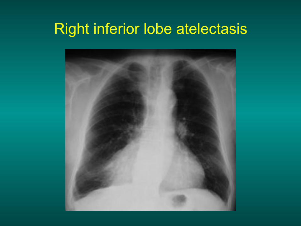
Right inferior lobe atelectasis
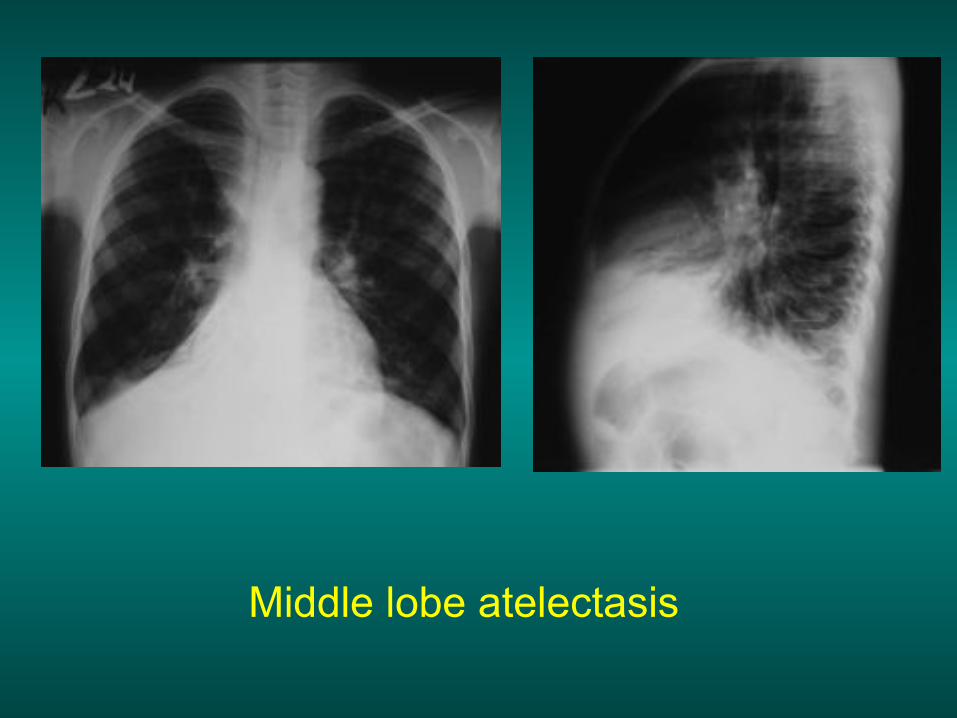
Middle lobe atelectasis
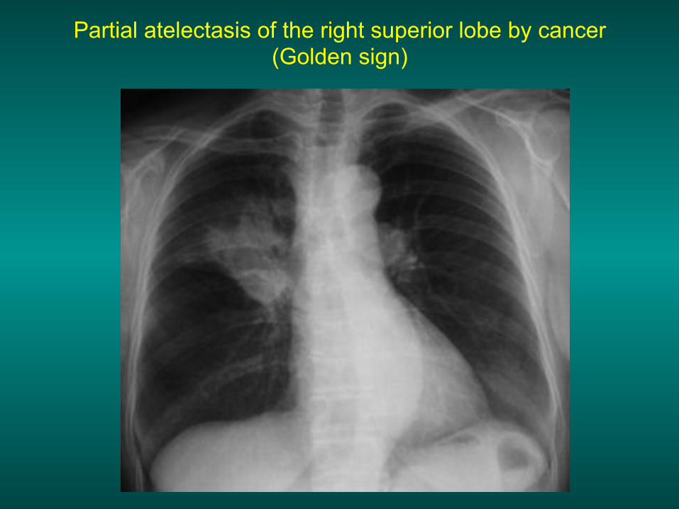
Partial atelectasis of the right superior lobe by cancer (Golden sign)
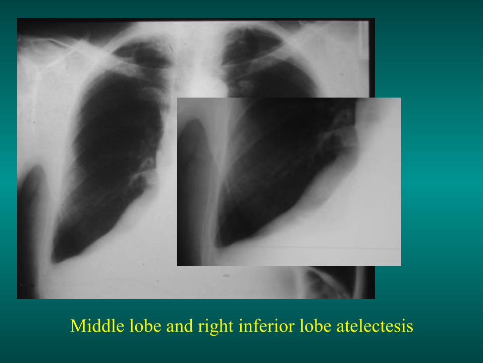
Middle lobe and right inferior lobe atelectesis

Man, 56 years old. High fever, right abdominal and thoracic pain, Muscular defense of the right hypochondrium,
x-ray: Middle lobe atelectasis

Liver abcess: the reduction of right hemidiaphragm mobility leads to atelectasis above the diaphragm
«passive atelectasis»
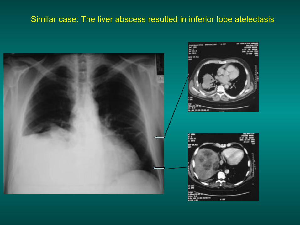
Similar case: The liver abscess resulted in inferior lobe atelectasis

Left superior lobe atelectasis by cancer
Tobacco = 60 pack-years. Haemoptysis. thoracic pain and dyspnea - AFB sputum x 3 negative
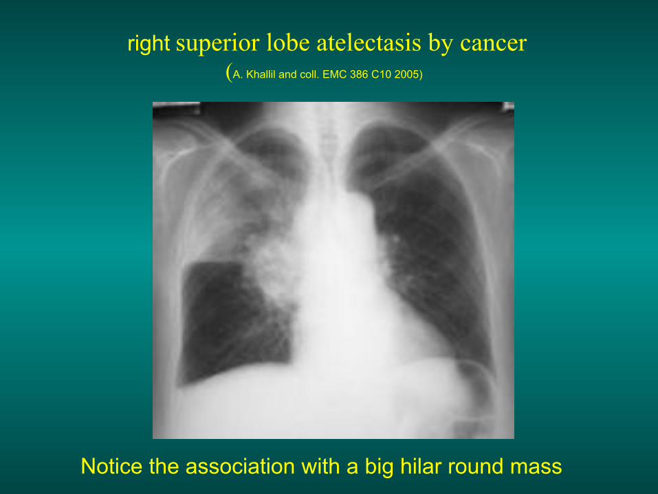
right superior lobe atelectasis by cancer
(A. Khallil and coll. EMC 386 C10 2005)
Notice the association with a big hilar round mass

Tobacco = 40 pack-years. Hemoptisy. Left anterior thoracic pain and cough.
Recent weight loss and asthenia - AFB sputum x 3 negative
Left superior lobe atelectasis by cancer Notice the round mass on the left hilus

The association of an atelectasis with a round mass
strongly suggests cancer
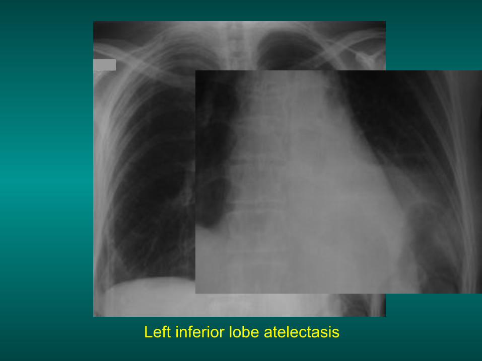
Left inferior lobe atelectasis
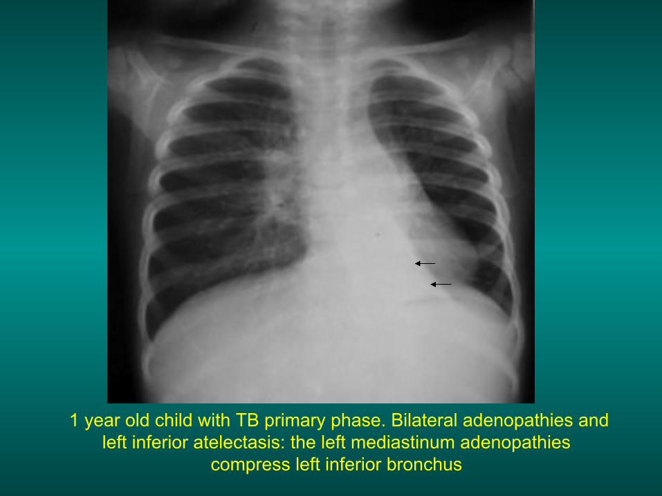
1 year old child with TB primary phase. Bilateral adenopathies and
left inferior atelectasis: the left mediastinum adenopathies compress left inferior bronchus
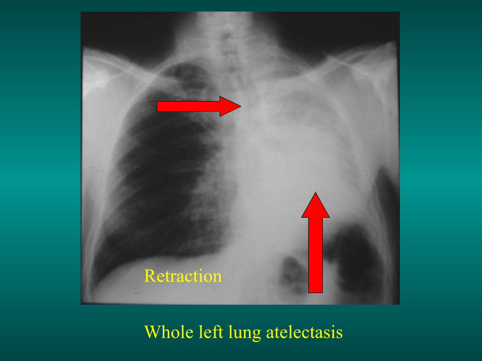
Whole left lung atelectasis
Retraction

Pleural effusion
Left atelectasis
Retraction
Pushing back
Draining bronchus TB cavity +++ - bacterial non-TB abscess +


TB cavity Notice the draining bronchus and right axillar infiltrate

TB cavity with a draining bronchus
Bronchectasis
This is a bronchus disease characterised by a permanent increase of the bronchus calibre. The cartilaginous framework of the bronchus wall is destroyed or broken up.
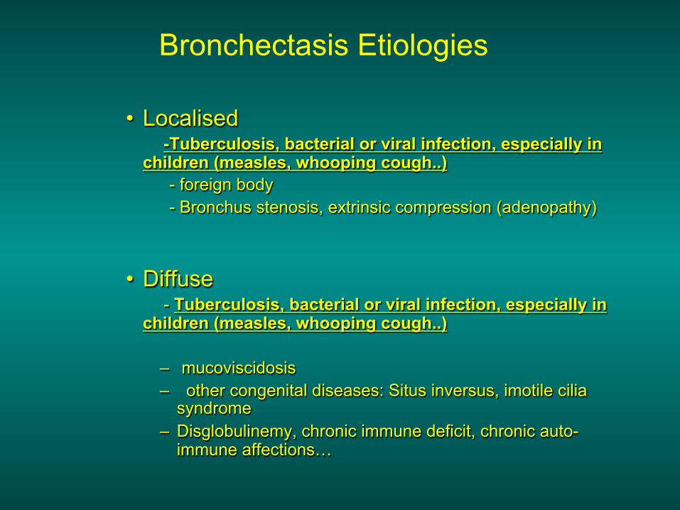
Bronchectasis Etiologies
• Localised -Tuberculosis, bacterial or viral infection, especially in
children (measles, whooping cough..) - foreign body - Bronchus stenosis, extrinsic compression (adenopathy)
• Diffuse - Tuberculosis, bacterial or viral infection, especially in
children (measles, whooping cough..)
– mucoviscidosis – other congenital diseases: Situs inversus, imotile cilia
syndrome – Disglobulinemy, chronic immune deficit, chronic auto-
immune affections…
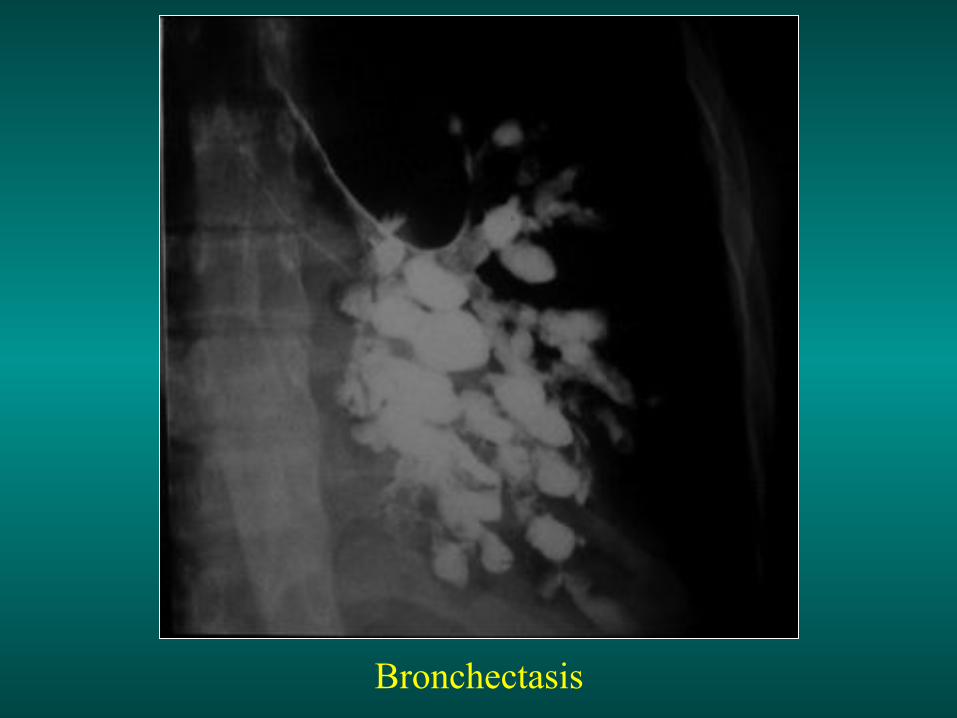
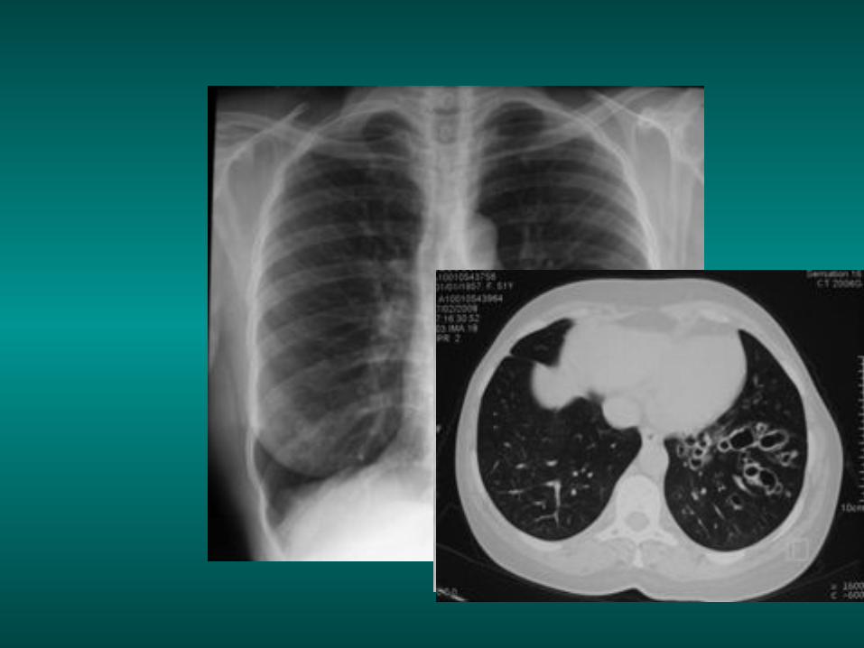
Bronchectasis
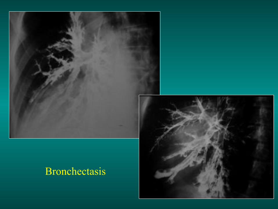
Bronchectasis
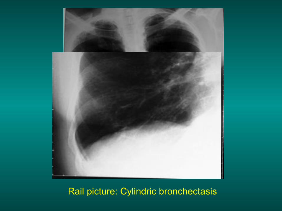
Rail picture: Cylindric bronchectasis

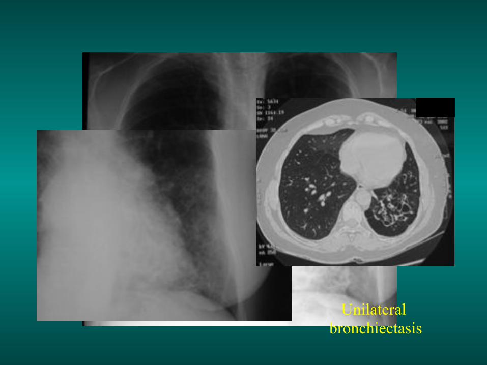
Unilateral bronchiectasis

Bilateral bronchectasis
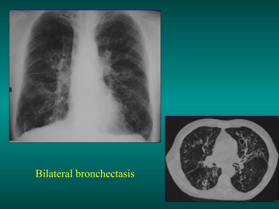
Bilateral bronchectasis
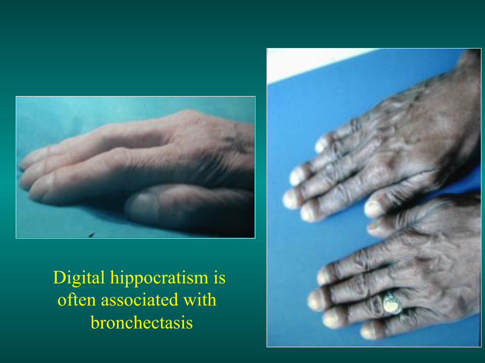
Digital hippocratism is often associated with
bronchectasis
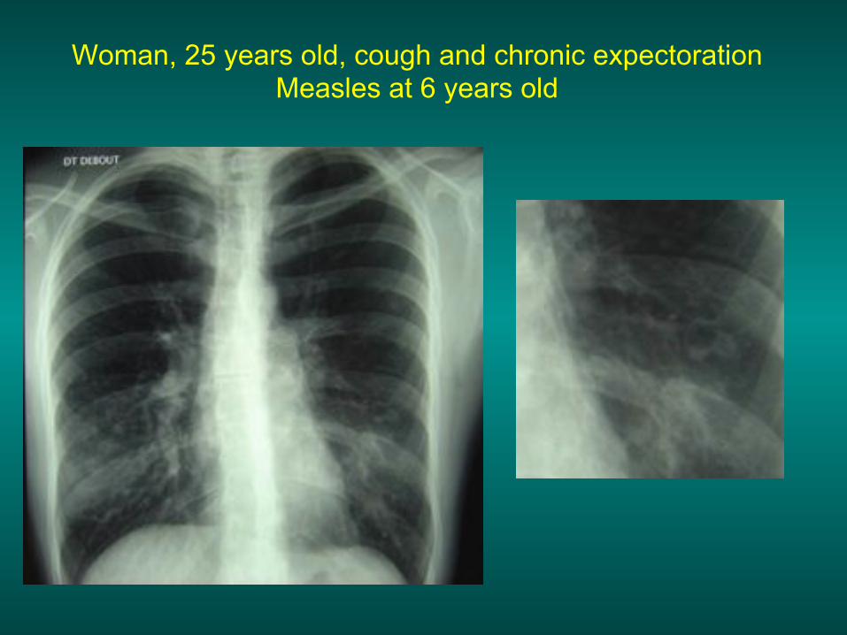
Woman, 25 years old, cough and chronic expectoration Measles at 6 years old


Woman, 54 years old, recurrent severe bronchopneumonia at 2 years old

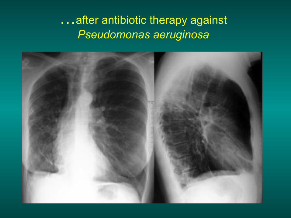
…after antibiotic therapy against Pseudomonas aeruginosa

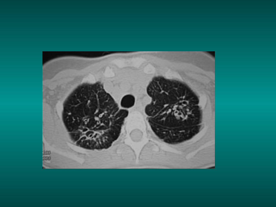
Young woman, 20 years old, recurrent bronchus infections from a
very early age, and gradual respiratory deficiency
MUCOVISIDOSIS (1 case/ 2000 births in Europe)
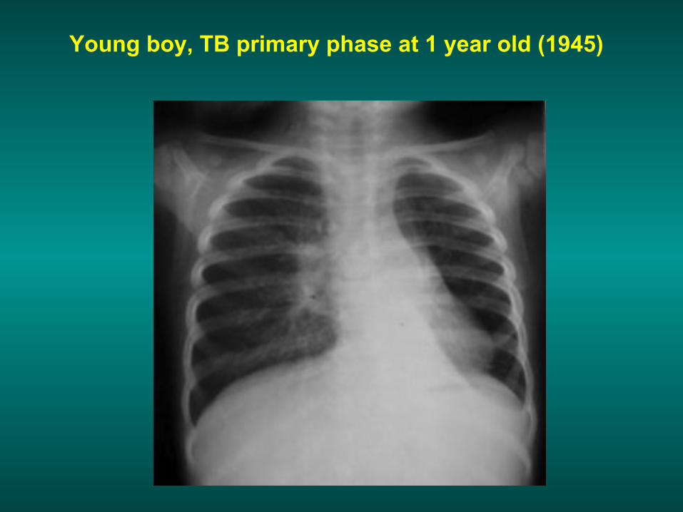
Young boy, TB primary phase at 1 year old (1945)
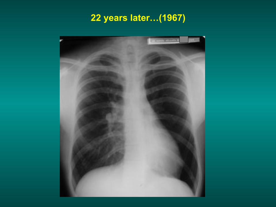
22 years later…(1967)
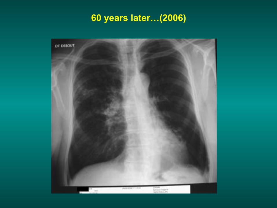
60 years later…(2006)
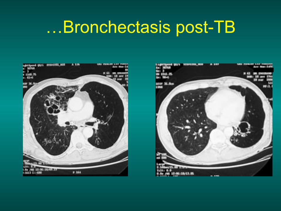
…Bronchectasis post-TB