cardiogenic shock - iconcept press · nada cemerlic adjic, slobodan dodic clinic of cardiology,...
TRANSCRIPT
Cardiogenic Shock Milovan Petrovic, Bojan Vujin, Gordana Panic
Nada Cemerlic Adjic, Slobodan Dodic Clinic of Cardiology, Institute of Cardiovascular Diseases Vojvodina
University of Novi Sad, Serbia

1 Prolog
Shock is a life-threatening condition that occurs due to circulatory insufficiency which leads to inade-quate tissue perfusion with cellular metabolism disorders. Cardiogenic shock is a major, and frequently fatal, complication of a variety of acute and chronic disorders that impair the ability of the heart to main-tain adequate tissue perfusion (Sharma et al., 2005). Cardiogenic shock may be divided in to four major groups: myopathic (damaged myocardial contractility), arrhythmogenic (serious cardiac arrhythmias lead to shock), mechanical (valvular regurgitation) and obstructive lesions (mitral and aortic stenosis, tumor and thrombus in the left atrium, the artificial valve dysfunction, hypertrophic obstructive cardiomyopa-thy). Miscellaneous group of cardiogenic shock include cardiac tamponade, constrictive pericarditis, pulmonary embolism and aortic dissection. This paper will outline the causes, diagnosis, pathophysiolo-gy, and treatment of cardiogenic shock with a focus on CS complicating myocardial infarction (MI). Car-diogenic shock (CS) occurs in ≈5% to 8% of patients hospitalized with ST-elevation myocardial infarc-tion (STEMI), but it can also be caused by mechanical complications, such as acute mitral regurgitation or rupture of either the ventricular septal or free walls. However, any cause of acute, severe left or right ventricular dysfunction may lead to CS. Early revascularization for CS improves survival substantially and new mechanical approaches to treatment are available. The mortality rate remains high (≈50%) de-spite intervention, and half of the deaths occur within the first 48 hours. The 3- and 6-year survival rates with the early revascularization are 41.4% and 32.8% with persistence of treatment benefit (Reynolds & Hochman, 2008). Mortality can range from 10% to 80% depending on demographic, clinical, and hemo-dynamic factors. These factors include age, clinical signs of peripheral hypoperfusion, anoxic brain dam-age, left ventricle ejection fraction (LVEF), and stroke work. Hemodynamic data are predictive of short-term but not long-term mortality. Revascularization provides benefit at every level of risk. Among CS patients undergoing percutaneous coronary intervention (PCI), age, time from symptom onset to PCI, and post-PCI TIMI (Thrombolysis in Myocardial Infarction) flow grade are independent predictors of mor-tality (Reynolds & Hochman, 2008).
2 Incidence
After decades of remarkable stability in the incidence of CS, it appears that the incidence is on the decline in parallel with increasing rates of use of primary PCI for acute MI. CS continues to complicate approxi-mately 5% to 8% of STEMI (Fox, et al., 2007; Babaev et al., 2005) and 2.5% of non-STEMI cases (Hasdai et al., 2000). This translates to 40 000 to 50 000 cases per year in the United States (Thom et al., 2006).
3 Etiology
MI with left ventricle (LV) failure remains the most common cause of CS. It is critical to exclude com-plicating factors that may cause shock in MI patients. Chief among these are the mechanical complica-tions: ventricular septal rupture, contained free wall rupture, and papillary muscle rupture. Mechanical complications must be strongly suspected in patients with CS complicating nonanterior MI, particularly a
first MI. Any cause of acute, severe LV or right ventricle (RV) dysfunction may lead to CS. Acute my-opericarditis, tako-tsubo cardiomyopathy, and hypertrophic cardiomyopathy may all present with ST ele-vation, release of cardiac markers, and shock in the absence of significant coronary artery disease. Stress-induced cardiomyopathy, also known as apical ballooning or tako-tsubo cardiomyopathy, is a syndrome of acute LV dysfunction after emotional or respiratory distress that leads to CS in 4.2% of cases (Gianni et al., 2006). Idiopathic dilated cardiomyopathy presenting in CS is more common. Acute valvular regur-gitation, typically caused by endocarditis or chordal rupture due to trauma or degenerative disease, may also cause CS. Aortic dissection may lead to CS via acute, severe aortic insufficiency or MI. Acute stress in the setting of aortic or mitral stenosis can also cause shock. Cardiac tamponade or massive pulmonary embolism can present as cardiogenic shock without associated pulmonary congestion. Also sustained ar-rhythmia may be included in etiology of cardiogenic shock.
4 Pathophysiology
Cardiogenic shock is characterized by systolic and diastolic dysfunction. Patients who develop cardiogen-ic shock from acute myocardial infarction consistently have evidence of progressive myocardial necrosis with infarct extension. Decreased coronary perfusion pressure and increased myocardial oxygen demand play a role in the vicious cycle that leads to cardiogenic shock. Patients suffering from cardiogenic shock often have multivessel coronary artery disease with limited coronary blood flow reserve. Ischemia remote from the infarcted zone is an important contributor to shock. Myocardial diastolic function is also im-paired, because ischemia causes decreased myocardial compliance, thereby increasing left ventricular filling pressure, which may lead to pulmonary edema and hypoxemia.
Cardiogenic shock is the result of temporary or permanent derangements in the entire circulatory system. Left ventricle pump failure is the primary insult in most forms of CS, but other parts of the circu-latory system contribute to shock with inadequate compensation or additional defects. Many of these ab-normalities are partially or completely reversible, which may explain the good functional outcome in most survivors. Tissue hypoperfusion, with consequent cellular hypoxia, causes anaerobic glycolysis, the accumulation of lactic acid, and intracellular acidosis. Also, myocyte membrane transport pumps fail which decreases transmembrane potential and causes intracellular accumulation of sodium and calcium, resulting in myocyte swelling. If ischemia is severe and prolonged, myocardial cellular injury becomes irreversible and leads to myonecrosis, which includes mitochondrial swelling, the accumulation of dena-tured proteins and chromatin, and lysosomal breakdown. These events induce fracture of the mitochon-dria, nuclear envelopes, and plasma membranes. Additionally, apoptosis (programmed cell death) may occur in peri-infarcted areas and may contribute to myocyte loss. The activation of inflammatory cas-cades, oxidative stress, and stretching of the myocytes produces mediators that overpower inhibitors of apoptosis, thus activating the apoptosis (Figure 1).
4.1 Left Ventricles
Compensatory mechanisms activated when cardiac output is reduced include sympathetic stimulation to increase heart rate and contractility, and renal fluid retention to increase preload. These compensatory mechanisms may become maladaptive and can create a vicious cycle that further worsens systolic dys-function. Vasoconstriction to maintain blood pressure increases myocardial afterload, impairing cardiac

Figure 1: Current concept of CS pathophysiology-Myocardial injury causes systolic and diastolic dysfunction. A decrease in CO leads to a decrease in systemic and coronary perfusion. This exacer-bates ischemia and causes cell death in the infarct border zone and the remote zone of myocardium. Inadequate systemic perfusion triggers reflex vasoconstriction, which is usually insufficient. Systemic inflammation may play a role in limiting the peripheral vascular compensatory response and may con-tribute to myocardial dysfunction. Whether inflammation plays a causal role or is only an epiphenom-enon remains unclear. Revascularization leads to relief of ischemia. It has not been possible to demonstrate an increase in CO or LVEF as the mechanism of benefit of revascularization; however, revascularization does significantly increase the likelihood of survival with good quality of life. IL-6 indicates interleukin-6; TNF-α, tumor necrosis factor-α; and LVEDP, LV end-diastolic pressure (Reynolds & Hochman, 2008).
performance and increasing myocardial oxygen demand. Myocardial ischemia increases myocardial stiff-ness, increasing LV end-diastolic pressure and, thus, myocardial wall stress at a given end-diastolic vol-ume. The increased LV stiffness limits diastolic filling and may result in pulmonary congestion, causing hypoxemia and worsening the imbalance of oxygen delivery and oxygen demand in the myocardium, resulting in further ischemia and myocardial dysfunction. The interruption of this cycle of myocardial dysfunction and ischemia forms the basis for CS therapeutic regimens. A key to understanding the patho-physiology and the treatment of CS is to realize that the areas of nonfunctional but viable myocardium can also cause or contribute to the development of CS after MI. This reversible dysfunction can be de-scribed in two main categories: stunning and hibernation. Myocardial stunning represents postischemic dysfunction that persists despite restoration of normal blood flow; myocardial performance eventually recovers completely. The direct evidence of myocardial stunning in humans has recently been obtained by demonstrating normal perfusion using positron emission tomography scanning and N-ammonia in pa-tients with persistent wall motion abnormalities after angioplasty for acute coronary syndromes. The pathogenesis of stunning has not been conclusively established but appears to involve a combination of
oxidative stress, perturbation of calcium homeostasis, and decreased myofilament responsiveness to cal-cium. The intensity of stunning is determined primarily by the severity of the antecedent ischemic insult. Myocardial hibernation can be seen as an adaptive response in which segments with severely reduced coronary blood flow reduce their contractile function to restore equilibrium between flow and function, minimizing the potential for ischemia or necrosis. Function in such segments can be normalized by im-proving blood flow. Repetitive episodes of myocardial stunning can coexist with or mimic myocardial hibernation. The consideration of myocardial stunning and hibernation is important because of their ther-apeutic implications. Both stunned and hibernating myocardiums retain inotropic reserve and can respond to catecholamines. The function of hibernating myocardium can improve with revascularization and func-tion of stunned myocardium can improve in time. The notion that some myocardial tissue may recover its function has underscored the importance of expeditious initiation of supportive measures, including both medications and intraaortic balloon counterpulsation, to maintain blood pressure and cardiac output in patients with CS. The presence of reversible myocardial dysfunction also has prognostic implications, supported by data from the SHOCK trial, in which most survivors had only class I or class II heart failure (Reynolds & Hochman, 2008).
The degree of myocardial dysfunction that initiates CS is often, but not always, severe. LV dys-function in shock reflects new irreversible injury, reversible ischemia, and damage from prior infarction. The unique position of the heart as an organ that benefits from low blood pressure via afterload reduction and also suffers from low blood pressure via compromise of coronary flow creates a situation in which changes in hemodynamic may be simultaneously beneficial and detrimental. As depicted, a decrease in coronary perfusion lowers cardiac output, which further decreases perfusion of the heart and other vital organs. Coronary flow may be additionally compromised by atherosclerosis of vessels other than the in-farct artery. Metabolic derangements occur in the remote myocardium and in the infarct region. Hy-poperfusion causes release of catecholamines, which increase contractility and peripheral blood flow, but catecholamines also increase myocardial oxygen demand and have proarrhythmic and myocardiotoxic effects (Reynolds & Hochman, 2008).
In light of the complex pathophysiology of CS, it is not surprising that in many cases, severe im-pairment of contractility does not lead to shock, and conversely, LVEF may be only moderately de-pressed in CS. LVEF is similar in the acute phase of CS and 2 weeks later, when functional status is quite different. Among patients in shock, however, LVEF remains a prognostic indicator. Approximately half of all CS patients have small or normal LV size, which represents the failure of the adaptive mechanism of acute dilation to maintain stroke volume in the early phase of MI. Progressive LV dilation (remodel-ing) in the chronic phase can be maladaptive. Serial echocardiography has demonstrated a small increase in LV end-diastolic volume in 2-week survivors of CS (15-mL change in median LV end-diastolic vol-ume). Contractile function can be assessed with echocardiography or LV angiography. In addition, the indwelling pulmonary artery (PA) catheter allows ongoing evaluation of CO in response to changes in therapy and volume status. Diastolic function is more difficult to assess. It is likely that the abnormalities of ventricular relaxation and compliance contribute to CS in some, if not all cases (Reynolds & Hochman, 2008).
4.2 Right Ventricle
RV dysfunction may cause or contribute to CS. Predominant RV shock represents only 5% of cases of CS complicating MI. RV failure may limit LV filling via a decrease in CO, ventricular interdependence, or

both. The treatment of patients with RV dysfunction and shock has traditionally focused on ensuring ade-quate right-sided filling pressures to maintain CO and adequate LV preload; however, patients with CS due to RV dysfunction have very high RV end-diastolic pressure, often >20 mm Hg. This elevation of RV end-diastolic pressure may result in shifting of the interventricular septum toward the LV cavity, which raises left atrial pressure but impairs LV filling due to the mechanical effect of the septum bowing into the LV. This alteration in geometry also impairs LV systolic function. Therefore, the common prac-tice of aggressive fluid resuscitation for RV dysfunction in shock may be misguided. Inotropic therapy is indicated for RV failure when CS persists after RV end-diastolic pressure has been optimized. RV end-diastolic pressure of 10 to 15 mm Hg has been associated with higher output than lower or higher pres-sures, but marked variability exists in optimal values. Inhaled nitric oxide (NO) may be useful to lower pulmonary vascular resistance and promote forward flow. Both pericardiectomy and the creation of atrial septal defects have been used in extreme cases.
Shock due to isolated RV dysfunction carries nearly as high a mortality risk as LV shock. The benefit of revascularization was similar.
4.3 Cardiovascular Mechanism of Cardiogenic Shock
The main mechanical defect in cardiogenic shock is a shift to the right for the left ventricular end-systolic pressure-volume curve (Figure 2, 3) because of a marked reduction in contractility. As a result, at a simi-lar or even lower systolic pressure, the ventricle is able to eject less blood volume per beat. Therefore, the end-systolic volume is usually greatly increased in persons with cardiogenic shock. The stroke volume is decreased, and to compensate for this, the curvilinear diastolic pressure-volume curve also shifts to the right, with a decrease in diastolic compliance. This leads to increased diastolic filling, which is associated with an increase in end-diastolic pressure. The attempt to enhance cardiac output by this mechanism comes at the cost of having a higher left ventricular diastolic filling pressure, which ultimately increases myocardial oxygen demand and causes pulmonary edema. As a result of decreased contractility, the pa-tient develops elevated left and right ventricular filling pressures and low cardiac output. Mixed venous oxygen saturation falls because of the increased tissue oxygen extraction, which is due to the low cardiac output. This, combined with the intrapulmonary shunting that is often present, contributes to substantial arterial oxygen desaturation.
4.4 Peripheral Vasculature, Neurohormones and Inflammation in Cardiogenic Shock
Hypoperfusion of the extremities and vital organs is a hallmark of CS. The decrease in CO caused by MI and sustained by ongoing ischemia triggers release of catecholamines, which constrict peripheral arteri-oles to maintain perfusion of vital organs. Vasopressin and angiotensin II levels increase in the setting of MI and shock, which leads to improvement in coronary and peripheral perfusion at the cost of increased afterload, which may further impair myocardial function. Activation of the neurohormonal cascade pro-motes salt and water retention; this may improve perfusion but exacerbates pulmonary edema. The reflex mechanism of increased systemic vascular resistance (SVR) is not fully effective. MI can cause the sys-temic inflammatory response syndrome (SIRS) and suggest that inappropriate vasodilatation as part of SIRS results in impaired perfusion of the intestinal tract, which enables transmigration of bacteria and sepsis. SIRS is more common with increasing duration of shock1 - even though levels of interleu-
1 http://circ.ahajournals.org/content/117/5/686.full

Figure 2: Left ventricular pressure-volume (PV) curves are derived from pressure and volume in-formation found in the cardiac cycle diagram (see left panel of figure). To generate a PV curve for the left ventricle, the left ventricular pressure (LVP) is plotted against left ventricular (LV) volume at multiple time points during a complete cardiac cycle. When this is done, a PV curve is generated (right panel of figure) (Klabunde, 2011).
Figure 3: Effects of acute left ventricular failure (loss of inotropy) on left ventricular pres-sure-volume curve (Klabunde, 2011).
kin-6 and tumor necrosis factor-" have been found to be elevated on admission among MI patients who were initially in Killip class I and later developed CS (Reynolds & Hochman, 2008). Cytokine levels rise more dramatically over the 24 to 72 hours after MI. Tumor necrosis factor-" and interleukin-6 have myo-cardial depressant action. Tumor necrosis factor-" also induces coronary endothelial dysfunction, which may further diminish coronary flow. Other circulating factors (complement, procalcitonin, neopterin, C-reactive protein, and others) have been reported to contribute to SIRS in CS. Excess NO may also con-

tribute to SIRS. MI is associated with increased expression of inducible NO synthase, which leads to ex-cess NO, which causes vasodilatation, myocardial depression, and interference with catecholamine action (Reynolds & Hochman, 2008).
5 Diagnosis
CS is a state of end-organ hypoperfusion due to heart failure. Hypoperfusion may be manifest clinically by cool extremities, decreased urine output (< 20 ml/h), and/or alteration in mental status. Hemodynamic abnormalities form a spectrum that ranges from mild hypoperfusion to profound shock, and the short-term outcome is directly related to the severity of hemodynamic derangement. The definition of CS in-cludes hemodynamic parameters: persistent hypotension (systolic blood pressure SBP < 80 to 90 mm Hg or mean arterial pressure MAP 30 mm Hg lower than baseline) with severe reduction in cardiac index (CI) (< 1.8 L · min−1 · m−2 without support or < 2.0 to 2.2 L · min−1 · m−2 with support) and adequate or elevated filling pressure (e.g. left ventricular end-diastolic pressure LVEDP > 18 mm Hg or right ventric-ular end-diastolic pressure RVEDP > 10 to 15 mm Hg) (Table 1). The diagnosis is usually made with the help of pulmonary artery catheterization; however, Doppler echocardiography may also be used to con-firm elevation of LV filling pressures (Giannuzzi et al., 1994). Echocardiography also is the technique of choice to help in diagnostic CS with estimation of LV and RV motion, kinetic disorders and valvular set-tings. Coronary angiography helps us to see morphology of coronary artery lesions and to perform rapid primary percutaneous coronary intervention.
Hemodynamic Parameters In Cardiogenic Shock SBP < 80 to 90 mm Hg MAP 30 mm Hg lower than baseline CI < 1.8 L · min−1 · m−2 without support or < 2.0 to 2.2 L · min−1 · m−2 with support LVEDP, RVEDP LVEDP > 18 mm Hg RVEDP > 10 to 15 mm Hg
Table 1: Diagnostic hemodynamic parameters in cardiogenic shock
6 Patient with High Risk for CS
Risk factors for development of CS in the context of MI include older age, anterior MI, hypertension, diabetes mellitus, multivessel coronary artery disease, prior MI or angina, prior diagnosis of heart failure, STEMI, and left bundle-branch block (Lindholm et al., 2003).
7 Treatment
Treatment of cardiogenic shock as a complication of acute myocardial infarction includes hemodynamic stability achieved by medication therapy or circulatory and respiratory support and emergency revascular-isation using PCI or surgical revascularization (CABG) (Figure 4).
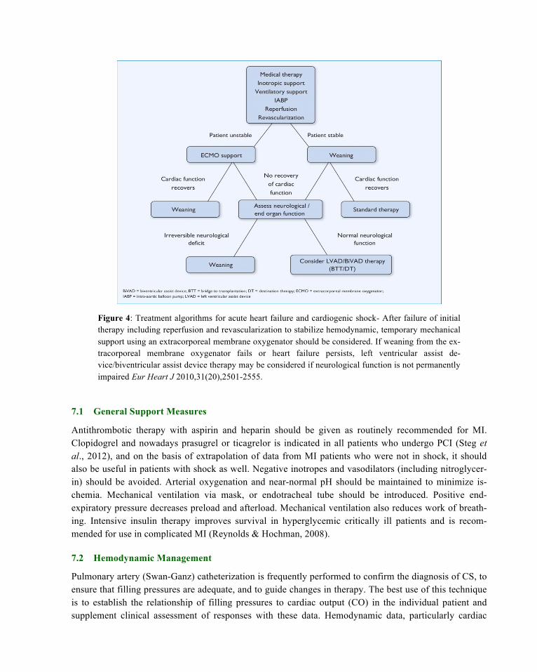
Figure 4: Treatment algorithms for acute heart failure and cardiogenic shock- After failure of initial therapy including reperfusion and revascularization to stabilize hemodynamic, temporary mechanical support using an extracorporeal membrane oxygenator should be considered. If weaning from the ex-tracorporeal membrane oxygenator fails or heart failure persists, left ventricular assist de-vice/biventricular assist device therapy may be considered if neurological function is not permanently impaired Eur Heart J 2010,31(20),2501-2555.
7.1 General Support Measures
Antithrombotic therapy with aspirin and heparin should be given as routinely recommended for MI. Clopidogrel and nowadays prasugrel or ticagrelor is indicated in all patients who undergo PCI (Steg et al., 2012), and on the basis of extrapolation of data from MI patients who were not in shock, it should also be useful in patients with shock as well. Negative inotropes and vasodilators (including nitroglycer-in) should be avoided. Arterial oxygenation and near-normal pH should be maintained to minimize is-chemia. Mechanical ventilation via mask, or endotracheal tube should be introduced. Positive end-expiratory pressure decreases preload and afterload. Mechanical ventilation also reduces work of breath-ing. Intensive insulin therapy improves survival in hyperglycemic critically ill patients and is recom-mended for use in complicated MI (Reynolds & Hochman, 2008).
7.2 Hemodynamic Management
Pulmonary artery (Swan-Ganz) catheterization is frequently performed to confirm the diagnosis of CS, to ensure that filling pressures are adequate, and to guide changes in therapy. The best use of this technique is to establish the relationship of filling pressures to cardiac output (CO) in the individual patient and supplement clinical assessment of responses with these data. Hemodynamic data, particularly cardiac
output, cardiac index, have powerful short-term prognostic value (Fincke et al., 2004). There has been a decline in PA catheter use. Clinical assessment with echocardiography is a reasonable alternative: Both PA systolic pressure and wedge pressure can be accurately estimated with Doppler echocardiography, and in particular, the finding of a short mitral deceleration time (≤140 ms) is highly predictive of pulmo-nary capillary wedge pressure ≥20 mm Hg in CS (Reynolds et al., 2006). The clinical examination and chest radiograph are not reliable predictors of pulmonary capillary wedge pressure.
7.3 Pharmacological Treatment
Pharmacological support includes inotropic and vasopressor agents. Vasopressors and inotropic agents are used because of their positive hemodynamic effect, but neither of them leads to permanent sympto-matic improvement, and many even reduce survival rate, which may be associated with cell dysfunction caused by these drugs (Thackray et al., 2002). A recent randomised study compared norepinephrine and dopamine in cardiogenic shock. Dopamine was associated with a higher mortality rate and more adverse effects, such as arrhythmia (De Backer et al., 2010). In hypotension with other signs of cardiogenic shock, norepinephrine is recommended as the first choice. It should be initially administered in low doses and gradually titrated until systolic pressure reaches values of over 80 mmHg. After that, dobutamine can be administered together with norepinephrine for better contractility (Steg et al., 2012).
7.4 Reperfusion
The survival benefit of early revascularization in CS, reported in several observational studies, was shown convincingly in the randomized SHOCK trial, which found a 13% absolute increase in 1-year sur-vival in patients assigned to early revascularization (Reynolds & Hochman, 2008). Revascularization could be percutaneous or surgical. Percutaneous coronary intervention is superior to fibrinolytic therapy. Fibrinolysis is recommended if PCI is not feasible or if delayed.
Stenting and glycoprotein IIb/IIIa inhibitors were independently associated with improved out-comes in patients undergoing PCI for CS in multiple registries, including the large ACC-National Cardi-ovascular Data Registry (Klein et al., 2005).
The optimal revascularization strategy (i.e., percutaneous or surgical, single or multivessel PCI) for patients with multivessel coronary artery disease and CS is not clear. This is of particular importance because multivessel disease is common. According to the ACC/AHA guidelines for revascularization in shock, in patients with multivessel disease, revascularization of the noninfarct related artery may be nec-essary to maximize myocardial perfusion. Alternatively, in patients with multivessel disease and particu-larly left main disease, emergency CABG as a primary reperfusion strategy may be preferred (Levine et al., 2011). The ability to achieve complete revascularization may be strongly associat-ed with improved in-hospital survival in patients with cardiogenic shock (Hussain et al., 2011).
7.5 Mechanical Circulatory Support: Intraaortic Balloon Pump
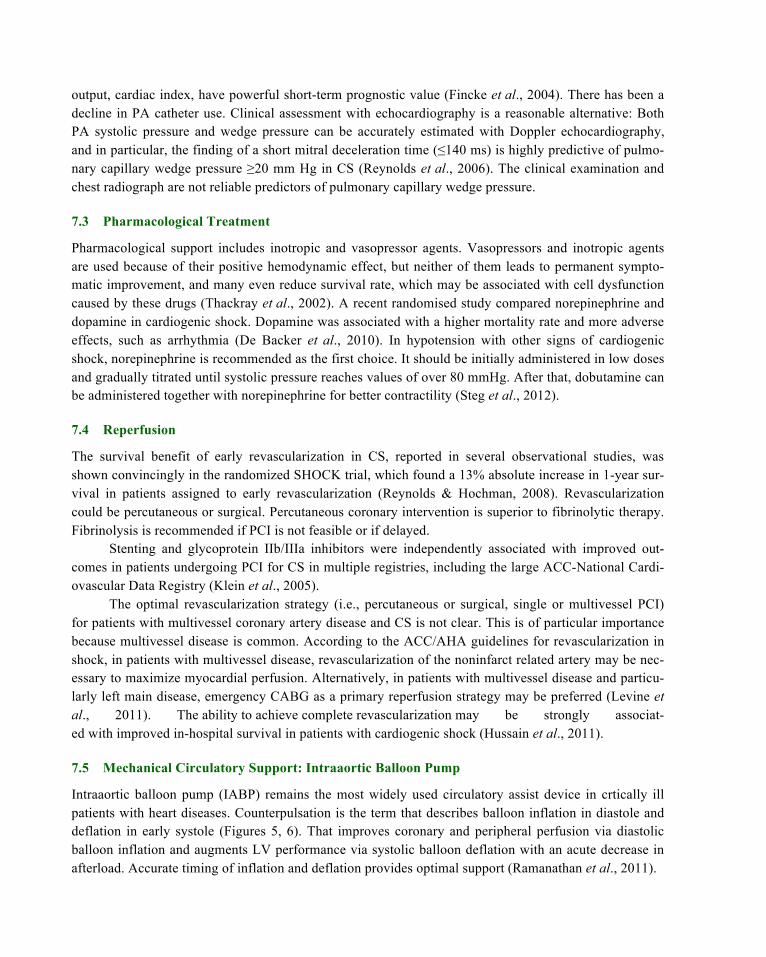
Intraaortic balloon pump (IABP) remains the most widely used circulatory assist device in crtically ill patients with heart diseases. Counterpulsation is the term that describes balloon inflation in diastole and deflation in early systole (Figures 5, 6). That improves coronary and peripheral perfusion via diastolic balloon inflation and augments LV performance via systolic balloon deflation with an acute decrease in afterload. Accurate timing of inflation and deflation provides optimal support (Ramanathan et al., 2011).
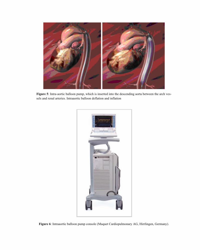
Figure 5: Intra-aortic balloon pump, which is inserted into the descending aorta between the arch ves-sels and renal arteries. Intraaortic balloon deflation and inflation
Figure 6: Intraaortic balloon pump console (Maquet Cardiopulmonary AG, Hirrlingen, Germany).
The primary goal of IABP treatment is to improve ventricular performance of failing heart by fa-cilitating an increasing myocardial oxygen supply and decrease myocardial oxygen demand. Although these effects are predominantly associated with enhancement of LV performance, IABP may also have favorable effects on RV function by complex mechanisms including accentuation RV myocardial blood flow, unloading of LV causing reduction in left atrial and pulmonary vascular pressure and RV afterload (Miller, 2011). Not every patient has a hemodynamic response to IABP; response predicts better outcome (Ramanathan et al., 2011). TACTICS study showed that IABP does not contribute to reduction of intra-hospital mortality, but it brings about improvement in six-month mortality rate (Ohman et al., 2005). Re-cent meta-analyses evidenced in relation to survival rate when using IABP in cardiogenic shock (Bahekar et al., 2012). IABP-SHOCK II study showed that use of intraaortic balloon counterpulsation did not sig-nificantly reduce 30-day mortality in patients with cardiogenic shock complicating acute myocardial in-farction for whom an early revascularization strategy was planned (Thiele et al., 2012). IABP support should be instituted as quickly as possible, even before any transfer for revascularization if a skilled oper-ator is available and insertion can be performed quickly. Complications associated with IABP are less common in the modern era; in the largest series, the overall and major complication rates were 7.2% and 2.8%, respectively. Risk factors for complications include female sex, small body size, and peripheral vascular disease (Urban et al., 2004). IABP therapy is considered to be a class IIb indication (European Society of Cardiology guidelines) for the management of cardiogenic shock (Steg et al., 2012).
7.6 Mechanical Circulatory Support: Ventricular Assist Devices
In profound cardiogenic shock, blood flow and perfusion pressure must be restored urgently to prevent permanent damage to the brain, liver, kidneys, and gut. Adequate right ventricular function is necessary to avoid central venous hypertension and end-organ venous congestion. In mechanical engineering, a fail-ing pump is repaired or replaced provided that the rest of the system remains in working order. The same strategy can be applied in patients with heart failure using modern circulatory support technology. The landmark REMATCH study (Rose et al., 2001; Westaby et al., 2012) established a long-term role for left ventricular assist devices (LVADs) to relieve symptoms and improve longevity in terminally ill patients with cardiomyopathy who were not eligible for a cardiac transplant. Temporary mechanical circulatory support with LV assist devices (LVADs) is theoretically appealing to interrupt the vicious spiral of is-chemia, hypotension, and myocardial dysfunction, allowing for recovery of stunned and hibernating my-ocardium and reversal of neurohormonal derangements. Device-related complications and irreversible organ failure remain major limitations. LVAD support involves circulation of oxygenated blood through a device that drains blood from the left side of the heart and returns blood to the systemic arteries with pulsatile or continuous flow. A variety of surgically implanted continuous-flow and pulsatile blood pumps have proven to be very effective postinfarction (Westaby et al., 2012). The advantage of these devices is that central cannulation of atria, ventricles, and great arteries can bypass and unload the failing ventricle and provide blood flow of up to 10 l/min. For left ventricular support, the ventricular assist de-vice (VAD) drains the left atrium or ventricle and pumps blood into the aorta. For right ventricular by-pass, the right atrium is normally used for VAD inflow with blood pumped into the main pulmonary ar-tery, thereby avoiding peripheral vascular complications. Outcome depends on myocardial viability fol-lowing revascularization, pre-existing left ventricular dysfunction, and potential for recovery in stunned or hibernating myocardium. In contrast to percutaneously inserted systems, all centrally implanted pumps

can be kept in situ for periods ranging from weeks to several years. Temporary pumps can be replaced by long-term implanted LVADs if the native heart does not recover (Westaby et al., 2012).
Data from the US INTERMACS provides important insights into the overall success of blood pumps in cardiogenic shock (Holman et al., 2009). Shock accounted for 42% of 483 VAD implants and, not surprisingly, these patients had a worse survival profile than those who underwent elective destination therapy. Patients who only required left ventricular support had the best outlook (50% survival at 12 months). When biventricular support was needed, survival fell to 35%. Prognosis was poor for those re-ceiving isolated right ventricular support or a total artificial heart (Holman et al., 2009).
Percutaneous LVADs are also available. Shock may already be apparent at the time of pPCI, or can be predicted from angiographic or echocardiographic findings. In both circumstances, two percutane-ously implanted blood pumps are available to the cardiologist without the need for immediate interven-tion by surgeons. The Tandem Heart (Cardiac Assist, Inc, Pittsburgh, PA) (Figure 7) removes blood from the left atrium using a cannula placed through the femoral vein and into the left atrium via transseptal puncture. Blood is then returned to a systemic artery, usually the femoral, with retrograde perfusion of the abdominal and thoracic aorta (Reynolds & Hochman, 2008). Another percutaneous device, the Impella 2.5 (Abiomed, Inc, Danvers, Mass) (Figure 8) pulls blood from the left ventricle through an inlet area near the tip and expels blood from the catheter into the ascending aorta. The pump can be inserted via a standard catheterization procedure through the femoral artery, into the ascending aorta, across the valve and into the left ventricle (Henriques et al., 2006). Extracorporal membrane oxygenation (ECMO) (Fig-ure 9) is used to sustain physiological levels of blood flow in cardiogenic shock, as a rescue system dur-ing cardiopulmonary resuscitation and to provide both cardiac and pulmonary support for patients with hypoxia (Smedira et al., 2001). An ECMO circuit consists of a centrifugal blood pump, membrane oxy-genator, and a heparin-coated circuit. ECMO is the simplest and most-rapid method of restoring systemic blood flow and, with contemporary peripheral arteriovenous cannulation techniques, can now be used in the catheterization laboratory or emergency room (Westaby et al., 2012). Cannulation of the femoral ar-tery and vein are achieved rapidly by the Seldinger technique, using specifically designed perfusion can-nulas over a guidewire, even during cardiac massage or PPCI. The tip of the femoral arterial cannula is advanced to the aortoiliac junction and the venous drainage pipe advanced into the lower right atrium. Systemic heparinization is used to achieve an activated clotting time of 150–180 s. Flow rates are initiat-ed at 2–3 l/min using dopamine to raise mean blood pressure to > 55 mmHg. Once the circuit is estab-lished, the flow rate can be progressively increased to 3.5–4.0 l/min, thus restoring a normal cardiac index and mean blood pressure to >70 mmHg (Westaby et al., 2012). Because venoarterial ECMO increases left ventricular afterload and wall stress, cardiac contractility must be maintained to avoid left ventricular distension, clot on the akinetic myocardium, or pulmonary hypertension using an IABP and inotropes (Westaby et al., 2012). ECMO can provide an effective bridge to urgent cardiac transplantation or to pro-longed LVAD support.
Mechanical circulatory support can sustain life during profound postinfarction cardiogenic shock and is an important adjunct to coronary angioplasty in rapidly deteriorating patients. Neither the IABP nor percutaneously inserted LVADs have been shown to improve survival in established cardiogenic shock. By contrast, both ECMO and centrally implanted blood pumps show the capacity to salvage more than half of those patients who would otherwise die (Westaby et al., 2012).
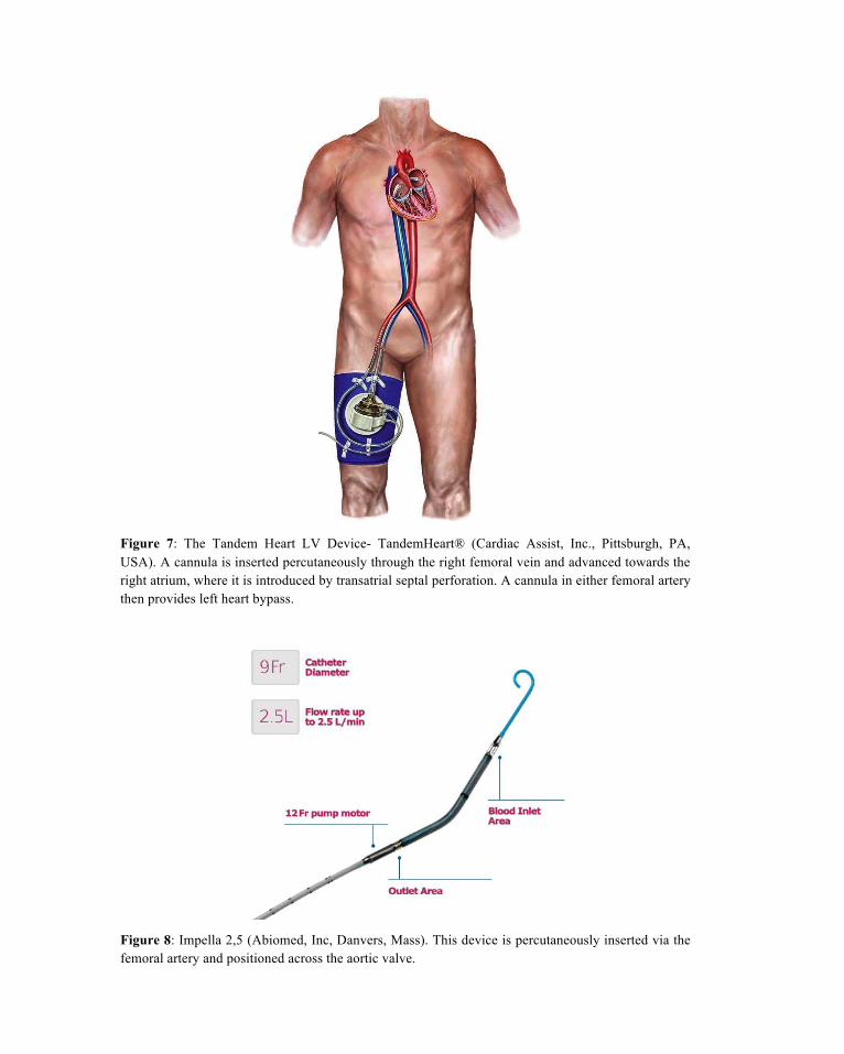
Figure 7: The Tandem Heart LV Device- TandemHeart® (Cardiac Assist, Inc., Pittsburgh, PA, USA). A cannula is inserted percutaneously through the right femoral vein and advanced towards the right atrium, where it is introduced by transatrial septal perforation. A cannula in either femoral artery then provides left heart bypass.
Figure 8: Impella 2,5 (Abiomed, Inc, Danvers, Mass). This device is percutaneously inserted via the femoral artery and positioned across the aortic valve.
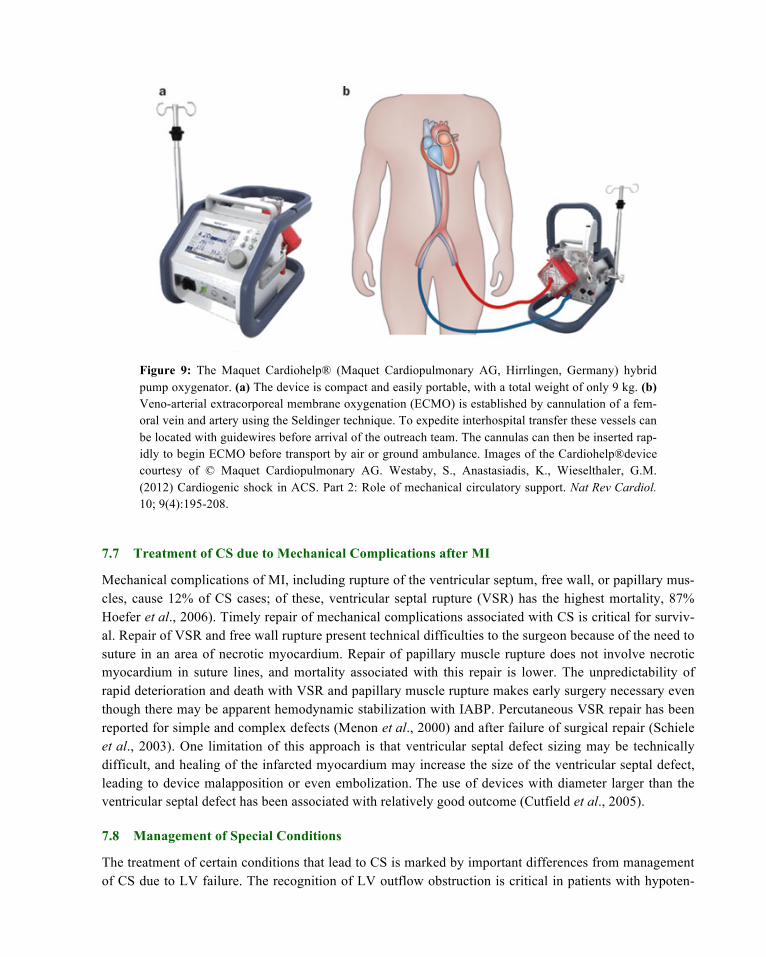
Figure 9: The Maquet Cardiohelp® (Maquet Cardiopulmonary AG, Hirrlingen, Germany) hybrid pump oxygenator. (a) The device is compact and easily portable, with a total weight of only 9 kg. (b) Veno-arterial extracorporeal membrane oxygenation (ECMO) is established by cannulation of a fem-oral vein and artery using the Seldinger technique. To expedite interhospital transfer these vessels can be located with guidewires before arrival of the outreach team. The cannulas can then be inserted rap-idly to begin ECMO before transport by air or ground ambulance. Images of the Cardiohelp®device courtesy of © Maquet Cardiopulmonary AG. Westaby, S., Anastasiadis, K., Wieselthaler, G.M. (2012) Cardiogenic shock in ACS. Part 2: Role of mechanical circulatory support. Nat Rev Cardiol. 10; 9(4):195-208.
7.7 Treatment of CS due to Mechanical Complications after MI
Mechanical complications of MI, including rupture of the ventricular septum, free wall, or papillary mus-cles, cause 12% of CS cases; of these, ventricular septal rupture (VSR) has the highest mortality, 87% Hoefer et al., 2006). Timely repair of mechanical complications associated with CS is critical for surviv-al. Repair of VSR and free wall rupture present technical difficulties to the surgeon because of the need to suture in an area of necrotic myocardium. Repair of papillary muscle rupture does not involve necrotic myocardium in suture lines, and mortality associated with this repair is lower. The unpredictability of rapid deterioration and death with VSR and papillary muscle rupture makes early surgery necessary even though there may be apparent hemodynamic stabilization with IABP. Percutaneous VSR repair has been reported for simple and complex defects (Menon et al., 2000) and after failure of surgical repair (Schiele et al., 2003). One limitation of this approach is that ventricular septal defect sizing may be technically difficult, and healing of the infarcted myocardium may increase the size of the ventricular septal defect, leading to device malapposition or even embolization. The use of devices with diameter larger than the ventricular septal defect has been associated with relatively good outcome (Cutfield et al., 2005).
7.8 Management of Special Conditions
The treatment of certain conditions that lead to CS is marked by important differences from management of CS due to LV failure. The recognition of LV outflow obstruction is critical in patients with hypoten-
sion, because diuretics and inotropic agents exacerbate obstruction. Treatment of CS with hypertrophic obstructive cardiomyopathy includes volume resuscitation. Outflow obstruction may also be seen in some cases of tako-tsubo cardiomyopathy when extensive akinesis/dyskinesis of apical zones occurs with hy-perkinesis of remaining myocardium. Therapy is guided by echocardiography and clinical response. IABP may provide circulatory support. β-Blockade is often not indicated in this circumstance because it exacerbates LV dysfunction. Vasopressors and inotropes improve function in the stunned myocardium and may therefore be useful when outflow obstruction is not visualized. Low doses should be initiated, with careful monitoring of the response (Reynolds & Hochman, 2008).
8 CS as an Iatrogenic Illness
Approximately three fourths of patients with CS complicating MI develop shock after hospital presenta-tion (Holzer et al., 2004). In some, medication use contributes to the development of shock. Several dif-ferent classes of medications used to treat MI have been associated with shock, including β-blockers, an-giotensin-converting enzyme inhibitors, nitrates and morphine. Although early use of each of these medi-cations is associated with only a small excess risk of CS, the large number of patients treated with these therapies translates into a substantial potential number of events (Jeger et al., 2006; Meine et al., 2005). The timing of CS (early after medication initiation) in the placebo-controlled, randomized trials of β-blockade and angiotensin-converting enzyme inhibition combined with their mechanisms of action indi-cate that they may contribute to CS development in those at high risk. Diuretics may also cause or con-tribute to shock in patients with MI (Chen et al., 2005; Hochman, 2003). Acute pulmonary edema is a state of redistribution of intravascular volume into extracellular space in the lungs. When hemodynamic stability is tenuous, the additional decrease in plasma volume caused by diuretics in patients without prior heart failure may induce shock. Tachycardia is often compensatory for lower stroke volume but is not appreciated as such. Treatment with β-blockade lowers heart rate and stroke volume, leading to frank shock. Decompensation may also occur when patients who are reliant on compensatory vasoconstriction are treated with angiotensin-converting enzyme inhibitors, particularly intravenously and early. Nitrates would be expected to have a similar effect but did not in the only systematic study, which used oral, low-dose treatment. Volume expansion may be deleterious when used to excess or when RV filling pressure is already elevated, because the RV may become volume overloaded with shift of the septum causing im-pairment in LV filling and contraction. A trial of a low diuretic dose coupled with low-dose nitrates and positional measures to decrease preload (e.g., seated position with legs down) should be attempted in pa-tients with MI and pulmonary edema to avoid precipitating shock (Reynolds & Hochman, 2008).
8 Conclusion
CS following acute myocardial infarction is a potentially treatable illness with a reasonable chance for fully recovery. An early invasive approach can increase short- and long-term survival and can result in excellent quality of life. Revascularization is associated with some benefit at every level of risk.
References
Babaev, A., Frederick, P.D., Pasta, D.J., et al. (2005). Trends in management and outcomes of patients with acute myo-cardial infarction complicated by cardiogenic shock. JAMA, 294(4), 448–454.
Bahekar, A., Singh, M., Singh, S., et al. (2012). Cardiovascular outcomes using intra-aortic balloon pump in high-risk acute myocardial infarction with or without cardiogenic shock: a meta-analysis. J Cardiovasc Pharmacol Ther, 17(1),44–56.
Chen, Z.M., Pan, H.C., Chen, Y.P., et al. (2005). Early intravenous then oral metoprolol in 45,852 patients with acute my-ocardial infarction: randomized placebo-controlled trial. Lancet, 366(9497), 1622–1632.
Cutfield, N.J., Ruygrok, P.N., Wilson, N.J., et al. (2005). Transcatheter closure of a complex postmyocardial infarction ventricular septal defect after surgical patch dehiscence. Intern Med J, 35(2), 128–130.
De Backer, D., Biston, P., Devriendt, J., et al. (2010). Comparison of dopamine and norepinephrine in the treatment of shock. N Engl J Med,362,779–89.
Fincke, R., Hochman, J.S., Lowe, A.M., et al. (2004). Cardiac power is the strongest hemodynamic correlate of mortality in cardiogenic shock: a report from the SHOCK trial registry. J Am Coll Cardiol, 44(2), 340–348.
Fox, K.A., Anderson, F.A. Jr, Dabbous, O.H., et al. (2007). Intervention in acute coronary syndromes: do patients undergo intervention on the basis of their risk characteristics? The Global Registry of Acute Coronary Events (GRACE). Heart, 93(2), 177–182.
Gianni, M., Dentali, F., Grandi, A.M., et al. (2006). Apical ballooning syndrome or takotsubo cardiomyopathy: a systemat-ic review. Eur Heart J, 27(13), 1523–1529.
Giannuzzi, P., Imparato, A., Temporelli, P.L. et al. (1994). Doppler-derived mitral deceleration time of early filling as a strong predictor of pulmonary capillary wedge pressure in postinfarction patients with left ventricular systolic dys-function. J Am Coll Cardiol, 23(7), 1630–1637.
Hasdai, D., Harrington, R.A., Hochman, J.S., et al. (2000). Platelet glycoprotein IIb/IIIa blockade and outcome of cardio-genic shock complicating acute coronary syndromes without persistent ST-segment elevation. J Am Coll Cardiol, 36(3), 685–692.
Henriques, J.P., Remmelink, M., Baan, J. Jr, et al. (2006). Safety and feasibility of elective high-risk percutaneous coro-nary intervention procedures with left ventricular support of the Impella Recover LP 2.5. Am J Cardiol, 97(7), 990–992.
Hochman, J.S. (2003). Cardiogenic shock complicating acute myocardial infarction: expanding the paradigm. Circulation, 107(24), 2998–3002.
Hoefer, D., Ruttmann, E., Poelzl, G., et al. (2006). Outcome evaluation of the bridge-to-bridge concept in patients with cardiogenic shock. Ann Thorac Surg, 82(1), 28–33.
Holman, W. L. et al. (2009) INTERMACS: interval analysis of registry data. J Am Coll Surg, 208(5), 755–761.
Holzer, R., Balzer, D., Amin, Z., et al. (2004). Transcatheter closure of postinfarction ventricular septal defects using the new Amplatzer muscular VSD occluder: results of a U.S. registry. Catheter Cardiovasc Interv, 61(2), 196–201.
Hussain, F., Philip, R.K., Ducas, A.R. et al. (2011). The ability to achieve complex revascularization is associated with improve in-hospital survival in cardiogenic shock due to myocardial infarction: Manitoba cardiogenic shock registry investigators, Catheter cardiovasc Interv, 78(4),540-548
Jeger, R.V., Harkness, S.M., Ramanathan, K., et al. (2006). Emergency revascularization in patients with cardiogenic shock on admission: a report from the SHOCK trial and registry. Eur Heart J, 27(6), 664–670.
Klabunde, R.E. (2011) Cardiovascular Physiology Concepts; 2nd edition; Lippincott Williams & Wilkins ISBN 9781451113846
Klein LW, Shaw RE, Krone RJ, et al. (2005). Mortality after emergent percutaneous coronary intervention in cardiogenic shock secondary to acute myocardial infarction and usefulness of a mortality prediction model. Am J Cardiol, 96(1), 35–41.
Levine, G.N., Bates, E.R., Blankenship, J.C., et al. (2011). ACCF/AHA/SCAI Practice Guideline for Percutaneous Coro-nary Intervention, Circulation, 124(23), e574-e651
Lindholm, M.G., Kober, L., Boesgaard, S., et al. (2003). Cardiogenic shock complicating acute myocardial infarction: prognostic impact of early and late shock development. Eur Heart J, 24(3), 258–265.
Meine, T.J., Roe, M.T., Chen, A.Y., et al. (2005). Association of intravenous morphine use and outcomes in acute coronary syndromes: results from the CRUSADE Quality Improvement Initiative. Am Heart J, 149(6), 1043–1049.
Menon, V., Webb, J.G., Hillis, L.D., et al. (2000). Outcome and profile of ventricular septal rupture with cardiogenic shock after myocardial infarction: a report from the SHOCK Trial Registry: Should we emergently revascularize Occluded Coronaries in cardiogenic shock? J Am Coll Cardiol, 36(3s1), 1110–1116.
Miller, R.D. Miller’s anesthesia. In: Nyhan D Johns RA eds. (1991/2007). Anesthesia for Cardiac Surgery. Elsevier
Ohman, E.M., Nanas, J., Stomel, R.J., et al., for the TACTICS Trial (2005). Thrombolysis and counterpulsation to improve survival in myocardial infarction complicated by hypotension and suspected cardiogenic shock or heart failure: re-sults of the TACTICS Trial. J Thromb Thrombolysis, 19(1), 33-39.
Ramanathan, K., Farkouh, M.E., Cosmi, J. et al. (2011). Rapid complete reversal of systemic hypoperfusion after intra-aortic balloon pump counterpulsation and survival in cardiogenic shock complicating an acute myocardial infarc-tion. Am Heart J, 162(2):268-75.
Reynolds, H.R., Anand, S.K., Fox, J.M., et al. (2006). Restrictive physiology in cardiogenic shock: observations from echo-cardiography. Am Heart J, 151(4), 890 e9–e15.
Reynolds, H.R., Hochman, J.S. (2008). Cardiogenic Shock Current Concepts and Improving Outcomes. Circulation, 117(6), 686-697
Rose, E.A. et al. (2001). Long-term use of a left ventricular assist device for end-stage heart failure. N Engl J Med 345, 1435–1443.
Schiele, T.M., Kozlik-Feldmann, R., Sohn, H.Y., et al. (2003). Transcatheter closure of a ruptured ventricular septum fol-lowing inferior myocardial infarction and cardiogenic shock. Catheter Cardiovasc Interv, 60(2), 224–228.
Sharma, S., Zevitz, M. (2005). Cardiogenic shock. eMedicine. Available at: http://www.emedicine.com/med/topic285.htm
Smedira, N.G. et al. (2001). Clinical experience with 202 adults receiving extracorporeal membrane oxygenation for car-diac failure: survival at five years. J Thorac Cardiovasc Sug 122(1), 92–102.
Steg., G., James, S. K., Atar, D., et al. (2012). ESC Guidelines for the management of acute myocardial infarction in pa-tients presenting with ST-segment elevation. European Heart Journal, 33(20), 2569–2619.
Thackray, S., Easthaugh, J., Freemantle, N., et al. (2002). The effectiveness and relative effectiveness of intravenous ino-tropic drugs acting through the adrenergic pathway in patients with heart failure-a meta-regression analysis. Eur J Heart Fail,4(4),515–529.
Thiele, H., Zeymer, U., Neumann, F.J., et al. for the IABP-SHOCK II trial investigators (2012). Intraaortic balloon support for myocardial infarction with cardiogenic shock. N Engl J Med, 367,1287-96
Thom, T., Haase, N., Rosamond, W., et al. (2006). Heart disease and stroke statistics—2006 update: a report from the American Heart Association Statistics Committee and Stroke Statistics Subcommittee. Circulation, 113(6), e85–e151.
Urban, P.M., Freedman, R.J., Ohman, E.M., et al. (2004). In-hospital mortality associated with the use of intra-aortic bal-loon counterpulsation. Am J Cardiol, 94(2), 181–185.
Westaby, S., Anastasiadis, K., Wieselthaler, G.M. (2012) Cardiogenic shock in ACS. Part 2: Role of mechanical circulato-ry support. Nat Rev Cardiol, 10,9(4), 195-208.