cardiovascular and pulmonary systems
DESCRIPTION
Cardiovascular and pulmonary systems. Mid Session Quiz -25%. Next week Will be on WebCT assessments From 9 am 25/8/08 5 pm 29/8/08 Multiple choice and matching Practice test (question types) up now, practice (content) on companion website for text. - PowerPoint PPT PresentationTRANSCRIPT
Cardiovascular and pulmonary systems

Mid Session Quiz -25%
• Next week• Will be on WebCT assessments• From 9 am 25/8/08 5 pm 29/8/08• Multiple choice and matching• Practice test (question types) up now, practice (content) on
companion website for text.• Covers all lecture, lab, text and reading materials from
weeks 1-5• Time limit = ½ hour• Grades will be released automatically• Contact me if tech problems
Today
• Cardiovascular– System review– Acute adaptations to exercise– Chronic adaptations to exercise
• Pulmonary– System review– Acute adaptations to exercise– Chronic adaptations to exercise
Major Cardiovascular Functions
Major Cardiovascular Functions
• Delivers oxygen to active tissues • Aerates blood returned to the lungs • Transports heat, a byproduct of cellular
metabolism, from the body’s core to the skin
• Delivers fuel nutrients to active tissues • Transports hormones, the body’s
chemical messengers
CV system
• Consists of;– Blood ~ 5L or 8% body mass
• 55% plasma• 45% formed elements (99%RBC, 1%WBC)
– Heart- pump– Arteries- High pressure transport– Capillaries- Exchange vessels– Veins- Low pressure transport
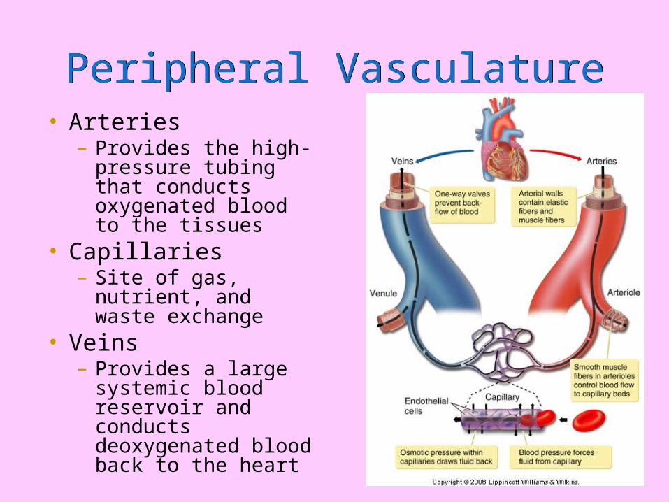
Peripheral VasculaturePeripheral Vasculature• Arteries
– Provides the high-pressure tubing that conducts oxygenated blood to the tissues
• Capillaries– Site of gas, nutrient,
and waste exchange• Veins
– Provides a large systemic blood reservoir and conducts deoxygenated blood back to the heart

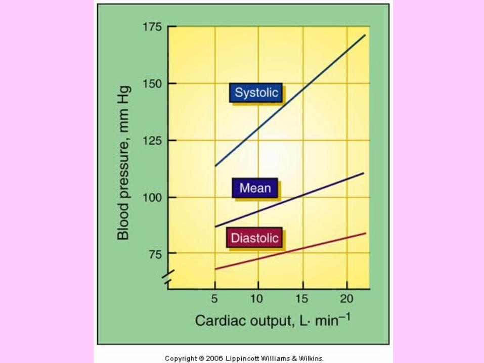
Blood Pressure Blood Pressure
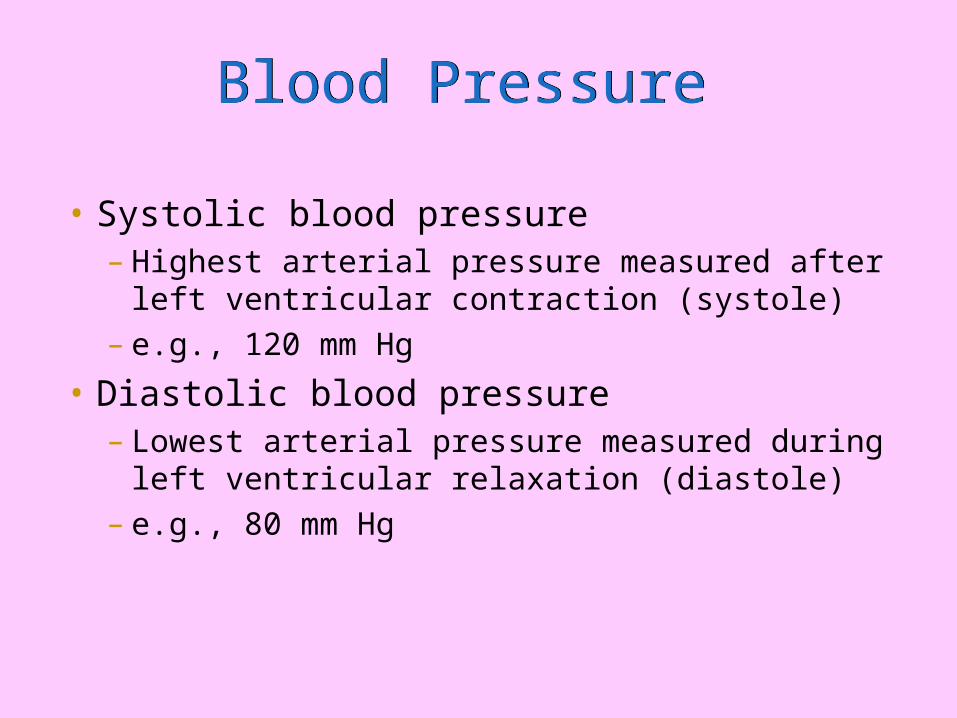
• Systolic blood pressure– Highest arterial pressure measured after left
ventricular contraction (systole)– e.g., 120 mm Hg
• Diastolic blood pressure– Lowest arterial pressure measured during
left ventricular relaxation (diastole)– e.g., 80 mm Hg
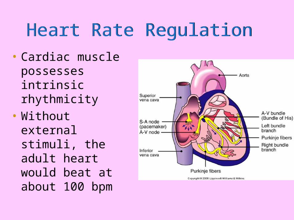
Heart Rate Regulation Heart Rate Regulation
• Cardiac muscle possesses intrinsic rhythmicity
• Without external stimuli, the adult heart would beat at about 100 bpm
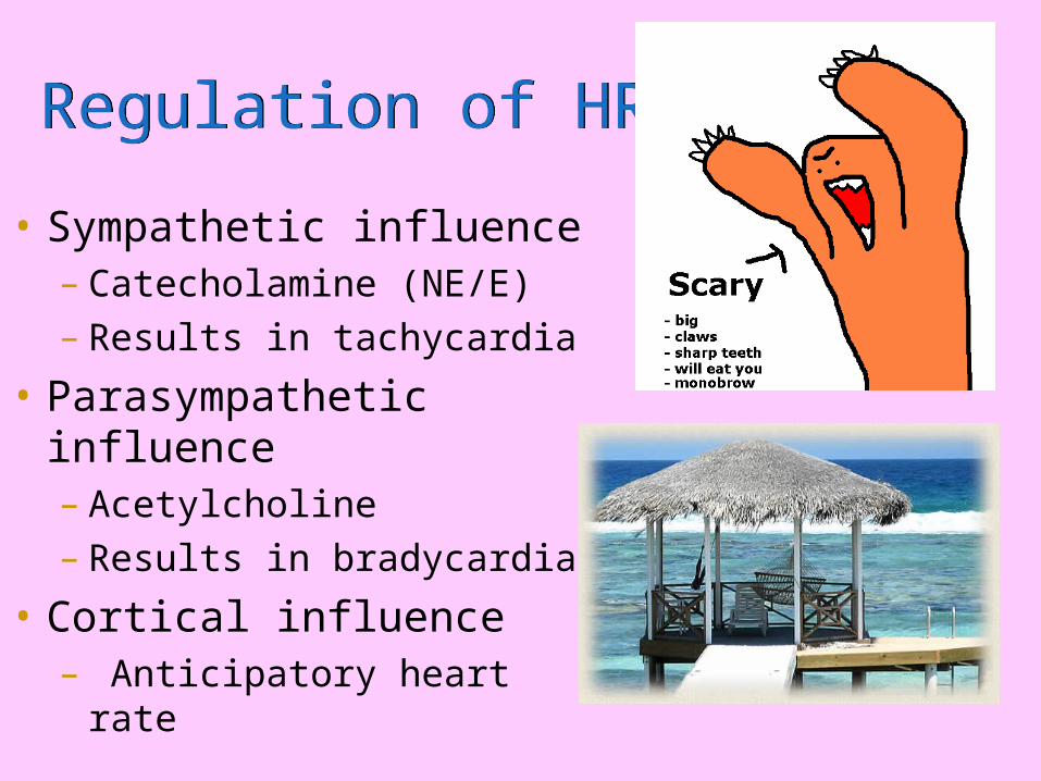
Regulation of HRRegulation of HR
• Sympathetic influence – Catecholamine (NE/E)– Results in tachycardia
• Parasympathetic influence– Acetylcholine – Results in bradycardia
• Cortical influence– Anticipatory heart rate
CV system during exercise
Acute Adaptations
Chronic adaptations
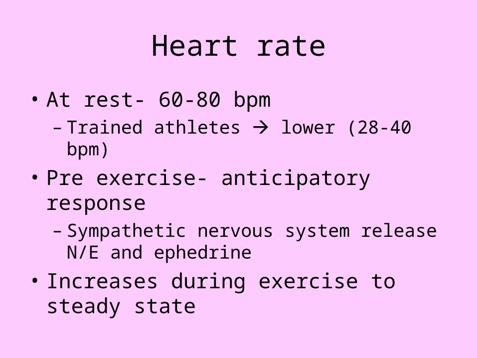
Heart rate
• At rest- 60-80 bpm– Trained athletes lower (28-40 bpm)
• Pre exercise- anticipatory response– Sympathetic nervous system release N/E and
ephedrine
• Increases during exercise to steady state
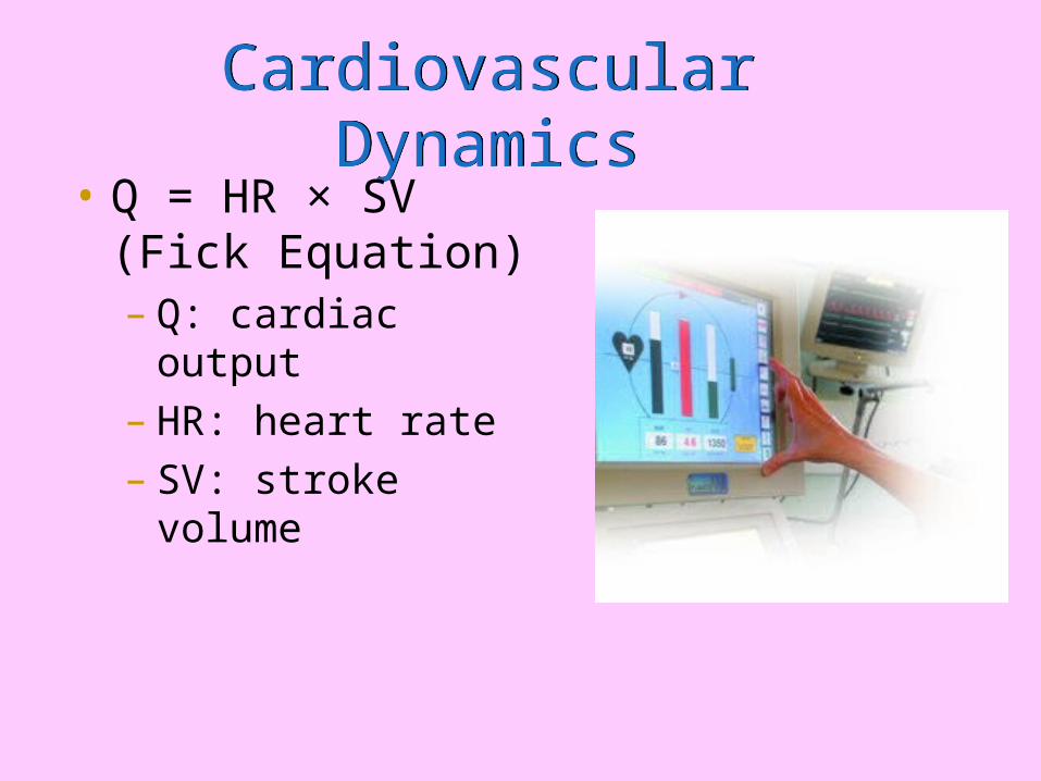
Cardiovascular DynamicsCardiovascular Dynamics• Q = HR × SV (Fick
Equation)– Q: cardiac output– HR: heart rate– SV: stroke volume
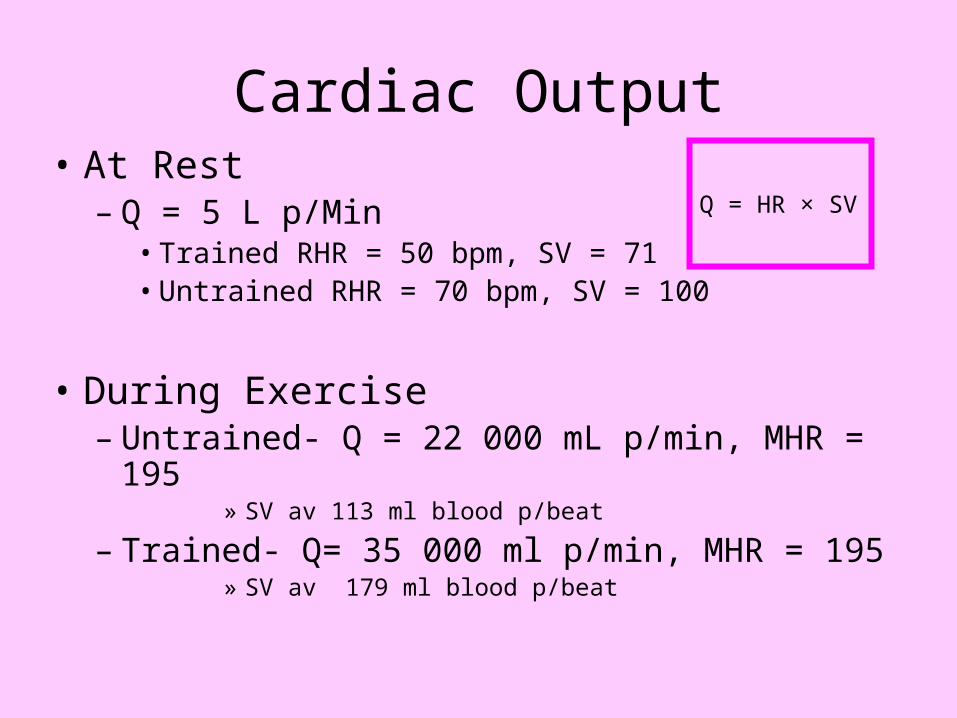
Cardiac Output• At Rest
– Q = 5 L p/Min• Trained RHR = 50 bpm, SV = 71• Untrained RHR = 70 bpm, SV = 100
• During Exercise– Untrained- Q = 22 000 mL p/min, MHR = 195
» SV av 113 ml blood p/beat
– Trained- Q= 35 000 ml p/min, MHR = 195» SV av 179 ml blood p/beat
Q = HR × SV
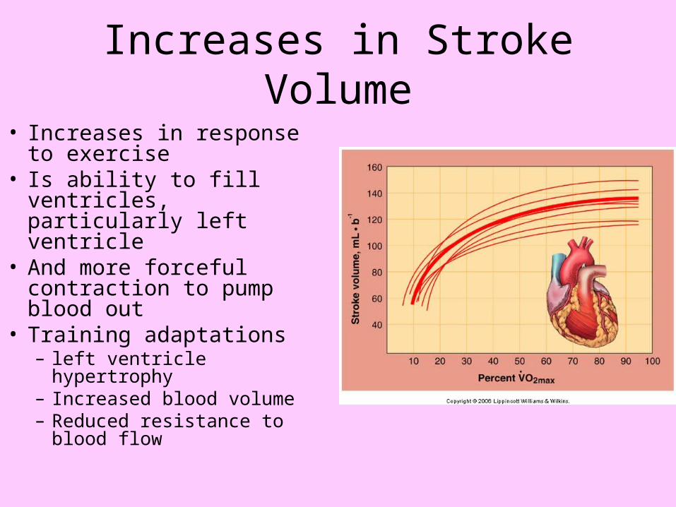
Increases in Stroke Volume
• Increases in response to exercise
• Is ability to fill ventricles, particularly left ventricle
• And more forceful contraction to pump blood out
• Training adaptations– left ventricle hypertrophy– Increased blood volume– Reduced resistance to
blood flow

Training Adaptations: Heart
• Eccentric hypertrophy – Slight thickening in left
ventricle walls– Increases left ventricular
cavity size
Therefore increases stroke
volume
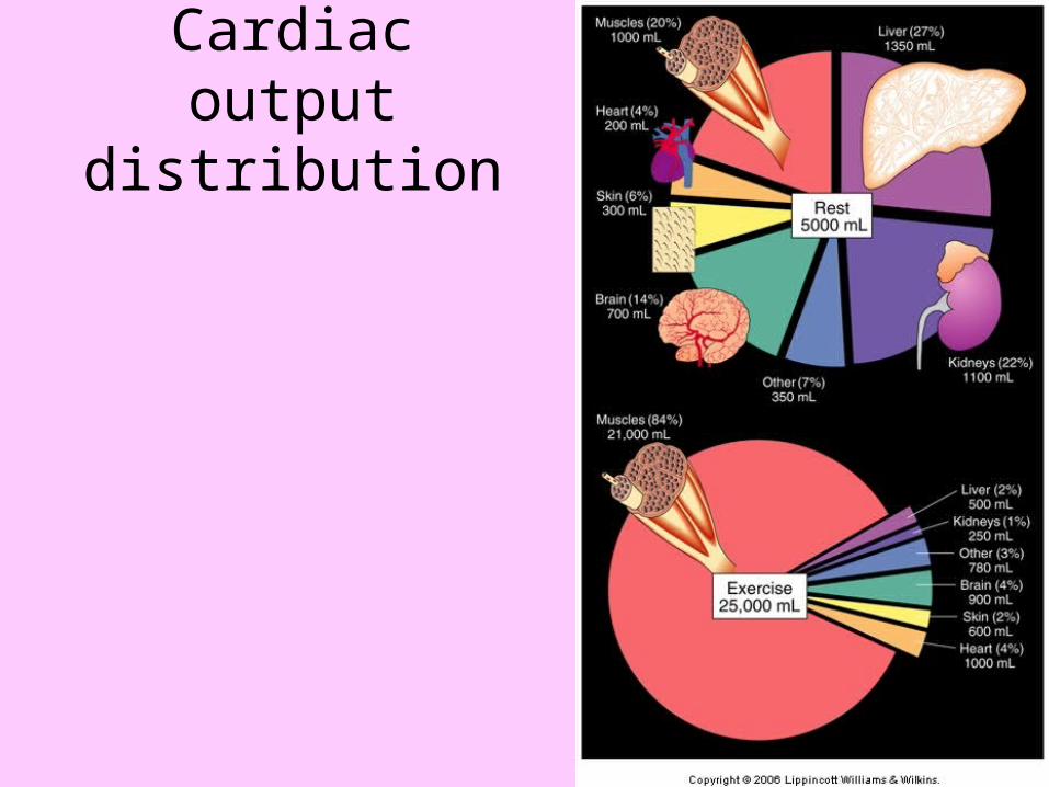
Cardiac output distribution
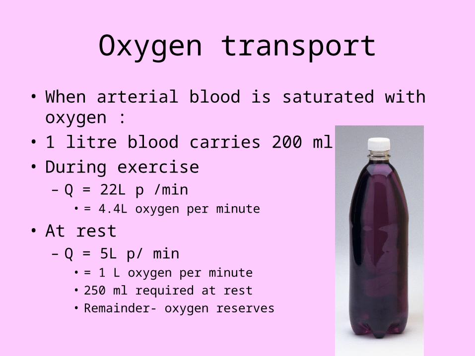
Oxygen transport
• When arterial blood is saturated with oxygen :• 1 litre blood carries 200 ml oxygen • During exercise
– Q = 22L p /min• = 4.4L oxygen per minute
• At rest– Q = 5L p/ min
• = 1 L oxygen per minute• 250 ml required at rest• Remainder- oxygen reserves
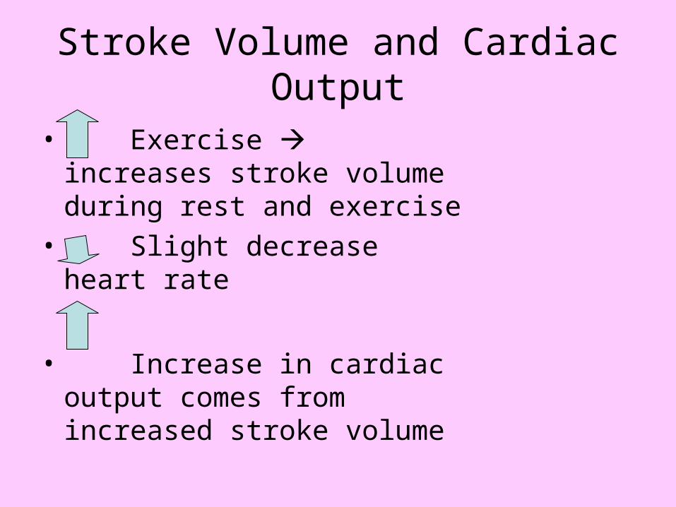
Stroke Volume and Cardiac Output
• Exercise increases stroke volume during rest and exercise
• Slight decrease heart rate
• Increase in cardiac output comes from increased stroke volume
Heart Rate
• Elite athletes have a lower heart rate relative to training intensity than sedentary people
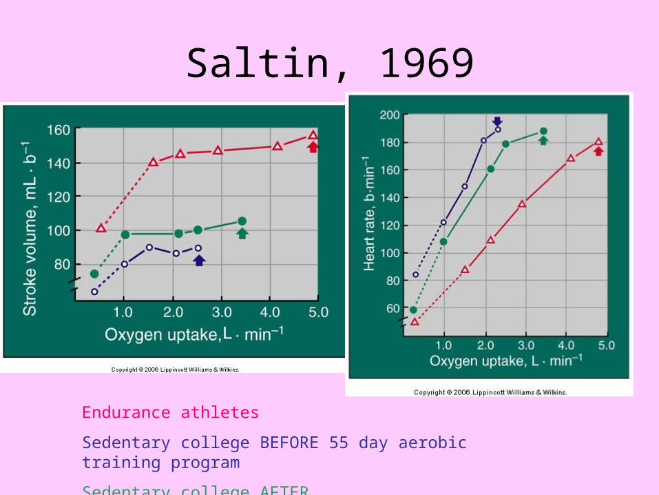
Saltin, 1969
Endurance athletes
Sedentary college BEFORE 55 day aerobic training program
Sedentary college AFTER
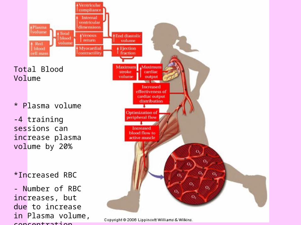
Total Blood Volume
* Plasma volume
-4 training sessions can increase plasma volume by 20%
*Increased RBC
- Number of RBC increases, but due to increase in Plasma volume, concentration stays the same
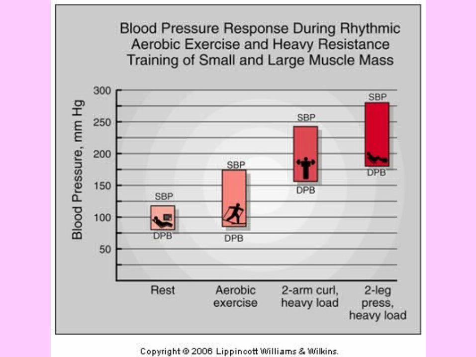
Blood Pressure
• Aerobic exercise reduces systolic and diastolic BP at rest and during exercise
• Particularly systolic– Caused by decrease in catecholamines
• Another reason for exercise to be prescribed for those with hypertension
• Resistance training not recommended due to acute high BP it causes
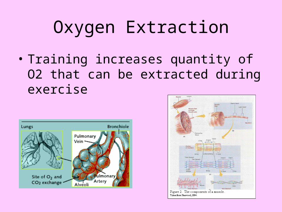
Oxygen Extraction
• Training increases quantity of O2 that can be extracted during exercise
Chronic Adaptations to Exercise- Chapter 10
Cardiovascular adaptations to training are extremely important for improving endurance exercise performance, and preventing cardiovascular diseases.
The more important of these adaptations are, Size of heart ventricular volumes total blood volume
- plasma volume - red cell mass
systolic and diastolic blood pressures maximal stroke volume maximal cardiac output extraction of oxygen
Factors Affecting Chronic adaptations
• Initial CV fitness• Training:
– Frequency- 3 x p/week• Only slightly higher gains for 4 or 5 times p/week
– Intensity• Most critical• Minimum is 130/ 140 bpm = (av) 50-55% Vo2 max/ 70% HR max • Higher = better
– Time • Or duration- 30 min is minimum
– Type• Specificity
Pulmonary System
Pulmonary Structure and Function
Pulmonary Structure and Function
• The ventilatory system– Supplies oxygen required in metabolism – Eliminates carbon dioxide produced in
metabolism – Regulates hydrogen ion concentration [H+]
to maintain acid-base balance
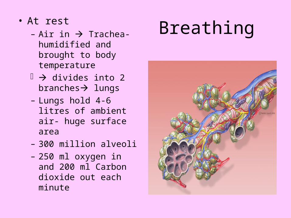
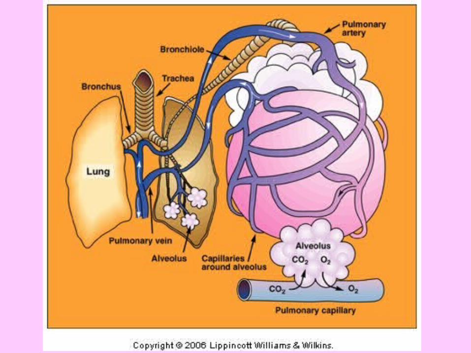
Breathing• At rest– Air in Trachea-
humidified and brought to body temperature
divides into 2 branches lungs
– Lungs hold 4-6 litres of ambient air- huge surface area
– 300 million alveoli– 250 ml oxygen in and
200 ml Carbon dioxide out each minute
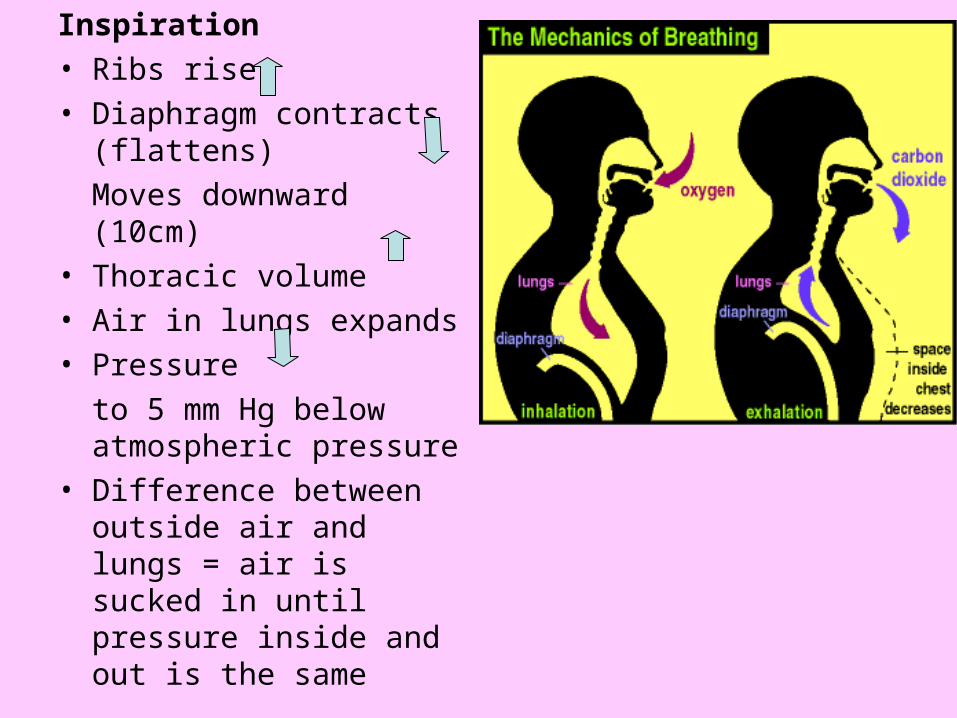
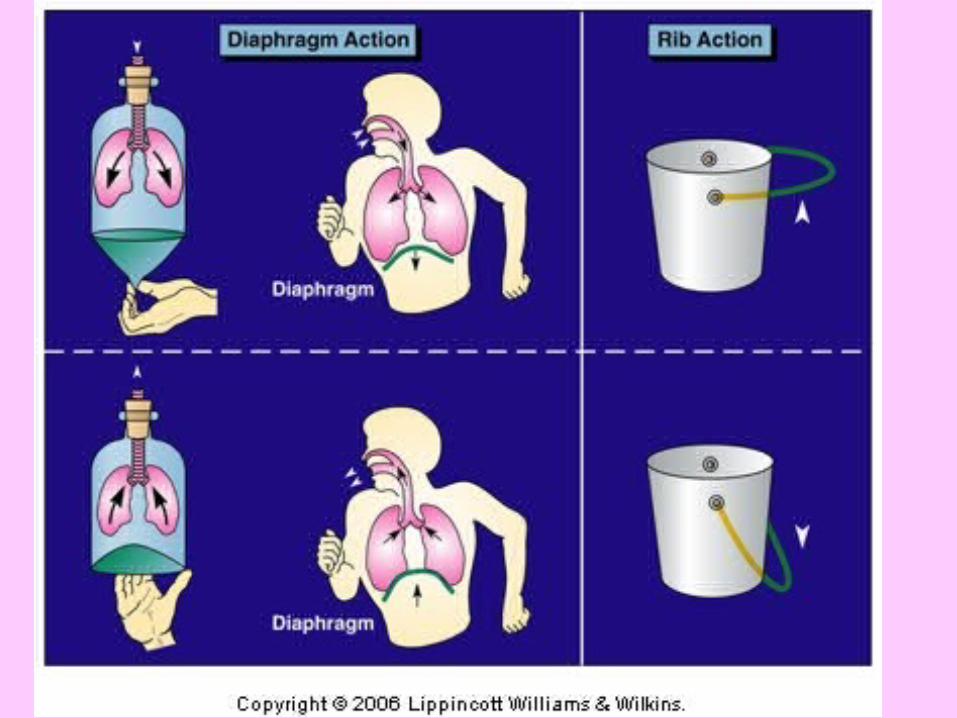
Inspiration• Ribs rise• Diaphragm contracts
(flattens)
Moves downward (10cm)• Thoracic volume• Air in lungs expands • Pressure
to 5 mm Hg below atmospheric pressure
• Difference between outside air and lungs = air is sucked in until pressure inside and out is the same

Expiration
• Ribs move back down• Diaphragm relaxes (rises)• Thoracic volume• Pressure • Difference between outside air and lungs = air
is pushed out until pressure inside and out is the same
Pulmonary system during exercise

Lung VolumesLung Volumes
• Static lung volume tests– Evaluate the dimensional component for
air movement within the pulmonary tract, and impose no time limitation on the subject
• Dynamic lung volume tests– Evaluate the power component of
pulmonary performance during different phases of the ventilatory excursion
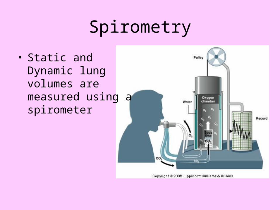
Spirometry
• Static and Dynamic lung volumes are measured using a spirometer
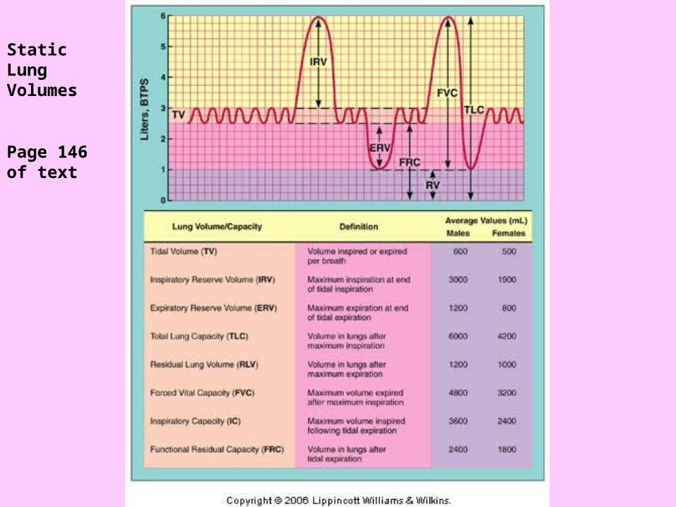
Static Lung Volumes
Page 146 of text
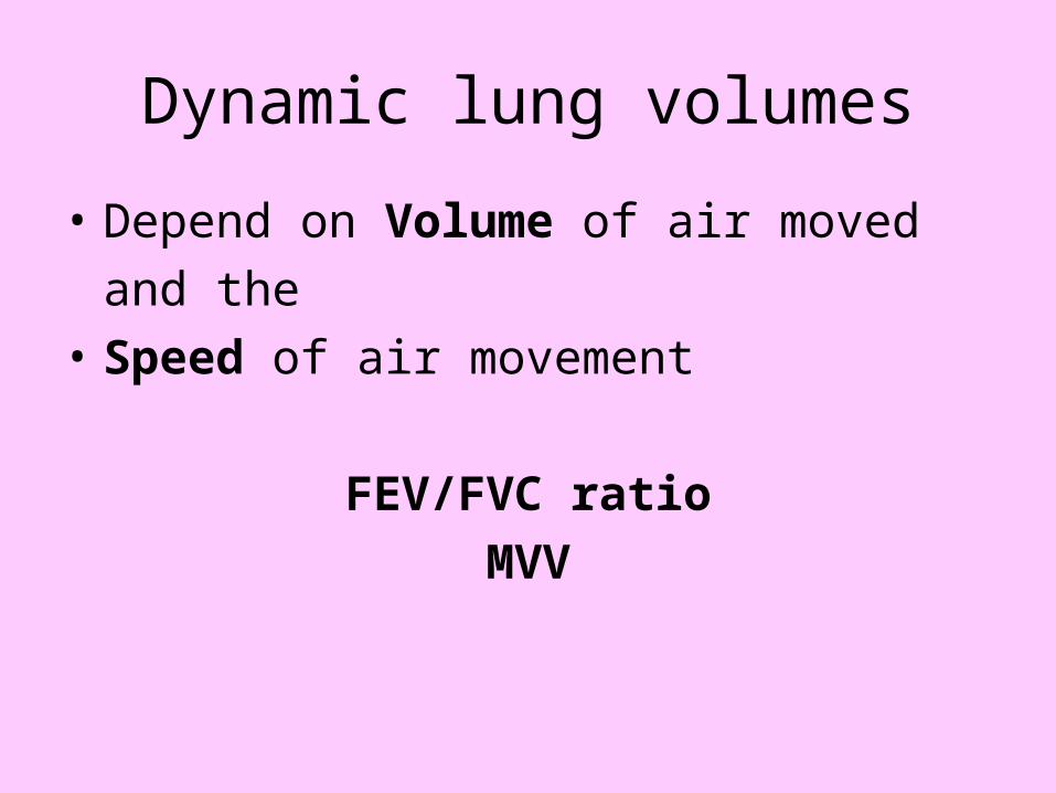
Dynamic lung volumes
• Depend on Volume of air moved
and the
• Speed of air movement
FEV/FVC ratio
MVV
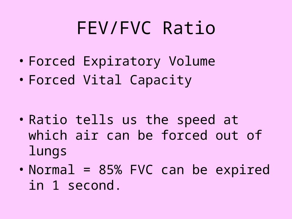
FEV/FVC Ratio
• Forced Expiratory Volume
• Forced Vital Capacity
• Ratio tells us the speed at which air can be forced out of lungs
• Normal = 85% FVC can be expired in 1 second.
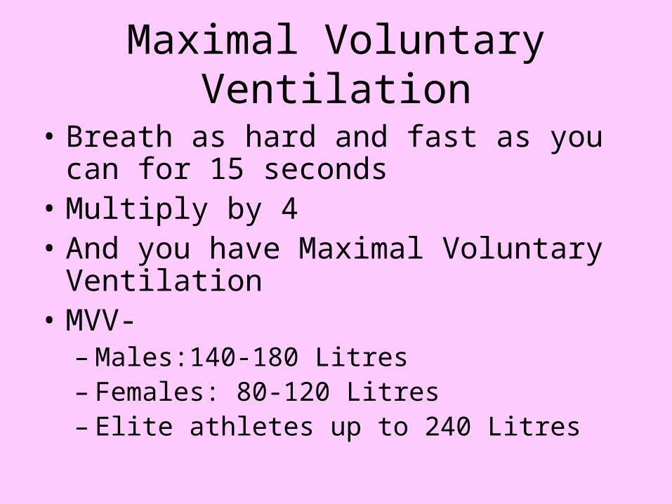
Maximal Voluntary Ventilation
• Breath as hard and fast as you can for 15 seconds
• Multiply by 4• And you have Maximal Voluntary
Ventilation• MVV-
– Males:140-180 Litres– Females: 80-120 Litres– Elite athletes up to 240 Litres
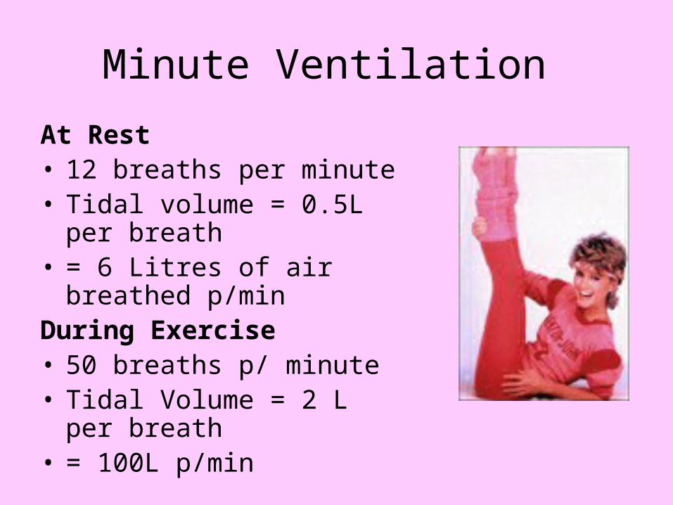
Minute Ventilation
At Rest• 12 breaths per minute• Tidal volume = 0.5L per
breath• = 6 Litres of air breathed
p/minDuring Exercise• 50 breaths p/ minute• Tidal Volume = 2 L per
breath• = 100L p/min

Alveolar VentilationAlveolar Ventilation
• Minute ventilation is just total amount of air
• Alveolar ventilation refers to the portion of minute ventilation that mixes with the air in the alveolar chambers
• Minute ventilation minus anatomical dead space (150-200 ml)- the air that is in the trachea, bronchi etc
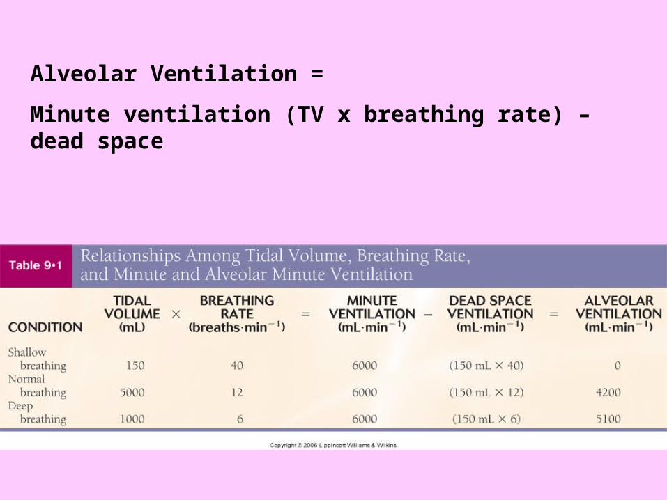
Alveolar Ventilation =
Minute ventilation (TV x breathing rate) – dead space

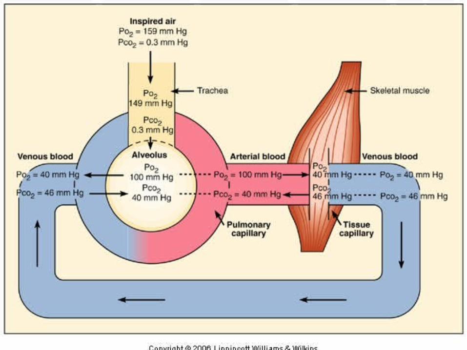
Gas exchange

Gas Exchange in the Body Gas Exchange in the Body
• The exchange of gases between the lungs and blood, and their movement at the tissue level, takes place passively by diffusion
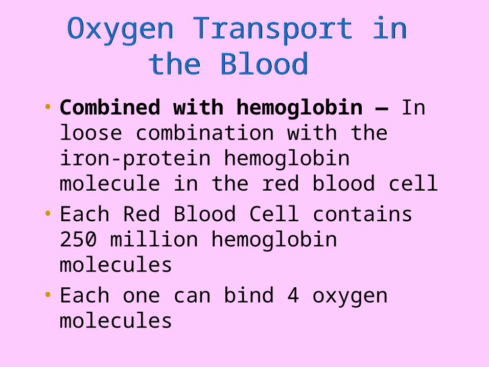
Oxygen Transport in the Blood Oxygen Transport in the Blood
• Combined with hemoglobin — In loose combination with the iron-protein hemoglobin molecule in the red blood cell
• Each Red Blood Cell contains 250 million hemoglobin molecules
• Each one can bind 4 oxygen molecules
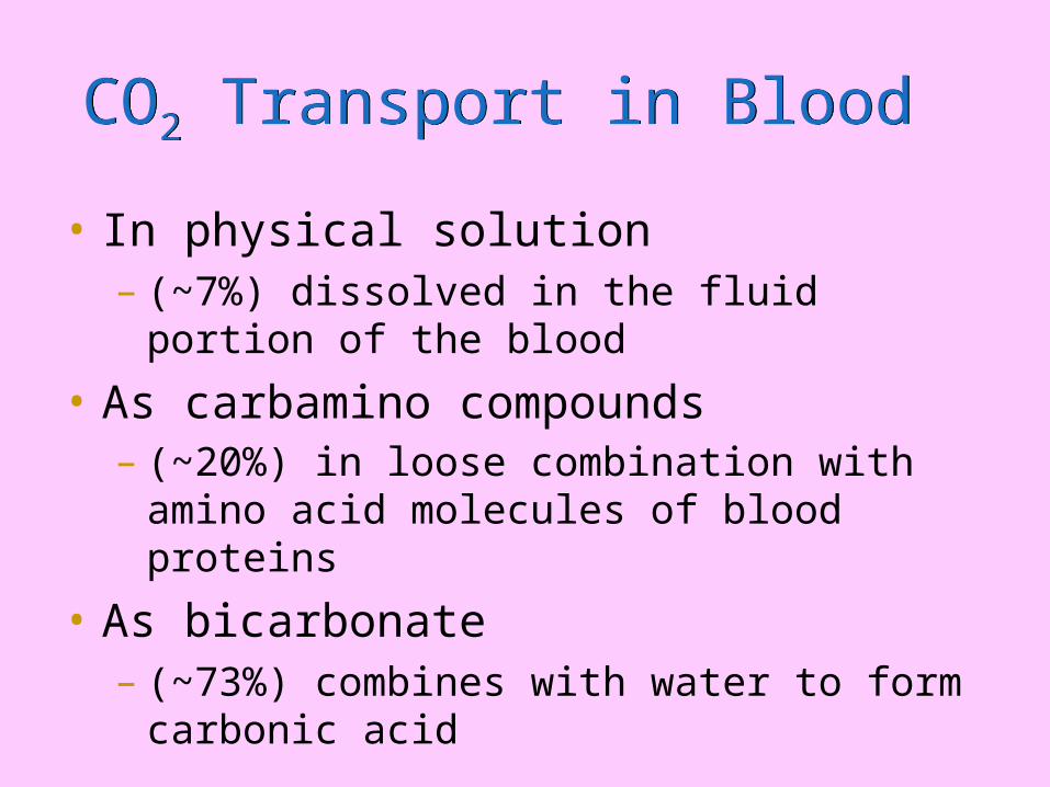
CO2 Transport in Blood CO2 Transport in Blood
• In physical solution– (~7%) dissolved in the fluid portion of the
blood
• As carbamino compounds – (~20%) in loose combination with amino acid
molecules of blood proteins
• As bicarbonate– (~73%) combines with water to form carbonic
acid
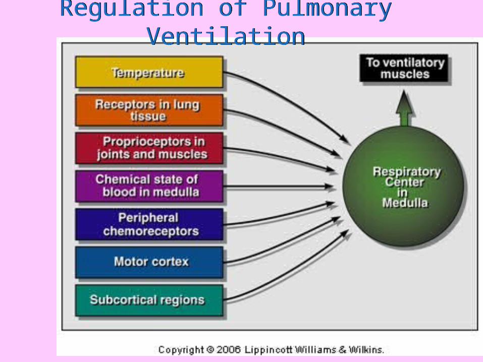
Regulation of Pulmonary VentilationRegulation of Pulmonary Ventilation
Regulation at rest: Plasma Pco2 and H+ Concentration
Regulation at rest: Plasma Pco2 and H+ Concentration
• The partial pressure of CO2 provides the most potent respiratory stimulus at rest
• [H+] in the cerebrospinal fluid bathing the central chemoreceptors provides a secondary stimulus driving inspiration
Ventilatory Regulation During Exercise
Ventilatory Regulation During Exercise
• Chemical control– Po2
– Pco2
– [H+]
• Nonchemical control
• Neurogenic factors– Cortical influence– Peripheral influence
Ventilation in steady rate exercise
Ventilation in steady rate exercise
• Of oxygen ( V E/ V O2)
– Quantity of air breathed per amount of oxygen consumed
– Remains relatively constant during steady-rate exercise- 25 L air breathed per 1L o2 consumed at 55% Vo2 max
• Of carbon dioxide ( V E/ V CO2)
– Remains relatively constant during steady-rate exercise
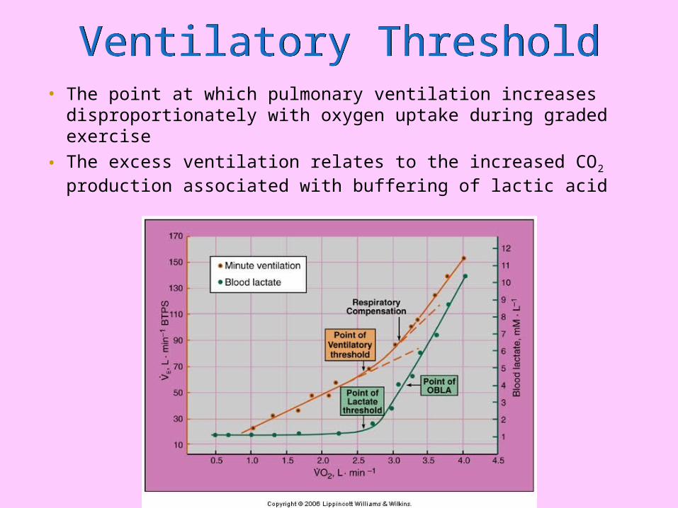
• The point at which pulmonary ventilation increases disproportionately with oxygen uptake during graded exercise
• The excess ventilation relates to the increased CO2 production associated with buffering of lactic acid
Ventilatory ThresholdVentilatory Threshold
Pulmonary adaptations to Exercise
Adaptations to
Maximal exercise
• Minute ventilation increases
• Increased oxygen uptake

Submaximal Exercise
• Ventilatory muscles stronger
• Ventilatory equivalent for oxygen
( V E/ V O2) reduces indicates breathing efficiency– This leads to
• Reduced fatigue in ventilatory muscles• O2 that would have been used by those muscles
can be used by skeletal muscle.
Pulmonary Adaptations
• Increased tidal volume
• Decreased breathing frequency
• Increased time between breaths (Increased time for oxygen to get into bloodstream)
• Therefore less oxygen in exhaled air
Summary
• Need to know– Cardiac and pulmonary Structure and
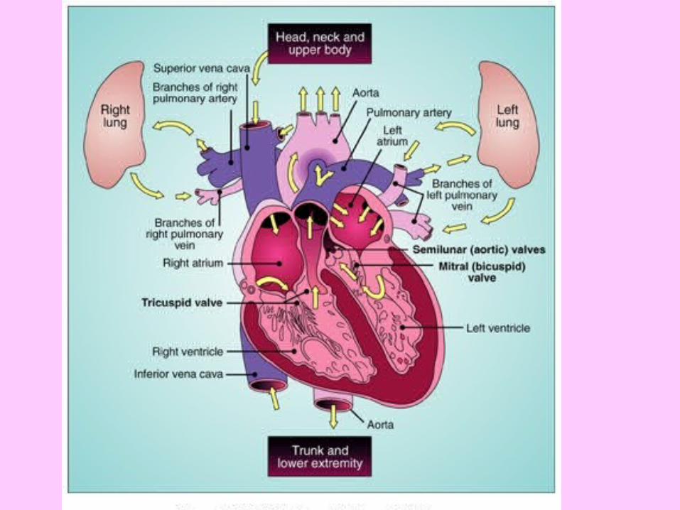
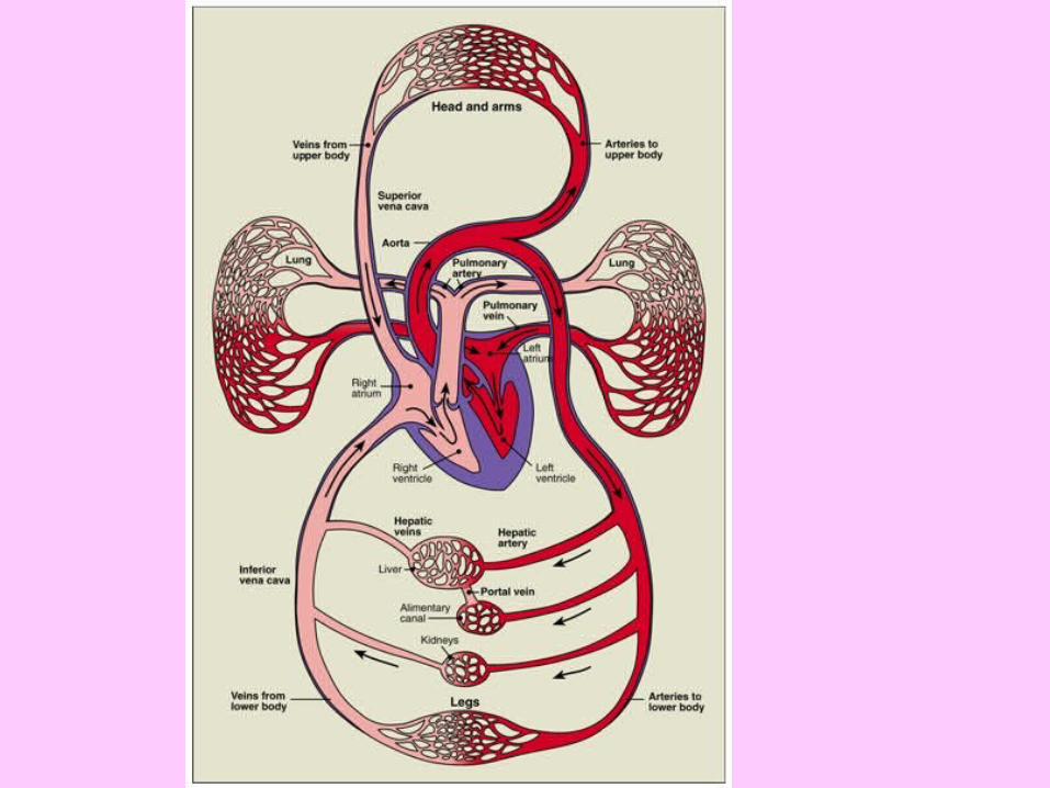
Function• Veins/arteries/cappilaries• Flow of blood through the heart• Alveoli bronchii etc• Flow of inspired air and pulmonary exchange
– Acute adaptations to exercise– Chronic adaptations to exercise