case of back pain 53 year old, right handed lady, hotelier 53 year old, right handed lady, hotelier...
TRANSCRIPT

Case of Back PainCase of Back Pain
53 year old, right handed lady, hotelier 53 year old, right handed lady, hotelier 3 day history of severe lower back pain 3 day history of severe lower back pain
and weakness in her legsand weakness in her legs bending over at work and had noticed a bending over at work and had noticed a
mild back pain, which progressed mild back pain, which progressed Night and rest pain, leg radiation, worse Night and rest pain, leg radiation, worse
with movement. Unable to walkwith movement. Unable to walk

Case of Back PainCase of Back Pain
Sep 05Haematologists shoulder Sep 05Haematologists shoulder pains, lymphadenopathy and rash, pains, lymphadenopathy and rash, fatigue, 7 kg weight loss in 6 monthsfatigue, 7 kg weight loss in 6 months
l-node < 1cm ALP 210 Rheum l-node < 1cm ALP 210 Rheum referralreferral
Subsequently admitted Subsequently admitted Ex In pain restricted spine ? leg Ex In pain restricted spine ? leg
weakness and altered sensation feet weakness and altered sensation feet
Case of Back PainCase of Back Pain
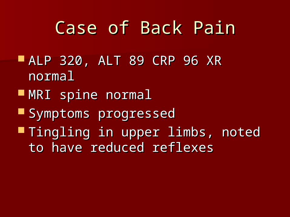
ALP 320, ALT 89 CRP 96 XR normalALP 320, ALT 89 CRP 96 XR normal MRI spine normalMRI spine normal Symptoms progressed Symptoms progressed Tingling in upper limbs, noted to Tingling in upper limbs, noted to
have reduced reflexes have reduced reflexes

Case of Back PainCase of Back Pain
CSF protein 2.55 gCSF protein 2.55 g ?Guillan-Barre?Guillan-Barre Transferred to neurologyTransferred to neurology IV Ig, Rehab, FVC, vitals monitoringIV Ig, Rehab, FVC, vitals monitoring Campylobacter IgG and IgA 160Campylobacter IgG and IgA 160 EBV +ve EBV +ve
GB syndromeGB syndrome
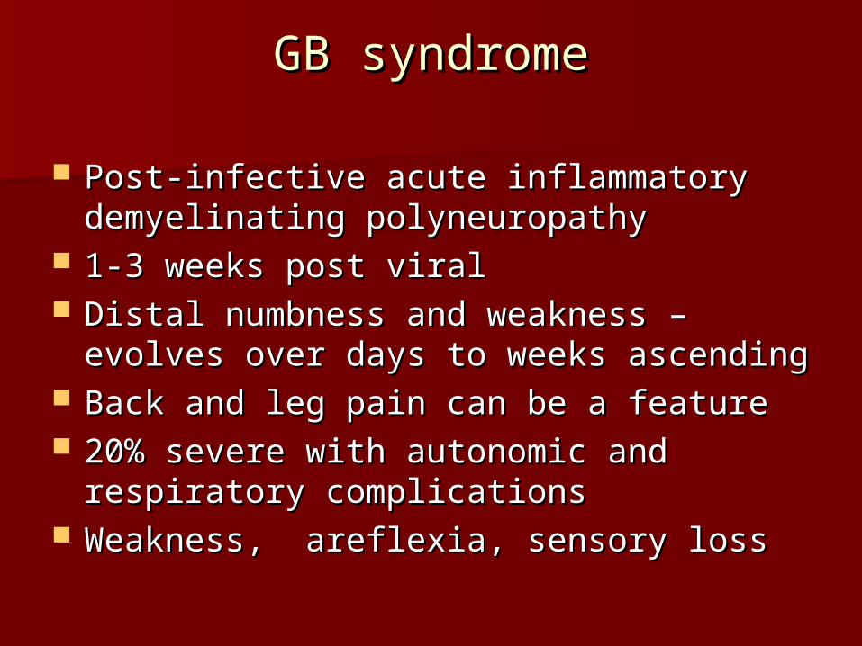
Post-infective acute inflammatory Post-infective acute inflammatory demyelinating polyneuropathydemyelinating polyneuropathy
1-3 weeks post viral1-3 weeks post viral Distal numbness and weakness – evolves Distal numbness and weakness – evolves
over days to weeks ascending over days to weeks ascending Back and leg pain can be a featureBack and leg pain can be a feature 20% severe with autonomic and 20% severe with autonomic and
respiratory complicationsrespiratory complications Weakness, areflexia, sensory loss Weakness, areflexia, sensory loss

GB syndromeGB syndrome
Rare – ocular and ataxia – Miller-Rare – ocular and ataxia – Miller-Fisher syndromeFisher syndrome
NCS: slowing of conduction or blockNCS: slowing of conduction or block CSF: 1-3g/lCSF: 1-3g/l IV Ig, supportive, ventilation, IV Ig, supportive, ventilation,
plasmapharesis, rehab plasmapharesis, rehab

BACK PAINBACK PAIN
Jaya RavindranJaya Ravindran
RheumatologistRheumatologist

CausesCauses
Simple mechanical eg ligamentous strainSimple mechanical eg ligamentous strain Degenerative disease with/without neural, Degenerative disease with/without neural,
cord or canal compromisecord or canal compromise Metabolic – osteoporosis, Pagets Metabolic – osteoporosis, Pagets Inflammatory – Ankylosing spondylitisInflammatory – Ankylosing spondylitis Infective – bacterial and TBInfective – bacterial and TB NeoplasticNeoplastic Others, (trauma,congenital)Others, (trauma,congenital) VisceralVisceral

Red flagsRed flags
– Age <20 or >50 with back pain Age <20 or >50 with back pain for the 1for the 1stst time time
– Thoracic pain >50 yrsThoracic pain >50 yrs- Pain following a violent Pain following a violent
injury/traumainjury/trauma- Unremitting, progressive painUnremitting, progressive pain

Red flagsRed flags
- Past or current history of cancerPast or current history of cancer- On Steroids or On Steroids or
immunosuppressantsimmunosuppressants- Drug abuser or +ve HIVDrug abuser or +ve HIV- Systemic symptoms - fever, Systemic symptoms - fever,
appetitie and weight loss, malaise appetitie and weight loss, malaise

Red flagsRed flags
- Bilateral leg radiation, Bilateral leg radiation, sensory/motor/sphincter symptoms sensory/motor/sphincter symptoms
- Pain predominantly at nightPain predominantly at night

Inflammatory flagsInflammatory flags
- Morning stiffness and pain >30 mins -1 hrMorning stiffness and pain >30 mins -1 hr- Better with activityBetter with activity- Peripheral joint involvementPeripheral joint involvement- Anterior uveitisAnterior uveitis- PsoriasisPsoriasis- Inflammatory bowel diseaseInflammatory bowel disease- Recent GI or GU infectionRecent GI or GU infection- Family historyFamily history

MyotomesMyotomes
C5 Deltoid, biceps (biceps jerk)C5 Deltoid, biceps (biceps jerk) C6 Wrist extensors, biceps (biceps, C6 Wrist extensors, biceps (biceps,
brachioradialis jerk) brachioradialis jerk) C7 Wrist flexors, finger extensors, C7 Wrist flexors, finger extensors,
triceps (triceps jerk)triceps (triceps jerk) C8 Finger flexor, thumb extensors C8 Finger flexor, thumb extensors
(triceps jerk)(triceps jerk) T1 finger abductorsT1 finger abductors
MyotomesMyotomes
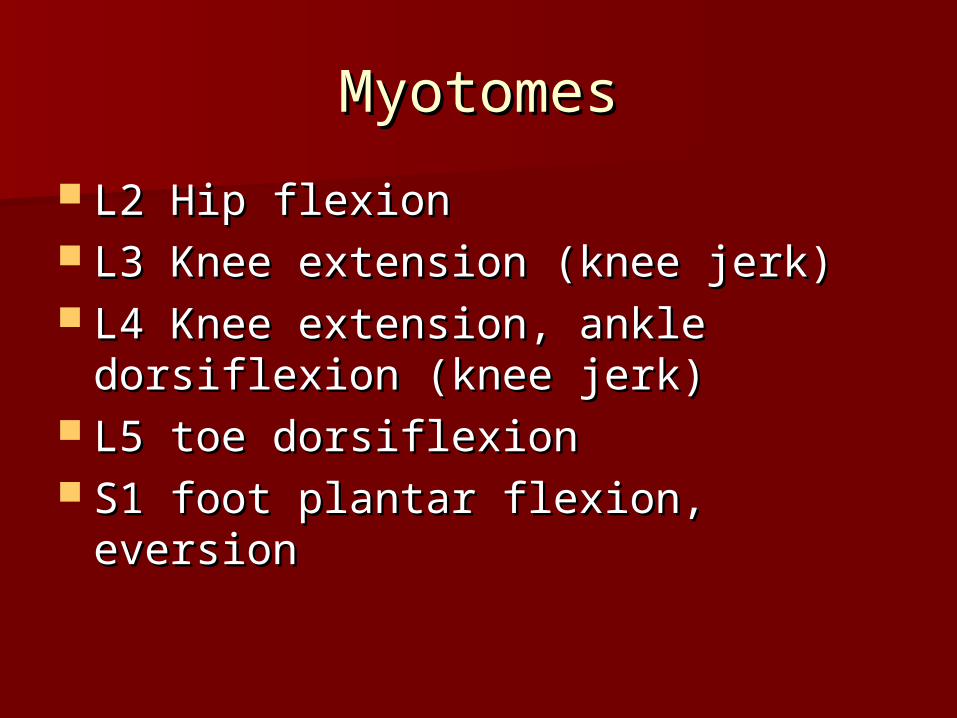
L2 Hip flexionL2 Hip flexion L3 Knee extension (knee jerk)L3 Knee extension (knee jerk) L4 Knee extension, ankle dorsiflexion L4 Knee extension, ankle dorsiflexion
(knee jerk)(knee jerk) L5 toe dorsiflexionL5 toe dorsiflexion S1 foot plantar flexion, eversionS1 foot plantar flexion, eversion

DDEERRMMAATTOOMMEESS
ExaminationExamination
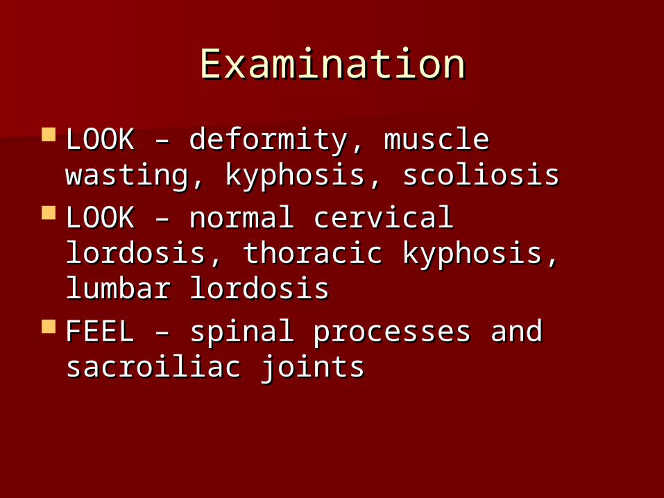
LOOK – deformity, muscle wasting, LOOK – deformity, muscle wasting, kyphosis, scoliosiskyphosis, scoliosis
LOOK – normal cervical lordosis, LOOK – normal cervical lordosis, thoracic kyphosis, lumbar lordosisthoracic kyphosis, lumbar lordosis
FEEL – spinal processes and FEEL – spinal processes and sacroiliac jointssacroiliac joints

ExaminationExamination
MOVE – Lumbar flexion MOVE – Lumbar flexion Schober’s test – marks at “dimples Schober’s test – marks at “dimples
of Venus” and 10 cm above. Measure of Venus” and 10 cm above. Measure at maximal flexion – usually 5 cm at maximal flexion – usually 5 cm
MOVE – Lumbar lateral flexionMOVE – Lumbar lateral flexion MOVE – Cervical flexion/extension, MOVE – Cervical flexion/extension,
lateral rotation and flexion, thoracic lateral rotation and flexion, thoracic rotation rotation

ExaminationExamination
Sciatic stretch (patient supine) - Sciatic stretch (patient supine) - Straight leg raise and dorsiflexion of Straight leg raise and dorsiflexion of foot - pain in calf and posterior thigh foot - pain in calf and posterior thigh between 30-70between 30-70o o – low lumbar (L5/S1) – low lumbar (L5/S1) lesion or sciatic irritationlesion or sciatic irritation
Femoral stretch (patient prone) – Femoral stretch (patient prone) – knee is flexed and then hip extended knee is flexed and then hip extended – pain in anterior thigh – high lumbar – pain in anterior thigh – high lumbar (L2-L4) lesion(L2-L4) lesion

ImagingImaging
XR – tumour, fracture, infection, XR – tumour, fracture, infection, inflammationinflammation
Bone scan – increased turnover eg Bone scan – increased turnover eg infection, metastatic disease, infection, metastatic disease, fractures, Pagetsfractures, Pagets
MRI – soft tissue, discs, facet joint, MRI – soft tissue, discs, facet joint, nerve roots, cord, inflammationnerve roots, cord, inflammation

Degenerative disease and Degenerative disease and sciaticasciatica
Very commonVery common Facet joint OA, disc disease, Facet joint OA, disc disease,
osteophyteosteophyte Mechanical back painMechanical back pain Sciatica – most resolve NB persistent, Sciatica – most resolve NB persistent,
neurology, bilateral, red flagsneurology, bilateral, red flags Analgesia, PT, pain clinicsAnalgesia, PT, pain clinics

Degenerative disease and Degenerative disease and sciaticasciatica

MalignancyMalignancy
Unremittting, progressive and night painUnremittting, progressive and night pain Systemic symtomsSystemic symtoms Past hx CaPast hx Ca Breast, bronchus, thyroid, kidney, prostate Breast, bronchus, thyroid, kidney, prostate
and myeloma/plasmacytoma and myeloma/plasmacytoma Osteolytic (prostate osteoblastic)Osteolytic (prostate osteoblastic) XR can be normal in early stages – further XR can be normal in early stages – further
imaging if suspicion highimaging if suspicion high Predilection for vertebral body and pediclesPredilection for vertebral body and pedicles

Malignancy Malignancy
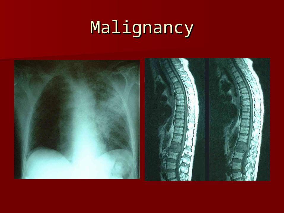
MalignancyMalignancy

InfectionInfection discitis, osteomyelitis, and epidural discitis, osteomyelitis, and epidural
abscess. abscess. hematogenously spread hematogenously spread most often Staphylococcus aureus.most often Staphylococcus aureus. Gram-negative rods in postoperative or Gram-negative rods in postoperative or
immunocompromised patientsimmunocompromised patients normal skin flora is less commonly isolated normal skin flora is less commonly isolated
in postoperative patients. in postoperative patients. Postoperative patients develop symptoms Postoperative patients develop symptoms
2 to 4 weeks after surgery after an initial 2 to 4 weeks after surgery after an initial improvement in pain. improvement in pain.

InfectionInfection Pseudomonas organisms in intravenous Pseudomonas organisms in intravenous
drug users. drug users. Mycobacterium tuberculosis in developing Mycobacterium tuberculosis in developing
nations and immigrant population. Fungal nations and immigrant population. Fungal infections are rare. infections are rare.
Only one third have fever and 3% to 15% Only one third have fever and 3% to 15% present with neurologic deficit.present with neurologic deficit.
Infections typically involve the Infections typically involve the intervertebral disc and vertebral body intervertebral disc and vertebral body endplateendplate

InfectionInfection Radiographic changes at 2 to 4 weeksRadiographic changes at 2 to 4 weeks bone scan can be positive as early as 2 bone scan can be positive as early as 2
days 75% specific. days 75% specific. MRI appearance is decreased T1- and MRI appearance is decreased T1- and
increased T2-weighted signal in the increased T2-weighted signal in the infected disk. Enhancement after infected disk. Enhancement after gadoliniumgadolinium
InfectionInfection Conservative treatment of antibiotics, rigid Conservative treatment of antibiotics, rigid
bracing to prevent deformity and control bracing to prevent deformity and control painpain
Surgery : neurologic deficit, presence of Surgery : neurologic deficit, presence of abscess, extensive bone loss with kyphosis abscess, extensive bone loss with kyphosis and instability, failure of blood work and and instability, failure of blood work and biopsy to isolate any organism, excision of biopsy to isolate any organism, excision of a sinus tract, or no response to a sinus tract, or no response to conservative treatment.conservative treatment.

InfectionInfection

InfectionInfection

OsteoporosisOsteoporosis

DEXADEXA

T scoresT scores

OsteoporosisOsteoporosis
Diagnostic Criteria for Osteoporosis Established by the World Health Organization Based on Comparison to Young Adult Mean Bone Density*
Normal
Bone density is within 1 SD of the young adult mean
Osteopenia
Bone density is within 1 to 2.5 SD below the young adult mean
Osteoporosis
Bone density is 2.5 SD or more below the young adult mean
Severe (established) osteoporosis
Bone density is more than 2.5 SD below the young adult mean and there has been one or more osteoporotic fractures
*One standard deviation (SD) represents about a 10% to 12% decline in bone density.
Low bone densityLow bone density
Differential Diagnosis of Low Bone Density
Osteoporosis
Primary
Secondary
Osteomalacia
Osteogenesis imperfecta
Marrow-based diseases (eg, myeloma, mastocytosis)

Osteoporosis - risksOsteoporosis - risks
History of low trauma # - colles, NOF, History of low trauma # - colles, NOF, vertebral, sacral or pelvic insufficiency vertebral, sacral or pelvic insufficiency
SteroidsSteroids Maternal history of NOF #Maternal history of NOF # Gonadal hormone deficiencyGonadal hormone deficiency Ca deficiencyCa deficiency Prolonged immobilityProlonged immobility Low BMILow BMI Alcohol and smoking Alcohol and smoking

Causes of low bone densityCauses of low bone density
Secondary Causes of Osteoporosis
Endocrine Neoplasm Congenital Miscellaneous
Hyperparathyroidism Multiple myeloma Osteogenesis Rheumatoid arthritis
Hyperthyroidism Lymphoma Homocystinuria Gastrectomy
Cushing’s syndrome Mastocytosis Gaucher’s disease Cirrhosis
Hypopituitarism Renal failure
Hyperprolactinemia Malabsorption (sprue)

Vertebral fracturesVertebral fractures

OsteoporosisOsteoporosis

Osteoporosis Osteoporosis
BisphosphonatesBisphosphonates SERMsSERMs StrontiumStrontium TeriparatideTeriparatide CalcitoninCalcitonin Lifestyle factorsLifestyle factors Ca and Vit DCa and Vit D

7-dehydrocholesterol sunlight cholecalciferol 7-dehydrocholesterol sunlight cholecalciferol (diet)(diet) liverliver
25-hydroxycholecalciferol 25-hydroxycholecalciferol kidney kidney 11-hydroxylase -hydroxylase 1,25-dihydroxycholecalciferol 1,25-dihydroxycholecalciferol (-)(-)
increased GI Ca2+ absorptionincreased GI Ca2+ absorption Ca2+Ca2+ Bone resorptionBone resorption
ThyroidThyroid (-)(-) Parathyroid Gland PTHParathyroid Gland PTH Renal Ca2+ Renal Ca2+
(-)(-) Calcitonin Calcitonin reabsorptionreabsorption

Spinal stenosisSpinal stenosis
Canal or foraminal narrowing with Canal or foraminal narrowing with possible subsequent neural possible subsequent neural compressioncompression
Cause Cause Ligamanetum flavum hypertrophyLigamanetum flavum hypertrophy, ,
facet joint hypertrophy, vertebral facet joint hypertrophy, vertebral body osteophytes, herniated discbody osteophytes, herniated disc
Rare: Pagets, AS, acromegalyRare: Pagets, AS, acromegaly

Spinal stenosisSpinal stenosis
SymptomsSymptoms– Age - >50Age - >50– Dull aching pain in the lower back and legsDull aching pain in the lower back and legs– Exertional leg pain/weakness/numbnessExertional leg pain/weakness/numbness– Symptoms relieved leaning forward, sitting or lyingSymptoms relieved leaning forward, sitting or lying
ExaminationExamination– May be normalMay be normal– Normal sensation and powerNormal sensation and power– Reflexes normal or slightly reducedReflexes normal or slightly reduced– Normal foot pulsesNormal foot pulses

Spinal stenosisSpinal stenosis
Spinal stenosisSpinal stenosis
Conservative – analgesics, NSAIDs, Conservative – analgesics, NSAIDs, PT, epiduralPT, epidural
Surgery – laminectomy Surgery – laminectomy (+arthrodesis)(+arthrodesis)

Cauda Equina SyndromeCauda Equina Syndrome
Back pain, lower limb weakness, saddle Back pain, lower limb weakness, saddle anaesthesia, sphincter disturbance, anaesthesia, sphincter disturbance, impotence impotence
Causes – usually disc, rarely tumour, Causes – usually disc, rarely tumour, abscess, advanced AS abscess, advanced AS
Diminished sensation L4 to S2 (sacral Diminished sensation L4 to S2 (sacral numbness), weakness ankle and plantar numbness), weakness ankle and plantar dorsiflexion, loss ankle jerks, urinary dorsiflexion, loss ankle jerks, urinary retention, loss anal toneretention, loss anal tone
Urgent MRI and surgical decompressionUrgent MRI and surgical decompression

Cauda Equina SyndromeCauda Equina Syndrome

PagetsPagets
PagetsPagets
Pain, deformityPain, deformity Skull, long bone, vertebra, pelvis, Skull, long bone, vertebra, pelvis,
near hipnear hip Neurologic compromiseNeurologic compromise Planned surgeryPlanned surgery ?ALP 2X ULN?ALP 2X ULN Rare: high output failureRare: high output failure

ASAS
The Concept of Spondyloarthropathy
Disease Subgroups
1. Ankylosing spondylitis
2. Reactive arthritis (Reiter’s syndrome)
3. Enteropathic arthritis
4. Psoriatic arthritis
5. Undifferentiated spondyloarthropathy
6. Juvenile spondyloarthropathy
All These Diseases Share Rheumatologic Features
• Sacroiliac and spinal (axial) involvement
• Enthesitis at long attachments of ligaments and tendons causing: Achilles tendonitis and plantar fasciitis, syndesmophyte formation (“bamboo spine”), sacroiliitis (due to a combination of enthesitis and synovitis), and periosteal reaction (“whiskering”) at gluteal tuberosity and other parts of pelvis and other sites
• Peripheral, often asymmetric, inflammatory arthritis and dactylitis (“sausage” digits)
Share Extra-articular Features
• Propensity to ocular inflammation (acute anterior uveitis conjunctivitis)
• Mucocutaneous lesions, variable for the subgroups
• Rare aortic incompetence or heart block
• Lack of association with rheumatoid factor and rheumatoid nodules
Share Genetic Predisposition
• Strong association with HLA-B27 gene
• Familial clustering

ASAS
NSAIDsNSAIDs Sulphasalazine – peripheral jointsSulphasalazine – peripheral joints PTPT Anti-TNFAnti-TNF

ASAS

ASAS

ASAS

THE ENDTHE END
THANK-YOUTHANK-YOU