cerebrovascular disease. section 1 general consideration cerebrovascular disease: any abnormality of...
TRANSCRIPT
Cerebrovascular DiseaseCerebrovascular Disease
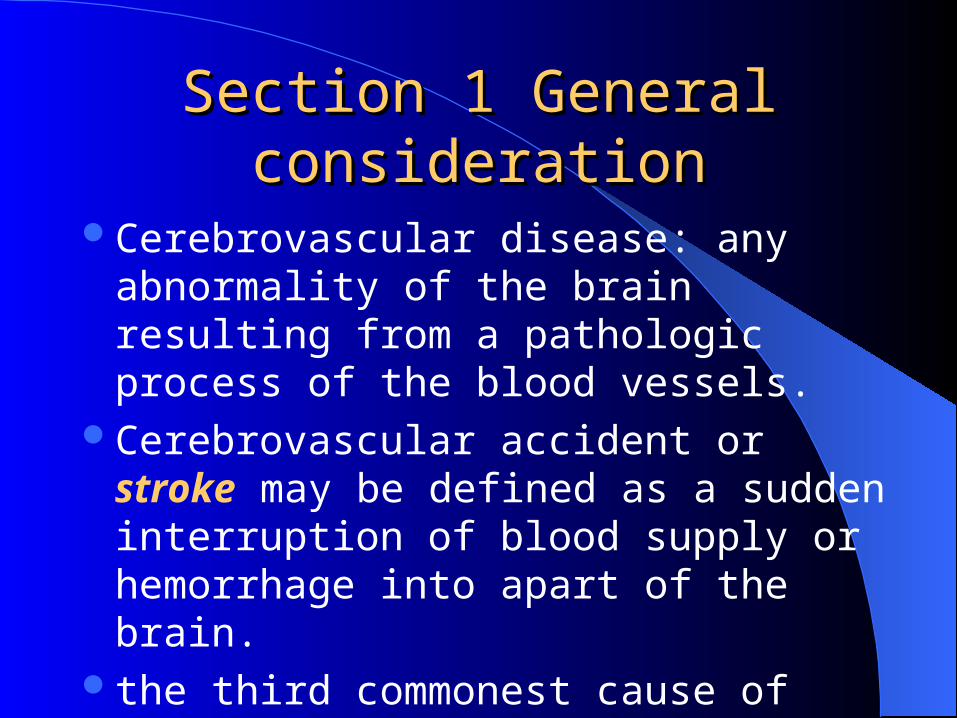
Section 1 General Section 1 General considerationconsideration
Cerebrovascular disease: any abnormality of the brain resulting from a pathologic process of the blood vessels.
Cerebrovascular accident or stroke may be defined as a sudden interruption of blood supply or hemorrhage into apart of the brain.
the third commonest cause of death
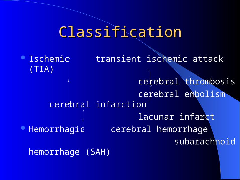
ClassificationClassification
Ischemic transient ischemic attack (TIA)
cerebral thrombosis
cerebral embolism cerebral infarction
lacunar infarct Hemorrhagic cerebral hemorrhage
subarachnoid hemorrhage (SAH)
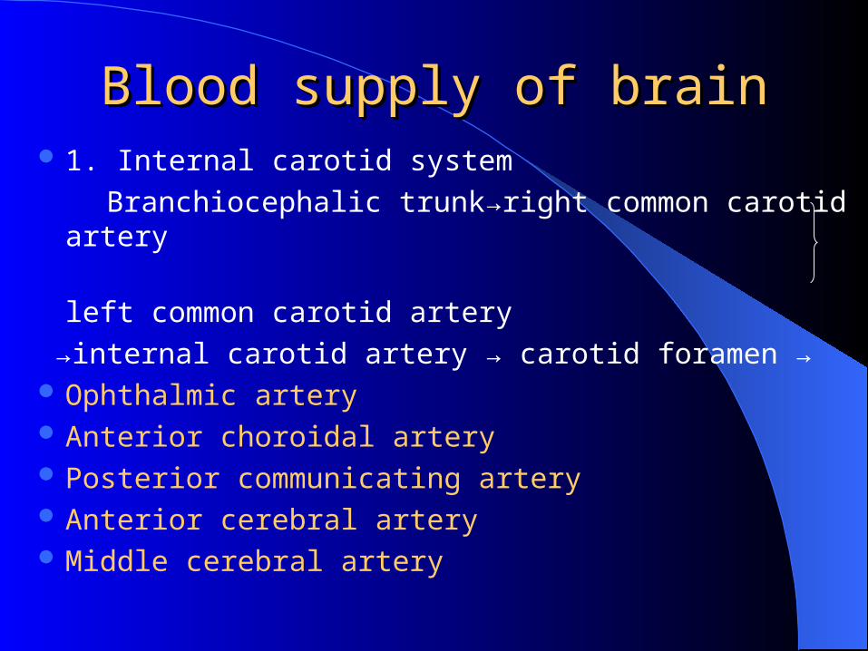
Blood supply of brainBlood supply of brain 1. Internal carotid system
Branchiocephalic trunk→right common carotid artery
left common carotid artery
→internal carotid artery → carotid foramen → Ophthalmic artery Anterior choroidal artery Posterior communicating artery Anterior cerebral artery Middle cerebral artery
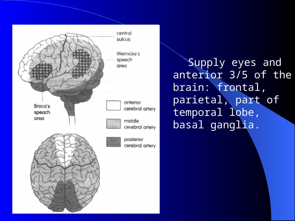
Supply eyes and anterior 3/5 of the brain: frontal, parietal, part of temporal lobe, basal ganglia.
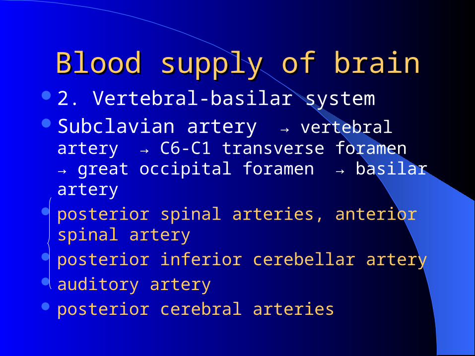
Blood supply of brainBlood supply of brain2. Vertebral-basilar systemSubclavian artery → vertebral artery → C6-C1
transverse foramen → great occipital foramen → basilar artery
posterior spinal arteries, anterior spinal artery posterior inferior cerebellar artery auditory artery posterior cerebral arteries
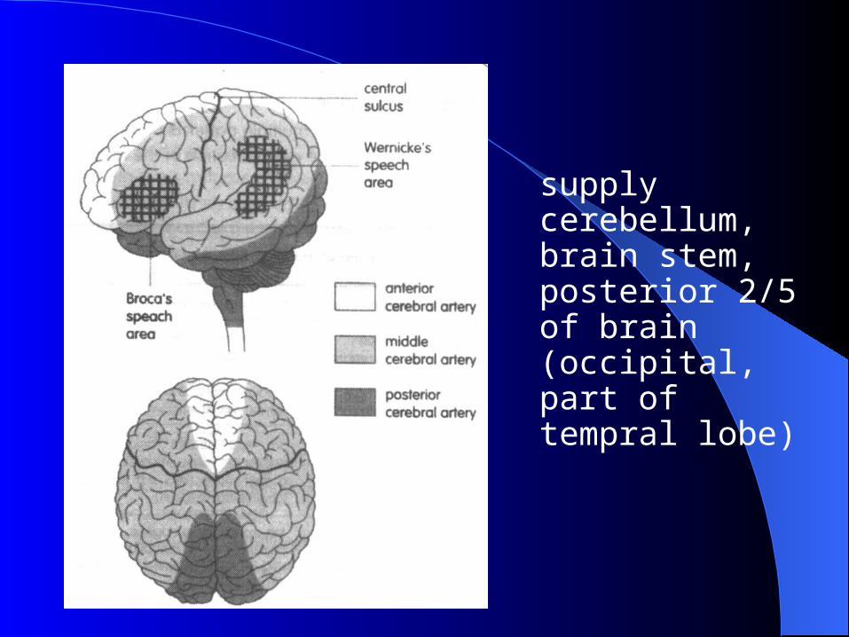
supply cerebellum, brain stem, posterior 2/5 of brain (occipital, part of tempral lobe)
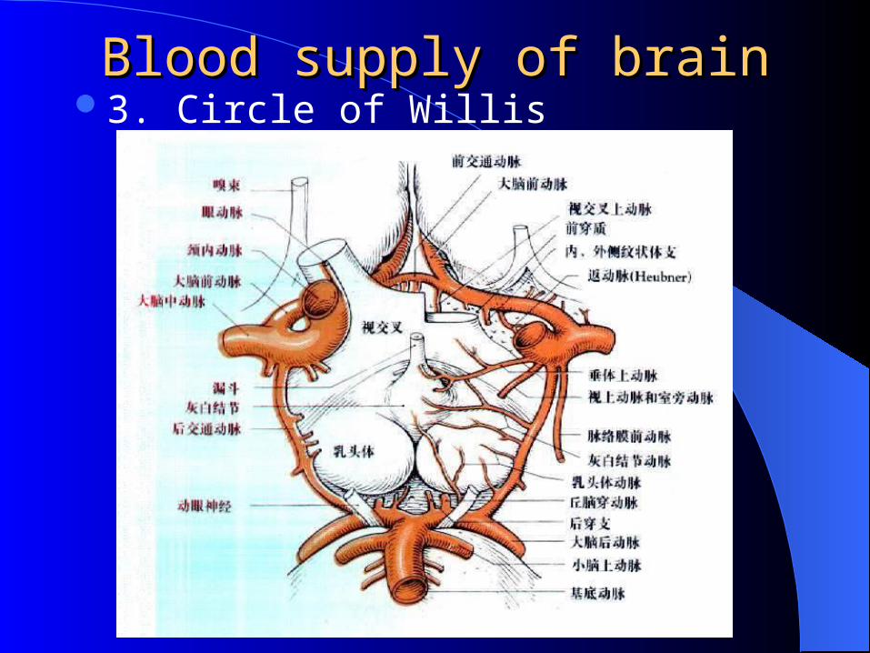
3. Circle of WillisBlood supply of brainBlood supply of brain
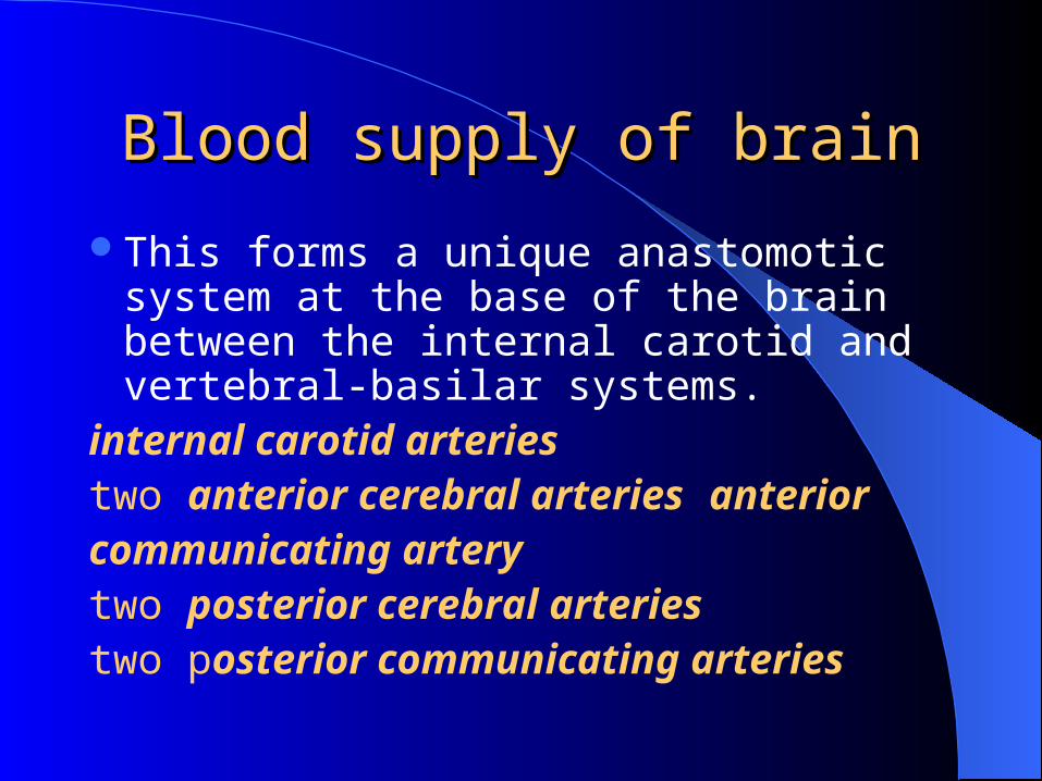
This forms a unique anastomotic system at the base of the brain between the internal carotid and vertebral-basilar systems.
internal carotid arteries two anterior cerebral arteries anteriorcommunicating arterytwo posterior cerebral arteriestwo posterior communicating arteries
Blood supply of brainBlood supply of brain
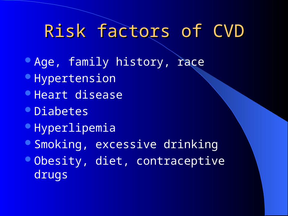
Age, family history, raceHypertensionHeart diseaseDiabetesHyperlipemiaSmoking, excessive drinkingObesity, diet, contraceptive drugs
Risk factors of CVDRisk factors of CVD
Section 2 TIASection 2 TIA
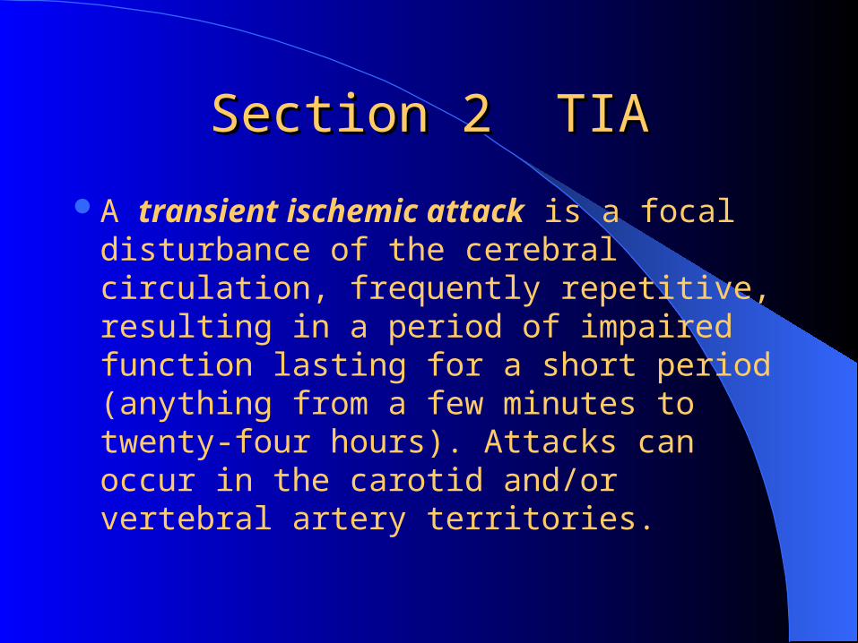
A transient ischemic attack is a focal disturbance of the cerebral circulation, frequently repetitive, resulting in a period of impaired function lasting for a short period (anything from a few minutes to twenty-four hours). Attacks can occur in the carotid and/or vertebral artery territories.
Etiology Etiology
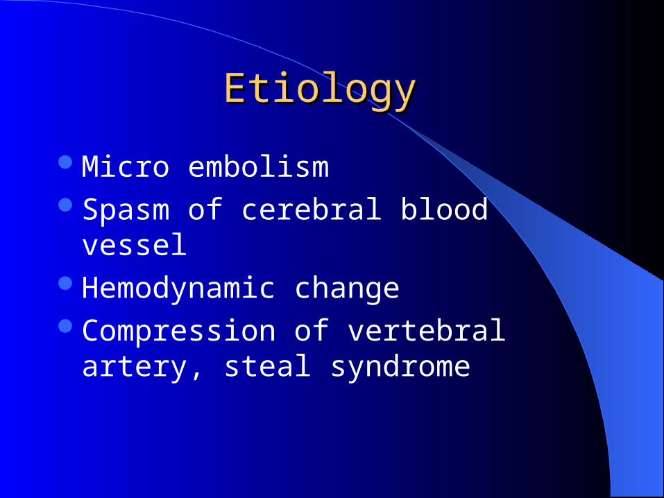
Micro embolismSpasm of cerebral blood vesselHemodynamic changeCompression of vertebral artery, steal
syndrome
Clinical featureClinical feature
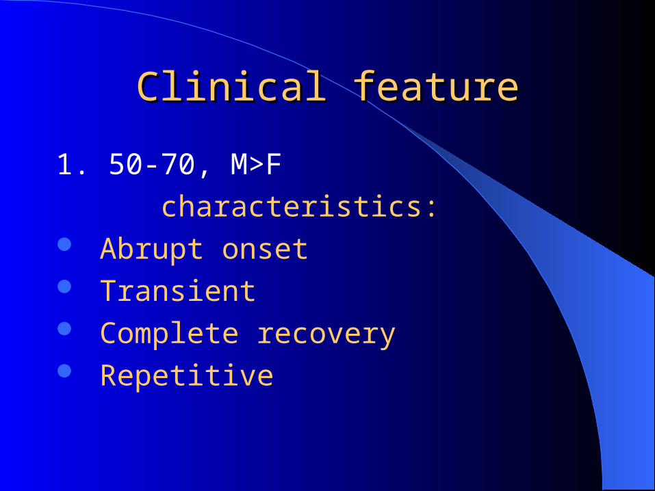
1. 50-70, M>F
characteristics: Abrupt onset Transient Complete recovery Repetitive
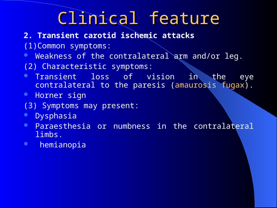
2. Transient carotid ischemic attacks(1)Common symptoms: Weakness of the contralateral arm and/or leg.(2) Characteristic symptoms: Transient loss of vision in the eye contralateral to the
paresis (amaurosis fugax). Horner sign(3) Symptoms may present: Dysphasia Paraesthesia or numbness in the contralateral limbs. hemianopia
Clinical featureClinical feature
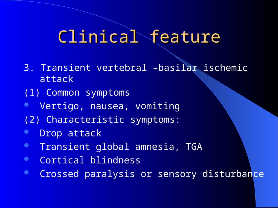
3. Transient vertebral –basilar ischemic attack
(1) Common symptoms Vertigo, nausea, vomiting
(2) Characteristic symptoms: Drop attack Transient global amnesia, TGA Cortical blindness Crossed paralysis or sensory disturbance
Clinical featureClinical feature
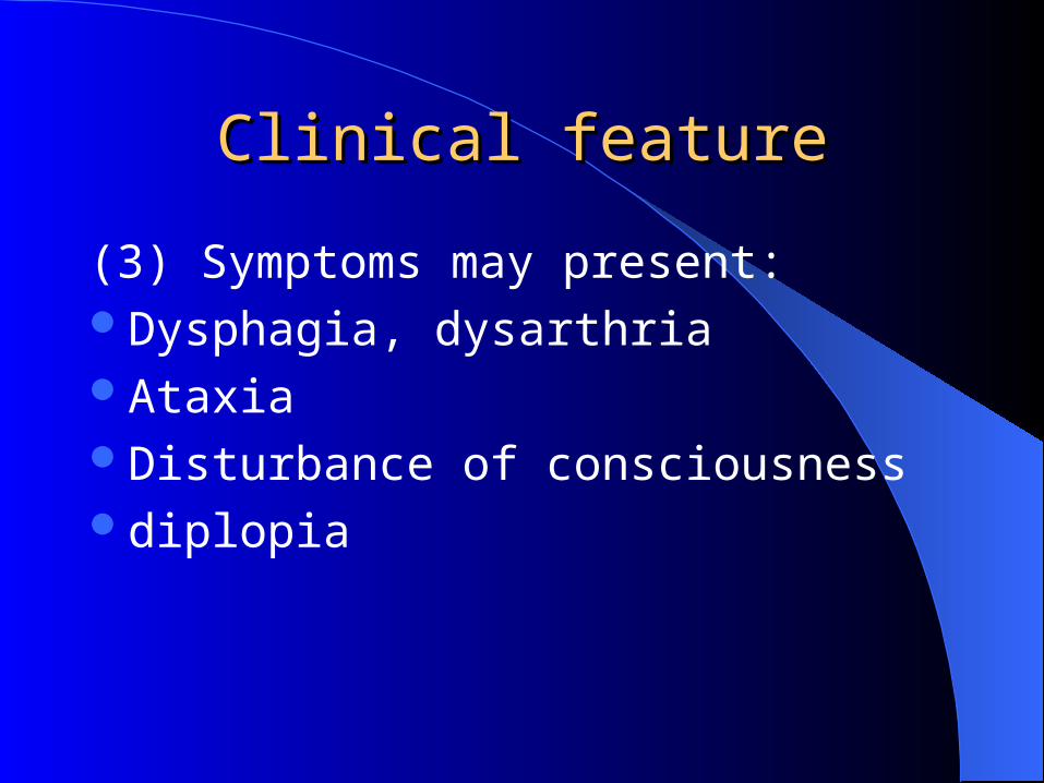
(3) Symptoms may present:Dysphagia, dysarthriaAtaxiaDisturbance of consciousnessdiplopia
Clinical featureClinical feature
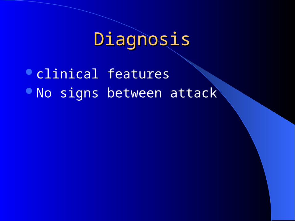
Diagnosis Diagnosis
clinical features No signs between attack
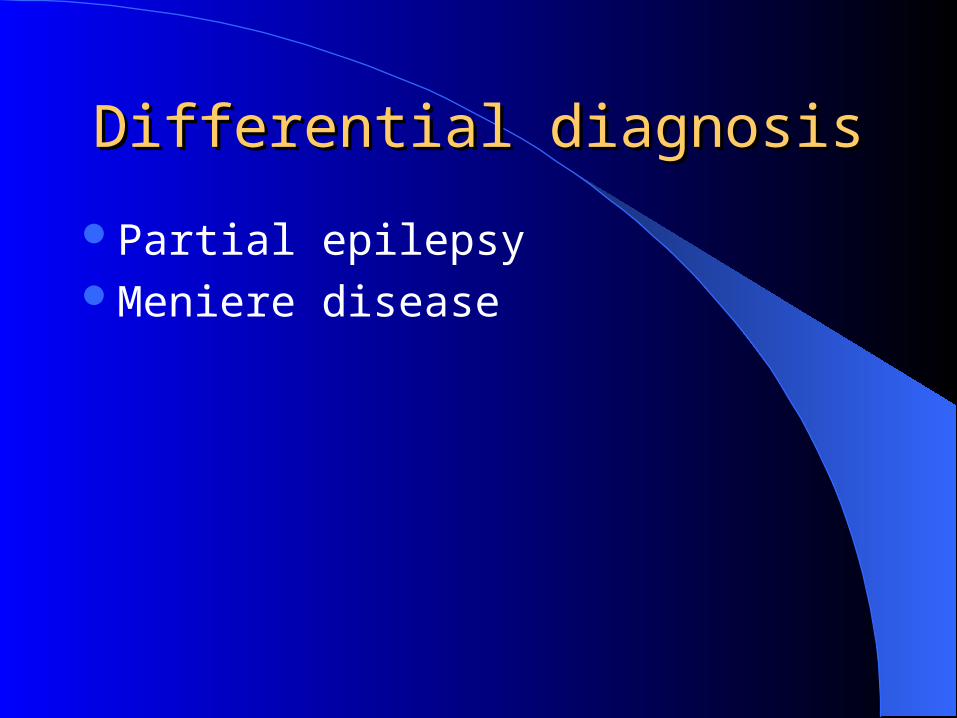
Differential diagnosisDifferential diagnosis
Partial epilepsyMeniere disease
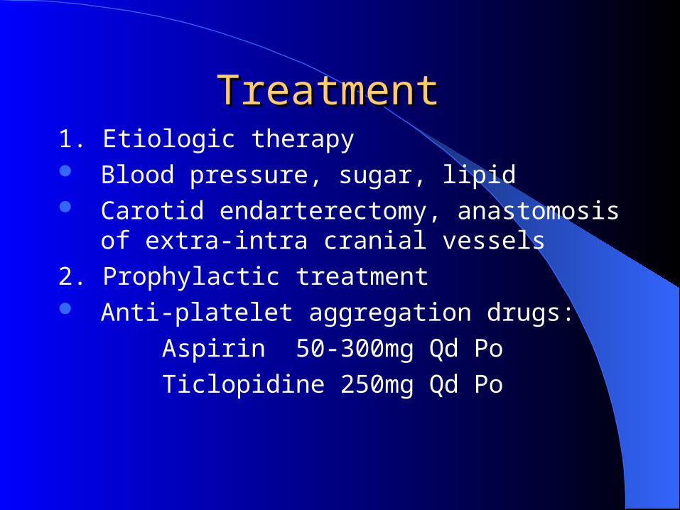
Treatment Treatment 1. Etiologic therapy Blood pressure, sugar, lipid Carotid endarterectomy, anastomosis of extra-
intra cranial vessels
2. Prophylactic treatment Anti-platelet aggregation drugs:
Aspirin 50-300mg Qd Po
Ticlopidine 250mg Qd Po
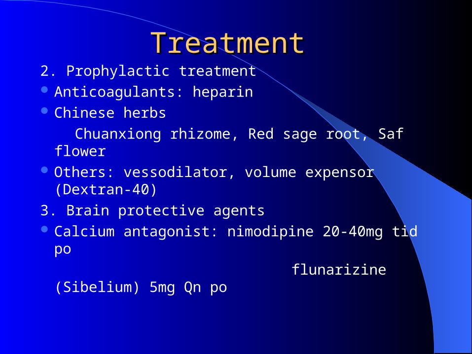
2. Prophylactic treatment Anticoagulants: heparin Chinese herbs
Chuanxiong rhizome, Red sage root, Saf flower Others: vessodilator, volume expensor (Dextran-
40)
3. Brain protective agents Calcium antagonist: nimodipine 20-40mg tid po
flunarizine (Sibelium) 5mg Qn po
Treatment Treatment
Prognosis Prognosis
1/3 → repetitive attack1/3 → remission1/3 → cerebral infarction
Section 3 Cerebral ThrombosisSection 3 Cerebral Thrombosis
infarction of an area of the brain secondary to arterial occlusion by thrombosis of a major vessel with insufficient collateral circulation.
Etiology Etiology atherosclerosis Arteritis: such as leptospirosis, rheumatic
feverrare cause:
congenital vascular malformation, polycythemia
blood hypercoagulability
Pathology Pathology
Vessel: carotid > middle > posterior > anterior > vertebral-basilar
Super-early stage: 1-6 hourNecrosis → cyst White infarct Red infarct: hemorrhagic infarct
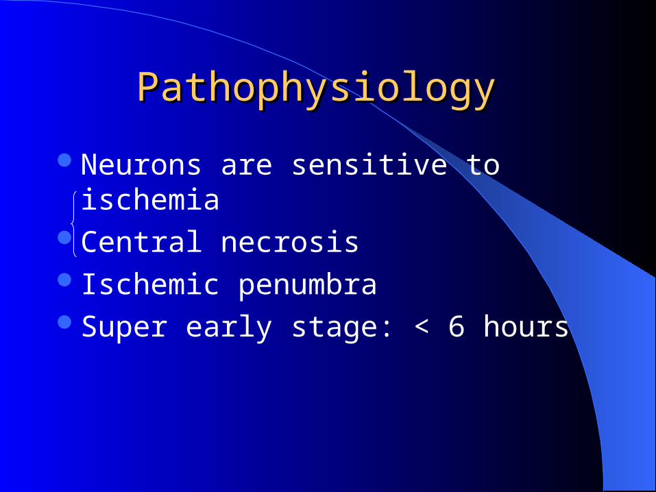
Pathophysiology Pathophysiology
Neurons are sensitive to ischemiaCentral necrosisIschemic penumbraSuper early stage: < 6 hours
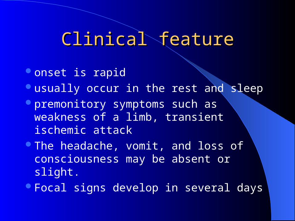
Clinical featureClinical feature
onset is rapid usually occur in the rest and sleep premonitory symptoms such as weakness of
a limb, transient ischemic attack The headache, vomit, and loss of
consciousness may be absent or slight. Focal signs develop in several days
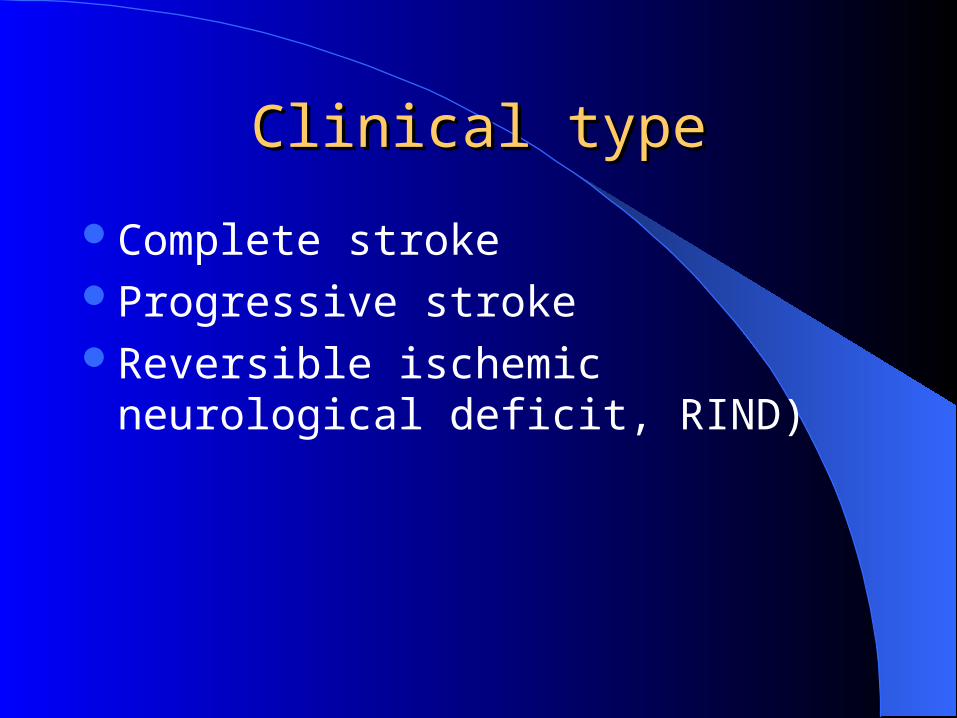
Clinical typeClinical type
Complete strokeProgressive strokeReversible ischemic neurological deficit,
RIND)

Clinical syndromeClinical syndrome
1. Internal carotid arteryMay have no signs (if the collateral supply,
from the other side, is good )amaurosis fugax, uniocular blindnessHorner's syndrome may present in the side of
the occlusion. contralateral hemiplegia and hemianesthesia.
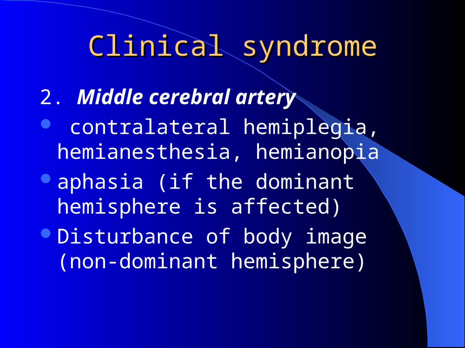
2. Middle cerebral artery contralateral hemiplegia, hemianesthesia,
hemianopiaaphasia (if the dominant hemisphere is
affected)Disturbance of body image (non-dominant
hemisphere)
Clinical syndromeClinical syndrome
3. Anterior cerebral artery contralateral hemiplegia, the leg frequently
being more affected than the arm. paracentral lobule: regulation of sphincter
function, retention or incontinencemental symptoms: apathy, euphoria
Clinical syndromeClinical syndrome
4. Posterior cerebral arterycontralateral hemianopia or quadrantanopia thalamic syndrome: contralateral hemianesthesia,
thalamic pain, ataxia, tremor, athetosis
Clinical syndromeClinical syndrome
5. Vertebro-basilar artery
(1) Main trunknausea, vomiting, tetraplegia, coma, death
(2) Weber syndromeUnilateral lesion of midbrainIpsilateral oculomotor nerve paralysis, contra
lateral hemiplegia
Clinical syndromeClinical syndrome
(3) locked-in syndromeBilateral infarction in the basis pontisTetraplegia, can not speak, can not swallow Conscious Can only respond by vertical gaze and
blinking
Clinical syndromeClinical syndrome
6. posterior inferior cerebellar artery Wallenberg's syndrome, Lateral medullary
syndromeVertigo, vomiting, nystagmusCrossed sensory disturbanceIpsilateral Horner sign Dysphagia, dysarthriaIpsilateral ataxia
Clinical syndromeClinical syndrome
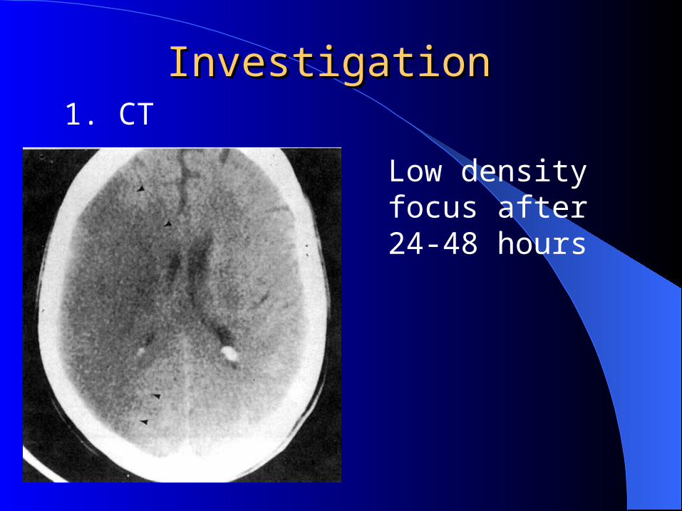
Investigation Investigation 1. CT
Low density focus after 24-48 hours
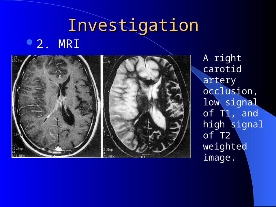
2. MRI Investigation Investigation
A right carotid artery occlusion, low signal of T1, and high signal of T2 weighted image.
3. Lumbar punctureNormal.Large infarct: pressure ↑Hemorrhagic infarction: RBC
4. DSA
5. TCD
Investigation Investigation
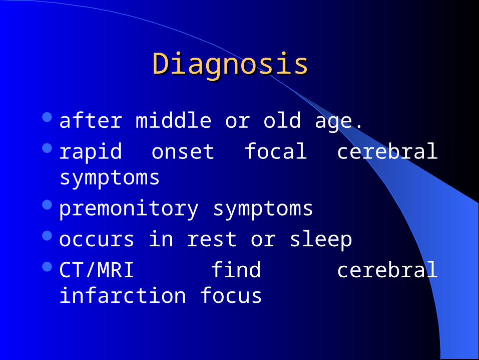
Diagnosis Diagnosis
after middle or old age.rapid onset focal cerebral symptomspremonitory symptomsoccurs in rest or sleepCT/MRI find cerebral infarction focus
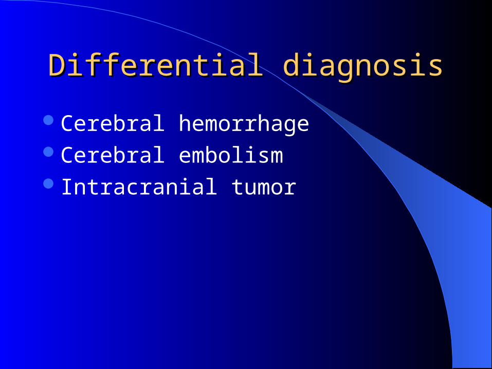
Differential diagnosisDifferential diagnosis
Cerebral hemorrhageCerebral embolismIntracranial tumor
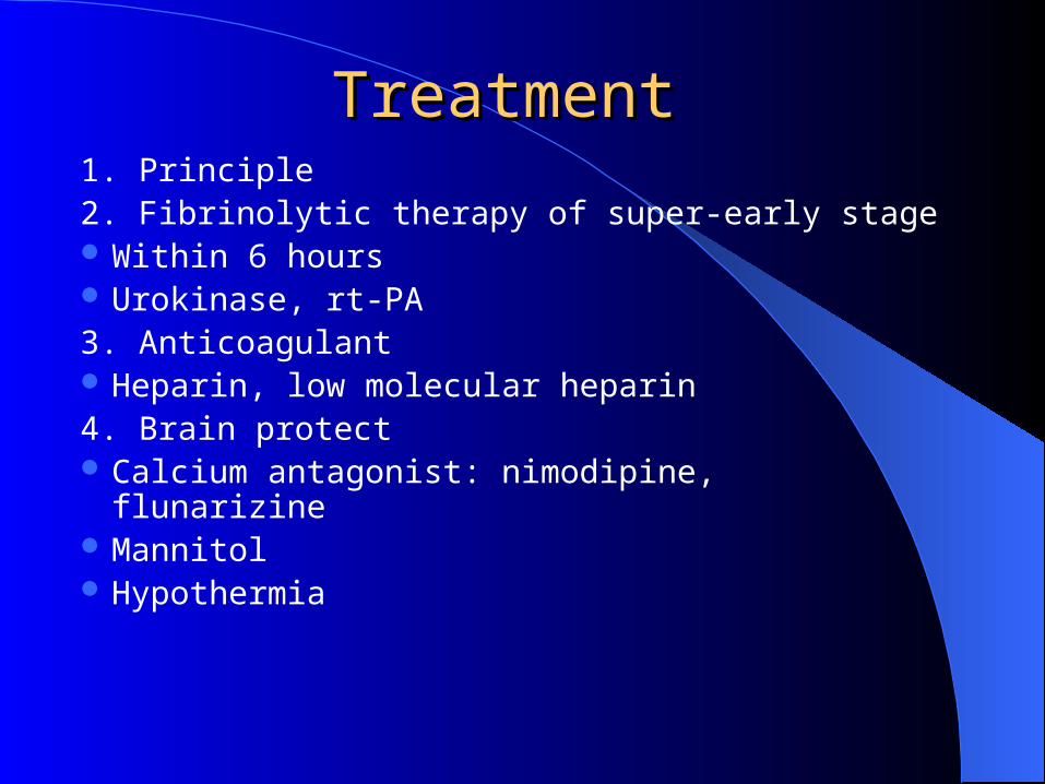
Treatment Treatment 1. Principle2. Fibrinolytic therapy of super-early stage Within 6 hours Urokinase, rt-PA3. Anticoagulant Heparin, low molecular heparin4. Brain protect Calcium antagonist: nimodipine, flunarizine Mannitol Hypothermia
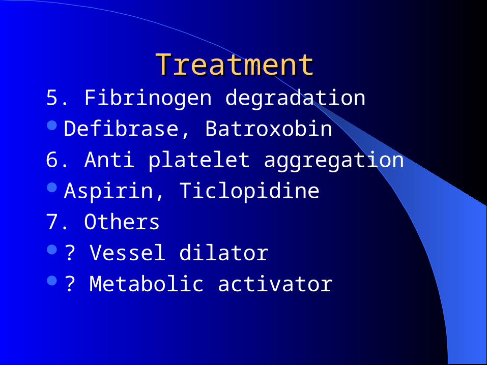
5. Fibrinogen degradationDefibrase, Batroxobin
6. Anti platelet aggregationAspirin, Ticlopidine
7. Others? Vessel dilator? Metabolic activator
Treatment Treatment
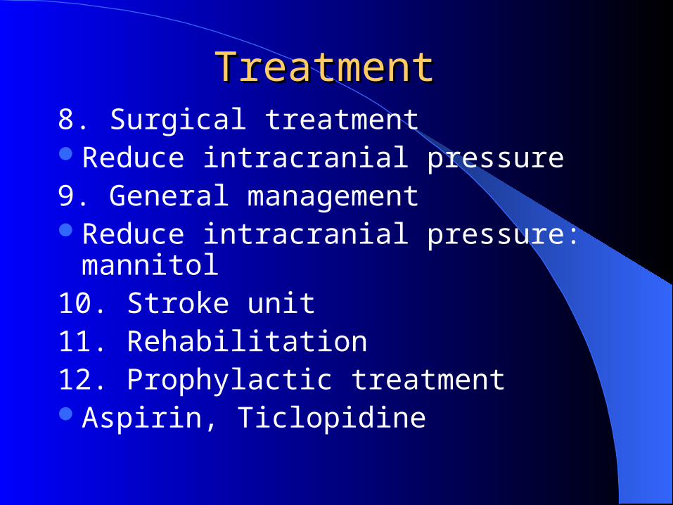
8. Surgical treatmentReduce intracranial pressure9. General managementReduce intracranial pressure: mannitol 10. Stroke unit11. Rehabilitation 12. Prophylactic treatmentAspirin, Ticlopidine
Treatment Treatment

Lacunar infarctLacunar infarct
Pathology Pathology
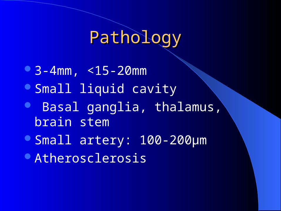
3-4mm, <15-20mmSmall liquid cavity Basal ganglia, thalamus, brain stemSmall artery: 100-200μmAtherosclerosis
Clinical featureClinical feature
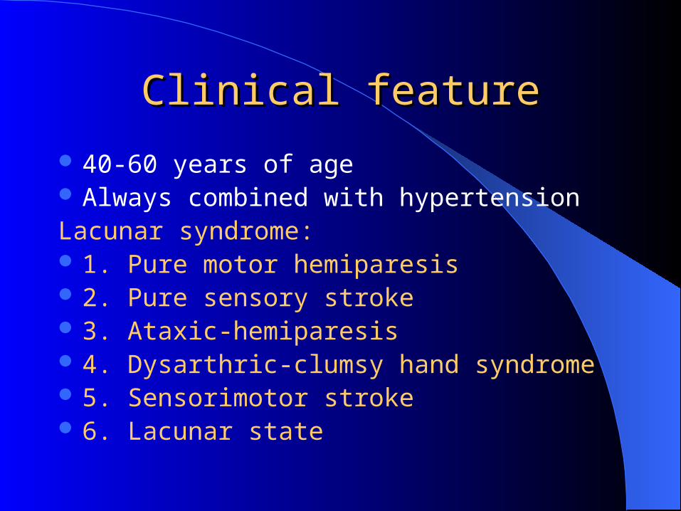
40-60 years of age Always combined with hypertensionLacunar syndrome: 1. Pure motor hemiparesis 2. Pure sensory stroke 3. Ataxic-hemiparesis 4. Dysarthric-clumsy hand syndrome 5. Sensorimotor stroke 6. Lacunar state
Cerebral embolismCerebral embolism
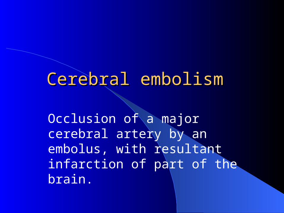
Occlusion of a major cerebral artery by an embolus, with resultant infarction of part of the brain.
Etiology Etiology
Cardiac cause: Atrial fibrillation, rheumatic valve disease,
endocarditis, atrial myxoma, myocardial infarction
Non-cardiac: Atherosclerosis plaque, pus embolus, fat
embolus, tumor embolusEmbolus of unknown origin
Clinical featureClinical feature
Left middle cerebral arteryabrupt onset, maximum disability occurring
at onceIn some cases, there is rapid improvement The primary disease, such as rheumatic
heart disease
Treatment Treatment
CerebrovasodilatorsAnticoagulant therapyTreatment of primary disease