changes in transcription pattern lead to a marked decrease ... · 1 + + 13+0 hygroma colli cysticum...
TRANSCRIPT
PHYSIOLOGICAL RESEARCH • ISSN 0862-8408 (print) • ISSN 1802-9973 (online) 2018 Institute of Physiology of the Czech Academy of Sciences, Prague, Czech Republic Fax +420 241 062 164, e-mail: [email protected], www.biomed.cas.cz/physiolres
Physiol. Res. 67: 79-91, 2018
Changes in Transcription Pattern Lead to a Marked Decrease in COX, CS and SQR Activity After the Developmental Point of the 22nd Gestational Week H. KOLAROVA1, J. KRIZOVA1, M. HULKOVA1, H. HANSIKOVA1, H. HULKOVA2, V. SMID3, J. ZEMAN1, T. HONZIK1, M. TESAROVA1 1Department of Pediatrics and Adolescent Medicine, First Faculty of Medicine, Charles University and General University Hospital in Prague, Prague, Czech Republic, 2Institute of Inherited Metabolic Disorders, First Faculty of Medicine, Charles University and General University Hospital in Prague, Prague, Czech Republic, 3Institute of Medical Biochemistry and Laboratory Diagnostics, First Faculty of Medicine, Charles University and General University Hospital in Prague, Prague, Czech Republic
Received October 29, 2016 Accepted June 19, 2017 On-line November 10, 2017 Summary Tissue differentiation and proliferation throughout fetal development interconnect with changes in the oxidative phosphorylation system (OXPHOS) on the cellular level. Reevaluation of the expression data revealed a significant increase in COX4 and MTATP6 liver transcription levels after the 22nd gestational week (GW) which inspired us to characterize its functional impact. Specific activities of cytochrome c oxidase (COX), citrate synthase (CS), succinate-coenzyme Q reductase (SQR) and mtDNA determined by spectrophotometry and RT-PCR were studied in a set of 25 liver and 18 skeletal muscle samples at 13th to 29th GW. Additionally, liver hematopoiesis (LH) was surveyed by light microscopy. The mtDNA content positively correlated with the gestational age only in the liver. The activities of COX, CS and SQR in both liver and muscle isolated mitochondria significantly decreased after the 22nd GW in comparison with earlier GW. A continuous decline of LH, not correlating with the documented OXPHOS-specific activities, was observed from the 14th to the 24th GW indicating their exclusive reflection of liver tissue processes. Two apparently contradictory processes of increasing mtDNA transcription and decreasing OXPHOS-specific activities seem to be indispensable for rapid postnatal adaptation to high energy demands. The inadequate capacity of mitochondrial energy production may be an important factor in the mortality of children born before the critical developmental point of the 22nd GW.
Key words Human fetal development • Mitochondrial biogenesis • Liver • Skeletal muscle • mtDNA Corresponding author M. Tesařová, Department of Pediatrics and Adolescent Medicine, First Faculty of Medicine, Charles University and General University Hospital in Prague, Ke Karlovu 2, Prague 2, 120 00, Czech Republic. Fax: +420 2 24967099. E-mail: [email protected] Introduction
Mitochondrial biogenesis is a target of long-term interest in various physiological or pathological situations, such as endurance training or heart failure (Wallace 1992), with specific attention to its pharmacological modulation (Whitaker et al. 2016). However, the functioning of mitochondrial biogenesis during fetal development still represents a highly unexplored field with only a few relevant reports available to date. A better understanding of the molecular mechanisms involved in mitochondrial biogenesis during this extremely dynamic period may help us not only to improve the diagnosis of mitochondrial disorders but also to provide better care to very premature neonates

80 Kolarova et al. Vol. 67 (Wenchich et al. 2002, Honzik et al. 2008).
Studies on rat models in the early 80s postulated that the activities of respiratory enzymes increase in the course of the human development, and significant differences in activity rate occur between fetal and adult life in both tissue homogenates (Warshaw 1969, Mackler et al. 1970, Jakovcic et al. 1971, Sordahl et al. 1972) and in isolated mitochondria (Jakovcic et al. 1971, Pollak et al. 1975). We previously showed that the activities of several oxidative phosphorylation system (OXPHOS) enzymes in isolated mitochondria were significantly lower in immature neonates and suggested that the low functional activity of the mitochondria is responsible for their high mortality rate (Wenchich et al. 2002, Honzik et al. 2008). However, contrary to the previous observations, no positive correlation between OXPHOS activities and gestational age was found (Honzik et al. 2008, Pejznochova et al. 2010). Moreover, the extensive report on OXPHOS activities in homogenate samples originating from five different human tissues did not show any correlation with the age of gestation (Minai et al. 2008).
To explain the regulation of mitochondrial function on a genomic level, we performed a study of the expression pattern of several proteins involved in mitochondrial DNA (mtDNA) transcription and regulation both in human fetal liver and skeletal muscle (Pejznochova et al. 2010). The increasing mitochondrial biogenesis as a preparation to postnatal life was demonstrated based on significant changes in mtDNA, mRNA and protein molecular levels throughout the studied period. These results were in accordance with the hypothesis that prenatal preparation leans mainly on a transcriptional level spearheading for more flexible post-transcriptional regulation early after birth (Cuezva et al. 1997). Our data, however, were not considered in an organ-specific manner, and it was further indicated that developing tissues, especially liver and skeletal muscle, differ in the control of mitochondrial biogenesis depending on their specific energy demands and the phase of development (Pejznochova et al. 2010).
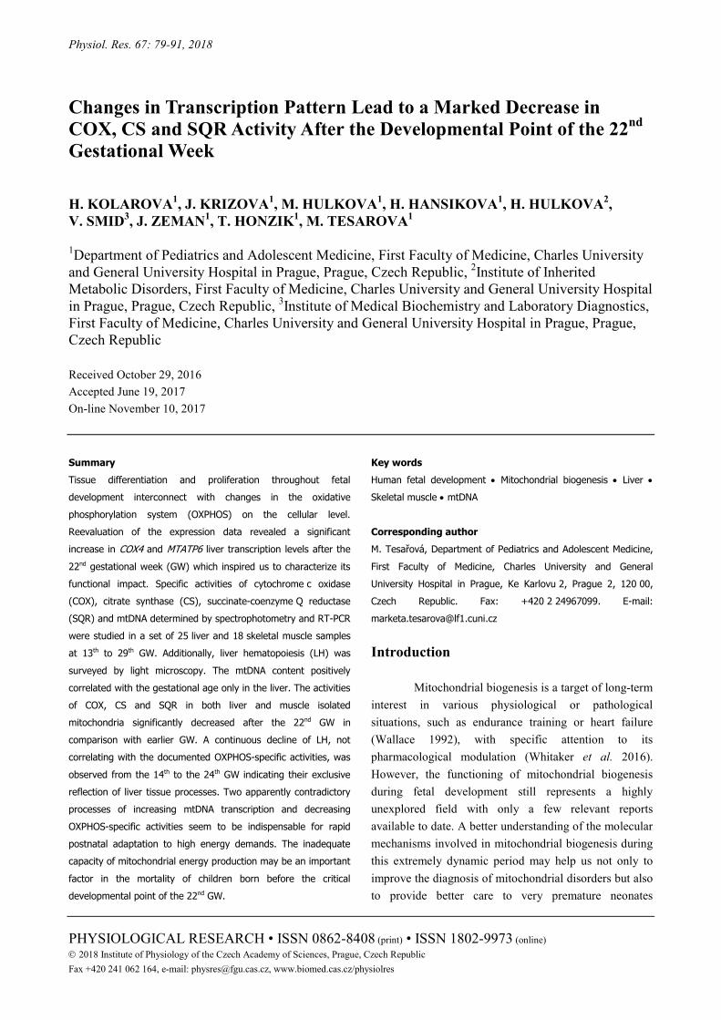
We further examined our expression data (Pejznochova et al. 2010) using the quadratic regression model and we revealed a significant increase in COX4 and MT-ATP6 transcription levels in the liver after the 22nd gestational week (GW) (Fig. 1). This finding prompted us to further examine enzyme activities in two distinctly developing fetal tissues, liver and skeletal
muscle. In the present study, the specific activities of three mitochondrial enzymes, cytochrome c oxidase (COX), citrate synthase (CS), succinate-coenzyme Q reductase (SQR), and mtDNA amount were studied in human fetal tissues between the 14th and 29th GW. Data on COX, CS and mtDNA during the human fetal development were already published in our previous report (Pejznochova et al. 2010). In the present work, a different statistical evaluation of enzyme activities is used. To determine whether ongoing liver hematopoiesis (LH) influences our data, we surveyed a consecutive series of fetal livers. Moreover, to determine whether marked changes of transcription capacity will be reflected in mitochondrial function, we decided to divide our results into two specific groups with a 22nd GW limit. To our knowledge, no study has described both OXPHOS and LH mass throughout this particular period. Methods Ethics
The work described has been carried out in accordance with the Code of Ethics of the World Medical Association (Declaration of Helsinki) after approval was granted by the Committee of Medical Ethics at the General University Hospital in Prague. Informed consent was obtained for experimentation with human samples. Material
A set of 25 and 18 samples of human fetal liver and skeletal muscle, respectively, were collected subsequently to the termination of pregnancy for genetic indications unrelated to OXPHOS deficiency (Table 1). Informed consent was obtained from women prior to tissue samples collection. Small blocks of liver and skeletal muscle tissue were taken at autopsy not later than 60 min post-mortem and immediately frozen and stored at -80 °C until used. The fetal gestational age varied from the 13th to 29th GW and there are 1-5 samples from each analysed GW in this period. The same set of liver and muscle samples was already used for our previous analyses (Pejznochova et al. 2010). However, in the latter paper, there was enough material for further analyses and the set was larger, including 26 liver and 25 muscle samples. In this paper, another 5 liver samples from GW 16th to 19th were included for additional analysis of hematopoietic activity (Table 1).
2018 COX, CS and SQR Changes Follow the 22nd Gestational Week 81
Fig. 1. Expression levels of (A) COX4 and (B) MT-ATP6 mRNA (a.u.; quadratic regression model). In the liver, there was a significant increase in COX4 and MT-ATP6 transcription levels after the 22nd week of gestation (p=0.001 and p=0.002, respectively). Adapted from Mrhalkova et al. 2010. Isolation of mitochondria
Mitochondria from the liver and muscle were isolated from 5 % tissue homogenates (w/v) prepared from frozen tissues according to the standard differential centrifugation procedure (Rickwood 1987). STE buffer was used for isolating liver tissue contained 0.25 M sucrose, 10 mM Tris-HCl, 2 mM EDTA, 2 μg/ml aprotinin, and 0.4 μM PMSF, pH 7.4; KTEA buffer for muscles contained 150 mM KCl, 50 mM Tris-HCl, 2 mM EDTA, and 2 μg/ml aprotinin, pH 7.5 (Makinen and Lee 1968). Freshly isolated mitochondria were used for activity evaluation and the rest of samples was kept at -80 °C for other analyses.
Respiratory enzyme activities Activities of respiratory enzymes COX, SQR,
and CS were measured spectrophotometrically in isolated mitochondria as described elsewhere (Rustin et al. 1994). Protein concentration was determined by the method of Lowry et al. (1951). MtDNA content
The quantification of mtDNA was executed by the method previously described in detail by Pejznochova et al. (2008). Hematopoietic mass
Small blocks of liver tissue were fixed in 10 % paraformaldehyde and embedded in paraffin. Serial ultrathin sections (6 µm) were cut and fixed doubly stained with haematoxylin and eosin.
Six representative sections from each fetal liver sample were chosen for light microscopy. The whole area of the section was systematically examined, and every third frame was photographed using stratified sampling method (Hamilton 1995). Five images were taken for each out of 6 sections, which led to a complete yield of 30 images for one fetal liver sample. All photographs were taken under constant conditions at 40× magnification of the objective.
The mass of LH was expressed as a proportion of hematopoietic islands to photographed field of view. The cell surface was quantified as the mean optical density of analysed areas as determined by the densitometric program cellSens Dimension 1.6 (Olympus Soft Imaging Solutions GmbH, Münster, Germany). Vessels and fixation artefacts were excluded from the measurement. Considering mild variations of a preparation thickness and intensity of staining, the program adjustment of density evaluation for each preparation needed to be made. Statistical methods
Correlation of selected observed variables was examined due to the lack of normality of the data, using a non-parametric Spearman correlation coefficient and a corresponding test of its statistical significance. Differences between groups of subjects were tested by a non-parametric two-sample Wilcoxon test, p-values less than 0.05 were considered as statistically significant. Analyses were conducted using R statistical package, version 3.1.1.

82 Kolarova et al. Vol. 67 Table 1. List of indications for pregnancy termination in individual fetuses.
No of sample Liver Muscle Gestational age (weeks + days)
Indication for pregnancy termination
1 + + 13+0 Hygroma colli cysticum 2 +* 13+5 Trisomy 21 3 + 13+6 Trisomy 21 4 +* + 14+2 Trisomy 21 5 * 15+5 Trisomy 18 6 + 16+0 Intrauterine infection 7 * 16+4 Trisomy 18 8 + 16+4 Trisomy 18 9 + + 17+4 Meningocele 10 * 17+6 Diaphragmatic hernia 11 + + 18+0 Trisomy 21 12 + 18+0 Gastroschisis 13 * 18+2 Fluidothorax 14 +* + 19+0 Spontaneous abortion, oligohydramnion 15 * 19+0 Congenital malformation 16 +* + 19+4 Trisomy 21 17 + + 19+4 Trisomy 21 18 +* + 20+5 Renal agenesis, oligohydramnion 19 + + 20+0 Unknown 20 + + 20+1 Sacrococcygeal teratoma 21 + + 20+3 Intrauterine infection 22 + 20+5 Omphalocele 23 + + 21+3 Limb deformity 24 + 21+3 Meningocele 25 +* + 22+1 IUGR, oligohydramnion 26 + + 22+2 Renal agenesis, oligohydramnion 27 + + 23+2 Trisomy 21 28 +* + 23+5 Urinary tract malformation 29 + + 25+4 Sacrococcygeal teratoma 30 + + 28+3 Meningocele
No – number, IUGR – Intrauterine Growth Restriction, * – samples used for additional analyses of liver hematopoiesis.
Results
Specific activities of the three mitochondrial enzymes COX, CS, and SQR and the quantity of mtDNA were studied in fetal tissues between 13th and 29th GW. In the present study, compared to previous results (Pejznochova et al. 2010), the quantity of mtDNA was positively correlated with the age of gestation only in the
liver (p=0.011) and not in muscle, a fact we ascribe to the diminution of a sample set and the use of a different statistical evaluation (Fig. 2). To compare the activities of these enzymes in distinctly developing tissues, both liver and skeletal muscle samples were used for the analysis. We recognize that these two tissues represent a heterogeneous population of cell types, and our data need to be considered in this concept.

2018 COX, CS and SQR Changes Follow the 22nd Gestational Week 83
Fig. 2. Time-specific differences in mitochondrial DNA (mtDNA) content per cell (a.u.) in the liver and skeletal muscle (Spearman’s correlation). The quantity of mtDNA positively correlated with the age of gestation only in the liver and not in the muscle (p=0.011). COX, CS and SQR specific activities as a function of gestational age
To determine whether there are age-dependent differences during the human development, the specific activities of COX, CS and SQR were determined in isolated mitochondria. Specific activities of COX were observed in the range of 5.22-580.89 nmol.min-1.mg-1 in the fetal liver, and in the fetal skeletal muscle, the range was 20.5-196.2 nmol.min-1.mg-1. Activities of SQR ranged in interval 14.5-273.2 nmol.min-1.mg-1 and 5.9-67.4 nmol.min-1.mg-1 and for CS, it was 9.37-256 nmol.min-1.mg-1 and 15.7-189.7 nmol.min-1.mg-1 in liver and muscle, respectively. Although no significant correlation with the age of gestation was observed for all studied enzymes in both prenatal tissues, the profile of enzymatic activities shown in Figure 3 indicates a similar pattern of decreasing specific activity in both tissues. The higher levels were consistently observed in the liver. Group subdivision at the 22nd GW
According to the pattern of increasing gene expression after the 22nd GW (Fig. 1), this period was chosen as a critical point for a group subdivision. This way, we could evaluate specific activities and their ratios as a dependent variable of changes in the transcription rate. After dividing the data into two subgroups with the 22nd GW age limit, significant differences were revealed both in liver and muscle. The specific activities of COX,
CS and SQR showed a significant decrease following the GW 22 in the liver (Fig. 4A, C, E) (p=0.003, p=0.003 and p=0.005, respectively). Activities of both COX and CS in muscle also showed a significant decrease (p=0.003, p=0.034, respectively), but of a lesser magnitude, except for SQR, which did not significantly differ before and after the 22nd GW (Fig. 4B, D, F). Determination of hematopoietic mass
Hematopoiesis represents an important process that takes place in the developing fetal liver. LH was determined in liver samples of human fetuses at the 14th to 24th GW (Fig. 5). Altogether, 12 samples of the human fetal liver were examined by light microscopy. Twelve ultrathin tissue sections were examined under light microscopy. The amount of hematopoietic mass was expressed as a proportion of hematopoietic cells to the liver tissue.
A significant negative correlation was observed between the LH mass and gestational age (q=-0.602, p=0.042). Stem cells were observed only in the extravascular location; however, the mature cells migrated into the sinusoids and were not included in our evaluation. Hematopoiesis was fully established around the third month, and the hematopoietic islands occupied considerable spaces between the parenchymal cells, altering the architecture of hepatic cords (Fig. 5A). Hepatic blood cell formation declined continually throughout the development from the maximum percentage of 15 % at the outset of the studied period and it was apparent that by the end of the second trimester, hematopoiesis diminished and became only focal (Fig. 5B, C). Unusually high level of LH was found in three patients, which may be due to their primary diagnosis of oligohydramnios (Fig. 5C). Moreover, there was no correlation between OXPHOS specific activities and LH mass (data not shown).
Discussion
Given its extreme time-specific variability, the period of fetal development represents an exciting but rather challenging field. Tissue differentiation and proliferation are interconnected with the changes of metabolism, including OXPHOS on the cellular level. The main objectives of this study were to further characterize several markers of mitochondrial biogenesis throughout the human fetal development with its time and tissue-specific changes.

84 Kolarova et al. Vol. 67
Fig. 3. The relationship between the age of gestation and the enzyme-specific activities (nmol.min-1.mg-1) in mitochondrial fraction (Spearman’s correlation). In liver, there was no significant correlation between gestational age and all three cytochrome c oxidases (COX) (p=0.342) (A), citrate synthase (CS) (p=0.161) (B) and succinate-coenzyme Q reductase (SQR) (p=0.124) (C). Insignificant results were also present in the muscle for all COX (p=0.082) (A), CS (p=0.216) (B) and SQR (p=0.766) (C). Black points – liver; blank points – skeletal muscle.
COX is a terminal enzyme of the mitochondrial electron transfer chain determining the functional status of mitochondria. It represents a multicomponent structure with rather complicated biogenesis not only due to its dual genetic origin but also because of the existence of tissue and age-specific isoforms of its subunits (Bonne et al. 1993, Rohdich and Kadenbach 1993). The SQR or Complex II is the only complex encoded only by nuclear DNA. Both the tricarboxylic acid cycle and respiratory chain share its enzymatic function; therefore, it is a suitable factor informing us about the overall metabolic function of the cell, including the fatty acid β-oxidation. CS, a mitochondrial matrix enzyme, is one of the three regulatory steps of the tricarboxylic acid cycle and, similarly as SQR, it is frequently used as important normalization factor for OXPHOS enzymatic activities. However, CS seems to be inappropriate for this application when two different organs are being compared, as there exists important inter-tissue variability between its specific activity (Fernandez-Vizzara et al. 2011). According to our observations, CS and SQR also seem to be inappropriate normalization parameters or markers of mitochondrial biogenesis within the fetal tissues throughout their development (Fig. 6).
Human mtDNA are self-replicating circular molecules present in mitochondria in variable numbers of copies. MtDNA content is an important marker of mitochondrial biogenesis (Malik et al. 2011). It was previously shown that the quantity of mtDNA increases throughout the development (Heerdt and Augenlicht 1990). Early studies on the rat liver (10-21 days of gestation) or bovine heart models (first and third trimester) determined that also COX and SQR activities increased throughout fetal development in tissue homogenate (Jakovcic 1971, Mackler et al. 1971, Marin-Garcia 1994) or in isolated mitochondria (Jakovcic 1971, Pollak 1975). Moreover, Marin-Garcia et al. later proved that COX activity in the human heart homogenates significantly grows during fetal life (6th to 17th GW) (Marin-Garcia et al. 2000).

2018 COX, CS and SQR Changes Follow the 22nd Gestational Week 85
Fig. 4. Tissue-specific differences in COX, SQR and CS enzyme activities (nmol.min-1.mg-1) in isolated liver and muscle mitochondria divided into two groups with a critical point at the 22nd gestational week (GW). In the fetal liver tissue, the specific activities of all studied enzymes, cytochrome c oxidase (COX), citrate synthase (CS) and succinate-coenzyme Q reductase (SQR), presented significant differences between the two categories (Wilcoxon test) (A, C, E). COX was the most age-dependent enzyme with a significant decrease within the groups in the liver (p=0.003) (A). Moreover, both CS and SQR demonstrated a similar significant decrease in the liver (p=0.003 and p=0.005, for CS and SQR, respectively) (C, E). In the muscle, the second group showed significantly lower levels after the 22nd GW for COX (p=0.003) (B) and CS (p=0.034) (D).

86 Kolarova et al. Vol. 67
Fig. 5. The hematopoietic mass (%) in 12 liver samples in the course of fetal development from the 14th to 24th gestational week (GW) (Spearman’s correlation). hematopoiesis was fully established around the third month and the hematopoietic islands occupied considerable spaces between the parenchymal cells, altering the architecture of hepatic cords (A). Hepatic blood cell formation declined continually throughout the development, and by the end of the second trimester, it became only focal, with sparse hematopoietic islands (B, C). The correlation between hematopoiesis and gestational age presented with a significant negative decrease (q=-0.602, p=0.042) (C). We found no correlation of COX, CS and SQR enzymatic activities with the age of gestation in both human liver and muscle isolated mitochondria within the second trimester of gestation. In both tissues, we observed high variability of enzymatic activities that can be explained by small number of samples in each GW and/or by heterogeneity of clinical conditions that led to pregnancy termination (Table 1). Nevertheless, none of clinical conditions was prevalent in any GW.
Interestingly, following the pattern of increasing gene expression, we could observe similar time-specific changes in both studied tissues after the 22nd GW. Specific activities of COX, CS and SQR were significantly lower in the group following the critical point of the 22nd week of development, although overall, the specific activities were lower in the muscle tissue (Fig. 3). At birth, the full switch from glycolysis to oxidative phosphorylation relies on the mitochondrial biogenesis (Valcarce et al. 1994). However, GW 22 to 23 seems to be a limit of viability (Ogawa et al. 2013). A profound change in the transcription pattern was observed for several COX subunits (Fig. 1A, Mrhalkova et al. 2010) after the 22nd GW, and a similar observation was made for several ATP synthase subunits (Fig. 1B, Mrhalkova et al. 2010). The expression level of mitochondrially encoded gene MT-ATP6 was distinctively increasing after the 22nd GW in both the liver and muscle and a parallel increase of nuclearly-encoded ATP5G2 was shown in liver, although compared to mitochondrially-encoded genes, levels of both ATP5G2 and ATP5O were four-fold lower (Mrhalkova et al. 2010). High levels of mitochondrial mRNAs seem to be a mechanism to compensate the relatively less efficient translation compared to a high nuclear elongation rate, which might be caused by significantly greater ribosomal spacing (Thames et al. 2000). Lightowlers et al. (1990) suggested that it is either inefficient translation or the rapid turnover of COX8L protein that is responsible for the isoform switch within bovine heart tissue. Similarly, COX6AL destabilization

2018 COX, CS and SQR Changes Follow the 22nd Gestational Week 87
led to increased mRNA degradation within skeletal muscle development. However, other COX subunits that do not undergo isoform switching, such as COX5A, were observed to be relatively stable up to >15-20 h (Thames et al. 2000). The accumulation of transcripts must, therefore, be accomplished rather by increased transcriptional rate than the increased half-life of mRNAs (Izquierdo et al. 1995b, Ostronoff et al. 1996).
It is hypothesized that protein expression in the liver is exerted mainly after birth (Cuezva et al. 1997). Similarly, no major difference in protein levels or enzyme-specific activities of all respiratory complexes were noted in the study examining 5 fetal tissues (Minai et al. 2008). We could have clearly observed in the liver tissue a marked decrease in COX activity after the 22nd GW. However, at the same time, its transcript levels started increasing. From these inverse processes and considering the stability of COX subunit protein levels throughout the studied period, we can speculate that around the 22nd GW, possibly regulatory changes in COX (OXPHOS) biogenesis are proceeding especially on the posttranslational level.
Based on our results, we suggest that sufficient levels of mtDNA and mitochondrially encoded transcripts that are accumulated throughout gestation but mainly after the 22nd GW are crucial for the adequate onset of respiration after birth. Translational activity reflected in enzyme specific activity would rather stay behind the scene to establish more effective energy production early after birth.
In the work of Pejznochova et al. (2010), it was suggested that the fetal liver and muscle differ in the control of mitochondrial biogenesis. The correlation of mtDNA and COX specific activity informed us about the effectiveness of the mtDNA template utilization. We showed that there was a significant difference between liver and muscle when the enzyme specific activity and mtDNA are correlated (Fig. 6A). A significant negative correlation between mtDNA and specific activity of COX is only present in the liver. One can presume that the gene
Fig. 6. The correlation between COX-, CS- and SQR-specific activity (nmol.min-1.mg-1) and mtDNA amount (a.u.) in both liver and muscle tissue. There was a significant difference between the studied tissues only for cytochrome c oxidase (COX)-specific activity (p=0.000) (A). A significant negative correlation between citrate synthase (CS)-specific activity, and the mitochondrial DNA (mtDNA) content is present in both the liver (p=0.008) and muscle (p=0.049) (B). On the contrary, between COX- and succinate-coenzyme Q reductase (SQR)-specific activity and mtDNA amount it was present only in the liver (p=0.000, p=0.010) and not in the muscle tissue (A, C).

88 Kolarova et al. Vol. 67 dosage in the liver may be one of the factors affecting gene expression; however, it does not determine the final COX activity.
This is also supported by the fact that while mtDNA levels are higher in the muscle, the enzyme specific activities are significantly lower in the muscle than in the liver (Fig. 2, Fig. 3A, B, C). These results are in line with the work of Fernandez-Vizzara et al. (2011), who suggested that while in adult skeletal muscle, the gene dosage determines COX activity; in other tissues, including the adult liver, other levels of mtDNA expression regulation must have a more relevant contribution to the regulation of the final OXPHOS activity. Nevertheless, it only speaks in favor of the fact that similar mechanisms regulate mitochondrial gene expression in fetal and in adult organisms.
It is evident from our results that all of the studied enzymes follow the same trend. Similarly, the overall activity of CS and SQR did not change throughout the studied period in either liver or muscle, but it was significantly lower in the liver after the 22nd GW. The mitochondrial oxidation of fatty acids plays a critical role in energy metabolism after birth (Oey et al. 2005). However, it was only recently proven that β-oxidation is active also during fetal life, as it was long thought that the major energy supply comes from glucose. In the last trimester of intra-uterine life, fat accumulation becomes more important in the human fetus due to its important function in energy storage and temperature maintenance. Some of the deposited fatty acids will accrue from fetal lipogenesis, but the great bulk of fetal lipid is derived from the maternal circulation. This is further supported by observations in the placenta where the activity of long-chain 3-hydroxyacyl-CoA dehydrogenase and short-chain 3-hydroxyacyl-CoA dehydrogenase were inversely correlated with gestational age (Oey et al. 2005). In this way, SQR as a more multifunctional enzyme may follow the same trend as other enzymes of the respiratory chain, leaving space for more effective β-oxidation early after birth. We suggest that metabolic pathways work inter-connectively in the organism and that the enzymes of tricarboxylic acid cycle need to develop hand in hand with OXPHOS.
Although differences exist in the regulation of liver and muscle mitochondrial biogenesis, we presume that both tissues need to be prepared for increased energy demands after birth. The study of three cattle muscles suggested that hoofed animals are more mature relative to other mammals, as they get up and start to walk within
hours after birth and the development of fast phasic muscle composed of type II glycolytic fibers is more important at birth (Gagniére et al. 1999). In a physiologic human neonate, hypertonia is a compensatory mechanism for the change of external conditions and the effect of gravity. Since postural muscles that keep the adequate muscle tonus consist mainly of type I oxidative fibers, rich in mitochondria, a sufficient OXPHOS capacity early after birth becomes crucial. This is further supported by the fact that in preterm infants with immature OXPHOS (Honzik et al. 2008), hypotonia is a frequent feature found at birth.
The studied parameters undergo important changes throughout the second trimester, especially in the liver. However, other specialized processes, such as LH, are limited to the fetal period, and their own mitochondrial functional status may be responsible for our observations. Throughout embryonic development, hematopoiesis represents a highly dynamic process, taking place in several different localizations. The blood formation in the human liver begins in embryos of approximately 12 mm in length, i.e. around the 6th GW (Minot et al. 1912) and it becomes fully established around the third month of intrauterine life (Zamboni 1965, Brugnara and Platt 2003). In our study, we observed a moderate, continuous decline of LH mass from the 14th to 24th GW, with a maximum of 15 % at the beginning of the studied period (Fig. 5). These results would indicate that participation of hematopoiesis in the overall liver metabolism is rather limited. This is further supported by the fact that hematopoietic cells in fetal liver rely mainly on glycolysis and have both low mitochondrial content and amounts of respiratory chain complexes (Imanirad et al. 2013, Simsek et al. 2010). Additionally, there were no correlations between OXPHOS-specific activities and LH mass (data not shown). We concluded that the documented developmental changes of respiratory chain enzyme activities are not affected by concomitant hematopoiesis and, above all, reflect processes in liver tissue.
In conclusion, the period of fetal development represents an exciting but rather challenging field. With great developments in perinatal intensive care, the limit of viability was shifted back to GW 22 to 23 (Ogawa et al. 2013). A better understanding of the molecular mechanisms involved in mitochondrial biogenesis during this extremely dynamic period may help us in improving diagnostics and in providing better care of very premature neonates (Wenchich et al. 2002, Honzik et al. 2008).

2018 COX, CS and SQR Changes Follow the 22nd Gestational Week 89
Within the course of the second trimester, profound tissue-specific changes of mitochondrial energy metabolism are underway. In our study, we observed that specific activities of COX, CS and SQR were significantly lower following the 22nd week of development; however, mtDNA levels continuously increased. These processes seem to be indispensable for rapid postnatal adaption and increasing energy demands. We conclude that the inability of OXPHOS to easily adapt to extrauterine life may be an important factor in the mortality of children born before this critical developmental point. Conflict of Interest There is no conflict of interest.
Acknowledgements This work was supported by grants GAČR 14-36804G from the Grant Agency of the Czech Republic, by institutional research support from Charles University in Prague (PROGRES Q26/LF1/3, SVV 260367, UNCE 204011) and from the Ministry of Health of the Czech Republic (RVO-VFN 64165). The authors are thankful to Vaclav Capek for the statistical treatment of the data. Abbreviations GW – gestational week, LH – liver hematopoiesis, OXPHOS – oxidative phosphorylation system, mtDNA – mitochondrial DNA, COX – cytochrome c oxidase, CS – citrate synthase, SQR – succinate-coenzyme Q reductase.
References BONNE G, SEIBEL P, POSSEKEL S, MARSAC C, KADENBACH B: Expression of human cytochrome c oxidase
subunits during fetal development. Eur J Biochem 217: 1099-1107, 1993. BRUGNARA C, PLATT OS: The neonatal erythrocyte and its disorders. In: Nathan's and Oski's Hematology of
Infancy and Childhood. NATHAN DG, ORKIN SK, LOOK AT, GINSBURG D (eds), Saunders Elsevier, Philadelphia, 2003, pp 19-26.
CUEZVA JM, OSTRONOFF LK, RICART J, LÓPEZ DE HEREDIA M, DI LIEGRO CM, IZQUIERDO JM: Mitochondrial biogenesis in the liver during development and oncogenesis. J Bioenerg Biomembr 29: 365-377, 1997.
FERNANDEZ-VIZARRA E, ENRIQUEZ JA, PEREZ-MARTOS A, MONTOYA J, FERNANDEZ-SILVA P: Tissue-specific differences in mitochondrial activity and biogenesis. Mitochondrion 11: 207-213, 2011.
GAGNIÈRE H, PICARD B, JURIE C, GEAY Y: Comparison of foetal metabolic differentiation in three cattle muscles. Reprod Nutr Dev 39: 105-112, 1999.
HAMILTON PW, ALLEN DC: Morphometry in histopathology. J Pathol 175: 369-379, 1995. HONZIK T, WENCHICH L, BOHM M, HANSIKOVA H, PEJZNOCHOVA M, ZAPADLO M, PLAVKA R,
ZEMAN J: Activities of respiratory chain complexes and pyruvate dehydrogenase in isolated muscle mitochondria in premature neonates. Ear Hum Dev 84: 269-276, 2008.
HEERDT BG, AUGENLICHT LH: Changes in the number of mitochondrial genomes during human development. Exp Cell Res 186: 54-59, 1990.
IMANIRAD P, KARTALAEI PS, CRISAN M, VINK C, YAMADA-INAGAWAA T, DE PATER E, KUREK D, KAIMAKIS P, VAN DER LINDEN R, SPECK N, DZIERZAK E: HIF1α is a regulator of hematopoietic progenitor and stem cell development in hypoxic sites of the mouse embryo. Stem Cell Res 12: 24-35, 2013.
IZQUIERDO JM, RICART J, OSTRONOFF LK, EGEA G, CUEZVA JM: Changing patterns of transcriptional and post-transcriptional control of beta-F1-ATPase gene expression during mitochondrial biogenesis in liver. J Biol Chem 270: 10342-10350, 1995.
JAKOVCIC S, HADDOCK J, GETZ GS, RABINOWITZ M, SWIFT H: Mitochondrial development in liver of foetal and newborn rats. Biochem J 121: 341-347, 1971.
LOWRY OH, ROSEBROUGH NJ, FARR AL, RANDALL RJ: Protein measurement with the Folin phenol reagent. J Biol Chem 193: 265-275, 1951.
LUIS AM, IZQUIERDO JM, OSTRONOFF LK, SALINAS M, SANTAREN JF, CUEZVA JM: Translational regulation of mitochondrial differentiation in neonatal rat liver. Specific increase in the translational efficiency of the nuclear-encoded mitochondrial beta-F1-ATPase mRNA. J Biol Chem 268: 1868-1875, 1993.

90 Kolarova et al. Vol. 67 MACKLER B, GRACE R, DUNCAN HM: Study of mitochondrial development during embryogenesis in the rat. Arch
Biochem Biophys 144: 603-610, 1971. MAKINEN M, LEE CP: Biochemical studies of skeletal muscle mitochondria: I. Microanalysis of cytochrome content,
oxidative and phosphorylative activities of mammalian skeletal muscle mitochondria. Arch Biochem Biophys 126: 75-82, 1968.
MALIK AN, SHAHNI R, RODRIGUEZ-DE-LEDESMA A, LAFTAH A, CUNNINGHAM P: Mitochondrial DNA as a non-invasive biomarker: Accurate quantification using real time quantitative PCR without co-amplification of pseudogenes and dilution bias. Biochem Biophys Res Commun 412: 1-7, 1990.
MARIN-GARCIA J, ANANTHAKRISHNAN R, AGRAWAL N, GOLDENTHAL MJ: Mitochondrial gene expression during bovine cardiac growth and development. J Mol Cell Cardiol 26: 1029-1036, 1994.
MARIN-GARCIA J, ANANTHAKRISHNAN R, GOLDENTHAL MJ: Heart mitochondrial DNA and enzyme changes during early human development. Mol Cell Biochem 210: 47-52, 2000.
MINAI L, MARTINOVIC J, CHRETIEN D, DUMEZ F, RAZAVI F, MUNNICH A, ROTIG A: Mitochondrial respiratory chain complex assembly and function during human fetal development. Mol Genet Metab 94: 120-126, 2008.
MINOT CS: Development of the blood, the vascular system and the spleen. In: Manual of Human Embryology. KEIBEL F, MALL FP (eds), J. B. Lippincott, Philadelphia, 1912, pp 498-534.
MRHALKOVA A: Study of Expression and Maturation of Mitochondrial Oxidative Phosphorylation System During Mammal's Prenatal Period; Diploma Thesis of Charles University, Prague 2010.
OEY NA, DEN BOER ME, WIJBURG FA, VEKEMANS M, AUGE J, STEINER C: Long-chain fatty acid oxidation during early human development. Pediatr Res 57: 755-759, 2005.
OGAWA M, MATSUDA Y, KANDA E, KONNO J, MITANI M, MAKINO Y: Survival rate of extremely low birth weight infants and its risk factors: case-control study in Japan. ISRN Obstet Gynecol 2013: 873563, 2013.
OSTRONOFF LK, IZQUIERDO JM, ENRIQUEZ JA, MONTOYA J, CUEZVA JM: Transient activation of mitochondrial translation regulates the expression of the mitochondrial genome during mammalian mitochondrial differentiation. Biochem J 316: 183-191, 1996.
PEJZNOCHOVA M, TESAROVA M, HONZIK T, HANSIKOVA H, MAGNER M, ZEMAN J: The developmental changes in mitochondrial DNA content per cell in human cord blood leukocytes during gestation. Physiol Res 57: 947-955, 2008.
PEJZNOCHOVA M, TESAROVA M, HANSIKOVA H, MAGNER M, HONZIK T, VINSOVA K, HAJKOVA Z, HAVLICKOVA V, ZEMAN J: Mitochondrial DNA content and expression of genes involved in mtDNA transcription, regulation and maintenance during human fetal development. Mitochondrion 10: 321-329, 2010.
POLLAK JK: The maturation of the inner membrane of foetal rat liver mitochondria. Biochem J 150: 477-488, 1975. RICKWOOD D: Isolation and characteristics of intact mitochondria. In: Mitochondria: A Practical Approach I.
WILSON MT, DARLEY-USMAR VM (eds), IRL Press, Oxford, 1987, pp 1-16. ROHDICH F, KADENBACH B: Tissue-specific regulation of cytochrome c oxidase efficiency by nucleotides.
Biochemistry 32: 8499-8503, 1993. RUSTIN P, CHRETIEN D, BOURGERON T, GERARD B, ROTIG A, SAUDUBRAY JM, MUNNICH A:
Biochemical and molecular investigations in respiratory chain deficiencies. Clin Chim Acta 228: 35-51, 1994. SIMSEK T, KOCABAS F, ZHENG J, DEBERARDINIS RJ, MAHMOUD AI, OLSON EN, SCHNEIDER JW,
ZHANG CC, SADEK HA: The distinct metabolic profile of hematopoietic stem cells reflects their location in a hypoxic niche. Cell Stem Cell 7: 380-390, 2010.
SORDAHL LA, CROW CA, KRAFT GH, SCHWARTZ A: Some ultrastructural and biochemical aspects of heart mitochondria associated with development: fetal and cardiomyopathic tissue. J Mol Cell Cardiol 4: 1-10, 1972.
VALCARCE C, IZQUIERDO JM, CHAMORRO, CUEZVA JM: Mammalian adaptation to extrauterine environment: mitochondrial functional impairment caused by prematurity. Biochem J 303: 855-862, 1994.
WALLACE DC: Mitochondrial genetics: a paradigm for aging and degenerative diseases? Science 256: 628-632, 1992. WARSHAW JB: Cellular energy metabolism during fetal development. I. Oxidative phosphorylation in the fetal heart.
J Cell Biol 41: 651-657, 1969.

2018 COX, CS and SQR Changes Follow the 22nd Gestational Week 91
WENCHICH L, ZEMAN J, HANSIKOVA H, PLAVKA R, SPERL W, HOUSTEK J: Mitochondrial energy metabolism in very premature neonates. Biol Neonate 81: 229-235, 2002.
WHITAKER RM, CORUM D, BEESON CC, SCHNELLMANN RG: Mitochondrial biogenesis as a pharmacological target: a new approach to acute and chronic diseases. Annu Rev Pharmacol Toxicol 56: 229-249, 2016.
ZAMBONI L: Electron microscopic studies of blood embryogenesis in humans. II. The hemopoietic activity in the fetal liver. J Ultrastruct Res 12: 525-541, 1965.