chapter 19 the cardiovascular system: the blood principles of human anatomy and physiology, 11e 1
TRANSCRIPT

Chapter 19 The Cardiovascular System: The Blood
Principles of Human Anatomy and Physiology, 11e1

INTRO
Principles of Human Anatomy and Physiology, 11e2
The cardiovascular system consists of the:Blood VesselsHeartBlood
a connective tissue, is composed of plasma and formed elements (cells and cell fragments, etc.).

COMPONENTS OF BLOOD
Principles of Human Anatomy and Physiology, 11e3
Blood consists of 55% plasma
91.5% water and 8.5% solutes (proteins, nutrients, enzymes, hormones, respiratory gases, electrolytes, and waste products.)
45% formed elements

Components of Blood
Principles of Human Anatomy and Physiology, 11e4

Blood Plasma Proteinsalbumin
maintain blood osmotic pressureglobulins (immunoglobulins)
antibodies bind to foreignsubstances called antigens
form antigen-antibody complexesfibrinogen
for clotting
Principles of Human Anatomy and Physiology, 11e 5

Functions of Blood
Principles of Human Anatomy and Physiology, 11e6
Transportation O2, CO2, metabolic wastes, nutrients, heat &
hormonesRegulation
pHbody temperature
coolant properties of water vasodilatation of surface vessels dump
heatwater content of cells by interactions with
dissolved ions and proteinsProtection from disease & loss of blood

Physical Characteristics of Blood
Principles of Human Anatomy and Physiology, 11e7
Thicker (more viscous) than waterTemperature of 100.4 degrees FpH 7.48 % of total body weightBlood volume varies
5 to 6 liters in average male4 to 5 liters in average femalehormonal negative feedback systems
maintain constant blood volume and osmotic pressure
FORMATION OF BLOOD CELLS
Principles of Human Anatomy and Physiology, 11e8
Called- Hematopoiesis or hemopoiesisHow? From stem cells (pluripotent become
myeloid and lymphoidWhere? In bone marrowRegulated by- growth factors

Principles of Human Anatomy and Physiology, 11e9

ObjectivesCompare and contrast erythrocytes and
leukocytes.ID various leukocytes based on structure
and function.Describe the steps hemostasis and factors
affecting whether one will occur.

Red Blood Cells or Erythrocytes (Figure 19.4a)
Principles of Human Anatomy and Physiology, 11e11
Contain oxygen-carrying protein hemoglobin that gives blood its red color1/3 of cell’s weight is hemoglobin
Biconcave diskincreased surface area/volume ratio flexible shape for narrow passagesno nucleus or other organelles
no cell division or mitochondrial ATP formationNormal RBC count
male 5.4 million/drop ---- female 4.8 million/dropnew RBCs enter circulation at 2 million/second

Hormones
Principles of Human Anatomy and Physiology, 11e12
increase the number of cell and platelet precursors.
Hemoglobin
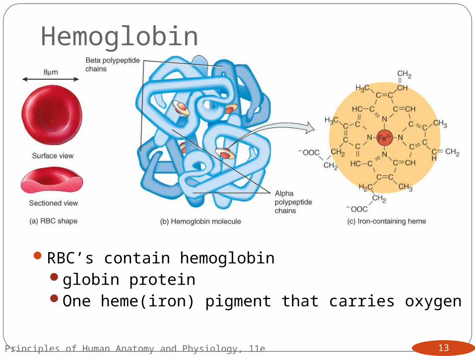
RBC’s contain hemoglobinglobin proteinOne heme(iron) pigment that carries oxygen
Principles of Human Anatomy and Physiology, 11e 13
Hemoglobin Transport
Principles of Human Anatomy and Physiology, 11e14
can carry 4 oxygen molecules from lungs to tissue cells
transports CO2 waste from tissue cells to lungs for release
transports nitric oxide & super nitric oxide helping to regulate BP through vasoconstriction and vasodilation, respectively.

Erythropoiesis:
Principles of Human Anatomy and Physiology, 11e15
What is it? The creation of erythrocytes (RBC’s)Where does it happen? in adult red bone marrow
of certain bonesWhat causes it? stimulus for erythropoiesis is
hypoxia (Figure 19.6).Major steps include:
1. Proerythroblast starts to produce hemoglobin2. Nucleus is ejected & a reticulocyte is formed 3. Reticulocytes escape from bone marrow into the
blood4. Remaining organelles get ejected to become a
mature RBC
WHITE BLOOD CELLS
Principles of Human Anatomy and Physiology, 11e16
Leukocytes (white blood cells or WBCs) are nucleated cells and do not contain hemoglobin.
Two principal types are granular (neutrophils, eosinophils, basophils)
and agranular (lymphocytes and monocytes) do not
have cytoplasmic granules. They differentiate into macrophages (fixed and wandering).
Leukocytes have surface proteins, as do erythrocytes.

WBC Physiology
Principles of Human Anatomy and Physiology, 11e17
Less numerous than RBCs5000 to 10,000 cells per drop of blood
Only 2% of total WBC population is in circulating blood at any given timerest is in lymphatic fluid, skin, lungs, lymph nodes &
spleen

Principles of Human Anatomy and Physiology, 11e18
WBC Type FunctionNeutrophils and wandering or fixed macrophages
phagocytosis
Eosinophils combat the effects of histamine in allergic reactions, phagocytize antigen-antibody complexes, and combat parasitic worms
Basophils develop into mast cells that liberate heparin, histamine, and serotonin in allergic reactions that intensify the inflammatory response
B lymphocytes differentiate into tissue plasma cells that produce antibodiesin response to the presence of foreign substances called antigens
T lymphocytes destroy foreign invaders directly

WBC examination
Principles of Human Anatomy and Physiology, 11e19
A differential white blood cell count is a diagnostic test in which specific white blood cells are enumerated. Because each type of WBC plays a different role, determining the percentage of each type in the blood assists in diagnosing the condition.

Neutrophil (Granulocyte)
Principles of Human Anatomy and Physiology, 11e 20

Eosinophil (Granulocyte)
Principles of Human Anatomy and Physiology, 11e 21
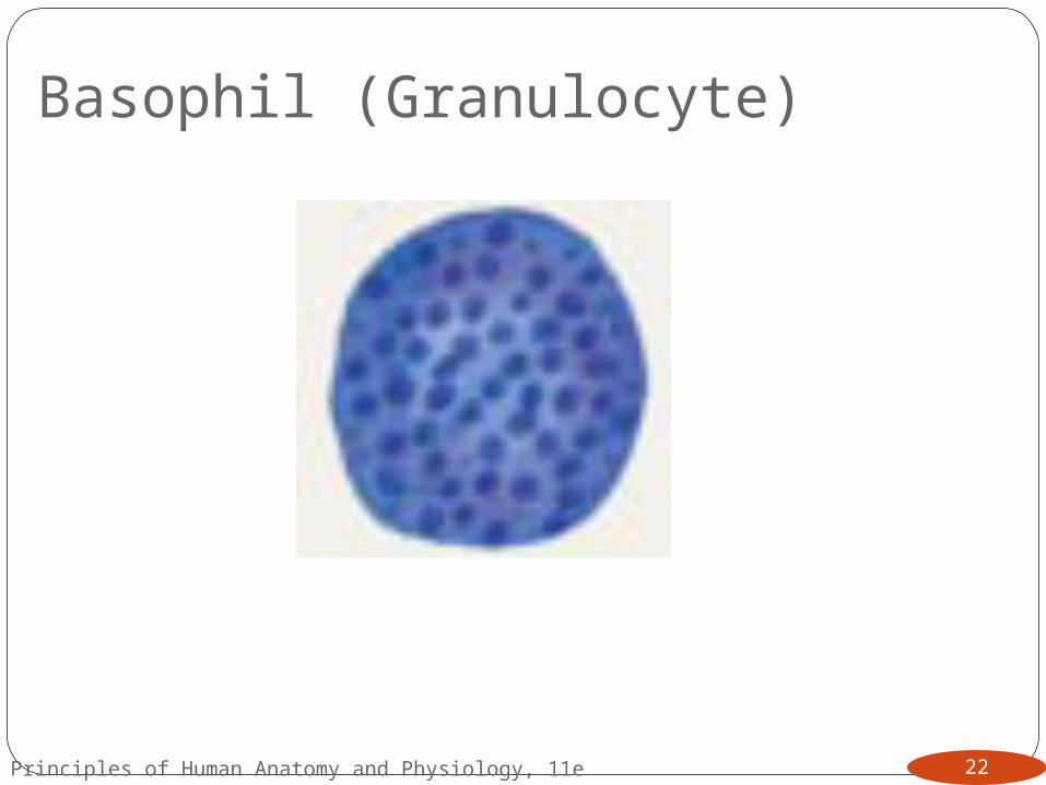
Basophil (Granulocyte)
Principles of Human Anatomy and Physiology, 11e 22

Lymphocyte (Agranulocyte)
Principles of Human Anatomy and Physiology, 11e 23
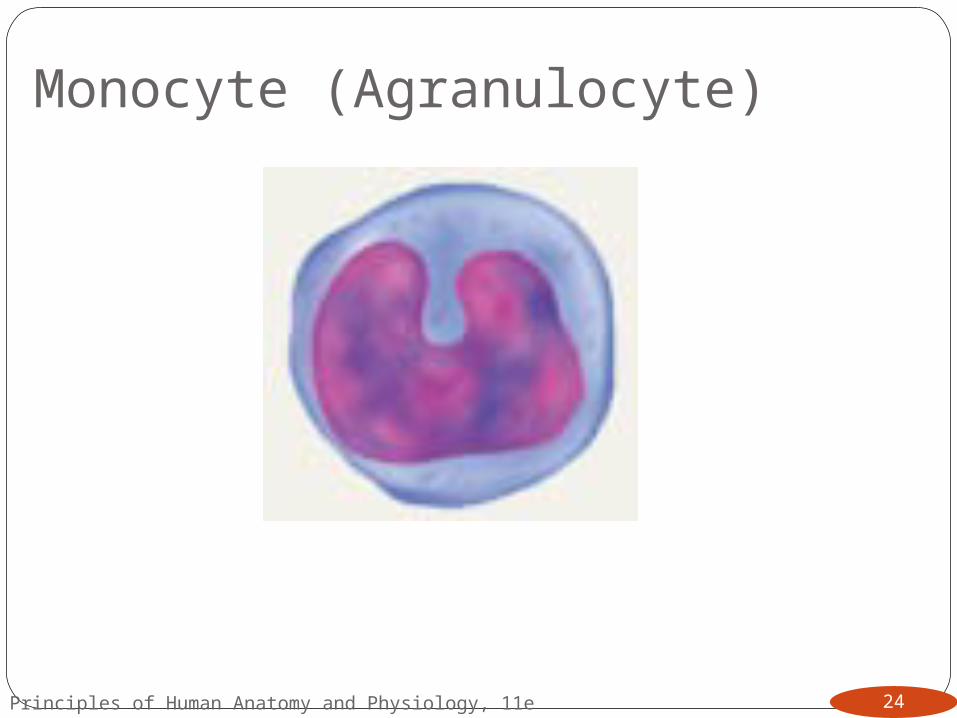
Monocyte (Agranulocyte)
Principles of Human Anatomy and Physiology, 11e 24
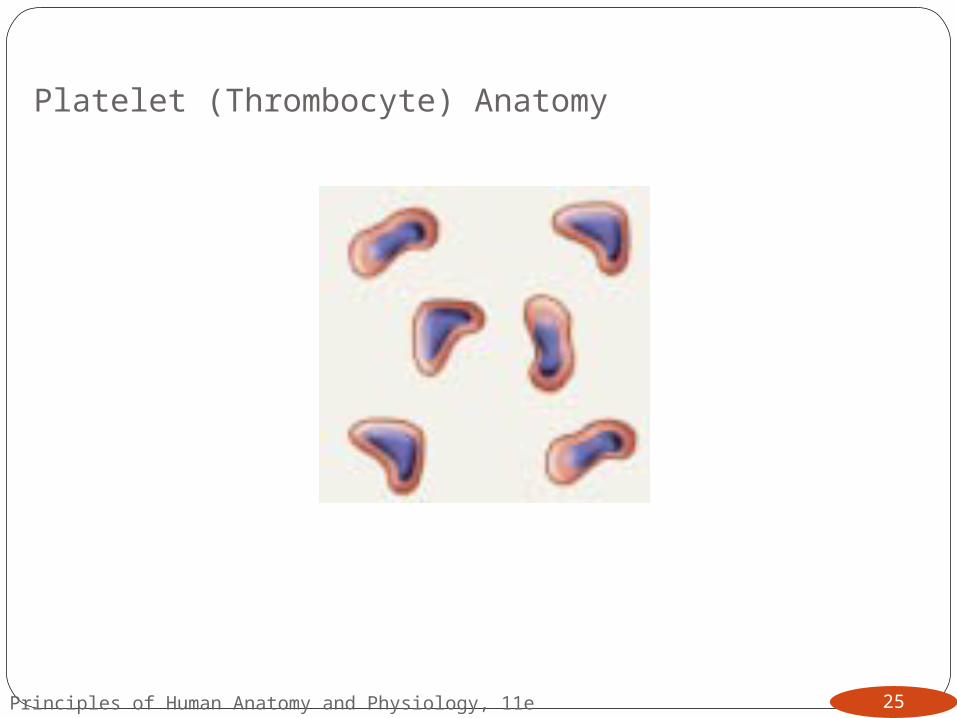
Platelet (Thrombocyte) Anatomy
Principles of Human Anatomy and Physiology, 11e 25

HEMOSTASIS
Principles of Human Anatomy and Physiology, 11e26
A clot is a gel consisting of a network of insoluble protein fibers (fibrin) in which formed elements of blood are trapped (Figure 19.10).
The chemicals involved in clotting are known as coagulation (clotting) factors; most are in blood plasma, some are released by platelets, and one is released from damaged tissue cells (Table 19.4).
Blood clotting involves a cascade of reactions that may be divided into three stages: formation of prothrombinase (prothrombin activator), conversion of prothrombin into thrombin, and conversion of soluble fibrinogen into insoluble fibrin (Figure 19.11).

The Blood Clotting ProcessMajor Steps:
1. Platelets become sticky when at the edge of a broken blood vessel and clump at the site.
2. Platelets then release protein clotting factor, thromboplastin.
3. Thromboplastin converts prothrombin (which is already in the blood) to the enzyme thrombin.
4. Thrombin then converts a plasma protein called fibrinogen into a sticky mesh of fibrin filaments (clot).
Both vitamin K and Ca are important in the clotting process
Principles of Human Anatomy and Physiology, 11e27
Role of Vitamin K in Clotting
Principles of Human Anatomy and Physiology, 11e28
Normal clotting requires adequate vitamin Kfat soluble vitamin absorbed if lipids are
presentabsorption slowed if bile release is insufficient
Required for synthesis of 4 clotting factors by hepatocytesfactors II (prothrombin), VII, IX and X
Produced by bacteria in large intestine

Intravascular Clotting
Principles of Human Anatomy and Physiology, 11e29
Thrombosisclot (thrombus) forming in an unbroken blood vessel
forms on rough inner lining of BVif blood flows too slowly (stasis) allowing clotting factors to
build up locally & cause coagulationmay dissolve spontaneously or dislodge & travel
Embolus clot, air bubble or fat from broken bone in the blood
pulmonary embolus is found in lungsLow dose aspirin blocks synthesis of thromboxane
A2 & reduces inappropriate clot formationstrokes, TIAs and myocardial infarctions

Anticoagulants and Thrombolytic Agents
Principles of Human Anatomy and Physiology, 11e30
Anticoagulants suppress or prevent blood clottingheparin
administered during hemodialysis and surgerywarfarin (Coumadin)
antagonist to vitamin K so blocks synthesis of clotting factorsslower than heparin
stored blood in blood banks treated with citrate phosphate dextrose (CPD) that removes Ca+2
Thrombolytic agents are injected to dissolve clotsdirectly or indirectly activate plasminogenstreptokinase or tissue plasminogen activator (t-PA)

Blood Groups and Blood Types
Principles of Human Anatomy and Physiology, 11e31
RBC surfaces are marked by genetically determined glycoproteins & glycolipids agglutinogens or isoantigensdistinguishes at least 24 different blood groups
ABO, Rh, Lewis, Kell, Kidd and Duffy systems
RH blood groups
Principles of Human Anatomy and Physiology, 11e32
AntigenPeople with Rh on RBC surface are Rh+. Normal
plasma contains no anti-Rh antibodiesAntibodies develop only in Rh- blood type & only
with exposure to the antigentransfusion of positive bloodduring a pregnancy with a positive blood type fetus
Transfusion reaction upon 2nd exposure to the antigen results in hemolysis of the RBCs in the donated blood

DISORDERS: HOMEOSTATIC IMBALANCES
Principles of Human Anatomy and Physiology, 11e33
Anemia Sickle-cell Hemophilia Disseminated intravascular clotting Acute leukemiachronic leukemia