chest radiography for prevalence surveys & active case ... · ¾chest radiography –...
TRANSCRIPT

Chest Radiography for Chest Radiography for prevalence surveys & active prevalence surveys & active
case detectioncase detection
Dr Narayan PENDSERadiologist Visiteur Médécin, Univ Hospital GenevaExpert, Radiology – WHO/STB, WHO/EHT [email protected]

Chest Radiography – Technologies and their use in prevalence surveys Chest Radiography in the field
Prepared for WHO/STB/TBS Guiding documents for planning, policy making, implementing and optimizing use of radiography in field conditions, particularly for prevalence surveys and for active case detection of TB


Aspects covered
Basics of radiography (field vs. indoor)Appropriate settings / prerequisites Costing Available technologies, comparison Manpower needs Criteria for selection Recommended specificationsInterpretation, radiation protection Practical tips References for specific areas

CXR in TB/TB-HIV
“Chest radiographs should be performed in all potential recipients – WHO Global TB Programme and UNAIDS, meeting report, 1998CXR plays an important role in Dx of TB in PLHIV and can also be an entry-point to Dx non-TB chest diseases –WHO, SN & EP TB, Recommendations for HIV-prevalent and resource-constrained settings, 2007

Recommendations (same document)CXR presentations of TB in HIV patients are now well characterized (A-IV)... significant role in shortening delays in Dx..... should be performed early...(A-II)Limitations like non-availability, difficulty in interpretation, need to be addressed (A) Research needed to identify innovative ways to enhance ability.......and to evaluate novel imaging techniques that might replace conventional radiography (A)

Case definitionSputum positive
2 sputum smear positive results 1 sputum smear + CXR s/o TB1 sputum smear + culture positivity
Sputum negativeSputum negative, culture +2 negative smears, CXR s/o TB
Not definite case (but active TB suggested by CXR)
TB Care with TB-HIV co-management IMAI 2007, Ethiopian Prevalence Survey Research Protocol 2009

Basics of Radiography
Generation Recording Displaying
X-ray generator X-ray tube X-ray table or stand Processing facility


Options
Conventional RadiographyConventional Radiography – automatic film processing Computed Radiography Direct Digital Radiography
Indirect Direct
Digital Imaging

Conventional Radiography
Time tested Cheap – initial cost, consumable-offset*SimpleDurable, sturdy, low maintenance Archival, Reproducibility Dark RoomThroughput time
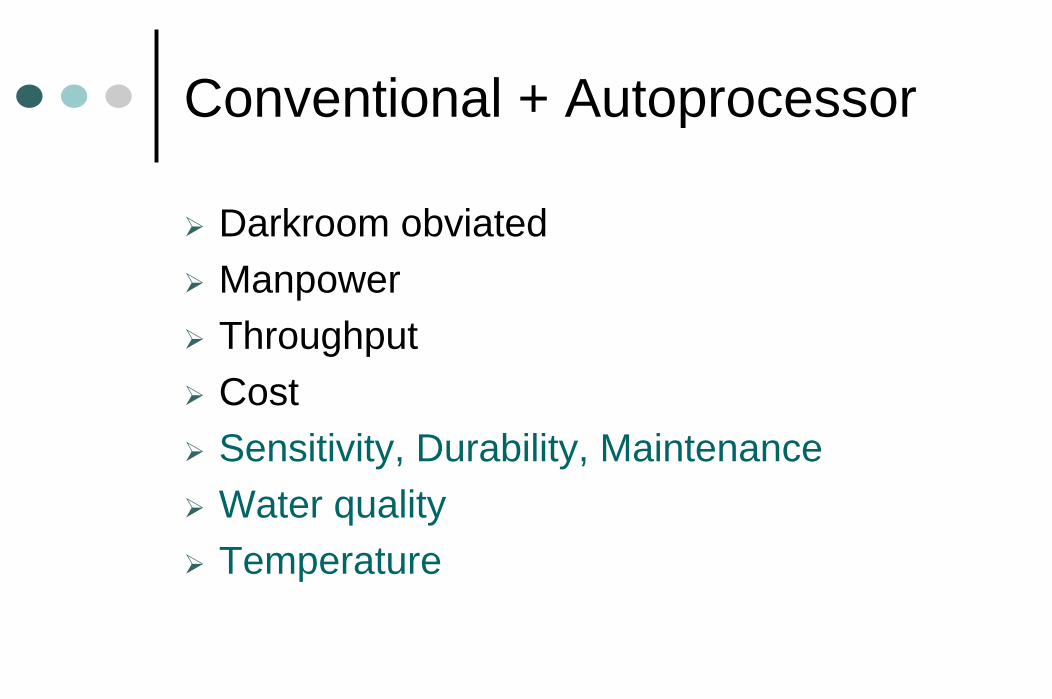
Conventional + Autoprocessor
Darkroom obviated Manpower Throughput Cost Sensitivity, Durability, Maintenance Water qualityTemperature

Digital Radiography*Digital Radiography*Analog to Digital conversion or Direct Advantages
Economical* : cost of films & chemicals, dark roomArchival : data archiving, documentation, storage spaceCommunicable : electronic transmission Robust & consistent Flexible : post processing possible
Disadvantages Infrastructure needsInterfacing Over interpretation mimics

Radiation – CR/ DR/ Film Screen, Trade off & repeat rates, overall low in DICostTechniques – Film Digitizer, Digital Camera/Video

Techniques Computed Radiography (CR)
Indirect digital Photostimulator Storage Phosphor to acquire image (IP)CR Reader Digital electronics/workstation for display & storage
Direct & Direct Digital Radiography (DR, DDR)Conversion of x-rays to direct signal captureFPD - Thin Film Transistors (TFT – amorphous Se / Si, CsI)Total electronic image capturePhotons - Pixel - Display

CR Image acquisition

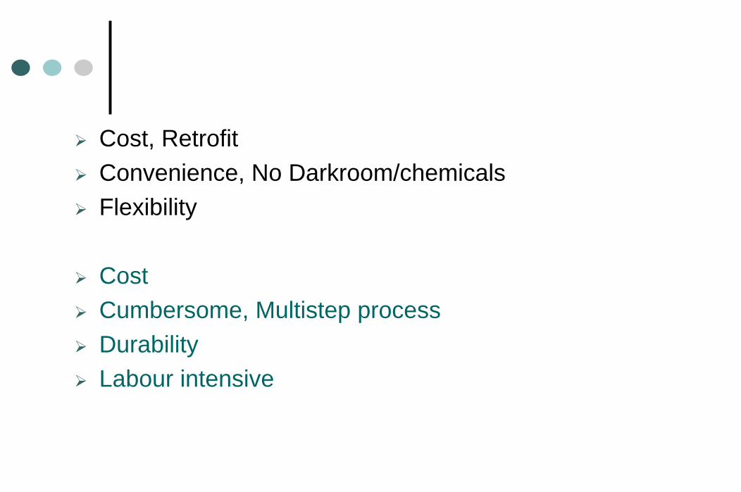
Cost, Retrofit Convenience, No Darkroom/chemicals Flexibility
CostCumbersome, Multistep process Durability Labour intensive

DR Image acquisition

Array of light sensitive detectors covered with light emitting x-ray phopshorLight generated by x-rays converted into charge Charge stored in capacitorsProcessing with read out of TFT arrayNO USER INTERVENTION

Flat panel detector systems – FPD Charge coupled device systems – CCD & CMOS Slot scanning type systems Photon counting type systems

FPD- X-ray convertor material on TFT matrixEach element has capacitor & switching transistorRead out from each detector

CCD & CMOS : x-ray conversion to visible light using scintillators or phosphors -> channelled by mirrors/prisms Slot scanning systems : like CT ScannogramsPhoton counting systems : Like slot scanning but use crystalline Se as scintillate. High DQE and SNR

Post processing- Data

Post processing - Display

Limitations

CR vs DR
Attribute CR DR
Positioning ++++ ++Replacement of FSR ++++ -System cost +++ +Patient throughput + +++Ease of use + ++++PACS compatibility ++ +++

Nutshell
CR : Retrofit advantage Cost advantage Quality comparable
DR : Superior quality and throughputGood for higher average number of case
5 year horizon for CR rule at the moment

Proposed algorithm for selection

Teleradiology
Teleradiology is the transmission of images and associated data between locations for the purpose of primary interpretation or consultation and/or clinical review Local benefits : Faster, 24x7 reporting Regional : Linking of centres for resource sharingNational : Increased capacity, sharing of expertise, reaching the unreached

Not an alternative to local capacity building Pre requisite : local expertise for service delivery Bridging a gap or for expert opinions Picture Archiving and Communication System (PACS)

Computer Aided Diagnosis
Information, Knowledge, WisdomSuccess in Mammography, Nodule detection in CTAssist tool – not for interpretation Not a substitute for education and training Tried for pulmonary nodules, ILDComplexities : A vs D, segmentation, QA, standard positioning

Inter Observer variability 222 images, 20 radiologists Standard definitionsAgreement possibleTB – smaller / older studies

RADIATION !
Deterministic – dose related Stochastic – not dose related
No “safe” level* – should be understood in the context of benefit versus risk

A Perspective: Other Risks People A Perspective: Other Risks People TakeTake
Deaths/10,000/yearDeaths/10,000/year
Smoking (all causes)Smoking (all causes) 3030MiningMining 6.06.0ConstructionConstruction 3.93.9Cardiac Cardiac CathCath (all causes)(all causes) 3.33.3Driving a car (Driving a car (avgavg, one year), one year) 2.42.4BoatingBoating 0.50.5AP lumbar spineAP lumbar spine 0.060.06Chest XChest X--ray Exam (AP +Lat)ray Exam (AP +Lat) 0.020.02

Pregnancy & Radiography
Advice from HPA – RCR
For most Dx (incl. CXR) associated risk of childhood CA are very low and acceptable when compared with natural risk
Radiation doses resulting from Dx procedures present a negligible risk of induced hereditary disease in descendants of the unborn child

ACR Guidelines
Some procedures (incl. CXR in 1st & 2nd trimester) render so low exposures that pregnancy status need not be considered for a “medically indicated”exam, as long as good radiation practice is ensured
3rd trimester : with good technique exposure remains very low
No risk of deterministic effect < 50mGy

Occupational Exposure RisksOccupational Exposure Risks
Occupational radiation exposure mostly scatter from Occupational radiation exposure mostly scatter from patientpatientAt 1 meter, occupational exposure (if no apron is worn) At 1 meter, occupational exposure (if no apron is worn) is 0.1% of that which enters the patientis 0.1% of that which enters the patientMinimizing patient dose minimizes your doseMinimizing patient dose minimizes your doseOccupational Protection: Occupational Protection: The Cardinal RulesThe Cardinal Rules::
TimeTimeDistanceDistanceShieldingShielding

Where does exposure come from:Where does exposure come from:Population ExposurePopulation Exposure
% of% of NumberNumber mRmR perper PopulationPopulationExamExam ExamsExams of Examsof Exams ExamExam Exposure (R)Exposure (R)
Port Chest 10 15,000 12 180PA Chest 15 24,000 18 432Body CT 17 28,000 1600 44,800Head CT 8 12,000 8000 96,000GI Fluoro 2.5 4,000 7500 30,000Spec/Card 8 13,000 50,000650,000

Radiology Exposures and DosesRadiology Exposures and Doses

THANK YOU !