cis-acting sequences required encapsidation …jvi.asm.org/content/65/6/3309.full.pdf ·...
TRANSCRIPT

Vol. 65, No. 6JOURNAL OF VIROLOGY, June 1991, p. 3309-33160022-538X/91/063309-08$02.00/0Copyright C) 1991, American Society for Microbiology
cis-Acting Sequences Required for Encapsidation ofDuck Hepatitis B Virus Pregenomic RNA
RUSSELL C. HIRSCH,1 DANIEL D. LOEB,2 JONATHAN R. POLLACK,' AND DON GANEM2*Departments of Microbiology and Immunology2 and Biochemistry and Biophysics,' University of California Medical
Center, San Francisco, California 94143-0502
Received 27 December 1990/Accepted 22 March 1991
Hepadnavirus reverse transcription requires that pregenomic RNA first be selectively packaged into acytoplasmic core particle. This process presumably requires the presence of specific recognition sequences onthe pregenomic RNA. To define the cis-acting sequences required for pregenome encapsidation in the duckhepatitis B virus (DHBV), we assayed the packaging efficiency of a series of pregenomic RNA deletion mutantsand hybrid DHBV/lacZ fusion transcripts. The 5' boundary of the packaging signal lies within the precoreregion, starting approximately 35 nucleotides from the cap site of pregenomic RNA; thus, the DR1 sequencerequired for proper viral DNA synthesis is not included in this signal. To define the 3' boundary of theencapsidation signal, fusion transcripts bearing foreign (lacZ) sequences fused to DHBV at different sites 3' tothe pregenomic RNA start site were examined. A surprisingly large region of the DHBV genome proved to berequired for packaging of such chimeras, which are efficiently encapsidated only when they contain the first1,200 to 1,400 nucleotides of DHBV pregenomic RNA. However, mutant genomes bearing insertions within thisregion are packaged efficiently, making it likely that the actual recognition elements for encapsidation aresmaller discontinuous sequences located within this region.
Hepadnaviruses are small, hepatotropic DNA viruses thatcan produce persistent infections of hepatocytes in a varietyof animal hosts (for review, see reference 10). In suchinfections, their DNA genomes are replicated via reversetranscription of an RNA intermediate (25). As in retrovi-ruses, hepadnavirus reverse transcription does not occur ina soluble form in the cytoplasm but rather takes place in asubviral core particle composed of the major capsid (C)protein, the polymerase (P) protein, and the RNA template(9). Hence, a key step in the replicative cycle of the virus isthe selective encapsidation of the appropriate viral RNA intosubviral core particles.The basic steps in the hepadnavirus life cycle are now well
established. Following viral entry, the genome is deliveredto the nucleus, where it is transcribed into two classes ofRNAs (Fig. 1): subgenomic transcripts, which serve asmRNAs for viral surface proteins, and genome-length RNAs(4, 7). Genomic RNAs are bifunctional; they serve as bothmRNA (for C and P proteins) and as the templates forreverse transcription (4, 10). Concomitant with or followingthe translation of these proteins, genomic transcripts areassembled into nascent subviral cores, whereupon reversetranscription begins (8).
Until recently, little was known about the proteins thatmediate the packaging of viral RNA into cores and about theparticular features of the genomic RNA that dictate itsselective encapsidation. We have recently employed a ge-netic approach to demonstrate that in the duck hepatitis Bvirus (DHBV), the P gene product is required not only forreverse transcription but also for the efficient packaging ofthe genomic RNA into viral cores: mutant viruses unable tosynthesize P gene products produce empty cores lackingviral RNA (13). Similar findings for the human hepatitis Bvirus (1, 12) have also been made. Presumably, the packag-ing of genomic RNA occurs as a result of specific interac-
* Corresponding author.
tions between the P gene product (or a protein complexcontaining the P gene product) and a cis-acting encapsidationsignal on the genomic RNA.The structure and location of the DHBV encapsidation
signal are unknown. To map and characterize this signal, wehave analyzed the encapsidation efficiency of RNAs pro-duced from a series of DHBV deletion mutants and fromgene fusions between DHBV and heterologous sequences.In this article, we report that this signal contains essentialelements that are dispersed over a surprisingly large region,extending from the precore region through the first third ofthe P open reading frame.
MATERIALS AND METHODS
Materials. Enzymes were purchased from New EnglandBioLabs and were used according to the manufacturer'sprotocols. Radionuclides were purchased from AmershamCorporation.
Plasmids and plasmid construction. pPC1 is a tandemdimer of the European strain of DHBV (24) bearing an 8-bpClaI linker insertion in the AccI site at nucleotide (nt)position 2577. To generate pPC1, an NcoI (DHBV nt posi-tion 2351) to EcoRI fragment (DHBV nt position 3020) ofpD3-SP65, a unit-length DHBV genome inserted at theEcoRI site of pSP65 (Promega Biotec), was subcloned intopSP65 to yield pD3N/R. pD3N/R was linearized with AccIand blunted with Klenow, and an 8-bp ClaI linker wasinserted with T4 DNA ligase to yield pD3N/R-ClaI. TheNcoI to EcoRI fragment of pD3N/R-ClaI was recloned intopD3-SP65 cut with NcoI and EcoRI to yield a unit-lengthDHBV genome bearing a ClaI linker insertion in the AccIsite at nt position 2577. This genome was reconstructed intoa tandem dimer, pPC1. pPC2 was generated in precisely thesame manner except that two ClaI linkers were cloned intothe AccI site.For construction of deletion mutants (mutants Al to A5) in
the DHBV precore region, we generated pD3-Xho-PCS.
3309
on July 22, 2018 by guesthttp://jvi.asm
.org/D
ownloaded from

3310 HIRSCH ET AL.
FIG. 1. Genetic and transcriptional map of DHBV. The innercircles represent the partially double-stranded DNA genome. The 5'end of the full-length minus strand is linked to a terminal protein(solid oval). The 5' end of the plus strand is in a fixed position andis linked to an oligoribonucleotide (wavy line), whereas the 3' end ofthe plus strand is variably situated as indicated by the dotted line.The three thin lines represent the major transcripts seen in infectedcells: the 2.1-kb mRNA encoding presurface antigen, the 1.8-kbmRNA encoding surface antigen, and the 3.3-kb RNA serving bothas the template for reverse transcription and as the mRNA encodingthe core antigen and the P gene product. The thick outer barsindicate the coding regions for the presurface/surface, precore/core,and P proteins. An indicates the mRNA poly(A) tail.
pD3-Xho-PCS was generated by cloning a synthetic oligo-nucleotide containing PstI, ClaI, and SpeI sites (in thatorder) into a unique XhoI site that had been created at nt
position 2548 of pD3-SP65 by using site-specific mutagene-
sis. pD3-Xho-PCS was digested with PstI and SpeI and thensubjected to digestion with exonuclease III and nuclease Si(double-strand nested deletion kit; Pharmacia). The extent ofthe deletions was verified by DNA sequencing.For construction of overlength linear genomes with dele-
tions only in the 5'-terminal redundancy (e.g., A2-5' andA5-5'), pDl.5G (an overlength DHBV genome betweenpositions 1658 and 3020) was cut with ScaI, treated with calfintestinal alkaline phosphatase and digested with NsiI. Theresultant fragments were ligated with either A2 or A5 ge-
nomes that had been cut with NsiI and treated with calfintestinal alkaline phosphatase, yielding a tandem dimericgenome bearing the appropriate deletion only in the regioncorresponding to the 5'-terminal redundancy. For construc-
tion of overlength linear genomes with deletions only in the3'-terminal redundancy (e.g., A2-3' and A5-3'), pD.5G (a
partial DHBV genome between positions 1658 and 3020) wascut with EcoRI and treated with calf intestinal alkalinephosphatase. The resultant fragment was ligated with eitherA2 or A5 genomes that had been digested with Scal, treated
with calf intestinal alkaline phosphatase, and then cut with
EcoRI. The ligation yielded overlength DHBV genomes (1.5
mers between positions 1658 and 3020) bearing the appropri-
ate deletion only in the region corresponding to the 3'-terminal redundancy.The DHBV/lacZ fusion transcripts (pseudopregenomes)
were derived from pCMV-DHBV9 (6) as the source of theCMV-IE promoter-driven DHBV sequences and from pON3(22) as the source of the Escherichia coli lacZ sequences andthe simian virus 40 polyadenylation signal. Pseudoprege-nomes p136-X and p319-N were derived, respectively, frompCMV-DHBV9 from which sequences from XbaI (DHBV ntposition 2662) or from NsiI (DHBV nt position 2845) toBamHI (polylinker) were removed and were replaced with a3.0-kb HpaI-XbaI fragment from pON3. p508-H was gener-ated by removing the Hindlll (DHBV nt position 14) toBamHI (polylinker) fragment of pCMV-DHBV9 and replac-ing it with the 2.5-kb ClaI-XbaI fragment of pON3. p814-Swas derived from pCMV-DHBV9 from which the sequencebetween SmaI (DHBV nt position 320) and BamHI (poly-linker) has been replaced with a 2.2-kb EcoRV-XbaI frag-ment of pON3. p1212-R was generated by removing theEcoRV (DHBV nt position 718) to BamHI (polylinker)fragment of pCMV-DHBV9 and replacing it with the 1.8-kbAvaII-XbaI fragment of pON3. p1396-T and p1780-K werederived, respectively, from pCMV-DHBV9 from which se-quences between TthlllI (DHBV nt position 902) or KpnI(DHBV nt position 1290) and BamHI (polylinker) wereremoved and replaced with the 1.4-kb SacI-XbaI fragment ofpON3. p1780-K-src was generated by excising the 821-bpBglIl (DHBV nt position 391) to XhoI (DHBV nt position1212) fragment and replacing it with the 800-bp MluI-BglIllfragment of the chicken c-src gene (16).pD3-0X118 was constructed by cutting pD3-SP65 with
NsiI (DHBV nt position 2845), generating blunt ends bytreatment with T4 DNA polymerase, and inserting a 118-bpHaeIII fragment (nt 4758 to nt 4876) of 0X174 DNA.
Cell culture and transfections. LMH avian hepatoma cells(6) were grown in Dulbecco's minimum essential medium-Ham's nutrient mixture F12 supplemented with 10% fetalcalf serum and were passaged every 3 to 4 days at a 1:3dilution. DNA transfections were performed by the calciumphosphate coprecipitation method exactly as described pre-viously (11).RNA preparation and RNA analysis. Polyadenylated total
cellular RNA was extracted from LMH cells 72 h posttrans-fection as previously described (11, 17). Encapsidated RNAwas isolated from LMH cells 72 h posttransfection bypurifying cytoplasmic core particles with polyethylene gly-col precipitation followed by proteinase K digestion andphenol extraction as described previously (11, 17). Primerextension analysis of pregenomic RNA with a synthetic18-base oligonucleotide (spanning positions 2658 to 2640within the core gene) was carried out as described previously(17). RNase protection analysis of pregenomic RNA andsynthesis of [a-32P]CTP-labeled RNA probes was carried outas described previously (13). Probe 4421inP was transcribedfrom plasmid p4421inP, which has been described previously(13). Probe lacZ-P108 was transcribed from plasmid placZ-P108. Plasmid placZ-P108 was generated by cloning the425-bp MluI-MluI fragment of pON3 into the HincII site ofpGEM 3Z (Promega Biotec); linearization with HindIII andtranscription with T7 RNA polymerase (Promega Biotec)yields a 475-nt transcript with about 50 nonhybridizingpolylinker nucleotides and 425 nt of lacZ that are comple-mentary to the fragments of lacZ cloned into all of theDHBV/lacZ fusion pseudopregenomes. Probe D3-X/R-Pwas transcribed from plasmid pD3-X/R-P. Plasmid pD3-X/R-P was generated by cloning the 358-bp XbaI (DHBV nt
J. VIROL.
on July 22, 2018 by guesthttp://jvi.asm
.org/D
ownloaded from

SEQUENCES FOR DHBV PREGENOMIC RNA ENCAPSIDATION 3311
position 2662) to EcoRI (DHBV nt position 3020) fragmentinto the HinclI site ofpGEM 3Z such that linearization withHindlll and transcription with T7 RNA polymerase yields a414-nt transcript with 56 nonhybridizing polylinker nucleo-tides. Since this probe is derived from sequences within theterminal redundancy of pregenomic RNA, it is complemen-tary to 358 nt at the 5' end of pregenomic RNA and 140 nt atthe 3' end of pregenomic RNA.Note that both the American (19) and European (24)
strains of DHBV have been employed in these studies. Inexperiments where complementation is performed, the wild-type (WT) donor is always of the same strain as the mutantrecipient.
RESULTS
Experimental strategy. To define the cis-acting site re-quired for DHBV genomic RNA encapsidation, we con-structed mutant DHBV genomes and assayed their ability topackage genomic RNA. To assay for RNA packaging, themutant DNAs were transfected into the avian hepatoma cellline LMH (6) by calcium phosphate-mediated transfection.Following transfection, we isolated total cellular poly(A)+RNA from one-half of each sample of transfected cells; fromthe other half, we purified cytoplasmic core particles andextracted their nucleic acids. The genomic RNA present inequal portions of each preparation was then quantitated byprimer extension or RNase mapping; the ratio of encapsi-dated to total RNA is a reflection of the packaging efficiencyof the mutant pregenome (13, 17).
Several of the mutations we have constructed affect notonly the cis-acting regions of pregenomic RNA but also thecoding regions for the C and/or P gene products, both ofwhich are essential for genomic RNA encapsidation (1, 3,13). Accordingly, in these cases, the mutant genomes werecotransfected with a WT genome to provide (in trans) theviral proteins required for encapsidation (17). Although thistrans complementation is less efficient than WT packaging(in which P proteins act preferentially in cis to encapsidatetheir own pregenomic RNAs [13]), it is sufficient to allowanalysis of such constructions.
Sequences residing within the terminal redundancy of ge-nomic RNA are required for encapsidation. Our first clue tothe location of cis-acting encapsidation sequences within thepregenome came from mutants PC1 and PC2 (Fig. 2).Mutants PC1 and PC2 bear, respectively, a single or double(tandem) insertion of an 8-bp ClaI linker at an AccI sitewithin the terminal redundancy of the genomic RNA. Ac-cordingly, as depicted in Fig. 2b, these insertions wererepresented at both ends of the mutant pregenome. Trans-fection of cells with these mutants results in levels ofprogeny DNA synthesis that are drastically reduced com-pared with those of the WT (data not shown). Primerextension analysis of total cellular poly(A)+ RNA from cellstransfected with PC1 (Fig. 2a, lane 3) and with PC2 (data notshown) revealed that they accumulate levels of properlyinitiated genomic RNA equivalent to that of the WT (lane 1;note that the PC1 extension product is 8 nt longer than theWT extension product because of the linker insertion). Butwhen we examined the encapsidated RNA from cells trans-fected with these mutants, we found that PC1 (lane 4) issignificantly impaired compared with the WT (lane 2) inpregenome encapsidation and that encapsidated PC2 RNA isessentially undetectable (data not shown).Mutants PC1 and PC2 are able to complement core-
defective mutants with WT efficiency, and cotransfection
a
b
Mutant: WT Pcim fm G A T C
RNA: T C T C
5'end of ',
pregenome
1 2 3 4 5 6 7 8
-4.-
DR 151
CIa linker(s)
DR 2 DR 1CIa linker(s)
Cla linker(s)
FIG. 2. Primer extension analysis of total and encapsidatedpregenomic RNA in transfected cells. (a) Cells were transfectedwith either WT DHBV DNA (lanes 1 and 2) or PC1 DNA (lanes 3and 4). From one half of the transfected cells, total cellular poly(A)+RNA (T) was isolated, and from the other half, cytoplasmic cores(C) were purified and the nucleic acid was extracted. The genomicRNA present in equal portions of each nucleic acid preparation wasquantitated by primer extension of an end-labeled oligonucleotideprimer complementary to the 5' region of the C open reading frame.Lanes 5 through 8, sequence ladder of WT DHBV DNA genomegenerated with the same primer utilized for primer extension. Thebands representing the 5' ends of WT pregenomic RNA and PC1pregenomic RNA are indicated by a solid arrow and a broken arrow,respectively. The PC1 extension product is 8 nt longer than the WTextension product because of the linker insertion. (b) Schematicrepresentation of mutants PC1 and PC2 and of the strategy forprimer extension. Mutants PC1 and PC2 harbor, respectively, singleand tandem insertions of an 8-bp ClaI linker at the AccI site withinthe terminal redundancy of pregenomic RNA. Primer extension wasperformed with a 5'-end-labeled primer (solid circle) complementaryto the 5' region of the C gene and to the identical sequence in thedownstream terminal redundancy of the RNA. Extension productsother than the authentic 5' end of pregenomic RNA are due to strongstops to extension from the downstream priming site. The intensityof the extension product that is longer than the authentic 5' end ofpregenomic RNA is greatly decreased in the pool of encapsidatedRNA relative to the pool of total cellular poly(A)+ RNA because ofthe 3'-to-5' degradation of pregenomic RNA by RNase H duringreverse transcription.
with WT genomes ruled out the possibility that precoreinsertions had created a dominant negative phenotype. Ad-ditionally, both mutants can complement a P- mutant ge-nome as effectively as WT DHBV, indicating that levels of Pgene expression are normal for this mutant (data not shown).Since the failure of PCI and PC2 to encapsidate theirgenomic RNA is not attributable to aberrant C or P proteinsynthesis, we conclude that their phenotype is due to adefect in a cis-acting signal for encapsidation.To further define the extent of this signal, we engineered a
set of mutants, Al to A5, bearing deletions within the regionof the genome that comprises the terminal redundancy ofgenomic RNA (Fig. 3a). Recircularized monomeric genomesbearing these lesions were transfected into LMH cells; thus,in these mutants (as in PCI), the lesions are represented inboth ends of pregenomic RNA. Total or encapsidated RNAwas then examined by RNase protection (Fig. 3b and c). Theuniformly labeled RNA probe used in this RNase protectionassay, 4421inP, contains a linker not found in pregenomicRNA (Fig. 3c). Therefore, following annealing to genomicRNA and removal of single-stranded regions by RNasetreatment, two product fragments, of 430 and 300 nt, will be
VOL. 65, 1991
on July 22, 2018 by guesthttp://jvi.asm
.org/D
ownloaded from

3312 HIRSCH ET AL.
a Pregenome llR I Acc I
AAGAA- T~-'-ACcccKP T:::A2T:CT
I 10 20 30 40 50
MI 1ANrI PACKAGING;
AI)R I
xlI * * +++
A 2 4 __
o -
i) (Genonie:RNA:
70n0
WT A\
C C1 T (: T
do
430) --
300 j*
1 2 3 4 5 6 7 #9 10It
C
IIFimpI.la etC
I'II)cl flomli
core-1- -- 430 bp 14300bl1)*
FIG. 3. RNA packaging by mutants bearing deletions in bothterminal redundancies. (a) Schematic representation of the 5' end ofpregenomic RNA. The extent of the deletion in mutant genomesADR1 and Al to A5 are indicated by the double-headed arrows.Mutant ADR1 is a precise deletion of the DR1 sequence in the5'-terminal redundancy of pregenomic RNA. Since mutants Al to A5bear deletions in the terminal redundancy of the genomic RNA, thelesion is represented twice in the RNA: once in the 5'-terminalredundancy (as depicted in this figure) and once in the 3'-terminalredundancy (data not shown). The encapsidation efficiency of themutant genomes relative to that of the WT is indicated by + and -
signs as follows: ++ +, equivalent to WT; +, <30% of WT; -, nodetectable encapsidated RNA. (b) Cells were transfected with WTDHBV DNA or mutant DNA; from one-half of each sample, totalcellular poly(A)+ RNA was isolated, and from the other half,cytoplasmic cores were purified and the nucleic acid was extracted.The genomic RNA present in equal portions of each preparation wasquantitated by nuclease protection as described for panel c. Lanes:1, size standards; 2, full-length (780 nt) undigested probe; 3, probedigested in absence of added sample RNA; 4, 6, 8, and 10, fragmentsprotected by core RNA (C) from cells transfected with WT, A2, A3,and A5 DNA, respectively; 5, 7, 9, and 11, fragments protected bytotal cellular poly(A)+ RNA (T) from cells transfected with WT, A2,A3, and A5 DNA, respectively. (c) Nuclease protection assay. SP6transcription of p4421inP generates a uniformly labeled 780-nt RNAprobe (wavy line) that contains a linker (black box) not found inpregenomic RNA. Following annealing to pregenomic RNA andremoval of single-stranded regions by nuclease treatment, twoproduct fragments of 430 and 300 nt are generated.
generated. As expected, in WT-transfected cells, abundantquantities of genomic RNA are present in the pool of totalcellular poly(A)+ RNA (Fig. 3b, lane 5). Analysis of encap-sidated RNA from the same cells confirms that a largefraction of the genomic RNA is found within core particles(lane 4). Similar analysis of deletion mutants A2, A3, and A5reveals that A2 encapsidates its genomic RNA at WT effi-ciency (lanes 6 and 7), while A3 is greatly decreased inpackaging efficiency (lanes 8 and 9), and in A5 (lanes 10 and11), encapsidated RNA is nearly undetectable. Deletionmutant Al, like A2, encapsidates at WT efficiency, anddeletion mutant A4, like A5, is greatly decreased in encapsi-dation efficiency (data not shown). Furthermore, since dele-tion mutants Al to A5 are able to complement core-defectivemutants as efficiently as WT, we conclude that the failure ofmutants A3, A4, and A5 to package their genomic RNA atWT efficiency is due to a defect in their cis-acting encapsi-dation signal. These data indicate that sequences within theterminal redundancies of the genomic RNA are required forencapsidation.
Sequences residing in the 5'-terminal redundancy are re-quired for encapsidation. In order to determine whethersequences within one or both of the terminal redundanciesare recognized by the packaging apparatus, deletion mutantsA2 and A5 were reconstructed into overlength linear ge-nomes such that the effects of deletions in the 5' and 3'copies of the terminal redundancy could be assayed inde-pendently (Fig. 4a). LMH cells were transfected with mu-tants A2 or A5 bearing deletions in either the 5'-terminalredundancy (A2-5' and A5-5'; Fig. 4a) or in the 3'-terminalredundancy (A2-3' and A5-3'; Fig. 4a), and total or encapsi-dated RNA was examined by RNase protection assay withthe same uniformly labeled RNA probe, 442linP, depicted inFig. 3b and c.
Recall that mutant A5, bearing a deletion in both the 5'-and 3'-terminal redundancies, is defective for RNA encap-sidation. Analysis of mutants A5-5' and A5-3' reveals thatboth mutants accumulate abundant quantities of genomicRNA in the pool of total cellular poly(A)+ RNA (Fig. 4b,lanes 8 and 10). (The 5' deletions were cloned as tandemdimers, while the 3' deletions were cloned as 1.5-mers. Forunknown reasons, we consistently observe lower levels ofpregenomic RNA in total cellular poly(A)+ RNA in cellstransfected with dimeric genomes than in cells transfectedwith 1.5-mer genomes (compare lanes 4 and 8 with lanes 6and 10). This is also true for WT DHBV genomes.) Exami-nation of the encapsidated RNA from the same cells revealsthat mutant A5-5' (Fig. 4b, lane 7), like mutant A5, isdefective for RNA encapsidation, while mutant A5-3' (lane9) encapsidates at WT efficiency. Analysis of encapsidatedand total cellular poly(A)+ RNA from mutants A2-5' (lanes 3and 4) and A2-3' (lanes 5 and 6) reveals that, like that ofmutant A2, their genomic RNA is encapsidated at WTefficiency. This demonstrates that sequences required in cisfor pregenome encapsidation lie in the 5'-terminal redun-dancy within the precore region.The results obtained from the previous experiments sug-
gested that the DR1 sequence is not required for encapsida-tion. To explicitly address this, we generated mutant ADR1by engineering a precise deletion of the DR1 sequence withinthe 5'-terminal redundancy (Fig. 3a). Analysis of encapsi-dated and total cellular poly(A)+ RNA from cells transfectedwith ADR1 revealed that it is encapsidated at WT efficiency(data not shown). Taken together, the data suggest that the5' boundary of the packaging signal lies within the precoreregion approximately 35 nt downstream of the genomic RNA
A 3
IL S
J. VIROL.
on July 22, 2018 by guesthttp://jvi.asm
.org/D
ownloaded from

SEQUENCES FOR DHBV PREGENOMIC RNA ENCAPSIDATION 3313
aDRI DR2 DR I
MU:TANT
A5-3l
-MS
.'5-3
b(;enr)me:
RNA
711 -8 )
A-2S'C 2-3' V5-5 A!L;-
C T (: T C C
430 -*-
4"m- _i
1 2 3 4 5 6 7 8 9
FIG. 4. RNA encapsidation by mutant DNAin either the 5' or 3' copy of the genomic RNA tei(a) Schematic diagram of pregenomic RNA. Diand A5 (Fig. 3a) were reconstructed into cloned cbearing the corresponding deletion in either thedancy (A2-5' and A5-5') or the 3'-terminal reduA5-3'). The position and relative size of themutants are depicted by the double-headed arrmthe pregenomic RNA of these mutants to tindicated by a Yes or a No. (b) RNase proteperformed exactly as described in the legendLanes: 1, full-length undigested probe (780 nt); 2the absence of added sample RNA; 3, 5, 7,protected by core RNA (C) from cells transfecteA5-5', or A5-3' DNA, respectively; 4, 6, 8,protected by total cellular poly(A)+ RNA (T) frcwith A2-5', A2-3', A5-5', and A5-3' DNA, respe
cap site and that sequences 5' of this pcDR1, are not required in cis for encapsidal
Sequences distributed over a large segm
nomic RNA are required to mediate encapskRNA sequence into viral cores. Having defirary of the packaging signal, we sought tboundary. To this end, we constructed a
lacZ fusion pseudopregenomes (Fig. Sa)ability to be encapsidated. These are preg(which various segments from the 5' en
pregenomic RNA are fused to 3'-terminal fithe constructs initiate at the cap site of pregare promoted by a CMV-IE promoter an(lated at a downstream poly(A) site derived J40. The pseudopregenomes were constructotal size of the RNA is nearly equivalent 1
DHBV pregenomic RNA. They are designof the number of nucleotides of the 5' en
pregenome contained in the construct. (TI
numbered such that nucleotide 1 corresponds to the 5' end ofauthentic pregenomic RNA.) Plasmids encoding pseudopre-genomes were cotransfected with WT DHBV DNA as a
PACKAGING donor for C and P gene products, and their ability to beencapsidated was assayed by nuclease protection using alacZ-specific probe. Annealing of the lacZ-specific probe toany of the DHBV/lacZ fusions results in protection of a425-nt fragment. In cells transfected with DHBV/lacZ fu-sions containing 508, 814, 1,212, and 1,396 bp of DHBVsequence, normal quantities of lacZ-containing RNA appearin the total cellular poly(A)+ RNA (Fig. 5b, lanes 4, 6, 8, and10). Analysis of encapsidated RNA from the same transfec-tants (lanes 3, 5, 7, and 9) reveals that DHBV/lacZ fusionscontaining up to 814 nt of DHBV sequence are not encapsi-dated (lane 5), fusions with 1,212 nucleotides of DHBV areinefficiently encapsidated (lane 7), and fusions with 1,396
.41nucleotides of DHBV are encapsidated with WT efficiency
T (lane 9).In light of what is known about the encapsidation signal in
HBV (15) and in retroviruses (2, 20, 21), we were surprisedby the large extent of the DHBV genome required to mediateefficient packaging of a foreign RNA into subviral coreparticles. We were initially concerned that the additionallacZ sequences present in constructs possessing less than1,396 bp of the 5' end of the DHBV pregenome might
_ specifically inhibit the encapsidation machinery. To addressthis concern, we constructed mutant 1780-K-src (Fig. 5a),which is analogous to mutant 319-N except that lacZ se-
lO quences between nt 319 and 1705 have been replaced with aLs bearing deletions fragment of the same size from the chicken c-src generminal redundancy. (1780-K-src also retains 78 nt of DHBV sequence betweeneletion mutants A2 the src and lacZ sequences). Analysis of total cellular)verlength genomes poly(A)+ RNA and encapsidated RNA from cells trans-5'-terminal redun- fected with 1780-K-src reveals that it, like mutant 319-N, isindancy (A2-3' and defective for encapsidation (data not shown). Therefore, thedeletions in these failure of chimeric RNAs possessing less than 1,212 nt ofows. The ability of DHBV sequence to be encapsidated is not due to an inhib-:ction analysis was itory effect of lacZ sequences but to some defect in thefor Fig. 3b and c. encapsidation signal.f, probe digested in Finally, to determine whether the continuity of the entire, and 9, fragments 5' 1,396 nt is required for a functional packaging signal, wed with A2-5', A2-3', interrupted it by inserting a fragment of foreign DNA.and 10, fragments Specifically, we inserted a 118-nt fragment of 0X174 DNAm cells transfected into the C gene at position 318 of the pregenomic RNA toctively. yield mutant D3-0X118 (Fig. 6a). Since this insertion is 3' of
the DHBV polyadenylation signal (represented by the soliddiamond in Fig. 6a), it is found only at the 5' end of
asition, including pregenomic RNA. To determine whether D3-0X118 couldtion. be encapsidated, it was cotransfected with WT as a donor forlent of the prege- the C gene product, and pregenomic RNA in total cellulardation of a foreign poly(A)+ RNA and cytoplasmic cores was quantitated byned the 5' bound- RNase protection. For this experiment, we used a 414-nto identify the 3' uniformly labeled RNA probe, D3-X/R-P, which overlapsseries of DHBV/ the site of insertion of the 0X174 DNA in mutant D3-0X118.and tested their Following annealing to this probe and digestion with RNase,enome analogs in the 5' end of WT genomic RNA protects an RNA of 358 nt,d of the DHBV whereas the 5' end of D3-0X118 RNA protects fragments ofragments of lacZ; 183 and 175 nt. Since probe D3-X/R-P overlaps the termi-Jenomic RNA but nally redundant part of the pregenome, it also anneals to thed are polyadeny- 3' ends of both WT and D3-0X118 pregenomic RNA,from simian virus resulting in protection of a fragment of approximately 140 nt.ted such that the Examination of the total cellular poly(A)+ RNA in cellsto the size of WT transfected with WT and D3-0X118 DNA reveals that bothated on the basis WT (Fig. 6b, lane 5) and mutant (lane 9) genomes areid of WT DHBV abundantly transcribed and that their RNAs generated pro-he constructs are tected fragments of appropriate size. As expected, a sub-
VOL. 65, 1991
on July 22, 2018 by guesthttp://jvi.asm
.org/D
ownloaded from

3314 HIRSCH ET AL.
a1 136
(:N1v~~~~~~~~~~~~~~~~~~~PA1 319
........pA1( 588S>oo--- S -p5
..............o-- u- W-...-pAt1 ~~~~~814
1212
(MV S///SSSSsSf Sf 'S __ _pA1396
1 1780
I .42rfs >
L 1II - -
1705 178(0
Mlutalnt Packaging ?
136-X -
Iv\.\
319-N - 4 _- I
425 --_
588-11 _
814-S _
Sf(0-IlI 014-S 1212-11 i131-I%%t.F_%T%661 661 1 66 1
I' 1' '11 (' 1-(
0m. ;.
12
2 3 4 ; bl 7 S l)
1212-R +
1396-1 +++
1780-K +t
('1 -6' r+++
1780-K-src -
FIG. 5. Encapsidation of DHBV/lacZ fusion pseudopregenomes. (a) Schematic representation of DHBV/IacZ fusion pseudopregenomes.The pseudopregenomes consist of a CMV-IE promoter driving various segments from the 5' end of the DHBV pregenomic RNA, which arefused to 3'-terminal fragments of lacZ and are polyadenylated by a downstream simian virus 40 polyadenylation signal. The hatched linesrepresent segments of DHBV pregenomic RNA, the solid lines represent 3-terminal segments of lacZ, and the stippled line represents asegment of the chicken c-src gene. The pseudopregenomes are numbered such that nucleotide 1 corresponds to the 5' end of authenticpregenomic RNA and are designated on the basis of the number of nucleotides of the 5' end of WT DHBV pregenome that they contain. Theencapsidation efficiency of the pseudopregenomes relative to that of the WT is indicated by + and - signs as follows: + + +, equivalent toWT; +, -30% of WT; -, no detectable encapsidated RNA. (b) RNase protection data. Cells were cotransfected with WT DNA and with DNAencoding pseudopregenomes, and the genomic RNA present in equal portions of preparations of total cellular poly(A)+ RNA andencapsidated RNA were quantitated by nuclease protection with a lacZ-specific probe. Annealing of the lacZ-specific probe to any of theDHBV/lacZ fusions results in protection of 425 nt of the 475-nt full-length lacZ probe. Lanes: 1, full-length undigested probe; 2, probedigested in absence of added sample RNA; 3, 5, 7, and 9, fragment protected by core RNA (C) from cells cotransfected with WT DNA plus508-H, 814-S, 1212-R, and 1396-T DNA, respectively; 4, 6, 8, and 10, fragments protected by total cellular poly(A)+ RNA from cellscotransfected with WT DNA plus 508-H, 814-S, 1212-R, and 1396-T DNA, respectively.
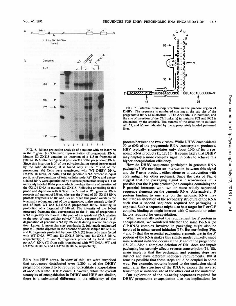
stantial amount ofWT RNA was found in cores (lane 4), butno D3-0X118 RNA was packaged since D3-0X118 is defec-tive for core biosynthesis (lane 8). Analysis of assembledcores (lane 6) and total cellular poly(A)+ RNA (lane 7) fromcells cotransfected with WT and D3-0X118 DNA revealsthat the WT and mutant genomic RNAs are encapsidatedwith identical efficiency. These data suggest that although asurprisingly large portion of the DHBV genome appears tobe required to mediate efficient packaging, it is likely that thecis-acting encapsidation signals of DHBV are composed ofsmaller discontinuous sequences located within this regionof the genome.
DISCUSSION
The finding that genomic but not subgenomic viral RNAsare encapsidated suggested early on that structural featuresof these RNAs mediated their selective recognition andpackaging (8). In this article, we have further characterizedthe cis-acting recognition sequences in DHBV. Our resultsindicate that they function to mediate encapsidation at veryhigh efficiency (-50%) and that they are distributed over aca. 1,200-nt region at the 5' end of pregenome, a region thatis not present in the subgenomic RNAs.We have previously shown that a mutant DHBV genome
bearing a 2-bp insertion at the AccI site in the precore region
is fully infectious and replicates with WT efficiency (5). Ittherefore came as a surprise that mutants bearing 8-bp and16-bp insertions at the same AccI site were drasticallyreduced in their encapsidation efficiency. Interestingly,Junker-Niepmann et al. (15) have pointed out that thisparticular part of the precore region contains a potentialstem-loop structure that is also found at a similar position inthe mammalian hepadnaviruses (Fig. 7). As shown in Fig. 7,the AccI site, in boldface, is found within the stem of thisputative stem-loop structure. While insertion of 2 bp at thissite would likely not significantly alter the stem-loop struc-ture, the 8- and particularly the 16-bp insertions might beexpected to more drastically disrupt the structure of thisregion, particularly that of the bulge region in the 5' stem.While we have no direct evidence to prove that this structureexists in the RNA, it is interesting to note that mutant A2-5',which deletes nucleotides up to but not including the base ofthe stem-loop, is encapsidated at WT efficiency (Fig. 4 a andb), while mutants A3 and A5-5', which delete increasingamounts of the 5' half of the stem, have increasingly delete-rious effects on encapsidation (see Fig. 7). Further studiesare required to investigate the possible role played by thispredicted stem-loop structure in pregenome encapsidation.
Recent work of Junker-Niepmann et al. (15) indicates thatfor HBV, a 137-nt sequence from the 5' end of pregenomicRNA is sufficient to mediate encapsidation of heterologous
J. VIROL.
on July 22, 2018 by guesthttp://jvi.asm
.org/D
ownloaded from
SEQUENCES FOR DHBV PREGENOMIC RNA ENCAPSIDATION 3315
a DR1S'1~
DR2 DR1-11L-........ 3'
118 bp 0X174 DNAinsert
b Genome:
RNA: F-S:..
x. .N .._. ...
358 --
183---
175 -*- *6
_e:_ WR.P
140 ---
1 2 3 4 5 6 7 8 9
FIG. 6. RNase protection analysis of a mutant with an insertionin the C gene. (a) Schematic representation of pregenomic RNA.Mutant D3-0X118 contains an insertion of a 118-nt fragment of0X174 DNA into the C gene at position 318 of the pregenomic RNA.Since this insertion is 3' of the polyadenylation signal (representedby the solid diamond), it is found only at the 5' end of thepregenome. (b) Cells were transfected with WT DHBV DNA,D3-0X118 DNA, or both, and the genomic RNA present in equalportions of preparations of total cellular poly(A)+ RNA and encap-sidated RNA were quantitated by nuclease protection using a 414-ntuniformly labeled RNA probe which overlaps the site of insertion ofthe 0X174 DNA in mutant D3-0X118. Following annealing to thisprobe and digestion with RNase, the 5' end of WT genomic RNAprotects a fragment of 358 nt, whereas the 5' end of D3-0X118 RNAprotects fragments of 183 and 175 nt. Since this probe overlaps theterminally redundant part of the pregenome, it also anneals to the 3'end of both WT and D3-0X118 pregenomic RNA, resulting inprotection of a fragment of 140 nt. The intensity of the 140-ntprotected fragment that corresponds to the 3' end of pregenomicRNA is greatly decreased in the pool of encapsidated RNA relativeto the pool of total cellular poly(A)+ RNA, because of the 3'-to-5'degradation of genomic RNA by RNase H during reverse transcrip-tion. Lanes: 1, molecular size standards; 2, full-length undigestedprobe; 3, probe digested in the absence of added sample RNA; 4, 6,and 8, fragments protected by core RNA (C) from cells transfectedwith WT DNA, WT and D3-0X118 DNA, and D3-0X118 DNA,respectively; 5, 7, and 9, fragments protected by total cellularpoly(A)+ RNA (T) from cells transfected with WT DNA, WT andD3-0X118 DNA, and D3-0X118 DNA, respectively.
RNA into HBV cores. In view of this, we were surprisedthat sequences distributed over 1,200 nt of the DHBVpregenome seemed to be required to facilitate encapsidationof lacZ RNA into DHBV cores. However, while the overallstrategies of encapsidation in DHBV and HBV are similar,there is a substantial difference in the efficiency of the
G U UU G
CG--CU55_ U-GUGA -G
55a0 U--GU7
A--UC--G
CA--U
U--A
AG- - UU--AG--C
35-40 NA- -U
DR I L-5G U......A3 U - -A
5'- AUACCCCUCUCUUCGGAGCUGCU UGUACCAUUGUUUA-3'* ww-A2-t15 95
FIG. 7. Potential stem-loop structure in the precore region ofDHBV. The sequence is numbered starting at the cap site of thepregenomic RNA as nucleotide 1. The AccI site is in boldface, andthe site of insertion of the ClaI linker(s) in mutants PC1 and PC2 isdesignated by the asterisk. The extents of the deletions in mutantsA2, A3, and A5 are indicated by the appropriately labeled patternedlines.
process between the two viruses. While DHBV encapsidates50 to 60% of the pregenomic RNA transcripts it produces,HBV typically encapsidates only about 10% of its prege-nomic RNA products (1, 12, 15). It seems likely that DHBVmay employ a more complex signal in order to achieve thishigher encapsidation efficiency.How do DHBV sequences participate in genomic RNA
packaging? We envision an interaction between this signaland the P gene product, either alone or in association withcore antigen (or other proteins). Since the data of Fig. 6suggest that the packaging signal is discontinuous, it ispossible that the P gene product (or a complex containing theP protein) interacts with two or more widely separatedsequence elements on the genomic RNA. Alternatively, Pprotein binding to one site on the genomic RNA mayfacilitate an alteration of the secondary structure of the RNAsuch that a second sequence required for packaging isexposed. Such a sequence might also be a target for P or C-Pcomplex binding or might interact with C subunits or otherfactors required for encapsidation.When we initially noted the requirement for P protein in
encapsidation, we wondered whether the same P-RNA (orC-P-RNA) complex involved in packaging might also beinvolved in minus-strand initiation (13). But our finding (Fig.4 and 5) that the essential packaging elements are in the 5'portion of the RNA makes this simple model unlikely, sinceminus-strand initiation occurs at the 3' end of the pregenome(18, 23). Also a complete deletion of DR1 does not impairpackaging but strongly affects reverse transcription (14, 18),again implying that the packaging and priming steps aredistinct and have different sequence requirements. But itremains possible that these steps could be coupled in someway. For example, proteins bound to the packaging signalmight alter the structure of the RNA to expose the reversetranscriptase initiation site at the other end of the molecule.Our exploration of the cis-acting sequences required for
DHBV pregenome encapsidation also has implications for
VOL. 65, 1991
lb
0.41 4yC-4 E-.
on July 22, 2018 by guesthttp://jvi.asm
.org/D
ownloaded from

3316 HIRSCH ET AL.
the potential of this virus to serve as a vector for the deliveryof foreign genes to the liver. In addition to its obvioushepatotropism and its known ability to produce persistentnoncytocidal infections, DHBV encapsidates RNA ex-tremely efficiently; these factors are clearly desirable at-tributes of a potential vector. On the other hand, thecomplexity of its encapsidation signal(s) and the limited sizeof the viral genome may severely limit the amount of foreignDNA that could be carried by the vector. Clearly, furtherdefinition of the minimal sequences required for efficientRNA packaging by DHBV will be needed to optimizestrategies for gene transfer mediated by this virus.
ACKNOWLEDGMENTS
We thank Roland Russnak for many helpful discussions during thecourse of this work.
This work was supported by grants from the National Institutes ofHealth.
REFERENCES1. Bartenschlager, R., M. Junker-Niepmann, and H. Schaller. 1990.
The P gene product of hepatitis B virus is required as astructural component for genomic RNA encapsidation. J. Virol.64:5324-5332.
2. Bender, M. A., T. D. Palmer, R. E. Gelinas, and A. D. Miller.1987. Evidence that the packaging signal of Moloney murineleukemia virus extends into the gag region. J. Virol. 61:1639-1646.
3. Birnbaum, F., and M. Nassal. 1990. Hepatitis B virus nucleo-capsid assembly: primary structure requirements in the coreprotein. J. Virol. 64:3319-3330.
4. Buscher, M., W. Reiser, H. Will, and H. Schaller. 1985. Tran-scripts and the putative RNA pregenome of duck hepatitis Bvirus: implications for reverse transcription. Cell 40:717-724.
5. Chang, C., G. Enders, R. Sprengel, N. Peters, H. E. Varmus,and D. Ganem. 1987. Expression of the precore region of anavian hepatitis B virus is not required for viral replication. J.Virol. 61:3322-3325.
6. Condreay, L. D., C. E. Aldrich, L. Coates, W. S. Mason, andT.-T. Wu. 1990. Efficient duck hepatitis B virus production byan avian liver tumor cell line. J. Virol. 64:3249-3258.
7. Enders, G. H., D. Ganem, and H. E. Varmus. 1985. Mapping themajor transcripts of ground squirrel hepatitis virus: the pre-sumptive template for reverse transcriptase is terminally redun-dant. Cell 42:297-308.
8. Enders, G. H., D. Ganem, and H. E. Varmus. 1987. 5'-terminalsequences influence the segregation of ground squirrel hepatitisvirus RNAs into polyribosomes and viral core particles. J.Virol. 61:35-41.
9. Fuetterer, J., and T. Hohn. 1987. Involvement of nucleocapsidsin reverse transcription: a general phenomenon? Trends Bio-chem. Sci. 12:92-95.
10. Ganem, D., and H. E. Varmus. 1987. The molecular biology ofthe hepatitis B viruses. Annu. Rev. Biochem. 56:651-694.
11. Hirsch, R., R. Colgrove, and D. Ganem. 1988. Replication ofduck hepatitis B virus in two differentiated human hepatoma celllines after transfection with cloned viral DNA. Virology 167:136-142.
12. Hirsch, R., and D. Ganem. Unpublished data.13. Hirsch, R., J. Lavine, L. Chang, H. Varmus, and D. Ganem.
1990. Polymerase gene products of hepatitis B viruses arerequired for genomic RNA packaging as well as for reversetranscription. Nature (London) 344:552-555.
14. Horwich, A. L., K. Furtak, J. Pugh, and J. Summers. 1990.Synthesis of hepadnavirus particles that contain replication-defective duck hepatitis B virus genomes in cultured HuH7cells. J. Virol. 64:642-650.
15. Junker-Niepmann, M., R. Bartenschlager, and H. Schaller. 1990.A short cis-acting sequence is required for hepatitis B viruspregenome encapsidation and sufficient for packaging of foreignRNA. EMBO J. 9:3389-3396.
16. Kmiecik, T. E., and D. Shalloway. 1987. Activation and suppres-sion of pp60c-src transforming ability by mutation of its primarysites of tyrosine phosphorylation. Cell 49:65-73.
17. Lavine, J., R. Hirsch, and D. Ganem. 1989. A system forstudying the selective encapsidation of hepadnavirus RNA. J.Virol. 63:4257-4263.
18. Loeb, D., and D. Ganem. Unpublished data.19. Mandart, E., A. Kay, and F. Galibert. 1984. Nucleotide se-
quence of a cloned duck hepatitis B virus genome: comparisonwith woodchuck and human hepatitis B virus sequences. J.Virol. 49:782-792.
20. Mann, R., and D. Baltimore. 1985. Varying the position of aretrovirus packaging sequence results in the encapsidation ofboth unspliced and spliced RNAs. J. Virol. 54:401-407.
21. Mann, R., R. Mulligan, and D. Baltimore. 1983. Construction ofa retrovirus packaging mutant and its use to produce helper-freedefective retroviruses. Cell 33:153-159.
22. Manning, W. C., and E. S. Mocarski. 1988. Insertional muta-genesis of the murine cytomegalovirus genome: one prominentalpha gene (ie2) is dispensable for growth. Virology 167:477-484.
23. Seeger, C., and J. Maragos. 1990. Identification and character-ization of the woodchuck hepatitis virus origin of DNA replica-tion. J. Virol. 64:16-23.
24. Sprengel, R., C. Kuhn, H. Will, and H. Schaller. 1985. Compar-ative sequence analysis of duck and human hepatitis B virusgenomes. J. Med. Virol. 15:323-333.
25. Summers, J., and W. S. Mason. 1982. Replication of the genomeof a hepatitis B-like virus by reverse transcription of an RNAintermediate. Cell 29:403-415.
J. VIROL.
on July 22, 2018 by guesthttp://jvi.asm
.org/D
ownloaded from