clinical relevance of oncogenic point mutations and chromosome … · mara lisa gaspar da silva...
TRANSCRIPT

Clinical relevance of oncogenic point mutations and chromosome copy number changes in
gastrointestinal stromal tumors
Mara Lisa Gaspar da Silva
Dissertation to a Master’s degree in Oncology
Porto, 2009

Mara Lisa Gaspar da Silva
Clinical relevance of oncogenic point mutations and chromosome copy number changes in gastrointestinal stromal tumors
DISSERTATION FOR APPLYING TO A
MASTER’S DEGREE IN ONCOLOGY
SUBMITTED TO THE INSTITUTE OF
BIOMEDICAL SCIENCES ABEL SALAZAR,
UNIVERSITY OF PORTO, AND THOMAS
JEFFERSON UNIVERSITY, USA
Supervisor:
Manuel António Rodrigues Teixeira, MD, PhD Department of Pathology and Molecular Immunology, ICBAS, University of Porto Co-supervisor:
Isabel M. S. Veiga dos Santos, MSc Department of Genetics, Portuguese Oncology Institute – Porto
ACKNOWLEDGMENTS To Professor Manuel Teixeira, for your valuable help, availability, and for giving me the
opportunity to focus my effort in cancer research. I felt that I could make a difference
helping people.
To Isabel, Manuela, Carla and Joana V., who trained me during my period at IPO.
To my dearest family, and to Joaninha, my best friend.
To my source of positive energy.
To those who cared, Joana S., Vera, Diogo, Bárbara, Paula. You made my days happier.
To the “Liga Portuguesa Contra o Cancro”, who supported me financially through 2008.
To the board of directors of the Portuguese Oncology Institute – Porto, and to the
directors of the Master's Program in Oncology provided by the Institute of Biomedical
Sciences Abel Salazar, University of Porto in collaboration with the Thomas Jefferson
University, USA.
To all the research team that participated in this project.
and
especially to my GAGC…
THANK YOU ALL!

Dedicated to all the patients who contributed to this research…

TABLE OF CONTENTS
SUMMARY 7
RESUMO 9
RESUMÉ 11
LIST OF ABBREVIATIONS 13
INTRODUCTION 15
Background 15
Epidemiology 15
Survival rates 16
Clinical features 16
Histological features 17
Immunohistochemical features 17
Signal transduction pathways 18
Molecular sub-classifications 20
Tumor progression and chromosome alterations 22
Prognostic factors 24
Therapeutic options in GIST 26
Surgery 26
Chemotherapy and/or radiotherapy 27
Imatinib treatment (first line therapy) 27
Sunitinib treatment (second line therapy) 29
AIMS 31
MATERIAL AND METHODS 32
Patient selection 32
DNA extraction from paraffin-embedded histological sections 32
DNA extraction from fresh-frozen tumor samples 33
DNA extraction from peripheral blood 33
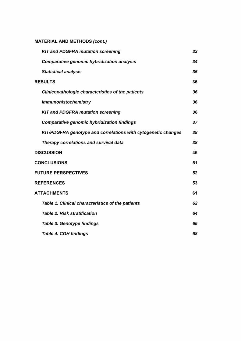
MATERIAL AND METHODS (cont.)
KIT and PDGFRA mutation screening 33
Comparative genomic hybridization analysis 34
Statistical analysis 35
RESULTS 36
Clinicopathologic characteristics of the patients 36
Immunohistochemistry 36
KIT and PDGFRA mutation screening 36
Comparative genomic hybridization findings 37
KIT/PDGFRA genotype and correlations with cytogenetic changes 38
Therapy correlations and survival data 38
DISCUSSION 46
CONCLUSIONS 51
FUTURE PERSPECTIVES 52
REFERENCES 53
ATTACHMENTS 61
Table 1. Clinical characteristics of the patients 62
Table 2. Risk stratification 64
Table 3. Genotype findings 65
Table 4. CGH findings 68
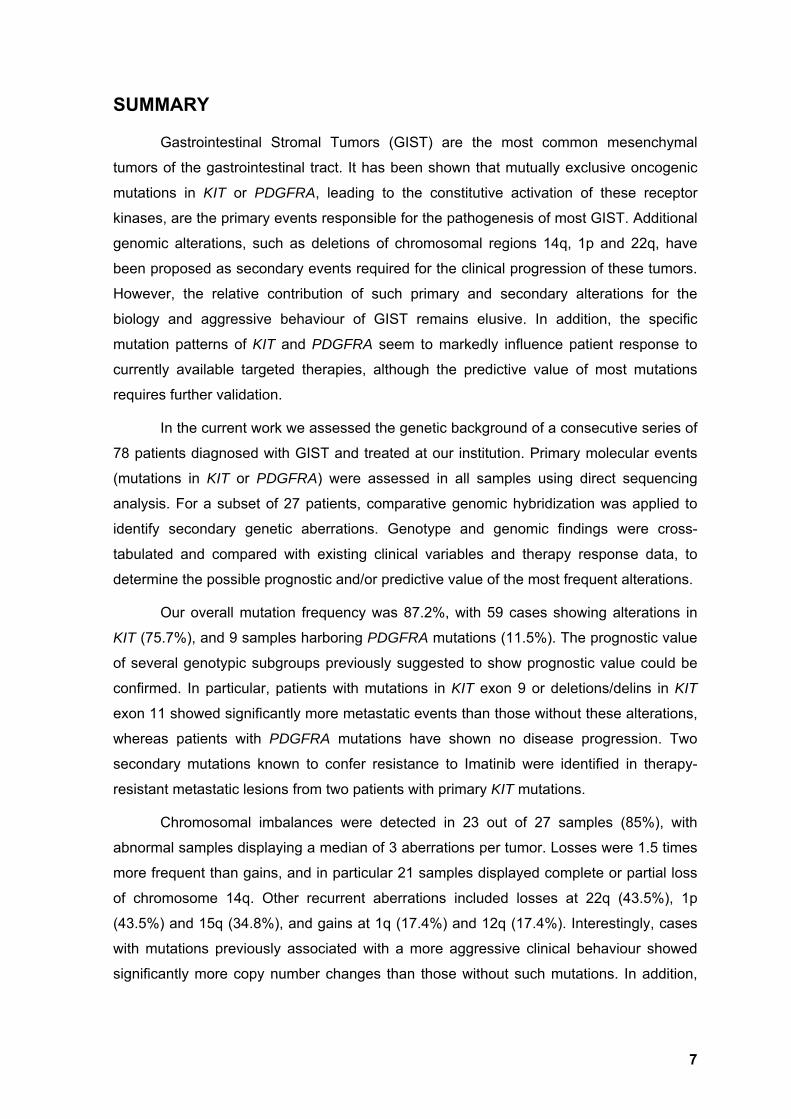
SUMMARY
Gastrointestinal Stromal Tumors (GIST) are the most common mesenchymal
tumors of the gastrointestinal tract. It has been shown that mutually exclusive oncogenic
mutations in KIT or PDGFRA, leading to the constitutive activation of these receptor
kinases, are the primary events responsible for the pathogenesis of most GIST. Additional
genomic alterations, such as deletions of chromosomal regions 14q, 1p and 22q, have
been proposed as secondary events required for the clinical progression of these tumors.
However, the relative contribution of such primary and secondary alterations for the
biology and aggressive behaviour of GIST remains elusive. In addition, the specific
mutation patterns of KIT and PDGFRA seem to markedly influence patient response to
currently available targeted therapies, although the predictive value of most mutations
requires further validation.
In the current work we assessed the genetic background of a consecutive series of
78 patients diagnosed with GIST and treated at our institution. Primary molecular events
(mutations in KIT or PDGFRA) were assessed in all samples using direct sequencing
analysis. For a subset of 27 patients, comparative genomic hybridization was applied to
identify secondary genetic aberrations. Genotype and genomic findings were cross-
tabulated and compared with existing clinical variables and therapy response data, to
determine the possible prognostic and/or predictive value of the most frequent alterations.
Our overall mutation frequency was 87.2%, with 59 cases showing alterations in
KIT (75.7%), and 9 samples harboring PDGFRA mutations (11.5%). The prognostic value
of several genotypic subgroups previously suggested to show prognostic value could be
confirmed. In particular, patients with mutations in KIT exon 9 or deletions/delins in KIT
exon 11 showed significantly more metastatic events than those without these alterations,
whereas patients with PDGFRA mutations have shown no disease progression. Two
secondary mutations known to confer resistance to Imatinib were identified in therapy-
resistant metastatic lesions from two patients with primary KIT mutations.
Chromosomal imbalances were detected in 23 out of 27 samples (85%), with
abnormal samples displaying a median of 3 aberrations per tumor. Losses were 1.5 times
more frequent than gains, and in particular 21 samples displayed complete or partial loss
of chromosome 14q. Other recurrent aberrations included losses at 22q (43.5%), 1p
(43.5%) and 15q (34.8%), and gains at 1q (17.4%) and 12q (17.4%). Interestingly, cases
with mutations previously associated with a more aggressive clinical behaviour showed
significantly more copy number changes than those without such mutations. In addition,
7
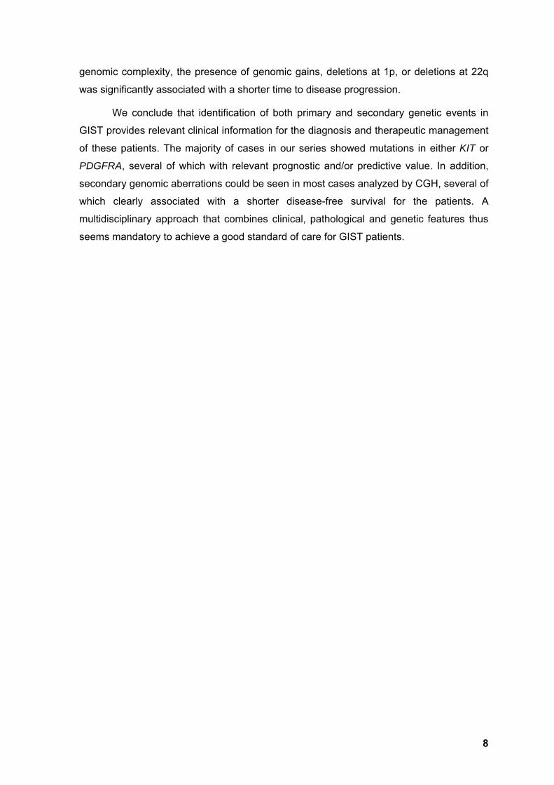
genomic complexity, the presence of genomic gains, deletions at 1p, or deletions at 22q
was significantly associated with a shorter time to disease progression.
We conclude that identification of both primary and secondary genetic events in
GIST provides relevant clinical information for the diagnosis and therapeutic management
of these patients. The majority of cases in our series showed mutations in either KIT or
PDGFRA, several of which with relevant prognostic and/or predictive value. In addition,
secondary genomic aberrations could be seen in most cases analyzed by CGH, several of
which clearly associated with a shorter disease-free survival for the patients. A
multidisciplinary approach that combines clinical, pathological and genetic features thus
seems mandatory to achieve a good standard of care for GIST patients.
8
RESUMO
Os GIST (Gastrointestinal Stromal Tumors) são os tumores das partes moles mais
comuns no tracto gastrointestinal. Mutações pontuais nos genes KIT ou PDGFRA,
promovendo a activação constitutiva destes receptores com actividade de cinase, são
consideradas as alterações genéticas responsáveis pela patogénese da maioria destes
tumores. Além destes eventos primários mutuamente exclusivos, várias alterações
genómicas secundárias, tais como delecções cromossómicas do 14q, 1p e 22q, parecem
ser requeridas para a progressão clínica destes tumores. No entanto, a contribuição
relativa das alterações primárias e secundárias para a biologia e comportamento
agressivo dos GIST ainda não foi determinada. Adicionalmente, os padrões específicos
de mutações dos genes KIT e PDGFRA parecem influenciar marcadamente a resposta
destes pacientes às actuais terapias existentes, apesar do valor predictivo das mesmas
não estar bem determinado.
Neste trabalho procurámos estabelecer o perfil genético de uma série consecutiva
de 78 doentes diagnosticados com GIST e tratados na IPO-Porto, realizando a pesquisa
de alterações primárias nos oncogenes KIT ou PDGFRA através de sequenciação
directa, e determinando eventos genómicos secundários num subgrupo de doentes
recorrendo à metodologia de hibridação comparativa do genoma (CGH). Os resultados
obtidos pelas duas técnicas de análise genética foram cruzados e comparados com as
variáveis clínicas e dados de resposta terapêutica dos pacientes, por forma a determinar
o possível valor prognóstico e/ou preditivo das alterações recorrentes.
A frequência total de mutações detectada na nossa série foi de 87.2%, tendo sido
observados 59 casos com alterações no KIT (75.7%) e 9 amostras com mutações no
PDGFRA (11.5%). O valor prognostico sugerido para alguns dos grupos genotípicos
observados pode ser confirmado na nossa série de amostras. Em particular, tumores com
mutações no exão 9 ou deleções/delins no exão 14 do gene KIT apresentaram uma taxa
de recorrência significativamente mais elevada que aqueles sem estas alterações,
enquanto que no grupo de pacientes com mutações no gene PDGFRA não houve
eventos de recorrência. A mesma mutação secundária de resistência a terapia foi
identificada em lesões metastáticas pós-tratamento de dois pacientes com mutações
primárias no gene KIT.
Das 27 amostras analisadas por CGH, 23 (85%) apresentaram alterações
cromossómicas. Foi detectada perda total ou parcial do cromossoma 14q em 93% destas
amostras, sendo ainda particularmente frequentes as perdas do 22q (43.5%), 1p (43.5%)
e 15q (34.8%). Foi possível verificar que o número de alterações cromossómicas
9
detectado nos tumores com mutações previamente associadas a pior prognóstico era
significativamente maior do que em amostras contendo outras mutações primárias. A
complexidade genómica, assim como a presença de ganhos genómicos, de delecções no
1p ou de perdas do 22q também foram significativamente associadas com um menor
intervalo de progressão da doença.
Foi assim possivel concluir que a identificação de eventos genéticos primários e
secundários em GIST proporciona informação clínica relevante para o diagnóstico e
decisão terapêutica nestes doentes. A maioria dos tumores estudados apresentaram
mutações nos oncogenes KIT ou PDGFRA, várias das quais associadas com uma pior
resposta à actual terapia dirigida a estes receptores. Adicionalmente, aberrações
genómicas secundárias foram observadas na maioria dos casos analisados por CGH,
algumas das quais claramente associadas com um menor intervalo livre de doença para
os doentes. Uma abordagem multidisciplinar combinando características clínicas,
patológicas e genéticas parece deste modo crucial para o diagnóstico e seguimento de
doentes com GIST.
10
RESUMÉ
Les tumeurs du stroma gastro-intestinale (GIST) sont les plus fréquents des
tumeurs mésenchymateuses du tube digestif. Il a été démontré que des mutations
oncogéniques mutuellement exclusifs en KIT ou PDGFRA, conduisant à l'activation
constitutive de ces récepteurs kinases, sont les événements génétiques responsables de
la pathogenèse de la plupart des GIST. Autres altérations génomiques, tels que les
suppressions de régions chromosomiques 14q, 1p et 22q, ont été proposées comme des
événements secondaires nécessaires pour la progression clinique de ces tumeurs.
Toutefois, la contribution relative des modifications primaires et secondaires pour la
biologie et comportement agressif des GIST reste insaisissable. En outre, les différents
mutations du KIT et PDGFRA semblent influencer la réponse du patients aux thérapies
actuellement disponibles, bien que la valeur prédictive de la plupart de ces mutations
nécessite plus validation.
Dans ce travail nous avons évalué le contenu génétique d'une série consécutive
de 78 patients diagnostiqués de GIST et traités dans notre institution. Événements
primaires (mutations du KIT ou PDGFRA) ont été évalués dans tous les lésions par
séquençage direct. Dans un sous-groupe de 27 patients, hybridation génomique
comparative a été appliquée pour identifier anomalies génétiques secondaires. Les
résultats génétiques ont été croisées et comparées avec les variables cliniques et de
réponse a là thérapie, afin de déterminer la possible valeur pronostique et/ou prédictif des
modifications les plus fréquentes.
Notre fréquence de mutation a été de 87,2%, avec 59 cas présentant des
altérations dans KIT (75,7%), et 9 dans PDGFRA (11,5%). Déséquilibres
chromosomiques ont été détectés dans 23 tumeurs (85%). Les pertes ont été 1,5 fois plus
fréquentes que les gains, en particulier dans le chromosome 14q (91%). Autres
aberrations récurrents inclus pertes à 22q (43,5%), 1p (43,5%) et 15q (34.8%), et gains à
1q (17,4%) et 12q (17.4%). Fait intéressant, les cas avec mutations déjà associé à un
comportement cliniques plus agressif ont montré beaucoup plus de modifications du
nombre de copies que ceux sans ces mutations. En outre, la présence de gains
génomiques, des suppressions 1p, ou suppressions à 22q ont été significativement
associés à la progression de cette maladie.
Nous concluons que l'identification des événements génétiques primaires et
secondaires du GIST fournit important renseignements cliniques pour le diagnostic et la
thérapeutique de ces patients. La majorité des cas dans notre série a montré mutations
dans KIT ou PDGFRA, dont quelques sont associés à la pire réponse à des
11

thérapeutiques ciblées. En outre, des aberrations génomiques secondaire on été vu dans
la plupart des cas analysés par CGH, dont certaines clairement associée à une courte
survie sans maladie. Des approches pluridisciplinaires qui combinent des caractéristiques
cliniques, pathologiques et génétiques semblent donc impératives de parvenir à un bon
niveau de soins pour les patients atteints de GIST.
12
LIST OF ABBREVIATIONS
ABL Abelson murine leukemia viral oncogene homolog
AKE Hypotonic Amino-(K) Potassium-EDTA Solution
Akt Intracelular signalling pathway Akt
AML Acute myeloid leukaemia
ANED Alive with no evidence of disease
ATP Adenosine-5'-triphosphate
AWD Alive with disease
BCR Breakpoint cluster region
CGH Comparative genomic hybridization
CDKN2A Cyclin-dependent kinase inhibitor 2A
CD34 Hematopoietic progenitor cell antigen
CML Chronic myelogenous leukemia
DAPI 4’,6-diamidino-2-phenylindole
Delins Deletion Insertion
DFD Dead from disease
DFS Disease-free survival
DNED Dead with no evidence of disease
DWD Dead with disease
DNA Deoxyribonucleic acid
DOG1 2-deoxyglucose-6-phosphate phosphatase 1
EDTA Etilenodiaminotetraacetic acid
FDA Food and Drugs Administration
FISH Fluorescent in situ hybridization
GI Gastrointestinal
GIST Gastrointestinal stromal tumor(s)
H&E Haematoxylin and eosin
13
HCL Hydrochloric acid
HPF High-power field
HSP90 Heat shock protein 90
ICC Interstitial cells of Cajal
KIT v-kit Hardy-Zuckerman 4 feline sarcoma viral oncogene homolog
LOH Loss of heterozygosity
MAP Mitogenic-activated-protein
MMR Mismatch repair system
mTOR mammalian target of Rapamycin
NF1 Neurofibromatosis type I
PCR Polymerase chain reaction
PDGFRA Platelet derived growth factor receptor alpha
PKC θ Protein kinase C theta
PS100 S100 protein
TP53 Tumor-suppressor gene p53
RNA Ribonucleic acid
RTK Receptor tyrosine kinase
SCF Stem cell factor
SMA Smooth muscle actin
SPSS Statistical Package for Social Sciences
SSCP Single Strand Conformational Polymorphism
STAT Signal transducer and activator of transcription
TK Tyrosine kinase
TSG Tumor-suppressor genes
VEGF Vascular endothelial growth factor
14
INTRODUCTION
Cancer is one of the worldwide main causes of death. Whereas 5 to 10 % of the
cases represent inherited conditions, most tumors arise from an altered somatic cell that
proliferates and originates a neoplastic clone through the acquisition and accumulation of
epigenetic and/or genetic alterations. These alterations usually target protooncogenes
(gain-of-function mutations), tumor-suppressor genes (TSG) (loss-of-function mutations)
or genes of the mismatch repair system (MMR) (loss-of-function mutations), leading to the
accumulation of replication errors in multiple other genes and metabolic pathways. Normal
cellular processes such as proliferation, differentiation, apoptosis, and adhesion become
compromised, eventually resulting in self-sufficiency of growth signals, insensitivity to anti-
growth stimuli, reduced apoptosis, sustained angiogenesis, limitless replicative potential
and finally tissue invasion and metastasis (Hanahan D and Weinberg RA, 2000).
Background
GIST (Gastrointestinal Stromal Tumors) represent the most common
mesenchymal tumors of the gastrointestinal tract (Yang J et al, 2008). For many years
these lesions were incorrectly classified as smooth muscle tumors and grouped with
leiomyomas, leiomyoblastomas or leiomyosarcomas due to their morphological similarities
(Corless CL et al, 2004; Rubin BP, 2006). This classification persisted until electron
microscopy studies showed that GIST lacked smooth muscle differentiation, leading
Mazur and Clark to propose the term “stromal tumor” to identify this distinct
clinicopathologic entity (Mazur MT and Clark HB, 1983). Later, in 1998,
immunohistochemistry studies in GIST revealed the absence of desmin expression
(characteristic of smooth muscle tumors) and the presence of KIT protein (CD117, absent
in leiomyomas and leiomyosarcomas) in approximately 95% of the samples (reviewed in
Corless CL et al, 2004 and Rubin BP, 2006). Most GIST were also shown to express the
hematopoietic blast antigen CD34 (Corless CL and Heinrich MC, 2008).
Epidemiology
Most gastrointestinal tumors have an epithelial origin, and as such GIST represent
only a small fraction (<1%) of the total spectrum of gastrointestinal cancers (Gupta P et al,
2008). Due to the misclassification problems in the early days, epidemiologic data for
GIST is incomplete and likely underestimates the true incidence of these tumors. Recent
studies estimate an annual incidence of 10 to 20 cases per million people (Du CY et al,
2008), with 3300 to 6000 new cases per year in the US (Corless CL and Heinrich MC,
15
2008). Incidence is identical in both sexes, with peak susceptibility between 40 to 60 years
of age. There is no epidemiologic data for these tumors in Portugal. Little information is
also available concerning risk factors associated with this neoplasia, even if NF1
(Neurofibromatosis type I), Carney Triad, and familial gastrointestinal stromal tumor
syndromes seem to confer an increased risk to develop GIST (Rubin BP et al, 2007).
Survival rates
Patients with primary, non-metastatic GIST in accessible anatomic locations are
routinely submitted to complete surgical resection. The 5-year survival for these patients
ranges from 42 to 65% (Rossi CR et al, 2003; DeMatteo RP et al, 2008). GIST often recur
locally, diffusing through the serosal surfaces. Advanced disease is characterized by a
distinct metastatic pattern, with the liver and the peritoneum as primary targets. GIST
rarely metastasize to the lungs, pleura, bones, brains, or lymph nodes (Rubin BP et al,
2007; Cichoż-Lach H et al, 2008).
Clinical features
A diagnosis of GIST involves a multidisciplinary approach that combines clinical,
pathological, and genetic features. GIST usually occur within the entire length of the
gastrointestinal tract (GI), predominantly in the stomach (~50%), followed by small
intestine (~25%), rectum and colon (~10%), and esophagus (~5%). These tumors can
also develop outside the GI, in locations such as the omentum, mesentery, pelvis and
retroperitoneum (~10% taken together), abdomen, uterus or vagina (Miettinen M and
Lasota J, 2001; Corless CL et al, 2004; Rubin BP, 2006, Gupta P et al, 2008). The clinical
presentation depends on the location and size of the tumor, but approximately 10 to 30%
of cases are asymptomatic and discovered incidentally in routine exams or non-related
surgeries. Symptomatic tumors may cause abdominal pain, early satiety, flatulence,
prolonged gastrointestinal bleeding, anemia of unknown origin, weight loss and vomiting,
among others (Cichoż-Lach H et al, 2008). Sporadic GIST may consist of solitary primary
lesions or multiple synchronous tumors. Hereditary tumors represent a minority of cases,
with usually several family members being affected, some presenting multiple primary or
metastatic lesions in different anatomic locations throughout their lifetime (Gupta P et al,
2008).
16
INTRODUCTION

Histological features
GIST have a median diameter of approximately 50 mm, ranging in size from small
10 mm lesions, usually discovered incidentally, to large tumors with up to 35 cm. They
commonly present as fleshy solid lesions with central foci of hemorrhage and/or necrosis.
These tumors are frequently solitary, but may arise as multiple nodules. GIST are divided
in three cytomorphological categories, namely spindle cell (70%), epithelioid (20%), or
mixed (Fletcher CD et al, 2002; Rubin BP et al, 2007). GIST are thought to arise from
interstitial cells of Cajal (ICC) or its precursors (Hirano K et al, 2008). ICC express CD34
and are known as the “pacemaker” cells of the gastrointestinal tract, as they
autonomously coordinate the peristalsis in these tissues (Rumessen JJ and Thuneberg L,
1996; Rubin BP, 2006). Both GIST cells and ICC have the dual characteristics of muscle
and neural cells (Du CY et al, 2008), also sharing several other morphologic,
immunohistochemical and molecular features (Miettinen M and Lasota J, 2006).
Immunohistochemical features
In 1995, Isozaki and collaborators showed that ICC expressed CD117 and that its
development was dependent on stem cell factor (SCF) signaling, through the kinase
receptor coded by the gene KIT (v-kit Hardy-Zuckerman 4 feline sarcoma viral oncogene
homolog) (Isozaki K et al, 1995). Hirota and collaborators quickly followed up on these
results and reported that 85 to 90% of GIST harbored mutations in KIT, and that
approximately 95% stained positive for the corresponding protein CD117 (Hirota S et al,
1998). Nowadays, CD117 staining remains the most specific and sensitive marker used
for the diagnosis of GIST (Miettinen M and Lasota J 2005a; Corless CL and Heinrich MC,
2008). KIT staining is usually strong and diffuse, with a citoplasmic, membranous or
paranuclear “dotlike” distribution (Rubin BP, 2006). Whereas expression of CD117 is a
well defined characteristic of GIST, other exams are required to reliably confirm a
suspected diagnosis due to the facts that: 5% of GIST are negative for this marker; other
tumors also express this protein, namely malignant glyomas and small-cell lung tumors
(Orosz Z et al, 2005); CD117 staining can differ within the same tumor and with the
therapy applied; an incorrect tittering of the antibody can mimic false positive results; KIT
is a transmembrane receptor (Figure 1) but in some tumors staining is predominantly
cytoplasmic (Corless CL and Heinrich MC, 2008).
Additional immunohistochemical markers are therefore useful for the correct
identification of GIST. Around 60 to 70% of these tumors are positive for CD34, depending
on tumor location: 80-85% of gastric lesions, 50% of those arising in the small intestine
17
INTRODUCTION

and 95-100% of oesophagus or rectum GIST show positivity for this marker. GIST may
also show positivity for smooth muscle actin (SMA; 30-40%), S100 protein (5%), desmin
(1-2%) and keratin (1-2%) (Rubin BP et al, 2007). Platelet derived growth factor receptor α
(PDGFRA), a transmembrane protein (Figure 1) that belongs to the same receptor
tyrosine kinase (RTK) family as KIT (and which shares several of its molecular features),
should also serve as an excellent marker to identify KIT negative GIST. However, this
protein is expressed in other mesenchymal tumors and currently available antibodies fail
to show the necessary specificity and sensibility (Corless CL and Heinrich MC, 2008).
Much more recently, gene expression profiling studies in GIST reported that
DOG1, a membrane surface protein of unknown function, was expressed in 98% of the
samples, including KIT negative GIST (West RB et al, 2004). As this protein was rarely
expressed in other tumors, it represents a very promising marker for differential diagnosis
of GIST, in particular those with unclear or negative KIT staining (Miettinen M and Lasota
J, 2006; Corless CL and Heinrich MC, 2008). Protein kinase C theta (PKC θ) is an
additional signaling molecule selectively expressed in ICC lineages and strongly and
specifically expressed in GIST. It plays an important role in the survival of lymphocytes T,
positively regulating the activation of T-cell receptor signaling pathways. It is still unclear if
PKC activity is dependent on KIT or PDGFRA activation or other independent
mechanisms, but it represents another promising immunomarker for the identification of
these tumors (Duesing et al, 2004).
Signal transduction pathways
The KIT protein, encoded by the KIT protooncogene located at chromosomal band
4q12, is a type III transmembrane receptor that belongs to the RTK family (Figure 2). It is
involved in several biological mechanisms including cell differentiation, proliferation,
adhesion and apoptosis (Rönnstrand L, 2004; Du CY et al, 2008), and is known to play an
important role in the development and maintenance of melanocytes, erythroblasts, mast
cells and ICC (Antonescu CR et al, 2003; Vu HA et al, 2005; Hirano K et al, 2008). The
protein structure of KIT consists in an extracellular (receptor) domain with five
immunoglobulin-like repeats, a transmembrane segment, and an intracellular (effector)
domain with multiple autophosphorylation sites that mediate tyrosine kinase (TK) activity
(Figure 1) (Hirota et al, 2003; Liu H et al, 2007; Corless CL and Heinrich MC, 2008). Upon
binding of the ligand SCF, the receptor homodimerizes and the TK domains are activated,
thus triggering the phosphorylation of downstream effectors within the MAP kinase, STAT
and/or phosphatidylinositol 3 (PI3)-kinase/AKT pathways (Figure 2).
18
INTRODUCTION

Figure 2. Signaling transduction pathways activated by KIT and PDGFRA receptors. Adapted from Rubin PR et al, 2007.
PDGFRAKIT
EXTRACELLULARDOMAIN
TRANSMEMBRANEDOMAIN
INTRACELLULARDOMAIN
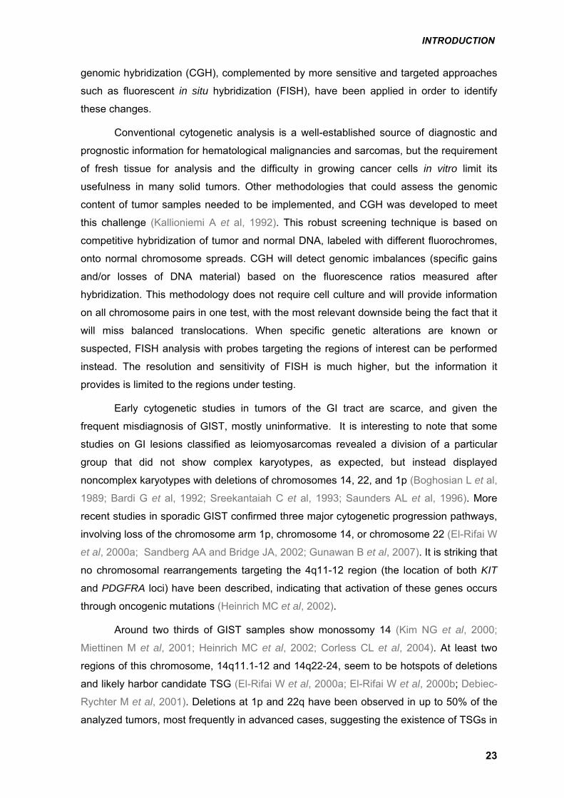
Figure 1. Structure of KIT and PDGFRA receptors with codifying exons and corresponding mutational frequencies found in GIST.
(85-90%) (5-10%)
19
INTRODUCTION
The PDFGRA protein, coded by the PDGFRA oncogene (also located at
chromosomal band 4q12), shares several structural homologies with KIT and is an
alternative activating receptor of its signaling cascade (Rubin BP et al, 2007). When
connected to its respective ligand (PDGF-A), it activates similar downstream signalling
molecules that promote, among other processes, cell proliferation and survival (Figure 2)
(Taylor ML and Metcalfe DD, 2000; Savage DG and Antman KH, 2002; Xiang Z et al,
2007).
Molecular sub-classifications
In 1998, Hirota and collaborators reported that 85 to 90% of GIST harbored
mutations in KIT (Hirota S et al, 1998). Later, it was shown that 5 to 10% of cases
presented PDGFRA mutations (Heinrich MC et al, 2003). Overall, up to 96% of GIST will
show mutations in one of these oncogenes (Heinrich MC et al, 2002; Hirota S et al, 2003;
Corless et al, 2005). These events are mutually exclusive, emerge in the early stages of
the carcinogenic process, and seem to determine the pathogenesis of GIST (Taylor ML
and Metcalfe DD, 2000; Savage DG and Antman KH, 2002).
Functional studies in cell lines transfected with KIT or PDGFRA showed that
mutant isoforms had constitutive kinase activity (SCF or PDGF-A independent
dimerization, respectively), which resulted in continuous activation of the downstream
signaling cascades (Hirota S et al, 1998; Tuveson et al, 2001; Heinrich MC et al, 2003;
Heinrich MC et al, 2006). It has also been shown that a negative control of KIT receptor
signaling is exerted through ubiquitination and fast proteosome degradation of the
receptor upon SCF ligation. Mutant isoforms, however, have much longer half-lives due to
the stabilization by the heat shock protein 90 (Corless CL and Heinrich MC, 2008). This
chaperone protein is responsible for the breakdown of many RTK as well as other
signaling molecules, and currently represents a promising target for tailored therapeutics.
The pathogenic role of KIT mutations has further been demonstrated as mutant isoforms
support the growth of stably transfected BA/F3 cells in nude mice (Hirota S et al, 1998),
mice engineered to express mutant KIT develop GIST and ICC hyperplasia (Sommer et
al, 2003; Rubin BP et al, 2005), and inhibition of KIT blocks the growth of GIST cell lines
(Tuveson et al, 2001; Nakatani et al, 2005; Heinrich MC et al, 2006; Tarn et al, 2006). It is
noteworthy that familial GIST syndrome has recently been found to be caused by heritable
KIT or PDGFRA mutations (Isosaki K et al, 2000; Corless CL and Heinrich MC, 2008).
20
INTRODUCTION

KIT mutations can be broadly assigned to one of two groups: 1) those that involve
the ‘regulatory’ regions responsible for modulating KIT enzymatic activity, and 2) those
that involve the enzymatic region itself (Yang J et al, 2008). Different mutations have
distinct biological and clinical implications (Corless CL and Heinrich MC, 2008). Four
different regions of KIT (composed of 21 exons) have been found to harbor mutations in
sporadic GIST (Figure 1). Exon 11 codes for the juxtamembrane intracellular domain that
is responsible for the inhibition of KIT dimerization in the absence of ligand. When
mutated (70% of cases), the receptor dimerizes and becomes constitutively activated.
Deletions and insertions predominate in the 5’ and duplications are found within the 3’ of
the exon. Tumors with exon 11 mutations, including in-frame deletions or insertions,
missense mutations or combinations thereof, occur throughout the entire GI tract (Tornillo
L and terracciano LM, 2006; Corless CL and Heinrich MC, 2008).
Exon 9 (coding for the extracellular domain) is mutated in 10% of the cases.
Although the pathogenic process is less clear, it is suggested that these mutations mimic
the conformational changes that occur upon ligation of the SCF (Liu H et al, 2007).
Tumors with exon 9 mutations are often characterized as high-risk or overtly malignant,
suggesting an inherently aggressive biology (Rubin BP et al, 2007), and approximately
95% localize in the small intestine. The mutation p.Ala502_Tyr503dup, described by Lux
and collaborators (2000), is particularly recurrent (10% of these cases).
Exon 13 codes the first portion of the tyrosine kinase domain and is mutated in
~1% of cases. The mechanism by which the receptor becomes activated in these cases is
yet to be discovered. Exon 17 codes for the kinase activation domain and when mutated
(0.5% of cases) promotes spontaneous kinase activity (Mol CD et al, 2003; Foster R et al,
2004). Xiang and collaborators reported that a KIT exon 17 mutant isoform (Asp816Val)
lacking the extracellular and transmembrane receptor domains still kept its kinase activity
(Xiang Z et al, 2007). All KIT exon 13 or 17 mutations identified to date are missense
changes (Rubin BP et al, 2007). Interestingly, missense mutations in residue 816 (aspartic acid) are common in
other KIT positive neoplasias, such as acute myeloid leukemia (AML), but were never
found in GIST. In opposition, exon 11 mutations, the most frequent in GIST, occur rarely
in other neoplasias. These observations suggest that KIT oncogene mutations are lineage
specific (Rubin BP, 2006; Tornillo L and Terracciano LM, 2006).
21
INTRODUCTION
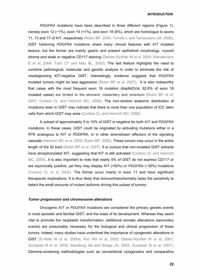
PDGFRA mutations have been described in three different regions (Figure 1),
namely exon 12 (~1%), exon 14 (<1%), and exon 18 (6%), which are homologue to exons
11, 13 and 17 of KIT, respectively (Rubin BP, 2006; Tornillo L and Terracciano LM, 2006).
GIST harboring PDGFRA mutations share many clinical features with KIT mutated
lesions, but the former are mainly gastric and present epithelioid morphology, myxoid
stroma and weak or negative CD117 staining (Debiec-Rychter M et al, 2004; Wardelmann
E et al, 2004; Tzen CY and Mau BL, 2005). The last feature highlights the need to
combine pathological, molecular and genetic analysis in order to eliminate the risk of
misdiagnosing KIT-negative GIST. Interestingly, evidence suggests that PDGFRA
mutated tumors might be less aggressive (Rubin BP et al, 2007). It is also noteworthy
that cases with the most frequent exon 18 mutation (Asp842Val, 62.6% of exon 18
mutated cases) are limited to the stomach, mesentery and omentum (Rubin BP et al,
2007; Corless CL and Heinrich MC, 2008). The non-random anatomic distribution of
mutations seen in GIST may indicate that there is more than one population of ICC stem
cells from which GIST may arise (Corless CL and Heinrich MC, 2008).
A subset of approximately 5 to 10% of GIST is negative for both KIT and PDGFRA
mutations. In these cases, GIST could be originated by activating mutations either in a
RTK analogous to KIT or PDGFRA, or in other downstream effectors of the signaling
cascade (Heinrich MC et al, 2002; Rubin BP, 2006). These tumors may occur in the entire
length of the GI tract (Rubin BP et al, 2007). It is curious that non-mutated GIST extracts
have phosphorylated KIT, suggesting that KIT is still activated (Corless CL and Heinrich
MC, 2008). It is also important to note that nearly 5% of GIST do not express CD117 or
are equivocally positive, yet they may display KIT (>50%) or PDGFRA (~30%) mutations
(Corless CL et al, 2004). The former occur mainly in exon 11 and have significant
therapeutic implications. It is thus likely that immunohistochemistry lacks the sensitivity to
detect the small amounts of mutant isoforms driving this subset of tumors.
Tumor progression and chromosome alterations
Oncogenic KIT or PDGFRA mutations are considered the primary genetic events
in most sporadic and familial GIST, and the basis of its development. Whereas they seem
vital to promote the neoplastic transformation, additional somatic alterations (secondary
events) are presumably necessary for the biological and clinical progression of these
tumors. Indeed, many studies have underlined the importance of cytogenetic alterations in
GIST (El-Rifai W et al, 2000a; Kim NG et al, 2000; Debiec-Rychter M et al, 2001;
Gunawan B et al, 2002; Sandberg AA and Bridge JA, 2002; Gunawan B et al, 2007).
Genome-screening methodologies such as conventional cytogenetics and comparative
22
INTRODUCTION
genomic hybridization (CGH), complemented by more sensitive and targeted approaches
such as fluorescent in situ hybridization (FISH), have been applied in order to identify
these changes.
Conventional cytogenetic analysis is a well-established source of diagnostic and
prognostic information for hematological malignancies and sarcomas, but the requirement
of fresh tissue for analysis and the difficulty in growing cancer cells in vitro limit its
usefulness in many solid tumors. Other methodologies that could assess the genomic
content of tumor samples needed to be implemented, and CGH was developed to meet
this challenge (Kallioniemi A et al, 1992). This robust screening technique is based on
competitive hybridization of tumor and normal DNA, labeled with different fluorochromes,
onto normal chromosome spreads. CGH will detect genomic imbalances (specific gains
and/or losses of DNA material) based on the fluorescence ratios measured after
hybridization. This methodology does not require cell culture and will provide information
on all chromosome pairs in one test, with the most relevant downside being the fact that it
will miss balanced translocations. When specific genetic alterations are known or
suspected, FISH analysis with probes targeting the regions of interest can be performed
instead. The resolution and sensitivity of FISH is much higher, but the information it
provides is limited to the regions under testing.
Early cytogenetic studies in tumors of the GI tract are scarce, and given the
frequent misdiagnosis of GIST, mostly uninformative. It is interesting to note that some
studies on GI lesions classified as leiomyosarcomas revealed a division of a particular
group that did not show complex karyotypes, as expected, but instead displayed
noncomplex karyotypes with deletions of chromosomes 14, 22, and 1p (Boghosian L et al,
1989; Bardi G et al, 1992; Sreekantaiah C et al, 1993; Saunders AL et al, 1996). More
recent studies in sporadic GIST confirmed three major cytogenetic progression pathways,
involving loss of the chromosome arm 1p, chromosome 14, or chromosome 22 (El-Rifai W
et al, 2000a; Sandberg AA and Bridge JA, 2002; Gunawan B et al, 2007). It is striking that
no chromosomal rearrangements targeting the 4q11-12 region (the location of both KIT
and PDGFRA loci) have been described, indicating that activation of these genes occurs
through oncogenic mutations (Heinrich MC et al, 2002).
Around two thirds of GIST samples show monossomy 14 (Kim NG et al, 2000;
Miettinen M et al, 2001; Heinrich MC et al, 2002; Corless CL et al, 2004). At least two
regions of this chromosome, 14q11.1-12 and 14q22-24, seem to be hotspots of deletions
and likely harbor candidate TSG (El-Rifai W et al, 2000a; El-Rifai W et al, 2000b; Debiec-
Rychter M et al, 2001). Deletions at 1p and 22q have been observed in up to 50% of the
analyzed tumors, most frequently in advanced cases, suggesting the existence of TSGs in
23
INTRODUCTION
this region that could be important in tumor progression (El-Rifai W et al, 2000a; Corless
CL et al, 2004; Lasota J et al, 2005; Tornillo L and Terracciano LM, 2006).
Interestingly, the number and type of chromosomal changes seems to correlate
with the clinical aggressiveness of the tumors (Heinrich MC et al, 2002). El-Rifai and
cooperators analyzed a subset of 95 GIST and concluded that the mean number of
changes increased from 2.6 in benign GIST to 7.5 in malignant primary tumors and up to
9 in metastatic lesions. These authors also showed that 9p deletions, 8q
gains/amplifcations, and 17q gains/amplifications, were found almost exclusively in high-
grade or metastatic GIST (El-Rifai W et al, 2000a). Indeed, specific losses at 1p, 9p and
11p are clearly associated with malignancy (Bergmann F et al, 1998; Kim NG et al, 2000;
Heinrich MC et al, 2003). A key gene in chromosome 9p is CDKN2A (p16INK4A), coding
for an important inhibitor of the cell cycle frequently inactivated in GIST (Rubin BP et al,
2007). Based on these findings, a putative genetic pathway leading to the development
and progression of GIST was proposed by Heinrich (2002): KIT or PDGFRA mutation
14q deletion 22q deletion 1p deletion 8q gain 11p deletion 9p deletion
17q gain.
Although such a genetic fingerprint for GIST can be roughly defined, the target
genes involved in these regions remain undiscovered (Heinrich MC et al, 2002). Also,
assuming that mutant KIT isoforms are capable of constitutive activation and
enhancement of target signaling cascades, the biological role (and mode of action) of
these secondary chromosomal events remains unclear. Strikingly, only a few very recent
reports have assessed both primary and secondary changes in the same samples, with
limited success (Anderson J et al, 2002; Assämäki R et al, 2007; Wozniak A et al, 2007).
Additional combined studies using molecular and cytogenetic methodologies are thus
needed to clarify the relative contribution of mutations and gross chromosomal changes to
the biological and clinical features of these tumors.
Prognostic factors
Considerable efforts have been placed in finding clinical, pathological or molecular
markers of prognosis that could distinguish indolent GIST from the more clinically
aggressive tumors, or possibly identify lesions with high metastatic potential (Liu XH et al,
2005; Martin J et al, 2005; Andersson J et al, 2006). This has proved a difficult task,
however, and few variables currently show predictive or prognostic value. Tumor size and
mitotic index remain the most relevant prognostic indicators in GIST, and the only
consensus criteria were established in the GIST Workshop convened at the United States
National Institute of Health in 2001 (Fletcher CD et al, 2002; Joensuu H, 2008).
24
INTRODUCTION

Whereas these parameters have proved clinically useful in the classification of
GIST, they are not fail-proof and additional markers are required. Indeed, it is not
guaranteed that a lesion with less than 2 cm and a small mitotic index will have a benign
or favorable course, as literature data shows that even these small tumors can
occasionally metastasize (Fletcher CD et al, 2002; Rubin BP, 2006; Yang J et al, 2008). In
view of these predicaments, many groups have shown reluctance in classifying GIST as
“benign”, since any GIST presenting clinical symptoms has the potential to behave in a
malignant fashion (Fletcher CD et al, 2002).
Tumor stage may also provide important information, as patients with resected
primary GIST that recur within the peritoneum or that metastasize to the liver have poor
prognosis (Fletcher CD et al, 2002), as do those with intra-abdominal dissemination
(DeMatteo RP et al, 2000). Rubin and collaborators (2006) have demonstrated that
anatomic location also shows prognostic significance, since gastric tumors and small-
bowel lesions with the same size have distinct clinical outcomes (the former presenting
better prognosis). Anatomic location also determines the risk of recurrence and
progression, a fact acknowledged in the 2007 National Comprehensive Cancer Network
risk stratification criteria (Demetri GD et al, 2007; Yang J et al, 2008). However, a study
comprising 1765 GIST does not support this approach (Miettinen M et al, 2005b).
Nevertheless, anatomic site was already considered as a prognostic factor in recent
studies (Corless CL and Heinrich MC, 2008). Other variables such as mucosal invasion,
tumor necrosis, and high cellularity have been statistically associated with an aggressive
phenotype, but these have been poorly reproducible and do not show consistent results
that can be used on an individual basis (Fletcher CD et al, 2002; Goh BK et al, 2008).
Interestingly, several studies indicate that the presence of KIT mutations confers a
malignant and more aggressive behavior to GIST (Ernst SI et al, 1998; Taniguchi M et al,
1999; Kim TW et al, 2004; Liu XH et al, 2005; Martin J et al, 2005; Cho S et al, 2006).
However, the presence of these alterations is not by itself able to differentiate the degree
of aggressiveness of these neoplasias or predict the likelihood of recurrence after
resection of a primary GIST (Rubin BP et al, 2001; Corless CL et al, 2002; Heinrich MC et
al, 2002; Heinrich MC et al, 2003). Moreover, small GIST (even tumors <10 mm) have
been shown to display KIT mutations, raising the question whether KIT mutations actually
influence outcome (DeMatteo RP et al, 2008). At the molecular level, KIT exon 11 point-mutations or insertions have been shown
to confer a favorable prognosis, and the former are even associated with longer
recurrence-free survival after surgical resection (Singer S et al, 2002). Some studies
report that deletions in exon 11 (Andersson J et al, 2006; Cho S et al, 2006), and more
25
INTRODUCTION

specifically in codons 557-558 (Wardelmann E et al, 2003; Martin J et al, 2005; Miettinen
M and Lasota J, 2006; DeMatteo RP et al, 2008) are associated with poor prognosis in
patients with completely resected GIST. Exon 9 mutations and exon 13 deletions have
also been associated with a poor prognosis (Lasota J et al, 2000; Sakurai S et al, 2001;
Antonescu CR et al, 2003; Lasota J et al, 2003). Tumors with PDGFRA mutations tend to
be less aggressive than those with KIT mutations, but they may also progress and lead to
patient death (Lasota J et al, 2004; Lasota J et al, 2006; Corless CL and Heinrich MC,
2008). In disagreement with these assumptions, a recent Taiwanese study of 134 GIST
(99% with KIT mutations, 1% with PDGFRA mutations) found no association between the
type or location of KIT mutations and progression-free survival rates (Yang J et al, 2008).
This study also raises the question of the prognostic significance of different mutations
according to racial differences, which requires further investigation.
It has been suggested that once GIST become metastatic, the specific kinase
genotype does not influence overall survival (Gold JS et al, 2007), and that the prognosis
at the time of clinical presentation is clearly influenced by additional genetic events
(Corless CL et al, 2004; Rubin BP, 2006). Additional studies are therefore required to
determine the possible association of molecular and/or cytogenetic alterations to the
prognosis of GIST, and their possible contribution for treatment recommendations (Raut
CP and DeMatteo RP, 2008).
Therapeutic options in GIST
Surgery
Surgical resection is the standard therapy for non-metastatic, operable GIST. In
primary tumors, the aim is complete resection without rupture of the tumor capsule, on
condition that the risk of dysfunction of the affected organ is low. However, 40 to 80% of
cases are inoperable or recur. After surgery, five year disease-free survival rates for
patients with primary GIST range from 43 to 65% (Di Matteo G et al, 2002; Rossi CR et al,
2003). Due to the specific metastatic pattern of GIST, lymphadenectomy is not advised,
with the exception of rare cases in which ganglia involvement is clear. Surgery is not
curative for recurrent or metastatic lesions (Corless CL and Heinrich MC, 2008).
26
INTRODUCTION
Chemotherapy and/or radiotherapy
Traditional chemotherapy and/or radiotherapy are not efficient in inoperable or
metastatic lesions. In general, only ~5% of these patients respond to treatment, and the
median survival is ~18 months (Corless CL and Heinrich MC, 2008; DeMatteo RP et al,
2008).
Imatinib treatment (first line therapy)
The inefficiency of the traditional therapeutic options for metastatic or unresectable
GIST and the knowledge that activating KIT mutations play a pivotal role in the
carcinogenesis of these tumors led to the application of targeted therapy for the protein
product of this oncogene. The selected drug, Imatinib mesylate (STI-571, commercial
name Gleevec or Glivec) is an orally bioavailable 2-phenylpyrimidine derivative,
developed originally in the 1990s to treat chronic myelogenous leukemia (CML). Indeed,
Imatinib was developed to inhibit the ABL kinase (BCR-ABL protein fusion) in CML cells
by occupying the ATP binding pocket of the ABL kinase domain (Antonescu CR, 2008;
Corless CL and Heinrich MC, 2008). It was later demonstrated that Imatinib had the
capacity to inhibit several other tyrosine kinases, namely KIT, PDGFRA and PDGFRB,
ARG, and ABL (Buchdunger E et al, 1996; Heinrich MC et al, 2002; Mol CD et al, 2004;
Dewar AL et al, 2005). ABL shares considerable homology with the type III receptor
tyrosine kinase family and, in the particular case of the KIT receptor, Imatinib was shown
to inhibit in vitro a mutant form of KIT commonly found in GIST, and also the growth of
cultured GIST cells harboring KIT mutations (Tuveson DA et al, 2001; Heinrich MC et al,
2006).
As for the ABL kinase, Imatinib inhibits KIT tyrosine kinase activity, preventing
phosphorylation of downstream effectors and thus suppressing the proliferation of GIST
(Savage DG and Antman KH, 2002; Scheijen B and Griffin JD 2002; Tamborini E et al,
2004). In 2000, Imatinib was administered to a patient with metastatic GIST, unresponsive
to multiple conventional therapies, and a reduction of 75% was verified in tumor size and
number of metastases (Joensuu H et al, 2001). In 2002, after multiple clinical trials, this
drug was approved by the Food and Drug Administration as a standard therapy for
patients with inoperable tumors or with metastatic disease, serving as a paradigmatic
example of a targeted therapy for a specific molecule in solid tumors (Rubin BP, 2006).
However, several studies quickly demonstrated an association between KIT and
PDGFRA mutations and different responses to Imatinib treatment (Heinrich MC et al,
2003; Corless CL et al, 2004; Gupta P et al, 2008; Heinrich MC et al, 2006). Indeed,
27
INTRODUCTION
patients with KIT exon 11 mutations have shown a more favorable response (83,5%)
relatively to the ones with KIT exon 9 mutations (48.7%) or the ones negative for both
genes (weak response). Interestingly, these results are not in accordance with previous in
vitro trials, in which it was possible to inhibit the growth of cell lineages of GIST with KIT
exon 9 mutations (Heinrich MC et al, 2002). Due to the small number of cases with KIT
exon 13 or 17 primary mutations, only limited correlations could be inferred (Lasota J et al,
2008). Interestingly, the missense mutation found in KIT exon 13 (Lys642Glu) is thought
to originate Imatinib resistance (Tornillo L and Terracciano LM, 2006), although in vitro
studies suggested inhibition of this isoform by Imatinib (Tuveson DA et al, 2001).
In agreement with in vitro assays, patients with the Asp842Val mutation in
PDGFRA respond weakly to Imatinib (Antonescu CR et al, 2003; Corless CL et al, 2004;
Corless CL et al, 2005). Mutations in exon 12, however, show good response to Imatinib
and the single rare mutation described in exon 14 showed in vitro sensitivity to Imatinib
(Tornillo L and Terracciano LM, 2006). More clinical studies are clearly needed to assess
the predictive value of these uncommon alterations, as well as to clarify the differences
between in vivo and in vitro assays.
During treatment with Imatinib, several patients eventually develop secondary
resistance that can be translated in growth of a nodule within preexisting, clinically
quiescent lesions, or widespread expansion of metastatic lesions throughout the liver or
abdominal cavity (Desai et al, 2007). Preliminary studies revealed the existence of, at
least, four mechanisms that can dramatically inhibit the action of this drug: 1) acquisition
of a secondary mutation in the TK domain of KIT or PDGFRA; 2) gene amplification
leading to KIT or PDGFRA over-expression; 3) functional resistance in GIST that present
kinase activity and that are sensitive to Imatinib in vitro, but not in vivo; 4) activation of
alternative kinases in the same signaling pathways (Corless CL et al, 2004). Tumors
showing evidence of KIT gene amplification have been occasionally described (Antonescu
CR et al, 2005; Debiec-Rytcher M et al, 2005). More rarely, there is downregulation of KIT
expression, suggesting the emergence of a KIT-independent phenotype. In their series of
resected GIST from Imatinib-treated patients, Agaram and colleagues identified several
mitotically active lesions that were p53 immunopositive and two tumors that had TP53
mutations (Agaram NP et al, 2007). It is likely that deregulation of the cell cycle through
such mutations contributes to Imatinib resistance. In the majority of primary resistance
cases, however, the mechanism for drug escape remains unknown. Acquisition of
secondary mutations that affect the site of Imatinib binding seems more frequent (Corless
CL and Heinrich MC, 2008). Several secondary point mutations were already identified
28
INTRODUCTION
and it was observed that generally they are present in the same allele affected by primary
mutations (Miettinen M and Lasota J, 2006).
Imatinib treatment (400-800 mg/day) is generally well tolerated and, in most cases,
patients achieve complete or partial remission. This treatment increased the 2-year
survival of patients with advanced or metastatic disease from 20% to 70% (Glabbeke MV
et al, 2006). Debiec-Rychter and colleagues found that the progression-free survival of
GIST patients with KIT exon 9 mutations was significantly better when they were treated
with 800 mg per day as compared with 400 mg per day (Debiec-Rytcher M et al, 2006).
However, long-term disease control is limited by the acquisition of resistance to this drug.
Due to the fact that such resistance is becoming a significant clinical problem in the
treatment of this neoplasia, several trials with new targeted drugs are underway,
underlining the importance of genetic studies aiming at identifying novel alterations with
prognostic or predictive value or that may work as therapeutic targets in GIST.
Sunitinib treatment (second line therapy)
Patients with inoperable GIST showing Imatinib resistance or intolerance had no
efficient alternative therapeutics until the introduction of SU11248 (Sunitinib Malate or
SUTENT, Pfizer, New York, USA), recently approved by the FDA as a second line
treatment in GIST. Sunitinib is an inhibitor of several TKs that shows anti-angiogenic and
anti-tumoral activity in several tumor models both in vitro and in vivo (Demetri GD et al,
2006). These effects have been associated to signaling block of KIT, PDGFRs, VEGFRs,
FLT3 and RET receptors (Corless CL and Heinrich MC, 2008). Despite the fact that both
Sunitinib and Imatinib target the ATP binding domain of the TKs, they belong to different
chemical classes and presumably present dissimilar characteristics and affinities of
binding (Demetri GD et al, 2006).
Nevertheless, the inhibition of several signaling pathways by Sunitinib might be
more efficient, given the fact that Imatinib has exclusive affinity to a limited number of TK
domains. Patients treated with Sunitinib show good results, namely an increase in
disease-free survival and overall survival rates. Future studies with Sunitinib in GIST are
however needed to investigate the molecular mechanisms through which the drug exerts
disease control after Imatinib failure (Demetri GD et al, 2006). Preliminary analysis
demonstrated that treatment with Sunitinib results in better anti-tumoral response rates
and clinical improvement in patients with KIT exon 9 primary mutations or KIT-negative
tumors compared with exon 11 mutated lesions (Demetri GD et al, 2006). This data
demonstrates that Sunitinib is an effective therapeutic option for patients with metastatic,
29
INTRODUCTION

Imatinib resistant GIST. As for Imatinib, however, mutations have already been described
(particularly in exon 17) that confer cross-resistance to Sunitinib. It is therefore predictable
that secondary resistance may develop in patients that initially respond to this second line
drug (Corless CL and Heinrich MC, 2008). The role of recurrent chromosome changes in
therapy resistance to Imatinib or Sunitinib is currently unknown.
30
INTRODUCTION

AIMS
A diagnosis of GIST requires a multidisciplinary approach that combines clinical,
pathological, and genetic features. Our main goal was to characterize primary and
secondary genetic events in a consecutive series of GIST, in order to contribute to their
correct diagnosis and to identify genetic aberrations with prognostic and/or predictive
value for these patients.
The specific aims of this project were:
o To identify primary genetic alterations in GIST by direct sequencing of the
oncogenes KIT and PDGFRA.
o To characterize the spectrum of chromosomal alterations (secondary genetic
events) in GIST using comparative genomic hybridization, a powerful whole-
genome screening methodology.
o To assess possible correlations between primary and secondary genetic events
and between these and the clinico-pathological features of GIST.
o To assess the prognostic value of genetic events (irrespective of therapy) and the
possible correlations between genetic events and response to therapy, namely
mutations associated with Imatinib or Sunitinib resistance.
31

MATERIAL AND METHODS Patient selection
A series of 78 patients diagnosed with GIST and submitted to surgery with curative
intent were included in this study. The majority of patients was diagnosed and treated at
the Portuguese Oncology Institute – Porto, with the exception of six cases that were
provided by other institutions. Patients had received no treatment prior to surgery. All
samples were obtained after informed consent. Fresh-frozen tumor samples of 27 patients
(conserved in RNA later) could be obtained. For all the remaining cases, genetic analyses
were performed in formalin-fixed, paraffin-embedded tissue sections. In all cases, H&E
sections from representative tissue blocks were reviewed by expert pathologists to
confirm a diagnosis of GIST and to evaluate relevant histopathological parameters. Other
clinical and demographic variables, such as age at diagnosis, gender, tumor size, and
tumor location (divided into stomach, small intestine, rectum and colon, and outside the
GI), were obtained (Table 1). Patients with tumors that eventually recurred or developed
metastatic lesions were treated with Imatinib in accordance with the guidelines followed at
the IPO-Porto. Second-line therapy for patients that progressed or were intolerant to
Imatinib was Sunitinib.
DNA extraction from paraffin-embedded histological sections
H&E stained sections for each formalin-fixed, paraffin-embedded tumor sample
were reviewed by pathologists and relevant areas with >80% neoplastic cells were
delimited. Additional 10μm unstained sections were sequentially obtained from the same
tissue blocks, placed in poli-L-lisine coated slides and dried at 37ºC overnight. Relevant
tumor areas were identified by overlapping these slides with the respective H&E sections,
and after humidification with buffer, tumor fragments were macrodissected using a sterile
blade and collected to 1.5 mL tubes. DNA isolation was performed using an adaptation of
the technique described by Lungu and colleagues (Lungu O et al, 1992).
Briefly, lysis buffer was prepared with 50 µl Tris-HCL 1M (pH 8.5), 10 µl of EDTA,
5µl of Tween 20 and 935 µl of bi-distillate water. A buffer volume proportional to the tumor
area and 4 µl of proteinase K (10mg/mL) were added, and samples were incubated 2
hours at 55ºC. The enzyme was inactivated by temperature shock (10 minutes at 95ºC
followed by 15 minutes on ice), and tubes were centrifuged 5 minutes at 14000 rpm. The
supernatant (containing the DNA) was transferred to a new 1.5 mL tube. Double-stranded
DNA concentration and purity were quantified using a NanoDrop ND-1000
32
spectrophotometer. DNA quality was evaluated by electrophoresis in ethidium-bromide
stained agarose gel 0.8% (p/v).
DNA extraction from fresh-frozen tumor samples
After physical disaggregation of the fresh-frozen tissue fragments using a sterile
blade, DNA was extracted following the salting-out-chloroform mixed methodology
described above.
DNA extraction from peripheral blood
In order to include a negative control in every set of amplifications, and to have
control electrophorograms to compare with the tumor sequences, DNA from a healthy
donor was extracted. A peripheral blood sample was collected inside sterilized tubes
containing EDTA and placed at 4ºC for immediate use. Lyses of erythrocytes was
achieved by adding hypotonic solution (AKE: NH4Cl 155mM; KHCO3 10mM; EDTA
0,1mM; pH 7,4) in a proportion of 10 times the available blood volume (3-5 mL). After 30
minutes incubation at 4ºC, tubes were centrifuged 10 minutes at 2000 rpm. The
supernatant was discarded and the process was repeated until the sediment was free of
haemoglobin. The resulting nuclear sediment was transferred to 1.5 mL tubes and
maintained at 80ºC until DNA extraction. Genomic DNA was isolated using the salting-out-
chloroform mixed technique (Müllenbach R et al, 1989). This protocol combines the
classic phenol/chloroform methodology with the salting-out technique, eliminating the
disadvantages of using phenol (classic technique), as well as reducing the DNA losses
that occur when DNA is precipitated with proteins (salting-out technique).
KIT and PDGFRA mutation screening
Using the DNA extracted from each sample, KIT (exons 9, 11, 13, and 17) and
PDGFRA (exons 12, 14, and 18) target sequences were amplified by polymerase chain
reaction (PCR) on a standard termocycler. Primers and conditions were as described in
the literature (Penzel R et al, 2005; Daum O et al, 2007). Negative controls were included
in every set of amplifications. PCR products were purified using the “GFX PCR DNA and
Gel Band Purification Kit” [GE Healthcare], according to the manufacturer’s recommendations, to remove non incorporated salts, enzymes, nucleotides and primers. Samples were
eluted in bi-distillate water and subsequently evaluated by electrophoresis in ethidium-
bromide stained agarose gel 2% (p/v).
33
MATERIAL AND METHODS
For the sequencing reactions, 30 to 90 ng of purified DNA were used. Primers and
conditions were as described in the literature (Penzel R et al, 2005; Daum O et al, 2007).
The resulting products were purified to remove excess dNTPs, labeled ddNTPs and non
incorporated primers using standard methods. Briefly, each sequencing product was
mixed with 2 µL of sodium acetate 3M and 50 µL of ethanol 96% (v/v). Tubes were
vortexed and kept on ice for 30 minutes. After that, samples were centrifuged 30 minutes
at 14000 rpm/4ºC. The supernatant was discarded and the sediment was washed with
250 µL of ethanol 70% (v/v), dried and eluted in deionized formamide. After denaturation
(5 minutes at 95ºC), the purified sequencing products were submitted to electrophoresis in
a capillary column. Direct sequencing was performed on an ABI PRISM 310 automatic
sequencer using the Big Dye Terminator Chemistry (Applied Biosystems, Foster City,
CA), according to the manufacturer’s recommendations. All results were confirmed with a
second independent analysis.
Comparative genomic hybridization analysis
Fresh-frozen tumor samples from 27 patients were analyzed by CGH, following the
procedure of Kallioniemi et al (1992), with modifications previously described (Ribeiro FR
et al, 2006). Briefly, test (tumor tissue) and reference (peripheral blood lymphocytes from
healthy donors) DNA was labeled in nick-translation reactions using SpectrumGreen and
SpectrumRed-conjugated nucleotides (Vysis, Downers Grove IL), respectively. Labeled
tumor and reference DNA were mixed with unlabeled Cot-1 DNA (Life Technologies,
Rockville, MD) and the probe mixture was denaturated and hybridized to commercially
available, normal metaphase slides (Vysis). After washing off excess probe, samples were
counterstained with 4’,6-diamidino-2-phenylindole (DAPI) in an antifade solution (Vector
Laboratories, Burlingame, CA). Samples were analyzed with a Cohu 4900 CCD camera
using an automated filter wheel coupled to a Zeiss Axioplan fluorescence microscope
(Zeiss, Oberkochen, Germany) and a CitoVysion system version 3.9 (Applied Imaging,
Santa Clara, CA). Data from ten cells was combined to generate average ratio profiles
with 99% confidence intervals for each sample. Aberrations were scored whenever the
sample profile and the standard reference profile at 99% did not overlap (Kirchhoff M et al,
1998). Description of the CGH copy number changes followed the guidelines suggested
by the International System for Human Cytogenetic Nomenclature 2005.
34
MATERIAL AND METHODS
Statistical analysis
Relevant clinical (gender, age, tumor size, tumor location and patient risk groups),
and genetic variables (mutation status and chromosomal imbalances) were cross-
tabulated in order to assess possible relationships, using the chi-square or Fisher’s exact
test. The number of chromosomal aberrations was compared within groups of samples
with different mutation genotypes using the non-parametric Mann-Whitney U test. Kaplan-
Meyer survival curves using log-rank test were computed for relevant clinical and genetic
events. A p-value lower than 0.05 was considered statistically significant. All statistical
analysis were performed using the Statistical Package for Social Sciences (SPSS)
software, version 15.
35
MATERIAL AND METHODS

RESULTS Clinicopathologic characteristics of the patients
A total of 78 patients diagnosed with GIST were enrolled in this study. Clinical and
demographic variables are summarized in Table 1. Out of the 78 patients, 32 were male
(41%) and 46 female (59%), with a median age at diagnosis of 61 years (ranging from 24
to 84). Tumor location was obtainable in 76 cases, from which 64 corresponded to primary
lesions. Nine metastatic lesions were analyzed due to lack of the primary sample. For the
three remaining patients, the type of tumor sample collected was unclear. A total of 36
tumors were located in the stomach (47.4%), 20 in the small intestine (26.3%), four in the
colon and one in the rectum (6.6%), and 15 outside the GI (19.7%). For four of the
patients, a second sample collected after disease progression could additionally be
assessed (Table 1), increasing the number of lesions submitted to sequencing analysis to
82. Tumor size was recorded in 70 cases and varied from 1.2 to 45 cm (average 8.8 cm).
Concerning cellular morphology, the series included 52 spindle cell tumors (73.2%), six
epithelioid lesions (8.5%), and 13 mixed tumors (18.3%). Based on the consensus criteria
proposed by Fletcher (2002), tumors in this series could be classified as low/very low risk
(n=16), intermediate risk (n=19) and high risk (n=37) (Table 2).
Immunohistochemistry
Expression of the KIT protein (CD117) was assessed in 74 cases. A total of 70
lesions (94.6%) showed a positive staining pattern, whereas two cases were negative and
two cases presented unclear results. Concerning other protein markers, 57 out of 70
cases (81.4%) showed positivity for CD34, 35 in 68 (51.5%) were positive for SMA, 6 out
of 66 (9.1%) for S100 protein, 4 out of 57 (7%) for desmin, and 41 out of 46 (89.1%) for
vimentin.
KIT and PDGFRA mutation screening
Samples from all 78 patients were screened for mutations within exons 9, 11, 13,
and 17 of the oncogene KIT. Mutations were detected in 59 tumors (75.7%), namely in
exon 11 (n=52, 66.7%) and exon 9 (n=7, 9%). No primary mutations were found in exons
13 or 17. All KIT negative cases (n=19) were then analyzed for mutations in exons 12, 14,
and 18 of PDGFRA. A total of nine samples (11.5%) showed mutations in this gene,
namely in exon 18 (n=6, 7.7%), exon 12 (n=2, 2.6%) and exon 14 (n=1, 1.3%).
36
Interestingly, CD117 staining was seen in six out of eight PDGFRA positive cases. The
overall mutation frequency for both genes in this series was 87.2% (68 out of 78 tumors).
A comprehensive list of the detected mutations, together with relevant clinical parameters,
is detailed in Table 3.
All KIT exon 9 positive cases harbored the same hotspot duplication
p.Ala502_Tyr503dup (Figure 3). Tumor location was available for all samples and varied
markedly, with three lesions arising in the small intestine and the remaining in the
stomach (n=2) or peritoneum (n=2). KIT exon 11 mutations were all in-frame, namely
deletions (n=16), insertions (n=1), delins (n=10), duplications (n=7), and missense (n=18).
Whereas missense mutations affected exclusively codons 556, 557, 560 and 576,
duplications clustered in the end of the exon and deletions were found mostly in the
beginning of the exon (Figure 4). Tumor location was available for 50 of these patients,
with most lesions appearing in the stomach (n=22, 44%), followed by those in the small
intestine (n=14, 28%), outside GI [peritoneum (n=8, 16%) or liver (n=2, 4%)], and colon or
rectum (n=4, 8%). Of note, two tumors with KIT exon 11 primary mutations and with an
initial positive response to Imatinib, acquired resistance and developed peritoneal or
hepatic metastases. Samples of a metastatic lesion of each patient could be analyzed,
and the same secondary mutation (p.Val654Glu) was found in KIT exon 13 in both cases
(Figure 5).
Regarding PDGFRA mutations, four positive cases for exon 18 displayed the
hotspot missense change p.Asp842Val (Figure 6), associated with primary resistance to
Imatinib, whereas two showed an in-frame deletion in this exon. The two mutations in
exon 12 were delins and missense, respectively, whereas the sole mutation in exon 14
was missense. All PDGFRA positive tumors were located in the stomach, except for one
in the small intestine and one in the peritoneum. The 10 tumors (12.8%) negative for both
KIT and PDGFRA mutations were located in the stomach (n=6), colon, small intestine,
and peritoneum (n=2).
Comparative genomic hybridization findings
Out of the 27 GIST submitted to whole-genome screening, 23 (85%) displayed
copy number changes (Table 4, Figure 7). Most abnormal samples displayed non-
complex profiles, with a median of three aberrations per tumor (ranging from one to 28
changes). Losses were 1.5 times more frequent than gains, and it is noteworthy that
complete or partial loss of chromosome 14q was seen in 21 samples (91.3%). In four
patients, loss of 14q was the sole copy number change detected. Other frequent changes
37
RESULTS
included losses at chromosomal regions 22q (43.5%), 1p (43.5%), and 15q (34.8%) and
gains at 1q (17.4%) and 12q (17.4%). All 23 cytogenetically abnormal GIST presented at
least one of the losses 1p, 14q, and 22q (Figures 8-9).
KIT/PDGFRA genotype and correlations with cytogenetic changes
Based on previous literature findings, samples submitted to CGH analysis were
divided according to mutation genotypes to test for possible correlations. Genomic results
were thus compared between samples with KIT exon 9 mutations (n=3), KIT exon 11
deletions/delins (n=8) or samples with no detectable mutations (n=1), totaling 12 cases
associated in the literature with bad prognosis, versus samples with KIT or PDGFRA
mutations not previously associated with a worse prognosis (n=11). Strikingly, the former
showed significantly more copy number changes (median of 6.5 versus 2 aberrations per
tumor, p=0.025, Mann-Whitney U test). The three cases with KIT exon 9 mutations
showed the most complex CGH profiles (median of 9 aberrations per tumor), followed by
those with exon 11 deletions/delins (median of 4 aberrations per tumor). It is noteworthy
that three of the four cases without detectable CGH alterations showed no mutations in
either KIT or PDGFRA. No significant associations were observed between specific copy
number changes and different mutation subgroups (Table 3). Indeed, tumors with
PDGFRA mutations showed the same pattern of alterations seen in those with KIT
mutations, even if genomic complexity was much reduced (median of 2 vs. 6 alterations
per tumor, respectively).
Therapeutic correlations and survival data
Follow-up data (median of 30 months, ranging from 8 to 123 months) was
available in 74 cases. During this period, 26 patients (35%) showed disease progression
and were subsequently treated with Imatinib. According with available clinical records, 19
responded partially to this therapy, but most tumors eventually progressed. The latest
data indicates that six of the 26 patients died from their cancer, whereas one died from
non-related causes. Fifteen are alive with disease and only three remain free of disease
(after secondary surgery for metastatic events). Most of these samples showed KIT
mutations, namely in exon 11 (n=21) and exon 9 (n=3), with two patients showing no
mutations in either gene. The 48 patients that received no adjuvant therapy are currently
alive without evidence of disease, with the exception of three non disease-related deaths
(also without evidence of disease). Within this group, 41 tumors harbored mutations,
namely in KIT exon 11 (n=28), KIT exon 9 (n=4), PDGFRA exon 12 (n=2), PDGFRA exon
38
RESULTS
14 (n=1), and PDGFRA exon 18 (n=6). All PDGFRA positive patients are thus alive with
no evidence of disease.
Disease-specific survival curves could not be computed due to the reduced
number of valid death-from-disease events (n=4). Given the significant worse prognosis
seen in patients presenting metastatic disease at diagnosis (p=0.002, Figure 10a), five-
year disease-free survival curves were computed only for the subgroup of patients with
primary disease (n=60 valid cases, 12 progression events). Stratification according to
tumor location showed that lesions in the small intestine or colon progressed much more
frequently than those located in the stomach or outside the GI (p<0.001, Figure 10b). Most
progression events were also seen in lesions categorized as high risk, with those in the
low or intermediate groups showing significantly less recurrences (p=0.026, Figure 10c).
When patients were categorized based on genetic variables, namely the presence
of mutations in either KIT or PDGFRA, a trend was observed towards a more aggressive
outcome in patients with KIT mutations compared to those with PDGFRA mutations
(p=0.081, Figure 11a). Based on previous literature reports, patients were additionally
categorized according to specific mutations associated with worse prognosis.
Interestingly, patients with KIT exon 9 or KIT exon 11 deletions/delins (n=22 valid cases)
showed significantly worse progression-free intervals (p=0.031, Figure 11b) than those
showing mutations in PDGFRA or other mutations in KIT (n=31 cases). Within the
subgroup of patients with KIT exon 11 mutations (n=52), the number of progression
events in lesions with deletions/delins was significantly higher than those with other
mutations (p=0.003, Fisher’s test). In multivariate analysis including mutation status, tumor
location and risk groups, tumor location was selected in the Cox-regression model as the
best predictor of disease relapse (p=0.016, 95% confidence interval 1.6-100.7).
In the smaller subgroup of patients with CGH data and valid follow-up (n=26,
metastatic lesions included), genomic complexity, coded as more than 3 aberrations per
tumor, was very strongly associated with a worse outcome for the patients (p=0.003,
Figure 11c). The presence of genomic gains (p=0.020, Figure 11d), 1p deletions
(p=0.036, Figure 11e) or 22q deletions (p=0.031, Figure 11f) was also significantly
associated with a shorter progression-free period. Multivariate survival analysis in this
subset (using tumor location, risk groups, genomic gains and losses at 1p and 22q)
showed that the best predictor of progression was the presence of genomic gains (p=0.05,
95% CI 1.0-69.5).
39
RESULTS

a)
b)
Figure 3. Electrophorogram illustrating a partial sequence of KIT exon 9. a) normal control; b) mutation p.Ala502_Tyr503dup (arrow).
a)
b)
Figure 4. Electrophorogram illustrating a partial sequence of KIT exon 11. a)normal control; b) mutation p.Trp557_Lys558del (box and arrow).
40
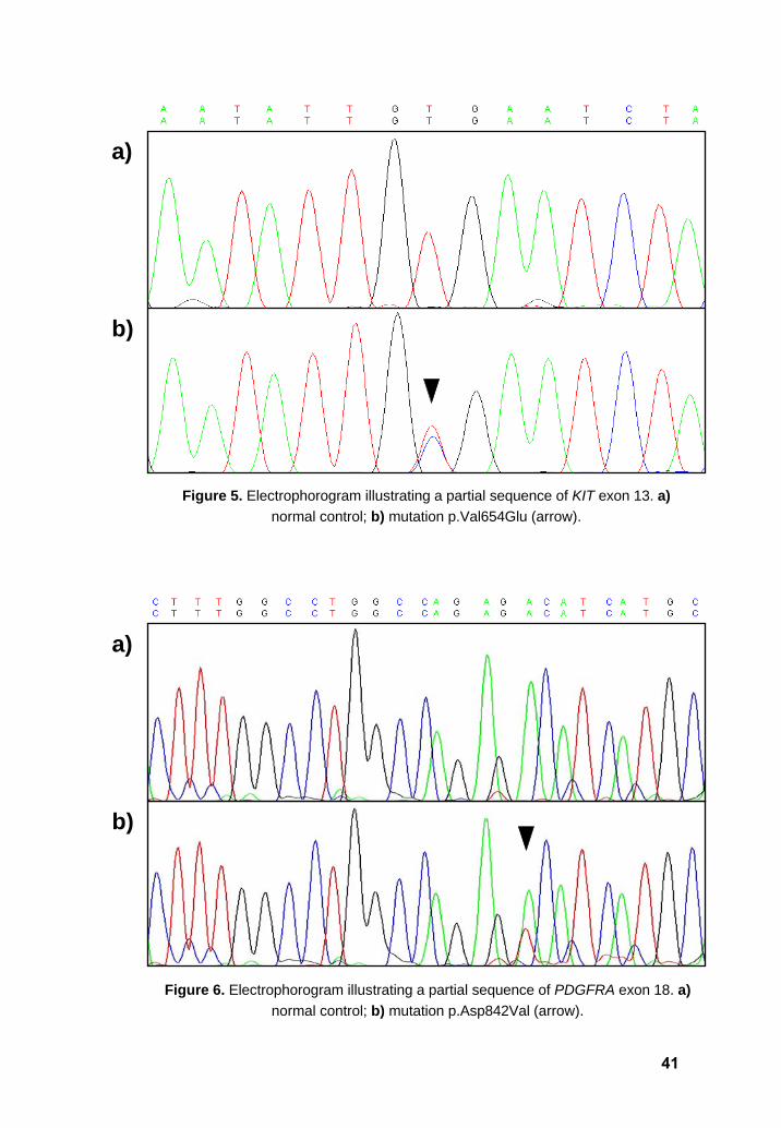
a)
b)
Figure 6. Electrophorogram illustrating a partial sequence of PDGFRA exon 18. a)normal control; b) mutation p.Asp842Val (arrow).
a)
b)
Figure 5. Electrophorogram illustrating a partial sequence of KIT exon 13. a)normal control; b) mutation p.Val654Glu (arrow).
41
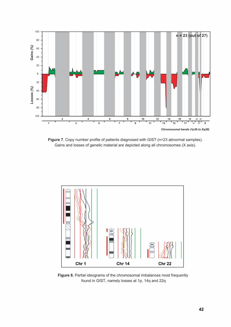
Gai
ns (%
)Lo
sses
(%)
Chromosomal bands (1p36 to Xq28)
80
0
60
12 12
1314
1516
1718
19
20
21
22
X34
56
78
910
11
20
60
40
100
40
20
80
100
n = 23 (out of 27)
Figure 7. Copy number profile of patients diagnosed with GIST (n=23 abnormal samples). Gains and losses of genetic material are depicted along all chromosomes (X axis).
Figure 8. Partial ideograms of the chromosomal imbalances most frequently found in GIST, namely losses at 1p, 14q and 22q
Chr 1 Chr 14 Chr 22Chr 1 Chr 14 Chr 22
42
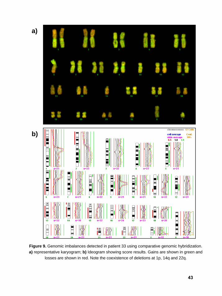
Figure 9. Genomic imbalances detected in patient 33 using comparative genomic hybridization. a) representative karyogram; b) Ideogram showing score results. Gains are shown in green and
losses are shown in red. Note the coexistence of deletions at 1p, 14q and 22q.
a)
b)
43
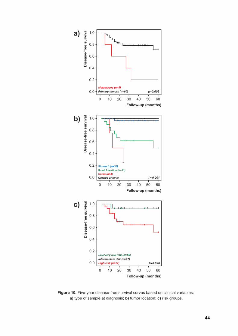
Figure 10. Five-year disease-free survival curves based on clinical variables: a) type of sample at diagnosis; b) tumor location; c) risk groups.
Dis
ease
-free
sur
viva
l
6050403020100
1.0
0.8
0.6
0.4
0.2
0.0
Follow-up (months)
p=0.026Intermediate risk (n=17) Low/very low risk (n=15)
High risk (n=27)
c)
Follow-up (months)
Dis
ease
-free
sur
viva
l
6050403020100
1.0
0.8
0.6
0.4
0.2
0.0 p<0.001Outside GI (n=5)
Stomach (n=30)Small Intestine (n=21)Colon (n=4)
b)
Follow-up (months)
Dis
ease
-free
sur
viva
l
6050403020100
1.0
0.8
0.6
0.4
0.2
0.0 p=0.002Primary tumors (n=60) Metastases (n=5)
a)
44
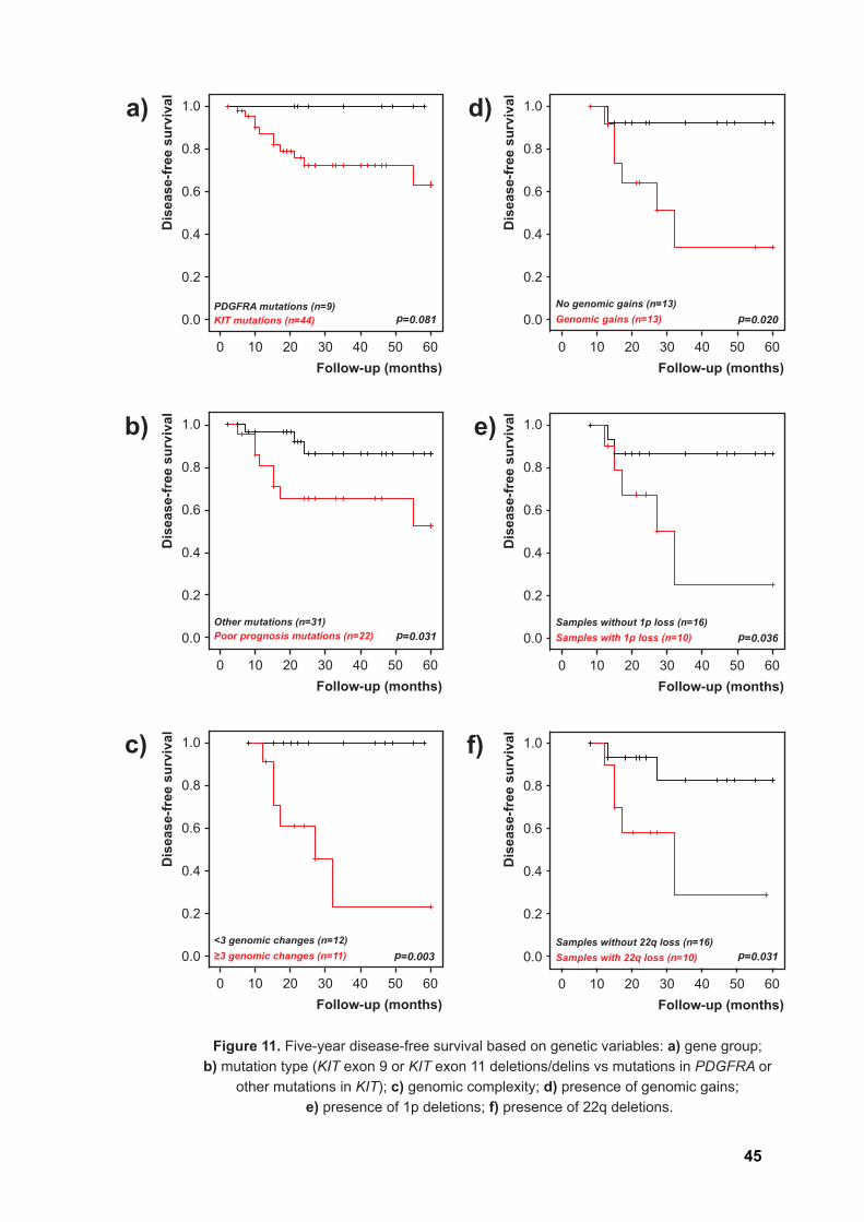
Figure 11. Five-year disease-free survival based on genetic variables: a) gene group; b) mutation type (KIT exon 9 or KIT exon 11 deletions/delins vs mutations in PDGFRA or
other mutations in KIT); c) genomic complexity; d) presence of genomic gains; e) presence of 1p deletions; f) presence of 22q deletions.
Dis
ease
-free
sur
viva
l
6050403020100
1.0
0.8
0.6
0.4
0.2
0.0
Follow-up (months)
p=0.081PDGFRA mutations (n=9) KIT mutations (n=44)
a)
Dis
ease
-free
sur
viva
l
6050403020100
1.0
0.8
0.6
0.4
0.2
0.0
Follow-up (months)
p=0.031Other mutations (n=31) Poor prognosis mutations (n=22)
b)
Dis
ease
-free
sur
viva
l
6050403020100
1.0
0.8
0.6
0.4
0.2
0.0
Follow-up (months)
p=0.036Samples without 1p loss (n=16) Samples with 1p loss (n=10)
e)
Dis
ease
-free
sur
viva
l
6050403020100
1.0
0.8
0.6
0.4
0.2
0.0
Follow-up (months)
p=0.020No genomic gains (n=13) Genomic gains (n=13)
d)
Dis
ease
-free
sur
viva
l
6050403020100
1.0
0.8
0.6
0.4
0.2
0.0
Follow-up (months)
p=0.031Samples without 22q loss (n=16) Samples with 22q loss (n=10)
f)
p=0.003<3 genomic changes (n=12) ≥3 genomic changes (n=11)
Dis
ease
-free
sur
viva
l
6050403020100
1.0
0.8
0.6
0.4
0.2
0.0
Follow-up (months)
c)
45
DISCUSSION
Recent years have seen important breakthroughs that resulted in better diagnostic,
prognostic and therapeutic tools for patients with gastrointestinal stromal tumors. Less
than 10 years ago, most GIST were misclassified as muscle neoplasias, with which they
shared morphological features, and it was not until the early 90’s that this independent
entity was properly acknowledged. More importantly, a distinctive genetic fingerprint was
uncovered that allowed targeted therapies for these aggressive tumors, bringing some
hope to patients whose live expectancy was otherwise much reduced. The current project
aimed to characterize the mutation profile of KIT and PDFGRA in a series of GIST
diagnosed and followed at our institution, and to determine the possible prognostic and/or
predictive value of these alterations. Secondary genetic changes were additionally
assessed using a whole-genome screening methodology to determine the relative
contribution of primary and secondary genetic events in GIST establishment and
progression.
Until 2007, tumor size and mitotic index remained the most important prognostic
indicators in GIST (Fletcher CD et al, 2002; Joensuu H, 2008). In our series, tumors
averaged 9 cm, and risk stratification based on these criteria grouped most lesions into
the high risk category (Table 2). However, it has been shown that even low risk GIST can
behave aggressively, progress and develop metastases. In accordance, two of the
patients in our low risk group developed metastases, with one of them dying from this
cause. Within the group of intermediate risk, most patients are currently alive with no
evidence of disease, whereas those in the group of high risk showed significantly more
progression events (Figure 10c). Indeed, only two of these patients are currently disease-
free (following additional surgical treatment). In 2007, anatomic location was also
considered of relevance and included in the determination of the risk of recurrence and
progression (Demetri GD et al, 2007; Yang J et al, 2008). In our series, lesions located in
the stomach (51.4%) or outside the GI (15.3%) showed no progression events, whereas
the majority of those in the small intestine (26.4%) or colon (6.9%) developed metastasis
in a significantly shorter period (p<0.001, Figure 10b).
The main contribution to this field, however, was the identification of the causative
role of KIT (and PDGFRA) receptors in these tumors (Hirota S et al, 1998), which not only
helped establish a correct diagnosis but also revealed important prognostic and predictive
value. Detection of KIT protein antigen (CD117) by immunohistochemistry is fundamental
for the diagnosis of GIST, as 95% of cases show positivity for this marker (Corless CL et
al, 2004). Interestingly, CD117 staining may show citoplasmic, membranous or
46

paranuclear “dotlike” distribution (Rubin BP, 2006). One possible reason for these
heterogeneous patterns is that different KIT mutations may express mutant isoforms with
preferred cellular distributions. Furthermore, many groups quickly suggested that the type
and molecular location of KIT or PDGFRA alterations is related with the risk of recurrence
or progression, and also therapy response in GIST (Ernst SI et al, 1998; Liu XH et al,
2005; Martin J et al, 2005; Andersson J et al, 2006; Cho S et al, 2006; Lasota J et al,
2006; DeMatteo RP et al, 2008; Corless CL and Heinrich MC, 2008).
In our series, expression of CD117 was assessed in 75 cases. A total of 71 lesions
showed a positive staining pattern (with considerable heterogeneity between different
lesions). Even if the positive rate is in accordance with the literature, further studies are
needed to clarify the dissimilar cellular distribution of KIT also observed in our study, as
several reports strangely refer a predominance of cytoplasmic staining (Corless CL and
Heinrich MC, 2008). Interestingly, it has been shown that within the 5% of GIST that do
not express CD117, oncogenic mutations of KIT (>50%) or PDGFRA (~30%) may be
found (Corless CL et al, 2004). Among our four CD117 expression-negative cases, one
showed indeed a PDGFRA mutation, but no mutations were found in the remaining three.
Our results for CD34, desmin, SMA, and S100 protein expression were also in
accordance with previous literature reports.
The overall frequency of KIT and PDGFRA mutations in GIST varies in different
studies, but is usually very high (Rubin BP, 2006). These mutually exclusive primary
events are critical in the pathogenesis of most GIST, as they promote the constitutive
activation of the receptors and initiate the downstream signaling pathways that translate
into aberrant cell proliferation, apoptosis, chemotaxis, and adhesion. Mutation screening
of both oncogenes was performed in 78 samples, and an overall mutation frequency of
87.2% (n=68) was obtained, with 59 cases harboring KIT mutations (75.7%) and 9 cases
showing PDGFRA mutations (11.5%). In particular, KIT mutations were detected in exons
11 (n=52, 66.7%) and 9 (n=7, 9%), whereas PDGFRA mutations were observed in exons
18 (n=6, 7.7%), 12 (n=2, 2.6%) and 14 (n=1, 1.3%) (Table 3).
A Portuguese study with a comparable series of cases (n=78) has been recently
published (Gomes AL et al, 2007), with the authors reporting an overall incidence of 63%
mutations, with 44 cases involving KIT (56%), namely in exons 11 (n=40, 51%) and 9
(n=4, 5%), and 5 cases involving PDGFRA (6%), specifically exons 12 (n=2, 3%) and 18
(n=3, 4%). The authors state that these results are in accordance with published ranges,
in particular those of the Iberian Peninsula (Martin J et al, 2005), even if the overall
incidence in other studies (and our own) is considerably higher (Rubin BP, 2006; Miettinen
M and Lasota J, 2006). Two main reasons likely account for this variability. The first refers
47
DISCUSSION
to the material used, as the previous Portuguese and Spanish studies screened formalin-
fixed, paraffin-embedded GIST (n=78 and n=162 cases, respectively), whereas 27 of our
lesions corresponded to fresh frozen tumors. It is known that DNA extracted from paraffin-
embedded tissue is more degraded, enhancing the probability of obtaining false negative
results. The second reason concerns the methodologies used for KIT mutational
screening. While we used PCR followed by direct sequencing in all cases, Gomes and
collaborators (2007) used a Single Strand Conformational Polymorphism (SSCP)
approach (a less sensitive method) for an initial screening, and only those samples
showing abnormal migration patterns were subjected to direct sequencing.
It has been suggested that the type and molecular location of different mutational
events in GIST carry distinct biological and clinical implications (Corless CL and Heinrich
MC, 2008; Lasota J et al, 2008). Indeed, different regulatory or effector domains of KIT
and PDGFRA receptors may be targeted, and thus oncogenic activation occurs through
distinct mechanisms. Mutations in the KIT extracellular regulatory domain, coded by exon
9, seem to mimic the conformational changes that follow SCF ligation. The most common
mutation found within this location (p.Ala502_Tyr503dup) corresponds to an insertion of
six nucleotides (Lux ML et al, 2000). All our samples with exon 9 mutations displayed this
hot-spot alteration (Figure 3). According to literature reports, tumors with KIT exon 9
mutations are mainly characterized as high-risk or overtly malignant, and approximately
95% are localized in the small intestine (Rubin BP et al, 2007). In our series, only two out
of the seven cases had origin in the small intestine and were classified as high risk. Both
patients are still alive (with disease persistence).
The major mutational hotspot in KIT is exon 11, which encodes the juxtamembrane
intracellular domain responsible for modulating KIT enzymatic activity (Yang J et al, 2008).
Mutations within this exon include in-frame deletions or insertions, missense mutations or
combinations thereof. These lesions may occur throughout the entire GI tract. KIT exon 11
deletions, and more specifically those affecting codons 557-558, have been linked to an
aggressive behavior comparing with missense and insertion mutations, independently of
Imatinib therapy (Wardelmann E et al, 2003; Martin J et al, 2005; Cho S et al, 2006;
Lasota J et al, 2008). In our series, 26 out of 52 mutations in this domain corresponded to
deletions or delins. Interestingly, 15 of these 26 patients showed disease progression,
(five died from their tumors), whereas only four patients in the group with insertions,
duplications or missense mutations showed disease progression (p=0.003). Most of the
deceased patients had tumors classified as high risk, but at least one was considered low
risk at diagnosis. It is also noteworthy that in two patients with primary KIT exon 11
mutations treated with Imatinib, and in which additional metastases developed, the same
48
DISCUSSION

secondary mutation in KIT exon 13 (p.Val654Glu), known to confer drug resistance
(Debiec-Rychter M et al, 2005), was detected (Figure 5).
GIST harboring PDGFRA mutations share many clinical features with KIT mutated
lesions, but are mainly gastric and present weak or negative CD117 staining
(Wardelmann E, 2004). Evidence suggests that PDGFRA mutated tumors might be less
aggressive (Rubin BP et al, 2007). In our series, three important observations should be
highlighted: from the nine cases with PDGFRA mutations, 3 showed weak or only focal
CD117 staining; most tumors (n=7) were located in the stomach; and all patients are
currently alive with no evidence of disease. Interestingly, the hotspot p.Asp842Val
mutation was detected in four of the cases (Figure 6). This mutation has been associated
with primary resistance to Imatinib (Heinrich MC et al, 2003), but these patients were
classified in the low and intermediate risk groups, showed no signs of progression, and
were not treated with Imatinib thus far.
KIT and PDGFRA negative GIST account for 5 to 10% of all cases and may occur
in the entire length of GI tract (Rubin BP et al, 2007). It is likely that analogous receptor
tyrosine kinases or other downstream effectors of their signaling cascades harbor
activating mutations in these lesions (Heinrich MC et al, 2002; Rubin BP, 2006). The ten
tumors in our series negative for both KIT and PDGFRA mutations were mostly located in
the stomach (n=6) and were classified as very low, low (n=2), intermediate, or high risk
(n=5). Only one high risk patient died as a consequence of the disease, whereas two
others died of unrelated causes.
In addition to the early mutation events activating KIT or PDGFRA, cytogenetic
studies have shown several secondary genetic changes associated with GIST
progression (El-Rifai W et al, 2000a; Sandberg AA and Bridge JA, 2002; Gunawan B et al,
2007). Few studies so far have performed genotype and genome analysis in the same
samples, preventing a reliable assessment of correlations between primary and
secondary genetic events, or their combined prognostic/predictive value (Anderson J et al,
2002; Assämäki R et al, 2007; Wozniak A et al, 2007). In our work, 85% of the analyzed
GIST displayed copy number changes (Figure 7). Complete or partial deletions of
chromosome 14 were seen in 91% of the abnormal cases, and in four patients this was
the sole change detected. Additional recurrent aberrations included losses at 22q (43.5%),
1p (43.5%) and 15q (34.8%), as well as gains at 1q (17.4%) and 12q (17.4%) (Table 4,
Figure 7). Based on previous literature findings, samples submitted to CGH analysis were
divided according to mutation genotypes. Interestingly, lesions harboring mutations
associated with a bad prognosis (n=12) showed significantly more copy number changes
than those without such mutations (median of 6.5 versus 2 aberrations per tumor,
49
DISCUSSION
p=0.025). The presence of gains, deletions at 1p, and deletions at 22q could also be
associated with a shorter disease-free survival for these patients. In accordance with other
groups, genomic complexity (more than 3 aberrations per tumor) was also significantly
associated with a worse outcome for the patients. It is also noteworthy that tumors with
PDGFRA mutations showed the same pattern of alterations seen in those with KIT
mutations (namely losses of 14q, 22q, and 1p, and gains at 10q), even if the complexity
was much reduced (median of 2 vs. 6 alterations per tumor, respectively).
Taken together, these findings suggest that secondary chromosome changes play
a more relevant role than it was recognized until now, and that cytogenetic screening
might be required in the future to fully characterize the malignant potential of GIST. It is
also noteworthy that of the four cases without detectable copy number changes, three
samples were negative for KIT or PDGFRA mutations (Table 4). As CD117 staining for
these three samples was focal or absent, one must consider the possibility that these
lesions have been misclassified as GIST. Interestingly, histological evaluation of a recent
metastatic lesion of one of these cases resulted in a diagnosis of leiomiosarcoma (patient
number 62).
The introduction of targeted therapy against the tyrosine kinase domain of KIT
(and related receptors) remarkably increased the overall survival of patients with
metastatic or inoperable GIST. The molecular location and nature of KIT and PDGFRA
activating mutations is known to influence the likelihood of clinical response to this drug. In
our series, 26 patients (35%) showed disease progression after initial surgery and were
subsequently treated with Imatinib. All but two of these tumors showed KIT exon 11
mutations, in particular deletions/delins. Strikingly, this group was also the most complex
regarding genomic alterations, together with lesions harboring KIT exon 9 mutations.
Patients with inoperable GIST and showing intolerance or resistance to Imatinib now have
the possibility to be treated with the second line drug Sunitinib. Preliminary data suggest
this drug might even be more efficient, given the fact that Imatinib has exclusive affinity to
a limited number of TK domains (Demetri GD et al, 2006). Unfortunately, tumors treated
with this second line therapy also tend to progress, namely by acquiring KIT exon 17
mutations (Corless CL and Heinrich MC, 2008). Due to the fact that such resistance is
becoming a significant clinical problem in the treatment of this neoplasia, several trials
with new targeted drugs are underway, underlining the importance of genetic studies
aimed at identifying novel alterations with prognostic or predictive value or that may work
as therapeutic targets in GIST.
50
DISCUSSION

CONCLUSIONS
During the past decade, GIST have emerged from historic anonymity to center
stage in the field of solid tumor management. Progresses concerning their biology and
genetic background, namely the discovery of a defining molecular and
immunohistochemical profile, laid the groundwork for the first largely successful targeted
therapy protocols in solid tumors. In this study we report the genetic characterization of a
consecutive series of 78 patients diagnosed with GIST, in which primary mutation events
in KIT or PDGFRA, as well as secondary chromosomal changes (for a subset of 27
patients), were assessed and correlated with available clinico-pathological and therapy
response data.
Using a direct sequencing approach, we conclude that most GIST show activating
mutations in either KIT (75.7%) or PDGFRA (11.5%). The importance of a molecular-
based approach in the diagnosis of GIST is well illustrated in the fact that within the group
of cases with negative or doubtful positivity for CD117, one harboured a PDGFRA
mutation. We further show that the majority of GIST display chromosomal alterations,
namely deletions at chromosomal regions 14q, 1p and 22q, and that a significantly higher
number of these secondary genetic aberrations can be seen in tumors with mutation
genotypes associated with aggressiveness, such as those harboring KIT exon 9 mutations
and KIT exon 11 deletions/delins.
The worse outcome previously associated with KIT exon 11 deletions/delins was
additionally demonstrated by the fact that most patients with disease progression showed
these mutations. Genomic complexity, the presence of gains, losses at 1p, or losses at
22q, could also significantly distinguish patients at a higher risk of disease progression. In
opposition, all patients with PDGFRA mutations, associated in the literature with less
aggressive phenotypes, are currently alive with no evidence of disease. These also
displayed significantly less aberrations as detected by CGH.
We deem to have successfully demonstrated the relevant contribution of genetic
analysis in the diagnosis and risk stratification of patients with GIST. Our findings further
suggest that cytogenetic screening is a valuable tool that should be included in future
studies, as secondary chromosomal changes were strongly associated with the clinical
aggressiveness of these tumors. Additional follow-up is required to fully assess the
prognostic and predictive value of several other recurrent genetic features.
51
FUTURE PERSPECTIVES
The present work focused on the genetic characterization of a series of patients
diagnosed with GIST, assessing both primary mutation events as well as secondary
genomic aberrations. Given the relatively small number of patients analyzed by CGH, the
correlations between genotype and genomic data were limited. The reduced follow-up
period for patients under Imatinib therapy further prevented a reliable determination of the
predictive value of several genetic alterations. To clarify these issues and achieve a more
complete characterization of our series, several steps are being implemented and
prepared, namely:
o To update and monitor patient status and response to first line and second line
therapies, in order to reliably determine the prognostic and/or predictive value of
genetic changes in this series of GIST.
o To monitor Imatinib or Sunitinib resistance in treated patients and assess if new
molecular events leading to this resistance have occurred in the respective tumors.
o To apply the FISH methodology to assess the most common secondary alterations
in all samples for which whole-genome screening (CGH) could not be applied.
52

REFERENCES
Agaram NP, Besmer P, Wong GC, Guo T, Socci ND, Maki RG, DeSantis D, Brennan MF, Singer S, DeMatteo RP, Antonescu CR. Pathologic and molecular heterogeneity in imatinib-stable or imatinib-responsive gastrointestinal stromal tumors. Clin Cancer Res 2007, 13:170-81.
Andersson J, Sjögren H, Meis-Kindblom JM, Stenman G, Aman P, Kindblom LG. The complexity of KIT gene mutations and chromosome rearrangements and their clinical correlation in gastrointestinal stromal (pacemaker cell) tumors. Am J Pathol 2002, 160:15-22.
Andersson J, Bümming P, Meis-Kindblom JM, Sihto H, Nupponen N, Joensuu H, Odén A, Gustavsson B, Kindblom LG, Nilsson B. Gastrointestinal stromal tumors with KIT exon 11 deletions are associated with poor prognosis. Gastroenterology 2006, 130:1573-81.
Antonescu CR, Sommer G, Sarran L, Tschernyavsky SJ, Riedel E, Woodruff JM, Robson M, Maki R, Brennan MF, Ladanyi M, DeMatteo RP, Besmer P. Association of KIT exon 9 mutations with nongastric primary site and aggressive behavior: KIT mutation analysis and clinical correlates of 120 gastrointestinal stromal tumors. Clin Cancer Res 2003, 9:3329-37.
Antonescu CR, Besmer P, Guo T, Arkun K, Hom G, Koryotowski B, Leversha MA, Jeffrey PD, Desantis D, Singer S, Brennan MF, Maki RG, DeMatteo RP. Acquired resistance to imatinib in gastrointestinal stromal tumor occurs through secondary gene mutation. Clin Cancer Res 2005, 11:4182-90.
Antonescu CR. Targeted therapies in gastrointestinal stromal tumors. Semin Diagn Pathol 2008, 25:295-303.
Assämäki R, Sarlomo-Rikala M, Lopez-Guerrero JA, Lasota J, Andersson LC, Llombart-Bosch A, Miettinen M, Knuutila S. Array comparative genomic hybridization analysis of chromosomal imbalances and their target genes in gastrointestinal stromal tumors. Genes Chromosomes Cancer 2007, 46:564-76.
Bardi G, Johansson B, Pandis N, Heim S, Mandahl N, Bak-Jensen E, Frederiksen H, Andrén-Sandberg A, Mitelman F. Recurrent chromosome aberrations in abdominal smooth muscle tumors. Cancer Genet Cytogenet 1992, 62:43-6.
Bergmann F, Gunawan B, Hermanns B, Höer J, Schumpelick V, Füzesi L. Cytogenetic and morphologic characteristics of gastrointestinal stromal tumors. Recurrent rearrangement of chromosome 1 and losses of chromosomes 14 and 22 as common anomalies. Verh Dtsch Ges Pathol 1998, 82:275-8.
Boghosian L, Dal Cin P, Turc-Carel C, Rao U, Karakousis C, Sait SJ, Sandberg AA. Three possible cytogenetic subgroups of leiomyosarcoma. Cancer Genet Cytogenet 1989, 43:39-49.
Buchdunger E, Zimmermann J, Mett H, Meyer T, Müller M, Druker BJ, Lydon NB. Inhibition of the Abl protein-tyrosine kinase in vitro and in vivo by a 2-phenylaminopyrimidine derivative. Cancer Res 1996, 56:100-4.
Cho S, Kitadai Y, Yoshida S, Tanaka S, Yoshihara M, Yoshida K, Chayama K. Deletion of the KIT gene is associated with liver metastasis and poor prognosis in patients with gastrointestinal stromal tumor in the stomach. Int J Oncol 2006, 28:1361-7.
Cichoz-Lach H, Kasztelan-Szczerbińska B, Słomka M. Gastrointestinal stromal tumors: epidemiology, clinical picture, diagnosis, prognosis and treatment. Pol Arch Med Wewn 2008, 118:216-21.
53
Corless CL, Fletcher JA, Heinrich MC. Biology of gastrointestinal stromal tumors. J Clin Oncol 2004, 22:3813-25.
Corless CL, Schroeder A, Griffith D, Town A, McGreevey L, Harrell P, Shiraga S, Bainbridge T, Morich J, Heinrich MC. PDGFRA mutations in gastrointestinal stromal tumors: frequency, spectrum and in vitro sensitivity to Imatinib. J Clin Oncol 2005, 23:5357-64.
Corless CL, Heinrich MC. Molecular pathobiology of gastrointestinal stromal sarcomas. Annu Rev Pathol 2008, 3:557-86.
Daum O, Grossmann P, Vanecek T, Sima R, Mukensnabl P, Michal M. Diagnostic morphological features of PDGFRA-mutated gastrointestinal stromal tumors: molecular genetic and histologic analysis of 60 cases of gastric gastrointestinal stromal tumors. Ann Diagn Pathol 2007, 11:27-33.
Debiec-Rychter M, Lasota J, Sarlomo-Rikala M, Kordek R, Miettinen M. Chromosomal aberrations in malignant gastrointestinal stromal tumors: correlation with c-KIT gene mutation. Cancer Genet Cytogenet 2001, 128:24-30.
Debiec-Rychter M, Wasag B, Stul M, De Wever I, Van Oosterom A, Hagemeijer A, Sciot R. Gastrointestinal stromal tumours (GISTs) negative for KIT (CD117 antigen) immunoreactivity. J Pathol 2004, 202:430-8.
Debiec-Rychter M, Cools J, Dumez H, Sciot R, Stul M, Mentens N, Vranckx H, Wasag B, Prenen H, Roesel J, Hagemeijer A, Van Oosterom A, Marynen P. Mechanisms of resistance to imatinib mesylate in gastrointestinal stromal tumors and activity of the PKC412 inhibitor against imatinib-resistant mutants. Gastroenterology 2005, 128:270-9.
Debiec-Rychter M, Sciot R, Le Cesne A, Schlemmer M, Hohenberger P, van Oosterom AT, Blay JY, Leyvraz S, Stul M, Casali PG, Zalcberg J, Verweij J, Van Glabbeke M, Hagemeijer A, Judson I. EORTC Soft Tissue and Bone Sarcoma Group; The Italian Sarcoma Group; Australasian GastroIntestinal Trials Group. KIT mutations and dose selection for imatinib in patients with advanced gastrointestinal stromal tumours. Eur J Cancer 2006, 42:1093-103.
DeMatteo RP, Lewis JJ, Leung D, Mudan SS, Woodruff JM, Brennan MF. Two hundred gastrointestinal stromal tumors: recurrence patterns and prognostic factors for survival. Ann Surg 2000, 231:51-8.
Dematteo RP, Gold JS, Saran L, Gönen M, Liau KH, Maki RG, Singer S, Besmer P, Brennan MF, Antonescu CR. Tumor mitotic rate, size, and location independently predict recurrence after resection of primary gastrointestinal stromal tumor (GIST). Cancer 2008, 112:608-15.
Demetri GD, van Oosterom AT, Garrett CR, Blackstein ME, Shah MH, Verweij J, McArthur G, Judson IR, Heinrich MC, Morgan JA, Desai J, Fletcher CD, George S, Bello CL, Huang X, Baum CM, Casali PG. Efficacy and safety of sunitinib in patients with advanced gastrointestinal stromal tumour after failure of imatinib: a randomised controlled trial. Lancet 2006, 368:1329-38.
Demetri GD, Benjamin RS, Blanke CD, Blay JY, Casali P, Choi H, Corless CL, Debiec-Rychter M, DeMatteo RP, Ettinger DS, Fisher GA, Fletcher CD, Gronchi A, Hohenberger P, Hughes M, Joensuu H, Judson I, Le Cesne A, Maki RG, Morse M, Pappo AS, Pisters PW, Raut CP, Reichardt P, Tyler DS, Van den Abbeele AD, von Mehren M, Wayne JD, Zalcberg J. NCCN Task Force. NCCN Task Force report: management of patients with gastrointestinal stromal tumor (GIST)--update of the NCCN clinical practice guidelines. J Natl Compr Canc Netw 2007, 5:S1-29.
54
REFERENCES
Desai J, Shankar S, Heinrich MC, Fletcher JA, Fletcher CD, Manola J, Morgan JA, Corless CL, George S, Tuncali K, Silverman SG, Van den Abbeele AD, van Sonnenberg E, Demetri GD. Clonal evolution of resistance to imatinib in patients with metastatic gastrointestinal stromal tumors. Clin Cancer Res 2007, 13:5398-405.
Dewar AL, Cambareri AC, Zannettino AC, Miller BL, Doherty KV, Hughes TP, Lyons AB. Macrophage colony-stimulating factor receptor c-fms is a novel target of imatinib. Blood 2005, 105:3127-32.
Di Matteo G, Pescarmona E, Peparini N, Di Matteo FM, Zeri KP, Mascagni D, Mele R, Maturo A, Redler A, De Antoni E. Histopathological features and clinical course of the gastrointestinal stromal tumors. Hepatogastroenterology 2002, 49:1013-6.
Du CY, Shi YQ, Zhou Y, Fu H, Zhao G. The analysis of status and clinical implication of KIT and PDGFRA mutations in gastrointestinal stromal tumor (GIST). J Surg Oncol 2008, 98:175-8.
Duensing A, Joseph NE, Medeiros F, Smith F, Hornick JL, Heinrich MC, Corless CL, Demetri GD, Fletcher CD, Fletcher JA. Protein Kinase C theta (PKCtheta) expression and constitutive activation in gastrointestinal stromal tumors (GISTs). Cancer Res 2004, 64:5127-31.
El-Rifai W, Sarlomo-Rikala M, Andersson LC, Knuutila S, Miettinen M. DNA sequence copy number changes in gastrointestinal stromal tumors: tumor progression and prognostic significance. Cancer Res 2000a, 60:3899-903.
El-Rifai W, Sarlomo-Rikala M, Andersson LC, Miettinen M, Knuutila S. High-resolution deletion mapping of chromosome 14 in stromal tumors of the gastrointestinal tract suggests two distinct tumor suppressor loci. Genes Chromosomes Cancer 2000b, 27:387-91.
Ernst SI, Hubbs AE, Przygodzki RM, Emory TS, Sobin LH, O'Leary TJ. KIT mutation portends poor prognosis in gastrointestinal stromal/smooth muscle tumors. Lab Invest 1998, 78:1633-6.
Fletcher CD, Berman JJ, Corless C, Gorstein F, Lasota J, Longley BJ, Miettinen M, O'Leary TJ, Remotti H, Rubin BP, Shmookler B, Sobin LH, Weiss SW. Diagnosis of gastrointestinal stromal tumors: A consensus approach. Hum Pathol 2002, 33:459-65.
Foster R, Griffith R, Ferrao P, Ashman L. Molecular basis of the constitutive activity and STI571 resistance of Asp816Val mutant KIT receptor tyrosine kinase. J Mol Graph Model 2004, 23:139-52.
Goh BK, Chow PK, Yap WM, Kesavan SM, Song IC, Paul PG, Ooi BS, Chung YF, Wong WK. Which is the optimal risk stratification system for surgically treated localized primary GIST? Comparison of three contemporary prognostic criteria in 171 tumors and a proposal for a modified Armed Forces Institute of Pathology risk criteria. Ann Surg Oncol 2008, 15:2153-63.
Gold JS, van der Zwan SM, Gönen M, Maki RG, Singer S, Brennan MF, Antonescu CR, De Matteo RP. Outcome of metastatic GIST in the era before tyrosine kinase inhibitors. Ann Surg Oncol 2007, 14:134-42.
Gomes AL, Gouveia A, Capelinha AF, de la Cruz D, Silva P, Reis RM, Pimenta A, Lopes JM. Molecular alterations of KIT and PDGFRA in GISTs: evaluation of a Portuguese series. J Clin Pathol 2008, 61:203-8.
Gunawan B, Bergmann F, Höer J, Langer C, Schumpelick V, Becker H, Füzesi L. Biological and clinical significance of cytogenetic abnormalities in low-risk and high-risk gastrointestinal stromal tumors. Hum Pathol 2002, 33:316-21.
55
REFERENCES
Gunawan B, von Heydebreck A, Sander B, Schulten HJ, Haller F, Langer C, Armbrust T, Bollmann M, Gasparov S, Kovac D, Füzesi L. An oncogenetic tree model in gastrointestinal stromal tumours (GISTs) identifies different pathways of cytogenetic evolution with prognostic implications. J Pathol 2007, 211:463-70.
Gupta P, Tewari M, Shukla HS. Gastrointestinal stromal tumor. Surg Oncol 2008, 17:129-38.
Hanahan D, Weinberg RA. The hallmarks of cancer. Cell 2000, 100:57-70.
Heinrich MC, Rubin BP, Longley BJ, Fletcher JA. Biology and genetic aspects of gastrointestinal stromal tumors: KIT activation and cytogenetic alterations. Hum Pathol 2002, 33:484-95.
Heinrich MC, Corless CL, Duensing A, McGreevey L, Chen CJ, Joseph N, Singer S, Griffith DJ, Haley A, Town A, Demetri GD, Fletcher CD, Fletcher JA. PDGFRA activating mutations in gastrointestinal stromal tumors. Science 2003 299:708-10.
Heinrich MC, Corless CL, Blanke CD, Demetri GD, Joensuu H, Roberts PJ, Eisenberg BL, von Mehren M, Fletcher CD, Sandau K, McDougall K, Ou WB, Chen CJ, Fletcher JA. Molecular correlates of imatinib resistance in gastrointestinal stromal tumors. J Clin Oncol 2006, 24:4764-74.
Hirano K, Shishido-Hara Y, Kitazawa A, Kojima K, Sumiishi A, Umino M, Kikuchi F, Sakamoto A, Fujioka Y, Kamma H. Expression of stem cell factor (SCF), a KIT ligand, in gastrointestinal stromal tumors (GISTs): a potential marker for tumor proliferation. Pathol Res Pract 2008, 204:799-807.
Hirota S, Isozaki K, Moriyama Y, Hashimoto K, Nishida T, Ishiguro S, Kawano K, Hanada M, Kurata A, Takeda M, Muhammad Tunio G, Matsuzawa Y, Kanakura Y, Shinomura Y, Kitamura Y. Gain-of-function mutations of c-kit in human gastrointestinal stromal tumors. Science 1998, 279:577-80.
Hirota S, Ohashi A, Nishida T, Isozaki K, Kinoshita K, Shinomura Y, Kitamura Y. Gain-of-function mutations of platelet-derived growth factor receptor alpha gene in gastrointestinal stromal tumors. Gastroenterology 2003, 125:660-7.
Isozaki K, Hirota S, Nakama A, Miyagawa J, Shinomura Y, Xu Z, Nomura S, Kitamura Y. Disturbed intestinal movement, bile reflux to the stomach, and deficiency of c-kit-expressing cells in Ws/Ws mutant rats. Gastroenterology 1995, 109:456-64.
Isozaki K, Terris B, Belghiti J, Schiffmann S, Hirota S, Vanderwinden JM. Germline-activating mutation in the kinase domain of KIT gene in familial gastrointestinal stromal tumors. Am J Pathol 2000, 157:1581-5.
Joensuu H, Roberts PJ, Sarlomo-Rikala M, Andersson LC, Tervahartiala P, Tuveson D, Silberman S, Capdeville R, Dimitrijevic S, Druker B, Demetri GD. Effect of the tyrosine kinase inhibitor STI571 in a patient with a metastatic gastrointestinal stromal tumor. N Engl J Med 2001, 344:1052-6.
Joensuu H. Risk stratification of patients diagnosed with gastrointestinal stromal tumor. Hum Pathol 2008, 39:1411-9.
Kallioniemi A, Kallioniemi OP, Sudar D, Rutovitz D, Gray JW, Waldman F, Pinkel D. Comparative genomic hybridization for molecular cytogenetic analysis of solid tumors. Science 1992, 58:818-21.
Kim NG, Kim JJ, Ahn JY, Seong CM, Noh SH, Kim CB, Min JS, Kim H. Putative chromosomal deletions on 9P, 9Q and 22Q occur preferentially in malignant gastrointestinal stromal tumors. Int J Cancer 2000, 85:633-8.
56
REFERENCES
Kim TW, Lee H, Kang YK, Choe MS, Ryu MH, Chang HM, Kim JS, Yook JH, Kim BS, Lee JS. Prognostic significance of c-kit mutation in localized gastrointestinal stromal tumors. Clin Cancer Res 2004, 10:3076-81.
Kirchhoff M, Gerdes T, Rose H, Maahr J, Ottesen AM, Lundsteen C. Detection of chromosomal gains and losses in comparative genomic hybridization analysis based on standard reference intervals. Cytometry 1998, 31:163-73.
Lasota J, Carlson JA, Miettinen M. Spindle cell tumor of urinary bladder serosa with phenotypic and genotypic features of gastrointestinal stromal tumor. Arch Pathol Lab Med 2000, 124:894-7.
Lasota J, Dansonka-Mieszkowska A, Sobin LH, Miettinen M. A great majority of GISTs with PDGFRA mutations represent gastric tumors of low or no malignant potential. Lab Invest 2004, 84:874-83.
Lasota J, Wozniak A, Kopczynski J, Dansonka-Mieszkowska A, Wasag B, Mitsuhashi T, Sarlomo-Rikala M, Lee JR, Schneider-Stock R, Stachura J, Limon J, Miettinen M. Loss of heterozygosity on chromosome 22q in gastrointestinal stromal tumors (GISTs): a study on 50 cases. Lab Invest 2005, 85:237-47.
Lasota J, Corless CL, Heinrich MC, Debiec-Rychter M, Sciot R, Wardelmann E, Merkelbach-Bruse S, Schildhaus HU, Steigen SE, Stachura J, Wozniak A, Antonescu C, Daum O, Martin J, Del Muro JG, Miettinen M. Clinicopathologic profile of gastrointestinal stromal tumors (GISTs) with primary KIT exon 13 or exon 17 mutations: a multicenter study on 54 cases. Mod Pathol 2008, 21:476-84.
Liu H, Chen X, Focia PJ, He X. Structural basis for stem cell factor-KIT signaling and activation of class III receptor tyrosine kinases. EMBO J 2007, 26:891-901.
Liu XH, Bai CG, Xie Q, Feng F, Xu ZY, Ma DL. Prognostic value of KIT mutation in gastrointestinal stromal tumors. World J Gastroenterol 2005, 11:3948-52.
Lungu O, Sun XW, Felix J, Richart RM, Silverstein S, Wright TC Jr. Relationship of human papillomavirus type to grade of cervical intraepithelial neoplasia. JAMA 1992, 267:2493-6.
Lux ML, Rubin BP, Biase TL, Chen CJ, Maclure T, Demetri G, Xiao S, Singer S, Fletcher CD, Fletcher JA. KIT extracellular and kinase domain mutations in gastrointestinal stromal tumors. Am J Pathol 2000, 156:791-5.
Martín J, Poveda A, Llombart-Bosch A, Ramos R, López-Guerrero JA, García del Muro J, Maurel J, Calabuig S, Gutierrez A, González de Sande JL, Martínez J, De Juan A, Laínez N, Losa F, Alija V, Escudero P, Casado A, García P, Blanco R, Buesa JM; Spanish Group for Sarcoma Research. Deletions affecting codons 557-558 of the c-KIT gene indicate a poor prognosis in patients with completely resected gastrointestinal stromal tumors: a study by the Spanish Group for Sarcoma Research (GEIS). J Clin Oncol 2005, 23:6190-8.
Mazur MT, Clark HB. Gastric stromal tumors. Reappraisal of histogenesis. Am J Surg Pathol 1983, 7:507-19.
Miettinen M, Lasota J. Gastrointestinal stromal tumors-definition, clinical, histological, immunohistochemical, and molecular genetic features and differential diagnosis. Virchows Arch 2001, 438:1-12.
Miettinen M, Lasota J. Gastrointestinal stromal tumors: review on morphology, molecular pathology, prognosis, and differential diagnosis. Arch Pathol Lab Med 2006, 130:1466-78.
57
REFERENCES

Miettinen M, Lasota J. KIT (CD117): a review on expression in normal and neoplastic tissues, and mutations and their clinicopathologic correlation. Appl Immunohistochem Mol Morphol 2005a, 13:205-20.
Miettinen M, Sobin LH, Lasota J. Gastrointestinal stromal tumors of the stomach: a clinicopathologic, immunohistochemical, and molecular genetic study of 1765 cases with long-term follow-up. Am J Surg Pathol 2005b, Jan 29:52-68.
Mol CD, Lim KB, Sridhar V, Zou H, Chien EY, Sang BC, Nowakowski J, Kassel DB, Cronin CN, McRee DE. Structure of a c-kit product complex reveals the basis for kinase transactivation. J Biol Chem 2003, 278:31461-4.
Müllenbach R, Lagoda PJ, Welter C. An efficient salt-chloroform extraction of DNA from blood and tissues. Trends Genet 1989, 5:391.
Nakatani H, Kobayashi M, Jin T, Taguchi T, Sugimoto T, Nakano T, Hamada S, Araki K. STI571 (Glivec) inhibits the interaction between c-KIT and heat shock protein 90 of the gastrointestinal stromal tumor cell line, GIST-T1. Cancer Sci 2005, 96:116-9.
Orosz Z, Tornóczky T, Sápi Z. Gastrointestinal stromal tumors: a clinicopathologic and immunohistochemical study of 136 cases. Pathol Oncol Res 2005, 11:11-21.
Penzel R, Aulmann S, Moock M, Schwarzbach M, Rieker RJ, Mechtersheimer G. The location of KIT and PDGFRA gene mutations in gastrointestinal stromal tumours is site and phenotype associated. J Clin Pathol 2005, 58:634-9.
Raut CP, DeMatteo RP. Prognostic factors for primary GIST: prime time for personalized therapy? Ann Surg Oncol 2008, 15:4-6.
Ribeiro FR, Jerónimo C, Henrique R, Fonseca D, Oliveira J, Lothe RA, Teixeira MR. 8q gain is an independent predictor of poor survival in diagnostic needle biopsies from prostate cancer suspects. Clin Cancer Res 2006, 12:3961-70.
Rönnstrand L. Signal transduction via the stem cell factor receptor/c-Kit. Cell Mol Life Sci 2004, 61:2535-48.
Rossi CR, Mocellin S, Mencarelli R, Foletto M, Pilati P, Nitti D, Lise M. Gastrointestinal stromal tumors: from a surgical to a molecular approach. Int J Cancer 2003, 107:171-6.
Rubin BP, Singer S, Tsao C, Duensing A, Lux ML, Ruiz R, Hibbard MK, Chen CJ, Xiao S, Tuveson DA, Demetri GD, Fletcher CD, Fletcher JA. KIT activation is a ubiquitous feature of gastrointestinal stromal tumors. Cancer Res 2001, 61:8118-21.
Rubin BP, Antonescu CR, Scott-Browne JP, Comstock ML, Gu Y, Tanas MR, Ware CB, Woodell J. A knock-in mouse model of gastrointestinal stromal tumor harboring kit K641E. Cancer Res 2005, 65:6631-9.
Rubin BP. Gastrointestinal stromal tumours: an update. Histopathology 2006, 48:83-96.
Rubin BP, Heinrich MC, Corless CL. Gastrointestinal stromal tumour. Lancet 2007, 369:1731-41.
Rumessen JJ, Thuneberg L. Pacemaker cells in the gastrointestinal tract: interstitial cells of Cajal. Scand J Gastroenterol Suppl 1996, 216:82-94.
Sakurai S, Oguni S, Hironaka M, Fukayama M, Morinaga S, Saito K. Mutations in c-kit gene exons 9 and 13 in gastrointestinal stromal tumors among Japanese. Jpn J Cancer Res 2001, 92:494-8.
Sandberg AA, Bridge JA. Updates on the cytogenetics and molecular genetics of bone and soft tissue tumors gastrointestinal stromal tumors. Cancer Genet Cytogenet 2002, 135:1-22.
58
REFERENCES
Saunders AL, Meloni AM, Chen Z, Sandberg AA, Lauwers GY. Two cases of low-grade gastric leiomyosarcoma with monosomy 14 as the only change. Cancer Genet Cytogenet 1996, 90:184-5.
Savage DG, Antman KH. Imatinib mesylate-a new oral targeted therapy. N Engl J Med 2002, 346:683-93.
Scheijen B, Griffin JD. Tyrosine kinase oncogenes in normal hematopoiesis and hematological disease. Oncogene 2002, 21:3314-33.
Singer S, Rubin BP, Lux ML, Chen CJ, Demetri GD, Fletcher CD, Fletcher JA. Prognostic value of KIT mutation type, mitotic activity, and histologic subtype in gastrointestinal stromal tumors. J Clin Oncol 2002, 20:3898-905.
Sommer G, Agosti V, Ehlers I, Rossi F, Corbacioglu S, Farkas J, Moore M, Manova K, Antonescu CR, Besmer P. Gastrointestinal stromal tumors in a mouse model by targeted mutation of the Kit receptor tyrosine kinase. Proc Natl Acad Sci U S A 2003, 100:6706-11.
Sreekantaiah C, Davis JR, Sandberg AA. Chromosomal abnormalities in leiomyosarcomas. Am J Pathol 1993, 142:293-305.
Tamborini E, Bonadiman L, Greco A, Albertini V, Negri T, Gronchi A, Bertulli R, Colecchia M, Casali PG, Pierotti MA, Pilotti S. A new mutation in the KIT ATP pocket causes acquired resistance to imatinib in a gastrointestinal stromal tumor patient. Gastroenterology 2004, 127:294-9.
Taniguchi M, Nishida T, Hirota S, Isozaki K, Ito T, Nomura T, Matsuda H, Kitamura Y. Effect of c-kit mutation on prognosis of gastrointestinal stromal tumors. Cancer Res 1999, 59:4297-300.
Tarn C, Skorobogatko YV, Taguchi T, Eisenberg B, von Mehren M, Godwin AK. Therapeutic effect of imatinib in gastrointestinal stromal tumors: AKT signaling dependent and independent mechanisms. Cancer Res 2006, 66:5477-86.
Taylor ML, Metcalfe DD. Kit signal transduction. Hematol Oncol Clin North Am 2000, 14:517-35.
Tornillo L, Terracciano LM. An update on molecular genetics of gastrointestinal stromal tumours. J Clin Pathol 2006, 59:557-63.
Tuveson DA, Willis NA, Jacks T, Griffin JD, Singer S, Fletcher CD, Fletcher JA, Demetri GD. STI571 inactivation of the gastrointestinal stromal tumor c-KIT oncoprotein: biological and clinical implications. Oncogene 2001, 20:5054-8.
Tzen CY, Mau BL. Analysis of CD117-negative gastrointestinal stromal tumors. World J Gastroenterol 2005, 11:1052-5.
Vu HA, Xinh PT, Kikushima M, Zhu Y, Tokuhara M, Tani M, Shimizu T, Saito K, Tokunaga K, Sato Y. A recurrent duodenal gastrointestinal stromal tumor with a frameshift mutation resulting in a stop codon in KIT exon 13. Genes Chromosomes Cancer 2005, 42:179-83.
Wardelmann E, Losen I, Hans V, Neidt I, Speidel N, Bierhoff E, Heinicke T, Pietsch T, Büttner R, Merkelbach-Bruse S. Deletion of Trp-557 and Lys-558 in the juxtamembrane domain of the c-kit protooncogene is associated with metastatic behavior of gastrointestinal stromal tumors. Int J Cancer 2003, 106:887-95.
Wardelmann E, Hrychyk A, Merkelbach-Bruse S, Pauls K, Goldstein J, Hohenberger P, Losen I, Manegold C, Büttner R, Pietsch T. Association of platelet-derived growth factor receptor alpha mutations with gastric primary site and epithelioid or mixed cell morphology in gastrointestinal stromal tumors. J Mol Diagn 2004, 6:197-204.
59
REFERENCES
West RB, Corless CL, Chen X, Rubin BP, Subramanian S, Montgomery K, Zhu S, Ball CA, Nielsen TO, Patel R, Goldblum JR, Brown PO, Heinrich MC, van de Rijn M. The novel marker, DOG1, is expressed ubiquitously in gastrointestinal stromal tumors irrespective of KIT or PDGFRA mutation status. Am J Pathol 2004, 165:107-13.
Wozniak A, Sciot R, Guillou L, Pauwels P, Wasag B, Stul M, Vermeesch JR, Vandenberghe P, Limon J, Debiec-Rychter M. Array CGH analysis in primary gastrointestinal stromal tumors: cytogenetic profile correlates with anatomic site and tumor aggressiveness, irrespective of mutational status. Genes Chromosomes Cancer 2007, 46:261-76.
Xiang Z, Kreisel F, Cain J, Colson A, Tomasson MH. Neoplasia driven by mutant c-KIT is mediated by intracellular, not plasma membrane, receptor signaling. Mol Cell Biol 2007, 27:267-82.
Yang J, Du X, Lazar AJ, Pollock R, Hunt K, Chen K, Hao X, Trent J, Zhang W. Genetic aberrations of gastrointestinal stromal tumors. Cancer 2008, 113:1532-43.
60
REFERENCES

ATTACHMENTS
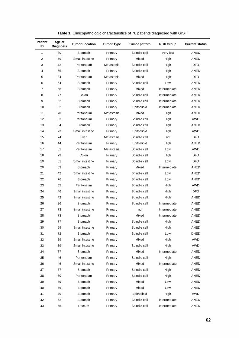
Patient ID
Age at Diagnosis Tumor Location Tumor Type Tumor pattern Risk Group Current status
1 80 Stomach Primary Spindle cell Very low ANED
2 59 Small intestine Primary Mixed High ANED
3 42 Peritoneum Metastasis Spindle cell High DFD
4 65 Stomach Primary Spindle cell High ANED
5 84 Peritoneum Metastasis Mixed High DFD
6 64 Stomach Primary Spindle cell Low ANED
7 58 Stomach Primary Mixed Intermediate ANED
8 77 Colon Primary Spindle cell Intermediate ANED
9 62 Stomach Primary Spindle cell Intermediate ANED
10 52 Stomach Primary Epithelioid Intermediate ANED
11 70 Peritoneum Metastasis Mixed High ANED
12 53 Peritoneum Primary Spindle cell High AWD
13 34 Stomach Primary Spindle cell High ANED
14 73 Small intestine Primary Epithelioid High AWD
15 74 Liver Metastasis Spindle cell nd DFD
16 44 Peritoneum Primary Epithelioid High ANED
17 61 Peritoneum Metastasis Spindle cell Low AWD
18 73 Colon Primary Spindle cell High DFD
19 61 Small intestine Primary Spindle cell Low DFD
20 53 Stomach Primary Mixed Intermediate ANED
21 42 Small intestine Primary Spindle cell Low ANED
22 76 Stomach Primary Spindle cell Low ANED
23 65 Peritoneum Primary Spindle cell High AWD
24 46 Small intestine Primary Spindle cell High DFD
25 42 Small intestine Primary Spindle cell High ANED
26 26 Stomach Primary Spindle cell Intermediate ANED
27 73 Small intestine Primary nd Intermediate ANED
28 73 Stomach Primary Mixed Intermediate ANED
29 77 Stomach Primary Spindle cell High ANED
30 69 Small intestine Primary Spindle cell High ANED
31 72 Stomach Primary Spindle cell Low DNED
32 59 Small intestine Primary Mixed High AWD
33 59 Small intestine Primary Spindle cell High AWD
34 77 Stomach Primary Mixed Intermediate ANED
35 46 Peritoneum Primary Spindle cell High ANED
36 46 Small intestine Primary Mixed Intermediate ANED
37 67 Stomach Primary Spindle cell High ANED
38 30 Peritoneum Primary Spindle cell High ANED
39 69 Stomach Primary Mixed Low ANED
40 66 Stomach Primary Mixed Low ANED
41 49 Stomach Primary Epithelioid High AWD
42 52 Stomach Primary Spindle cell Intermediate ANED
43 58 Rectum Primary Spindle cell Intermediate ANED
Table 1. Clinicopathologic characteristics of 78 patients diagnosed with GIST
62
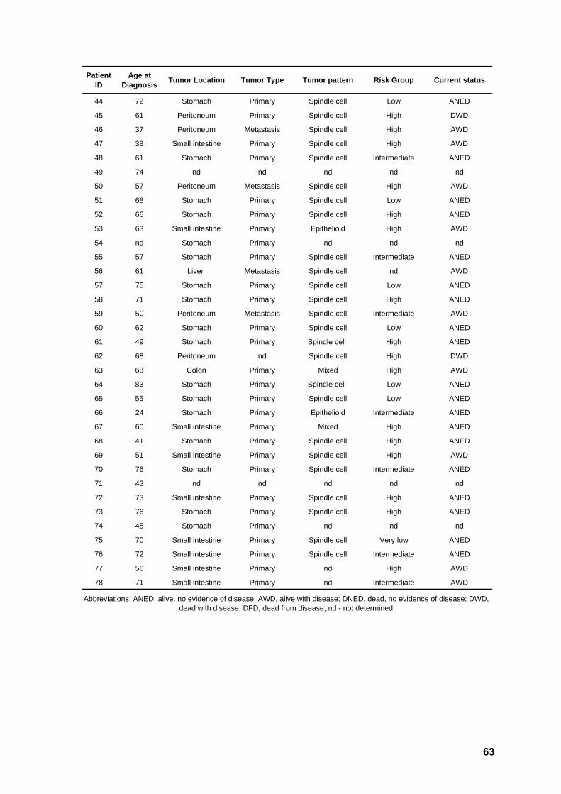
Patient ID
Age at Diagnosis Tumor Location Tumor Type Tumor pattern Risk Group Current status
44 72 Stomach Primary Spindle cell Low ANED
45 61 Peritoneum Primary Spindle cell High DWD
46 37 Peritoneum Metastasis Spindle cell High AWD
47 38 Small intestine Primary Spindle cell High AWD
48 61 Stomach Primary Spindle cell Intermediate ANED
49 74 nd nd nd nd nd
50 57 Peritoneum Metastasis Spindle cell High AWD
51 68 Stomach Primary Spindle cell Low ANED
52 66 Stomach Primary Spindle cell High ANED
53 63 Small intestine Primary Epithelioid High AWD
54 nd Stomach Primary nd nd nd
55 57 Stomach Primary Spindle cell Intermediate ANED
56 61 Liver Metastasis Spindle cell nd AWD
57 75 Stomach Primary Spindle cell Low ANED
58 71 Stomach Primary Spindle cell High ANED
59 50 Peritoneum Metastasis Spindle cell Intermediate AWD
60 62 Stomach Primary Spindle cell Low ANED
61 49 Stomach Primary Spindle cell High ANED
62 68 Peritoneum nd Spindle cell High DWD
63 68 Colon Primary Mixed High AWD
64 83 Stomach Primary Spindle cell Low ANED
65 55 Stomach Primary Spindle cell Low ANED
66 24 Stomach Primary Epithelioid Intermediate ANED
67 60 Small intestine Primary Mixed High ANED
68 41 Stomach Primary Spindle cell High ANED
69 51 Small intestine Primary Spindle cell High AWD
70 76 Stomach Primary Spindle cell Intermediate ANED
71 43 nd nd nd nd nd
72 73 Small intestine Primary Spindle cell High ANED
73 76 Stomach Primary Spindle cell High ANED
74 45 Stomach Primary nd nd nd
75 70 Small intestine Primary Spindle cell Very low ANED
76 72 Small intestine Primary Spindle cell Intermediate ANED
77 56 Small intestine Primary nd High AWD
78 71 Small intestine Primary nd Intermediate AWD
Abbreviations: ANED, alive, no evidence of disease; AWD, alive with disease; DNED, dead, no evidence of disease; DWD, dead with disease; DFD, dead from disease; nd - not determined.
63
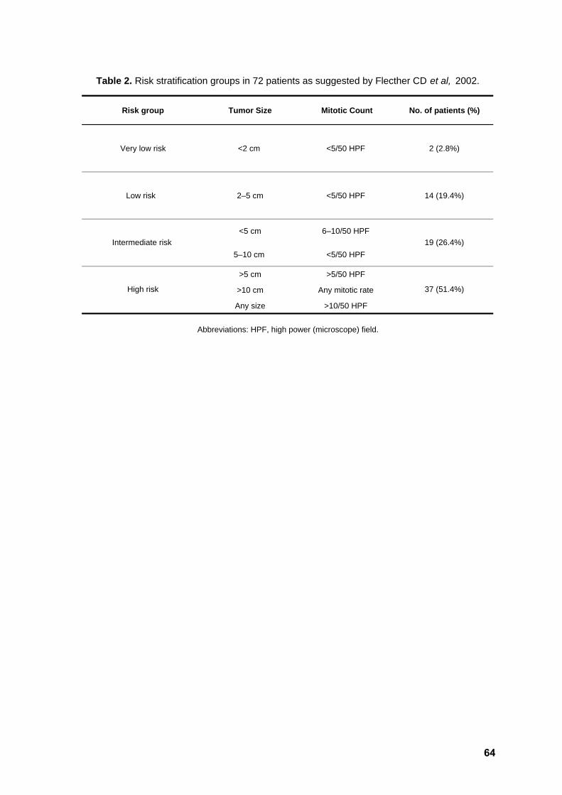
Risk group Tumor Size Mitotic Count No. of patients (%)
Very low risk <2 cm <5/50 HPF 2 (2.8%)
Low risk 2–5 cm <5/50 HPF 14 (19.4%)
<5 cm 6–10/50 HPF
5–10 cm <5/50 HPF
>5 cm >5/50 HPF
>10 cm Any mitotic rate
Any size >10/50 HPF
Table 2. Risk stratification groups in 72 patients as suggested by Flecther CD et al, 2002.
Abbreviations: HPF, high power (microscope) field.
19 (26.4%)
37 (51.4%)
Intermediate risk
High risk
64

Patient ID Risk Group Gene Exon Mutation* Type
2 High KIT 9 p.Ala502_Tyr503dup duplication
12 High KIT 9 p.Ala502_Tyr503dup duplication
17 Low KIT 9 p.Ala502_Tyr503dup duplication
17 (met) Low KIT 9 p.Ala502_Tyr503dup duplication
22 Low KIT 9 p.Ala502_Tyr503dup duplication
53 High KIT 9 p.Ala502_Tyr503dup duplication
66 Intermediate KIT 9 p.Ala502_Tyr503dup duplication
72 High KIT 9 p.Ala502_Tyr503dup duplication
3 High KIT 11 p.Trp557_Lys558del deletion
4 High KIT 11 p.Lys550_Gln556del deletion
18 High KIT 11 p.Tyr570_Leu576del deletion
23 High KIT 11 p.Asp579del deletion
27 Intermediate KIT 11 p.Trp557_Glu561del deletion
34 Intermediate KIT 11 p.Asp579del deletion
35 High KIT 11 p.Val560del deletion
42 Intermediate KIT 11 p.Val559_Glu561del deletion
45 High KIT 11 p.Glu554_Val559del deletion
46 High KIT 11 p. Val559_Asp572del deletion
49 nd KIT 11 p.Glu554_Val561del deletion
50 High KIT 11 p.Pro551_Glu554del deletion
60 Low KIT 11 p.Lys550_Lys558del deletion
67 High KIT 11 p.Met552_Trp557del deletion
69 High KIT 11 p.Lys550_Glu554del deletion
11 High KIT 11 p.Asn567_Tyr578delinsGluAsp delins
13 High KIT 11 p.Tyr553_Leu576delinsAsnCysLeuHis LeuTyrSerSerGln delins
15 nd KIT 11 p.Gln556_Val559delinsHis delins
19 Low KIT 11 p.Val559_Ile571delinsVal delins
32 High KIT 11 p.Glu554_Lys558delinsGlu delins
33 High KIT 11 p.Trp557_Val559delinsPhe delins
39 Low KIT 11 p.Pro551_Gln556delinsThr delins
41 High KIT 11 p.Gln556_Val559delinsProThrVal delins
43 Intermediate KIT 11 p.Lys558_Thr574delinsAsnArgSer delins
47 High KIT 11 p.Asn567_Leu576delinslle delins
10 Intermediate KIT 11 p.Asp572_His580dup duplication
28 Intermediate KIT 11 p.Leu576_Arg588dup duplication
Table 3. Genotype status of KIT and PDGFRA in 84 samples from 78 patients diagnosed with GIST
65

Patient ID Risk Group Gene Exon Mutation* Type
36 Intermediate KIT 11 p.Pro573_Tyr578dup duplication
55 Intermediate KIT 11 p.Thr574_Asp579dup duplication
59 Intermediate KIT 11 p.Asp572_ Pro573dup duplication
68 High KIT 11 p.Ile571_Asp579dup duplication
71 nd KIT 11 p.Ile571_Asp579dup duplication
70 Intermediate KIT 11 p.Pro585_Arg586insThrThrGlnLeu ProTyrAspHisLysTrpGluPhePro insertion
5 High KIT 11 p.Trp557Arg missense
14 High KIT 11 p.Trp557Gly missense
14 (met) High KIT 11 p.Trp557Gly missense
25 High KIT 11 p.Trp557Arg missense
26 Intermediate KIT 11 p.Val559Asp missense
29 High KIT 11 p.Val559Asp missense
30 High KIT 11 p.Val559Ala missense
40 Low KIT 11 p.Trp557Arg missense
44 Low KIT 11 p.Val559Asp missense
52 High KIT 11 p.Val559Asp missense
54 nd KIT 11 p.Val559Asp missense
56 nd KIT 11 p.Leu576Pro missense
57 Low KIT 11 p.Val559Asp missense
58 High KIT 11 p.Asp579del missense
63 High KIT 11 p.Val560Asp missense
73 High KIT 11 p.Val559Ala missense
75 Very low KIT 11 p.Trp557Arg missense
76 Intermediate KIT 11 p.Leu576Pro missense
77 High KIT 11 p.Val560Glu missense
78 Intermediate KIT 11 p.Trp557Arg missense
5 (met) High KIT 13 p.Val654Glu missense
56 (met) nd KIT 13 p.Val654Glu missense
21 Low PDGFRA 12 p.Ser566_Glu571delinsArg delins
64 Low PDGFRA 12 p.Val561Asp missense
20 Intermediate PDGFRA 14 p.Asn569Tyr missense
7 Intermediate PDGFRA 18 p.Met844_Ser847del deletion
16 High PDGFRA 18 p.Met844_Ser847del deletion
6 Low PDGFRA 18 p.Asp842Val missense
9 Intermediate PDGFRA 18 p.Asp842Val missense
48 Intermediate PDGFRA 18 p.Asp842Val missense
66
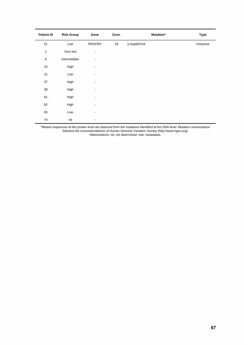
Patient ID Risk Group Gene Exon Mutation* Type
51 Low PDGFRA 18 p.Asp842Val missense
1 Very low -
8 Intermediate -
24 High -
31 Low -
37 High -
38 High -
61 High -
62 High -
65 Low -
74 nd -
*Mutant sequences at the protein level are deduced from the mutations identified at the DNA level. Mutation nomenclature followed the recommendations of Human Genome Variation Society (http://www.hgvs.org).
Abbreviations: nd, not determined; met, metastasis.
67

Patie
nt
IDG
ene
Exon
Mut
atio
n*M
utat
ion
type
CG
H fi
ndin
gs**
2K
IT9
p.A
la50
2_Ty
r503
dup
dupl
icat
ion
rev
ish
dim
(X)(
p11p
22),d
im(1
)(p1
3p36
),enh
(1)(
q21)
,enh
(1)(
q32)
,dim
(2)(
q11q
37),e
nh(3
)(p2
1),
enh(
3)(q
21),e
nh(3
)(q2
8q29
),enh
(6)(
p21)
,enh
(7)(
q22)
,enh
(7)(
q36)
,enh
(8)(
p11p
23),e
nh(9
)(p1
2p13
), en
h(9)
(q22
q34)
,enh
(10)
(q22
q26)
,dim
(11)
(p14
p15)
,enh
(11)
(q13
q14)
,enh
(12)
(p13
),enh
(12)
(q13
), en
h(12
)(q2
2q24
),dim
(13)
(q),d
im(1
4)(q
),dim
(15)
(q15
q26)
,enh
(16)
,enh
(17)
,enh
(19)
,enh
(20)
(q),d
im(2
1)(q
)
53K
IT9
p.A
la50
2_Ty
r503
dup
dupl
icat
ion
rev
ish
dim
(1)(
p13p
36),d
im(6
)(q1
2q27
),dim
(9)(
p21p
24),d
im(1
4)(q
),dim
(18)
(p11
q21)
,dim
(19)
(p13
), en
h(20
)(p1
2p13
),enh
(20)
(q),d
im(2
2)(q
)
17K
IT9
p.A
502_
Y50
3dup
dupl
icat
ion
rev
ish
dim
(1)(
p13p
36),d
im(2
)(p2
5q13
),dim
(11)
,enh
(12)
(p12
p13)
,dim
(13)
(q),d
im(1
5)(q
),enh
(21)
(q)
3K
IT11
p.Tr
p557
_Lys
558d
elde
letio
nre
v is
h en
h(Y
),dim
(1)(
p21p
36),e
nh(1
)(q2
1q23
),enh
(1)(
q42q
44),d
im(2
)(q1
1q37
),enh
(6)(
p12p
25),
dim
(6)(
q24q
27),e
nh(8
)(p2
1p22
),enh
(12)
(q21
q24)
,dim
(13)
(q),d
im(1
4)(q
),dim
(15)
(q21
q26)
, en
h(17
)(q1
1q24
),dim
(18)
(q21
q23)
,dim
(22)
(q)
4K
IT11
p.Ly
s550
_Gln
556d
elde
letio
nre
v is
h en
h(6)
(q26
q27)
,dim
(10)
,dim
(14)
(q),d
im(1
5)(q
),dim
(22)
(q)
42K
IT11
p.V
al55
9_G
lu56
1del
dele
tion
rev
ish
dim
(14)
(q),d
im(1
5)(q
),dim
(22)
(q)
58K
IT11
p.A
sp57
9del
dele
tion
rev
ish
dim
(14)
(q)
60K
IT11
p.Ly
s550
_Lys
558d
elde
letio
nre
v is
h di
m(1
1)(p
),dim
(14)
(q),d
im(2
2)(q
)
13K
IT11
p.Ty
r553
_Leu
576d
elin
sAsn
Cys
LeuH
is
LeuT
yrS
erS
erG
lnde
lins
rev
ish
dim
(14)
(q)
33K
IT11
p.Tr
p557
_Val
559d
elin
sPhe
delin
sre
v is
h di
m(1
)(p1
3p36
),enh
(1)(
q21q
44),d
im(2
)(p)
,enh
(2)(
q14q
36),e
nh(3
),enh
(4),e
nh(5
)(p1
2p15
), en
h(5)
(q12
q35)
,enh
(6)(
p12p
25),d
im(6
)(q1
6q27
),enh
(7)(
p13p
22),e
nh(7
)(q2
1q35
),enh
(8)(
p22p
23),
enh(
8)(q
13q2
4),e
nh(1
0)(p
13q2
2),e
nh(1
0)(q
24),e
nh(1
1)(p
14p1
5),d
im(1
1)(q
22q2
3),
enh(
12)(
q14q
22),d
im(1
3)(q
),dim
(14)
(q),d
im(1
5)(q
),dim
(18)
(q),d
im(2
1)(q
),dim
(22)
(q)
43K
IT11
p.Ly
s558
_Thr
574d
elin
sAsn
Arg
Ser
delin
sre
v is
h di
m(1
)(p1
2p36
),dim
(2)(
p22p
23),d
im(3
)(q1
2q22
),dim
(4)(
p12p
16),d
im(7
)(p)
,dim
(9)(
q22q
32)
10K
IT11
p.A
sp57
2_H
is58
0dup
dupl
icat
ion
rev
ish
dim
(X)(
q21q
28),d
im(1
4)(q
)
70K
IT11
p.P
ro58
5_A
rg58
6ins
ThrT
hr
Gln
LeuP
roTy
rAsp
His
LysT
rpG
luP
heP
roin
serti
onre
v is
h di
m(1
4)(q
),enh
(20)
(q)
5K
IT11
p.Tr
p557
Arg
mis
sens
ere
v is
h di
m(1
)(p1
3p36
),dim
(3)(
p21p
25),d
im(1
4)(q
22q3
2),d
im(1
5)(q
12q2
2),e
nh(1
7)(q
21q2
5),d
im(2
2)(q
)
Tabl
e 4.
Chr
omos
omal
imba
lanc
es a
nd m
olec
ular
alte
ratio
ns d
etec
ted
in 2
7 G
IST
subm
itted
to C
GH
ana
lysi
s.
68
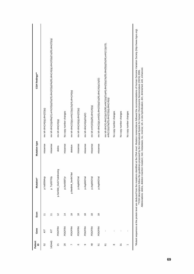
Patie
nt
IDG
ene
Exon
Mut
atio
n*M
utat
ion
type
CG
H fi
ndin
gs**
52K
IT11
p.V
al55
9Asp
mis
sens
ere
v is
h di
m(1
4)(q
),dim
(22)
(q)
14(m
et)
KIT
11p.
Trp
557G
lym
isse
nse
rev
ish
dim
(1)(
p36q
21),e
nh(1
0)(q
22q2
3),d
im(1
0)(q
24q2
6),d
im(1
4)(q
),dim
(15)
(q21
q26)
,dim
(22)
(q)
21P
DG
FRA
12p.
Ser
566_
Glu
571d
elin
sArg
delin
sre
v is
h di
m(1
4)(q
)
20P
DG
FRA
14p.
Asn
569T
yrm
isse
nse
No
copy
num
ber c
hang
es
7P
DG
FRA
18p.
Met
844_
Ser
847d
el
dele
tion
rev
ish
dim
(12)
(p),e
nh(1
2)(q
12q2
4),d
im(1
4)(q
)
6P
DG
FRA
18p.
Asp
842V
alm
isse
nse
rev
ish
dim
(14)
(q),d
im(2
2)(q
)
9P
DG
FRA
18p.
Asp
842V
alm
isse
nse
rev
ish
dim
(14)
(q22
q32)
48P
DG
FRA
18p.
Asp
842V
alm
isse
nse
rev
ish
enh(
10)(
q26)
,dim
(14)
(q)
51P
DG
FRA
18p.
Asp
842V
alm
isse
nse
rev
ish
dim
(1)(
p),e
nh(5
),dim
(10)
(q21
q26)
,dim
(14)
(q12
q32)
61-
rev
ish
dim
(X),d
im(1
)(p1
2p36
),enh
(1)(
q21q
44),d
im(3
)(q1
3q29
),dim
(9)(
q22q
34),e
nh(1
1)(p
15),
enh(
11)(
q13q
25),d
im(1
3)(q
),dim
(14)
(q)
8-
No
copy
num
ber c
hang
es
31-
No
copy
num
ber c
hang
es
1-
No
copy
num
ber c
hang
es
*Mut
ant s
eque
nces
at t
he p
rote
in le
vel a
re d
educ
ed fr
om th
e m
utat
ions
iden
tifie
d at
the
DN
A le
vel.
Mut
atio
n no
men
clat
ure
follo
wed
the
reco
mm
enda
tions
of H
uman
Gen
ome
Var
iatio
n S
ocie
ty (h
ttp://
ww
w.h
gvs.
org)
. **
CG
H d
escr
iptio
ns fo
llow
the
guid
elin
es p
ropo
sed
by th
e In
tern
atio
nal S
yste
m fo
r Hum
an C
hrom
osom
e N
omen
clat
ure
(ISC
N) 2
005.
Abb
revi
atio
ns: d
elin
s, d
elet
ion
inse
rtion
mut
atio
n; m
et, m
etas
tasi
s; re
v, re
vers
e; is
h, in
situ
hyb
ridiz
atio
n; d
im, d
imin
ishe
d; e
nh, e
nhan
ced.
69