mutations in cen3 cause aberrant chromosome … · mutations in cen3 cause aberrant chromosome...
TRANSCRIPT
Copyright 0 1989 by the Genetics Society of America
Mutations in CEN3 Cause Aberrant Chromosome Segregation During Meiosis in Saccharomyces cerm'siae
Arlene Gaudet and Molly Fitzgerald-Hayes
Department of Biochemistry, University of Massachusetts, Amherst, Massachusetts 01003 Manuscript received May 23, 1988
Accepted for publication December 1, 1988
ABSTRACT We investigated the structural requirements of the centromere from chromosome ZZZ (CEN3) of
Saccharomyces cerevisiae by analyzing the ability of chromosomes with CEN3 mutations to segregate properly during meiosis. We analyzed diploid cells in which one or both copies of chromosome ZZZ carry a mutant centromere in place of the wild-type centromere and found that some alterations in the length, base composition and primary sequence characteristics of the central A+T-rich region (CDE 11) of the centromere had a significant effect on the ability of the chromosome to segregate properly through meiosis. Chromosomes containing mutations which delete a portion of CDE I1 showed a high rate of premature disjunction at meiosis I. Chromosomes containing point mutations in CDE I or lacking CDE I appeared to segregate properly through meiosis; however, plasmids carrying centromeres with CDE I completely deleted showed an increased frequency of segregation to nonsister spores.
T HE centromere is that portion of the chromo- some which interacts with elements of the mi-
totic spindle to allow segregation of the chromosomes during cell division. Centromere DNA ( C E N ) was first isolated from the yeast Saccharomyces cerevisiae (CLARKE and CARBON 1980). These sequences allow plasmids containing autonomously replicating se- quences (ARS) to segregate to both daughter cells during most cell divisions. Mutational analysis of the centromere DNA has identified which sequences are important for proper mitotic function (CARBON and CLARKE 1984; PANZERI et al. 1985; MCGREW, DIEHL and FITZGERALD-HAYES 1986; GAUDET and FITZGER- ALD-HAYES 1987). Less is known about the require- ments for centromere function through meiosis. Meiosis consists of a reductional division (meiosis I), in which the replicated sister chromatids remain to- gether as homologous chromosomes and move to opposite poles, and an equational division (meiosis II), in which the sister chromatids segregate to opposite poles. There is evidence indicating that correct chro- mosome movement throughout meiosis requires a functional centromere to segregate the chromosomes at both meiotic divisions (CLARKE and CARBON 1985). The centromere may also be involved in maintaining the association between sister chromatids during the first meiotic division (CLARKE and CARBON 1984; CUMBERLEDGE and CARBON 1987).
Centromere DNA for 11 of the chromosomes of the yeast Saccharomyces cerevisiae have been analyzed and found to share three sequence elements, referred to as conserved centromere DNA elements (CDE) (FITZGERALD-HAYES et al. 1982; FITZGERALD-HAYES,
Genetics 121: 477-489 (March, 1989)
CLARKE and CARBON 1982; PANZERI and PHILIPPSEN 1982; MAINE, SUROSKY and TYE 1984; HIETER et al. 1985a; NEITZ and CARBON 1985; PANZERI et al. 1985; MANN and DAVIS 1986) (Figure 1). The A+T-rich central core element, CDE 11, is flanked by two dif- ferent short conserved sequences, CDE I (8 bp) and CDE 111 (25 bp). Although no consensus sequence exists among the CDE I1 regions of different yeast centromeres, they do share three features of general sequence organization. First, they are similar in length, ranging from 78 to 86 bp. Second, the base composition is always greater than 90% adenine plus thymine. Third, the CDE I1 sequences contain stretches of A and T residues 5-7 bp in length.
Mutational analysis has demonstrated that sequence elements CDE I, I1 and 111 all contribute to wild-type mitotic centromere function (FITZGERALD-HAYES and CARBON 1982; CLARKE and CARBON 1983; CARBON and CLARKE 1984; PANZERI et al. 1985; MCGREW, DIEHL and FITZGERALD-HAYES 1986; CUMBERLEDGE and CARBON 1987; GAUDET and FITZGERALD-HAYES 1987). Correct mitotic chromosome segregation oc- curs with very high fidelity in yeast; a nondisjunction event takes place on the average only once in 100,000 cell divisions (HARTWELL et al. 1982). The function of each mutant centromere can be measured by com- paring the nondisjunction frequency of a chromosome bearing an altered centromere with that of an analo- gous chromosome with a wild-type centromere. Al- though centromeres from which CDE I and most of CDE I1 have been removed retain some mitotic cen- tromere function (CARBON and CLARKE 1984; GAU- DET and FITZGERALD-HAYES 1987), some point mu-

478 A. Gaudet and M. Fitzgerald-Hayes
GTCATATG I I
GGATCCCGGGMTT I I
m I " 1 I 1
Mitotic Stability
CEN 3
BCTl
COI
CAT
RB76a
RB76b
x35
B58A I
BS58
X69
X78
P130-I
P 130-3
BS154
I X
I x 10-5
I x 10-4
2 x
6 x
6 x
4 x
2 x
7 x 10-3
I x 10-3
2 x 10-4
I x 10-4
2 x 10⁻³
6 x
W X h o I linker or SalI/XhoI junction - Inverted 45% A+T
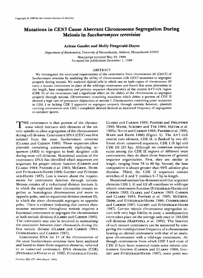
34 bp, 68% A+T FIGURE 1 .-Schematic representation of centromere mutants and mitotic stability of substituted chromosomes. The wild-type sequence of
CEN3 and the CDE I and 111 consensus sequences are shown at the top. The mitotic stability of chromosomes containing each mutant centromere was determined by quantitative mating (GAUDET and FITZGERALDHAYES 1987) and is presented as the fraction of mating competent cells/cell. The mutant sequences of CDE I11 (BCT1) and CDE I (CO1 and CAT) are shown. The RB76 deletion extends from the center of the RsaI site just upstream of CDE I and removes CDE I without deleting any bases of CDE 11. An 8-bp B a m H I linker marks the deletion and produces the following sequence:
RsaI BamHI CDE I1 ---TACT CGGATCCG ATGATA---
Substitution strains carrying the RB76 mutation were obtained for both orientations of the CEN segment in the chromosome. RB76a has CDE 111 adjacent to the B flanking region (CLARKE and CARBON 1983). RB76b contains the same fragment inserted into JC313 in the reverse orientation, such that CDE I11 is adjacent to pBR322 DNA and the A flanking region. B58AI is a deletion of CDE I and 26 bases of CDE 11. This mutation places pBR322 sequences adjacent to the remaining portion of CDE 11; the right-most 14 bp of adjoining pBR322 sequences are shown on the figure. The length of CDE I, I1 and 111 and insertion DNA are drawn approximately to scale. Other details of the mutant constructions and calculation of mitotic stability values are given in MATERIALS AND METHODS and GAUDET and FITZGERALD-HAYES (1987).
tations in CDE I11 result in complete loss of mitotic et al. 1988). The most dramatic effect is seen when centromere function (MCGREW, DIEHL and FITZGER- the central C residue in the CDE 111 dyad symmetry ALD-HAYES 1986; NG and CARBON 1987; HEGEMANN is changed to a T (MCGREW, DIEHL and FITZGERALD-

Chromosome Segregation in Meiosis 479
HAYES 1986). Chromosomes containing this single base pair alteration are strikingly unstable and segre- gate as acentric chromosomes during mitosis. This result suggests that CDE I11 constitutes a recognition site for a centromere binding protein. Recently, NG and CARBON (1 987) used exonuclease protection and a gel retention assay to demonstrate specific binding of protein(s) to CDE 111. Additional details regarding yeast centromeres are reviewed in CLARKE and CAR- BON (1 985) and FITZGERALD-HAYES (1 987).
Although CDE I is not absolutely necessary to mi- totic centromere function, deletion of this region does cause a 60-fold decrease in mitotic chromosome sta- bility (CARBON and CLARKE 1984; PANZERI et al. 1985; NG, CUMBERLEDGE and CARBON 1986; CUMBERLEDGE and CARBON 1987). However, chromosomes with CDE I deleted still segregate properly in most cell divisions. In contrast, deletions of CDE I1 reduce centromere function by 100-4000-fold depending upon the length of the deletion (CARBON and CLARKE 1984; GAUDET and FITZGERALD-HAYES 1987). Dele- tions have a more severe effect than insertions (GAU- DET and FITZGERALD-HAYES 1987), however, length is not the only important factor for mitotic centromere function. When DNA deleted from CDE I1 was re- placed with other sequences without changing the wild-type spacing between CDE I and CDE 111, A+T- rich DNA was found to impart the most nearly wild- type function (CUMBERLEDGE and CARBON 1987; M. FITZGERALD-HAYES, unpublished data). Apparently, A+T-rich DNA lacking runs of A and T residues can confer at least partial function. Finally, our previous results also indicate that in mitosis different regions of CDE I1 may be functionally equivalent since iden- tical insertions in two different positions in CDE I1 result in similar chromosome stabilities (GAUDET and FITZGERALD-HAYES 1987).
In this study our primary objective was to determine which features of the centromere sequence are nec- essary for normal meiotic chromosome segregation. We analyzed the meiotic products of strains contain- ing insertion, deletion and replacement mutations in CDE I1 and compared them to the results of mutations in CDE I and 111, as diagrammed in Figure 1, to evaluate the role of each region in meiotic chromo- some segregation. These mutations fall into four classes which include: (1) mutations affecting only CDE I (CO1, CAT, RB76a and RB76b); (2) a deletion removing CDE I and part of CDE I1 (B58AI); (3) insertion, deletion and replacement mutations in CDE 11 (X35, BS58, X69, X78, P130-1, P130-3, and BS 154); and (4) a single base pair change in CDE 111 (BCTl). The details concerning construction, mitotic function and sequence analyses of all but the CDE 1 mutants have been described previously (MCGREW,
DIEHL and FITZGERALD-HAYES 1986; GAUDET and FITZGERALD-HAYES 1987).
We found that deletions in CDE I1 caused an in- creased frequency of premature separation of sister chromatids. Mutations in CDE I, which have little effect on mitotic chromosome movement, did not cause chromosomes to disjoin prematurely in meiosis. In light of previous work (CUMBERLEDGE and CARBON 1987) suggesting that plasmids carrying centromeres deleted for CDE I segregated to nonsister spores at a higher frequency than plasmids with wild-type centro- meres, we tested two of our CDE I mutant centro- meres on plasmids. We found that plasmids with CDE I deleted showed an increased frequency of nonsister segregation, suggesting that they were defective in maintaining sister chromatid contact through meiosis I.
MATERIALS AND METHODS
Yeast strains: The yeast strains used in these experiments are listed in Figure 2. Some mutations were analyzed in two different strains. Strains containing chromosomes with a substituted centromere also carried a functional URA3 gene within 400 bp of the centromere as well as additional mark- ers on chromosome I I I , including the MAT locus which encodes mating type information. All strains also contained one or two independent centromere-linked genes on other chromosomes. TRPl is tightly linked to CEN4 (0.45 cM) in strain MH2/YP3; MET14 is linked to C E N l l (2.6 cM) and ADEl is linked to CENl (4 cM) in strains SB7883-1C and J17/S150 (MORTIMER and SCHILD 1980) (Figure 2). Plas- mids were propagated in a derivative of strain MH2/YP3 which carried a substituted wild-type CEN3 with adjacent URA3 gene in place of the wild-type centromere on one copy of chromosome IIZ. In this strain, the URA3 marker gene was used to indicate sister spores.
Centromere mutations and genomic substitution vec- tors: All of the mutants used in this study are derivatives of the centromere from chromosome IIZ (CEN3) (Figure 1). Only one available CEN3 CDE 111 mutant, BCTl, is mitot- ically stable enough to make meiotic analysis practical. BCTl contains a single C to T mutation in CDE I11 that was produced by bisulfite mutagenesis as described in MCGREW, DIEHL and FITZGERALD-HAYES (1986). The same technique was used to produce CO 1, which has a single C to T change in CDE I that creates a new NdeI site, GTCA- TATG (M. FITZGERALD-HAYES, unpublished data). The mutant CAT was constructed by cleaving at the unique NdeI site in CO 1, treating the linear molecules with Klenow DNA polymerase I and religating. Although this procedure should have resulted in a 2-bp insertion, sequencing revealed the CAT mutation to be GTCATTATG (Figure 1). Mutant RB76 (a gift of R. BAKER) is a deletion of CDE I which was derived from CO1 by deleting sequences between the NdeI site and the upstream RsaI site (see CEN3 sequence in FITZCERALD-HAYES, CLARKE and CARBON 1982) and insert- ing an 8-bp BamHI linker (CGGATCCG). The sequences of the altered regions, verified by dideoxynucleotide (SANGER, NICKLEN and COULSEN 1977) or MAXAM-GILBERT (MAXAM and GILBERT 1980) DNA sequencing, are shown in Figure 1.
Wild-type CEN3 DNA contains a CDE I 1 region 84 bp in length. Deletion mutant X35 was constructed by removing

480 A. Gaudet and M. Fitzgerald-Hayes
SB7883-1C HATa/HATa, trpl-289dtrpl-289, ura3-52/ura3-52, LEU2/leu2-3, HIS4/his4-519, CRY1 /crylr, met14/HET14, adel/ADEl
MH2/YP3 HATa/HATa, trplAfTRP1. ura3-52/ura3-52, LEU2/leu2-3, HIS3/his3, ade2-101Jade2-1010, LYS2/lys2
J17/S150 HATa/HATa, trpl-289/trpl-289, ura3-52/ura3-52, Lrm2/leu2-3, metl4/HET14, adel/AQEl
Chromosome I I I markers:
HIS4 LEU2 CEr3 UATa I I I
his4 I
leu2 URA3 CEN* SUP11 I L I 4
MATa I
Independent centromere markers:
CENll HET14 I ’
CEN4 Ll’l I
CENI @El
Centromere Strain
cEN3 co1 CAT RB76a RB76b B58AIS B58AI x7 8 x35 x35s X69 X69S BS58 P130-1 P130-3 BS154 BCTlS Pl/P3
SB7883-1C, MH2ffP3, J17/S150 MH2/YP3 MH2/YP3 MH2/YP3 MH2/YP3 MH2/YP3 SB7883 - 1C SB7883-1C SB7883-1C MH2/YP3
MH2/YP3 SB7883-IC
SB7883-1C, J17/S150 SB7883-1C, J17/S150 SB7883-1C, J17/S150 SB7883 - 1C MH2/YP3 SB7883-1C
FIGURE 2.”Strain genotypes. Genotypes of the strains used for transformation with the mutant (or wild-type) CEN sequences are shown. The relative location of the chromosome IZZ markers and independent centromere markers on chromosomes X I , N a n d Z are illustrated (not to scale). Genomic transplacement of the mutant centromere, URA3 and, in some cases, SUPl I was targeted into one copy of chromosome IZZ in the case of heterocentric strains. CEN* indicates the transplaced centromere. Homocentric strains were obtained by crossing two Ura+ haploid progeny from a heterocentric diploid. The strains in which each centromere was analyzed are shown at the bottom of the figure.
57 bp of CDE I1 using Ba131 followed by insertion of an XhoI linker (CCTCGAGG). The deletion mutation in B58AI removed all of CDE I and the first 26 bp of CDE 11. The BS58 mutation has 48 bp of CDE I1 deleted and replaced with a 22-bp palindromic sequence, thereby creating a CDE I1 region 58 bp in length. A replacement mutant was con- structed by inserting a 34-bp synthetic oligonucleotide of 68% A+T (a gift of FUMIKYO NAGAWA) into the XhoI site of X35 to produce X69. Deletion mutant X78 was con- structed by removal of 14 bp of CDE I1 by Ba131 treatment and insertion of the XhoI linker. P130-1 contains a 46-bp insert in the first Aha111 site of CDE 11, while P130-3 contains the same insert in the third AhaIII site of CDE 11. In each case the 46-bp insert expanded the CDE I1 regions
in these mutants to 130 bp. BS 154 contains a partial dupli- cation of CDE I1 and 22 bp of synthetic palindromic DNA, which separate CDE I and CDE 111 by 154 bp. The sequence of each mutation was verified by dideoxynucleotide sequenc- ing. Other details concerning these constructions are re- ported in GAUDET and FITZGERALD-HAYES (1987).
Mutant centromeres were introduced directly onto chro- mosome ZZZ in place of the wild-type CEN3 by inserting each into the unique BamHI site of a genomic replacement vector, either JC3 13 (CLARKE and CARBON 1983) or pJUP, a deriv- ative ofJC3 13 modified by J. MCGREW to contain the SUPl 1 suppressor tRNA gene (J. MCGREW, Z. XIAO and M. FITZ- GERALD-HAYES, manuscript in preparation) in the Hind111 site of the B flanking region. The CO1, CAT and RB76

Chromosome Segregation in Meiosis 48 1
mutations were inserted into pJC313 on a 390-bp Sau3A fragment (see FITZGERALD-HAYES, CLARKE and CARBON 1982). Since RB76 has a BamHI linker in place of CDE I, the BamHI site was first methylated in vitro with BamHI methylase prior to cleavage with SauJA. Other details of subcloning into pJC3 13 are presented in MCGREW, DIEHL and FITZGERALD-HAYES (1 986) and GAUDET and FITZGER- ALD-HAYES (1 987).
We constructed diploid yeast strains in which the cen- tromere on one copy of chromosome ZZZ was replaced with a mutant or wild-type centromere tightly linked to a URA3 gene (heterocentric) (CARBON and CLARKE 1984). By cross- ing haploid progeny of these strains we constructed addi- tional strains in which both copies of chromosome ZZZ con- tained substituted centromeres, having either identical CEN mutations (homocentric) or different CEN mutations (mixed mutants). P1/P3 was constructed by crossing a P130-1 hap- loid with a P130-3 haploid, each carrying the URA3 gene adjacent to the mutant centromere. All mutant strains were verified by SOUTHERN (1975) hybridization (data not shown). Mutant centromeres containing a restriction site not found in the wild-type CEN3 DNA were analyzed by SOUTHERN hybridization for retention of that site in the substituted chromosome (RB76, X35, X78, B58A1, BCTlS and CO 1).
We also studied the effects of CDE I mutations (RB76 and CAT) when carried on replicating plasmids which also contained the yeast T R P l gene as a selectable marker. First, the CEN segments were subcloned on HzndIII/SalI frag- ments into the polylinker region of pUC8. Then, the 1.45- kb yeast TRPlIARSl EcoRI fragment was inserted into the EcoRI site of each plasmid. Plasmids chosen for study all had T R P l oriented such that transcription proceeded away from the CEN segment (R. BAKER, unpublished data).
Chromosome mitotic stability assay: The mitotic stability results presented in Figure 1 and Table 3 were determined by means of the quantitative mating assay (DUTCHER and HARTWELL 1982). Diploid cells which lose one copy of chromosome ZZZ become mating competent (2n - 1: MATa or MATa). Samples to be tested were grown for 10 genera- tions in liquid YPD (1% yeast extract, 2% peptone, 2% dextrose), mixed with an excess of a haploid tester strain of opposite mating type, filtered onto sterile membranes (HA Millipore Corp.) and incubated at 32” for 4 hr. Cells were resuspended in sterile water and serial dilutions plated onto minimal agar plates (Difco yeast nitrogen base containing 2% glucose) allowing only mated cells to grow (GAUDET and FITZGERALD-HAYES 1987).
Colony color assay: We modified a plasmid-based colony color assay (HIETER et al. 1985b) to allow the detection of loss or nondisjunction of chromosome ZZZ (J. MCGREW, Z. XIAO and M. FITZGERALD-HAYES, manuscript in prepara- tion). Several strains were constructed which carry the sup- pressor tRNA ( S U P l l ) gene adjacent to the mutant cen- tromere (designated by the “S” in the strain name, X35S, B58AIS, X69S and BCTlS). Cells homozygous for the ade2- 101 mutation accumulate a red-pigmented intermediate and produce colonies which are dark red. The presence of SUPl 1 results in partial suppression of the ade2-101 (ochre) mutation, causing a decrease in the concentration of the red pigment. In diploids, one copy of SUPl 1 results in a pink colony and two copies in a white colony. In haploids, one or two copies of SUPl 1 results in a white colony. Diploid and haploid colonies lacking SUP11 are red. Color assay plates (Difco yeast nitrogen base containing 2% dextrose, 20 mg/ liter tryptophan, 20 mg/liter uracil, 0.5% casamino acids) contained 6 mg/liter adenine, sufficient to allow growth of Ade- cells and a visible change in colony color.
Diploids heterozygous for a partially functional mutant centromere and the SUPl 1 gene produce pink colonies with red and white sectors resulting from nondisjunction of the mutant chromosomes during colony growth. The degree of sectoring is proportional to the frequency of nondisjunction of the mutant chromosomes. The color assay allowed us to select and sporulate colonies containing primarily diploid (2n) rather than aneuploid (272 + 1) cells. Since the abnormal meiotic events analyzed here often produce spores inviable due to lack of chromosome ZZZ, the presence of extra copies of chromosome ZZZ in 2n + 1 aneuploids compromises the analysis. Following dissection, the meiotic products can also be analyzed using the color assay. Colonies arising from haploid cells containing two copies of chromosome ZZZ (one with SUPl 1 and one without) can be detected by the pres- ence of red sectors in an otherwise white colony.
Sporulation and dissection: Prior to sporulation, diploid cella were grown on YPD plates or on agar plates lacking uracil but containing 0.67% yeast nitrogen base and 2% dextrose, supplemented with 20 mg/liter tryptophan, 20 mg/liter adenine and 0.5% casamino acids. Cells were washed twice and sporulated in 1% potassium acetate at room temperature with agitation for 4-5 days. The resulting tetrads were treated with 50% glusulase (New England Nuclear) for 10 min and dissected using a light microscope equipped with a micromanipulator. The haploid meiotic products were separated and allowed to form colonies on YPD agar, and subsequently tested for the presence of diagnostic markers by replica-plating onto the appropriate minimal media agar.
SOUTHERN analysis: Yeast genomic DNA was isolated, cleaved by EcoRI and, in some cases, by an appropriate second restriction enzyme and separated by electrophoresis through a 1 % agarose gel. DNA fragments were transferred to nitrocellulose by the method of SOUTHERN (1975) and hybridized with centromere DNA or a fragment from the centromere-flanking region of chromosome ZZZ labeled by nick-translation as previously described (GAUDET and FITZ- GERALD-HAYES 1987).
RESULTS
Predicted results of abnormal meiotic segrega- tion: Analysis of the segregation of genetic markers to haploid progeny allows us to detect certain abnor- mal meiotic events. Since a frequent consequence of centromere mutations is mitotic nondisjunction, we wanted to know whether the mutant chromosomes exhibit meiotic nondisjunction as well. We can detect nondisjunction events at meiosis I and estimate the frequency of nondisjunction at meiosis 11. We can also accurately measure segregation of mutant chromo- somes to nonsister spores, an indication that the sister chromatids separated prematurely at the first meiotic division. We chose to analyze these events by tetrad dissection in order to distinguish between nondisjunc- tion at meiosis I and premature disjunction.
For these analyses we substituted mutant or wild- type CEN3 DNA for the native centromere on one copy of chromosome ZZZ. Each substitution also con- tained a URA3 gene within 400 bp of the centromere to identify sister chromatids with substituted centro- meres. Sister spores were identified by the presence of tightly centromere linked markers on other chro-

482 A. Gaudet and M. Fitzgerald-Hayes
A -ERERICATDN B WOMOSOMEFEPUCITDN
NORMAL MEDSLP 1 PREMATLRE DslUVCflON 1 @ RE-BNI- @
MEIOSIS I
MEIOSIS I - K))Iocoas SEPARATE @@@a MUTANT SISTER CcIRou*;TDs SEPARATE PREMATURELY
MEIOSIS II - SISTER CHROMAATIDS SEPARATE Q)aampJ@a@ @ @
MEIOSIS I1
t- In sklw spom T+ In mhtu SPUN ED msEcToRs YmITEWllH
wm
C c " E f W L b X T D N D OROM090MEFEPUCATION
K)H)m AT MEOSIS I 1 NoMsluNcTDNAT
M E W I S I1 1
MEIOSIS I MEIOSIS I @:mm=-o @-=NE@
MEIOSIS II MEIOSIS I1
SISTER CkROMAllDS SEPARATE MUTANT CHAOMATlDS FAIL TO DISK)*I @@DB@(jJ@@ WIT€ WITH REDSECTORS REDsEcxrs
WHITE WITH ED ED wm

Chromosome Segregation in Meiosis 483
mosomes, T R P l on chromosome N (strain MHz/ YP3), MET14 on chromosome XI and ADEl on chro- mosome Z (strains J17/S150 and SB7883-1C). TRPl is very tightly linked to CEN4 (within 0.5 cM) and, as expected, we did not observe any TRPlIURA3 tetra- types in any of our strains with substituted wild-type centromeres. MET14 is physically located within 1.5 kb of C E N l l (FITZGERALD-HAYES et al. 1982; YEH, CARBON and BLOOM 1986), however, a genetic dis- tance of 2.6 cM has been reported (MORTIMER and SCHILD 1980). T o ensure that we did not score recom- bination between MET14 and C E N l l as cases of non- sister segregation of URA3, we required the concur- rence o f A D E l with MET14 to determine sister spores.
Figure 3A shows a normal meiosis in which the Ura+ chromosomes segregate to sister spores (identi- fied by Trp+). Since each spore receives one copy of chromosome ZZZ with one copy of mating type infor- mation, all of the progeny can mate with a haploid tester strain. In strains where both URA3 and SUPl 1 are carried adjacent to the mutant centromere, the two Ura+ (Supl 1+) colonies will be white and the two Ura- (Supl 1-) colonies will be red. Figure 3B shows the expected pattern if a premature disjunction event occurs resulting in segregation of the mutant chro- mosomes to nonsister spores. The diagnostic pheno- type is the presence of Ura+ in nonsister spores in a tetrad containing one dead spore. One of the Ura+ spores will be disomic for chromosome ZZZ, which can be verified by two characteristics. If the two copies of chromosome ZZZ in this spore (Ura’ and Ura-) contain opposite mating type information, the cells will be nonmaters. Second, in strains with SUPl 1 adjacent to the mutant centromere, loss of the Supl 1+ chromo- some due to nondisjunction during colony growth will produce red sectors in a white colony.
Nondisjunction events at meiosis I are indicated by tetrads with two Ura+ spores in sisters and two dead spores, as shown in Figure 3C. The live spores will be disomes and those which contain both MATa and MATa chromosomes will be distinguishable by their inability to mate with a haploid tester strain. In strains carrying SUPl 1 on one copy of chromosome ZZZ, di-
somy can be verified by the sectoring colony color assay, as discussed in the previous paragraph.
We tried to estimate the incidence of nondisjunc- tion at meiosis I1 (see Figure 3D). In this case, the analysis is complicated by random spore death, which occurs in our strains with wild-type centromeres at a frequency of 5-1 0%. Since the color assay does not allow us to distinguish between one and two copies of SUP11 in a haploid, we cannot always verify disomy in Ura+ cells. Therefore, the frequency of occurrence of the diagnostic phenotype (two Ura- sister spores and one Ura+ spore) must be compared to the fre- quency expected from random spore death.
Spore viability and URA3 distribution: Spore via- bility of strains carrying wild-type centromeres ranged from 90 to 95%. Spore viability was also high, 89- 95%, for most of the heterocentric strains examined. Strains containing deletion mutants X35 and BS58 exhibited reduced spore viability (70% and 82%, re- spectively), indicative of abnormal meiotic events pro- ducing inviable spores lacking a copy of chromosome zzz.
The distribution of the URA3 gene was also noted in the meiotic products. Sporulation of heterocentric strains should yield two Ura+ spores and two Ura- spores. In three cases, B58A1, B58AIS and X35S, some crosses resulted in tetrads displaying a 4+:0- or 3+: 1- Ura+ distribution indicating that extra copies of chromosome ZZZ were present prior to sporulation, making these useless for analysis of chromosome seg- regation. Therefore, multiple crosses were performed and strains sporulated to obtain samples which were not aneuploid prior to sporulation.
CDE I1 deletions result in premature disjunction of sister chromatids: We found that chromosomes with deletions in CDE I1 (X35, X35S and BS5S) exhibited an elevated level of premature disjunction (12-13% for heterocentric stains, Table 1). A 34-bp insertion into the X35 centromere, which expanded the CDE I1 region from 35 to 69 bp (X69) (Figure l), resulted in a statistically significant ( P > 95%) (SOKAL and ROHLF 198 1) improvement in meiotic function (compare the percentage of nonsister segregation for
FIGURE 3,”Predicted genotypes of haploid products after normal (A) and several possible abnormal (B-D) meiotic segregation events. MAT is located 25 cM from CEN3 and recombination between MAT and CEN3 can be expected to occur in approximately 40-60% of the asci. (B) Premature disjunction at meiosis I, in which one copy of the mutant chromatid separates from its sister and migrates to the opposite pole at meiosis I, results in three viable spores. Two of these spores are Ura’ and occur as nonsister spores (nonsister segregation). The spore which has a Ura- sister contains both a Ura+ and a Ura- copy of chromosome III . In disomic haploids, if both mating types are present, the resulting cells are nonmaters. In SUPl I strains, if a cell contains both a Ura- (Supl 1-) copy and a Ura+ (Supl 1+) copy of chromosome I I I , disomy can be verified by the sectoring colony color assay. (C) If homologs of chromosome III fail to disjoin at meiosis I, two live spores result. These spores are Ura+ sisters, both disomic for chromosome III . Disomy can be verified by the sectoring colony color assay and/or nonmating phenotype. (D) Nondisjunction of the mutant copy of chromosome III at meiosis I1 results in three viable spores, two of which are Ura- sister spores. The nonsister spore is Ura+ and disomic for chromosome III. If both chromosomes are of different mating types, the resulting cells are nonmaters. If both chromosomes are of the same mating type, the phenotype resulting from nondisjunction at meiosis I1 cannot be distinguished from that resulting from failure of a Ura+ spore to germinate due to factors unrelated to centromere function. This occurs at a rate of approximately 5-10% in strains with wild-type centromeres. Therefore, the rates at which this phenotype is observed in the mutants must be compared to background rates for wild-type strains.

484 A. Gaudet and M. Fitzgerald-Hayes
TABLE 1
Nonsister segregation in asci with three viable spores
Heterocentrics Homocentrics" URA3-MET14 or LEU2-MET14 or LEU2-
URA3-TRPI TRPl
No. Centromere scoredb NS' %NS scored NS %NS
WTCEN3 28 0 0 32 0 0
CDE I
No.
co 1 17 0 0 ND ND
CAT 26 0 0 22 0 0 RB76a 25 0 0 30 0 0 RB76b 26 0 0 27 0 0
CDE I+II B58AI 21 1 5 ND ND B58AIS 22 1 5 ND ND
x 3 5 15 2 13 10 2 20 x 3 5 s 49 6 12 32 5 16 BS58 26 3 12 ND ND
X69 50 2 4 31 2 6 X69S 38 1 3 31 2 6 X78 24 0 0 11 0 0 P130-1 39 0 0 46 0 0 P130-3 31 0 0 ND ND
BS154 34 0 0 16 0 0
CDE I I
CDE III BCT 1 S 38 1 3 17 1 6
CDE I I / I I I P1/P3 46 1 2 NA
ND, not determined; NA, not applicable. a Since homocentrics carried a functional URA3 gene on each
copy of chromosome I I I , segregation of CEN3 (URA3) relative to MET14 or TRPl could not be determined. Each homocentric strain, however, was heterozygous for LEU2, which lies within 5 cM of CEN3; therefore, we used LEU2 to identify sister chromatids. Non- sister segregation of LEU2 relative to MET14 or TRPI can result either from premature disjunction of a mutant chromosome or a combination of LEU2-CEN3 recombination and death of the fourth spore for reasons unrelated to centromere function.
Total number of tetrads with three or four viable spores. Number of tetrads showing nonsister segregation. These are
represented by three live spores, two of which are Ura+ (or Leu+) in nonsisters. The Ura+ (Leu+) spore which has a live Ura- (Leu-) sister is a disome (see Figure 3B).
X35 and X69; Table 1). A single tetrad (of 38 scored) exhibiting nonsister segregation was obtained for the heterocentric strain bearing the centromere with the CDE 111 point mutation, BCTlS (Table 1). No non- sister segregation was observed for the four strains bearing either point mutations or deletions affecting only CDE I (CO1, CAT, RB76a and RB76b; Table 1). However one nonsister tetrad was observed for each strain in which the deletion removed a significant segment of CDE I1 as well as CDE I (B58AI and B58AIS; Table 1).
All cases of nonsister segregation in the heterocen- tric strains occurred in tetrads having three viable spores, indicating premature separation of the Ura' chromatids and segregation to nonsister spores (Fig- ure 3B). In most cases disomic spores could be verified by the presence of nonmaters or the sectoring colony
color assay. Since the Ura- chromatids segregated to sister spores, these asci each contained one dead spore lacking any copy of chromosome ZZZ (see Figure 3B). No cases of nonsister segregation were observed in any strain containing a wild-type substituted centro- mere. However, it must be noted that our assay would not detect low levels of premature disjunction in wild- type strains. Similar values were obtained for strains which differed only by the presence or absence of SUP11 (X35 and X35S, B58AI and B58AIS, X69 and B69S; see Table l), indicating that the presence of the SUP1 1 gene itself did not noticeably affect cen- tromere function.
Since the homocentric strains carried URA? adja- cent to the substituted centromeres on both copies of chromosome ZZZ, URA? segregation relative to MET14 or TRPl could not be determined. Instead we used LEU2, which is located within 5 cM of CEN? on chromosome ZZZ, to follow mutant sister chromatids. Table 1 presents the frequency of nonsister segrega- tion of LEU2 for homocentric asci with three viable spores (corresponding values for homocentric asci with four viable spores are given in Table 2). We believe that the values in Table 1 represent the ratio of mutant sister chromatids which disjoined prema- turely and segregated to nonsister spores. An alter- native explanation is that a recombination event oc- curred between LEU2 and CEN?, and independently, a spore was rendered inviable for reasons unrelated to centromere function. However, the probability of these events happening in the same tetrad is very low, the product of the LEU2/CEN? recombination rate (approximately 10%) (MORTIMER and SCHILD 1980) and the frequency of random death of a Leu- spore (<5%). We did not observe nonsister segregation in any wild-type or CDE I mutant asci having three viable spores. Table 1 shows that nonsister segregation oc- curred in 20% and 16% of the asci for X35 and X35S, respectively. Lower frequencies (4-6%) occurred in X69, X69S, and BCTlS asci. These values are con- sistent with those observed for heterocentric strains.
Recombination near substituted centromeres: We determined the frequency of recombination between LEU2 and CEN? (URA?) for all heterocentric asci which did not experience abnormal meiotic chromo- some segregation. With the exception of X35 and X35S, the frequencies ranged between 0 and 12.5%, in good agreement with the literature value of ap- proximately 10% (MORTIMER and SCHILD 1980). For X35 and X35S we observed values of 21 and 18%, respectively, which is twofold higher than expected, but not statistically significant ( P < 0.95). In order to be sure that the values reported for X35 and X35S represent actual LEU2/CEN? recombination, we were careful not to score tetrads in which the mutant chro- matids segregated to nonsister spores. Recombination

Chromosome Segregation in Meiosis 485
between the MAT locus and the centromere on chro- mosome IZZ ranged between 25 and 61 % for most strains, in agreement with published values (46% (MORTIMER and SCHILD 1980). However, for several insertion mutants, we observed recombination fre- quencies significantly higher than expected. Hetero- centric insertion mutants X78, P130-1 and BS154 and homocentric mutants X78 and BS154 experi- enced MATICEN3 (LEU2) recombination frequencies of 69 to 83%, statistically higher than expected ( P > 0.95) (SOKAL and ROHLF 1981). While we have no explanation for these increases, similar results for centromere mutants have been reported previously (CUMBERLEDGE and CARBON 1987).
We also analyzed the incidence of LEU2ICEN3 and MATICEN3 recombination for all tetrads which underwent a premature disjunction event. As a group, asci which experienced nonsister segregation showed essentially normal frequencies of MATICEN3 and LEU2ICEN3 recombination. Of 30 tetrads which ex- perienced premature disjunction, 15 showed a recom- bination event between the MAT locus and CEN3 (heterocentric strains) or between the MAT locus and LEU2 (homocentric strains). Of these 15, three cases of recombination involved the chromosome which disjoined prematurely. In several other cases the as- sortment of markers did not permit us to determine whether the prematurely separating chromatid was involved in the recombination event. Therefore, these numbers most likely underrepresent the number of prematurely separating chromatids which recom- bined. Two tetrads (of 30) showed a recombination event between LEU2 and CEN3, both of which in- volved the prematurely separating chromosomes. While these numbers are not large enough to draw definitive conclusions, they suggest that chromatid separation occurred after homolog pairing and recom- bination. However, we cannot rule out the possibility that a prematurely separated chromatid subsequently paired and recombined with its homolog.
Nonsister segregation in homocentric asci with four viable spores: Since homocentric strains carry the URA3 gene adjacent to both mutant centromeres, recombination between LEU2 and the mutant centro- meres could not be determined directly. For these strains, nonsister segregation of LEU2 relative to TRPl or MET14 was scored instead (Table 2). There- fore, the values given in Table 2 represent nonsister segregation of LEU2 resulting from: (1) LEU2ICEN3 recombination (9.8%) (MORTIMER and SCHILD 1980) and (2) premature disjunction and nonsister segrega- tion of both pairs of mutant sister chromatids. We observed frequencies of LEU2 nonsister segregation ranging from 4.0 to 13.0% for all samples except X69S, in reasonable agreement with the expected contribution from LEU2ICEN3 recombination. How-
TABLE 2
Nonsister segregation of LEU2 in homocentric asci with four viable spores
LEUZIMETl4 or LEUZITRPI
Centromere S NS %NS
W T CEN3 24 1 4.0
CDE I CAT 19 2 9.5 RB76a 11 1 8.5 RB76b 14 1 6.6
x 3 5 s 20 2 9.1 X69 20 3 13.0 X69S 15 12 44.4 P130-1 32 2 5.9 BS 154 10 1 9.1
CDE I1
CDE III I I P 1/P3 29 4 12.1
S, sister segregation; NS, nonsister segregation.
ever, the frequency we observed for X69S was very high (44.4%). We cannot determine whether this value represents an increase in the LEU2ICEN3 re- combination rate or premature disjunction and non- sister segregation of both pairs of sister chromatids. However, only X69S (and not X69) exhibited such a high value. Moreover, of the 14 tetrads showing non- sister segregation for X69S (Tables 1 and 2), 12 had four viable spores. I f both pairs of sister chromatids separate prematurely and the (now) unpaired chro- matids segregate randomly at the second meiotic di- vision, the resulting tetrads would contain two, three or four viable spores, depending on how the chro- matids assort. Since we know of no mechanism to segregate unpaired chromatids at the second meiotic division, we believe that samples in which spore via- bility is uniformly high are unlikely to result from premature disjunction. Therefore, we believe that this strain experienced a higher than normal frequency of LEU2ICEN3 recombination, for reasons undeter- mined at this time.
Frequency of nondisjunction at meiosis I and 11: Table 3 compares the data on nondisjunction at meiosis I and I1 with chromosome mitotic stability values. We observed instances of nondisjunction at meiosis I ranging from 2 to 5% for the X35, X35S and X69 heterocentric strains and the mixed mutant P1/P3, which were all verified by the presence of disomic Ura+ spores. For the homocentric strains, only X35 and X35S showed nondisjunction events at meiosis I which could be verified by the presence of nonmaters. This result is consistent with the values obtained for this mutation in the heterocentric strains. We did not observe events of this kind in the control strain (CEN3) or in any of the other heterocentric or homocentric strains. However, it should be noted that

486 A. Gaudet and M. Fitzgerald-Hayes
TABLE 3
Estimate of nondisjunction at meiosis I and 11
Heterocentric strains Homocentric strains
Class' Class'
Centromere stability" scoredb I 11 scoredb I 11 Mitotic No. ~ No. ~
WTCEN3 1 X 31 0 2 35 0 2
CDE I co 1 1 X 18 o 2 ND CAT 1 X 31 0 2 22 0 1 RB76a 6 X 25 0 0 30 0 6 RB76b 6 X 26 0 4 27 0 4
B58AI 2 X IO-' 23 0 2 (1) ND B58AIS 22 0 0 ND
CDE I+II
CDE II x 3 5 4 X IO-' 21 1 3 (1 ) 19 1 4 x 3 5 s 4 X IO-* 61 2 10 45 1 4
I X 53 I 3 ( 1 ) 32 o o
x 7 8 6 X 25 0 1 1 1 0 0 ~ 1 3 0 - I 2 x 1 0 - 4 41 o I 53 0 4
BS58 7 X 10⁻³ 36 o I X69 X69S 1 X 41 0 3 31 0 2
ND
P130-3 1 X 37 0 3 ND
BS154 2 X IO-' 38 0 0 21 0 3
BCTlS 2 X IO-' 41 0 3 21 0 4 CDE III
MIXED PI/P3 51 1 5
ND, not determined. ' Mitotic stability is fraction of mating competent cells as deter-
mined by the quantitative mating assay (GAUDET and FITZCERALD HAYES 1987).
'Total number of asci scored including asci with two viable spores.
' Class I = nondisjunction at meiosis I . These are represented by two viable sister spores, both Ura' (heterocentrics) or both Leu' (homocentrics). In each case both spores were verified as disomes, either by the color assay and/or the presence of two nonmaters. Class I1 = nondisjunction at meiosis I1 or failure of a spore to germinate (5-10% in strains with wild-type centromeres). These are represented by three viable spores, two Ura- in sisters and one Ura+ (heterocentrics) or three viable spores two of which are either Leu+ or Leu- in sisters (homocentrics). The value in parentheses indicates the number of asci containing one verified disome whose siblings were sisters.
tetrad dissection is not sensitive enough to detect low levels of meiotic nondisjunction (<1%).
Table 3 also presents the percentage of asci with three viable spores in which two were Ura- and one was Ura+, the phenotype which would result from a nondisjunction event at meiosis 11. However, we stress that this phenotype can also result from failure of a Ura+ spore to germinate (random spore death). Therefore, in this case the values shown for the mu- tants must be compared to those given for strains with wild-type centromeres (approximately 5-10%). In general, levels significantly higher than that expected from random spore death indicate aberrant segrega- tion of chromosomes with mutant centromeres. Some of our strains showed elevated levels of this phenotype
TABLE 4
Meiotic segregation of plasmids carrying mutant and wild-type centromeres
No. show- Segregation
ing relative to LrR.43
No. 2+:2- Trp+ Percent Centromere dissected distribution Sister Nonsister nonsister
CEN3 95 27 25 2 7 CAT a7 25 23 2 a RB76 233 36 27 9 25
(heterocentric strains RB76b, X35 and X35S and homocentric strains RB76a, RB76b, X35, BS154 and BCT 1 S). However, statistical analysis (SOKAL and ROHLF 198 1) indicated that the levels are not suffi- ciently elevated to conclude that these mutant chro- mosomes are undergoing nondisjunction at meiosis 11. CDE I mutations cause impairment of plasmid
segregation: We found that deletion of CDE I had no detectable effect on the meiotic segregation of whole chromosomes in either heterocentric or homocentric strains (Tables 1 and 3). These results are in contrast with previous work showing that deletion of CDE I from plasmid-borne centromere DNA causes a high frequency of distribution of plasmids to nonsister spores (NG, CUMBERLEDCE and CARBON 1986; CUM- BERLEDGE and CARBON 1987). T o determine if plas- mids with CDE I mutations behave differently in meiosis than whole chromosomes with the same cen- tromere mutations, we analyzed CEN3, CAT and RB76 on pUC plasmid derivatives (Table 4). Each plasmid also carried a TRPl gene. The host strain was our heterocentric CEN3 control strain, in which the URA3 marker adjacent to CEN3 served as the cen- tromere linked gene for sister spore determination. The results (Table 4) indicate that when the plasmid carrying the wild-type centromere segregated 2+:2-, the plasmid-borne Trp+ marker was usually found in sister spores. Two instances of nonsister segregation (7%) were observed for the CEN3 plasmid, possibly caused by the extremely small size of the plasmid (approximately 3 kb), as reported previously (FITZ- GERALD-HAYES et al. 1982). The plasmid with the CAT centromere showed a similar level of nonsister segregation (8%). However, plasmids carrying the CDE I deletion mutant centromere, RB76, segregated to nonsister spores in nine out of 36 tetrads (25%), a significant ( P > 0.95) (SOKAL and ROHLF 1981) in- crease over the wild-type value.
DISCUSSION
The CDE I and I11 sequence homology shared by all S. cerevisiae centromeres isolated to date implies that these regions are required for chromosome seg-

Chromosome Segregation in Meiosis 487
regation. While the CDE I1 regions of different yeast centromeres share no sequence homology, the simi- larity of length and structural characteristics suggest that this region is also involved in some aspect of centromere function. Although CDE I and some of CDE I1 can be deleted without abolishing mitotic centromere function, CDE 111 has been shown to be absolutely required. Since centromeres with muta- tions in CDE I and I1 function at least partially in mitosis, we questioned whether the integrity of these sequences might be more critical for chromosome segregation in meiosis than in mitosis.
Using heterozygous genetic markers on several chromosomes, we analyzed the effect of mutations in CDE I, I1 and I11 on meiotic chromosome segregation. We found that the worst meiotic defects were caused by deletions in CDE 11. The primary result of deleting CDE I1 DNA was premature separation of sister chro- matids and segregation to nonsister spores. Hetero- centric deletion mutants X35, X35S and BS58 exhib- ited relatively high rates (1 2-1 3%) of nonsister seg- regation (Table 1). Homocentric deletion mutants X35 and X35S segregated to nonsister spores in 16- 20% of the tetrads examined, suggesting that CDE I1 plays a role in maintaining the contact between sister chromatids until the second meiotic division.
In this study we also analyzed the behavior of chro- mosomes containing insertion mutations in CDE I1 (P130-1, P130-3 and BS154). We observed no evi- dence for premature disjunction of chromosomes with these mutant centromeres in either heterocentric or homocentric strains (Table 1). However, we observed one tetrad displaying nonsister segregation (out of 46 analyzed) for the mixed mutant Pl/P3. This effect could either be due to the centromere mutations or to having different centromeres on each homolog. CLARKE and CARBON (1983) showed that reversing the orientation of CEN3 or replacing CEN3 with C E N l l on one copy of chromosome 111 did not affect centromere function. Therefore, we believe that this instance of nonsister segregation resulted from the insertion into CDE 11.
We tested the requirement for proper spacing be- tween CDE I and CDE I11 by replacing a segment of CDE I1 DNA deleted in X35 with 34 bp of synthetic A+T DNA and observed a statistically significant ( P > 95%) decrease in the frequency of premature dis- junction (X69, Table 1). However, chromosomes con- taining the X69 centromere still disjoin prematurely at a rate of 3-4% (heterocentric strains) and 6% (homocentric strains), indicating that the artificial re- placement DNA does not completely compensate for the deleted DNA in maintaining sister chromatid con- tact. In both X35 and X69 the base composition is similar (74% and 7 1 %, respectively), but the spacing between CDE I and I11 and the arrangement of the
A+T DNA are different. The DNA inserted into X69 is not arranged in runs of A and T residues as found in wild-type CEN DNA. In a recent study on the meiotic segregation of plasmid-borne mutant centro- meres, insertion of pBR322 DNA or A+T-rich DNA (without stretches of As and Ts) resulted in improved, but not wild-type, meiotic function (CUMBERLEDGE and CARBON 1987). In both cases the spacing between CDE I and I11 was essentially wild type. These results suggest that, while the length of CDE I1 may be critical to meiotic function, the arrangement of bases is also an important characteristic.
We used tetrad dissection rather than random spore analysis in order to distinguish between nondisjunc- tion at meiosis I and premature disjunction. Dissection precludes analysis of large sample sizes, therefore, we could not detect low levels of nondisjunction/prema- ture disjunction in wild-type centromere strains. How- ever, a study by SORA, LUCCHINI and MACNI (1982) determined the rate of spontaneous occurrence of disomy due to errors at meiosis I and 11. They found that the incidence of spores disomic for chromosome V in a strain with wild-type centromeres was 0.64 X
This value is actually the sum of nondisjunction at meiosis I and nonsister segregation due to prema- ture disjunction. Although we used different strains and some strain variation is likely, it is probable that in our control strains levels of nondisjunction at meiosis I and premature disjunction are also very low. If so, some of our chromosome mutations have a pronounced effect on the rate of chromosome ZZZ nondisjunction at meiosis I and premature disjunc- tion.
Previous studies have shown that mutations in both CDE I1 and CDE I11 can have a profound effect on mitotic chromosome disjunction (GAUDET and FITZ- GERALDHAYES 1987; CLARKE and CARBON 1985). However, a specific CDE mutation may, or may not, have the same effect on meiotic chromosome segre- gation. For example, the CDE I1 replacement mutant X69 has a mitotic nondisjunction rate of only 1 in 1000 cell divisions, yet shows nonsister segregation (premature disjunction) in 3-4% of the tetrads ana- lyzed. Similarly, chromosomes containing the deletion mutant BS58 function well during mitosis (having a nondisjunction rate of 7 per 1000 cell divisions), yet show nonsister segregation in 12% of the tetrads examined (Table 1). In contrast, the CDE I11 point mutant BCTlS is mitotically more unstable (20 non- disjunction events per 1000 cell divisions) than BS58, but exhibited nonsister segregation in only 3% of the meiotic cell divisions. These results suggest that yeast centromere DNA contains mitotic and meiotic func- tions that can be uncoupled by mutations introduced into CDE 11.
Nondisjunction at meiosis I was verified in the CDE

488 A. Gaudet and M. Fitzgerald-Hayes
I1 deletion mutants, X35 and X35S, the CDE I1 replacement mutant X69 and mixed mutant Pl/P3, indicating that the integrity of CDE I1 is important for optimal centromere function at meiosis I . Rates of nondisjunction at meiosis I1 were also estimated. How- ever, it is difficult to determine with precision the actual rate of nondisjunction at meiosis I1 since the phenotype used to score this event can also be caused by failure of a Ura+ spore to germinate. Therefore, the frequency at which this phenotype occurs in mu- tant strains must be compared to values for wild-type strains (CEN3) which are isogenic except for the cen- tromere mutation. This rate is elevated in some of the mutant strains (including RB76a, RB76b, X35, BS154 and BCTlS), however, the increase is not statistically significant ( P < 0.95). Since spore inviability can vary somewhat from strain to strain, we cannot say with certainty that these mutations are causing an increased rate of nondisjunction at meiosis 11. However, these results tend to support roles for all the CDE regions in meiosis 11.
I n this study we found that deletion of CDE I1 DNA (X35) caused measurable premature disjunction of chromosome ZZZ whether the mutation was carried on one (heterocentric) or both (homocentric) copies of the chromosome. Previously a similar centromere mu- tation was observed to cause aberrant meiotic segre- gation only in homocentric and not in heterocentric strains suggesting that the presence of the wild-type homolog was sufficient to complement the CDE I1 deletion in trans (CARBON and CLARKE 1984). One reason for this difference may be that the mutations analyzed in the two studies were not the same, and specific CDE I1 sequences present in one, but absent from the other, might account for the effect. As yet nothing is known about the protein(s), helical confor- mation or other factors important for CDE I1 func- tion, so it is difficult to evaluate this possibility without further study. We also considered that, in our strains, gene conversion events in the heterocentric strains could be responsible for replacing the wild-type CEN? sequences with mutant CEN DNA thereby changing the heterocentric configuration to a homocentric one. It is known that gene conversion involving the cen- tromere region does occur, however the conversion tracts are frequently 1000 bp or longer, continuous and often involve the co-conversion of nearby markers (SYMINGTON and PETES 1988). If conversion of the centromere were occurring at a detectable frequency we would expect one or both of the markers (URA3 or SUPl 1) closely linked to the mutant centromeres to undergo conversion along with the CEN DNA in at least some of the tetrads. I n fact, in this entire study we observed only one tetrad where URA3 and SUP1 1 were no longer linked and no instances of conversion of both URA3 and SUPl 1.
The results reported here are in contrast to a pre- vious study showing that homocentric strains contain- ing deletions of CDE I either produce a high percent- age of inviable spores (CARBON and CLARKE 1984), or could not be sporulated at all, requiring that genetic analysis be performed on plasmids (CUMBERLEDGE and CARBON 1987). We also experienced difficulty spor- ulating homocentric strains with CDE I mutations but were eventually successful and found no indication of premature disjunction at meiosis I for these chromo- somes (CAT and RB76). Aware of the discrepancy with previous work, we also analyzed the segregation of plasmids bearing CDE I mutations (Table 4). We discovered that centromeres lacking CDE I cause de- tectable abnormal meiotic segregation when present on plasmids but not when carried on whole chromo- somes.
There are several possible explanations for this result. The meiotic defect observed for CDE I muta- tions may be amplified in plasmids due to the circular nature or small size of these molecules. We have observed previously that some centromere mutations which cause a decrease in plasmid mitotic stability fail to have a detectable effect on chromosome mitotic disjunction (GAUDET and FITZGERALD-HAYES 1987). I n addition, plasmids present at one copy per cell lack a homolog, which may affect their proper meiotic segregation.
Alternatively, the chromosome could segregate properly in meiosis despite the lack of a centromeric CDE I because a sequence homologous to CDE I, positioned elsewhere on the chromosome, can provide a compensatory function. I n fact, CDE I homologs have been identified in the yeast genome at noncen- tromeric locations (BRAM and KORNBERG 1987; R. BAKER, unpublished data). I n addition, the region adjacent to CEN6 is known to contain a sequence homologous to the centromeric CDE I (PANZERI and PHILIPPSEN 1982; PANZERI et al. 1985).
The sequences immediately upstream of the CDE I deletion could also affect the ability of a plasmid or chromosome to segregate properly during meiosis. One study of CEN6 mutants showed that a plasmid with a CDE I deletion segregated normally through meiosis, while an analogous plasmid retaining 1 bp of CDE I but having different adjacent sequences exhib- ited abnormal meiotic segregation (PANZERI et al. 1985). I n some mutants it is possible that the se- quences brought adjacent to CDE I1 might contain sufficient homology to function (completely or par- tially) as a CDE I analog.
I n conclusion, the results reported here indicate that a minimum length CDE I1 is required for proper meiotic function. Insertions into this region have a less dramatic effect. Large deletions of CDE I1 result in premature separation of sister chromatids before

Chromosome Segregation in Meiosis 489
the second meiotic division. Insertion of A+T-rich, random sequence DNA into one of the deletion mu- tants only partially compensated for the wild-type deleted DNA. We can conclude from these results that a minimum segment of CDE I1 DNA, arranged in the characteristic pattern of runs of A and T residues, is required to maintain optimal sister chro- matid contact until the second meiotic division. Dele- tion of CDE I had no apparent effect on the meiotic segregation of whole chromosomes whether present on one or both copies of chromosome ZZZ. However, the same mutation carried on a plasmid resulted in a higher rate of nonsister segregation than wild-type CEN plasmids, supporting the hypothesis that CDE I is involved in maintenance of sister chromatid contact until meiosis 11.
We are grateful to CAROL NEWLON, LYNN MILLER and JOHN
CARBON for their careful reading of the manuscript and advice. We are grateful to RICHARD BAKER for construction of RB76, and sequencing and subcloning of RB76, CO1 and CAT. We wish to thank JEFFREY MCGREW for construction, subcloning and mitotic stability analysis of some of the mutants, and to LEZLIE DENSMORE for help with the tetrad dissections. This work was supported by U.S. Public Health Service grant GM 32257 from the National Institutes of Health to M.F.H.
LITERATURE CITED
BRAM, R. J., and R. D. KORNBERG, 1987 Isolation of a Saccharo- myces cereuisiae centromere DNA-binding protein, its human homolog, and its possible role as a transcription factor. Mol. Cell. Biol. 7: 403-409.
CARBON, J., and L. CLARKE, 1984 Structural and functional analy- sis of a yeast centromere (CEN3). J. Cell Sci. Suppl. 1: 43-58.
CLARKE, L., and J. CARBON, 1980 Isolation of a yeast centromere and construction of functional small circular chromosomes. Nature 287: 504-509.
CLARKE, L., and J. CARBON, 1983 Genomic substitutions of cen- tromeres in Saccharomyces cereuisiae. Nature 305: 23-28.
CLARKE, L., and J. CARBON, 1985 The structure and function of yeast centromeres. Annu. Rev. Genet. 19: 29-56.
CUMBERLEDGE, S., and J. CARBON, 1987 Mutational analysis of meiotic and mitotic centromere function in Saccharomyces cere- uisiae. Genetics 117: 203-21 2.
DUTCHER, S. K . , and L. H. HARTWELL, 1982 The role of Saccha- romyces cereuisiae cell division cycle genes in nuclear fusion. Genetics 100: 175-184.
FITZGERALD-HAYES, M., 1987 Yeast centromeres. Yeast 3: 187- 200.
FITZGERALD-HAYES, M., and J. CARBON, 1982 Identification of DNA sequences required for mitotic stability of centromere plasmids in yeast. Recent Adv. Yeast Mol. Biol. 1: 1-12.
FITZGERALD-HAYES, M., L. CLARKE and J. CARBON, 1982 Nucleotide sequence comparisons and functional analysis of yeast centromere DNAs. Cell 2 9 235-244.
FITZGERALD-HAYES, M., J.-M. BUHLER, T . G. COOPER and J. CAR- BON, 1982 Isolation and subcloning analysis of functional centromere DNA (CENI 1 ) from Saccharomyces cereuisiae chro- mosome XI. Mol. Cell. Biol. 2: 82-87.
GAUDET, A , , and M. FITZGERALD-HAYES, 1987 Alterations in the
adenine-plus-thymine-rich region of CEN3 affect centromere function in Saccharomyces cereuisiae. Mol. Cell. Biol. 7: 68-75.
HARTWELL, L. H., S. K. DUTCHER, J. S. WOOD and B. GARVIK, 1982 The fidelity of mitotic chromosome reproduction in S. cereuisiae. Recent Adv. Yeast Mol. Biol. 1: 28-38.
HEGEMANN, J. H., J. H. SHERO, G. COTTAREL, P. PHILIPPSEN and P. HIETER, 1988 Mutational analysis of centromere DNA from chromosome VI of Saccharomyces cereuisiae. Mol. Cell. Biol. 8: 2523-2535.
HIETER, P., C. MANN, M. SNYDER and R. W. DAVIS, 1985a Mitotic stability of yeast chromosomes: a colony color assay that meas- ures nondisjunction and chromosome loss. Cell 4 0 381-392.
HIETER, P., D. PRIDMORE, J. H. HEGEMANN, M. THOMAS, R. W. DAVIS and P. PHILIPPSEN, 1985b Functional selection and analysis of yeast centromeric DNA. Cell 42: 9 13-92 1.
MAINE, G. T., R. T . SUROSKY and B.-K. TYE, 1984 Isolation and characterization of the centromere from chromosome V (CEN5) of Saccharomyces cereuisiae. Mol. Cell. Biol. 4 86-91.
MANN, C., and R. W. DAVIS, 1986 Structure and sequence of the centromeric DNA of chromosome 4 in Saccharomyces cereuisiae. Mol. Cell. Biol. 6: 241-245.
MAXAM, A. M., and W. GILBERT, 1980 Sequencing end-labeled DNA with base-specific chemical cleavages. Methods Enzymol. 65: 497-560.
MCGREW, J., B. DIEHL and M. FITZGERALD-HAYES, 1986 Single base-pair mutations in centromere element 111 cause aberrant chromosome segregation in Saccharomyces cereuisiae. Mol. Cell. Biol. 6 530-538.
MORTIMER, R. K., and D. SCHILD, 1980 Genetic map of Saccha- romyces cereuisiae. Microbiol. Rev. 44: 5 19-57 1.
NEITZ, M., and J. CARBON, 1985 Identification and characteriza- tion of the centromere from chromosome XIV in Saccharomyces cerevisiae. Mol. Cell. Biol. 5: 2887-2893.
NG, R., and J. CARBON, 1987 Mutational and in vitro protein- binding studies on centromere DNA from Saccharomyces cere- uisiae. Mol. Cell. Biol. 7: 4522-4534.
NG, R., S. CUMBERLEDGE and J. CARBON, 1986 Structure and function of centromeres, pp. 225-239, in Yeast Cell Biology. Alan R. Liss, New York.
PANZERI, L., and P. PHILIPPSEN, 1982 Centromeric DNA from chromosome VI in Saccharomyces cereuisiae strains. EMBO J. 1: 1605-1611.
PANZERI, L., L. LANDONIO, A. STOTZ and P. PHILIPPSEN, 1985 Role of conserved sequence elements in yeast centro- mere DNA. EMBO J. 4: 1867-1873.
SANGER, F., S. NICKLEN and A. R. COULSEN, 1977 DNA sequenc- ing with chain terminating inhibitors. Proc. Natl. Acad. Sci. USA 74: 5463-5467.
SOKAL, R. R., and F. J. ROHLF, 1981 Biometry: The Principles and Practice of Statistics in BiologuA Research, Ed. 2. W. H. Freeman, New York.
SORA, S., G. LUCCHINI and G. E. MAGNI, 1982 Meiotic diploid progeny and meiotic nondisjunction in Saccharomyces cereuisiae. Genetics 101: 17-33.
SOUTHERN, E. M., 1975 Detection of specific sequences among DNA fragments separated by gel electrophoresis. J. Mol. Biol. 98: 503-5 17.
SYMINGTON, L., and T . PETES, 1988 Expansions and contractions of the genetic map relative to the physical map of yeast chro- mosome 111. Mol. Cell. Biol. 8: 595-604.
YEH, E., J. CARBON and K. BLOOM, 1986 Tightly centromere- linked gene (SPOI5) essential for meiosis in the yeast Saccha- romyces cereuisiae. Mol. Cell. Biol. 6: 158-167.
Communicating editor: D. BOTSTEIN