concussion: diagnosis and treatment - medicine.utah.edu · 29th pmr update, canyons resort, ... pd,...
TRANSCRIPT
Concussion: diagnosis and treatment
Rodolfo Savica, MD, MSc, PhD
University of Utah
January 29,8 201529th PMR Update, Canyons Resort, Park City, UT
Disclosure
Relevant Financial Relationships
None
Off Label Usage
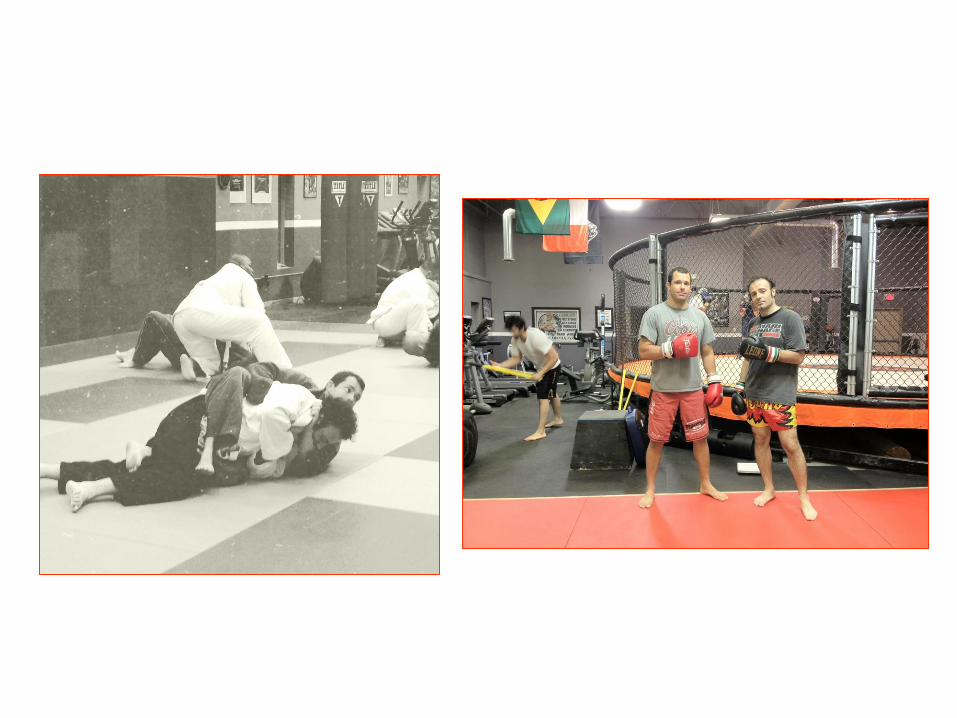
Disclosure
Personal disclosure
Being “concussed” many times
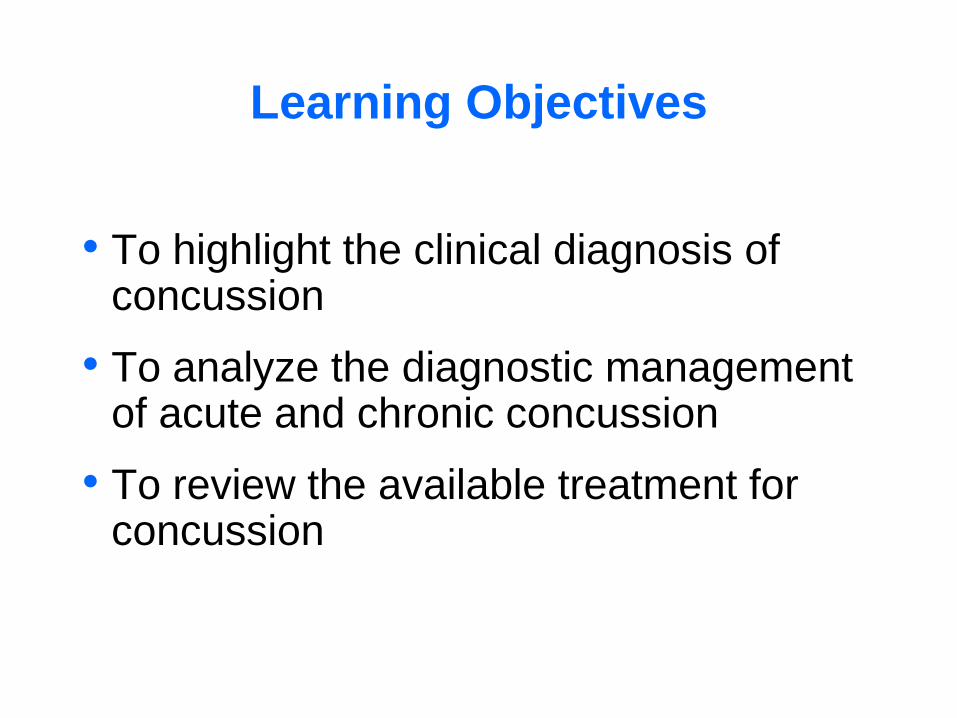
Learning Objectives
• To highlight the clinical diagnosis of concussion
• To analyze the diagnostic management of acute and chronic concussion
• To review the available treatment for concussion
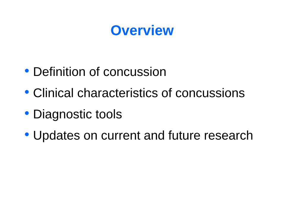
Overview
• Definition of concussion
• Clinical characteristics of concussions
• Diagnostic tools
• Updates on current and future research
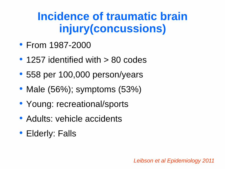
Incidence of traumatic brain injury(concussions)
• From 1987-2000
• 1257 identified with > 80 codes
• 558 per 100,000 person/years
• Male (56%); symptoms (53%)
• Young: recreational/sports
• Adults: vehicle accidents
• Elderly: Falls
Leibson et al Epidemiology 2011
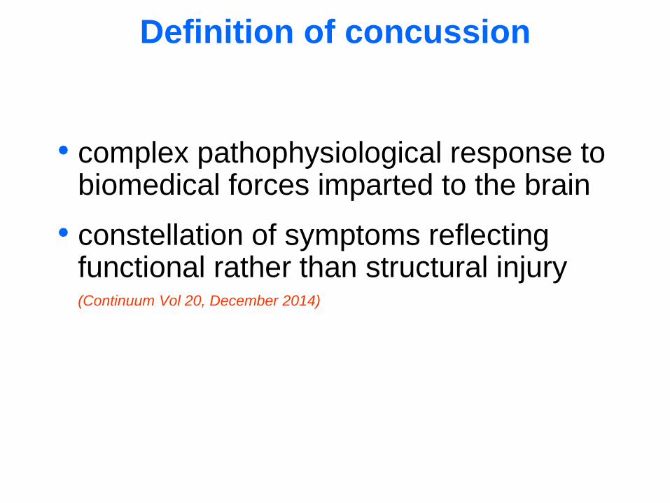
Definition of concussion
• complex pathophysiological response to biomedical forces imparted to the brain
• constellation of symptoms reflecting functional rather than structural injury(Continuum Vol 20, December 2014)
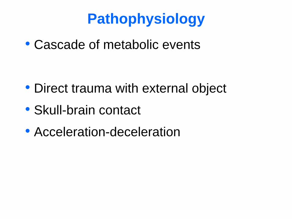
Pathophysiology
• Cascade of metabolic events
• Direct trauma with external object
• Skull-brain contact
• Acceleration-deceleration

Pathophysiology
• Heterogeneous symptoms
• Lack of anatomical correspondence

Clinical Scenario
• 18 ys, male
• Backflips with wakeboard
• Football linebacker
• Transient unilateral blindness
• Confusion
• Complete recovery
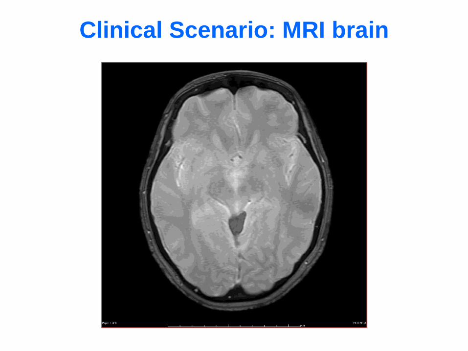
Clinical Scenario: MRI brain
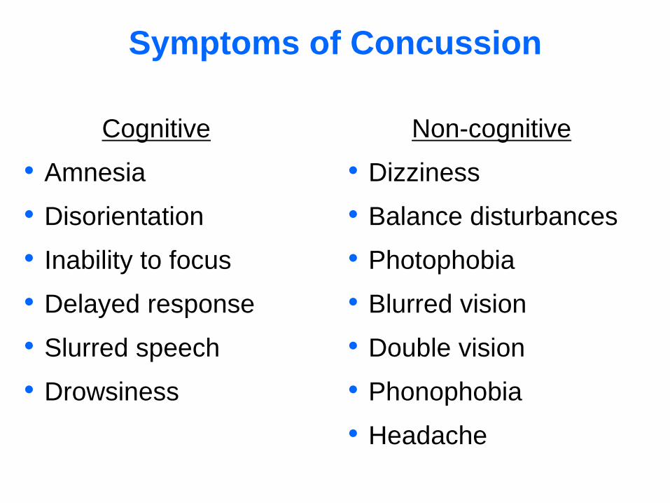
Symptoms of Concussion
Cognitive
• Amnesia
• Disorientation
• Inability to focus
• Delayed response
• Slurred speech
• Drowsiness
Non-cognitive
• Dizziness
• Balance disturbances
• Photophobia
• Blurred vision
• Double vision
• Phonophobia
• Headache

Symptoms of Concussion
Affective
• Emotional lability
• Depression
• Anxiety
• Mania
• Irritability
Sleep
• Increased latency
• Awakenings
• Increased sleep time
• Decreased sleep time
• RBD
• Nightmares
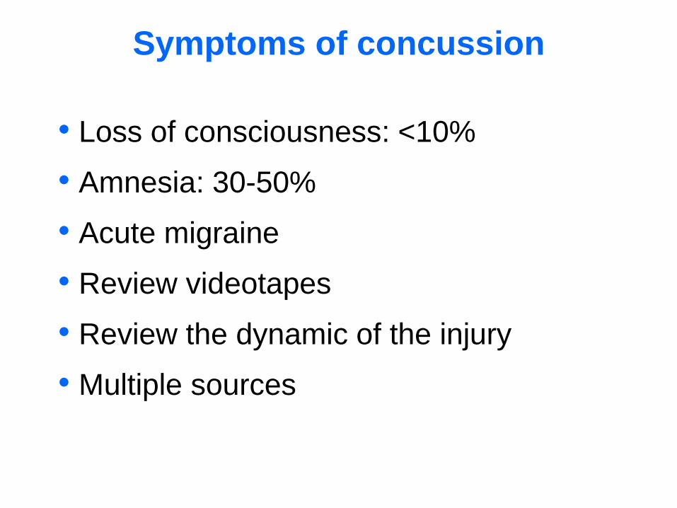
Symptoms of concussion
• Loss of consciousness: <10%
• Amnesia: 30-50%
• Acute migraine
• Review videotapes
• Review the dynamic of the injury
• Multiple sources
Aggravating factors
• Repetitive concussions
• Severity
• Duration of loss of consciousness
• Younger age
• Sex
• Pre-existing migraine, ADHD, learning disabilities, anxiety, depression

Contributing factors
• Sleep deprivation
• Fatigue
• Dehydration
• Illness
• Medications
• Illicit drugs
• Recovering from previous concussion

Concussions Clinical Timeline
• Concussion: transient alteration of brain function as the direct result of a biomechanical force. Days to weeks
• Post-concussion syndrome: complexpathophysiology, both biological and psychological, that occurs after the concussion is over. Months to years
• Chronic effects: unknown pathophysiology, unclear epidemiology. Chronic TraumaticEncephalopathy (CTE), depression,parkinsonism, cognitive decrement. Lifetime
Kutcher J. MD, University of Michigan

Post-concussion syndrome
• No reliable tests for diagnosis
• Clinical history: detailed concussionshistory
• Review videotapes
• Review the dynamic of the injury
• Multiple sources
Chronic Effects
• Multiple concurrent diseases
• Mild cognitive impairment
• Parkinsonism
• Chronic migraine
• Dementias
• ALS
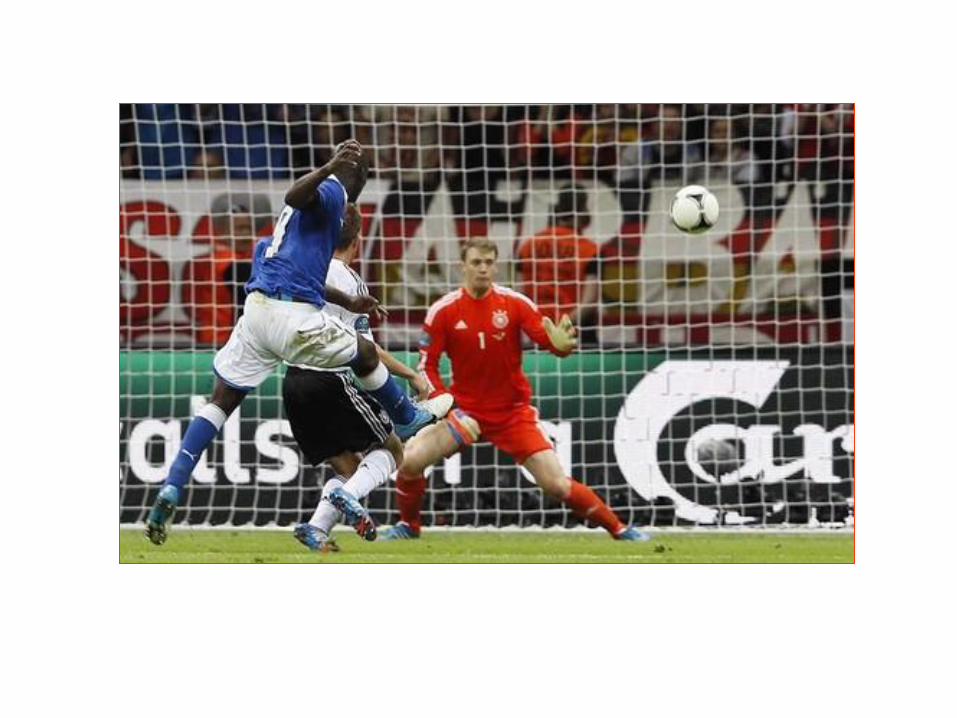
World Cup Final 2014Argentina-Germany
• https://www.youtube.com/watch?v=HlwY69oVL8I
• https://www.youtube.com/watch?v=v4utKclNpkI
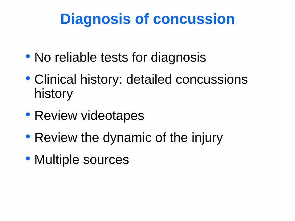
Diagnosis of concussion
• No reliable tests for diagnosis
• Clinical history: detailed concussionshistory
• Review videotapes
• Review the dynamic of the injury
• Multiple sources

Biomarkers
• Indicator of presence or severity of a disease:
Specific
Sensitive
Predictive
Robust
Non invasive
Non expensive
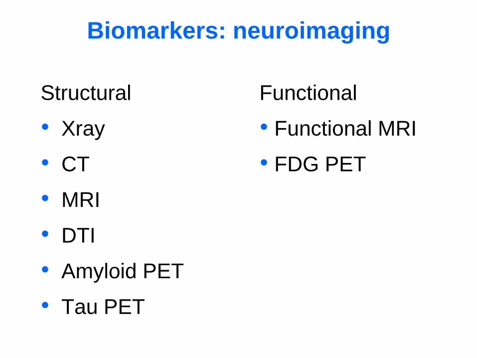
Biomarkers: neuroimaging
Structural
• Xray
• CT
• MRI
• DTI
• Amyloid PET
• Tau PET
Functional
• Functional MRI
• FDG PET
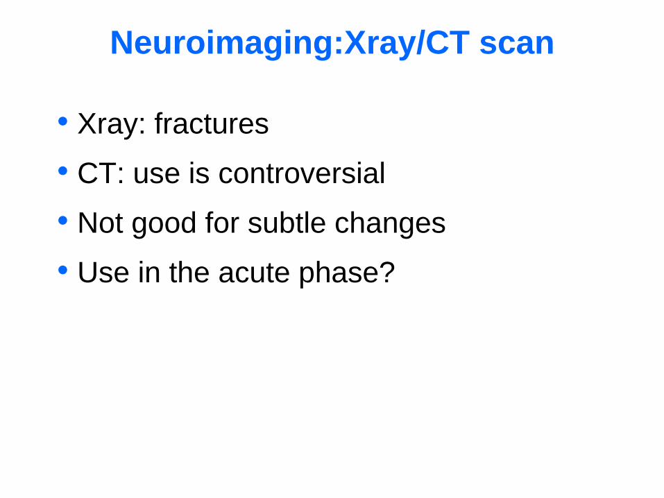
Neuroimaging:Xray/CT scan
• Xray: fractures
• CT: use is controversial
• Not good for subtle changes
• Use in the acute phase?
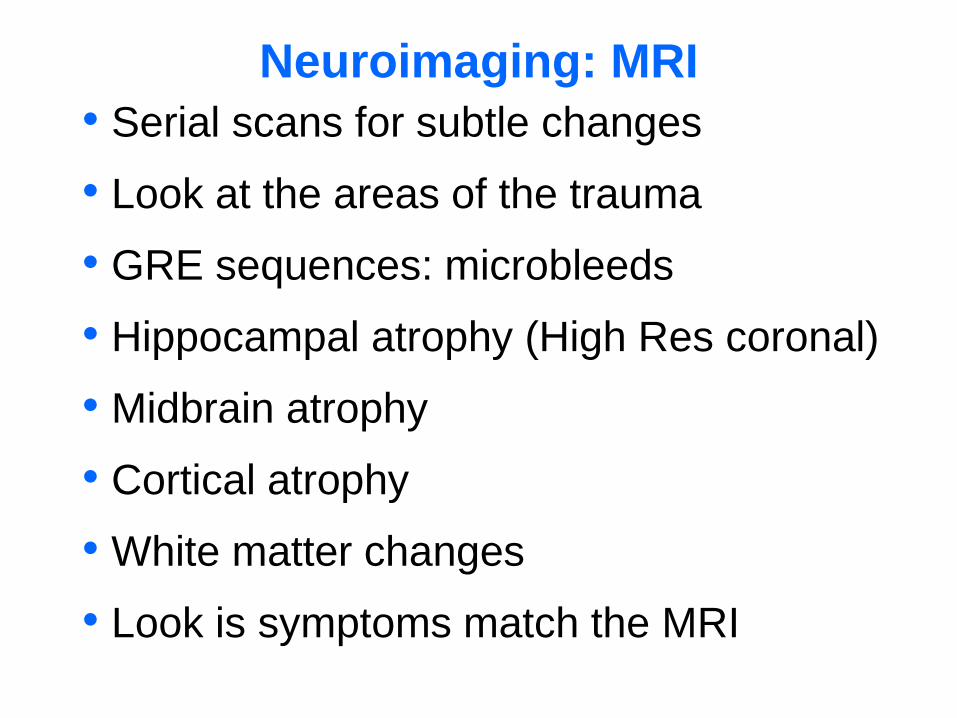
Neuroimaging: MRI
• Serial scans for subtle changes
• Look at the areas of the trauma
• GRE sequences: microbleeds
• Hippocampal atrophy (High Res coronal)
• Midbrain atrophy
• Cortical atrophy
• White matter changes
• Look is symptoms match the MRI
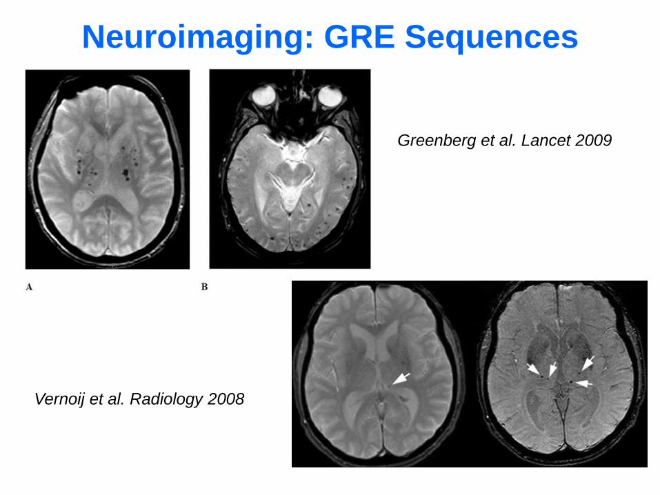
Neuroimaging: GRE Sequences
Greenberg et al. Lancet 2009
Vernoij et al. Radiology 2008

Normal AD
hippocampal atrophynormal hippocampi
Neuroimaging: Hippocampal Atrophy
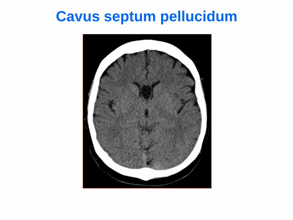
Cavus septum pellucidum
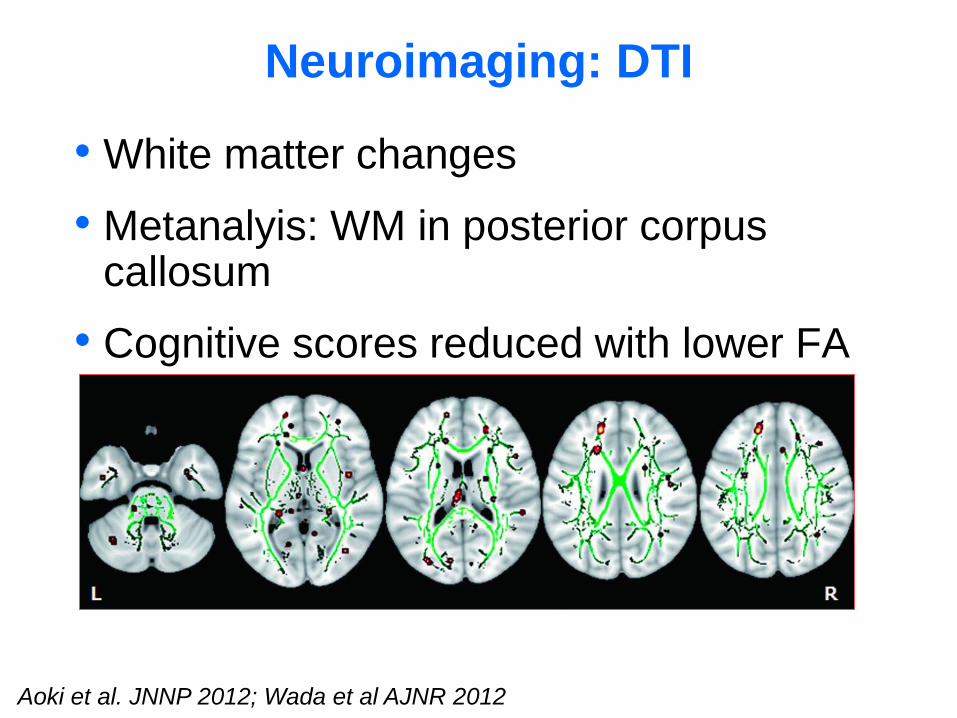
Neuroimaging: DTI
• White matter changes
• Metanalyis: WM in posterior corpus callosum
• Cognitive scores reduced with lower FA
Aoki et al. JNNP 2012; Wada et al AJNR 2012
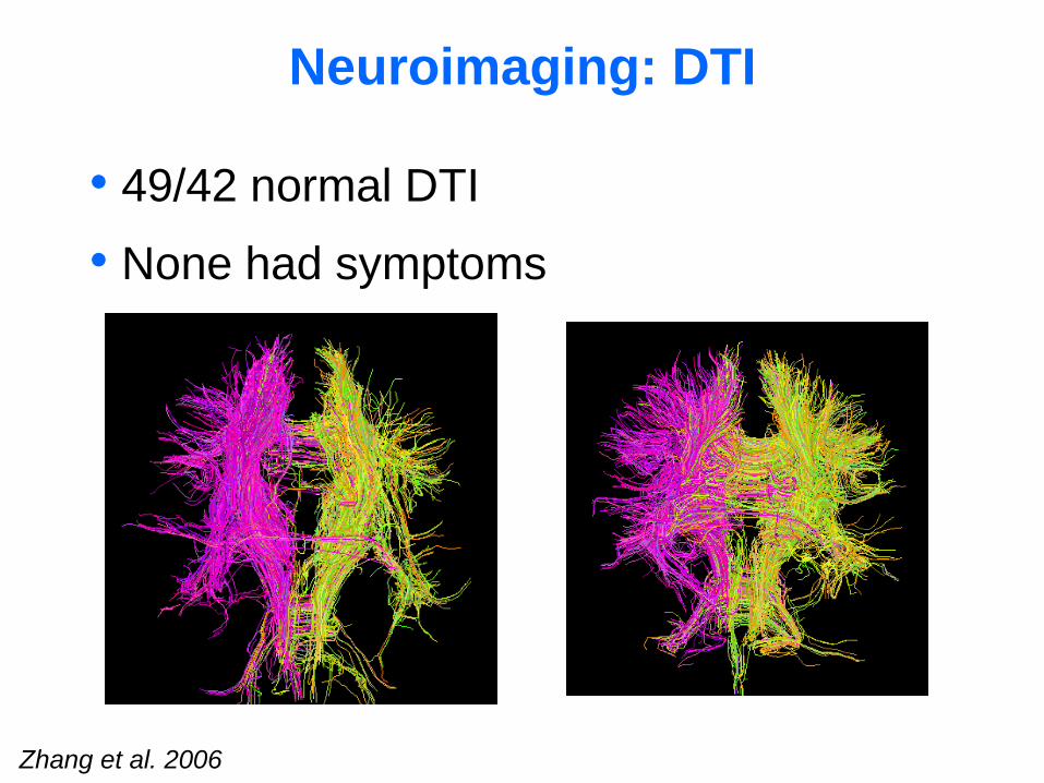
Neuroimaging: DTI
Zhang et al. 2006
• 49/42 normal DTI
• None had symptoms
Maruyama et al. Neuron 2013
Tau Β-Amyloid
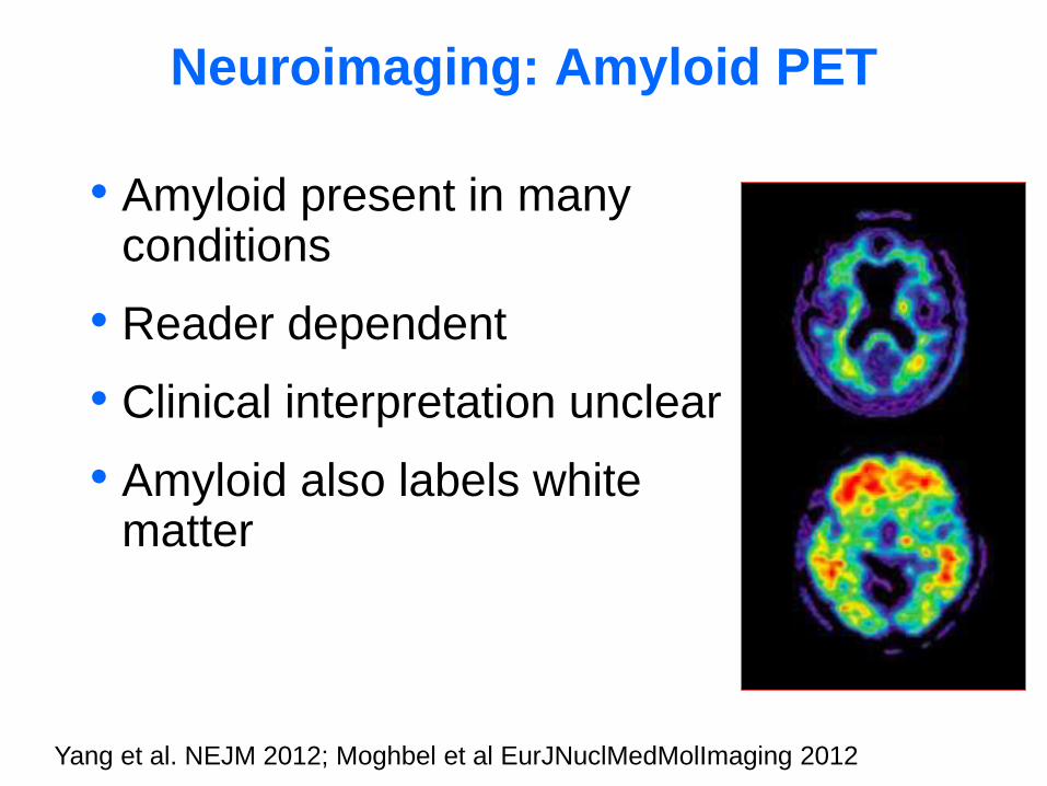
Neuroimaging: Amyloid PET
• Amyloid present in manyconditions
• Reader dependent
• Clinical interpretation unclear
• Amyloid also labels white matter
Yang et al. NEJM 2012; Moghbel et al EurJNuclMedMolImaging 2012
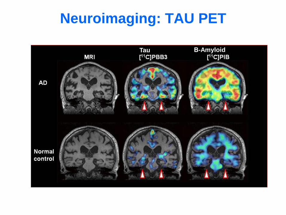
Maruyama et al. Neuron 2013
Tau Β-Amyloid
Neuroimaging: TAU PET
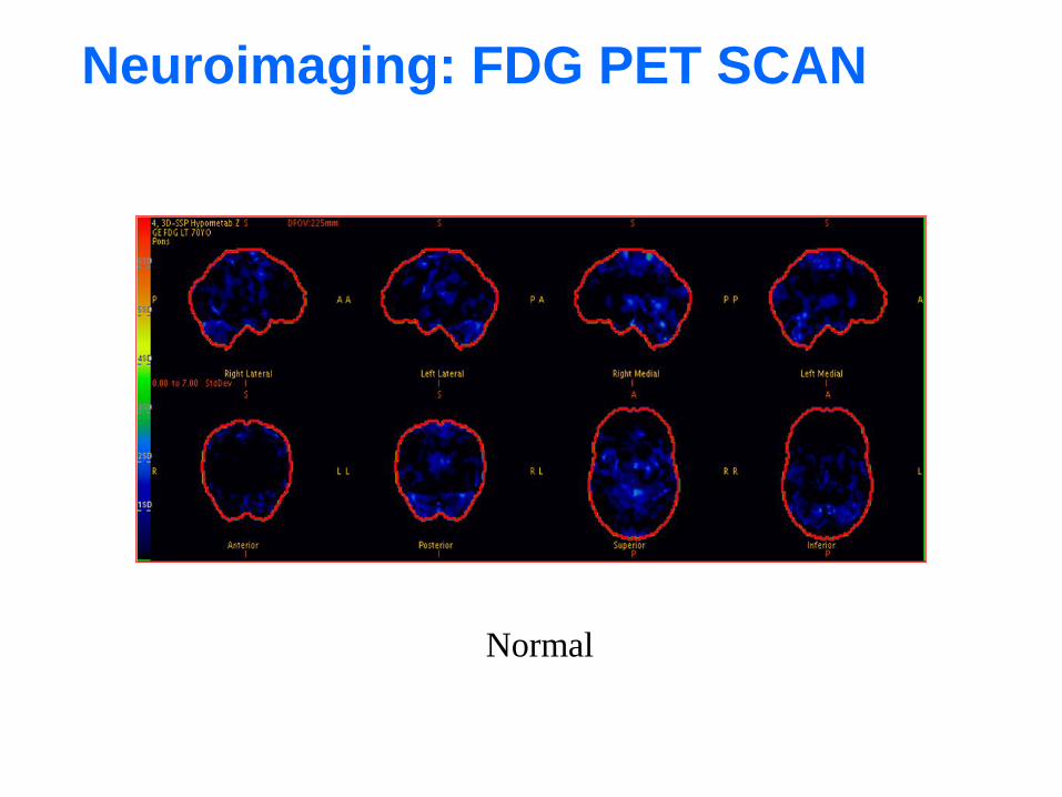
Normal
Neuroimaging: FDG PET SCAN
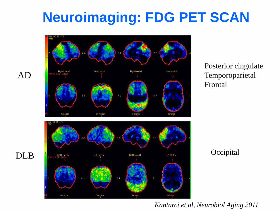
DLB
ADPosterior cingulate
Temporoparietal
Frontal
Kantarci et al, Neurobiol Aging 2011
Occipital
Neuroimaging: FDG PET SCAN
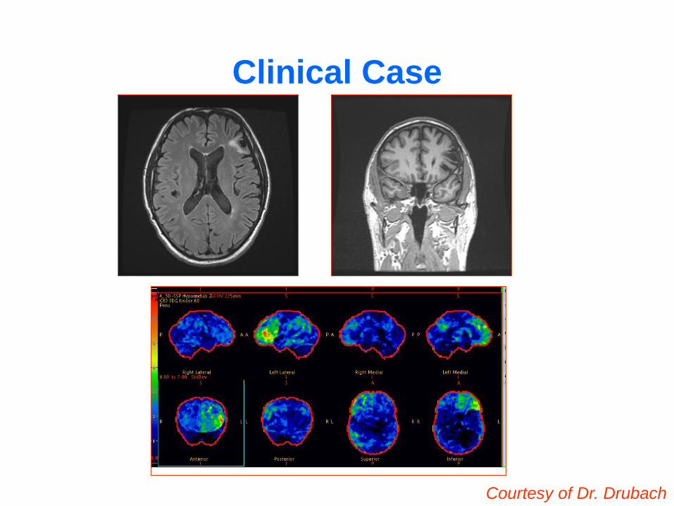
Clinical Case
Courtesy of Dr. Drubach
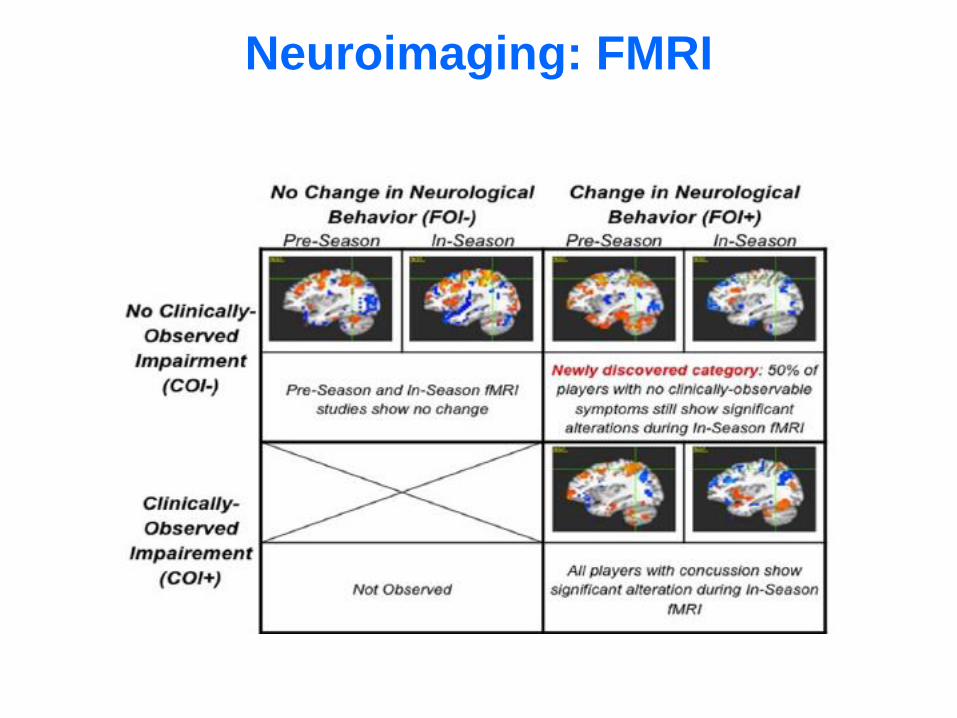
Neuroimaging: FMRI
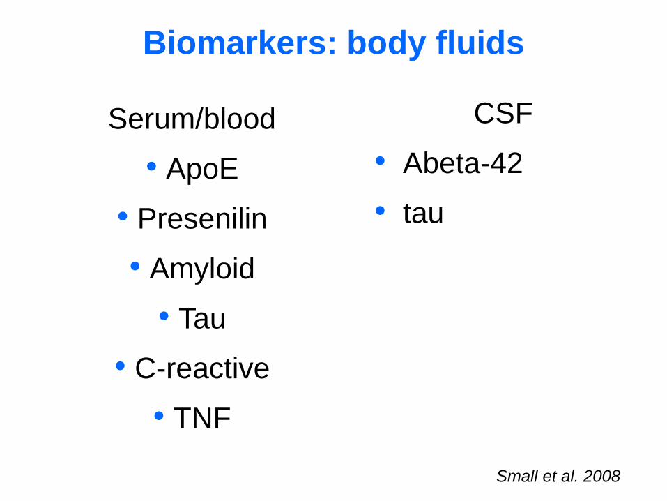
Biomarkers: body fluids
CSF
• Abeta-42
• tau
Serum/blood
• ApoE
• Presenilin
• Amyloid
• Tau
• C-reactive
• TNF
Small et al. 2008

Head Trauma and ALS
• First description in 1897 (Erb W. 1897)
• 3 fold increase in subjects with previous HT (Chen et al Am J Epidemiol 2007)
• Increased risk of ALS in soccer players (Chiò et al. Neurology 2005)
• 40 fold increase of prevalence in football players (Abel E. Perceptual Motor Skills 2007)

Head trauma and AD
• Pooled analysis of 11 case-control studies that investigated HT and AD
• The relative risk for dementia was 1.82
• Stronger association in cases with family hx of dementia and males (Mortimer et al. Int J Epidemiol 1991)

Head trauma and PD
• Head trauma and future risk of PD: Lag-time between HT and PD: 21 years; OR = 4.3 (Bower et al. Neurology 2003)
• HT injury with amnesia/loss of consciousness increased the risk for PD: OR = 3.8 (Goldman et al. Ann Neurol 2006)

Head trauma and genetic predisposition
• FAME and SEARCH : 476 (89+387) cases and 576 controls (387+189)
• Rep 1: a promoter increasing a-synuclein
• HT not associated with increased risk of PD
• HT was associated with PD and Rep1 FAME: OR, 5.4; 95% CI, 1.5-19; SEARCH OR, 2.3; 95% CI, 0.6-9.2pooled OR, 3.5; 95% CI 1.4-9.2, (p interaction = 0.02).
• 4.9 years earlier (p = 0.03).
Ann of Neurology 2012
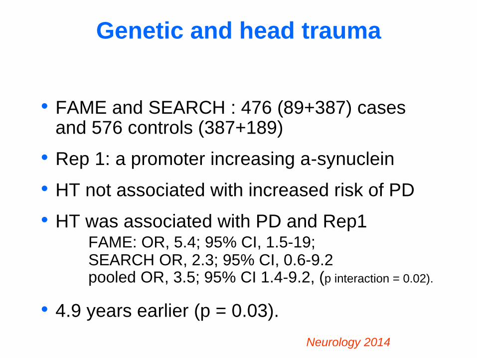
Genetic and head trauma
• FAME and SEARCH : 476 (89+387) cases and 576 controls (387+189)
• Rep 1: a promoter increasing a-synuclein
• HT not associated with increased risk of PD
• HT was associated with PD and Rep1 FAME: OR, 5.4; 95% CI, 1.5-19; SEARCH OR, 2.3; 95% CI, 0.6-9.2pooled OR, 3.5; 95% CI 1.4-9.2, (p interaction = 0.02).
• 4.9 years earlier (p = 0.03).
Neurology 2014
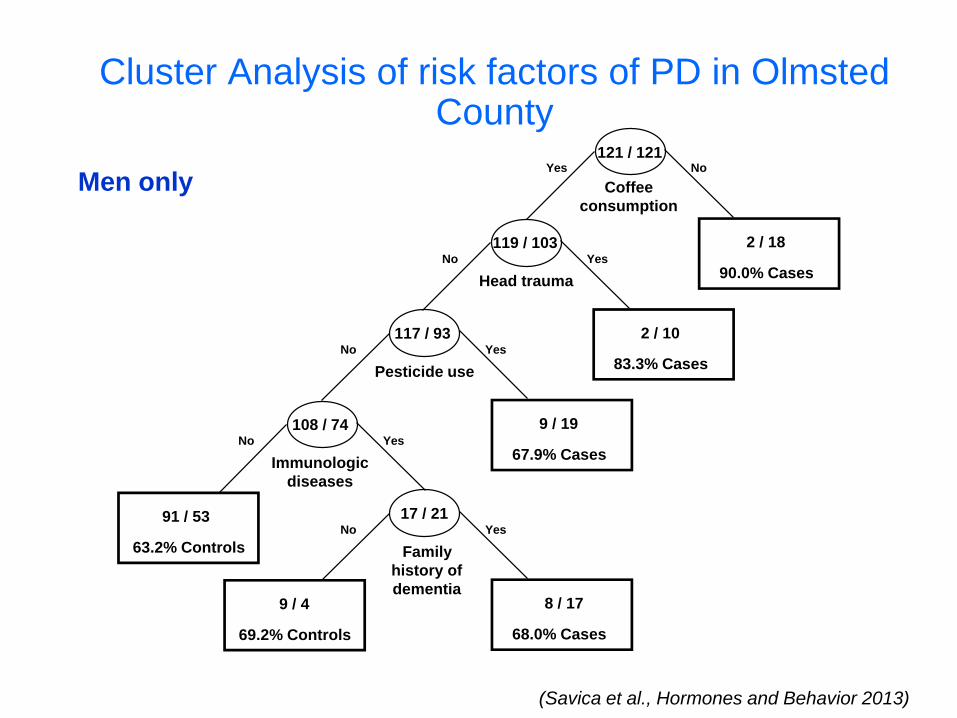
Cluster Analysis of risk factors of PD in Olmsted County
(Savica et al., Hormones and Behavior 2013)
Men only
121 / 121NoYes
Coffee
consumption
119 / 103 2 / 18
90.0% CasesYesNo
2 / 10
83.3% Cases
Head trauma
117 / 93YesNo
9 / 19
67.9% Cases
Pesticide use
108 / 74YesNo
Immunologic
diseases
17 / 21YesNo
Family
history of
dementia8 / 17
68.0% Cases
9 / 4
69.2% Controls
91 / 53
63.2% Controls
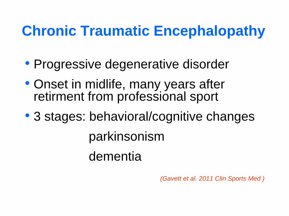
Chronic Traumatic Encephalopathy
• Progressive degenerative disorder
• Onset in midlife, many years after retirment from professional sport
• 3 stages: behavioral/cognitive changes
parkinsonism
dementia
(Gavett et al. 2011 Clin Sports Med )
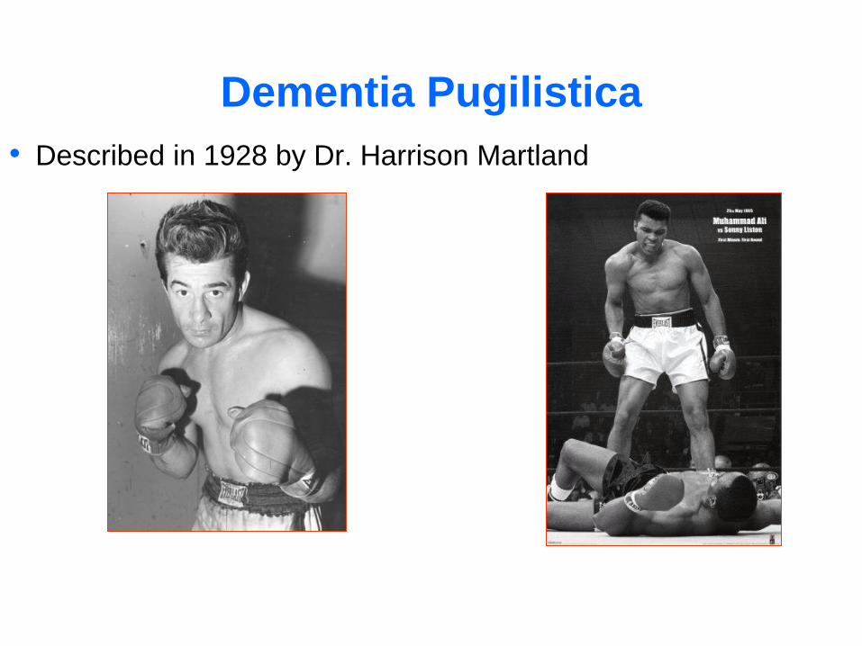
Dementia Pugilistica
• Described in 1928 by Dr. Harrison Martland

CTE Diagnosis
• It is not possible to diagnose CTE without pathological confirmation
• Signs and symptoms are identical to PD, Fronto-temporal Dementia, AD, and ALS (Gavett et al. 2011 Clin Sports Med )
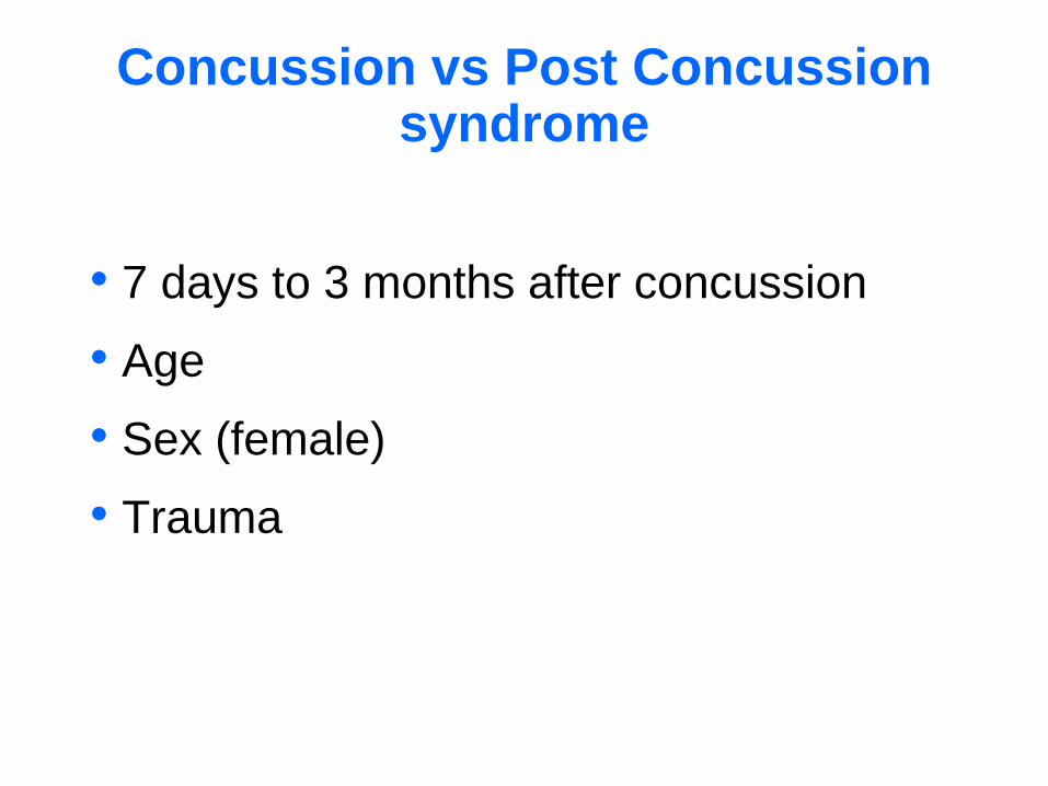
Concussion vs Post Concussion syndrome
• 7 days to 3 months after concussion
• Age
• Sex (female)
• Trauma
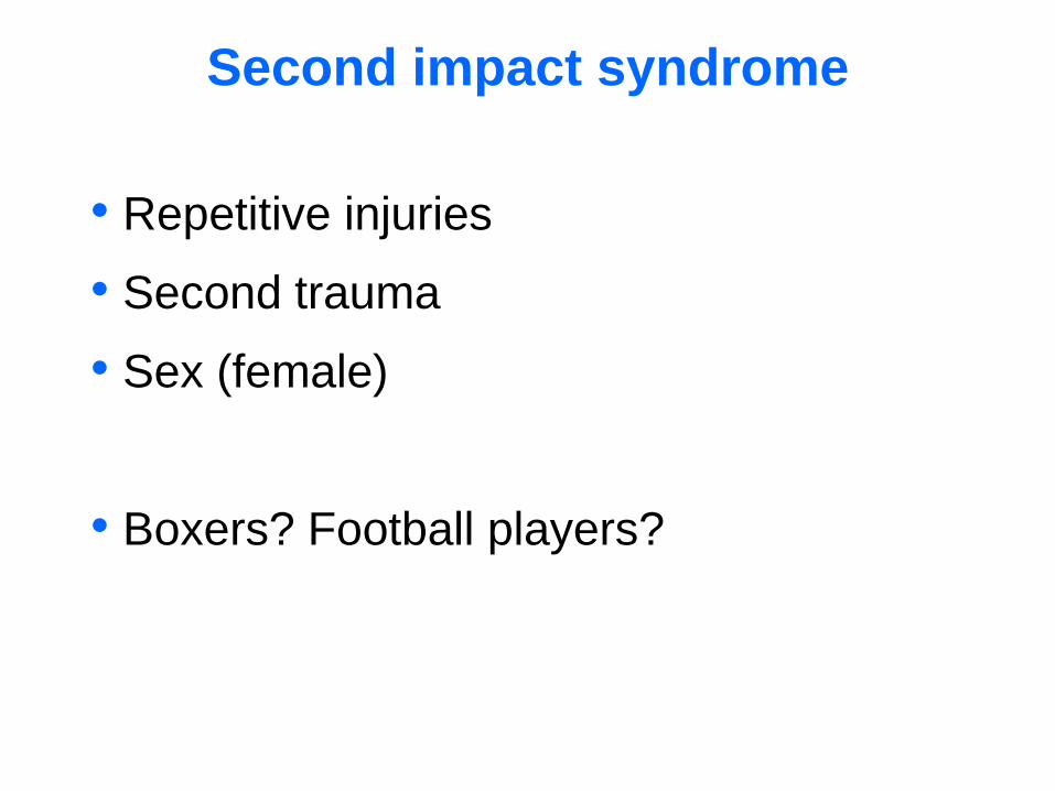
Second impact syndrome
• Repetitive injuries
• Second trauma
• Sex (female)
• Boxers? Football players?
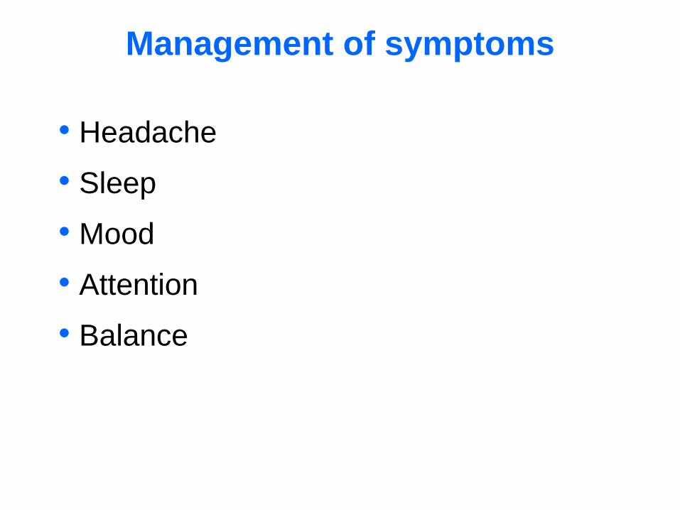
Management of symptoms
• Headache
• Sleep
• Mood
• Attention
• Balance

Post-traumatic headache (PTH) is just headache?
• Headache in athletes: 58%
• Triggers: sleep deprivation, emotional stress, skipping meals,excercise, travel, altitude, etc…
• Benign exertional headache
• Airplane headache
• Altitude headache
• Swimmer/diver headacheNCAA Task Force
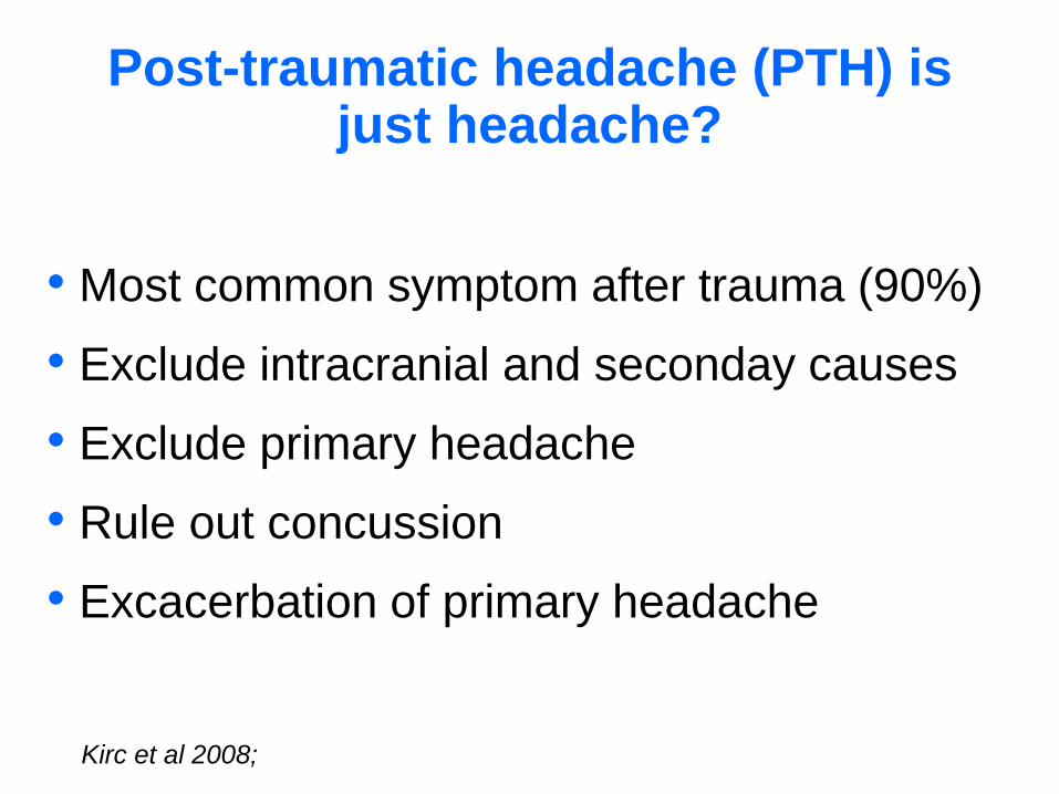
Post-traumatic headache (PTH) is just headache?
• Most common symptom after trauma (90%)
• Exclude intracranial and seconday causes
• Exclude primary headache
• Rule out concussion
• Excacerbation of primary headache
Kirc et al 2008;

Mimickers of PTH
• Dehydration
• Hypoglicemia
• Hypertension
• Hypertermia
• Drugs (prescription and not prescription)

Drugs causing headache in athletes
• Alcohol
• NSAIDs
• Anabolic Steroids
• Nicotine
• Antibiotics
• Nitrazepam
• Antihypertensives
• Oral Contraceptives
• Caffeine
• Sympathomimetics
• Corticosteroids
• Theophylline
• Dipyridamole
• Vasodilator Agents
• Analgesics
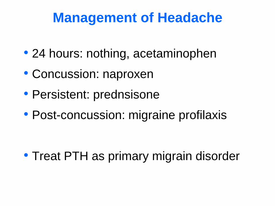
Management of Headache
• 24 hours: nothing, acetaminophen
• Concussion: naproxen
• Persistent: prednsisone
• Post-concussion: migraine profilaxis
• Treat PTH as primary migrain disorder

Management of Sleep
• 24 hours: nothing
• Concussion: melatonin
• Persistent: zolpidem
• Post-concussion: tricyclic
• Quietapine
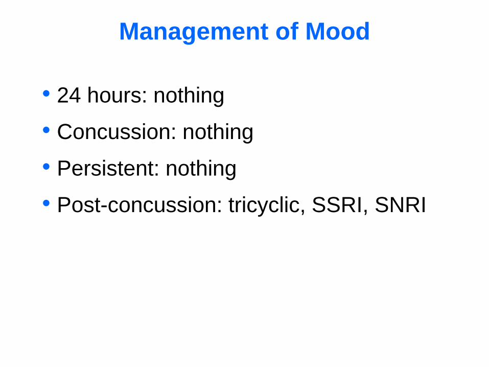
Management of Mood
• 24 hours: nothing
• Concussion: nothing
• Persistent: nothing
• Post-concussion: tricyclic, SSRI, SNRI

Management of Attention
• 24 hours: nothing
• Concussion: nothing
• Persistent: nothing
• Post-concussion: like ADD

Management of Neurodegeneration
• Parkinsonism
• Dementias
• ALS
Use the current state of the art treatment

High school football form 1946-1956 and risk of neurodegeneration
Savica et al 2013
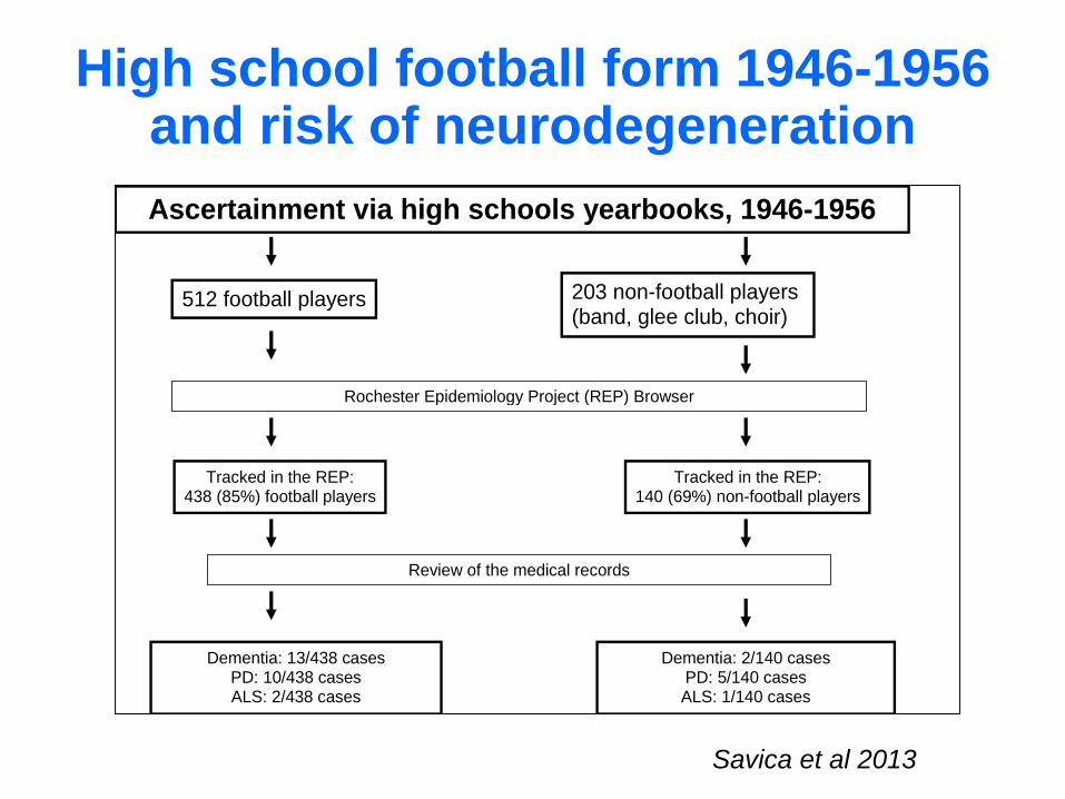
High school football form 1946-1956 and risk of neurodegeneration
Ascertainment via high schools yearbooks, 1946-1956
Rochester Epidemiology Project (REP) Browser
Tracked in the REP: 438 (85%) football players
Review of the medical records
Dementia: 13/438 cases PD: 10/438 cases ALS: 2/438 cases
512 football players 203 non-football players (band, glee club, choir)
Tracked in the REP: 140 (69%) non-football players
Dementia: 2/140 cases PD: 5/140 cases ALS: 1/140 cases
Savica et al 2013
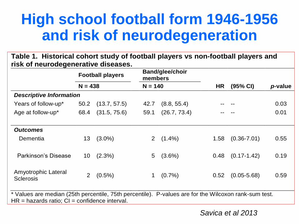
High school football form 1946-1956 and risk of neurodegeneration
Table 1. Historical cohort study of football players vs non-football players and risk of neurodegenerative diseases.
Football players Band/glee/choir members
N = 438 N = 140 HR (95% CI) p-value
Descriptive Information
Years of follow-up* 50.2 (13.7, 57.5) 42.7 (8.8, 55.4) -- -- 0.03
Age at follow-up* 68.4 (31.5, 75.6) 59.1 (26.7, 73.4) -- -- 0.01
Outcomes
Dementia 13 (3.0%) 2 (1.4%) 1.58 (0.36-7.01) 0.55
Parkinson’s Disease 10 (2.3%) 5 (3.6%) 0.48 (0.17-1.42) 0.19
Amyotrophic Lateral Sclerosis
2 (0.5%) 1 (0.7%) 0.52 (0.05-5.68) 0.59
* Values are median (25th percentile, 75th percentile). P-values are for the Wilcoxon rank-sum test. HR = hazards ratio; CI = confidence interval.
Savica et al 2013
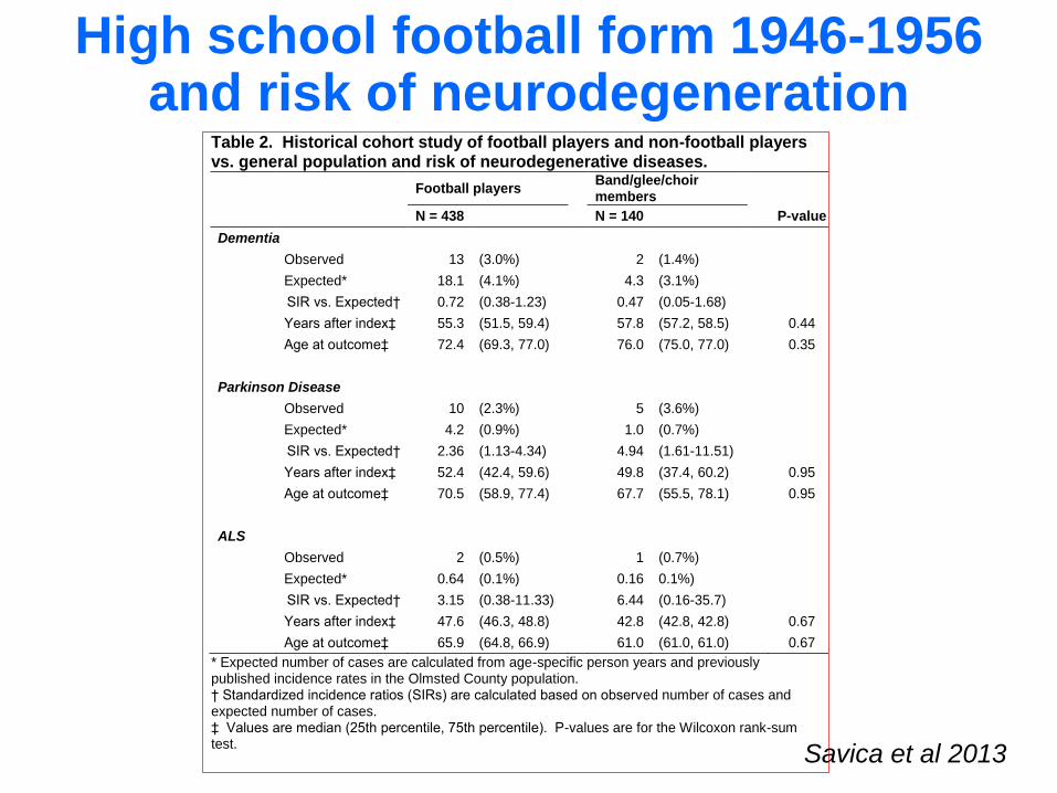
High school football form 1946-1956 and risk of neurodegeneration
Table 2. Historical cohort study of football players and non-football players vs. general population and risk of neurodegenerative diseases.
Football players Band/glee/choir members
N = 438 N = 140 P-value
Dementia
Observed 13 (3.0%) 2 (1.4%)
Expected* 18.1 (4.1%) 4.3 (3.1%)
SIR vs. Expected† 0.72 (0.38-1.23) 0.47 (0.05-1.68)
Years after index‡ 55.3 (51.5, 59.4) 57.8 (57.2, 58.5) 0.44
Age at outcome‡ 72.4 (69.3, 77.0) 76.0 (75.0, 77.0) 0.35
Parkinson Disease
Observed 10 (2.3%) 5 (3.6%)
Expected* 4.2 (0.9%) 1.0 (0.7%)
SIR vs. Expected† 2.36 (1.13-4.34) 4.94 (1.61-11.51)
Years after index‡ 52.4 (42.4, 59.6) 49.8 (37.4, 60.2) 0.95
Age at outcome‡ 70.5 (58.9, 77.4) 67.7 (55.5, 78.1) 0.95
ALS
Observed 2 (0.5%) 1 (0.7%)
Expected* 0.64 (0.1%) 0.16 0.1%)
SIR vs. Expected† 3.15 (0.38-11.33) 6.44 (0.16-35.7)
Years after index‡ 47.6 (46.3, 48.8) 42.8 (42.8, 42.8) 0.67
Age at outcome‡ 65.9 (64.8, 66.9) 61.0 (61.0, 61.0) 0.67
* Expected number of cases are calculated from age-specific person years and previously published incidence rates in the Olmsted County population. † Standardized incidence ratios (SIRs) are calculated based on observed number of cases and expected number of cases. ‡ Values are median (25th percentile, 75th percentile). P-values are for the Wilcoxon rank-sum test. Savica et al 2013
Conclusions
• Concussion is a complex and heterogenous syndrome
• No certain diagnosis…in vivo
• Desperate need of good science
• In practice: use the available evidence and treat the symptoms regardless of concussion

Thank You
Messina, Italy

Salt Lake City, UT

Rochester, MN

Questions?