connectivetissuegrowthfactor–specificmonoclonalantibody...
TRANSCRIPT

Connective Tissue Growth Factor–Specific Monoclonal Antibody
Therapy Inhibits Pancreatic Tumor Growth and Metastasis
Nadja Dornhofer,1,5
Suzanne Spong,4Kevin Bennewith,
1Ali Salim,
1,2Stephen Klaus,
4
Neeraja Kambham,3Carol Wong,
4Fiona Kaper,
1Patrick Sutphin,
1Rendall Nacalumi,
2
Michael Hockel,5Quynh Le,
1Michael Longaker,
2George Yang,
2
Albert Koong,1and Amato Giaccia
1
Departments of 1Radiation Oncology, 2Plastic Surgery, and 3Pathology, Stanford University School of Medicine, Stanford; 4FibroGen, Inc.,South San Francisco, California; and 5Department of Obstetrics and Gynecology, University of Leipzig, Leipzig, Germany
Abstract
Pancreatic cancer is highly aggressive and refractory to mostexisting therapies. Past studies have shown that connectivetissue growth factor (CTGF) expression is elevated in humanpancreatic adenocarcinomas and some pancreatic cancer celllines. To address whether and how CTGF influences tumorgrowth, we generated pancreatic tumor cell lines that over-express different levels of human CTGF. The effect of CTGFoverexpression on cell proliferation was measured in vitro inmonolayer culture, suspension culture, or soft agar, andin vivo in tumor xenografts. Although there was no effect ofCTGF expression on proliferation in two-dimensional cultures,anchorage-independent growth (AIG) was enhanced. Thecapacity of CTGF to enhance AIG in vitro was linked toenhanced pancreatic tumor growth in vivo when these cellswere implanted s.c. in nude mice. Administration of aneutralizing CTGF-specific monoclonal antibody, FG-3019,had no effect on monolayer cell proliferation, but blockedAIG in soft agar. Consistent with this observation, anti-CTGFtreatment of mice bearing established CTGF-expressingtumors abrogated CTGF-dependent tumor growth andinhibited lymph node metastases without any toxicity ob-served in normal tissue. Together, these studies implicateCTGF as a new target in pancreatic cancer and suggest thatinhibition of CTGF with a human monoclonal antibody maycontrol primary and metastatic tumor growth. (Cancer Res2006; 66(11): 5816-27)
Introduction
Pancreatic cancer continues to be one of the most lethalcancers. More then 30,000 new cases of pancreatic cancer arediagnosed annually in the U.S. Mortality rates for this disease havenot changed significantly in 30 years and approach 100% within5 years after diagnosis. Current treatment options are limited dueto the lack of diagnostic biomarkers and a lack of obvioussymptoms until the tumor stage is already advanced. Approxi-mately 10% to 20% of patients have surgically resectable diseaseat presentation, but even in these cases, the 5-year survival rate isonly 20% (1). In patients with advanced disease, gemcitabine isconsidered to be a first-line option. However, gemcitabine only
modestly improves survival with a 1-year survival rate of <20% (1).Clearly, there exists a need for more effective targeted therapeu-tics to treat pancreatic cancer by targeting gene products that willalter the malignant progression of pancreatic cancer or itsresponse to therapy.
Connective tissue growth factor (CTGF) is a member of the CCN[CYR61 (cysteine-rich 61) / CTGF / NOV (nephroblastoma over-expressed)] family of secreted proteins, which are characterizedas cysteine-rich matricellular proteins that each contain fourmodular domains displaying homology to insulin-like growthfactor–binding proteins (domain 1), a von Willebrand factor typeC repeat (domain 2), a thrombospondin type 1 repeat (domain 3),and a cysteine knot domain (domain 4), respectively (2). Thecysteine knot domain contains heparin-binding sites that mediatebinding to extracellular matrix and cell surface heparan sulfateproteoglycans (3). CTGF is an immediate early gene that is potentlyinduced by a variety of stimuli that regulate extracellular matrixdeposition, tissue remodeling, and neovascularization, includingplatelet-derived growth factor, transforming growth factor (TGF)-h,basic fibroblast growth factor, vascular endothelial growth factor(VEGF), and hypoxia in fibroblasts or endothelial cells (4–7). CTGFexhibits a diverse range of cellular functions including celladhesion, stimulation of cell migration, and potentiation of growthfactor–induced DNA synthesis (8).
CTGF interacts with integrin receptors including avh3, aIIbh3,a6h1, and amh2 (3, 9–11) and has been reported to be a ligand forlow-density lipoprotein-related protein 1 (LRP-1); interacts withLRP-5 to inhibit Wnt signaling (12–14) and can interact directlywith several growth factors including TGF-h (49). Taken together,past studies indicate that the mechanism of action of CTGF relatesto its capacity to modulate and amplify a variety of biologicalprocesses by binding directly to mitogenic, fibrogenic, andangiogenic factors that are important in inflammation, fibrosis,tumor growth, and tumor metastasis.
Elevated CTGF levels have been detected in a number of cancersincluding pancreatic (15), breast (16), glioblastoma (17, 18),esophageal (19), melanoma (20), chondrosarcoma (21), oralsquamous cell cancer (22), acute lymphoblastic leukemia (23),rhabdomyosarcoma (24), and hepatocellular carcinoma (25), butits direct role in tumor suppression or progression has not beeninvestigated in pancreatic cancer nor with therapeutic agents withthe capacity to inhibit CTGF function in vivo . An increase in CTGFwas reported to be associated with decreased survival of patientswith breast cancer (16), glioblastoma (18), or adenocarcinoma ofthe esophagus (19), and increased breast cancer bone metastasisin a mouse model (26). In contrast, high levels of CTGF wereassociated with better survival in patients with esophageal
Requests for reprints: Amato Giaccia, Division of Radiation and Cancer Biology,Department of Radiation Oncology, Stanford University School of Medicine, Room1255, CCSR South, 269 Campus Drive, Stanford, CA 94305. Phone: 650-723-7366;E-mail: [email protected].
I2006 American Association for Cancer Research.doi:10.1158/0008-5472.CAN-06-0081
Cancer Res 2006; 66: (11). June 1, 2006 5816 www.aacrjournals.org
Research Article
Research. on June 25, 2015. © 2006 American Association for Cancercancerres.aacrjournals.org Downloaded from
Research. on June 25, 2015. © 2006 American Association for Cancercancerres.aacrjournals.org Downloaded from
Research. on June 25, 2015. © 2006 American Association for Cancercancerres.aacrjournals.org Downloaded from

squamous cell carcinoma (19) and chondrosarcoma (21), anddecreased metastasis in a colon cancer mouse model (27).Likewise, in lung adenocarcinoma, reduced CTGF expression wascorrelated with advanced disease stage and decreased survival, andexpression of CTGF in lung cancer cell lines suppressed metastasisin a mouse tail vein injection model (28).
Whereas elevated levels of CTGF have been detected in theabove tumor types, pancreatic carcinomas are especially notewor-thy because a hallmark of their histopathology is desmoplasia. In aprevious study by Wenger et al., 15 of 19 samples from pancreatictumors exhibited an average 59-fold enhancement of CTGF mRNAexpression, compared with a 4.5-fold increase in chronic pancre-atitis (15). CTGF transcript levels were grossly correlated with thedegree of fibrosis and collagen expression, consistent with knownCTGF bioactivity. In a study of 25 patients with pancreatic cancer,Hartel et al. reported a 46-fold increase in CTGF mRNA levels inpancreatic cancer tissue compared with normal tissue (29).Furthermore, they found a positive correlation between desmo-plastic reaction and CTGF mRNA level and concluded that thedesmoplastic reaction might account for better survival of patientswith elevated CTGF expression observed in this study. In contrast,Ryu and coworkers identified CTGF as a member of a pancreaticcancer invasion–specific gene cluster (30). In this study, the CTGFmessage was localized to pancreatic tumor cells rather than tostromal or endothelial cells (31). However, CTGF expression hasbeen seen in both tumor cells and associated fibroblasts,endothelial cells, pancreatic stellate cells, and vascular smoothmuscle cells (15, 29), not permitting clear insight into thesignificance of the cellular origin of CTGF expression. However,because CTGF has been found to be expressed in both the stromaland tumoral compartments and is a potential driver of desmo-plasia, CTGF represents a unique target in pancreatic tumorigen-esis. Although we and others (15) have found that most patientswith pancreatic cancer exhibit elevated levels of CTGF comparedwith controls, no study has directly investigated the role of CTGF inpancreatic cancer growth or the potential of targeting CTGF fortherapy.
In this study, we investigated the influence of overexpressingCTGF on pancreatic cancer cell growth in vitro and in vivo , and theeffect of inhibiting CTGF to control pancreatic tumor growth. Ourdata suggest that tumor cell–derived CTGF enhances pancreatictumor growth, whereas inhibition of CTGF with a humanmonoclonal antibody (mAb) reduces pancreatic tumor growthand metastasis. Therefore, CTGF may be a new target for thetreatment of pancreatic cancer.
Materials and Methods
Patient samples. Human tissue from patients with pancreatic cancerwas obtained during Whipple procedures at Stanford University. All
patients signed an informed consent approved by the Stanford Institutional
Review Board (in accord with an assurance filed with and approved by the
U.S. Department of Health and Human Services). Representative paraffinblocks with carcinoma and normal pancreas were selected from each of
eight cases. The immunohistochemical stains were done using rabbit
polyclonal antibody directed against mouse CCN2/Fisp 12 which cross-reacts with human CTGF (provided by L. Lau; ref. 32). Serial sections of
4 Amol/L were obtained from the selected paraffin blocks, deparaffinized in
xylene, and hydrated in a graded series of alcohols. Heat-induced antigen
retrieval was carried out by microwave pretreatment in citric acid buffer(10 mmol/L, pH 6.0) for 10 minutes. The CCN2 antibody was used at a
dilution of 1:400. The endogenous peroxidase was blocked and the DAKO
Envision System (DAKO Corporation, Carpinteria, CA) was used fordetection; diaminobenzidine was used as a chromogen.
The intensity of the staining was scored on a scale of 0 to 3+: where 0, no
staining; 1, weak staining; 2, moderate staining; and 3, strong staining. If the
staining in a particular case was variable, the percentage of tumor andnormal tissue with a specific intensity score was also recorded. Scoring was
done separately for normal pancreas, areas of chronic pancreatitis, and
carcinoma; both epithelial elements and stroma were scored.
CTGF ELISA. Cell supernatants were collected in DMEM supplementedwith 1% penicillin/streptomycin, 100 Ag/mL low molecular weight heparin,
and 0.25% bovine serum albumin. CTGF levels in culture supernatants,
blood plasma, and urine were measured using a sandwich ELISA that
detects whole CTGF and the NH2-terminal fragment of CTGF that persistsin body fluids and cell culture supernatants after proteolytic cleavage of the
hinge domain (33).
Cell lines and tissue culture. The human pancreatic cancer cell lines,MIA PaCa-2 and PANC-1, were grown in DMEM supplemented with 10%
fetal bovine serum (FBS) and 1% penicillin/streptomycin, whereas Su86.86
pancreatic cancer cells were grown in RPMI 1640 containing 10% FBS. To
generate pancreatic cancer cell lines overexpressing CTGF, we stablytransfected MIA PaCa-2 cells with pShuttle-CMV-Puro-CTGF vector
(an adenoviral construct encoding full-length CTGF and the puromycin
antibiotic-resistance gene) using LipofectAMINE 2000 reagent (Invitrogen
Life Technologies, Carlsbad, CA) according to the manufacturer’s instruc-tions. Control transfections were done with the same vector lacking the
CTGF cDNA sequence. Individual clones were selected based on their
resistance to puromycin, and CTGF mRNA and protein levels weredetermined by quantitative real-time PCR (qRT-PCR) and ELISA or Western
blot assay, respectively. A series of CTGF-expressing or vector control clones
were isolated and characterized, and representative clones that exhibited
different levels of CTGF expression and protein production were identified.Stable transfectants were cultured in DMEM supplemented with 10% FBS,
1% penicillin/streptomycin and 1.5 Ag/mL puromycin. All cell lines were
cultured in a 5% CO2 humidified atmosphere at 37jC.CTGF mAb. FG-3019 is a fully human IgG1n mAb recognizing domain 2
of human and rodent CTGF, and was obtained from FibroGen, Inc. (South
San Francisco, CA). FG-3019 was purified under cyclic guanosine 3¶,5¶-monophosphate conditions and formulated in a 25 mmol/L histidine buffer(pH 6.0). In some experiments, polyclonal human IgG from Cohn’s fraction
of human serum (Sigma, St. Louis, MO) purified by protein A sepharose was
used as a control.
In vitro growth curve. Cells were plated at a density of 5 � 104 in 6 cm
dishes. Every 3 or 4 days, cells were trypsinized, counted and 5 � 104 cells
were replated. To investigate the effect of a CTGF-specific antibody (FG-3019)
on tumor cell growth, growth curves were also determined in the presence of
20 and 40 Ag/mL of FG-3019 or control human IgG. For growth in suspension,
2.5 � 105 cells were plated on ultra-low cluster plates (Costar, Cambridge,
MA) which have a covalently bound hydrogel layer that effectively inhibits
cellular attachment. Photographs were taken 4 days after cells were plated
using a Leica MZ6 microscope with 10� and 40� objectives. Growth curves
were obtained by plating 2.5� 105 tumor cells into each of 12 wells, allowing
the aggregates to form over a 48-hour period, and trypsinizing the tumor cell
aggregates in triplicate wells daily with subsequent cell counting.
Soft agar assay. In duplicate experiments, f3,000 cells from each clone
were resuspended in 2 mL of 0.35% Noble Agar (Difco Laboratories, Detroit,MI) containing 10% FBS (Life Technologies) and 10% newborn calf serum
(Life Technologies). Each embedded cell mixture was overlaid on 1.5 mL of
0.7% Noble Agar in six-well plates, and a 1.5 mL top layer of 0.7% Noble Agar
was added to each well to prevent evaporation. In some experiments, humancontrol IgG or FG-3019 was added at a concentration of 100 Ag/mL to the
cell layer containing 1,500 cells in 2 mL. Plates were incubated for 11 days in
a humidified incubator at 37jC, 5% CO2. The number of colonies was
enumerated by counting a 1.5 � 1.5 cm grid under a microscope. Total colonycounts were extrapolated to the entire plate based on the ratio of the surface
area of each well to the surface area of the grid. Colony morphologies were
captured with a Nikon camera mounted onto an inverted microscope that wasset at �20 magnification.
CTGF in Pancreatic Cancer
www.aacrjournals.org 5817 Cancer Res 2006; 66: (11). June 1, 2006
Research. on June 25, 2015. © 2006 American Association for Cancercancerres.aacrjournals.org Downloaded from

qRT-PCR. For confirmation of CTGF mRNA expression, we did qRT-PCR.We obtained cDNA by reverse transcription of 1 Ag of DNase-treated total
RNA from each sample using random hexamer priming in 50 AL reactions
according to the manufacturer’s recommendations (TaqMan reverse
transcription reagent kit; Applied Biosystems, Foster City, CA). Weproceeded with qRT-PCR using the Applied Biosystems Prism 7900HT
sequence detection system. A nonmultiplexed SYBR Green assay in which
each cDNA sample was evaluated at least in triplicate and 20 AL reactions
was used for all target transcripts. Expression values were normalized tohuman glyceraldehyde-3-phosphate dehydrogenase. qRT-PCR primers were
designed using Primer Express version 2.0.0 (Applied Biosystems) and
tested to confirm appropriate product size and optimal concentrations. All
primer sequences are available on request.Animal experiments/s.c. pancreatic tumor growth. To establish s.c.
xenografts, 6- to 8-week-old male nude mice (nu/nu , f25 g) or severe
combined immunodeficient mice (f28 g; Charles River BreedingLaboratories, Wilmington, MA) were used. Cells were grown to subcon-
fluency and 2 � 107 or 1 � 107 cells in 0.1 mL DMEM + 10% FBS were s.c.
injected into the flank of the animals. Mice were maintained in a pathogen-
free environment; food and water were given ad libitum . Housing andall procedures were done with approval of the Institutional Animal Care
and Use Committee at Stanford University. Tumor size was measured
in three dimensions with a caliper ruler and tumor volume was calcu-
lated by multiplication of the three dimensions divided by 2 (volume = a �b � c / 2). At the end of the experiments, mice were sacrificed using a
CO2 chamber consistent with the 2000 report of the American Veterinary
Medical Association Panel on Euthanasia. Xenografts were then excisedand fixed in 10% neutral buffered formaldehyde or embedded in optimal
cutting temperature compound (Sakura Finetek USA, Inc., Torrance, CA)
and stored at �80jC until processing.
Immunohistochemistry (Ki-67 and CD31). Sequential 4 Amol/Lparaffin sections were stained with rabbit anti-Ki-67 antibody (1:50;
Zymed, San Francisco, CA). The detection was done by using biotinylated
secondary antibodies in combination with horseradish peroxidase–
coupled streptavidin (Jackson ImmunoResearch, West Grove, PA) andthe substrate 3,3¶-diaminobenzidine (Research Genetics/Invitrogen). CD31
staining was done on frozen optimal cutting temperature–embedded
tumor sections using rat anti-mouse (platelet/endothelial cell adhesion
molecule 1; 1:50; BD PharMingen, Bedford, MA). All sections werecounterstained with hematoxylin, dehydrated, and mounted using
synthetic nonaqueous mounting medium.
Terminal deoxynucleotidyl transferase–mediated dUTP nick-endlabeling staining for apoptotic cells in tumor sections. Apoptosisin formalin fixed, paraffin-embedded tumor slides was assessed by the
principle of terminal deoxynucleotidyl transferase–mediated dUTP nick-end
labeling (TUNEL) to detect fragmented DNA in apoptotic cells. TheDeadEnd Fluorometric TUNEL System (Promega, Madison, WI) was used.
Sections were treated according to the manufacturer’s recommendations.
Briefly, sections were deparaffinized and rehydrated, permeabilized in
proteinase K, and treated with terminal deoxynucleotidyl transferaseincubation buffer at 37jC for 60 minutes in the dark. Sections were
counterstained with 4¶-6-diamidino-2-phenylindole (DAPI; Sigma).
Evaluation of immunohistochemical slides. Using the antimouse
Ki-67 antibody and the TUNEL reagent, we determined the percentage ofKi-67-, and TUNEL-positive tumor cells in relation to all tumor cells. For
this purpose, four representative areas (high-power fields) of tumor sections
were randomly selected. Evaluation was done with a Nikon Eclipse E800microscope with �400 magnification. The percentage of Ki-67- and TUNEL-
positive cells was determined by manually counting at least 400 cells per
high power field. Indices of cell proliferation and apoptosis were expressed
Figure 1. Immunohistochemical staining of patient samples. A to D, pancreatic tissue samples were taken from eight patients with pancreatic cancer and werestained for CTGF by immunohistochemistry. Intensity of the staining was scored on a scale of 0 to 3+, with 3+ being the strongest staining. A, normal pancreas;B, pancreatitis; C, pancreatic carcinoma; D, overview of sections from all eight patients.
Cancer Research
Cancer Res 2006; 66: (11). June 1, 2006 5818 www.aacrjournals.org
Research. on June 25, 2015. © 2006 American Association for Cancercancerres.aacrjournals.org Downloaded from

as the ratio of Ki-67- and TUNEL-positive cells, respectively, and normalizedto the vector control–transfected cell line, VA2. Using CD31 staining, the
number of vessels was counted in equivalent areas of a low power field
(�100) in at least four randomly selected low-power fields per tumor
section. Mean values for all five areas and SDs were calculated.Statistical analysis. Statistical evaluation was done using unpaired
Student’s t test for comparison between two values where appropriate. All
statistical tests were two-sided. P < 0.05 was considered statistically
significant.
Results
CTGF is overexpressed in pancreatic cancer tissue. Severalstudies have shown CTGF to be elevated in pancreatic cancertissue samples. We did immunohistochemical staining of CTGFexpression in noninvolved normal pancreas tissue (Fig. 1A), areasof chronic pancreatitis (Fig. 1B), and tumor tissue (Fig. 1C) in eachcase derived from the same patient. CTGF was overexpressed in allpancreatic cancer samples. In five of eight patients, CTGF stainingwas scored as level 3 (levels 0-3) in pancreatic cancer cells, whereasstaining was scored as levels 1 to 2 in healthy pancreatic tissue orpancreatitis. Interestingly, the highest CTGF staining was found inthe cytoplasm of pancreatic cancer cells, rather than in thesurrounding stromal cellular and connective tissue elements.Generation of MIA PaCa-2-derived cell lines overexpressing
CTGF. To understand the effect of CTGF on pancreatic tumorgrowth, we used a genetic approach in which we comparedisogenic pancreatic tumor cells that lack constitutive CTGFexpression in vitro with those in which the CTGF gene had beenintroduced under the control of a CMV promoter. In this manner,the effects of CTGF on the same cells could be determined anddifferences due to other genetic alterations are minimized. We usedthe human MIA PaCa-2 pancreatic cancer cell line to examineCTGF expression because it does not exhibit TGF-h-responsiveness(34) and does not constitutively produce endogenous CTGF (15).Stable clones of MIA PaCa-2 cells overexpressing different levels ofCTGF (CE8, CA9, CD2, and CB4) or vector control cells (VA2, VA3,VB4, VA6, and VB1) that do not express CTGF in vitro as confirmedby CTGF-specific ELISA were generated (see Fig. 2A and B). Wealso determined VEGF levels produced by the selected clonesbecause VEGF is known to be overexpressed in many tumor celltypes (35), to enhance tumorigenicity in pancreatic tumor models(36) and to interact with CTGF (ref. 37; Fig. 2A and B). To rule outeffects of differing VEGF levels on CTGF-dependent tumor cellgrowth, we picked three clones with comparably low VEGFsecretion (VA2, CE8, and CA9; Fig. 2A) for further studies andconfirmed expression of CTGF mRNA in these clones by qRT-PCR(Fig. 2C). As expected, no CTGF mRNA expression was found in thevector control cell line VA2, whereas there were 40-fold and 180-fold inductions of mRNA, respectively, in the clones transfectedwith CTGF. MIA PaCa-2 clones with similar VEGF levels butdiffering CTGF expression levels were then tested for growthin vitro and in vivo .CTGF expression does not influence in vitro (monolayer)
proliferation rate of pancreatic cancer cells. To study theinfluence of CTGF expression on cell proliferation in vitro , wedetermined the monolayer growth rate of the VEGF-matched, non–CTGF-expressing cell line (VA2), and the two CTGF-expressing celllines (CE8 and CA9). Over 13 days, there were no significantdifferences in in vitro cell growth rates (Fig. 3A). Therefore, CTGFdoes not have any apparent paracrine or autocrine effect on MIAPaCa-2 tumor cell proliferation in two-dimensional growth assays.
CTGF promotes anchorage-independent tumor cell growthof MIA PaCa-2 cells. Anchorage-independent growth (AIG) is acommon characteristic of cancer cells. Although unable to induceAIG independently, CTGF is required for the induction of AIG byTGF-h (38). MIA PaCa-2 cells reportedly have a colony-formingefficiency in soft agar of f19% (39). To determine the effect ofCTGF expression on AIG in MIA Paca-2 cells, VEGF-matched pairsof vector-transfected control cells and CTGF-expressing cells (VA2,CA9, and VA6, CD2) were analyzed for their ability to grow insoft agar (Fig. 3B ; *, P < 0.001) and in ultra-low attachment plates(Fig. 3C). The latter have a covalently bound hydrogel layer thatinhibits cell attachment, allowing cells to grow in suspension (40).Although CTGF had little effect on monolayer growth, both thenumber and size of colonies were increased when the same cellswere grown in soft agar (Fig. 3B). Multicellular masses also formed
Figure 2. MIA PaCa-2–derived clones. A and B, MIA PaCa-2 cells were stablytransfected with an empty vector (VA2, VB4, VA3, VA6, and VB1) or vectorcontaining human CTGF (CE8, CA9, CD2, and CB4). Clones were analyzed forlevels of secreted CTGF and VEGF by sandwich ELISA of cell culturesupernatants. As expected, CTGF was not detected in supernatants from celllines transfected with empty vector and differing levels of CTGF were expressedby cell lines transfected with human CTGF expression plasmid. Levels ofsecreted VEGF varied up to 8-fold between the clones. C, clones withcomparably low VEGF expression were chosen for further studies and analyzedfor relative CTGF mRNA expression by qPCR.
CTGF in Pancreatic Cancer
www.aacrjournals.org 5819 Cancer Res 2006; 66: (11). June 1, 2006
Research. on June 25, 2015. © 2006 American Association for Cancercancerres.aacrjournals.org Downloaded from

in the ultra-low attachment plates after 48 hours of culture, withthe CTGF-expressing tumor cell aggregates displaying increasedproliferation over time (Fig. 3C ; *, P < 0.001). These results indicatethat CTGF could increase the AIG of MIA PaCa-2 cells.CTGF overexpression supports tumor xenograft growth.
Because the transfected clones showed different levels of VEGFexpression, we next investigated the influence of VEGF expressionon tumor growth in our system. We implanted three vector controlclones (VA2, VA3, and VB4) expressing different VEGF levels innude mice. As shown in Fig. 4A , the growth of MIA PaCa-2 vectorcontrol clones was VEGF-dependent in the absence of CTGF. Thesedata indicate that CTGF-deficient tumor cells such as MIA PaCa-2grow in a VEGF-dependent manner. To determine the effects ofelevated CTGF expression on tumor cell growth in vivo , three lowVEGF-expressing MIA PaCa-2 clones with differing CTGF expres-sion levels were injected s.c. into the flanks of nude mice. As shownin Fig. 4B , there was little growth of the vector control clone thatdoes not express CTGF. Clones expressing CTGF exhibitedsignificantly enhanced tumor growth that was directly related toCTGF protein levels (*, P V 0.01). Representative photographsof tumors are shown in Fig. 4C . Similar results were obtainedwith severe combined immunodeficient mice (data not shown)
indicating no difference in CTGF-enhanced tumor growth with thehost strain. To determine whether the expression levels of CTGFobserved in vitro translated into higher circulating levels of CTGFin vivo , CTGF levels were measured in the plasma (41) and urine(33) of hosts bearing tumors derived from MIA PaCa-2 clonesexpressing different levels of CTGF. The level of CTGF present inboth urine and plasma of tumor-bearing hosts paralleled thein vitro CTGF expression levels observed in culture supernatants ofthe respective MIA PaCa-2 clones (Fig. 4D). Animals implantedwith high CTGF-expressing clones exhibited high levels ofcirculating and urinary CTGF. CTGF levels were undetectable inhosts implanted with the non–CTGF-expressing vector controlcells. Taken together, these data suggest that CTGF promotestumor growth in immunodeficient mice, and that CTGF producedby pancreatic cancer cells could be detected in the plasma andurine of pancreatic tumor–bearing mice.CTGF increases proliferation and decreases apoptosis in
pancreatic tumors. We examined tumor sections for effects onproliferation using Ki-67 staining, apoptosis by TUNEL staining,and neovascularization by CD31 staining. We found that the levelsof Ki-67-positive cells correlated with tumor growth and CTGFlevels (Fig. 4E). The percentage of Ki-67-positive cells in tumors
Figure 3. Effect of CTGF expression on in vitro monolayer and AIG of MIA PaCa-2–derived clones. A, influence of CTGF on monolayer growth rate of MIAPaCa-2–derived clones. Vector-transfected (VA2) and CTGF-expressing (CE8 and CA9) clones were tested for their proliferation rates in vitro . Cells were plated intriplicate at the same starting cell density (5 � 104) and counted in regular intervals over a period of 13 days. B, effect of CTGF expression on tumor cell growth insoft agar. Vector control (VA2 and VA6) and CTGF-expressing (CA9 and CD2) MIA PaCa-2 cells were plated in soft agar and allowed to form colonies over an11-day period before counting the colonies. The VA2-CA9 and VA6-CD2 pairs are matched for similar VEGF expression (see Fig. 2A and B). Bars, SE; *, P < 0.001(Student’s t test). C, effect of CTGF expression on AIG of cells plated in ultra-low attachment plates. Photographs were taken after 72 hours with �10 and �40magnification. Tumor cell aggregates were trypsinized and counted daily for the growth curves; points, mean from triplicate wells; bars, FSE; *, P < 0.001(Student’s t test).
Cancer Research
Cancer Res 2006; 66: (11). June 1, 2006 5820 www.aacrjournals.org
Research. on June 25, 2015. © 2006 American Association for Cancercancerres.aacrjournals.org Downloaded from

Figure 4. Influence of CTGF expression on tumor xenograft growth. A, s.c. tumor xenograft growth of MIA PaCa-2 clones transfected with empty vector–expressingdifferent levels of VEGF. B, s.c. tumor xenograft growth of MIA PaCa-2 transfectants expressing either empty control vector (VA2) or human CTGF (CE8 and CA9) innude mice. Tumor volume was calculated at weekly intervals with caliper measurements. Points, mean; bars, FSE; *, P V 0.01 (Student’s t test). C, macroscopicphotographs of vector-transfected (VA2) and CTGF-expressing (CE8 and CA9) s.c. xenografts excised from nude mice. D, CTGF levels detected in urine andplasma samples from mice implanted with non–CTGF-expressing (VA2) or CTGF-expressing (CE8 and CA9) xenografts. CTGF levels were determined by sandwichELISA. E, Ki-67 staining of paraffin-embedded tumor sections from vector control (VA2) and CTGF-expressing (CE8) xenografts were used to assess tumor cellproliferation (brown, proliferating cells; �200 magnification). Columns, mean percentage of proliferating cells; bars, FSE; *, P < 0.01 (Student’s t test). F, TUNEL andDAPI staining of paraffin-embedded tumor sections of vector control (VA2) and CTGF-expressing (CE8) xenografts were used to assess apoptosis (green, �200magnification). Columns, mean percentage of apoptotic cells; bars, F SE; *, P < 0.01 (Student’s t test). G, CD31 staining of paraffin-embedded tumor sections of vectorcontrol (VA2) and CTGF-expressing (CE8 and CA9) xenografts were used to assess blood vessel density (brown, endothelial cells; �100 magnification). Columns,mean number of blood vessels per field of view; bars, FSE.
CTGF in Pancreatic Cancer
www.aacrjournals.org 5821 Cancer Res 2006; 66: (11). June 1, 2006
Research. on June 25, 2015. © 2006 American Association for Cancercancerres.aacrjournals.org Downloaded from

generated with the non–CTGF-expressing cell line (VA2) was 11%compared with 18% (P < 0.01) in tumors of the CTGF-expressingcell line CE8. The percentage of apoptotic cells in tumors generatedwith the VA2 cell line was 2.2%, whereas for tumors generated withCE8, the percentage of apoptotic cells was significantly lower at0.7% (Fig. 4F ; P < 0.01). Thus, the ratio of proliferation to apoptosisfor VA2 is 5 (11/2.2) and for CE8 is 26 (18/0.7). No difference inneovascularization was found between the three cell lines (Fig. 4G).Taken together, CTGF expression induces a significant increase inproliferation and a significant inhibition of apoptosis in pancreatictumor xenografts, leading to a nearly 4-fold increase in theproliferation/apoptosis ratio. These data suggest that the tumorgrowth advantage of CTGF-overexpressing cells is due to enhancedcell growth and diminished cell death.The neutralizing CTGF-specific mAb FG-3019 does not
influence monolayer tumor cell proliferation in vitro. Toconfirm that increased tumor growth and progression in vivo areCTGF-dependent, we investigated the effects of CTGF inhibitionin vitro and in vivo using a CTGF-specific human mAb FG-3019(FibroGen). We first determined the monolayer proliferation rateof the three cell lines we had previously assessed in xenografttransplant studies (VA2, CE8, and CA9) in the presence of FG-3019or control human IgG. As shown in Fig. 5A , FG-3019 did not exhibita significant effect on monolayer tumor cell growth over a timeperiod of 10 days in all three cell lines, consistent with the findingthat CTGF overexpression had little effect on cell growth on tissueculture plates.FG-3019 inhibits AIG. Because CTGF expression supports AIG,
we further examined the influence of CTGF inhibition by FG-3019on cells growing in soft agar. Soft agar assays were therefore donein the presence of FG-3019 or control human IgG over 10 days. Asshown in Fig. 5B (right columns), colony formation of a CTGF-overexpressing cell line can be inhibited down to the level of non–CTGF-expressing clones by treatment with FG-3019 compared withcontrol human IgG (*, P < 0.001). No effect of FG-3019 wasobserved on the growth of vector controls that do not expressCTGF (left columns). This further supports the hypothesis thatCTGF promotes AIG and shows that this effect could be inhibitedby blocking CTGF using a neutralizing human mAb.FG-3019 has a cytostatic effect on MIA PaCa-2-derived
tumor xenografts. As the expression of CTGF promotes tumorxenograft growth, and the in vitro soft agar experiments showed aninhibition of AIG with FG-3019, we further sought to investigate theeffect of CTGF inhibition on tumor growth in vivo . Based onprevious pharmacokinetic data generated with FG-3019 in normalmice (data not shown), we gave FG-3019 twice a week to animalsthat had been implanted with CTGF-expressing MIA PaCa-2 cells(CE8). When mean tumor size reached 150 to 200 mm3, animalswere stratified on the basis of tumor volume (such that the meantumor volume in each group was uniform) to either a treatment orvehicle control group, and antibody treatment was started.Antibody or neutral-buffered vehicle was injected i.p. twice a weekat a dose of 40 mg/kg. As shown in Fig. 5C , animals dosed with FG-3019 exhibited decreased tumor growth after f2 weeks of therapyin xenografts derived from a CTGF-expressing MIA PaCa-2 clonalcell line, whereas a continuous increase in tumor volume was seenin the vehicle control groups. These experiments show thatneutralization of CTGF in CTGF-expressing xenografts inhibitstumor growth in vivo . They further show that enhanced tumorgrowth of CTGF-expressing MIA PaCa-2 clones is CTGF-dependentand is not due to other genetic lesions.
FG-3019 treatment inhibits tumor growth and increasesapoptosis, but does not alter proliferation. To analyze thepotential mechanisms for the inhibitory effect of FG-3019, weexamined whether antibody treatment alters the proliferative,apoptotic, or angiogenic status of treated tumors. We excisedtumors from animals implanted with CTGF-expressing MIA PaCa-2cells that were treated or not treated with FG-3019. Tumor sectionswere analyzed for effects on apoptosis or proliferation as before,and for neoangiogenesis by staining for the endothelial cell markerCD31. No significant differences were seen in proliferation markersor CD31 staining in tumor xenografts from mice treated with orwithout FG-3019 (Fig. 5D and F). However, the percentage ofapoptotic cells increased 3-fold in the FG-3019 treatment group(Fig. 5E ; P < 0.01). The ratio of proliferation to apoptosis was 3-foldhigher in the control versus the FG-3019 group [30 (20.7/0.7)compared to 10 (23.5/2.4)]. Taken together, these data indicate thatantibody treatment inhibits tumor growth by decreasing the ratioof proliferation to apoptosis by 67%, mainly by inducing apoptosis.FG-3019 inhibits the pancreatic cancer tumor growth of two
endogenous CTGF-expressing pancreatic tumor cell lines.Thus far, all experiments described have been done with geneticallyengineered tumor cell clones originating from the pancreatic cancercell line MIA PaCa-2. We next investigated the effect of inhibitingCTGF on the wild-type pancreatic tumor cell lines Su86.86 andPANC-1, both of which endogenously express CTGF (Fig. 6A) in aTGF-h-inducible manner (ref. 15; data not shown). As seenpreviously with MIA PaCa-2 clones that overexpress CTGF, thein vitro monolayer growth rate of PANC-1 cells was not affectedusing CTGF antibody concentrations up to 100 Ag/mL (Fig. 6B). Forin vivo studies, PANC-1 or Su86.86 cells were s.c. injected into theflanks of nude mice. After a mean tumor size of 150 to 200 mm3 wasreached, animals were stratified on the basis of tumor volume (suchthat the mean tumor volume in each group was uniform) into atreatment or a control group such that the starting tumor volume ineach group was uniform. The treatment group was injected i.p. withFG-3019 (40 mg/kg) twice per week. As shown in Fig. 6C , PANC-1tumor growth was significantly decreased when treated withneutralizing CTGF-specific antibody. After 6 weeks of treatment,the mean tumor size of the treatment group was reduced by>50% relative to the control group treated with control human IgG(*, P < 0.01). A tumor growth delay of 2.5 weeks was also observedwhen comparing the growth curves at the half-maximal controltumor size. Su86.86 tumor growth was even more affected by FG-3019 treatment (Fig. 6D), with treated tumors 75% smaller thancontrol tumors after 6 weeks of treatment, and a tumor growthdelay of 5 weeks at the half-maximal control tumor size. The delayof Su86.86 tumor growth induced by inhibition of CTGF was ofcomparable magnitude to that induced by three injections of 100mg/kg of gemcitabine in a separate experiment (Fig. 6E).FG-3019 inhibits lymph node metastasis of PANC-1 tumor
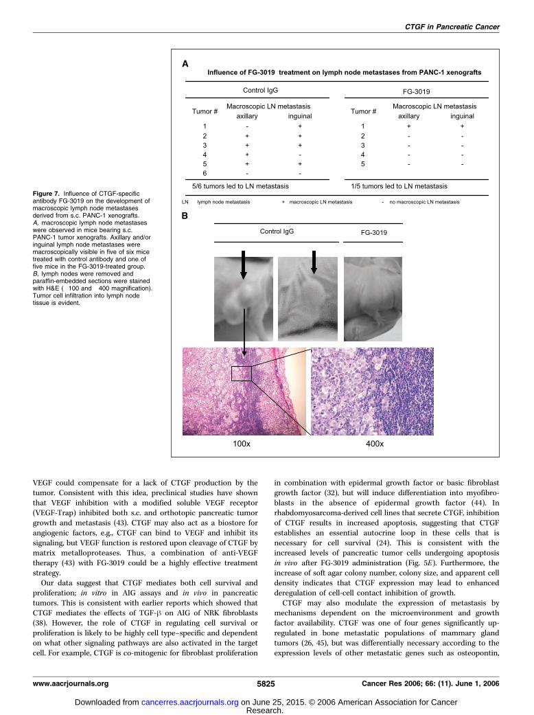
cells. It is noteworthy that in mice implanted with s.c. PANC-1tumors, five of six mice in the control group developed macro-scopically visible inguinal and/or axillary lymph node metastases,whereas only one of five mice in the antibody treatment groupdeveloped macroscopically visible metastases (Fig. 7A). Histologicexamination of excised lymph nodes showed that the macroscopicenlargement in control groups was due to tumor cell infiltration(Fig. 7B). These data suggest that inhibition of CTGF is growth-inhibitory for two wild-type pancreatic tumor types with endog-enous CTGF expression, and that CTGF inhibition is alsoantimetastatic.
Cancer Research
Cancer Res 2006; 66: (11). June 1, 2006 5822 www.aacrjournals.org
Research. on June 25, 2015. © 2006 American Association for Cancercancerres.aacrjournals.org Downloaded from
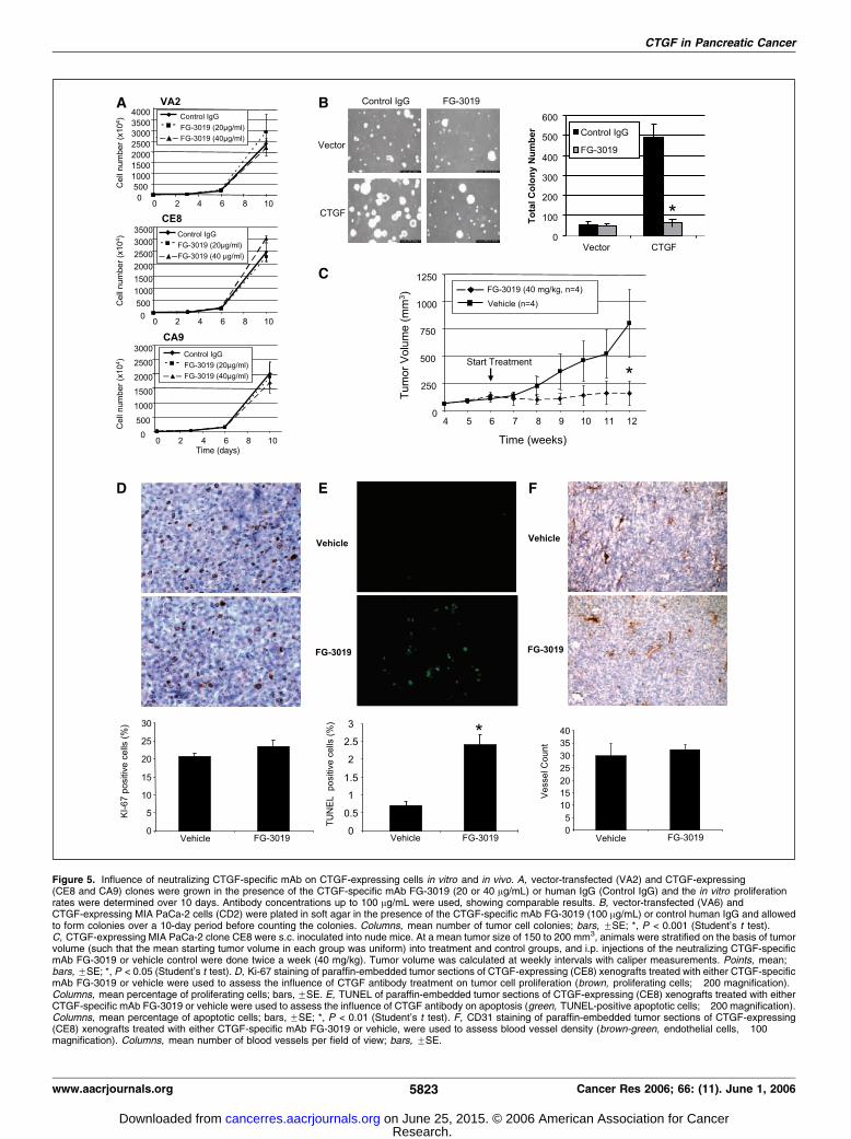
Figure 5. Influence of neutralizing CTGF-specific mAb on CTGF-expressing cells in vitro and in vivo. A, vector-transfected (VA2) and CTGF-expressing(CE8 and CA9) clones were grown in the presence of the CTGF-specific mAb FG-3019 (20 or 40 Ag/mL) or human IgG (Control IgG) and the in vitro proliferationrates were determined over 10 days. Antibody concentrations up to 100 Ag/mL were used, showing comparable results. B, vector-transfected (VA6) andCTGF-expressing MIA PaCa-2 cells (CD2) were plated in soft agar in the presence of the CTGF-specific mAb FG-3019 (100 Ag/mL) or control human IgG and allowedto form colonies over a 10-day period before counting the colonies. Columns, mean number of tumor cell colonies; bars, FSE; *, P < 0.001 (Student’s t test).C, CTGF-expressing MIA PaCa-2 clone CE8 were s.c. inoculated into nude mice. At a mean tumor size of 150 to 200 mm3, animals were stratified on the basis of tumorvolume (such that the mean starting tumor volume in each group was uniform) into treatment and control groups, and i.p. injections of the neutralizing CTGF-specificmAb FG-3019 or vehicle control were done twice a week (40 mg/kg). Tumor volume was calculated at weekly intervals with caliper measurements. Points, mean;bars, FSE; *, P < 0.05 (Student’s t test). D, Ki-67 staining of paraffin-embedded tumor sections of CTGF-expressing (CE8) xenografts treated with either CTGF-specificmAb FG-3019 or vehicle were used to assess the influence of CTGF antibody treatment on tumor cell proliferation (brown, proliferating cells; �200 magnification).Columns, mean percentage of proliferating cells; bars, FSE. E, TUNEL of paraffin-embedded tumor sections of CTGF-expressing (CE8) xenografts treated with eitherCTGF-specific mAb FG-3019 or vehicle were used to assess the influence of CTGF antibody on apoptosis (green, TUNEL-positive apoptotic cells; �200 magnification).Columns, mean percentage of apoptotic cells; bars, FSE; *, P < 0.01 (Student’s t test). F, CD31 staining of paraffin-embedded tumor sections of CTGF-expressing(CE8) xenografts treated with either CTGF-specific mAb FG-3019 or vehicle, were used to assess blood vessel density (brown-green, endothelial cells, �100magnification). Columns, mean number of blood vessels per field of view; bars, FSE.
CTGF in Pancreatic Cancer
www.aacrjournals.org 5823 Cancer Res 2006; 66: (11). June 1, 2006
Research. on June 25, 2015. © 2006 American Association for Cancercancerres.aacrjournals.org Downloaded from

Discussion
The most extensive literature to date regarding CTGF defines itsrole in wound-healing and fibrotic disease. However, several recentstudies implicate CTGF in tumor development and progression(16–19, 25, 26) and tumor cell survival (24). Our results show thatCTGF promotes anchorage-independent pancreatic cancer cellgrowth, which translates in vivo to enhanced tumor growth.Furthermore, anti-CTGF treatment with FG-3019 inhibited AIGin vitro , primary tumor growth in vivo , and the development ofmacroscopic lymph node metastases. Thus, CTGF may represent anovel target in pancreatic cancer and blocking its activity may alsoinhibit the growth of distant metastasis. This is especially relevantbecause patients are frequently diagnosed with advanced meta-static disease and there are few effective therapies for pancreaticcancer metastasis.
These results with CTGF in the context of pancreatic adeno-carcinomas are particularly noteworthy because a hallmark of theirhistopathology is desmoplasia, and overexpression of CTGF isassociated with increased tissue fibrosis. To date, only one smallclinical study has examined the relationship between CTGF,desmoplasia, and prognosis of pancreatic cancer (29). In this
study, CTGF mRNA was overexpressed mostly in connective tissuecells and high CTGF levels in tumor samples were associated withincreased tumor differentiation and patient survival. The authorshypothesized that elevated CTGF resulted in a better prognosis byinducing fibrosis and inhibiting metastasis. However, the factremains that >90% of pancreatic tumors have prominentdesmoplasia (30), and the aggressive nature of these tumorssuggests that this reaction may facilitate invasion rather thanprevent it (31, 42). In situ hybridization studies by Iacobuzio-Donahue et al. have shown CTGF mRNA expression within thetumor cells in one pancreatic cancer tumor specimen (31).Although our immunohistochemical and transplanted tumor datasuggest that tumor cell–derived CTGF is important for tumorgrowth and metastasis, other reports suggest that CTGF producedby stromal fibroblasts mediate tumor growth (15, 29). Ourxenograft studies indicate that CTGF overexpression couldpromote tumor growth and metastasis, but does not exclude thepossibility that stromal CTGF may have a significant role in tumorprogression. Although the production of CTGF by the stroma couldexplain why pancreatic tumors which possess low levels of CTGFstill grow and metastasize, our data suggest that elevated levels of
Figure 6. Influence of CTGF-specific antibody FG-3019 on the growth of xenografts derived from the pancreatic cancer cell lines, PANC-1 and Su86.86. A, thepancreatic cancer cell line PANC-1 expresses CTGF endogenously. B, the CTGF-specific mAb FG-3019 does not influence in vitro monolayer growth of PANC-1 cells(concentrations up to 100 Ag/mL). C, nude mice were s.c. inoculated with 107 PANC-1 cells, a pancreatic cancer cell line with endogenous CTGF expression. At amean tumor size of 150 to 200 mm3, animals were stratified on the basis of tumor volume (such that the mean starting tumor volume in each group was uniform)into treatment and control groups and i.p. injections of the neutralizing, CTGF-specific mAb FG-3019, or control IgG were done twice a week (40 mg/kg). Tumor volumewas calculated at weekly intervals with caliper measurements. Points, mean; bars, FSE; *, P < 0.01 (Student’s t test). D, nude mice were s.c. implanted with107 Su86.86 cells, a second pancreatic cancer cell line with endogenous CTGF expression. At a mean tumor size of 150 to 200 mm3, animals were randomizedinto treatment and control groups and i.p. injections of FG-3019 (40 mg/kg) or vehicle control were done twice per week. Tumor volume was calculated at weeklyintervals with caliper measurements. Points, mean; bars, FSE; *, P < 0.05 (Student’s t test). E, nude mice bearing Su86.86 tumors were given 100 mg/kg ofgemcitabine on days 1, 5, and 9 of the experiment. The growth delay induced by gemcitabine was of comparable magnitude to that obtained with 40 mg/kg of FG-3019.Tumor volume was calculated twice weekly with caliper measurements. Points, mean; bars, FSE; *, P < 0.05 (Student’s t test).
Cancer Research
Cancer Res 2006; 66: (11). June 1, 2006 5824 www.aacrjournals.org
Research. on June 25, 2015. © 2006 American Association for Cancercancerres.aacrjournals.org Downloaded from
VEGF could compensate for a lack of CTGF production by thetumor. Consistent with this idea, preclinical studies have shownthat VEGF inhibition with a modified soluble VEGF receptor(VEGF-Trap) inhibited both s.c. and orthotopic pancreatic tumorgrowth and metastasis (43). CTGF may also act as a biostore forangiogenic factors, e.g., CTGF can bind to VEGF and inhibit itssignaling, but VEGF function is restored upon cleavage of CTGF bymatrix metalloproteases. Thus, a combination of anti-VEGFtherapy (43) with FG-3019 could be a highly effective treatmentstrategy.
Our data suggest that CTGF mediates both cell survival andproliferation; in vitro in AIG assays and in vivo in pancreatictumors. This is consistent with earlier reports which showed thatCTGF mediates the effects of TGF-h on AIG of NRK fibroblasts(38). However, the role of CTGF in regulating cell survival orproliferation is likely to be highly cell type–specific and dependenton what other signaling pathways are also activated in the targetcell. For example, CTGF is co-mitogenic for fibroblast proliferation
in combination with epidermal growth factor or basic fibroblastgrowth factor (32), but will induce differentiation into myofibro-blasts in the absence of epidermal growth factor (44). Inrhabdomyosarcoma-derived cell lines that secrete CTGF, inhibitionof CTGF results in increased apoptosis, suggesting that CTGFestablishes an essential autocrine loop in these cells that isnecessary for cell survival (24). This is consistent with theincreased levels of pancreatic tumor cells undergoing apoptosisin vivo after FG-3019 administration (Fig. 5E). Furthermore, theincrease of soft agar colony number, colony size, and apparent celldensity indicates that CTGF expression may lead to enhancedderegulation of cell-cell contact inhibition of growth.
CTGF may also modulate the expression of metastasis bymechanisms dependent on the microenvironment and growthfactor availability. CTGF was one of four genes significantly up-regulated in bone metastatic populations of mammary glandtumors (26, 45), but was differentially necessary according to theexpression levels of other metastatic genes such as osteopontin,
Figure 7. Influence of CTGF-specificantibody FG-3019 on the development ofmacroscopic lymph node metastasesderived from s.c. PANC-1 xenografts.A, macroscopic lymph node metastaseswere observed in mice bearing s.c.PANC-1 tumor xenografts. Axillary and/oringuinal lymph node metastases weremacroscopically visible in five of six micetreated with control antibody and one offive mice in the FG-3019-treated group.B, lymph nodes were removed andparaffin-embedded sections were stainedwith H&E (�100 and �400 magnification).Tumor cell infiltration into lymph nodetissue is evident.
CTGF in Pancreatic Cancer
www.aacrjournals.org 5825 Cancer Res 2006; 66: (11). June 1, 2006
Research. on June 25, 2015. © 2006 American Association for Cancercancerres.aacrjournals.org Downloaded from

interleukin-11, and CXCR4 that increase bone metastases. CTGFhas also been shown to be elevated in breast cancer patients withpositive lymph nodes compared to patients with negative nodes(16, 46).
Several studies have shown a role for the TGF-h pathway inpancreatic cancer growth and metastasis (47). Domain 2 of CTGFhas also been shown to bind and potentiate TGF-h effects,suggesting that one possible mechanism for CTGF is throughamplifying the protumorigenic actions of TGF-h (48). However, thismay not entirely explain how CTGF promotes tumor growth andmetastasis in pancreatic tumor cells like MIA PaCa-2 that aredefective in TGF-h signaling. Therefore, it is likely that otherdomains of CTGF may affect pancreatic tumor growth. The domainstructure of CTGF has been associated with various interactionswith other growth factors and cell receptors. Domain 1 containshomology to insulin-like growth factor (IGF)–binding proteins andcould bind to IGF-I and IGF-II; domain 3 possesses thrombospon-din type 1–like repeats and could bind VEGF, whereas domain 4could bind to heparan sulfate proteoglycans and integrins (9, 23, 37,49, 50). Thus, there are a number of possible mechanisms wherebyCTGF interactions with other growth factors and cell surfacereceptors might affect pancreatic tumorigenesis.
Overall, this study supports the clinical investigation of anti-CTGF therapy. However, it will first be important to test the effectsof combining FG-3019 with standard treatments like gemcitabineand radiotherapy to determine the potential additive or synergisticeffects on primary tumor growth inhibition. We have alreadyshown that FG-3019 treatment alone produces an inhibition ofSu86.86 tumor growth that is similar to gemcitabine (Fig. 6E), andrecent clinical studies have indicated that combining targetedagents with chemotherapy or radiation therapy are resulting inimproved outcomes for patients. Thus, targeting CTGF with FG-3019 in combination with a cytotoxic agent such as gemcitabine or
radiation could be more efficacious than either treatment alone. Asmall animal study addressing this question is currently ongoing. Inaddition, one could consider anti-CTGF treatment either as anadjuvant treatment after initial surgical or radiation therapy, or as asecond-line treatment for gemcitabine-refractory patients, toprevent or inhibit metastasis of disseminated tumor cells byinhibiting AIG. In addition to therapy, another implication of thisstudy would be the use of CTGF as a biomarker. Becausecirculating CTGF levels in mouse plasma and urine paralleledCTGF expression levels in the tumor cell lines, CTGF might beuseful as a marker of disease. Consistent with this idea, CTGFexpression has been found to be prognostic for tumor progressionand survival in patients with glioma (18).
In summary, CTGF produced by pancreatic cancer cells seems toplay an important role in pancreatic tumor growth and metastasis.Our data clearly shows that CTGF promotes AIG in vitro as well astumor xenograft growth in vivo . Furthermore, treatment with theCTGF-specific antibody, FG-3019, inhibited the growth of tumorxenografts and metastases, without exhibiting noticeable sideeffects. These data provide a sound scientific rationale for furtherinvestigation into targeting CTGF in pancreatic cancer.
Acknowledgments
Received 1/10/2006; revised 3/2/2006; accepted 4/4/2006.Grant support: Else Kroner-Fresenius-Foundation (N. Dornhofer) and from the
National Cancer Institute (A. Giaccia). K. Bennewith is supported by a fellowship fromthe Canadian Institutes of Health Research.
The costs of publication of this article were defrayed in part by the payment of pagecharges. This article must therefore be hereby marked advertisement in accordancewith 18 U.S.C. Section 1734 solely to indicate this fact.
The authors thank Pauline Chu for her excellent technical assistance and Dr.Denise Chan for her help in preparing the manuscript; Dr. Lester Lau for kindlyproviding the antibody against CCN2/Fisp 12 for immunohistochemical analysis; andDrs. Barbara Bedogni, Janine Erler, Rachel Freiberg, Ester Hammond, Adam Krieg,Marianne Powell, Scott Welford, and Dawn Zinyk for their support and advice.
References
1. Li D, Xie K, Wolff R, Abbruzzese JL. Pancreatic cancer.Lancet 2004;363:1049–57.2. Bork P. The modular architecture of a new family ofgrowth regulators related to connective tissue growthfactor. FEBS Lett 1993;327:125–30.3. Chen CC, Chen N, Lau LF. The angiogenic factorsCyr61 and connective tissue growth factor induceadhesive signaling in primary human skin fibroblasts.J Biol Chem 2001;276:10443–52.4. Igarashi A, Okochi H, Bradham DM, Grotendorst GR.Regulation of connective tissue growth factor geneexpression in human skin fibroblasts and during woundrepair. Mol Biol Cell 1993;4:637–45.5. Grotendorst GR, Okochi H, Hayashi N. A noveltransforming growth factor h response element controlsthe expression of the connective tissue growth factorgene. Cell Growth Differ 1996;7:469–80.6. Shimo T, Kubota S, Kondo S, et al. Connective tissuegrowth factor as a major angiogenic agent that isinduced by hypoxia in a human breast cancer cell line.Cancer Lett 2001;174:57–64.7. Suzuma K, Naruse K, Suzuma I, et al. Vascularendothelial growth factor induces expression of con-nective tissue growth factor via KDR, Flt1, andphosphatidylinositol 3-kinase-akt-dependent pathwaysin retinal vascular cells. J Biol Chem 2000;275:40725–31.8. Takigawa M. CTGF/Hcs24 as a multifunctional growthfactor for fibroblasts, chondrocytes and vascular endo-thelial cells. Drug News Perspect 2003;16:11–21.
9. Gao R, Brigstock DR. Connective tissue growth factor(CCN2) induces adhesion of rat activated hepaticstellate cells by binding of its C-terminal domain tointegrin a(v)h(3) and heparan sulfate proteoglycan.J Biol Chem 2004;279:8848–55.10. Jedsadayanmata A, Chen CC, Kireeva ML, Lau LF,Lam SC. Activation-dependent adhesion of humanplatelets to Cyr61 and Fisp12/mouse connective tissuegrowth factor is mediated through integrin a(IIb)h(3).J Biol Chem 1999;274:24321–7.11. Schober JM, Chen N, Grzeszkiewicz TM, et al.Identification of integrin a(M)h(2) as an adhesionreceptor on peripheral blood monocytes for Cyr61(CCN1) and connective tissue growth factor (CCN2):immediate-early gene products expressed in atheroscle-rotic lesions. Blood 2002;99:4457–65.12. Gao R, Brigstock DR. Low density lipoproteinreceptor-related protein (LRP) is a heparin-dependentadhesion receptor for connective tissue growth factor(CTGF) in rat activated hepatic stellate cells. HepatolRes 2003;27:214–20.13. Segarini PR, Nesbitt JE, Li D, Hays LG, Yates JR III,Carmichael DF. The low density lipoprotein receptor-related protein/a2-macroglobulin receptor is a receptorfor connective tissue growth factor. J Biol Chem 2001;276:40659–67.14. Mercurio S, Latinkic B, Itasaki N, Krumlauf R, SmithJC. Connective-tissue growth factor modulates WNTsignalling and interacts with the WNT receptorcomplex. Development 2004;131:2137–47.15. Wenger C, Ellenrieder V, Alber B, et al. Expression
and differential regulation of connective tissue growthfactor in pancreatic cancer cells. Oncogene 1999;18:1073–80.16. Xie D, Nakachi K, Wang H, Elashoff R, Koeffler HP.Elevated levels of connective tissue growth factor,WISP-1, and CYR61 in primary breast cancers asso-ciated with more advanced features. Cancer Res 2001;61:8917–23.17. Pan LH, Beppu T, Kurose A, et al. Neoplastic cells andproliferating endothelial cells express connective tissuegrowth factor (CTGF) in glioblastoma. Neurol Res 2002;24:677–83.18. Xie D, Yin D, Wang HJ, et al. Levels of expression ofCYR61 and CTGF are prognostic for tumor progressionand survival of individuals with gliomas. Clin CancerRes 2004;10:2072–81.19. Koliopanos A, Friess H, di Mola FF, et al. Connectivetissue growth factor gene expression alters tumorprogression in esophageal cancer. World J Surg 2002;26:420–7.20. Kubo M, Kikuchi K, Nashiro K, et al. Expression offibrogenic cytokines in desmoplastic malignant mela-noma. Br J Dermatol 1998;139:192–7.21. Shakunaga T, Ozaki T, Ohara N, et al. Expression ofconnective tissue growth factor in cartilaginous tumors.Cancer 2000;89:1466–73.22. Moritani NH, Kubota S, Nishida T, et al. Suppres-sive effect of overexpressed connective tissue growthfactor on tumor cell growth in a human oralsquamous cell carcinoma-derived cell line. CancerLett 2003;192:205–14.
Cancer Research
Cancer Res 2006; 66: (11). June 1, 2006 5826 www.aacrjournals.org
Research. on June 25, 2015. © 2006 American Association for Cancercancerres.aacrjournals.org Downloaded from

23. Vorwerk P, Wex H, Hohmann B, Mohnike K, SchmidtU, Mittler U. Expression of components of the IGFsignalling system in childhood acute lymphoblasticleukaemia. Mol Pathol 2002;55:40–5.24. Croci S, Landuzzi L, Astolfi A, et al. Inhibition ofconnective tissue growth factor (CTGF/CCN2) expres-sion decreases the survival and myogenic differentiationof human rhabdomyosarcoma cells. Cancer Res 2004;64:1730–6.25. Zeng ZJ, Yang LY, Ding X, Wang W. Expressions ofcysteine-rich61, connective tissue growth factor and Novgenes in hepatocellular carcinoma and their clinicalsignificance. World J Gastroenterol 2004;10:3414–8.26. Kang Y, Siegel PM, Shu W, et al. A multigenicprogram mediating breast cancer metastasis to bone.Cancer Cell 2003;3:537–49.27. Lin BR, Chang CC, Che TF, et al. Connective tissuegrowth factor inhibits metastasis and acts as anindependent prognostic marker in colorectal cancer.Gastroenterology 2005;128:9–23.28. Chang CC, Shih JY, Jeng YM, et al. Connective tissuegrowth factor and its role in lung adenocarcinomainvasion and metastasis. J Natl Cancer Inst 2004;96:364–75.29. Hartel M, Di Mola FF, Gardini A, et al. Desmoplasticreaction influences pancreatic cancer growth behavior.World J Surg 2004;28:818–25.30. Ryu B, Jones J, Hollingsworth MA, Hruban RH, KernSE. Invasion-specific genes in malignancy: serial analysisof gene expression comparisons of primary andpassaged cancers. Cancer Res 2001;61:1833–8.31. Iacobuzio-Donahue CA, Ryu B, Hruban RH, Kern SE.Exploring the host desmoplastic response to pancreaticcarcinoma: gene expression of stromal and neoplasticcells at the site of primary invasion. Am J Pathol 2002;160:91–9.32. Kireeva ML, Latinkic BV, Kolesnikova TV, et al. Cyr61and Fisp12 are both ECM-associated signaling mole-
cules: activities, metabolism, and localization duringdevelopment. Exp Cell Res 1997;233:63–77.33. Gilbert RE, Akdeniz A, Weitz S, et al. Urinaryconnective tissue growth factor excretion in patientswith type 1 diabetes and nephropathy. Diabetes Care2003;26:2632–6.34. Simeone DM, Pham T, Logsdon CD. Disruption ofTGFh signaling pathways in human pancreatic cancercells. Ann Surg 2000;232:73–80.35. Ferrara N. Vascular endothelial growth factor:basic science and clinical progress. Endocr Rev2004;25:581–611.36. Luo J, Guo P, Matsuda K, et al. Pancreatic cancer cell-derived vascular endothelial growth factor is biologicallyactive in vitro and enhances tumorigenicity in vivo . Int JCancer 2001;92:361–9.37. Inoki I, Shiomi T, Hashimoto G, et al. Connectivetissue growth factor binds vascular endothelial growthfactor (VEGF) and inhibits VEGF-induced angiogenesis.FASEB J 2002;16:219–21.38. Kothapalli D, Frazier KS, Welply A, Segarini PR,Grotendorst GR. Transforming growth factor h inducesanchorage-independent growth of NRK fibroblasts via aconnective tissue growth factor-dependent signalingpathway. Cell Growth Differ 1997;8:61–8.39. Yunis AA, Arimura GK, Russin DJ. Humanpancreatic carcinoma (MIA PaCa-2) in continuousculture: sensitivity to asparaginase. Int J Cancer1977;19:218–35.40. Douma S, Van Laar T, Zevenhoven J, Meuwissen R,Van Garderen E, Peeper DS. Suppression of anoikis andinduction of metastasis by the neurotrophic receptorTrkB. Nature 2004;430:1034–9.41. Roestenberg P, van Nieuwenhoven FA, Wieten L, et al.Connective tissue growth factor is increased in plasmaof type 1 diabetic patients with nephropathy. DiabetesCare 2004;27:1164–70.42. Iacobuzio-Donahue CA, Maitra A, Olsen M, et al.
Exploration of global gene expression patterns inpancreatic adenocarcinoma using cDNA microarrays.Am J Pathol 2003;162:1151–62.43. Fukasawa M, Korc M. Vascular endothelial growthfactor-trap suppresses tumorigenicity of multiplepancreatic cancer cell lines. Clin Cancer Res 2004;10:3327–32.44. Grotendorst GR, Duncan MR. Individual domains ofconnective tissue growth factor regulate fibroblastproliferation and myofibroblast differentiation. FASEBJ 2005;19:729–38.45. Van’t Veer LJ, Weigelt B. Road map to metastasis. NatMed 2003;9:999–1000.46. Jiang WG, Watkins G, Fodstad O, Douglas-Jones A,Mokbel K, Mansel RE. Differential expression of theCCN family members Cyr61, CTGF and Nov inhuman breast cancer. Endocr Relat Cancer 2004;11:781–91.47. Rowland-Goldsmith MA, Maruyama H, Matsuda K,et al. Soluble type II transforming growth factor-hreceptor attenuates expression of metastasis-associatedgenes and suppresses pancreatic cancer cell metastasis.Mol Cancer Ther 2002;1:161–7.48. Abreu JG, Ketpura NI, Reversade B, De RobertisEM. Connective-tissue growth factor (CTGF) modu-lates cell signalling by BMP and TGF-h. Nat Cell Biol2002;4:599–604.49. Kim HS, Nagalla SR, Oh Y, Wilson E, Roberts CT, Jr.,Rosenfeld RG. Identification of a family of low-affinityinsulin-like growth factor binding proteins (IGFBPs):characterization of connective tissue growth factor as amember of the IGFBP superfamily. Proc Natl Acad SciU S A 1997;94:12981–6.50. Hashimoto G, Inoki I, Fujii Y, Aoki T, Ikeda E, OkadaY. Matrix metalloproteinases cleave connective tissuegrowth factor and reactivate angiogenic activity ofvascular endothelial growth factor 165. J Biol Chem2002;277:36288–95.
CTGF in Pancreatic Cancer
www.aacrjournals.org 5827 Cancer Res 2006; 66: (11). June 1, 2006
Research. on June 25, 2015. © 2006 American Association for Cancercancerres.aacrjournals.org Downloaded from
CTGF in Pancreatic Cancer
In the article on CTGF in pancreatic cancer in the June 1, 2006issue of Cancer Research (1), the correct spelling of the tenthauthor’s name is Randall Nacamuli.
1. Dornhofer N, Spong S, Bennewith K, Salim A, Klaus S, Kambham N, Wong C, KaperF, Sutphin P, Nacamuli R, Hockel M, Le Q, Longaker M, Yang G, Koong A, Giaccia A.Connective tissue growth factor-specific monoclonal antibody therapy inhibitspancreatic tumor growth and metastasis. Cancer Res 2006;66:5816–27.
I2006 American Association for Cancer Research.doi:10.1158/0008-5472.CAN-66-15-COR2
Cancer Res 2006; 66: (15). August 1, 2006 7832 www.aacrjournals.org
Correction
2006;66:5816-5827. Cancer Res Nadja Dornhöfer, Suzanne Spong, Kevin Bennewith, et al. MetastasisAntibody Therapy Inhibits Pancreatic Tumor Growth and
Specific Monoclonal−Connective Tissue Growth Factor
Updated version
http://cancerres.aacrjournals.org/content/66/11/5816
Access the most recent version of this article at:
Cited articles
http://cancerres.aacrjournals.org/content/66/11/5816.full.html#ref-list-1
This article cites 50 articles, 24 of which you can access for free at:
Citing articles
http://cancerres.aacrjournals.org/content/66/11/5816.full.html#related-urls
This article has been cited by 13 HighWire-hosted articles. Access the articles at:
E-mail alerts related to this article or journal.Sign up to receive free email-alerts
Subscriptions
Reprints and
To order reprints of this article or to subscribe to the journal, contact the AACR Publications
Permissions
To request permission to re-use all or part of this article, contact the AACR Publications
Research. on June 25, 2015. © 2006 American Association for Cancercancerres.aacrjournals.org Downloaded from