cytochemical investigation of the digestive gland of two
TRANSCRIPT

Cytochemical Investigation of the Digestive Gland of TwoStrombidae Species (Strombus gigas and Strombus pugilis)in Relation to the NutritionJEAN-MARIE VOLLAND* AND OLIVIER GROSDepartement de Biologie, UMR 7138 SAE, Equipe Biologie de la mangrove, Universite des Antilles et de la Guyane, U.F.R des SciencesExactes et Naturelles, B.P. 592. 97159 Pointe-a-Pitre Cedex, Guadeloupe, France
KEY WORDS molluscs; cytochemistry; physiology; lysosomal digestion; EFTEM
ABSTRACT Strombus gigas and Strombus pugilis are threatened species and aquaculture rep-resents a good alternative solution to the fishing. In this study, we highlighted the intracellulardigestion process in the digestive gland of two Strombidae species, S. gigas and Strombus pugilis,by the cytochemical characterization of two lysosomal enzymes: acid phosphatase and arylsulfa-tase. In order to check the efficiency of artificial food digestion, we conducted the characterizationon freshly collected, starved and artificially fed individuals of S. pugilis. TEM observations of di-gestive gland sections from freshly collected individuals of both species revealed the presence ofacid phosphatase and arylsulfatase activity mostly located in the apical third of digestive cells.Both enzymes were also detected in artificially fed individuals. In response to the starvation, acidphosphatase is not produced anymore by digestive cells, while arylsulfatase is still present. To ourknowledge, this is the first cytochemical validation of intracellular digestion of artificial food inStrombidae. This study highlights the intracellular digestion of artificial food developed for Strom-bidae aquaculture. Moreover, we have shown that the lysosomal activity could be used as a feedindex. Microsc. Res. Tech. 00:000–000, 2012. VVC 2012 Wiley Periodicals, Inc.
INTRODUCTION
Strombidae are marine benthic Gastropods whichrepresent an important economic resource in the Ca-ribbean. At least four species represent a staple foodincluding Strombus gigas LINNAEUS, 1758 andStrombus pugilis LINNAEUS, 1758. In 2001, for exam-ple, 3,132 tones of S. gigas have been fished, which rep-resent more than 30 million USD (Aldana Aranda,2003). Due to an important fishing pressure, some spe-cies as S. gigas are threatened and some populationshave already completely disappeared like in the Yuca-tan (Mexico) for example. Natural stocks decrease rap-idly and international authorities have taken protec-tion measures (Adams, 1970; Aldana Aranda, 2003;Appeldoorn, 1987): S. gigas is included in the annex IIof the Convention on International Trade in Endan-gered Species (CITES) and also in the red list of theInternational Union for Conservation of Nature(IUCN). For these reasons, aquaculture represents agood alternative solution to the high pressure onStrombidae (Brito Manzano et al., 1998). While studiesreport the development in laboratory of fertilized eggsfrom the sea (Brito Manzano et al., 1999; Brito Man-zano and Aldana Aranda, 2004), to our knowledge nostudies report a complete reproduction cycle of Strom-bidae in the laboratory. Veliger larvae are obtained andgrown up to the metamorphosis by feeding with unicel-lular algae. Then juveniles are bred with artificial foodprovided by small pellets. Few studies have investi-gated the efficiency of unicellular algae and pellets forStrombidae breeding (Aldana Aranda and Suarez,1998; Aldana Aranda et al., 1997, 2007). In these publi-
cations, the authors have studied the digestive glandstructure by histological observations and the quantifi-cation of the growing rate of individuals. However, toour knowledge, no study reports an ultrastucturalinvestigation of the digestive gland of Strombidaeexcept our previous works (Gros et al., 2009; Vollandet al., 2010, 2012). Strombidae are among the few Gas-tropods which present a crystalline style. Such struc-ture is more common in Bivalvia and is often associ-ated to a continuous microphagous nutrition (Fretterand Graham, 1962). The few studies which focused onthe digestion physiology of Strombidae reported the na-ture of enzymes of the crystalline style (Alyakrinskaya,2001; Horiuchi and Lane, 1965, 1966). To our knowl-edge, no cytochemical investigation of the digestivegland of Strombidae has been conducted to date. Suchorgan has a key role in the digestion process and repre-sents with the stomach the most complex part of thedigestive tract (Owen, 1966). In this study, we high-lighted the intracellular digestion process in the diges-tive gland of two Strombidae species, S. gigas andStrombus pugilis, by the cytochemical characterization
*Correspondence to: Jean-Marie Volland, Departement de Biologie, UMR 7138SAE. Equipe Biologie de la mangrove, Universite des Antilles et de la Guyane,U.F.R des Sciences Exactes et Naturelles, B.P. 592. 97159 Pointe-a-Pitre Cedex,Guadeloupe, France. E-mail: [email protected]
Received 3 March 2012; accepted in revised form 26 April 2012; accepted inrevised form 26 April 2012
Contract grant sponsor: ECOS-Nord (Caracterisation reproductive, molecu-laire, ecologique Apicomplexa-Strombidae, implication pour la peche et l’aqua-culture dans la region Caraıbe et le Golfe du Mexique); Contract grant number:M09-A02
DOI 10.1002/jemt.22074
Published online inWiley Online Library (wileyonlinelibrary.com).
VVC 2012 WILEY PERIODICALS, INC.
MICROSCOPY RESEARCH AND TECHNIQUE 00:000–000 (2012)
of two lysosomal enzymes: acid phosphatase and aryl-sulfatase. In order to check the efficiency of artificialfood digestion, we conducted the characterization onfreshly collected, starved and artificially fed individu-als of Strombus pugilis.
MATERIAL AND METHODSExperimental Conditions
Individuals of S. gigas were collected during theauthorized fishing period by professional fishermen onor near Thalassia testudinum sea grass beds in Le Gos-ier, Guadeloupe (French West Indies). Strombus pugi-lis samples were collected by hands on sand area inSaint-Francois, Guadeloupe. Living materials wererapidly brought to the laboratory. Three S. gigas andthree Strombus pugilis (batch Bcontrol) individuals weredirectly processed for cytochemical detection of lysoso-mal enzymes. Six other Strombus pugilis were placedin two 400 L raceway filled with sand-filtered sea water(pH 5 8; temperature 5 268C; psu 5 36; photoperiod 512 h) which was continuously oxygenated using an airpump. Sea water was renewed twice a week and race-way was cleaned to avoid development of microalgaeand biofilm which could be used as food source. Onebatch named Bfed was fed ad libitum during 4 monthswith artificial food developed and provided by the CIN-VESTAV-IPN of Merida (Nutrition and Aquaculture ofMolluscs Laboratories). It has been controlled visuallythat food pellets were ingested by animals. The otherbatch named Bstarved was kept 5 months in starvation.After their respective experimental maintenance, indi-viduals from Bfed and Bstarved were sacrificed and proc-essed for cytochemistry.
Characterization of Lysosomal Activity
Acid Phosphatase. Small digestive gland pieceswere dissected and fixed for 2 h in 2% glutaraldehydeand 1% paraformaldehyde in cacodylate buffer (0.1 M;1,100 mOsm; pH 5 7.2). Pieces were then washed inthe same buffer and thick sections (50–100 lm) werecut in refrigerated cacodylate buffer using an OCTSlicer1. The Gomori (1952) acid phosphatase detectionmethod modified by Pasteels (1971) was used. Tissuesslices were incubated in a 3.3 mg mL21 sodium ß-gly-crophosphate solution saturated with lead nitrate.Incubation was realized at 378C during 25 min. Sliceswere then rinsed in acetate buffer (sodium acetate 50mM, acetic acid 15 mM; pH 5 5) and in 3.5% aceticacid before dehydratation through an ascending etha-nol series and embedded in a resin mixture composedof Epon (63.2%) and Araldite (36.8%). Controls werealso prepared by omitting sodium ß-glycrophosphate inthe incubation medium.
Arylsulfatase. We used a method adapted fromHopsu-Havu et al. (1965) described in Lewis andKnight (1992). Thick section (100–150 lm) of prefixeddigestive gland were obtained as described above andincubated 45 min at 378C in the reactive media (p-nitrocatechol sulfate 8 mg mL21, acetate buffer 60 mM,barium chloride 60 mM; pH 5 5.5). Slices were thenwashed in cacodylate buffer overnight before dehydra-tation and embedding in the same resin as describedabove. Control slices were incubated without p-nitroca-techol sulfate.
Light and Transmission Electron Microscopy
Semithin sections (0.5 lm thick) were cut from theresin-embedded samples and stained with 0.5% tolui-dine blue in 1% borax for light microscopy observation.Ultrathin sections (60 nm) were obtained from resinblocks and observed without supplementary contrastwith Energy Filtered Transmission Electron Micros-copy (EFTEM). Acquisitions in spectra mode (EELS,Electron Energy Loss Spectroscopy) were performedusing a LEO 912 Omega transmission electron micro-scope (LEO Electron Optics GmbH, Oberkochen, Ger-many) at 120 kV. Observation on image mode (ESI,Electron Spectroscopic Imaging) was accomplishedwith the ESIvision program (version 3.0 Soft-ImagingSoftware, SIS, GmbH, 48153 Munster). For the ele-mental cartography, the subtractive method of the‘‘three windows’’ was used (Jeanguillaume et al., 1978;Reimer et al., 1992).
Histochemical Detection of Lipids
Digestive gland samples of S. gigas and Strombuspugilis were fixed for 6 h in 6% paraformaldehyde insea water. Samples were then washed in sea water andtransferred in 30% sucrose in sea water. They werethen rapidly frozen in isopentane cooled at 2358C withliquid nitrogen. Cryosections of 10 lm realized with aCryo-cut1 (American Optical Corporation) werestained with black Soudan B as described by Gabe(1968).
RESULTSUltrastructure of the Digestive Cells
According to Gros et al. (2009), the digestive gland ofStrombidae is composed of an assemblage of digestivetubules and ducts. Digestive tubules are composed bythree cell types: short pyramidal crypt cells, vacuolatedcells, and the predominant long columnar digestivecells (Figs. 1C and 2A). This last cell type is the mostimportant in terms of number and volume in the diges-tive tubules. They form a unistratified epithelium andare regularly aligned (Fig. 1A). Digestive cells were onaverage 90 lm long in S. gigas and 80 lm long inStrombus pugilis. They present a basal nucleus andthree compartments can be distinguished in the cyto-plasm: the basal third, the median third, and the apicalthird (Figs. 1D, 2B, and 2C). The basal third is charac-terized by the presence of vesicles from 2 to 5 lm in di-ameter. Such vesicles appear empty on resin section,due to the sample preparation. Histochemical stainingwith black Soudan B of cryosections revealed thatthese structures are lipid droplets (data not shown).The median third of the cell is characterized by thepresence of one or few large granules (Figs. 1A, 1B, 1Dand 2B–2D). Such granules have a diameter of 4 tomore than 10 lm and their average diameter is 7 lmfor both species. Finally, the apical third of the cell iscomposed by two areas (Figs. 1D and 1E). The area 1,adjacent to the median third, present various vesicleswith a diameter of 0.5 to 5 lm. The area 2 presentssmall vesicles (less than 2 lm).The apical pole of the di-gestive cells, in contact with the tubule’s lumen, is bor-dered by short microvilli of 2 lm on average (Fig. 1D).
Microscopy Research and Technique
2 VOLLAND AND GROS
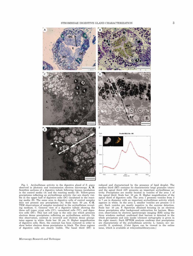
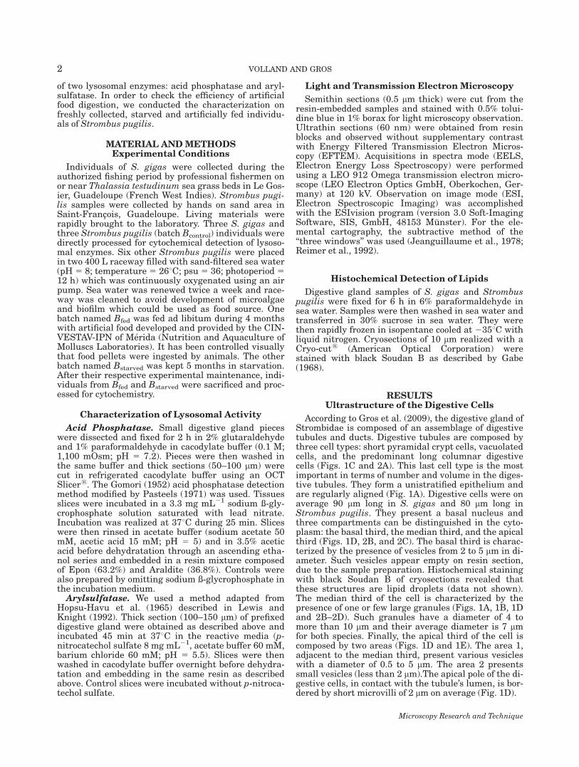
Fig. 1. Arylsulfatase activity in the digestive gland of S. gigasobserved in photonic and transmission electron microscopy. A, B.Semithin sections of a digestive tubule following tissues incubationin the control media (A) and the reacting media (B). Yellow-greenprecipitates reflecting an arylsulfatase activity (As) are clearly visi-ble in the upper half of digestive cells (DC) incubated in the react-ing media (B). The same area in digestive cells of control samplesdoes not present any precipitates (A). Scale bars: 50 lm. C–E.TEM observations of samples incubated in the arylsulfatase reveal-ing medium. C. General view of a digestive tubule showing thethree cell types: crypt cells (CC), vacuolated cells (VC), and diges-tive cells (DC). This last cell type is the only one which presentselectron dense precipitates reflecting an arylsulfatase activity. Onthis observation, contrast is inversed and electron dense precipi-tates appear in white. Scale bar: 20 lm. D. Higher magnificationof digestive cells. Here, the contrast has been restored in order toshow better electron dense precipitates in black. The three regionsof digestive cells are clearly visible. The basal third (BT) is
reduced and characterized by the presence of lipid droplet. Themedian third (MT) contains its characteristic large granules (stars)and the apical third (AT) presents an important arylsulfatase ac-tivity. Precipitates are mostly located in vesicles of the area 1 ofthe apical third. Scale bar: 10 lm. E. Higher magnification of theapical third of digestive cells. The area 1 presents vesicles from 3to 7 lm in diameter with an important arylsulfatase activity whichappears in white. In the area 2, smaller vesicles are present (1–3lm). Such vesicles are mostly negative to the enzyme detection.Scale bar: 10 lm. F. Spectrum obtained focusing on an electrondense precipitates after background subtraction for barium. More-over, observation by electron spectroscopic imaging (ESI) using thethree windows method, confirmed that barium is detected in thewhole precipitate and only inside (element cartography is given inthe right insert). Such EFTEM analysis confirms that precipitatesare characteristic of the arylsulfatase activity. L: lumen; mv: mi-crovilli; S: symbiont. [Color figure can be viewed in the onlineissue, which is available at wileyonlinelibrary.com.]
Microscopy Research and Technique
3STROMBIDAE DIGESTIVE GLAND CHARACTERIZATION
Cytochemical Detection of Arylsulfatase
Observations of semithin digestive gland sectionsfrom freshly collected individuals of both species fol-lowing cytochemical detection of arylsulfatase revealedthe presence of yellow-green precipitates located in thedigestive cells of the gland (Fig. 1B). Results presentedbelow were observed identically on the three replicatesfor both species. TEM observations of ultra-thin sec-tions confirmed that the arylsulfatase activity is onlylocated in the digestive cells. No electron dense precipi-tates were observed in vacuolated and crypt cells (Fig.1C). In digestive cells, arylsulfatase activity is mostlydetected in the area 1 of apical third of cells (Figs. 1Dand 1E). Most of small vesicles (0.5 to 2 lm) of the area2 of the apical third are not positive to the enzymedetection (Figs. 1D and 1E). The arylsulfatase activityis the most important in large vesicles (average 7 lm)
of the apical third in which the precipitate occupied thewhole vesicle (Figs. 1C–1E). While the large vesicles ofthe median third are not positive to the enzyme detec-tion, some large granules of this compartment presenta weak arylsulfatase activity. In such granules, the ac-tivity is limited to few restricted precipitates (Fig. 1D).The validity of the enzyme detection has been con-trolled twice, first, by observations of control slices(Fig. 1A) and secondly by EFTEM microanalysis of theprecipitates. Such analysis in spectrum mode con-firmed the presence of barium. In image mode, we alsochecked that barium was only located in the precipi-tates (Fig. 1F).
Cytochemical Detection of Acid Phosphatase
TEM observations of digestive gland sections fromfreshly collected individuals of both species revealed
Fig. 2. Acid phosphatase activity in the digestive gland ofS. pugilis observed from thin sections. A, B. Semithin sections of adigestive tubule following tissues incubation in the reacting media(A) and the control media (B) which does not contain the enzymesubstrate. On the (A), a partial view of a digestive tubule with thethree cell types is visible. In vacuolated cell (VC), which hosts thesymbionts (S), no precipitates are detected. In crypt cells (CC),spherocrystals (arrows) present electron dense precipitates reflectingan acid phosphatase activity. A group of digestive cells (DC) is alsovisible and present an important acid phosphatase activity mostlylocated in the apical third of cells (arrow heads). Scale bar: 20 lm. Incontrast, on the control (B), no precipitates are detected. Scale bar:20 lm. C. Observations focusing on digestive cells. Electron dense
precipitates (arrow heads) reflecting acid phosphatase activity aremostly detected in the apical third (AT) of cells. Precipitates are alsopresents in the large granules (stars) of the median third (MT). Thedetail of a granule is showed in the insert of (C). In the basal third(BT) lipid droplet are present but no enzyme activity is detectable.Small scattered precipitates (0.1–0.5 lm) are also present in thewhole cytoplasm of digestive cells. An acid phosphatase activity isalso detected in spherocrystals (arrows) of crypt cells. Precipitatesform a ring in the periphery of spherocrystals. Scale bar: 10 lm. D.Detail of the apical and median thirds of digestive cells showing theimportant acid phosphatase activity in the apical third. In somelarge granules an enzyme activity is present as small numerous pre-cipitates. Scale bar: 5 lm.
Microscopy Research and Technique
4 VOLLAND AND GROS
the presence of electron dense precipitates after cyto-chemical detection of acid phosphatase. Results pre-sented below were observed identically on the threereplicates for both species. The enzyme activity hasbeen detected mostly in the digestive cells but we alsofound precipitates in specific structures called spheroc-rystals located in the crypt cells (Figs. 2A and 2C).However, vacuolated cells did not show any acid phos-phatase activity. In the digestive cells, the enzyme wasmostly detected on the apical third of the cell, in smallvesicles of 0.5 to 2 lm in diameter (Fig. 2D). Some-times, acid phosphatase activity was also detectedin the large granules of the median third of the diges-tive cells (Fig. 2C). The specificity of electron dense pre-cipitates was confirmed by the observation of controls(Fig. 2B).
Starvation and Artificial Feeding
Preliminary starvation experiments of different pe-riod of time have been conducted and enzyme activityfor both acid phosphatase and arylsulfatase wasdetected by photonic microscopy even following severalweeks of experiments (data not shown). Consequently,we have chosen to use the oldest batches (i.e. 5 monthsstarved Strombus pugilis) for the cytochemical detec-tion of lysosomal activity. Concerning acid phospha-tase, TEM observations of ultrathin section revealedthe presence of precipitates in the digestive gland ofBcontrol as expected. On digestive gland of artificiallyfed individuals (Bfed) an acid phosphatase activity wasalso present. While no quantification were done, the ob-servation of precipitates revealed that they are lessnumerous and more scattered, which means thatenzyme activity is less important than in Bcontrol. Forboth Bcontrol and Bfed, the enzyme activity was locatedas described earlier. However, in Bstarved, we wereunable to detect any acid phosphatase activity in thedigestive gland of the three individuals (Table 1).
Concerning the arylsulfatase activity, even if itseemed stronger (precipitates more numerous) inthe digestive cells of Bcontrol individuals, we coulddetect the enzyme activity on the three individualsof the three batches (Table 1). The enzyme waslocated as described earlier for the freshly collectedindividuals.
DISCUSSION
The digestive gland of both Strombus gigas andS. pugilis is composed by an assemblage of digestivetubules and ducts. Tubules are formed by an epithe-lium containing three cell types as described by Groset al. (2009): crypt cells, vacuolated cells, and digestivecells. Digestive tubules of Caenogastropoda are knownto be composed by, at least, two cell types: digestive
cells and pyramidal crypt cells. This last cell typeregroups secreting cells, excreting cells, basophile cells,and calcium cells (Boghen and Farley, 1974; Devi et al.,1981; Fretter and Graham, 1962; Lutfy and Demain,1967; Merdsoy and Farley, 1973; Voltzow, 1994; Wig-ham, 1976). In the two Strombidae species, the mostnumerous cells are in the digestive tubules. They arealso known to be the largest cell type in the digestivetubules of Mollusca in general (Lobo-Da-Cunha, 2000;Lutfy and Demain, 1967; Owen, 1966). Many studieshave described their structure and ultrastructure inrelation with their function in Bivalvia (Henry, 1984;Morse et al., 1997; Pal, 1972), in Cephalopoda (Bou-caud-Camou and Roper, 1998; Semmens, 2002; Swiftet al., 2005; Pernice et al., 2009) and in Polyplacophora(Lobo-da-Cunha, 1997). Among Gastropods many stud-ies focused on the digestive cells in Opistobranchia(Coelho et al., 1998; Lobo-Da-Cunha, 2000; Taıeb,2001), in Pulmonata (Luchtel et al., 1997; Walker,1970, 1972), and in Prosobranchia (Boghen and Farley,1974; Fretter and Graham, 1962; Lutfy and Demain,1967; Merdsoy and Farley, 1973; Voltzow, 1994; Wig-ham, 1976). Such studies reported some general char-acteristics for the digestive cells of Mollusca because ofthe presence of microvilli and an important endocyticsystem at the apical pole. While the absorption anddigestion process seems to be proven, the secretingfunction remains hypothetical, except for someBivalves (Henry et al., 1991) and Cephalopods (Bou-caud-Camou and Roper, 1995) where it has been con-firmed. The long columnar shape and the presence ofnumerous vacuoles and large granules also belong tothe general characteristics of these cells. Such charac-teristics are also observed in the digestive cells ofStrombidae. However, the division of digestive cell intothree regions seems to be more specific to Strombidae.Even if few studies reported lipid droplets in digestivecells of molluscs’ digestive gland, they are not as impor-tant as in Strombidae in which a third of the cell isdedicated to such lipid storage (Dimitriadis andAndrews, 2000; Lobo-Da-Cunha, 2000). Otherwise, di-gestive cells have ever been suspected to have a storagefunction (Owen, 1966).
In the median and apical third of digestive cells, wehighlighted a lysosomal activity by the cytochemicaldetection of acid phosphatase and arylsulfatase. Thesetwo lysosomal enzymes are known to be markers of theintracellular digestion process (Holtzman, 1989). More-over, apical microvilli reflect an exchange processbetween the lumen and the digestive cell. Such obser-vations confirm that the intracellular digestion processoccurs in both apical and median third of digestive cellsof Strombidae. Arylsulfatase activity has been detectedin digestive cells of Aplysia deplians (Opistobranchia)(Lobo-Da-Cunha, 2000). This author reported anenzyme activity in the Golgi apparatus, in lysosomes,and a strong activity in heterolysosomes in the medianand apical regions of the digestive cell. In the bivalveMytilus galloprovincialis an arylsulfatase activity hasalso been detected, but limited to the Golgi apparatus(Dimitriadis et al., 2004). In the same species, anotherstudy reports an arylsulfatase activity in heterolyso-somes of the apical pole and in some lysosomal residualbodies (Robledo et al., 2006). Finally a fourth studyhighlighted the presence of arylsulfatase in ‘‘cyto-
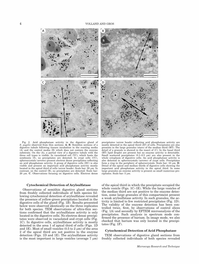
TABLE 1. Cytochemical detection of lysosomal enzymes activity inthe digestive gland of S. pugilis
Batchcontrol Batchfed Batchstarved
Acid phosphatase 11 1 2Arylsulfatase 11 1 1
Comparison of the batch of freshly collected individuals (Bcontrol), the batch fed 4months with artificial food (Bfed) and the batch starved 5 months (Bstarved). Thesemiquantitative characterization was done visually by observation of the num-ber and the size of precipitates detected on digestive glands section of the threereplicates individuals.
Microscopy Research and Technique
5STROMBIDAE DIGESTIVE GLAND CHARACTERIZATION
plasmic granules’’ of the digestive cells in the BivalveM. edulis and in the pulmonate Helix aspersa (Sumner,1969). In Strombidae, we observed an increasing size ofarylsulfatase positive vesicles from the apical pole tothe one-half of the cells. Such vesicles might representheterolysosomes at different digestion stages.
Acid phosphatase has also been detected by cyto-chemistry in the digestive cells of several molluscs. Inthe prosobranch Nucella lapillus and in the bivalveM. galloprovincialis, it has been detected in heterolyso-somes in the middle of the digestive cells, and, locallyin some residual bodies (Dimitriadis and Andrews,2000; Robledo et al., 2006). Sumner (1969) also reportedit detection in ‘‘cytoplasmic granules’’ of M. edulis di-gestive cells and in the whole cells in H. aspersa. In S.gigas and Strombus pugilis, acid phosphatase localiza-tion is more or less the same as arylsulfatase localiza-tion. Even of different sizes, the vesicules from the api-cal third of digestive cells present an activity for bothenzymes, confirming that such vesicles are lysosomesor heterolysosomes when their size exceeds 1 lm(Sumner, 1969). We also highlighted an acid phospha-tase activity in the large granules of the digestive cells.Such granules are named ‘‘blue granules’’ by Gros et al.(2009) because of their affinity for alcian blue, reflect-ing their proteoglycan content. They seem similar tothe large residual bodies described in the digestive cellsof other molluscs (Dimitriadis et al., 2004; Henry, 1984;Lobo-Da-Cunha, 2000; Taıeb, 2001). Lobo-Da-Cuhna(2000) highlighted that such granules result from thefusion of every heterolysosomes and lysosomal residualbodies of the cell. Lipofushine pigment has been high-lighted in such granules in Aplysia punctata and inMaoricrypta monoxyla and its function in a detoxifica-tion process has been proposed (Desouky, 2006; Dimi-triadis et al., 2004; Marigomez et al., 1990; Nelson andMorton, 1979; Taıeb, 2001).
Furthermore, we found an acid phosphatase activityin the spherocrystals of the crypt cells. Spherocrystalsare implied in regulation and detoxification of essentialtrace metals (George et al., 1980, 1982; Volland et al.,2012). Note that an alkaline phosphatase activity hasalready been detected in midgut cell spherocrystals ofthe arthropod Raillietiella sp. (Pentostomida: Cephalo-baenida) (Thomas et al., 1999). Among molluscs, a ß-glucuronidase activity has also been highlighted inspherocrystals of the basophile cells of the bivalve M.galloprovincialis (Dimitriadis et al., 2004). Theseauthors have proposed that the enzyme is presentunder an inactive form up to spherocrystals arereleased in the lumen in order to participate to theextracellular digestion. In Strombidae, the acid phos-phatase activity in crypt cells does not result of an in-tracellular digestion process since neither endocyticsystem nor lysosomal residual bodies have beendetected in this cell type.
Concerning the lysosomal activity during starvationand feeding with artificial food, we found that arylsul-fatase is detected whatever the nutritional status ofStrombus pugilis individuals. Although we were notable to detect any acid phosphatase activity in diges-tive cells of starved individuals, we detected it in indi-viduals artificially fed with pellets. Therefore, bothenzymes present different profiles when individualsare starved for a long period. In response to the starva-
tion, acid phosphatase is not produced anymore bydigestive cells, while arylsulfatase is still present. Asimilar study on H. aspersa highlighted that individu-als can survive 1 year in starvation and that the lysoso-mal enzyme activity is detected during all the experi-ment (Sumner, 1969). Among the five enzymes fol-lowed, only arylsulfatase and acid phosphatase activitydecreased or stopped. Another study, in the same spe-cies, reported that during the first 6 months of starva-tion, the organism used the stored energetic resources(heterophagy), before beginning to use its own tissuesas food source (autophagy) (Porcel et al., 1996).Autophagy of digestive cells has also been proposed asa response to a prolonged starvation in M. edulis(McVeigh et al., 2006). Such process could explain whyan arylsulfatase activity is still detected after severalmonths of starvation in Strombus pugilis. Moreover,Borges et al. (2004) have proposed that the digestivegland of H. aspersa can be used as principal food sourceduring starvation.
Controlling the growth of Strombidae is a major issuefor the aquaculture of this group. In this respect, it isessential to evaluate the nutritional status of individu-als and it has been shown that the shell growth andweight are not precise indices of optimal growth andassimilation (Lucas and Beninger, 1985). Even if it isvisually controlled that artificial food is eaten by indi-viduals, it seems very important to confirm that thefood is truly digested. The acid phosphatase activitydetected in digestive cells of artificially fed S. pugilishighlights the fact that pellets are truly metabolized byindividuals. To our knowledge, this is the first cyto-chemical validation of intracellular digestion of artifi-cial food in Strombidae. Indeed, artificially fed S. pugi-lis present a lysosomal activity qualitatively similar tocontrols whereas starved individuals did not presentacid phosphatase activity any more. This study high-lights the intracellular digestion of artificial food devel-oped for Strombidae aquaculture by the ‘‘Laboratoriode Biologia y Cultivo de Moluscos’’ (CINVESTAV, Uni-dad de Merida, Mexico). Moreover, we have shown thatthe lysosomal activity could be used as a feed index. Ina study focusing on S. gigas Aldana Aranda et al. (2011)investigated the potentiality of the digestive glandstructure and more specifically the quantity of ‘‘bluegranules’’ in digestive cells as a feed index. They foundthat such histochemical analysis of the digestive glandis a sensitive feed index that can be used as a tool toadapt the formulated feed and the feeding rate to theneeds of individuals. They also pointed that ‘‘the foodpellets seems to be digested in the stomach and reducedto a fine homogeneous powder that is transported to thedigestive gland tubules where it may be absorbed.’’ Ourcytochemical investigation of the Strombidae digestivegland has confirmed that such absorption occurs in di-gestive cells and induces an intracellular digestion pro-cess of the food. Optimization of Strombidae aquacul-ture requires a better understanding of intracellulardigestion process. Cytochemistry proves to be an appro-priate technique to investigate intracellular digestionin the digestive gland in relation to the nutrition status.Such cytochemical investigations could be conducted,for example, on individuals fed with different artificialfood mix in order to determine the most efficient interms of lysosomal digestion induction.
Microscopy Research and Technique
6 VOLLAND AND GROS

ACKNOWLEDGMENTS
The authors thank Dr. Dalila Aldana Aranda fromthe CINVESTAV-IPN Merida who provided the artifi-cial food for experiments. The authors are grateful toGislaine Frebourg and Jean-Pierre Lechaire (IFR 83Biologie Integrative, Universite Pierre et Marie Curie)for their help during EELS analysis.
REFERENCES
Adams JE. 1970. Conch fishing industry of Union Island, Grenadines,West Indies. Trop Sci 12:279–288.
Aldana Aranda D. 2003. El Caracol Strombus gigas: ConocimientoIntegral Para Su Manejo Sustentable En El Caraibe. Yucatan,Mexico: Impressos profesionales Aranda. pp. 165.
Aldana Aranda D, Frenkiel L, Zetina Zarate A, Garcia Santaella E,Rios E, Saenz R, Brulle T. 2007. Digestive gland as a feed index forjuveniles of Queen conch Strombus gigas reared with formulatedfood. Proc Gulf Caribbean Fish Inst 60:516–518.
Aldana-Aranda D, Patino-Suarez V, Brule T. 1997. Nutritional poten-tialities of Chlamydomonas coccoides and Thalassiosira fluviatilis,as measured by their ingestion and digestion rates by the queenconch larvae (Strombus gigas). Aquaculture 156:9–20.
Aldana Aranda D, Suarez VP. 1998. Overview of diets used in larvi-culture of three Caribbean conchs: Queen conch Strombus gigas,milk conch Strombus costatus and Fighting Conch Strombus pugi-lis. Aquaculture 167:163–178.
Alyakrinskaya IO. 2001. The dimensions, characteristics and func-tions of the crystalline style of molluscs. Biol Bull 28:523–535.
Appeldoorn RS. 1987. Assessment of mortality in an offshore popula-tion of queen conch, Strombus gigas L., in Southwest Puerto Rico.Fish Bull 85:797–804.
Boghen A, Farley J. 1974. Phasic activity in the digestive gland cellsof the intertidal prosobranch, Littorina saxatilis (Olivi) and its rela-tions to the tidal cycle. J Molluscan Stud 41:41–55.
Borges E, Vuaden FC, Cognato GDP, Fauth MDG, Bonan CD, TurcatoG, Rossi ICDC, Dias RD. 2004. Effects of starvation on haemolym-phatic glucose levels, glycogen contents and nucleotidase activitiesin different tissues of Helix aspersa (Muller, 1774) (Mollusca, Gas-tropoda). J Exp Zool 301:891–897.
Boucaud-Camou E, Roper CFE. 1995. Digestive enzymes in paral-arval cephalopods. Bull Mar Sci 57:313–327.
Boucaud-Camou E, Roper CFE. 1998. The digestive system of rhyn-choteuthion paralarvae (Cephalopoda: Ommastrephidae). Bull MarSci 62:81–87.
Brito Manzano N, Aldana Aranda D, Brule T. 1998. Effects of pho-toperiod on development, growth and survival of larvae of thefighting conch Strombus pugilis in the laboratory. Aquaculture167:27–34.
Brito Manzano N, Aldana Aranda D, Baqueiro Cardenas E. 1999. De-velopment, growth and survival of larvae of the fighting conchStrombus pugilis L. (Mollusca, Gastropoda) in the laboratory. BullMar Sci 64:201–203.
Brito Manzano N, Aldana Aranda D. 2004. Development, growth andsurvival of the larvae of queen conch Strombus gigas under labora-tory conditions. Aquaculture 242:479–487.
Coelho L, Prince J, Nolen TG. 1998. Processing of defensive pigmentin Aplysia californica: Acquisition, modification and mobilization ofthe red algal pigment, r-phycoerythrin by the digestive gland.J Exp Biol 201:425–438.
Desouky MMA. 2006. Tissue distribution and subcellular localizationof trace metals in the pond snail Lymnaea stagnalis with specialreference to the role of lysosomal granules in metal sequestration.Aquat Toxicol 77:143–152.
Devi C, Rao KH, Shyamasundari K. 1981. Observations on the histol-ogy and cytochemistry of the digestive gland in Pila virens(Lamarck) (Mollusca: Gastropoda). Proc Anim Sci 90:307–314.
Dimitriadis V, Domouhtsidou G, Cajaraville M. 2004. Cytochemicaland histochemical aspects of the digestive gland cells of the musselMytilus galloprovincialis (L.) in relation to function. J Mol Histol35:501–509.
Dimitriadis VK, Andrews EB. 2000. Ultrastructural and cytochemicalstudy of the digestive gland cells of the marine prosobranch molluscNucella Lapillus (L.) in relation to function. Malacologia 42:103–112.
Fretter V, Graham A. 1962. British Prosobranch molluscs: Their func-tional anatomy and ecology. London: The Ray Society Series. pp.756.
Gabe M. 1968. Techniques histologiques. Paris: Masson. pp. 1113.
George SG, Coombs TL, Pirie BJS. 1982. Characterization of metal-containing granules from the kidney of the common mussel, Myti-lus edulis [Abstract]. BBA Gen Subjects 716:61–71.
George SG, Pirie BJS, Coombs TL. 1980. Isolation and elementalanalysis of metal-rich granules from the kidney of the scallop,Pecten maximus (L.) [Abstract]. J Exp Mar Biol Ecol 42:143–156.
Gomori G. 1952. Microscopic histochemistry: Principles and practice.Chicago: University of Chicago Press. pp. 273.
Gros O, Frenkiel L, Aldana Aranda D. 2009. Structural analysisof the digestive gland of the queen conch Strombus gigas Lin-naeus, 1758 and its intracellular parasites. J Molluscan Stud75:59–68.
Henry M. 1984. Ultrastructure des tubules digestifs d’un Mollusquebivalve marin, la palourde Ruditapes decussatus L., en metabo-lisme de routine. I La cellule digestive. Vie Mar 6:7–15.
Henry M, Boucaud-Camou E, Lefor Y. 1991. Functional micro-anat-omy of the digestive gland of the scallop Pecten maximus (L.). AquatLiving Resour 4:191–202.
Hopsu VK, Arstila A, Glenner GG. 1965. A method for electron micro-scopic localization of aryl-sulphatase. Ann Med Exp Biol Fenn43:114–116.
Holtzman E. 1989. Lysosomes. New York: Plenum Press. pp. 445.Horiuchi S, Lane CE. 1965. Digestive enzymes of the crystalline style
of Strombus gigas Linne. I. Cellulase and some other carbohy-drases. Biol Bull 129:273–281.
Horiuchi S, Lane CE. 1966. Carbohydrases of the crystalline styleand hepatopancreas of Strombus gigas linne. Comp Biochem Phys-iol 17:1189–1197.
Jeanguillaume C, Tribbia P, Colliex C. 1978. About the use of electronenergy-loss spectroscopy for chemical mapping of thin foils withhigh spatial resolution. Ultramicroscopy 3:237–242.
Lewis PR, Knight DP. 1992. Cytochemical staining methods for elec-tron microscopy. Amsterdam: Elsevier. pp. 321.
Lobo-da-Cunha A. 1997. The peroxisomes of the hepatopancreas intwo species of chitons. Cell Tissue Res 290:655–664.
Lobo-Da-Cunha A. 2000. The digestive cells of the hepatopancreas inAplysia depilans (Mollusca, Opisthobranchia): Ultrastructural andcytochemical study. Tissue Cell 32:49–57.
Lucas A, Beninger PG. 1985. The use of physiological condition indi-ces in marine bivalve aquaculture. Aquaculture 44:187–200.
Luchtel DL, Martin AW, Deyrup-Olsen I, Boer HH. 1997. Gastropoda:pulmonata. In: Harrison WF, Kohn AJ, editors. Microscopic anat-omy of invertebrates. New York: Wiley-Liss. pp. 459–718.
Lutfy RG, Demain ES. 1967. The histology of the alimentary systemof Marisa cornuarietis. Malacologia 5:375–422.
Marigomez JA, Cajaraville MP, Angulo E. 1990. Cellular cadmiumdistribution in the common winkle, Littorina littorea (L.) deter-mined by X-ray microprobe analysis and histochemistry. Histo-chemistry 94:191–199.
McVeigh A, Moore M, Allen JI.Dyke P. 2006. Lysosomal responses tonutritional and contaminant stress in mussel hepatopancreatic di-gestive cells: A modelling study. Mar Environ Res 62:S433–S438.
Merdsoy B, Farley J. 1973. Phasic activity in the digestive gland cellsof the marine prosobranch gastropod, Littorina littorea (L.). J Mol-luscan Stud 40:473–482.
Morse MP, Zardas JD, Harrison FW, Kohn AJ. 1997. Bivalvia. Micro-scopic anatomy of invertebrates, Vol. 6A: Mollusca II. New York:Wiley-Liss. pp. 828.
Nelson L, Morton JE. 1979. Cyclic activity and epithelial renewal inthe digestive gland tubules of the marine prosobranch Maoricryptamonoxyla (Lesson). J Molluscan Stud 45:262–283.
Owen G. 1966. Digestion. In: Wilbur KM, Yonge CM, editors. Physiol-ogy of mollusca. New York: Academic Press. pp. 53–96.
Pal SG. 1972. The fine structure of the digestive tubules of Mya are-naria L. II Digestive cell. J. Molluscan Stud 40:161–170.
Pasteels JJ. 1971. Acidic phosphatase and golgian polarity in theabsorbing cells of the gill of Mytilus edulis—An electron microscopystudy. Histochemie 28:296–304.
Pernice ,M, Boucher J, Boucher-Rodoni R, Joannot P, Bustamante P.2009. Comparative bioaccumulation of trace elements betweenNautilus pompilius and Nautilus macromphalus (Cephalopoda:Nautiloidea) from Vanuatu and New Caledonia. Ecotoxicol EnvironSaf 72:365–371.
Porcel D, Bueno J, Almendros A. 1996. Alterations in the digestivegland and shell of the snail Helix aspersa Muller (Gastropoda, Pul-monata) after prolonged starvation. Comp Biochem Physiol A115:11–17.
Reimer L, Zepke U, Moesch J, Schulze-Hillert S, Ross-Messmer M,Probst W, Weimer E. 1992. EELSpectroscopy. Oberkochen: CarlZeiss.
Microscopy Research and Technique
7STROMBIDAE DIGESTIVE GLAND CHARACTERIZATION
Robledo Y, Marigomez I, Angulo E, Cajaraville MP. 2006. Glycosyla-tion and sorting pathways of lysosomal enzymes in mussel digestivecells. Cell Tissue Res 324:319–333.
Semmens JM. 2002. Changes in the digestive gland of the loliginidsquid Sepioteuthis lessoniana (lesson 1830) associated with feeding.J Exp Mar Biol Ecol 274:19–39.
Sumner AT. 1969. The distribution of some hydrolytic enzymes in thecells of the digestive gland of certain lamellibranchs and gastro-pods. J Zool 158:277–291.
Swift K, Johnston D, Moltschaniwskyj N. 2005. The digestive gland ofthe Southern Dumpling Squid (Euprymna tasmanica): Structureand function. J Exp Mar Biol Ecol 315:177–186.
Taıeb N. 2001. Distribution of digestive tubules and fine structure ofdigestive cells of Aplysia punctata (Cuvier, 1803). J Molluscan Stud67:169–182.
Thomas G, Stender-Seidel S, Bockeler W. 1999. Investigation of differ-ent ontogenetic stages of Raillietiella sp. (Pentastomida: Cephalo-baenida): Excretory functions of the midgut. Parasitol Res 85:274–279.
Volland JM, Frenkiel L, Aldana Aranda D, Gros O. 2010.Occurrence of sporozoa-like microorganisms in the digestivegland of various species of strombidae. J Molluscan Stud76:196–198.
Volland JM, Lechaire JP, Frebourg G, Aldana Aranda D, Ramdine G,Gros O. 2012. Insight of EDX analysis and EFTEM: Are spherocrys-tals located in strombidae digestive gland implied in detoxificationof trace metals? Microsc Res Tech 75:425–432.
Voltzow J. 1994. Gastropoda: Prosobranchia. In: Frederick W, Harri-son W, editors. Microscopic anatomy of invertebrates. New York:Wiley-Liss, Inc. pp. 111–252.
Walker G. 1970. The cytology, histochemistry, and ultrastructure ofthe cell types found in the digestive gland of the slug, Agriolimaxreticulatus (Muller). Protoplasma 71:91–109.
Walker G. 1972. The digestive system of the slug, Agriolimax reticula-tus (Muller): Experiments on phagocytosis and nutrient absorption.Proc Malacol Soc 40:33–43.
Wigham GD. 1976. Feeding and digestion in the marine prosobranchRissoa parva (da Costa). J Molluscan Stud 42:74–94.
Microscopy Research and Technique
8 VOLLAND AND GROS