decreasingstriatopallidalpathwayfunctionenhances ... ·...
TRANSCRIPT

Neurobiology of Disease
Decreasing Striatopallidal Pathway Function EnhancesMotivation by Energizing the Initiation of Goal-DirectedAction
X Fernanda Carvalho Poyraz,1 X Eva Holzner,1,5 X Matthew R. Bailey,1 Jozsef Meszaros,1 Lindsay Kenney,1
Mazen A. Kheirbek,1,4 X Peter D. Balsam,1,5* and Christoph Kellendonk1,2,3*1Department of Psychiatry and 2Department of Pharmacology, College of Physicians and Surgeons, Columbia University, New York, New York 10032,3Division of Molecular Therapeutics and 4Division of Integrative Neuroscience, New York State Psychiatric Institute, New York, New York 10032, and5Barnard College, New York, New York 10027
Altered dopamine D2 receptor (D2R) binding in the striatum has been associated with abnormal motivation in neuropsychiatric disor-ders, including schizophrenia. Here, we tested whether motivational deficits observed in mice with upregulated D2Rs (D2R-OEdev mice)are reversed by decreasing function of the striatopallidal “no-go” pathway. To this end, we expressed the G�i-coupled designer receptorhM4D in adult striatopallidal neurons and activated the receptor with clozapine-N-oxide (CNO). Using a head-mounted miniaturemicroscope we confirmed with calcium imaging in awake mice that hM4D activation by CNO inhibits striatopallidal function measuredas disinhibited downstream activity in the globus pallidus. Mice were then tested in three operant tasks that address motivated behavior,the progressive ratio task, the progressive hold-down task, and outcome devaluation. Decreasing striatopallidal function in the dorso-medial striatum or nucleus accumbens core enhanced motivation in D2R-OEdev mice and control littermates. This effect was due toincreased response initiation but came at the cost of goal-directed efficiency. Moreover, response vigor and the sensitivity to changes inreward value were not altered. Chronic activation of hM4D by administering CNO for 2 weeks in drinking water did not affect motivationdue to a tolerance effect. However, the acute effect of CNO on motivation was reinstated after discontinuing chronic treatment for 48 h.Used as a therapeutic approach, striatopallidal inhibition should consider the risk of impairing goal-directed efficiency and behavioraldesensitization.
Key words: calcium imaging; DREADD; indirect pathway; motivation; striatum
IntroductionMotivation can be defined as the activation of goal-directed be-havior. It involves a “directional” component, allowing subjects
to efficiently select a behavior leading to optimal outcome, and an“activational” component that initiates and maintains the vigorand persistence of actions (Salamone, 1988; Simpson and Bal-sam, 2015). Different striatal subregions have been implicated inmotivation, including the dorsomedial striatum (DMS) (Yin et
Received Feb. 8, 2016; revised April 5, 2016; accepted April 14, 2016.Author contributions: F.C.P., M.R.B., M.A.K., P.D.B., and C.K. designed research; F.C.P., E.H., L.K., and M.A.K.
performed research; F.C.P., M.R.B., J.M., and M.A.K. analyzed data; F.C.P., P.D.B., and C.K. wrote the paper.This work was supported by National Institute of Mental Health Grant R01MH093672 to C.K. and Grant R01MH
068073 to P.D.B. M.A.K. was supported by NYSTEM C029157 and National Institute of Mental Health K01MH099371.Behavioral analyses were performed with support from the Rodent Neurobehavioral Analysis Core at the New York
State Psychiatric Institute. We thank Dr. Jonathan Javitch and members of the C.K. laboratory for thoughtful discus-sions on the data presented in the manuscript; and Dr. Rene Hen for sharing reagents and resources.
The authors declare no competing financial interests.*P.D.B. and C.K. contributed equally to this manuscript.
Significance Statement
Motivation involves a directional component that allows subjects to efficiently select the behavior that will lead to an optimaloutcome and an activational component that initiates and maintains the vigor and persistence of actions. Striatal output pathwaysmodulate motivated behavior, but it remains unknown how these pathways regulate specific components of motivation. Here, wefound that the indirect pathway controls response initiation without affecting response vigor or the sensitivity to changes in thereward outcome. A specific enhancement in the activational component of motivation, however, can come at the cost of goal-directed efficiency when a sustained response is required to obtain the goal. These data should inform treatment strategies forbrain disorders with impaired motivation such as schizophrenia and Parkinson’s disease.
5988 • The Journal of Neuroscience, June 1, 2016 • 36(22):5988 – 6001

al., 2005a, b; Shiflett et al., 2010; Tai et al., 2012) and nucleusaccumbens core (NAc) (Corbit et al., 2001; Nowend et al., 2001;Lex and Hauber, 2010; Mai et al., 2012). However, it remainsunclear whether the activational and directional components ofmotivation can be dissociated (Bailey et al., 2015) and what therelative contributions of DMS and NAc are in regulating thesecomponents.
The striatum is the input nucleus of the basal ganglia (BG),which projects to BG output nuclei via two parallel pathways. Theindirect pathway is formed by striatopallidal neurons that pre-dominantly express dopamine D2 receptors (D2Rs). It projectsfrom the dorsal striatum to the external segment of the globuspallidus (GPe) or from the NA to the ventral pallidum (VP). Thelatter pallidal structures further connect to BG output nuclei, theinternal segment of the globus pallidus (GPi) and substantianigra (SNr) (Gerfen and Surmeier, 2011). In contrast, the directpathway is formed by striatonigral neurons that express dopa-mine D1 receptors (D1Rs) and project monosynaptically to BGoutput nuclei. However, this anatomical segregation is not com-plete because neurons expressing D1Rs also innervate the “indi-rect” pallidum (Fujiyama et al., 2011; Cazorla et al., 2014;Kupchik et al., 2015). Direct and indirect pathways are oftendescribed as “go” and “no-go” pathways because the former dis-inhibits, whereas the latter inhibits thalamocortical activity (Al-bin et al., 1989).
D2Rs are G�i-coupled receptors that largely inhibit neuronalfunction. In humans, dopaminergic function and D2R bindinghave been linked to motivation in healthy individuals (Tomer etal., 2008) and individuals with neuropsychiatric disorders, in-cluding drug addiction (Volkow et al., 2002) and Parkinson’sdisease (Pedersen et al., 2009; Thobois et al., 2010). Decreasedmotivation is a negative symptom of schizophrenia, and brainimaging studies have shown increased availability of D2Rs in thestriatum of drug-naive patients with schizophrenia (Laruelle,1998; Abi-Dargham et al., 2000; Howes and Kapur, 2009).
Using a mouse model of selective D2R hyperfunction (D2R-OEdev mice), we previously showed that striatal D2R upregula-tion starting in late embryonic development results in deficits inincentive motivation, replicating the negative symptoms ofschizophrenia (Drew et al., 2007; Ward et al., 2009). Moreover,D2R upregulation increases excitability of striatal projectionsneurons by downregulating inward-rectifier potassium channels(Kir channels) (Cazorla et al., 2012, 2014). We therefore testedwhether the motivation deficit of D2R-OEdev mice could be res-cued by decreasing indirect-pathway function to offset increasedexcitability of striatal projection neurons in these animals.
To this end, we used the designer G�i-coupled receptorhM4D, which previously had been used to decrease indirect-pathway function (Ferguson et al., 2011). Using in vivo calciumimaging, we verified that acutely activating G�i-coupled signalingin striatopallidal neurons disinhibits GPe activity. At the behav-ioral level, inhibiting striatopallidal neurons in the DMS and NAcenhanced motivation in D2R-OEdev and control littermates bymodulating initiation of the goal-directed action. However, en-hanced performance on a task that requires rapid repetition of abrief action came at the cost of goal-directed efficiency in a task
that requires an action of extended duration. Despite the effectson initiation of goal-directed action, response vigor and sensitiv-ity to changes in reward value were not affected.
Because disorders associated with impaired motivation areoften chronic conditions, we also investigated the behavioral ef-fects of chronically decreasing indirect-pathway function. Sur-prisingly, chronic hM4D activation did not affect motivation dueto a tolerance effect that was reversed 48 h after drug discontin-uation. These findings have important implications for drugtherapies to enhance motivation by targeting GPCRs and BGcircuitry.
Materials and MethodsExperimental subjectsAll animal protocols used in the present study were approved by theInstitutional Animal Care and Use Committees of Columbia Universityand New York State Psychiatric Institute. The generation of D2R-OEdev
mice has been described previously (Kellendonk et al., 2006). TetO-D2Rmice have been backcrossed for over 20 generations onto the C57BL/6Jbackground, and CaMKII�-tTA mice backcrossed onto the 129SveVTacbackground. To generate D2R-OEdev mice, tetO-D2R/C57BL/6 micewere crossed to CaMKII�-tTA/129SveVTac mice. Double transgenicmice express the transgenic D2R, and these animals were crossed toDrd2-Cre (ER44Gsat/Mmcd; GENSAT) mice backcrossed to C57BL/6Jto obtain the transgenic D2R-OEdev/Drd2-Cre mice. For experimentsthat included D2R-OEdev animals, controls were littermates of D2R-OEdev/Drd2-Cre mice that were positive for the Cre transgene but nega-tive for the TetO or tTa transgenes. For experiments that did not includeD2R-OEdev mice, animals were Drd2-Cre backcrossed onto theC57BL/6J background. Both male and female adult mice at least 8 weeksold were used in this study. Mice were housed (up to 5 animals per cage)under a 12:12 h light/dark cycle in a temperature-controlled environ-ment, and all behavioral testing was done during the light cycle. Food andwater were available ad libitum, except for experiments that requiredrestriction. Animals were food-deprived when being trained or tested inoperant behavioral tasks and maintained at 85% of their baseline weight.For chronic CNO treatment, mice had ad libitum access to CNO-treatedwater (0.25 mg/ml) instead of regular drinking water. Animals werewater-restricted for 16 h before experiments that involved acute oralCNO treatment.
Stereotactic viral injections and lens implantationFor all viral injection surgeries that did not involve lens implantation forcalcium imaging, mice were anesthetized with a mixture of ketamine andxylazine (100 mg/kg and 10 mg/kg), injected intraperitoneally, andplaced into a stereotaxic apparatus. For viral injection surgeries involvinglens implantation for calcium imaging, mice were anesthetized by inha-lation with isoflurane (3.0% for induction, 1.0% for maintenance) mixedwith oxygen (1 L/min). For all surgeries, body temperature was main-tained at 37°C with a heating pad. Surgical incision to expose the craniumwas made, and small cranial windows (�0.5 mm) were drilled at theappropriate sites. The AAV5/hSyn-DIO-hM4D-mCherry virus (Univer-sity of North Carolina) was used for Cre-dependent expression of thehM4D receptor at a titer of 10 13 particles/ml. It was delivered at anaverage rate of 100 nl/min using glass pipettes (tip diameter 10 –15 �m).All stereotactic coordinates were measured relative to bregma. A total of0.4 – 0.5 �l volume was delivered into each site for all injections. Two sitesof injection were used for bilaterally targeting the DMS to allow diffusionof the virus to the entire region (site A: anteroposterior: 1.3 mm, medio-lateral: �1.4 mm, dorsoventral: �3.3; site B: anteroposterior 0.9 mm,mediolateral �2.0 mm, dorsoventral �3.4 mm). The NAc was targetedbilaterally with one set of coordinates (anteroposterior: 1.7 mm, medio-lateral: �1.2 mm, dorsoventral: 4.0 mm). For all animals, except thoseused for calcium imaging, the skin incision was closed using tissue adhe-sive after viral injection, and animals were allowed to recover for 4 weeksbefore behavior experiments were initiated.
In mice used for calcium imaging of the GPe, after the AAV5/hSyn-DIO-hM4D-mCherry virus was injected into the DMS as described
Correspondence should be addressed to either Dr. Christoph Kellendonk or Dr. Peter D. Balsam, ColumbiaUniversity/New York State Psychiatric Institute, 1051 Riverside Drive, New York, NY 10032. E-mail:[email protected] or [email protected].
M. A. Kheirbek’s present address: Department of Psychiatry, Center for Integrative Neuroscience, University ofCalifornia, San Francisco, CA 94158.
DOI:10.1523/JNEUROSCI.0444-16.2016Copyright © 2016 the authors 0270-6474/16/365989-14$15.00/0
Carvalho Poyraz et al. • Striatopallidal Neurons Regulate Response Initiation J. Neurosci., June 1, 2016 • 36(22):5988 – 6001 • 5989

above, a 2 mm cranial window was drilled over the intended lens implan-tation site on the right hemisphere, followed by removal of the dura andaspiration of portions around the edges of the craniotomy. The AAV1/hSyn-GCaMP6f (University of Pennsylvania Vector Core) virus was theninjected into the GPe in discrete pulses, covering a 0.3 mm distance in thedorsoventral axis (anteroposterior 0.0 mm, mediolateral: 1.8 mm, dor-soventral: �3.9 to �4.1 mm). A gradient index lens, measuring 0.5 mmin diameter and �8.4 mm in length (Inscopix), was lowered into thesame site used for viral injection into the GPe with alternate retractions of0.5 mm for every 1 mm ventral increment to allow penetrated tissue toproperly settle around the lens. After implantation, the portion of thelens extending above the skull was fixed in place using dental cement andanchored by 3 screws attached to different plates on the skull. The lenswas covered with a silicone elastomer to protect the imaging surface fromexternal damage, and mice were allowed to recover from surgery. Thesilicone elastomer was removed 4 weeks after surgery for attachment of abaseplate (Inscopix) to support the miniature microscope (Inscopix) oneach animal’s head. For this procedure, mice were once again anesthe-tized with isoflurane. The silicone mold was removed, and the miniaturemicroscope with a baseplate attached and the 473 nm LED turned on waspositioned above the implanted lens and lowered with a micromanipu-lator until fluorescence could be detected. Once the microscope waspositioned for imaging at an adequate focal plane, the magnetic baseplatewas cemented around the lens adjoined to the dental cement previouslyplaced at the time of initial surgery. The microscope was subsequentlydetached, and a magnetic cover plate (Inscopix) was secured onto thebaseplate with a screw set to protect the lens until imaging.
Drug treatmentsCNO was provided by the National Institutes of Health as part of theNational Institutes of Mental Health Chemical Synthesis and Drug Sup-ply Program. For behavioral experiments, CNO was freshly dissolved insterile PBS on the day of treatment. For acute treatment, the drug wasprepared at 0.2 mg/ml and delivered at 2 mg/kg dose (i.p.) 30 min beforea behavioral task, unless otherwise indicated. Saline injections for controlconditions consisted of 10 ml/kg sterile PBS. Animals were kept in theirhome cages after injection with drug or saline before behavioral testing.For chronic treatment, CNO was dissolved in animals’ regular drinkingwater at a concentration of 0.25 mg/ml. Animals treated with CNOchronically had this CNO solution as their only source of drinking waterad libitum for at least 2 weeks. Except where otherwise indicated, CNOsolution in the drinking water was freshly prepared on the day beforestart of chronic treatment, and freshly prepared solution was replenishedas needed every 2– 4 d for the duration of chronic treatment. Vehiclechronic treatment consisted of regular mouse drinking water. For acuteoral CNO administration, animals were allowed to freely consume CNOsolution in drinking water at 0.25 mg/ml for 1 h before behavioral testing.Crossover designs for drug treatment were used for testing in all behav-ioral assays, unless otherwise indicated, with animals receiving CNO orsaline/vehicle on alternate days of testing, counterbalancing for geno-type, virally targeted brain region, sex, cage, and behavior testingchamber.
In vivo calcium imagingFor calcium imaging experiments, cellular activity and locomotor activ-ity were simultaneously measured while animals moved freely in an openfield. Before each imaging session, the miniature microscope was con-nected to the magnetic baseplate attached to each animal’s cranium andfixed in place by the baseplate screw set while the mouse was brieflyanesthetized with isoflurane. Each animal was placed back into its homecage for 20 min to recover from anesthesia before drug injection andimaging. During imaging sessions, mice were placed in acrylic activitychambers (42 cm long � 42 cm wide � 38 cm high) �1 min beforeoptical recording with the miniature microscope was initiated. The Etho-vision XT system (Noldus) was used to trigger both start and end ofrecording sessions using a TTL pulse converter. This software was alsoused to track each animal’s locomotor activity using a video cameraplaced over the open field area. On a single day of testing, an animal wassubjected to two imaging session starting 30 min after acute treatment
with saline or CNO. Drug treatments for paired session in one day con-sisted of either two sessions after saline treatment or one session aftertreatment with saline followed by a session after treatment with CNO.During each imaging session, calcium activity was recorded over a totalof 5 min.
Calcium imaging data processing and statistical analysisCalcium imaging recordings were acquired at 20 frames per second, andindependent cellular units were identified and processed using Mosaicsoftware (Inscopix). Briefly, videos were spatially and temporally binnedby a factor of 4, motion corrected, and independent cellular units wereidentified. First, movies were spatially down-sampled by a factor of 4. Tocorrect for brain movement, all frames in movie sequences acquiredfrom the same mouse at the same focal plane were registered to one singleframe and concatenated in time. To normalize fluorescence signals to theaverage fluorescence of the entire frame, we used the mean z-projectionimage of the entire movie as reference (F) to generate a movie represent-ing percentage-change-over-baseline (�F/F). Calcium transients fromindividual cells were isolated and identified with an automated cell-sorting algorithm that uses independent and principal component anal-yses of each �F/F movie. The independent components identified by thealgorithms were visually inspected for spatial configuration, and tempo-ral properties of calcium traces and those that appeared like calciumtransients from individual cell bodies were used for further analysis.Histograms for the average calcium activity of all cells imaged from onemouse at the same focal plane were generated for different drug condi-tions: after treatment with either saline or CNO or after two treatmentswith saline. Student’s t tests for paired data were used to compare calciumactivity between drug conditions. In addition, calcium transients fromindividual cells imaged over 5 min during different drug conditions wereseparated into 12 s time bins. For individual cell analysis, average calciumactivity in 12 s bins measured for each individual cell at the same focalplane, after treatment first with saline and then with CNO or after twotreatments with saline, were also compared using Student’s t tests forpaired data. Cells that displayed a significant increase or decrease inactivity in between sessions were identified, and the proportions of cellsthat became significantly more active or less active were compared usinga � 2 test to determine whether these proportions were different fromwhat would be expected by chance.
Behavioral assays: operant training and progressive ratio taskAll mice that underwent testing in the progressive ratio task started initiallever press training at least 2 weeks after viral injection surgery, andtesting in the progressive ratio task occurred at least 4 weeks after surgery.Training and testing were done in experimental chambers equipped withliquid dippers, retractable levers, a head entry detector in the food mag-azine, a house light, and an exhaust fan. Unless otherwise indicated, forevery session, the dipper was submerged into a tray containing evapo-rated milk so that raising the dipper provided a reward of one 0.01 mldrop of evaporated milk into the feeder trough.
Dipper training. For the first phase of training, animals learned toretrieve the milk reward from the feeder trough. Mice were placed insidethe chambers with the dipper in the raised position, providing access to adrop of evaporated milk. The dipper was retracted 10 s after the first headentry into the feeder trough. A variable intertrial interval between 1 and108 s ensued, followed by a new trial identical to the first. The sessionended after 30 min or 20 dipper presentations. On the following day,mice underwent another session similar to the first, except that the dip-per retraction was response-independent. During each trial, the dipperwas raised for 8 s and then lowered independently of whether mice hadmade a head entry. The session ended after 30 min or 20 dipper presen-tations. All mice underwent dipper training for 2 d before moving on toPavlovian training.
Pavlovian (auto-shaping) training. During the 1 h Pavlovian trainingsession, the lever was extended into the chamber for 6 s and immediatelyfollowed by a 5 s dipper presentation to allow mice to associate the leverextension with the milk reward. Each pairing was separated by a variableintertrial interval averaging 60 s, and dipper presentation always oc-curred at the termination of lever presentation, independent of whetheror not a lever press occurred.
5990 • J. Neurosci., June 1, 2016 • 36(22):5988 – 6001 Carvalho Poyraz et al. • Striatopallidal Neurons Regulate Response Initiation

Continuous reinforcement training. In the subsequent phase of train-ing, mice were required to press a lever to earn the reward. At the begin-ning of the session, the lever was extended into the chamber, and everylever press was reinforced. In this and all subsequent sessions, the rewardconsisted of raising the dipper with a drop of evaporated milk in it for 5 s.The lever was retracted after every two times the mouse earned a rewardand then was reextended after a variable intertrial interval, averaging 30 s.The session ended when the mouse earned 60 reinforcements or 1 helapsed. Mice continued to undergo daily continuous reinforcement ses-sions until they earned 60 rewards in two consecutive sessions. When allmice reached criterion, they were moved to random interval (RI)schedules.
RI training. In RI schedule training, the lever remained extendedthroughout the session, but lever presses were not reinforced until after arandom interval had elapsed. The average time between one reinforcerand the time at which the next one can be earned is defined by the RIschedule. All mice began on the RI 3 s schedule, meaning that a lever presswas reinforced only after 3 s on average elapsed from the start of a sessionor from the time of the last reward. When a mouse earned at least 40rewards in one session, the RI schedule was increased. The RI schedulesused were 3, 10, 15, and 20 s. When all mice reached the criterion of 40rewards in one session on the RI 20 s schedule, they began experimentaltesting on a progressive ratio schedule. Mice continued to undergo RI20 s sessions for 1–2 d in between each progressive ratio session to pre-vent lever pressing behavior from being extinguished after not earningmany rewards in progressive ratio sessions.
Progressive ratio task. The progressive ratio task directly assays operantmotivation by quantifying the amount of effort a subject is willing toexpend to earn a reward. The program was designed so that the firstreward was earned after two presses and the number of presses requiredto obtain each successive reward increased geometrically by doublingafter each reward. Each session could last up to 2 h but ended early if themouse did not press the lever for 3 min. Motivation was measured byrecording the number of rewards earned during a session, the total num-ber of lever presses made in a session, the breakpoint, or ratio require-ment at which animals stopped responding in a session, as well as thesession duration, or how long a subject continued to respond beforegiving up in a session.
Progressive hold-down taskFollowing the progressive ratio, all mice were tested in the progressivehold-down task (Bailey et al., 2015) in which rewards could be earned bycontinuously holding the lever in a depressed position for a criterionduration. Specific training for the progressive hold-down test consistedof variable interval hold-down (VIH) schedules. Each VIH session in-volved up to 40 trials. At the beginning of each trial, the required holdduration was drawn randomly from a distribution with a mean specifiedby the session. Each session ended when a mouse successfully com-pleted 40 trials or when 1 h elapsed. This hold requirement remained inplace until the subject was reinforced for completing the trial. During thefirst session, the distribution of required hold durations had a mean of0.5 s (VIH 0.5 s). When all mice in the cohort earned 40 rewards in onesession, they were moved to the subsequent VIH schedule, following thesequence VIH 0.5, 1, 2, 4, 6, 8, and 10 s. Animals were only tested in theprogressive hold-down test after all subjects in the cohort were able tocomplete all 40 trials in VIH 10 s schedule. For the progressive hold-down task, the first required hold duration was fixed at 3 s, and therequirement for subsequent hold durations was increased sequentially bya factor of 1.4. The sessions could last up to 2 h but ended early if themouse did not press the lever for 10 min. Motivation was assayed bymeasuring the number of lever presses made, how long subjects contin-ued to respond in a session, and the number of rewards earned in asession.
Outcome devaluationIn this task, mice were tested for their sensitivity to alterations in rewardvalue. For example, a manipulation that facilitates habit formation or itmakes animals less sensitive to within-session changes of reward value bysatiation ought to be reflected in a diminished impact of explicitly deval-
uing the reinforcer. The milk reward was devalued by allowing mice tohave unlimited access to milk for 30 min before operant testing. As acontrol, on different days, mice were given unlimited access to standardmouse chow for 1 h before testing. Mice were acutely treated with eitherCNO or saline 1 h after the start of prefeeding. Thirty minutes after thedrug administration, mice were placed in an operant chamber with anextended lever for 15 min, but lever presses were not reinforced through-out the session. Testing occurred on 4 different days in a randomizeddesign so that each mouse received each experimental manipulation once( prefeeding with milk or chow and drug treatment with either CNO orsaline). The number of lever presses made in each session was measuredfor assessment of outcome devaluation. Because this nonreinforced testextinguished pressing behavior, mice were trained in random ratio ses-sions for 2–3 d in between devaluation testing days to maintain high ratesof lever pressing. In a random ratio schedule, there is a constant proba-bility of reinforcement for each lever press. The specific schedule used,random ratio 20, required the animal to press the lever on average 20times before receiving a reward.
Open fieldFor open-field experiments that did not involve calcium imaging (de-scribed above), exploration and reactivity to an open field was assayed inacrylic activity chambers (42 cm long � 42 cm wide � 38 cm high)equipped with infrared photobeams for motion detection (Kinder Sci-entific). Mice were placed in the open field, and activity was automati-cally recorded for a specified amount of time (90 or 120 min). Forexperiments involving acute drug administration, the open-field systemwas programmed to pause recording 30 min after the start, at which pointmice were administered CNO or saline and placed back in the open field.Recording of locomotor activity resumed immediately after acute druginjections. For open-field experiments involving acute oral administra-tion of CNO (data not shown), animals were water-deprived and allowedto freely consume CNO-treated or untreated water for 1 h immediatelybefore being placed in the open field. Each animal was subjected to threetotal sessions, including control sessions, on different days. In one ses-sion, animals were given untreated water before testing; in another ses-sion, animals were given freshly prepared CNO-treated water beforetesting. In yet another session, animals were given CNO-treated waterthat had been prepared 1 month prior and stored at room temperature (1mo old CNO works as well as freshly prepared CNO; data not shown).
Histology and microscopyFor all histological analysis of brain tissue following behavioral experi-ments, mice were anesthetized with a mixture of ketamine and xylazine(100 mg/kg and 10 mg/kg, respectively) injected intraperitoneally andtranscardially perfused, first with PBS and then with 4% PFA. Followingperfusion, brains were postfixed in 4% PFA for 24 h and then transferredto PBS. Brains were then sliced into 50 �m sagittal or coronal sectionsusing a vibratome, and every section was collected. Immunohistochem-istry using fluorescence was performed on these free-floating sections bytreating sections first with blocking buffer (0.5% BSA, 5% horse serum,0.2% Triton X-100), followed by overnight incubation with one or moreprimary antibodies and subsequently with the appropriate secondaryantibodies conjugated to a fluorophore. The primary antibodies usedincluded rabbit anti-dsRed (1:250, Clontech, catalog #632496), mouseanti-RFP (1:1000, Abcam, catalog #AB65856), rabbit anti-Cre (1:2000)(Kellendonk et al., 1999), and goat anti-ChAT (1:100, Millipore, catalog#AB144P). Sections were washed with 0.2% Triton X-100 in betweenprimary and secondary antibody incubations and with 50 mM Tris-Cl,pH 7.4, before mounting. Sections were mounted on glass slides andsubsequently coverslipped for imaging with VectaShield containingDAPI (Vector Labs). For confirmation of spread of viral infection intargeted structures, images were acquired at 2.5� magnification using aHamamatsu camera attached to a Carl Zeiss epifluorescence microscope.For analysis of coexpression of fluorescent and immune-labeled pro-teins, images were acquired at 20� or 40� using a Nikon Ti Eclipseinverted microscope for scanning confocal microscopy. Micrographswere processed using ImageJ software (National Institutes of Health).
Carvalho Poyraz et al. • Striatopallidal Neurons Regulate Response Initiation J. Neurosci., June 1, 2016 • 36(22):5988 – 6001 • 5991

Behavioral data processing and statistical analysisAll data collected in the current study were processed with Excel (Mi-crosoft) or with custom scripts and functions written with MATLAB(The MathWorks). Statistical analyses were done with Prism 5 (Graph-Pad). All statistical tests were two-tailed, and � level was set to 0.05. Foroperant tests of motivation, the behavioral measures used for analysisincluded the total number of lever presses and reinforcers earned in asession, as well as the session duration, for the progressive ratio andprogressive hold-down tests. For outcome devaluation tests, total num-ber of lever presses made in a session was the parameter analyzed. Tocompare normally distributed parameters, such as reinforcers earned,number of lever presses, and breakpoint, under different conditions,repeated-measures ANOVA tests were used to test for statistical signifi-cance. The log-rank Mantel-Cox test, a nonparametric statistical test, wasused to compare whether or not the independent variables significantlyaffected session duration. To analyze the effect of drug on locomotoractivity in an open field, the sums of the total ambulatory distance froma time point 30 min after intraperitoneal injection until the end of eachsession were obtained. These measures obtained under different condi-tions were used to test for statistical significance between groups usingrepeated-measures ANOVA tests. Alternatively, we calculated the dis-tance traveled per minute 30 min after intraperitoneal injection as apercentage of baseline distance traveled per minute before acute treat-ment. We then performed two-way ANOVAs to compare locomotorresponse to acute treatment between groups.
ResultsActivating hM4D receptors in indirect-pathway mediumspiny neurons (MSNs) leads to increased neuronal activity inGPeWe first verified in vivo that the hM4D designer receptor can beused to inhibit striatopallidal spiny neurons. Previous work inbrain slices has shown that activating hM4D receptors in D2R-postive MSNs in the NAc decreases evoked IPSCs in the VP afteroptogenetic stimulation of the indirect pathway (Bock et al.,2013). We therefore hypothesized that decreasing function of theindirect pathway using the hM4D/CNO system should disinhibitneuronal activity in the GPe in vivo. To test this hypothesis, weinjected a GCaMP6f virus in the GPe and a Cre-dependent hM4Dvirus in the DMS of Drd2-Cre mice and further implanted amicrolens in the GPe (Fig. 1A). We then imaged activity of cells inthe GPe in three freely behaving animals after acutely treatingthese animals with CNO or saline. Figure 1B shows sample cal-cium traces for 10 neurons imaged after treatment with saline orCNO, illustrating the activity of individual GPe cells in responseto each drug condition.
We performed two different statistical analyses to test for theeffect of CNO on the activity of GPe neurons. First, the averagecalcium activity over 5 min of the same cells imaged during dif-ferent drug conditions was compared directly. As a control, com-parison of average activity of cells imaged after two consecutivesaline conditions (SAL-SAL) revealed a small decrease in activityin the second session compared with the first session (t(210) �2.308, p � 0.0220, n � 211) (Fig. 1C). In contrast, comparison ofthe average activity of cells imaged first after saline treatment andthen after CNO treatment (SAL-CNO) showed a robust increasein activity after CNO treatment compared with saline treatment(t(208) � 6.857, p � 0.0001, n � 209) (Fig. 1D). Increased GPe cellactivity after treatment with CNO was observed for all 3 miceimaged (SAL-SAL experiments: mouse 1: t(102) � 0.2217, p �0.8250, n � 103 cells; mouse 2: t(39) � 1.329, p � 0.1917, n � 40cells; and mouse 3: t(68) � 2.577, p � 0.0121, n � 69 cells; SAL-CNO experiments: mouse 1: t(97) � 4.803, p � 0.0001, n � 98;mouse 2: t(49) � 3.755, p � 0.0005, n � 50; and mouse 3: t(60) �3.225, p � 0.0020, n � 61).
Second, we confirmed these findings by obtaining the averageactivity of each cell in 12 s bins and analyzing the proportion ofcells that showed significantly increased or decreased activity af-ter each drug condition. For this analysis, we found in the SAL-SAL control condition that 3.37% of cells showed significantlyincreased activity and 3.79% of cells displayed significantly de-creased activity in the second SAL session (SAL-SAL, 211 cellsfrom 3 animals) (Fig. 1C, inset). In contrast, similar analysis forthe SAL-CNO experiments showed that 10.05% of cells imageddisplayed significantly increased activity, whereas only 0.48% ofcells showed significantly decreased activity in the CNO sessioncompared with the SAL session (SAL-CNO, 209 cells from 3 an-imals; Fig. 1D, inset). Analysis of these proportions for SAL-SALand SAL-CNO experiments demonstrated that the frequency ofcells showing increased activity after CNO treatment was greaterthan expected by chance (�2
2 � 8.889, p � 0.0117). These find-ings demonstrate that activation of the G�i-coupled receptorhM4D in indirect-pathway MSNs enhances GPe activity.
Decreasing function of the indirect pathway increasesmotivation in D2R-OEdev and control littermatesWe then analyzed the effects of inhibiting indirect pathway neu-rons of D2R-OEdev and control mice on motivated behavior. Weexpressed hM4D in the DMS and NAc because both subregionsof the striatum are known to support motivated behavior (Corbitand Balleine, 2011; Hilario et al., 2012; Burton et al., 2015) (Fig.2A,B). No fluorescence signal was observed in the brain struc-tures targeted by the direct pathway, the SNr and GPi (data notshown). Costaining for hM4D and Cre revealed that 95% ofhM4D-positive cells were immune-positive for Cre recombinase.Efficiency of transfection and recombination were high, as 86%of Cre-positive cells within the constraints of the virally infectedregion were immune-positive for hM4D (n � 150/175 Cre-positive cells from 4 mice). Cholinergic interneurons that repre-sent �2% of neurons in the striatum are also known to expressD2Rs (Dawson et al., 1990). We found that only 5% of neuronspositive for ChAT, a marker for cholinergic interneurons, werealso immune-positive for hM4D in the virally infected region(n � 6/110 ChAT-positive neurons from 4 mice), in agreementwith previous results using this BAC transgenic Cre mouse line(Kravitz et al., 2010; Gallo et al., 2015).
Mice were then tested in a progressive ratio schedule of rein-forcement, which measures how much effort an animal is willingto expend to obtain a reward. As previously reported, D2R-OEdev
mice showed a deficit in the progressive ratio task (Drew et al.,2007), as they stopped responding sooner than control litter-mates (�1
2 � 4.235, p � 0.0121; Fig. 2C), earned fewer rewards(t(20) � 2.692, p � 0.0140; Fig. 2D), had a lower breakpoint(t(20) � 2.535, p � 0.0197; Fig. 2E), and made fewer lever pres-ses (t(20) � 2.846, p � 0.0100; Fig. 2F). We then retested theanimals after treatment with saline or CNO using a within-subject design in which animals received treatment with saline orCNO on alternate days of testing. We found that both D2R-OEdev
and control mice continued to respond for significantly longertimes during a session after treatment with CNO compared withtreatment with saline (D2R-OEdev: �1
2 � 19.56, p � 0.0001; Con-trol: �1
2 � 9.671, p � 0.0019; Figure 2G). Both D2R-OEdev andcontrol mice earned more reinforcers after treatment with CNOcompared with saline (Fdrug(1,22) � 65.49, p � 0.0001,Fgenotype(1,22) � 7.526, p � 0.0119, Finteraction(1,22) � 2.285, p �0.1448; Figure 2G). Consistent with these results, both D2R-OEdev and control animals also had a higher breakpoint(Fdrug(1,22) � 55.87, p � 0.0001, Fgenotype(1,22) � 10.87, p � 0.0033,
5992 • J. Neurosci., June 1, 2016 • 36(22):5988 – 6001 Carvalho Poyraz et al. • Striatopallidal Neurons Regulate Response Initiation

Finteraction(1,22) � 10.23, p � 0.0041; Figure 2I) and showed in-creased total number of lever presses in sessions after CNO treat-ment compared with saline treatment (Fdrug(1,22) � 69.21, p �0.0001, Fgenotype(1,22) � 7.912, p � 0.0101, Finteraction(1,22) � 7.528,p � 0.0119; Figure 2J). However, while CNO robustly enhancedmotivation in both groups, D2R-OEdev mice still displayed de-creased performance in the task compared with control litter-mates while on the drug, as measured by how long animalscontinued to respond in the task (CNO condition: �1
2 10.49, p �0.0012; Fig. 2G), the number of reinforcers earned (Bonferronipost hoc test, p � 0.01; Fig. 2H), the breakpoint (Bonferroni posthoc test, p � 0.001; Fig. 2I), and the total number of lever pressesmade (Bonferroni post hoc test, p � 0.001; Fig. 2J). Moreover,although mice in both groups showed enhanced performance inthe progressive ratio task after CNO treatment, this enhancement
cannot be attributed to a general increase in response vigor be-cause, for each ratio requirement, mice in both groups showedunaltered press rates after treatment with CNO compared withsaline (F(1,44) � 1.894, p � 0.1446; Fig. 2K).
Optogenetic stimulation and genetic lesion studies suggestthat D2-MSNs inhibit motor activity (Kravitz et al., 2010; Du-rieux et al., 2012; Lenz and Lobo, 2013; Cazorla et al., 2014).Thus, we hypothesized that inhibiting indirect-pathway MSNs inthe DMS and NAc by acutely activating hM4D receptors wouldenergize motor behavior. To test this hypothesis, we trackedopen-field locomotion of D2R-OEdev and control littermates ex-pressing hM4D in indirect-pathway MSNs before and after treat-ment with CNO or saline. We found that both D2R-OEdev andcontrol littermates showed increased ambulation after treatmentwith CNO (F(1,22) � 32.36, p � 0.0001), which peaked �30 min
A
DMS
GPe GPi
SNrSTN
Time (s)0 200 400 200 400 600
Cell #
0
1
2
3
4
5
6
7
8
9
10
0600
Scaled calcium activity in 5 min-2 -1.5 -1 -0.5 0 0.5 1 1.5
Num
ber o
f cel
ls
0
5
10
15
20
25SAL-CNO
SALCNO
Scaled calcium activity in 5 min-2 -1.5 -1 -0.5 0 0.5 1 1.5
Num
ber o
f cel
ls
0
5
10
15
20
25
30
35SAL-SAL
SAL1SAL2
B
DC
SAL CNO
0
5
10
Increas
ed
Decrea
sed
%of
cel
ls
0
5
10
Increas
ed
Decrea
sed
%of
cel
ls
ΔF/F
Figure 1. Activating hM4D receptors in indirect-pathway MSNs leads to increased neuronal activity in GPe. A, Top, Diagram of coronal section showing targeted injection sites in DMS and GPe forcalcium imaging. Bottom, Micrograph of sagittal section showing region in the DMS and GPe virally targeted for in vivo calcium imaging of GPe cells expressing GCaMP6f (green) and indirect-pathway MSNs in the DMS expressing hM4D (red). Both red and green fluorescence can be observed in the GPe, corresponding to axon terminal fields of indirect-pathway MSNs expressing hM4D andGPe cell bodies expressing GCaMP6f, respectively. The lens was implanted in the GPe for imaging using a miniature microscope, and the track of the lens can be seen in the tissue ending at the levelof the GPe. Scale bar, 1 mm. B, Representative scaled calcium traces (�F/F) for 10 isolated GPe neurons imaged in a freely behaving mouse 30 min after treatment with saline and 30 min aftertreatment with CNO (SAL-CNO), in two separate 600 s imaging sessions, conducted on the same day without subjecting animals to anesthesia in between sessions. The scaling factors used forrepresentative cell traces 1–10 were 0.12, 0.18, 0.45, 0.29, 0.17, 0.41, 0.30, 0.86, 0.13, and 0.26, respectively. C, D, Histograms of average calcium activity for all cells imaged in (C) control SAL-SALexperiments, revealing a small decrease in activity in the second session compared with the first session (t(210) � 2.308, p � 0.0220, n � 211), and (D) SAL-CNO experiments, showing a robustincrease in activity after CNO treatment compared with saline treatment (t(208) �6.857, p �0.0001, n �209). Insets, Plots of percentage of cells that showed a significant decrease (red) or increase(green) in activity when (C, inset) comparing control paired sessions done after treatment with saline (SAL-SAL condition: 3.37% significantly increased, 3.79% significantly decreased), and (D,inset) comparing sessions done after treatment with saline to sessions after treatment with CNO (SAL-CNO condition: 10.05% significantly increased, 0.48% significantly decreased). A significantincrease in the proportion of cells that showed increased activity can be attributed to CNO treatment when comparing proportions across conditions ( p � 0.0117). A total of 211 cells for the SAL-CNOcondition and 209 cells for the SAL-SAL condition from 3 different animals were used to calculate all statistics reported above.
Carvalho Poyraz et al. • Striatopallidal Neurons Regulate Response Initiation J. Neurosci., June 1, 2016 • 36(22):5988 – 6001 • 5993

C D
10 20 30 40 50 60 70 80 90 100
110
120
0
1
2
3
Control-hM4D (CNO)Control-hM4D (saline)
D2R-OEdev-hM4D(CNO)D2R-OEdev-hM4D (saline)
Injection withCNO or saline
Time in open field (min)
Am
bula
tory
dis
tanc
e (m
)
G
K
0 20 40 60 80 100 1200
50
100
150
Time (min)
Perc
ent s
urvi
val
1 2 3 4 5 6 70
50
100
150
200
Control-hM4D (saline)Control-hM4D (CNO)D2R-OEdev-hM4D (saline)D2R-OEdev-hM4D (CNO)
Ratio #
Pres
s ra
te(p
ress
es/m
in)
0 20 40 60 80 100 1200
50
100
150 Control-hM4D (saline)D2R-OEdev-hM4D (saline)
Time (min)
Perc
ent s
urvi
val
0
2
4
6
8
10
*
# re
info
rcer
s ea
rned
Control-hM4D (saline)D2R-OEdev-hM4D (saline)
200
400
0
600
800
1000
**
# le
ver p
ress
es
Bre
akpo
int
(pre
ss ra
tio re
quire
men
t)
Saline CNO0
1000
2000
3000***
# le
ver p
ress
esD2R-OEdev-hM4DControl-hM4D
Saline CNO
Bre
akpo
int
(pre
ss ra
tio re
quire
men
t)
B
E F
H I J
L
A
Control-hM4D (saline)Control-hM4D (CNO)D2R-OEdev-hM4D (saline)D2R-OEdev-hM4D (CNO)
Saline CNO0
3
6
9
12***
# re
info
rcer
s ea
rned
DMS
NAc
DMS
NAc
0
500
1000
1500
2000 ***
0
200
400
600
*
Figure 2. Decreasing function of the indirect pathway increases motivation in D2R-OEdev and control littermates. A, Coronal section micrograph showing hM4D-mCherry expression in the DMSand NAc of a Drd2-Cre mouse. Scale bar, 1 mm. B, Superimposed traces of viral spread from coronal sections at �1.0 mm anterior to bregma for all animals (D2R-OEdev and control littermates)injected with the AAV5/hSyn-DIO-hM4D-mCherry virus into the DMS and NAc. Traces were overlaid onto a diagram of the corresponding coronal section adapted from Paxinos’ mouse brain atlas(Franklin and Paxinos, 2008). C, D2R-OEdev mice expressing hM4D and injected with saline showed a decreased performance in the progressive ratio task compared with control littermates,measured by survival functions for session duration ( p � 0.012) (C), total number of reinforcers earned ( p � 0.014) (D), breakpoint ( p � 0.0197) (E), and total number of lever presses made ( p �0.0100) (F ). G–J, Both D2R-OEdev and control mice expressing hM4D in indirect-pathway MSNs in the DMS and NAc showed enhanced performance in a second session of the progressive ratio taskafter treatment with CNO compared with saline, measured by survival functions for session duration (D2R-OEdev: p � 0.0001; control: p � 0.0019) (G), total number of reinforcers earned ( p �0.0001) (H ), breakpoint ( p � 0.0001) (I ), and total number of lever presses made ( p � 0.0001) (J ) for each session. When comparing performance across genotypes after treatment with CNO inthe progressive ratio task, D2R-OEdev continued to respond in the task for shorter times ( p�0.0012) (G), earned fewer reinforcers ( p�0.01) (H ), had a lower breakpoint ( p�0.001) (I ), and madefewer total lever presses ( p � 0.001) (J ) per session compared with control littermates. K, Analysis of animals’ press rate per ratio requirement shows that subjects of either genotype did not exhibita general increase in rate of pressing when treated with CNO compared with saline ( p � 0.1446). L, Plot of locomotion in an open field across time showed that both D2R-OEdev and control miceincreased ambulatory activity after treatment with CNO compared with treatment with saline ( p � 0.0001). The increase in locomotion reached maximum level �30 min after injection with CNOfor mice of both genotypes. No difference was observed between D2R-OEdev and control littermates in their response to CNO treatment ( p � 0.2513). Data from 12 animals per genotype were usedto calculate all statistics reported above using a crossover design. Error bars indicate SEM. *p � 0.05, **p � 0.01, ***p � 0.001.
5994 • J. Neurosci., June 1, 2016 • 36(22):5988 – 6001 Carvalho Poyraz et al. • Striatopallidal Neurons Regulate Response Initiation

after drug injection and remained ele-vated for the remainder of the testing ses-sion (Fig. 2L). There were no differencesbetween D2R-OEdev mice and control lit-termates in their locomotor response toCNO (F(1,22) � 1.388, p � 0.2513) (Fig.2L).
Inhibiting the indirect pathway ineither DMS or NAc leads toenhanced motivationMedial regions of the striatum have beenshown to support incentive motivation ininstrumental tasks of reinforcement (Cor-bit and Balleine, 2011; Hilario et al., 2012;Burton et al., 2015). In the results pre-sented above, we targeted both the DMSand NAc, and it was still unclear whetherthe DMS or NAc might have been specif-ically responsible for the effect. We there-fore injected Cre-dependent hM4D virusin Drd2-Cre mice bilaterally in either theDMS or NAc selectively. These animalswere then trained and tested in the pro-gressive ratio task. In animals injected inthe DMS, histology confirmed hM4D ex-pression in the DMS and in axon terminalfields in the GPe (Fig. 3A–C). Acutely ac-tivating G�i signaling in indirect-pathwayMSNs in the DMS enhanced motivationin mice, as they continued responding forlonger times (DMS: �1
2 � 7.745, p �0.0054; Fig. 3D), had higher breakpoints(DMS: t(7) � 4.147, p � 0.0043; Fig. 3E),and made a greater number of leverpresses (DMS: t(7) � 3.572, p � 0.0091;Fig. 3F). In animals injected in the NAc,histology confirmed hM4D expression inthe NAc and in axon terminal fields in theVP (Fig. 3H–J). Acutely inhibitingindirect-pathway MSNs in the NAc en-hanced motivation in mice, as they con-tinued responding for longer times (NAc:�1
2 � 3.427, p � 0.0641, statistical trend;Fig. 3K), had higher breakpoints (NAc:t(8) � 3.261, p � 0.0115; Fig. 3L), andmade a greater number of lever presses(NAc: t(8) � 2.495, p � 0.0232; Fig. 3M).In some animals of the NAc group, viralinjection spread unilaterally to the DMS(Fig. 3 I, J). When we analyzed perfor-mance in the progressive ratio task of onlythose mice in which the injection wascompletely restricted to the NAc, wefound it to be enhanced in each individualanimal. There were no differences in therelative responses to CNO between ani-mals expressing hM4D in indirect-pathway MSNs in the DMS and thoseexpressing hM4D in the NAc, measuredby survival functions for session duration(�1
2 � 0.09760, p � 0.7547; Fig. 3D,K),
E
A
GFD
0 40 80 1200
50
100
150 DMS (saline)DMS (CNO)
Time (min)
Perc
ent s
urvi
val
Saline CNO
Bre
akpo
int
(pre
ss ra
tio re
quire
men
t)
Saline CNO0
500
1000
1500 DMS
**
# le
ver p
ress
es
Chow Milk0
50
100
150
200
250
Devalued reward
**
# le
ver p
ress
es
0 40 80 1200
50
100
150NAc (saline)NAc (CNO)
Time (min)
Perc
ent s
urvi
val
Saline CNO0
500
1000
1500 NAc
*
# le
ver p
ress
es
Chow Milk0
50
100
150
200
250
Devalued reward
**
# le
ver p
ress
es
Saline CNO
Bre
akpo
int
(pre
ss ra
tio re
quire
men
t)
C
DMS DMS (saline)DMS (CNO)
NAc NAc (saline)NAc (CNO)
L NMK
I JH
DMSB DMS
GPeGPe
NAc VP NAc VP
DMS
NAc
SNr
GPe GPi
VP
0
200
400
600
800
1000*
0
200
400
600
800
1000
*
Figure 3. Inhibiting the indirect pathway in either DMS or NAc leads to enhanced motivation and does not affectsensitivity to reward devaluation. A, Sagittal section micrograph showing hM4D-mCherry expression in the DMS and GPe ofa representative D2-Cre mouse. Scale bar, 1 mm. B, C, Superimposed traces of viral spread from sagittal sections at �1.4and 2.0 mm right and left of bregma for all animals injected with the AAV5/hSyn-DIO-hM4D-mCherry virus into the DMS.Traces were overlaid onto a diagram of the corresponding sagittal section adapted from Paxinos’ mouse brain atlas(Franklin and Paxinos, 2008). D–F, Mice expressing hM4D in indirect-pathway MSNs selectively in the DMS showedenhanced performance in the progressive ratio task of motivation after treatment with CNO compared with saline, mea-sured by survival functions for session duration ( p � 0.0054) (D), breakpoint ( p � 0.0043) (E), and total number of leverpresses made in each session ( p � 0.0091) (F ). G, In a test of outcome devaluation, compared with a control condition inwhich mice were prefed chow, a food that had never been previously associated with lever pressing, mice expressing hM4Din the DMS showed similar decreased rates of pressing after prefeeding with evaporated milk and treatment with CNO orsaline (prefeeding: p � 0.0075; drug: p � 0.9699). H, Sagittal section micrograph showing hM4D-mCherry expression inthe NAc and VP. Scale bar, 1 mm. I, J, Superimposed traces of viral spread from sagittal sections at �1.1 and 1.3 mm rightand left of bregma for all animals injected with the AAV5/hSyn-DIO-hM4D-mCherry virus into the NAc. Traces were overlaidonto a diagram of the corresponding sagittal section adapted from Paxinos’ mouse brain atlas (Franklin and Paxinos, 2008).K–M, Mice expressing hM4D in indirect-pathway MSNs selectively in the NAc showed enhanced performance in theprogressive ratio task of motivation after treatment with CNO compared with saline, measured by survival functions forsession duration ( p � 0.0641, statistical trend) (K ), breakpoint ( p � 0.0115) (L), and total number of lever presses madein each session ( p � 0.0232) (M ). N, Mice expressing hM4D in the NAc were sensitive to reward value in the outcomedevaluation task in both drug treatment conditions, as they showed similar decreased rates of pressing after prefeedingwith evaporated milk and treatment with CNO or saline (prefeeding: p � 0.0037; drug: p � 0.5529). There were nodifferences in the relative responses to CNO between animals expressing hM4D in indirect-pathway MSNs in the DMS andthose expressing hM4D in the NAc, measured by survival functions for session duration ( p � 0.7547) (D, K ), breakpoint( p � 0.7766) (E, L), or total number of lever presses made in each session ( p � 0.7237) (F, M ). Data from 8 animalsexpressing hM4D in the DMS and 9 animals expressing hM4D in the NAc were used to calculate all statistics reported aboveusing a crossover design. Error bars indicate SEM. *p � 0.05, **p � 0.01.
Carvalho Poyraz et al. • Striatopallidal Neurons Regulate Response Initiation J. Neurosci., June 1, 2016 • 36(22):5988 – 6001 • 5995

breakpoint (t(15) � 0.2889, p � 0.7766; Fig. 3E,L), or total num-ber of lever presses made in each session (t(15) � 0.3602, p �0.7237; Fig. 3F,M).
Moreover, as observed for mice expressing hM4D in bothDMS and NAc, the response to CNO in mice expressing hM4Dseparately in each striatal subregion cannot be attributed to achange in response vigor: for each ratio requirement reached byall animals in the study, mice in each group showed similar ratesof lever pressing after treatment with CNO or saline (DMS:F(1,14) � 0.1406, p � 0.7126, saline: 27.30 � 9.972 presses/min,CNO: 26.71 � 4.087 presses/min, n � 8 mice; NAc: F(1,14) �0.005455, p � 0.9422, saline: 21.07 � 3.881 presses/min, CNO:19.75 � 3.183 presses/min, n � 9 mice) (data not shown).
We also investigated the contributions of the DMS and NAc tothe locomotor effect of acutely decreasing function of the indirectpathway. Both groups displayed increased ambulatory activityafter treatment with CNO (DMS: t(7) � 4.041, p � 0.0049, saline:83.62 � 4.125 cm/min, CNO: 151.2 � 19.28 cm/min, n � 8 mice;NAc: t(8) � 5.718, p � 0.0004, saline: 71.81 � 5.439 cm/min,CNO: 139.7 � 10.67 cm/min, n � 9 mice) (data not shown).
Inhibiting the indirect pathway in either DMS or NAc doesnot affect the sensitivity to reward devaluationWe then tested whether inhibiting indirect-pathway function inDMS or NAc alters the sensitivity to changes in the value of thereward. Since all animals were trained to press a lever to obtainevaporated milk as a food reward, we tested whether devaluationof the reward by prefeeding animals with evaporated milk woulddecrease their rate of operant responding in extinction trials. Wefound that, compared with prefeeding with chow, prefeedingwith milk resulted in reduced lever pressing after treatment withsaline as well as after treatment with CNO (DMS group: prefeed-ing: F(1,13) � 10.32, p � 0.0075, drug: F(1,13) � 0.001480, p �0.9699; Bonferroni post hoc tests: p � 0.05 for chow and milk;NAc group: prefeeding: F(1,14) � 12.10, p � 0.0037, drug:F(1,14) � 0.3697, p � 0.5529; Bonferroni post hoc tests: p � 0.05for chow and milk) (Fig. 3G,N). These findings show that acti-vating G�i signaling in indirect-pathway MSNs in either the DMSor NAc does not affect animals’ sensitivity to a change in the valueof the reward, suggesting that inhibition of indirect-pathwayfunction does not result in habitual lever pressing or a decreasedsensitivity to satiation.
Decreasing function of the indirect pathway energizesbehavior at the expense of goal-directed efficiency when asustained response is required to obtain the goalIncentive motivation is thought to consist of two components: adirectional and an activational component. The directional com-ponent involves selecting the actions most likely to reach a goal.The activational or arousal component regulates the initiation,vigor, and persistence of the action. A change in any or all of theseactivational components will affect performance on the progres-sive ratio schedule of reinforcement (Salamone and Correa, 2002;Bailey et al., 2015). Given that inhibiting indirect-pathway func-tion leads to hyperactivity in the open field and enhanced perfor-mance in the progressive ratio task, we asked whether thismanipulation alters specific activational components of motiva-tion. To this end, we tested D2R-OEdev and control mice express-ing hM4D in the indirect pathway of NAc and DMS in theprogressive hold-down task. This task requires mice to holddown a lever for progressively longer intervals of time to earneach subsequent reward (Bailey et al., 2015). Both D2R-OEdev
and control mice acquired the task successfully (data not shown).After acute treatment with CNO compared with saline, both ge-notypes continued to engage in the task for longer times (control:� 2 � 12.02, p � 0.005; D2R-OEdev: � 2 � 21.23, p � 0.0001;Fig. 4A). As in the progressive ratio task, they also made morelever presses (Fdrug(1,21) � 44.82, p � 0.0001, Fgenotype(1,21) �2.215, p � 0.1516, Finteraction(1,21) � 1.370, p � 0.2549) (Fig. 4B).However, in contrast to the progressive ratio task, both genotypesearned fewer rewards after being treated with CNO (Fdrug(1,21) �31.92, p � 0.0001, Fgenotype(1,21) � 0.8983, p � 0.3540,Finteraction(1,21) � 0.8442, p � 0.3686) (Fig. 4C). This observationis consistent with reduced efficiency at making longer presses,which we confirmed by calculating the proportion of rewardedpresses per hold requirement. This measure decreased for holdrequirements �8.2 s for CNO- versus saline-treated mice (F(1,21)
� 7.080, p � 0.0143) (Fig. 4D). Figure 4E shows a plot of thedistribution of press durations for all lever presses made on dif-ferent days of testing by one representative subject. Although thismouse made progressively longer hold-down presses on salinetreatment days, it made generally shorter and more frequentpresses on days when it was treated with CNO (Fig. 4E). Theseresults suggest that activating G�i signaling in indirect-pathwayMSNs in the DMS and NAc leads to increased motivation byenergizing repeated initiation of the goal-directed action, evenwhen this effect results in decreased efficiency in earning rewards.
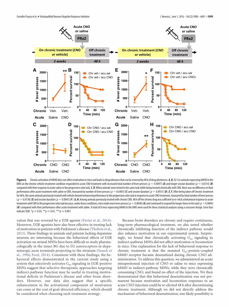
Chronic activation of hM4D does not affect motivation inmice and leads to drug tolerance that can be reversed by 48 hof drug abstinenceBecause neuropsychiatric diseases generally require long-termpharmacological treatment, we investigated whether continuousactivation of hM4D receptors in indirect-pathway MSNs in theDMS and NAc also enhances motivation. Chronic CNO treat-ment did not affect performance of mice expressing hM4D inindirect-pathway MSNs in either DMS or NAc in the progressiveratio task, measured by either session duration (DMS: � 2 �0.02327, p � 0.8788; NAc: � 2 � 0.8556, p � 0.3550; Fig. 5A),number of lever presses (DMS: t(7) � 0.8276, p � 0.4352, NAc:t(8) � 0.5896, p � 0.5717; Figure 5B), or breakpoint (DMS: t(7) �0.4952, p � 0.6357, NAc: t(8) � 0.2526, p � 0.8069; Figure 5C).Similar lack of effect was also observed for D2R-OEdev mice orcontrol littermates expressing hM4D in indirect-pathway MSNsin both the DMS and NAc (session duration: control: � 2 �0.2393, p � 0.6247, median survival: 19.5 min; D2R-OEdev: � 2 �3.690, p � 0.2969, median survival: 21.5 min; number of leverpresses: Fdrug(1,20) � 1.671, p � 0.2108, Fgenotype(1,20) � 0.01001,p � 0.9213, Finteraction(1,20) � 0.1.907, p � 0.1825, vehicle:363.667 � 61.667 min, CNO: 256.0 � 53.333 min, n � 6 mice pergroup) (data not shown). Importantly, the absence of a CNOeffect was not due to the different routes of administration(drinking water vs intraperitoneal injection) or instability ofCNO in water or room temperature (data not shown).
We then asked whether animals that are treated with CNO for2 weeks still exhibit a behavioral response to an acute intraperi-toneal injection of CNO. As a control experiment, we found thatwild-type animals expressing hM4D in the DMS receiving un-treated water (chronic vehicle) responded to acute CNO treat-ment with increased total number of lever presses (t(7) � 3.222,p � 0.0097) (Fig. 6A) and longer session duration (� 2 � 4.719,p � 0.0314) (Fig. 6B). In contrast, wild-type animals on chronictreatment with CNO did not exhibit an acute response to thedrug injection, measured by total number of lever presses (t(7) �0.4880, p � 0.6405) (Fig. 6C) and session duration (� 2 �
5996 • J. Neurosci., June 1, 2016 • 36(22):5988 – 6001 Carvalho Poyraz et al. • Striatopallidal Neurons Regulate Response Initiation

0.01117, p � 0.8931) (Fig. 6D). Comparable results were ob-tained for mice expressing hM4D in the NAc (data not shown).These findings show that a response to acute CNO in the progres-sive ratio task can no longer be elicited while animals are chron-ically treated with CNO in their drinking water.
We then tested the persistence of this tolerance effect afterdiscontinuing chronic CNO treatment. Mice expressing hM4Din the DMS that had been treated with CNO for 2 weeks weretaken off the CNO treatment and 48 h later injected intraperito-neally with CNO or saline. As expected, mice that were chroni-cally treated with vehicle (control condition) demonstratedenhanced motivation in response to acute CNO, measured bynumber of lever presses (t(7) � 3.261, p � 0.0138) (Fig. 6E) andsession duration (� 2 � 8.170, p � 0.0045) (Fig. 6F). Moreover,
48 h off chronic treatment with CNO wassufficient to re-elicit the behavioral re-sponse to acute treatment with CNOcompared with acute treatment with sa-line (number of lever presses: t(7) � 4.211,p � 0.0040; session duration: � 2 � 12.33,p � 0.0004) (Fig. 6G,H). Comparable re-sults were obtained for mice expressinghM4D in the NAc (data not shown). Anda similar recovery from CNO tolerancewas observed for ambulatory activity inthe open field (data not shown). Micetherefore recover their sensitivity to CNOafter 48 h of drug withdrawal.
DiscussionHere, we determined whether motiva-tional deficits induced by upregulation ofstriatal D2Rs starting in early develop-ment could be reversed by decreasingfunction of the indirect pathway in theadult. We found that acute activation ofthe G�i-coupled designer receptor hM4Din the indirect pathway increased motiva-tion in D2R-OEdev mice, as well as in con-trol littermates. This effect was due toenergized initiation of the behavioral re-sponse. Our manipulation facilitatedperformance when a brief response wasrequired to earn reward but came at thecost of goal-directed efficiency when aresponse of sustained duration was re-quired to earn the reward. The increasein response initiation was not due to ha-bitual lever pressing because sensitivityto changes in the value of the rewardremained intact. It was also not medi-ated by increased response vigor be-cause the rate of lever pressing duringprogressive ratio testing was unaltered.Selective manipulation of MSNs in ei-ther the DMS or NAc showed that bothstriatal subregions contribute to this ef-fect on motivation. Furthermore, wealso investigated the behavioral effectsof chronically decreasing function of theindirect pathway on motivation, and wefound that chronic activation of G�i sig-naling in indirect-pathway MSNs leadsto a behavioral desensitization that is re-
versible upon discontinuation of hM4D activation.
Inhibiting indirect-pathway function enhances motivation inD2R-OEdev and wild-type miceD2R-OEdev mice show deficits in motivation that are associatedwith enhanced MSN excitability. Because the hM4D receptor hasbeen previously used to decrease MSN excitability in rodents(Ferguson et al., 2011), we inferred that, if hyperexcitability ofindirect-pathway MSNs is at the origin of the motivational deficitof D2R-OEdev mice, activating hM4D receptors in this neuronalpopulation would rescue their motivational deficit. However, asboth D2R-OEdev and control mice showed increased perfor-mance in the progressive ratio task after treatment with CNO, it is
A
D
B
C
E
0 20 40 60 80 100 1200
50
100
150
Control-hM4D (saline)Control-hM4D (CNO)D2R-OEdev-hM4D (saline)D2R-OEdev-hM4D (CNO)
Time (min)
Perc
ent s
urvi
val
Saline CNO0
100
200
300
400 D2R-OEdev-hM4DControl-hM4D
***
# le
ver p
ress
es
3.0 4.2 5.9 8.2 11.5
16.1
22.6
31.6
44.3
0.0
0.2
0.4
0.6
0.8
1.0
Control-hM4D (saline)Control-hM4D (CNO)D2R-OEdev--hM4D (saline)D2R-OEdev-hM4D (CNO)
Hold requirement (sec)
Prop
ortio
n o f
rew
arde
dpr
esse
s in
tria
l
1 22 43 64 85 106
127
148
169
190
211
232
253
274
295
316
337
358
379
400
421
442
0
10
20
30
40
50
Day 1 salineDay 2 CNODay 3 salineDay 4 CNO
Press number
Hold
dur
atio
n (s
ec)
Saline CNO0
3
6
9Control-hM4DD2R-OEdev-hM4D
***
# rein
forc
ers ea
rne d
Figure 4. Decreasing function of the indirect pathway energizes behavior at the expense of goal-directed efficiency when asustained response is required to obtain the goal. A–D, D2R-OEdev and control littermates expressing hM4D in indirect-pathwayMSNs show decreased efficiency in the progressive hold-down task after treatment with CNO compared with treatment withsaline. Mice of both genotypes continued to respond for longer times when on CNO compared with saline, measured by survivalfunctions for session duration (control: p � 0.005, D2R-OEdev: p � 0.0001) (A). Mice on CNO made a greater number of leverpresses ( p � 0.0001) (B) and earned fewer reinforcers ( p � 0.0001) (C) in a session compared with saline. For each holdrequirement, mice treated with CNO showed lower efficiency in responding compared with treatment with saline, measured by theproportion of rewarded presses ( p � 0.0143) (D). E, Distribution of the press duration for all presses made on different days oftesting by one representative subject: while this mouse made progressively longer presses on saline treatment days, it madegenerally shorter presses and a greater number of presses on days when it was treated with CNO. Data from 12 animals pergenotype were used to calculate all statistics reported above using a crossover design. Error bars indicate SEM. ***p � 0.001.
Carvalho Poyraz et al. • Striatopallidal Neurons Regulate Response Initiation J. Neurosci., June 1, 2016 • 36(22):5988 – 6001 • 5997

unclear whether hM4D activation rescuedthe underlying neuronal mechanism thatcaused the motivational deficit ofD2R-OEdev mice or whether it enhancedmotivation by an independent mecha-nism. Moreover, in our own experiments,we could not detect an effect of hM4D ac-tivation on somatic excitability ofindirect-pathway neurons (data notshown). But consistent with an inhibitoryeffect of hM4D activation on synaptictransmission in the GPe, we found thatthis manipulation disinhibited GPe activ-ity in vivo. We propose that inhibitingfunction of the indirect pathway may rep-resent a more general strategy to amelio-rate deficits in motivation than justrescuing the deficit in D2R-OEdev mice.
Inhibiting indirect-pathway functionenergizes the initiation of behaviorUsing the progressive hold-down task,we demonstrated that inhibiting function of the indirect path-way energizes the initiation of behavior, which, however,comes at the expense of goal-directed efficiency. In the pro-gressive hold-down task, maximal efficiency requires animalsto suppress any tendency to initiate new behaviors to success-fully hold down a lever until a reward is obtained. Inhibition ofthe indirect pathway caused both D2R-OEdev and control miceto continue to engage in this task for longer times. However,acute CNO treatment also caused animals to make many re-sponses of short duration and thus earn fewer rewards in theprogressive hold-down task. These observations suggest thatinhibition of the indirect pathway in the NAc and DMS en-hances motivation by primarily regulating the readiness toinitiate behaviors rather than affecting processes related to theoptimal selection of specific behaviors or to processes thatregulate the vigor or persistence of actions. This increasedtendency to affect initiation of prepotent behavior could ac-count for the effects we observed in both operant tasks and inthe open field.
Our results are interesting in light of earlier studies showingthat D1R antagonists interferes with the initiation of actions(Choi et al., 2009, 2011). In these studies, pharmacologically in-duced blockade of D1R function, presumably inhibiting functionof the direct pathway, led to impairments of initiation, whereas inour study inhibiting indirect-pathway function led to enhancedaction initiation. Together, these results indicate that the initia-tion of action depends on a balance between activity in bothindirect and direct pathways, consistent with previous observa-tions that both pathways are active during the onset of new ac-tions (Cui et al., 2013).
It is possible, however, that the primary consequence of inhib-iting indirect-pathway function is not enhanced initiation but areduction in hold times, which would then lead to a faster reini-tiation of responding. This interpretation would be in line withthe observation that the D2R antagonist raclopride increases holdtimes (Fowler and Liou, 1994).
Moreover, although our data are consistent with an increasedaction initiation, altered sensitivity to reward cannot be fully ex-cluded. Whereas CNO enhanced the number of lever presses inthe progressive ratio task, we did not observe any enhancement oflever pressing in the devaluation task. This may be due to the fact
that a lower effort is required during the devaluation task or,alternatively, that no reward is delivered in the devaluation task.Enhanced responding in the progressive ratio task could there-fore also be influenced by an enhanced sensitivity toreinforcement.
Inhibiting indirect pathway in both DMS and NAcenhances motivationPrevious studies have shown that the NAc, and not the DMS,plays a key role in goal-directed behavior by regulating how ani-mals allocate effort to achieve specific outcomes based on therewarding value of those outcomes (Nowend et al., 2001; Mai etal., 2012). These studies often used instrumental tasks that wereinherently different from the progressive ratio task used here. Inthe latter studies, animals chose between a high-effort action thatled to a high-value reward and a low-effort action that led to alow-value reward. Lesioning or antagonizing D1R and D2R func-tion in the NAc made animals less motivated, choosing low-effortactions that led to smaller rewards (Nowend et al., 2001; Mai etal., 2012). It is likely, therefore, that the NAc plays a specific rolein effort-related choices. Our findings that acutely decreasingfunction of the indirect pathway selectively in either the NAc orDMS led to similar enhanced motivation, without affecting out-come devaluation, suggest that both subregions of the striatumwork together to regulate at least the initiation of motivated be-havior. Consistent with these observations, a number of studieshave established a role for the DMS in motivated behavior (Yin etal., 2005b; Shiflett et al., 2010; Hilario et al., 2012). Other behav-ioral assays of instrumental performance, such as tasks that mea-sure sensitivity to response contingencies or effort/valuerelationships, may potentially reveal dissociations between therole of the indirect pathway arising from the DMS and NAc inmotivation.
Possible therapeutic relevance of inhibitingindirect-pathway functionThe link between initiation of motor behaviors, motivation, andfunction of the indirect pathway is also relevant for Parkinson’spatients who present with both difficulty initiating motor actionsand deficits in motivation. In a rodent model of Parkinson’s dis-ease using 6-OHDA lesions, animals exhibited a deficit in moti-
A BDMS (vehicle)DMS (CNO)NAc (vehicle)NAc (CNO)
C
Vehicle CNO0
500
1000
1500 DMSNAc
# le
ver p
ress
es
Vehicle CNO0
200
400
600
800 DMSNAc
Bre
akpo
int
(pre
ss ra
tio re
quire
men
t)
0 20 40 60 80 100 1200
50
100
150
Perc
ent s
urvi
val
Figure 5. Chronically activating G�i signaling in indirect-pathway MSNs in either DMS and NAc does not affect motivation inmice using a crossover design. A, No effect of chronic CNO was observed in the survival function for average session duration in theprogressive ratio schedule for D2-Cre mice expressing hM4D in indirect-pathway MSNs in either DMS ( p � 0.8788) or NAc ( p �0.3550). B, The number of lever presses made in a progressive ratio session was not changed after chronic CNO treatment for miceexpressing hM4D in indirect-pathway MSNs in either DMS ( p � 0.4352) or NAc ( p � 0.5717). C, The breakpoint was also notchanged as a result of chronic CNO treatment for mice expressing hM4D in indirect-pathway MSNs in either DMS ( p � 0.6357) orNAc ( p�0.8069). A total of 8 mice in the DMS group and 9 mice in the NAc were used to calculate all statistics reported above. Errorbars indicate SEM.
5998 • J. Neurosci., June 1, 2016 • 36(22):5988 – 6001 Carvalho Poyraz et al. • Striatopallidal Neurons Regulate Response Initiation
vation that was reversed by a D2R agonist (Favier et al., 2014).Moreover, D2R agonists have also been effective in treating lackof motivation in patients with Parkinson’s disease (Thobois et al.,2013). These findings in animals and patients lacking dopamineneurons are interesting because the behavioral effects of D2Ractivation on striatal MSNs have been difficult to study pharma-cologically in the intact BG due to D2 autoreceptors in dopa-minergic axon terminals projecting to the striatum (Sesack etal., 1994; Ford, 2014). Consistent with these findings, the be-havioral effects demonstrated in the current study using asystem that selectively activates G�i signaling in D2R-positiveMSNs suggest that selective therapeutic approaches targetingindirect-pathway function may be useful in treating motiva-tional deficits in Parkinson’s disease and other brain disor-ders. However, our data also suggest that a specificenhancement in the activational component of motivationcan come at the cost of goal-directed efficiency, which shouldbe considered when choosing such treatment strategy.
Because brain disorders are chronic and require continuous,long-term pharmacological treatment, we also tested whetherchronically inhibiting function of the indirect pathway wouldalso enhance motivation in our experimental system. Surpris-ingly, we found that chronically activating G�i signaling inindirect-pathway MSNs did not affect motivation or locomotionin mice. One explanation for the lack of behavioral response tochronic treatment is that the mutated G�i-protein-coupledhM4D receptor became desensitized during chronic CNO ad-ministration. To address this question, we administered an acuteintraperitoneal injection of CNO or saline to mice expressinghM4D in indirect-pathway MSNs, while they were chronicallyconsuming CNO, and found no effect of the injection. We thendemonstrated that this behavioral desensitization was not per-manent because motivation and locomotion responses to anacute CNO injection could be re-elicited 48 h after discontinuingchronic treatment. Although we did not directly address themechanism of behavioral desensitization, one likely possibility is
0
500
1000
1500
2000 **
Chronic
Acute
Veh. Veh.
Saline CNO
# le
ver p
ress
es
0 40 80 1200
50
100
150 Chr veh / acu salChr veh / acu CNO
Time (min)
Perc
ent s
urvi
val
0 40 80 1200
50
100
150 Chr CNO / acu salChr CNO / acu CNO
Time (min)
Perc
ent s
urvi
val
0
500
1000
1500
2000
Chronic
Acute
CNO CNO
Saline CNO
# le
ver p
ress
es
0
500
1000
1500
2000 *
Chronic
Acute
Veh. Veh.
Saline CNO#
leve
r pre
sses
0
500
1000
1500
2000
**
Chronic
Acute
CNO CNO
Saline CNO
# le
ver p
ress
es
0 40 80 1200
50
100
150 Chr CNO / acu salChr CNO / acu CNO
Time (min)
Perc
ent s
urvi
val
0 40 80 1200
50
100
150 Chr veh / acu salChr veh / acu CNO)
Time (min)
Perc
ent s
urvi
val
On chronic treatment (CNO or vehicle)
2 weeks 48 h
PRx2
Off chronic treatment
Acute CNOor saline
On chronic treatment (CNO or vehicle)
2 weeks 48 h
Off chronic treatment
PRx2
Acute CNOor saline
A B
C D
E F
G H
Figure 6. Chronic activation of hM4D does not affect motivation in mice and leads to drug tolerance that can be reversed by 48 h of drug abstinence. A, B, D2-Cre animals expressing hM4D in theDMS on the chronic vehicle treatment condition responded to acute CNO treatment with increased total number of lever presses ( p � 0.0097) (A) and longer session duration ( p � 0.0314) (B)compared with their response to acute saline in the progressive ratio task. C, D, When animals were tested in the same task while being treated chronically with CNO, there was no difference in theirperformance after acute treatment with saline or CNO, measured by number of lever presses ( p � 0.6405) (C) and session duration ( p � 0.8931) (D). E, F, After being taken off chronic treatmentfor 48 h, the same animals previously treated with vehicle showed enhanced performance in the progressive ratio task in response to acute CNO treatment, measured by total number of lever presses( p � 0.0138) (E) and session duration ( p � 0.0045) (F ). G, H, Among animals previously treated with chronic CNO, 48 h off the chronic drug was sufficient to re-elicit a behavioral response to acutetreatment with CNO in the progressive ratio task because, under these conditions, mice made more lever presses ( p �0.0040) (G) and continued to respond for longer times in the task ( p �0.0004)(H ) compared with their performance after acute treatment with saline. A total of 8 mice expressing hM4D in the DMS were used for these statistical analyses using a crossover design. Error barsindicate SEM. *p � 0.05, **p � 0.01, ***p � 0.001.
Carvalho Poyraz et al. • Striatopallidal Neurons Regulate Response Initiation J. Neurosci., June 1, 2016 • 36(22):5988 – 6001 • 5999

that the G�i-coupled receptor becomes desensitized due to inter-nalization after chronic stimulation (Gainetdinov et al., 2004).Alternatively, behavioral desensitization may be mediated bycircuit-level adaptations. The fast recovery of the response sug-gests that structural anatomical rewiring may not be required, butthe desensitization effect could be mediated by circuit-level plas-ticity, which is known to occur at shorter timescales (Caroni et al.,2014). Thus, receptor desensitization or circuit adaptations are tobe taken into consideration when developing chronic therapiesfor neuropsychiatric disorders. Our results may explain whydrugs that target GPCRs may not improve motivation whentaken chronically by patients (Catapano and Manji, 2007).
In conclusion, the present study shows that the indirectpathway is important for regulating response initiation, a pro-cess that can be pharmacologically manipulated to increasebehavioral activation. However, inhibiting the indirect path-way may also lead to inefficient responding at the cost ofreduced reward outcome in situations in which a long dura-tion of action is required to obtain a goal. These findingsprovide insight into BG function and should inform the de-velopment of new treatments for neuropsychiatric disorderswith abnormal motivation.
ReferencesAbi-Dargham A, Rodenhiser J, Printz D, Zea-Ponce Y, Gil R, Kegeles LS,
Weiss R, Cooper TB, Mann JJ, Van Heertum RL, Gorman JM, Laruelle M(2000) Increased baseline occupancy of D2 receptors by dopamine inschizophrenia. Proc Natl Acad Sci U S A 97:8104 – 8109. CrossRefMedline
Albin RL, Young AB, Penney JB (1989) The functional anatomy of basalganglia disorders. Trends Neurosci 12:366 –375. CrossRef Medline
Bailey MR, Jensen G, Taylor K, Mezias C, Williamson C, Silver R, SimpsonEH, Balsam PD (2015) A novel strategy for dissecting goal-directed ac-tion and arousal components of motivated behavior with a progressivehold-down task. Behav Neurosci 129:269 –280. CrossRef Medline
Bock R, Shin JH, Kaplan AR, Dobi A, Markey E, Kramer PF, Gremel CM,Christensen CH, Adrover MF, Alvarez VA (2013) Strengthening the ac-cumbal indirect pathway promotes resilience to compulsive cocaine use.Nat Neurosci 16:632– 638. CrossRef Medline
Burton AC, Nakamura K, Roesch MR (2015) From ventral-medial todorsal-lateral striatum: neural correlates of reward-guided decision-making. Neurobiol Learn Mem 117:51–59. CrossRef Medline
Caroni P, Chowdhury A, Lahr M (2014) Synapse rearrangements uponlearning: from divergent-sparse connectivity to dedicated sub-circuits.Trends Neurosci 37:604 – 614. CrossRef Medline
Catapano LA, Manji HK (2007) G protein-coupled receptors in major psy-chiatric disorders. Biochim Biophys Acta 1768:976 –993. CrossRefMedline
Cazorla M, Shegda M, Ramesh B, Harrison NL, Kellendonk C (2012) Stria-tal D2 receptors regulate dendritic morphology of medium spiny neuronsvia Kir2 channels. J Neurosci 32:2398 –2409. CrossRef Medline
Cazorla M, de Carvalho FD, Chohan MO, Shegda M, Chuhma N, Rayport S,Ahmari SE, Moore H, Kellendonk C (2014) Dopamine D2 receptorsregulate the anatomical and functional balance of basal ganglia circuitry.Neuron 81:153–164. CrossRef Medline
Choi WY, Morvan C, Balsam PD, Horvitz JC (2009) Dopamine D1 and D2antagonist effects on response likelihood and duration. Behav Neurosci123:1279 –1287. CrossRef Medline
Choi WY, Campbell CM, Balsam PD, Horvitz JC (2011) Effects of corticaland striatal dopamine D1 receptor blockade on cued versus noncuedbehavioral responses. Behav Neurosci 125:705–713. CrossRef Medline
Corbit LH, Balleine BW (2011) The general and outcome-specific forms ofPavlovian-instrumental transfer are differentially mediated by the nu-cleus accumbens core and shell. J Neurosci 31:11786 –11794. CrossRefMedline
Corbit LH, Muir JL, Balleine BW (2001) The role of the nucleus accumbensin instrumental conditioning: evidence of a functional dissociation be-tween accumbens core and shell. J Neurosci 21:3251–3260. Medline
Cui G, Jun SB, Jin X, Pham MD, Vogel SS, Lovinger DM, Costa RM (2013)
Concurrent activation of striatal direct and indirect pathways during ac-tion initiation. Nature 494:238 –242. CrossRef Medline
Dawson VL, Dawson TM, Wamsley JK (1990) Muscarinic and dopaminer-gic receptor subtypes on striatal cholinergic interneurons. Brain Res Bull25:903–912. CrossRef Medline
Drew MR, Simpson EH, Kellendonk C, Herzberg WG, Lipatova O, FairhurstS, Kandel ER, Malapani C, Balsam PD (2007) Transient overexpressionof striatal D2 receptors impairs operant motivation and interval timing.J Neurosci 27:7731–7739. CrossRef Medline
Durieux PF, Schiffmann SN, de Kerchove d’Exaerde A (2012) Differentialregulation of motor control and response to dopaminergic drugs by D1Rand D2R neurons in distinct dorsal striatum subregions. EMBO J 31:640 – 653. CrossRef Medline
Favier M, Duran T, Carcenac C, Drui G, Savasta M, Carnicella S (2014)Pramipexole reverses Parkinson’s disease-related motivational deficits inrats. Mov Disord 29:912–920. CrossRef Medline
Ferguson SM, Eskenazi D, Ishikawa M, Wanat MJ, Phillips PE, Dong Y, RothBL, Neumaier JF (2011) Transient neuronal inhibition reveals opposingroles of indirect and direct pathways in sensitization. Nat Neurosci 14:22–24. CrossRef Medline
Ford CP (2014) The role of D2-autoreceptors in regulating dopamine neu-ron activity and transmission. Neuroscience 282C:13–22. CrossRefMedline
Fowler SC, Liou JR (1994) Microcatalepsy and disruption of forelimb usageduring operant behavior: differences between dopamine D1 (SCH-23390) and D2 (raclopride) antagonists. Psychopharmacology 115:24 –30. CrossRef Medline
Franklin KBJ, Paxinos G (2008) The mouse brain in stereotaxic coordinates.San Diego: Academic.
Fujiyama F, Sohn J, Nakano T, Furuta T, Nakamura KC, Matsuda W, KanekoT (2011) Exclusive and common targets of neostriatofugal projectionsof rat striosome neurons: a single neuron-tracing study using a viral vec-tor. Eur J Neurosci 33:668 – 677. CrossRef Medline
Gainetdinov RR, Premont RT, Bohn LM, Lefkowitz RJ, Caron MG (2004)Desensitization of G protein-coupled receptors and neuronal functions.Annu Rev Neurosci 27:107–144. CrossRef Medline
Gallo EF, Salling MC, Feng B, Moron JA, Harrison NL, Javitch JA, KellendonkC (2015) Upregulation of dopamine D2 receptors in the nucleus accum-bens indirect pathway increases locomotion but does not reduce alcoholconsumption. Neuropsychopharmacology 40:1609 –1618. CrossRefMedline
Gerfen CR, Surmeier DJ (2011) Modulation of striatal projection systemsby dopamine. Annu Rev Neurosci 34:441– 466. CrossRef Medline
Hilario M, Holloway T, Jin X, Costa RM (2012) Different dorsal striatumcircuits mediate action discrimination and action generalization. EurJ Neurosci 35:1105–1114. CrossRef Medline
Howes OD, Kapur S (2009) The dopamine hypothesis of schizophrenia: III.The final common pathway. Schizophr Bull 35:549 –562. CrossRefMedline
Kellendonk C, Tronche F, Casanova E, Anlag K, Opherk C, Schutz G (1999)Inducible site-specific recombination in the brain. J Mol Biol 285:175–182. CrossRef Medline
Kellendonk C, Simpson EH, Polan HJ, Malleret G, Vronskaya S, Winiger V,Moore H, Kandel ER (2006) Transient and selective overexpression ofdopamine D2 receptors in the striatum causes persistent abnormalities inprefrontal cortex functioning. Neuron 49:603– 615. CrossRef Medline
Kravitz AV, Freeze BS, Parker PR, Kay K, Thwin MT, Deisseroth K, KreitzerAC (2010) Regulation of parkinsonian motor behaviours by optoge-netic control of basal ganglia circuitry. Nature 466:622– 626. CrossRefMedline
Kupchik YM, Brown RM, Heinsbroek JA, Lobo MK, Schwartz DJ, Kalivas PW(2015) Coding the direct/indirect pathways by D1 and D2 receptors isnot valid for accumbens projections. Nat Neurosci 18:1230 –1232.CrossRef Medline
Laruelle M (1998) Imaging dopamine transmission in schizophrenia: a re-view and meta-analysis. Q J Nucl Med 42:211–221. Medline
Lenz JD, Lobo MK (2013) Optogenetic insights into striatal function andbehavior. Behav Brain Res 255:44 –54. CrossRef Medline
Lex B, Hauber W (2010) The role of nucleus accumbens dopamine in out-come encoding in instrumental and Pavlovian conditioning. NeurobiolLearn Mem 93:283–290. CrossRef Medline
Mai B, Sommer S, Hauber W (2012) Motivational states influence effort-
6000 • J. Neurosci., June 1, 2016 • 36(22):5988 – 6001 Carvalho Poyraz et al. • Striatopallidal Neurons Regulate Response Initiation
based decision making in rats: the role of dopamine in the nucleus accum-bens. Cogn Affect Behav Neurosci 12:74 – 84. CrossRef Medline
Nowend KL, Arizzi M, Carlson BB, Salamone JD (2001) D1 or D2 antago-nism in nucleus accumbens core or dorsomedial shell suppresses leverpressing for food but leads to compensatory increases in chow consump-tion. Pharmacol Biochem Behav 69:373–382. CrossRef Medline
Pedersen KF, Larsen JP, Alves G, Aarsland D (2009) Prevalence and clinicalcorrelates of apathy in Parkinson’s disease: a community-based study.Parkinsonism Relat Disord 15:295–299. CrossRef Medline
Salamone JD (1988) Dopaminergic involvement in activational aspects ofmotivation: effects of haloperidol on schedule-induced activity, feeding,and foraging in rats. Psychobiology 16:196 –206.
Salamone JD, Correa M (2002) Motivational views of reinforcement: impli-cations for understanding the behavioral functions of nucleus accumbensdopamine. Behav Brain Res 137:3–25. CrossRef Medline
Sesack SR, Aoki C, Pickel VM (1994) Ultrastructural localization of D2receptor-like immunoreactivity in midbrain dopamine neurons and theirstriatal targets. J Neurosci 14:88 –106. Medline
Shiflett MW, Brown RA, Balleine BW (2010) Acquisition and performanceof goal-directed instrumental actions depends on ERK signaling in dis-tinct regions of dorsal striatum in rats. J Neurosci 30:2951–2959. CrossRefMedline
Simpson EH, Balsam PD (2015) The behavioral neuroscience of motiva-tion: an overview of concepts, measures, and translational applications.Curr Top Behav Neurosci. Advance online publication. Retrieved Nov 25,2016. doi: 10.1007/7854_2015_402. CrossRef Medline
Tai LH, Lee AM, Benavidez N, Bonci A, Wilbrecht L (2012) Transient stim-ulation of distinct subpopulations of striatal neurons mimics changes inaction value. Nat Neurosci 15:1281–1289. CrossRef Medline
Thobois S, Ardouin C, Lhommee E, Klinger H, Lagrange C, Xie J, Fraix V,Coelho Braga MC, Hassani R, Kistner A, Juphard A, Seigneuret E, Chab-ardes S, Mertens P, Polo G, Reilhac A, Costes N, LeBars D, Savasta M,Tremblay L, et al. (2010) Non-motor dopamine withdrawal syndromeafter surgery for Parkinson’s disease: predictors and underlying mesolim-bic denervation. Brain 133:1111–1127. CrossRef Medline
Thobois S, Lhommee E, Klinger H, Ardouin C, Schmitt E, Bichon A, KistnerA, Castrioto A, Xie J, Fraix V, Pelissier P, Chabardes S, Mertens P, Que-sada JL, Bosson JL, Pollak P, Broussolle E, Krack P (2013) Parkinsonianapathy responds to dopaminergic stimulation of D2/D3 receptors withpiribedil. Brain 136:1568 –1577. CrossRef Medline
Tomer R, Goldstein RZ, Wang GJ, Wong C, Volkow ND (2008) Incentivemotivation is associated with striatal dopamine asymmetry. Biol Psychol77:98 –101. CrossRef Medline
Volkow ND, Wang GJ, Fowler JS, Thanos PP, Logan J, Gatley SJ, Gifford A,Ding YS, Wong C, Pappas N (2002) Brain DA D2 receptors predict re-inforcing effects of stimulants in humans: replication study. Synapse 46:79 – 82. CrossRef Medline
Ward RD, Kellendonk C, Simpson EH, Lipatova O, Drew MR, Fairhurst S,Kandel ER, Balsam PD (2009) Impaired timing precision produced bystriatal D2 receptor overexpression is mediated by cognitive and motiva-tional deficits. Behav Neurosci 123:720 –730. CrossRef Medline
Yin HH, Knowlton BJ, Balleine BW (2005a) Blockade of NMDA receptorsin the dorsomedial striatum prevents action-outcome learning in instru-mental conditioning. Eur J Neurosci 22:505–512. CrossRef Medline
Yin HH, Ostlund SB, Knowlton BJ, Balleine BW (2005b) The role of thedorsomedial striatum in instrumental conditioning. Eur J Neurosci 22:513–523. CrossRef Medline
Carvalho Poyraz et al. • Striatopallidal Neurons Regulate Response Initiation J. Neurosci., June 1, 2016 • 36(22):5988 – 6001 • 6001