design of three-dimensional biomimetic scaffoldsmolly/binder/design of three-dimensional... ·...
TRANSCRIPT

Review
Design of three-dimensional biomimetic scaffolds
Shawn C. Owen,1,2 Molly S. Shoichet1,2,3,4
1Department of Chemical Engineering and Applied Chemistry, University of Toronto, 200 College Street, Toronto,
Ontario, Canada M5S 3ES2Terrence Donnelly Centre for Cellular and Biomolecular Research, 160 College Street, Toronto, Ontario, Canada M5S 3E13Institute of Biomaterials and Biomedical Engineering, University of Toronto, 164 College Street, Toronto, Ontario, Canada M5S 3G94Department of Chemistry, University of Toronto, 80 Saint George Street, Toronto, Ontario, Canada M5S 3H6
Received 24 February 2010; revised 24 March 2010; accepted 25 March 2010
Published online 1 July 2010 in Wiley InterScience (www.interscience.wiley.com). DOI: 10.1002/jbm.a.32834
Abstract: A detailed understanding of the biophysical fea-
tures that affect cell growth and development is important in
guiding the design of biomimetic scaffolds. The cellular
microenvironment is a network of structural and functional
components that provide mechanical and chemical stimuli,
which influence cell function and fate. Important develop-
mental signals are conveyed to cells through interactions
with neighboring cells, the extracellular matrix (ECM), and
growth factors. Currently, there are number of approaches to
create 3D tissue models in vitro that allow for control over
cell adhesion, the physical properties of the surrogate matrix,
and the spatial distribution of growth factors. This review
describes some of the most significant biological features of
the ECM, and several engineering methods currently being
implemented to design and tune synthetic scaffolds to mimic
these features. VC 2010 Wiley Periodicals, Inc. J Biomed Mater Res
Part A: 94A: 1321–1331, 2010.
Key Words: extracellular matrix, scaffold materials, tissue
engineering, biological models, regenerative medicine
INTRODUCTION
Two-dimensional (2D) cell culture is commonly used tostudy cell function and behavior, providing a basic methodto explore biological mechanisms, cell differentiation, andtherapeutic efficacy before moving into more complex,in vivo models. However, cells cultured in traditional 2Dmodels fail to provide an accurate representation of cells insitu as they lack the contextual cues found in the nativethree-dimensional (3D) tissue.1,2 Several factors in the cellu-lar microenvironment provide important signals to cells,including interactions with neighboring cells, the extracellu-lar matrix, and soluble factors.
The extracellular matrix (ECM) is a heterogeneous compo-sition of proteoglycans, proteins, and signaling molecules thatwas originally known for its role in providing structural sup-port to cells and as a milieu for cell migration. Recent investi-gations of the ECM have clarified its role beyond an inertbackground to an active component in cell signaling.2–4 Begin-ning with embryogenesis and continuing throughout adult-hood, the ECM influences cell differentiation, proliferation,survival, and migration through both biochemical interactions(cell adhesion, presentation of growth factors) and mechanicalcues5 (stiffness, deformability). Successful understanding ofECM signals will facilitate the ability to guide cell behaviorand evaluate complex intracellular signaling pathways.
Considering the impact of the ECM on cellular behavior,a multidisciplinary paradigm shift is underway towards the
development of biomimetic 3D cell culture systems thatincorporate ECM molecules to recapitulate the native envi-ronment more accurately than 2D systems.6,7 Current 3Dmodels are made from polysaccharides, collagens, syntheticbiomaterials, spheroids of other cells, peptides, cell frag-ments, or decellularized ECM from living tissue.8 There area number of excellent reviews which discuss these naturaland synthetic scaffolds.9–14
Among the scaffolds currently in use, the majority areeither simple or ill-defined. Simple matrices include syn-thetic polymers such as poly(ethylene glycol) (PEG) andpoly(vinyl alcohol) (PVA), or naturally derived polymerssuch as chitosan and collagen. As these scaffolds are oftenmade from only one or two components, the physicochemi-cal properties can be controlled; however, many of thesepolymers have limited cellular recognition and, therefore,natural cell–matrix adhesions may be limited or completelyabsent. Any variation in adhesion will alter the signalingmechanisms that are important to many cellular processes.
At the other extreme are complex, ill-defined matricessuch as Matrigel15 or reconstituted tissue.16 These scaffoldsprovide factors that impact cell function; however, the inher-ent complexity of these scaffolds makes it difficult to under-stand cell signaling. Batch-to-batch variability hinders thereproducibility of experiments. Problems with biocompatibil-ity prevent implantation of the scaffolds into human patients,and the mechanical properties of these scaffolds are not
Correspondence to: M. S. Shoichet; e-mail: [email protected]
VC 2010 WILEY PERIODICALS, INC. 1321

easily manipulated. As such, the direct effect of these currentsurrogate ECMs on cell behavior remains ambiguous.
Appreciation of the complexity of the cell response toECM signaling has stimulated the development of 3D scaf-folds that imitate a range of ECM properties. 3D models canovercome the constraints of current 2D models by incorpo-rating both mechanical and biochemical components directlyinto the matrix. In the first part of this review, we outlineseveral biological features of the cellular environment thatare important in guiding cell fate. Later, we discuss note-worthy engineering advancements in designing scaffoldswith tunable components to allow control of matrix factorsthat affect cell function.
DEFINING THE NATURAL BIOPHYSICAL AND CHEMICAL
PROPERTIES OF THE EXTRACELLULAR MATRIX
All cells reside in a complex microenvironment that is tai-lored to guide their physiological functions. As shown inFigure 1, the complex 3D cellular environment provides me-chanical and biochemical signals that are important in guid-ing cell growth and function. Composition of the ECM dic-tates matrix stiffness, nutrient diffusion to tissues, and cell–
matrix interactions, including cell adhesion and migration.Nonstructural factors, such as cell density, cell–cell interac-tions, and bound or secreted signaling proteins, are also im-portant in guiding cell differentiation and function.
Structural elements of the ECM include a hydratedmeshwork of laminin, collagen, elastin, entactin (nidogen),proteoglycans, fibronectin, and various other constituents.17
The more fibrous components (e.g., collagen and elastin)provide architectural rigidity and tension for the cells, whilethe non-fibrous components (predominantly glycosamino-glycans) regulate turgor pressure, form intimate intracellu-lar connections, and modulate the binding, display, and ac-tivity of growth factors.18
The cellular environment is paramount: during embryo-genesis and differentiation into the three primary germ celllayers; in complex tissue and organ formation; throughoutadulthood in maintaining homeostasis; and in response toinsult.19 During early development highly organized chemi-cal gradients in the ECM guide cell migration to form thegastrula. Cell differentiation is further directed through mor-phogenesis and organogenesis by both cell–matrix and cell–cell interactions.20 Most cells in the body are maintained in
FIGURE 1. The complex 3D cellular environment provides mechanical and biochemical signals that guide cell function. The components of the
ECM dictate the stiffness of matrix and the types of cell–matrix adhesions. The matrix composition also determines the ease of nutrients to dif-
fuse to tissues, and the ability of cells to migrate through the matrix. Nonstructural factors such as cell density, cell–cell interactions, and bound
or secreted signaling proteins are important in guiding cell differentiation and function. Image copyright (2010) by Karyn Ho and Anne Hsieh.
[Color figure can be viewed in the online issue, which is available at www.interscience.wiley.com.]
1322 OWEN AND SHOICHET DESIGN OF THREE-DIMENSIONAL BIOMIMETIC SCAFFOLDS

a quiescent state following embryogenesis; however, prolif-eration and differentiation of some specialized cells (such ashematopoietic progenitor cells) are continually regulated byECM interactions. In addition, the ECM has been shown tobe instrumental in physiological response to wounding andinfection.21,22 The vital instructive cues in the cellularmicroenvironment include cell binding interactions, mechan-ical and structural support, and the presentation of regula-tory molecules.
Cellular adhesion to the ECMThe matrix environment in which cells are grown influencesthe type and extent of cellular adhesion, which in turnaffects cell proliferation.23 Integrins are the primary cellsurface receptors that are responsible for cell–matrix adhe-sion (Fig. 2). They are composed of two transmembraneunits—a large a subunit and a smaller b subunit—that formnon-covalent heterodimers in the presence of extracellularCa2þ.24 Various combinations of a- and b-subunits allow forthe formation of 24 different heterodimers, which determineligand specificity. Although some redundancy exists betweenintegrin pairs and their respective ligands, the loss of almostany integrin has deleterious effects.25 Most importantly,integrins not only act as anchors to the ECM but also trans-duce mechanochemical signals to the cell via intracellulartransduction. Initial binding of integrins often leads to theclustering of additional specialized adhesive proteins andlocal remodeling of cytoskeletal and cytoplasmic proteins.3
The resulting focal adhesions sensitize cells to mechanicalstimuli, including the rigidity and elasticity of the ECM.
Integrins bind a number of ‘‘insoluble’’ components ofthe ECM including laminin, elastin, and hyaluronan, amongothers.24 The types and concentrations of these insolublefactors provide signals that are disseminated by the integrinfamily, promoting activation of diverse cytoplasmic proteinsto control a number of cellular processes: differentiation,survival/apoptosis, cell polarity, gene regulation, actin orga-nization, proliferation, and cell migration.2,24–26 For example,the polarity of epithelial cells is essential in tissue organiza-tion for structural formation (such as ductal arrangement),and the directionality of product secretion (such as lacta-tion). It has been shown that epithelial cell integrins mustinteract with a laminin-rich basement membrane to formthe proper architecture and achieve normal cell func-tion.27,28 Cells cultured in a 2D environment lack basal andapical membrane differentiation, while cells cultured in a3D matrix may present appropriate integrins to maintainpolarity.29
An impressive list of integrins and their associated cyto-plasmic proteins was arranged by van der Flier and Sonnen-berg;24 however, no comprehensive index has been tabu-lated that links the extracellular binding of integrin dimerswith their respective cellular function. Progress in elucidat-ing the role of each integrin and its downstream regulationof cell behavior will aid in the design of more specializedECM surrogates that are specific to a desired cellular out-come. The use of defined ECM scaffolds will provide greaterinsight into the impact of integrin–matrix interactions.
Density and stiffness of the ECMCells are not only sensitive to ECM adhesion but also to itsdensity and stiffness. For example, cultured fibroblastsexhibited significantly different migration patterns when thedensity of the matrices was changed.30 When ECM densitywas increased, by increasing concentrations of collagen, themigration of fibroblasts was reduced. Thus, an inverse cor-relation between matrix density and cell migration wasobserved while matrix ligand and integrin receptor concen-trations were held constant.
Important work by Discher and colleagues demonstratedthe importance of matrix elasticity on stem cell fate.31 Mes-enchymal stem cells (MSCs) were cultured on collagen-coated gels that mimicked the elasticity of various tissues.The MSCs responded to gel elasticity by differentiating intolineages that corresponded to the stiffness of the nativeenvironment (Fig. 3). For example, MSCs cultured on softgels (�0.1–1 kPa), to mimic brain elasticity, developed aneuronal morphology, with filopodia branching and spread-ing. More importantly, the RNA profile of these cells showedan increased expression of the neuronal progenitor marker,nestin, and the neuron marker, bIII tubulin. Interestingly,medium stiffness gels (8–17 kPa), which mimic striatedmuscle elasticity, promoted differentiation to myogenic cells,
FIGURE 2. Integrins are composed of two transmembrane units—a
large a subunit and a smaller b subunit—that form noncovalent heter-
odimers that have high affinity for EMC ligands. Integrins not only act
as anchors to the ECM but also transduce mechanochemical signals
to the cell via intracellular transduction that lead to the remodeling of
cytoskeletal and cytoplasmic proteins. (Reproduced with permission
from Schwartz MA, Trends Cell Biol, 2001, 11, 466–470, Elsevier).
[Color figure can be viewed in the online issue, which is available at
www.interscience.wiley.com.]
REVIEW ARTICLE
JOURNAL OF BIOMEDICAL MATERIALS RESEARCH A | 15 SEP 2010 VOL 94A, ISSUE 4 1323
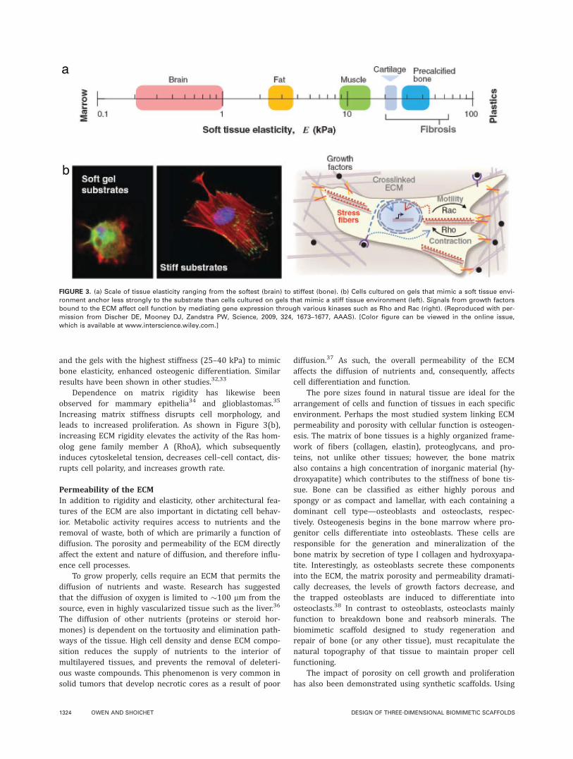
and the gels with the highest stiffness (25–40 kPa) to mimicbone elasticity, enhanced osteogenic differentiation. Similarresults have been shown in other studies.32,33
Dependence on matrix rigidity has likewise beenobserved for mammary epithelia34 and glioblastomas.35
Increasing matrix stiffness disrupts cell morphology, andleads to increased proliferation. As shown in Figure 3(b),increasing ECM rigidity elevates the activity of the Ras hom-olog gene family member A (RhoA), which subsequentlyinduces cytoskeletal tension, decreases cell–cell contact, dis-rupts cell polarity, and increases growth rate.
Permeability of the ECMIn addition to rigidity and elasticity, other architectural fea-tures of the ECM are also important in dictating cell behav-ior. Metabolic activity requires access to nutrients and theremoval of waste, both of which are primarily a function ofdiffusion. The porosity and permeability of the ECM directlyaffect the extent and nature of diffusion, and therefore influ-ence cell processes.
To grow properly, cells require an ECM that permits thediffusion of nutrients and waste. Research has suggestedthat the diffusion of oxygen is limited to �100 lm from thesource, even in highly vascularized tissue such as the liver.36
The diffusion of other nutrients (proteins or steroid hor-mones) is dependent on the tortuosity and elimination path-ways of the tissue. High cell density and dense ECM compo-sition reduces the supply of nutrients to the interior ofmultilayered tissues, and prevents the removal of deleteri-ous waste compounds. This phenomenon is very common insolid tumors that develop necrotic cores as a result of poor
diffusion.37 As such, the overall permeability of the ECMaffects the diffusion of nutrients and, consequently, affectscell differentiation and function.
The pore sizes found in natural tissue are ideal for thearrangement of cells and function of tissues in each specificenvironment. Perhaps the most studied system linking ECMpermeability and porosity with cellular function is osteogen-esis. The matrix of bone tissues is a highly organized frame-work of fibers (collagen, elastin), proteoglycans, and pro-teins, not unlike other tissues; however, the bone matrixalso contains a high concentration of inorganic material (hy-droxyapatite) which contributes to the stiffness of bone tis-sue. Bone can be classified as either highly porous andspongy or as compact and lamellar, with each containing adominant cell type—osteoblasts and osteoclasts, respec-tively. Osteogenesis begins in the bone marrow where pro-genitor cells differentiate into osteoblasts. These cells areresponsible for the generation and mineralization of thebone matrix by secretion of type I collagen and hydroxyapa-tite. Interestingly, as osteoblasts secrete these componentsinto the ECM, the matrix porosity and permeability dramati-cally decreases, the levels of growth factors decrease, andthe trapped osteoblasts are induced to differentiate intoosteoclasts.38 In contrast to osteoblasts, osteoclasts mainlyfunction to breakdown bone and reabsorb minerals. Thebiomimetic scaffold designed to study regeneration andrepair of bone (or any other tissue), must recapitulate thenatural topography of that tissue to maintain proper cellfunctioning.
The impact of porosity on cell growth and proliferationhas also been demonstrated using synthetic scaffolds. Using
FIGURE 3. (a) Scale of tissue elasticity ranging from the softest (brain) to stiffest (bone). (b) Cells cultured on gels that mimic a soft tissue envi-
ronment anchor less strongly to the substrate than cells cultured on gels that mimic a stiff tissue environment (left). Signals from growth factors
bound to the ECM affect cell function by mediating gene expression through various kinases such as Rho and Rac (right). (Reproduced with per-
mission from Discher DE, Mooney DJ, Zandstra PW, Science, 2009, 324, 1673–1677, AAAS). [Color figure can be viewed in the online issue,
which is available at www.interscience.wiley.com.]
1324 OWEN AND SHOICHET DESIGN OF THREE-DIMENSIONAL BIOMIMETIC SCAFFOLDS

centrifugation to process polycaprolactone (PCL) scaffolds,Lee and coworkers fabricated scaffolds with a gradient ofpore diameters ranging from �88–405 lm, and then exam-ined the interaction of cells (chondrocytes, osteoblasts, andfibroblasts) with the scaffolds in vitro.39 Chondrocyte andosteoblast growth was greatest in scaffolds with pore sizes380–405 lm, while fibroblast growth was greatest in scaf-folds with pore sizes 186–200 lm. Scaffolds with pore size290–310 lm encouraged the greatest degree of tissue infil-tration resulting in bone formation in vivo.
Degradation and remodeling of the matrixFollowing binding to the ECM, cells respond to the environ-ment by releasing different proteases. The type of concen-tration of protease released depends on the composition ofthe ECM and its sensitivity to enzymatic degradation. In thismanner, cells are defined by their environment, but alsosimultaneously remodel it. Seminal work by Bissell et al.has termed this type of cell–matrix synergy as ‘‘dynamicreciprocity.’’40
Most cells reside in a state of homeostasis, reaching fulldevelopment at the end of embryogenesis.41 Some cells,however, go through significant physiologic changes at muchlater stages of development, requiring remodeling of the cellenvironment. Among these are cells of the mammary gland,which branch into ducts and terminal lobular units (acini)during puberty, and again change during pregnancy, finallyreaching a fully developed state only after parturition.Epithelial mammary cells initially respond to hormonesecretion and the elasticity of their environment by growingsmall projections. This is followed by remodeling of theirenvironment through secretion of proteases, such as matrixmetalloproteinease (MMP), and enzymes, such as hyaluroni-dases.42 Degradation of the ECM changes the local modulus,decreases the number of cell–matrix adhesions, and alsoresults in the release of ECM fragments that may possess bi-ological activity. The cues that result from degradation arerelayed back to the cell, guiding subsequent behavior andfunction. Thus, the ability of cells to remodel their environ-ment, in concert with hormonal cues and reciprocal signal-ing, allows for proper functional development.
Cell–cell interactionsThe cellular microenvironment includes cell–cell interac-tions where cell density alone can influence cell func-tion.43,44 Moreover, different cell types invariably influencecell function. Cell–cell interactions are instrumental in reca-pitulating the native environment and promoting the mor-phogenesis of functional tissue. For example, mammary epi-thelial cells in situ maintain physical contact withneighboring myoepithelial cells via a combination of connec-tions, including adherens and gap junctions.45 Adherensjunctions generate the polarization of epithelial cells, leadingto the development of basal and apical membranes that arerequired for proper secretory function. Early investigationsby Okada showed that co-cultures of myoepithelial and epi-thelial cells, formed penetrating tubes into collagen gels, butmonocultures of either cell lacked structural correctness
(Fig. 4).46 Similar co-dependency between myoepithelial(arising from the mesoderm) and epithelial cells (arisingfrom the ectoderm) is seen in all glandular and vasculartissue.45
FIGURE 4. Light micrographs of two types of outgrowths seen from
co-cultures of myoepithelial and epithelial cells on floating collagen
gels. (a) Twelve days after culture, blunt outgrowths were seen from
the cell sheet, (b) 6 days after culture, pointed outgrowths were seen
originating at the edge of the cell sheet. Scale bar: 200 lm. (Repro-
duced with permission from Bennett DC, Armstrong BL, Okada SM,
Dev Biol, 1981, 87, 193–199, Elsevier).
REVIEW ARTICLE
JOURNAL OF BIOMEDICAL MATERIALS RESEARCH A | 15 SEP 2010 VOL 94A, ISSUE 4 1325

The importance of cell–matrix and cell–cell interactionsis apparent in the brain where neurons are surrounded bythe ECM and glial cells (oligodendrocytes and astrocytes).Astrocytes guide neuron migration during development, andpromote the myelination activity of oligodendrocytes whichact as insulating conduits to route synaptic signals.47 Inaddition, astrocytes provide biochemical support to neuronsby supplying nutrients and regulating the concentration ofions in the extracellular space. Importantly, there isdynamic, bi-directional signaling between astrocytes andneurons. Glutamate released from astrocytes influences thetransmission of signals between neurons at synaptic junc-tions; conversely neuronal activity stimulates glial fibrillaryacidic protein (GFAP) production in astrocytes.48
Cell–cell interactions are also required for appropriatephenotypic growth. Bhatia et al. have shown that hepato-cytes co-cultured with fibroblasts restore the appropriatehepatocellular phenotype.49 Further work has also shownthe dependency of liver sinusoidal endothelial cells (LSECs)on neighboring cells.50 Analysis demonstrated that theexpression of characteristic cell surface markers and prolif-eration were optimized when LSECs were maintained in co-cultures of both hepatocytes and fibroblasts. The stimula-tory cues that arise from these intimate neighbors includephysical junctions as well as secreted paracrine chemicalfactors, all of which are instrumental in determining cellfate.
The cellular environment is paramount in guiding cellgrowth and function. The combination of the structuralcharacteristics of the matrix, the types of cell–matrix adhe-sions, as well as other factors such as cell–cell interactions,and bound or secreted signaling proteins are all importantaspects of the cellular environment that must be regulatedfor proper cell function.
BIOENGINEERING APPROACHES TO TUNE
MATRIX PROPERTIES
Appreciating the significance of the cellular environmenthas led to numerous surrogate scaffolds with the expecta-tion that mixing cells into a porous matrix and adding solu-ble growth factors will result in functional tissue. However,in many cases, culturing cells in these surrogates has notled to the desired outcome. Successful fabrication of func-tional tissue analogs requires an awareness of the physical,biochemical, and cellular stimuli of the microenvironment.As such, researchers have begun to modulate many aspectsof synthetic matrices in an attempt to overcome the limita-tions of oversimplified or undefined matrices currently inuse.
Controlling the mechanical properties of the scaffoldUnderstanding the signals that guide cell fate lies at theinterface of biology, chemistry, and materials science andcomprise the field of tissue engineering.51 Advancements inbiomaterials and engineering have established a large set oftools to develop strategies to control the range of signalsthat affect tissue form and function. Several approacheshave made noteworthy improvements in tuning scaffolds to
recapitulate the modulus and ductility of a range of nativeenvironments. The most common approach to control scaf-fold rigidity is by varying the types and ratio of compo-nents. In an attempt to direct cartilage regeneration, Kuoet al. cultivated chondrocytes in a ternary, physical mixtureof natural and synthetic polymer scaffolds containing PEG/chitin/chitosan.52 The authors found that the regenerationof cartilaginous material could be controlled simply byadjusting the composition of the hybrid scaffold. A secondapproach to manipulate the mechanical properties of fullysynthetic hydrogel scaffolds was devised by Anseth and col-leagues by changing macromer concentrations in copolymerformulations.53 Photocrosslinking gels based on multi-vinylmacromers of PEG and PLA were fabricated to optimize thecompressive modulus of the gel to mimic physiological com-pressive loads. Varying the concentration of PEG macromerfrom 10 to 20%, resulted in gels with moduli ranging from60 to 500 kPa. Similarly, Borzacchiello and co-workers con-trolled the elastic moduli of natural matrices composed ofhyaluronic acid (HA) and collagen by changing the molecu-lar weight of HA in the system.54 Interpenetrating networksof HA with collagen resulted in firm gels that preserved theimportant biological and chemical properties of HA. Thesesimple approaches represent an important step in tuningthe physical properties of ECM mimetics; however, moreresearch is required to evaluate the effectiveness of thesescaffolds to control cell function.
A fascinating approach to modulate the rigidity of anECM substitute, derived by Chen et al. is to direct controlover contractile forces using microfabricated cantilevers.55
NIH 3T3 fibroblasts were cultured in collagen gels, and theoverall stiffness was measured and controlled by anchoredcantilevers. Varying the overall rigidity of the scaffold pro-vided a means to regulate changes in the protein expressionof embedded cells, whereas changes in the amount of fibril-lar actin, fibronectin, and tenascin C reflected responses tomechanical stress. This study highlights the dynamic reci-procity between cellular forces, ECM remodeling, and cellu-lar function. Importantly, the authors present a means toinduce stress gradients in tissue scaffolds that can be usedto control cell differentiation.
In a separate study, the migration of vascular smoothmuscle cells was examined with respect to matrix stiffnessusing defined polyacrylamide gels with moduli ranging from5 to 80 kPa.56 Analysis of cell behavior demonstrated duro-taxis—that is cell guidance up a stiffness gradient. More-over, cells aligned in the direction of the stiffness gradient.Cell morphology varied with the modulus of uniform (gradi-ent free) gels—the extent of cell spreading increased withincreasing stiffness.
In addition to meticulous iterations in scaffold design,high-throughput methods and computer simulations havebegun to emerge to facilitate the understanding of howmaterials affect cell function. For instance, Langer and col-leagues generated a rapid assay to characterize the interac-tions of human embryonic stem (hES) cells and a variety ofacrylate-based polymers.57 The group deposited 576 combi-nations of 25 different acrylate monomers on a layer of
1326 OWEN AND SHOICHET DESIGN OF THREE-DIMENSIONAL BIOMIMETIC SCAFFOLDS

poly(hydroxyethyl methacrylate) (pHEMA) and then hEScells were monitored for changes in cellular morphology,growth, and differentiation. Interestingly, the majority ofmonomers supported general cell attachment and growth,but also biased differentiation into a cytokeratin-positive,epithelial-like cell. Separately, Huang and Ingber designedcomputer simulations to model the activation of the cell sig-naling network in response to general mechanical stimuli,and predict the cell fates that may result.58 Their results re-emphasize the robust link between cell fate regulation andinteractions with physical surroundings, as well as intro-duce a simple means to conceptualize regulatory signalprocessing.
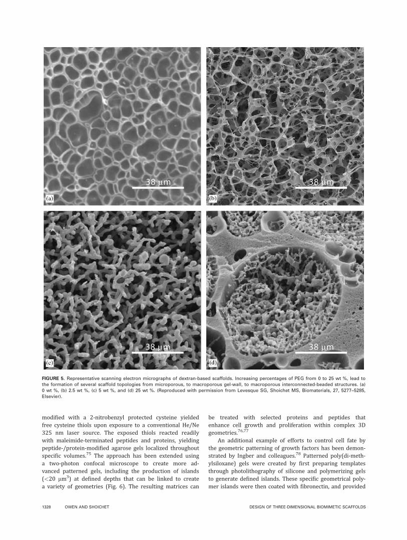
Tuning the porosity and permeability of the matrixThe need for a porous, interconnected matrix is apparent;however, defining pore dimensions and the degree of per-meability remains a significant challenge. One endeavor byShoichet and co-workers to control both the pore size andporosity of scaffolds used various concentrations of dextranand PEG.59 Formulations of 10 wt % dextran with increas-ing percentages of PEG from 0 to 25 wt %, led to the forma-tion of several scaffold topologies from microporous, tomacroporous gel-wall, to macroporous interconnected-beaded structures (Fig. 5). The interconnected-beaded scaf-folds contained pores with a median diameter of 41 lmthat were connected by narrower channels with a mediandiameter of 11 lm. An alternative approach from Hollisterand colleagues coupled solid free form (SFF) manufacturingwith sponge scaffold fabrication to cast an array of materi-als into porous architectures.60 Polyglycolide (PGA) andpolylactide (PLA) were formed into scaffolds using porogenleaching of NaCl or by solvent evaporation of chloroform.The resulting architecture featured global pore sizes of�100 lm, and local pore sizes ranging from 10 to 300 lm.Importantly, the architecture of scaffolds can be controlledto manipulate both global and local porosity and pore size.Mikos and co-workers developed a method of controllingthe porosity of chitosan scaffolds for osteogenic differentia-tion by incorporation of lysozyme at the material surface.Incubation with lysozyme degraded the polymer backbone,resulted in the formation of pores, and increased the overallporosity of the scaffold initially at 5–55% after 21 days.Bhatia and colleagues used lithography and microsyringedeposition to control the porosity of poly(DL-lactide-co-glyco-lide) (PLGA) scaffolds, and evaluated the advantages andlimitations of each fabrication technique.61 The majority ofresearch on scaffolds lies in fabrication of the material, forwhich several reviews have been written.9,62,63 A core chal-lenge of these technologies is to balance the integrity andmechanical support of scaffolds, while still allowing cellmigration, transport of nutrients, and removal of waste.
Regulating matrix degradationA central feature of several development processes is theability of cells to degrade and remodel their environment.Susceptibility of the substratum to proteolytic degradation,as well as its capacity to be remodeled, is essential in em-
bryonic development, angiogenesis, ductal formations, andwound healing. In recreating suitable matrices to recapitu-late these processes, it is imperative to include componentsthat allow for the natural remodeling of the ECM as seen inthe cells’ native environment. Efforts by Hubbell and col-leagues have led to the development of PEG acrylate poly-mer scaffolds that are modified with peptide cross link-ers.64,65 The peptide sequences contained in these materialsare sensitive to cleavage by specific proteases, such as colle-genase and plasmin. By also including cell adhesion pro-teins, these gels have shown great potential in allowing themigration of smooth muscle cells and fibroblasts.
In a separate approach, Mooney and colleagues exploredthe influence of matrix degradation rate on myoblast pheno-type.66 Scaffolds were engineered using alginate, and thedegradation rate was varied by partial oxidation of alginatebefore encapsulation of cells. Although the cellular prolifera-tion rate was highest in non-degradable gels, only myoblastscultured in degradable gels differentiated into multi-nucleated myofibers. Additional investigation may furtherclarify the role that scaffold degradation plays on cell differ-entiation and function.
Beyond degradation of the matrix in the cell microenvir-onment, the complete removal of scaffolds after the success-ful ex vivo generation of tissue could be advantageous. Scaf-fold removal would allow for the implantation of generatedtissue without the complications associated with physiologi-cal responses to the scaffold itself. Pioneering efforts in scaf-fold removal, or scaffold-less engineered tissues include thework of Okano, Matsuda,67 Auger,68 and others.
Incorporation of biochemical signalsVarious biochemical cues found in the ECM may include: in-soluble components (e.g., laminin, fibronectin); solublegrowth factors (e.g., neurotrophin-3, platelet-derived growthfactor); and matrix-bound factors (e.g., vascular endothelialgrowth factor). Inclusion of insoluble biochemical cues isessential for all ECM surrogates. The specific type and con-centration of factors should be based on the target cell.Hubbell et al. have constructed fibrin scaffolds while graft-ing heparin into the matrix during fibrinogen crosslink-ing.69,70 Specifically, the transglutaminase enzyme factorXIIIa was used to crosslink individual fibrinogen fibers, andto append a modified heparin protein to the backbone. Nat-ural heparin affinity is then used to bind a number ofgrowth factors, including vascular endothelial growth factor(VEGF), and has been used to guide the morphogenesis ofblood vessels. In contrast to the hyperpermeability oftenobserved when free VEGF is used to treat endothelial cells,the low concentrations incorporated into this polymer back-bone resulted in the formation of more normal vasculature.Current work by Prestwich has expanded the available ECMscaffold library by incorporating a number of growth factorsand cleavable crosslinking agents in hyaluronic acid basedmatrices.71–73
Innovative work by Shoichet and co-workers advancedmatrix design to include micropatterned biomolecular gra-dients into scaffolds for cell guidance.74 Agarose gels
REVIEW ARTICLE
JOURNAL OF BIOMEDICAL MATERIALS RESEARCH A | 15 SEP 2010 VOL 94A, ISSUE 4 1327
modified with a 2-nitrobenzyl protected cysteine yieldedfree cysteine thiols upon exposure to a conventional He/Ne325 nm laser source. The exposed thiols reacted readilywith maleimide-terminated peptides and proteins, yieldingpeptide-/protein-modified agarose gels localized throughoutspecific volumes.75 The approach has been extended usinga two-photon confocal microscope to create more ad-vanced patterned gels, including the production of islands(<20 lm3) at defined depths that can be linked to createa variety of geometries (Fig. 6). The resulting matrices can
be treated with selected proteins and peptides thatenhance cell growth and proliferation within complex 3Dgeometries.76,77
An additional example of efforts to control cell fate bythe geometric patterning of growth factors has been demon-strated by Ingber and colleagues.78 Patterned poly(di-meth-ylsiloxane) gels were created by first preparing templatesthrough photolithography of silicone and polymerizing gelsto generate defined islands. These specific geometrical poly-mer islands were then coated with fibronectin, and provided
FIGURE 5. Representative scanning electron micrographs of dextran-based scaffolds. Increasing percentages of PEG from 0 to 25 wt %, lead to
the formation of several scaffold topologies from microporous, to macroporous gel-wall, to macroporous interconnected-beaded structures. (a)
0 wt %, (b) 2.5 wt %, (c) 5 wt %, and (d) 25 wt %. (Reproduced with permission from Levesque SG, Shoichet MS, Biomaterials, 27, 5277–5285,
Elsevier).
1328 OWEN AND SHOICHET DESIGN OF THREE-DIMENSIONAL BIOMIMETIC SCAFFOLDS

adhesion sites for subsequently treated capillary endothelialcells. The size and shape of immobilized fibronectin islandsdictated the degree of cell spreading, and, therefore, guidedcell growth or apoptosis.
Multifaceted approaches in developing ECM surrogatesRecent scaffold designs have begun to incorporate both me-chanical and biochemical cues to support the phenotypicalgrowth and development of functional tissue. One importantaspect of current research is the attention on cell–cell inter-actions. For example, matrices are being designed to stabi-lize isolated hepatocytes in co-culture with fibroblasts.79
The PEG hydrogel-based scaffolds used in these studies con-sist of photo-patterned adhesive proteins specificallyselected based on hepatocyte integrin expression. Patternedhydrogels resulted in higher levels of albumin and urea, in-dicative of hepatocyte functionality.
In addition to adapting conventional polymers for scaf-fold synthesis, a number of novel materials are gaining pop-ularity for use as ECM surrogates. Among these are carbonnanotubes,80 silk nanofibers,81 and hydrogels.82 Althoughmany of these materials show promise as the next genera-tion of scaffolds for 3D cell culture, these surrogate extracel-lular matrices must be tuned to match the mechanical andchemical requirements specific to each target tissue toensure proper guidance of cell fate.
OUTLOOK
There is significant opportunity to advance the field furtherthrough the design of tunable scaffolds and the incorporationof multiple cell-based strategies. For example, little researchhas succeeded in developing scaffolds with mechanical prop-erties that can be tuned after fabrication. The ability toincrease scaffold rigidity after cells have been seeded wouldallow mechanical properties to be precisely tailored, whileavoiding complications during scaffold processing. Anotherpotential improvement would be the capacity to add cells intopatterned matrices following scaffold formation. In this man-ner, sensitive cells would not be exposed to detrimental proc-esses common during scaffold preparation.
Similarly, more research is needed to better understandcell–cell interactions in 3D environments. Further develop-ment of co-culture models and methods of cell seeding mayimprove the use of scaffolds for tissue engineering.
Elucidating the cellular response to environment andapplication of these mechanisms to the design of biomimeticscaffolds, offers great potential to control stem cell fate andguide tissue regeneration. Techniques that allow for finetuning of individual aspects of the cellular microenviron-ment will be essential in developing models to enhance ourunderstanding of the relationship between structure andfunction and as templates for complex tissues and organs.
ACKNOWLEDGMENTS
The authors are grateful to members of the Shoichet researchgroup for critical review of this manuscript and particularlyKaryn Ho for producing Figure 1. They thank NSERC and CIHRfor funding through the collaborative health research projectsprogram and the Canada Council for the Arts, Killam ResearchFoundation.
REFERENCES1. Schmeichel KL, Bissell MJ. Modeling tissue-specific signaling and
organ function in three dimensions. J Cell Sci 2003;116(Part 12):
2377–2388.
2. Cukierman E, Pankov R, Stevens DR, Yamada KM. Taking cell–ma-
trix adhesions to the third dimension. Science 2001;294:1708–1712.
3. Berrier AL, Yamada KM. Cell–matrix adhesion. J Cell Physiol
2007;213:565–573.
4. Lund AW, Yener B, Stegemann JP, Plopper GE. The natural and
engineered 3D microenvironment as a regulatory cue during stem
cell fate determination. Tissue Eng Part B Rev 2009;15:371–380.
5. Gieni RS, Hendzel MJ. Mechanotransduction from the ECM to the
genome: Are the pieces now in place? J Cell Biochem 2008;104:
1964–1987.
6. Prestwich GD. Evaluating drug efficacy and toxicology in three
dimensions: Using synthetic extracellular matrices in drug discov-
ery. Acc Chem Res 2008;41:139–148.
7. Griffith LG, Swartz MA. Capturing complex 3D tissue physiology
in vitro. Nat Rev Mol Cell Biol 2006;7:211–224.
8. Yamada KM, Cukierman E. Modeling tissue morphogenesis and
cancer in 3D. Cell 2007;130:601–610.
9. Madurantakam PA, Cost CP, Simpson DG, Bowlin GL. Science of
nanofibrous scaffold fabrication: Strategies for next generation
tissue-engineering scaffolds. Nanomed 2009;4:193–206.
10. Tibbitt MW, Anseth KS. Hydrogels as extracellular matrix mimics
for 3D cell culture. Biotechnol Bioeng 2009;103:655–663.
11. Hoffman AS. Hydrogels for biomedical applications. Adv Drug
Deliv Rev 2002;54:3–12.
12. Tsang VL, Bhatia SN. Fabrication of three-dimensional tissues.
Adv Biochem Eng Biotechnol 2007;103:189–205.
FIGURE 6. (a) Oblique and (b) side view of a hydrogel patterned using
muliphoton excitation to activate functional groups. Green squares
are 60 � 60 � 18 lm labeled with the green fluorescent dye AF488-
Mal. Circles are 50 lm in diameter and are labeled with the red fluo-
rescent dye AF546-Mal. (Reproduced with permission from Wosnick
JH, Shoichet MS, Chem Mater 2008, 20, 55–60, American Chemical
Society). [Color figure can be viewed in the online issue, which is
available at www.interscience.wiley.com.]
REVIEW ARTICLE
JOURNAL OF BIOMEDICAL MATERIALS RESEARCH A | 15 SEP 2010 VOL 94A, ISSUE 4 1329

13. Barnes CP, Sell SA, Boland ED, Simpson DG, Bowlin GL. Nano-
fiber technology: Designing the next generation of tissue engi-
neering scaffolds. Adv Drug Deliv Rev 2007;59:1413–1433.
14. Shoichet MS. Polymer scaffolds for biomaterials applications.
Macromolecules 2010;43:581–591.
15. Kleinman HK, Martin GR. Matrigel: Basement membrane matrix
with biological activity. Semin Cancer Biol 2005;15:378–386.
16. Choi JS, Yang HJ, Kim BS, Kim JD, Kim JY, Yoo B, Park K, Lee
HY, Cho YW. Human extracellular matrix (ECM) powders for
injectable cell delivery and adipose tissue engineering. J Control
Release 2009;139:2–7.
17. Laurencin CT, Nair LS. Nanotechnology and Tissue Engineering:
The Scaffold. Boca Raton: CRC Press; 2008.
18. Handel TM, Johnson Z, Crown SE, Lau EK, Sweeney M, Proudfoot
AE. Regulation of protein function by glycosaminoglycans: As
exemplified by chemokines. Annu Rev Biochem 2005;74:385–410.
19. Alison M. The Cancer Handbook. Chichester, West Sussex, Eng-
land, Hoboken, NJ: John Wiley & Sons; 2007. 1573 p.
20. Dye JF, Lawrence L, Linge C, Leach L, Firth JA, Clark P. Distinct
patterns of microvascular endothelial cell morphology are deter-
mined by extracellular matrix composition. Endo J Endo Cell Res
2004;11:151–167.
21. Shimizu Y, Rose DM, Ginsberg MH. Integrins in the Immune Sys-
tem. Advances in Immunology, Vol. 72. San Diego: Academic
Press Inc; 1999. p 325–380.
22. Schultz GS, Wysocki A. Interactions between extracellular matrix
and growth factors in wound healing. Wound Repair Regen 2009;
17:153–162.
23. Bissell MJ. Modeling molecular mechanisms of breast cancer and
invasion: Lessons from the normal gland. Biochem Soc Trans
2007;35(Part 1):18–22.
24. van der Flier A, Sonnenberg A. Function and interactions of integ-
rins. Cell Tissue Res 2001;305:285–298.
25. Schwartz MA. Integrin signaling revisited. Trends Cell Biol 2001;
11:466–470.
26. Mooney D, Hansen L, Vacanti J, Langer R, Farmer S, Ingber D.
Switching from differentiation to growth in hepatocytes: Control
by extracellular-matrix. J Cell Physiol 1992;151:497–505.
27. Nelson CM, Bissell MJ. Of extracellular matrix, scaffolds, and sig-
naling: Tissue architecture regulates development, homeostasis,
and cancer. Annu Rev Cell Dev Biol 2006;22:287–309.
28. Streuli CH, Schmidhauser C, Bailey N, Yurchenco P, Skubitz APN,
Roskelley C, Bissell MJ. Laminin mediates tissue-specific gene-
expression in mammary epithelia. J Cell Biol 1995;129:591–603.
29. Bissell MJ, Radisky DC, Rizki A, Weaver VM, Petersen OW. The
organizing principle: Microenvironmental influences in the normal
and malignant breast. Differentiation 2002;70:537–546.
30. Zaman MH, Trapani LM, Sieminski AL, Mackellar D, Gong H,
Kamm RD, Wells A, Lauffenburger DA, Matsudaira P. Migration of
tumor cells in 3D matrices is governed by matrix stiffness along
with cell–matrix adhesion and proteolysis. Proc Natl Acad Sci
USA 2006;103:10889–10894.
31. Engler AJ, Sen S, Sweeney HL, Discher DE. Matrix elasticity
directs stem cell lineage specification. Cell 2006;126:677–689.
32. Saha K, Keung AJ, Irwin EF, Li Y, Little L, Schaffer DV, Healy KE.
Substrate modulus directs neural stem cell behavior. Biophys J
2008;95:4426–4438.
33. Leipzig ND, Shoichet MS. The effect of substrate stiffness on
adult neural stem cell behavior. Biomaterials 2009;30:6867–6878.
34. Paszek MJ, Zahir N, Johnson KR, Lakins JN, Rozenberg GI, Gefen A,
Reinhart-King CA, Margulies SS, DemboM, Boettiger D. Tensional ho-
meostasis and themalignant phenotype. Cancer Cell 2005;8:241–254.
35. Ulrich TA, Pardo EMD, Kumar S. The mechanical rigidity of the
extracellular matrix regulates the structure, motility, and prolifera-
tion of glioma cells. Cancer Res 2009;69:4167–4174.
36. McClelland RE, Coger RN. Use of micropathways to improve oxy-
gen transport in a hepatic system. J Biomech Eng Trans Asme
2000;122:268–273.
37. Lenhard RE, Osteen RT, Gansler TS, American Cancer S. Clinical
Oncology. Atlanta, GA: American Cancer Society; 2001.
38. Lieberman JR, Friedlander GE. Bone Regeneration and Repair:
Biology and Clinical Applications. Totowa, NJ: Humana Press;
2005. 398 p.
39. Oh SH, Park IK, Kim JM, Lee JH. In vitro and in vivo characteris-
tics of PCL scaffolds with pore size gradient fabricated by a cen-
trifugation method. Biomaterials 2007;28:1664–1671.
40. Bissell MJ, Hall HG, Parry G. How does the extracellular-matrix
direct gene-expression. J Theor Biol 1982;99:31–68.
41. Engler AJ, Humbert PO, Wehrle-Haller B, Weaver VM. Multiscale
modeling of form and function. Science 2009;324:208–212.
42. Talhouk RS, Chin JR, Unemori EN, Werb Z, Bissell MJ. Protein-
ases of the mammary gland: Developmental regulation in vivo
and vectorial secretion in culture. Development 1991;112:439–449.
43. Liu WF, Chen CS. Cellular and multicellular form and function.
Adv Drug Deliv Rev 2007;59:1319–1328.
44. Desai RA, Gao L, Raghavan S, Liu WF, Chen CS. Cell polarity trig-
gered by cell–cell adhesion via E-cadherin. J Cell Sci 2009;122:
905–911.
45. Parmar H, Cunha GR. Epithelial-stromal interactions in the mouse
and human mammary gland in vivo. Endocr Relat Cancer 2004;
11:437–458.
46. Bennett DC, Armstrong BL, Okada SM. Reconstitution of branch-
ing tubules from two cloned mammary cell types in culture. Dev
Biol 1981;87:193–199.
47. Araque A, Carmignoto G, Haydon PG. Dynamic signaling between
astrocytes and neurons. Annu Rev Physiol 2001;63:795–813.
48. Pasti L, Volterra A, Pozzan T, Carmignoto G. Intracellular calcium
oscillations in astrocytes: A highly plastic, bidirectional form of
communication between neurons and astrocytes in situ. J Neuro-
sci 1997;17:7817–7830.
49. Khetani SR, Bhatia SN. Microscale culture of human liver cells for
drug development. Nat Biotechnol 2008;26:120–126.
50. March S, Hui EE, Underhill GH, Khetani S, Bhatia SN. Microenvir-
onmental regulation of the sinusoidal endothelial cell phenotype
in vitro. Hepatology 2009;50:920–928.
51. Langer R, Vacanti JP. Tissue engineering. Science 1993;260:
920–926.
52. Kuo YC, Ku IN. Cartilage regeneration by novel polyethylene oxide/
chitin/chitosan scaffolds. Biomacromolecules 2008;9:2662–2669.
53. Shapira-Schweitzer K, Seliktar D. Matrix stiffness affects sponta-
neous contraction of cardiomyocytes cultured within a PEGylated
fibrinogen biomaterial. Acta Biomaterialia 2007;3:33–41.
54. Xin XJ, Borzacchiello A, Netti PA, Ambrosio L, Nicolais L. Hyal-
uronic-acid-based semi-interpenetrating materials. J Biomater Sci
Polym Ed 2004;15:1223–1236.
55. Legant WR, Pathak A, Yang MT, Deshpande VS, McMeeking RM,
Chen CS. Microfabricated tissue gauges to measure and manipu-
late forces from 3D microtissues. Proc Natl Acad Sci USA 2009;
106:10097–10102.
56. Isenberg BC, DiMilla PA, Walker M, Kim S, Wong JY. Vascular
smooth muscle cell durotaxis depends on substrate stiffness gra-
dient strength. Biophys J 2009;97:1313–1322.
57. Anderson DG, Levenberg S, Langer R. Nanoliter-scale synthesis
of arrayed biomaterials and application to human embryonic
stem cells. Nat Biotechnol 2004;22:863–866.
58. Huang S, Ingber DE. Shape-dependent control of cell growth, dif-
ferentiation, and apoptosis: Switching between attractors in cell
regulatory networks. Exp Cell Res 2000;261:91–103.
59. Levesque SG, Lim RM, Shoichet MS. Macroporous interconnected
dextran scaffolds of controlled porosity for tissue-engineering
applications. Biomaterials 2005;26:7436–7446.
60. Taboas JM, Maddox RD, Krebsbach PH, Hollister SJ. Indirect solid
free form fabrication of local and global porous, biomimetic and
composite 3D polymer-ceramic scaffolds. Biomaterials 2003;24:
181–194.
61. Vozzi G, Flaim C, Ahluwalia A, Bhatia S. Fabrication of PLGA scaf-
folds using soft lithography and microsyringe deposition. Bioma-
terials 2003;24:2533–2540.
62. Tsang VL, Bhatia SN. Three-dimensional tissue fabrication. Adv
Drug Deliv Rev 2004;56:1635–1647.
63. Shin H. Fabrication methods of an engineered microenvironment
for analysis of cell–biomaterial interactions. Biomaterials 2007;28:
126–133.
64. Sawhney AS, Pathak CP, Hubbell JA. Bioerodible hydrogels based
on phoropolymerized poly(ethylene glycol)-co-poly(alpha-hydroxy
acid) diacrylate macromers. Macromolecules 1993;26:581–587.
1330 OWEN AND SHOICHET DESIGN OF THREE-DIMENSIONAL BIOMIMETIC SCAFFOLDS

65. Jo YS, Gantz J, Hubbell JA, Lutolf MP. Tailoring hydrogel degra-
dation and drug release via neighboring amino acid controlled
ester hydrolysis. Soft Matter 2009;5:440–446.
66. Boontheekul T, Hill EE, Kong HJ, Mooney DJ. Regulating myo-
blast phenotype through controlled gel stiffness and degradation.
Tissue Eng 2007;13:1431–1442.
67. Ohya S, Nakayama Y, Matsuda T. Thermoresponsive artificial
extracellular matrix for tissue engineering: Hyaluronic acid bio-
conjugated with poly(N-isopropylacrylamide) grafts. Biomacromo-
lecules 2001;2:856–863.
68. Auger FoA, Berthod Fo, Moulin Vr, Pouliot R, Germain L. Tissue-
engineered skin substitutes: From in vitro constructs to in vivo
applications. 2004;39(Part 3):263–275.
69. Schense JC, Hubbell JA. Cross-linking exogenous bifunctional
peptides into fibrin gels with factor XIIIa. Bioconjug Chem 1999;
10:75–81.
70. Hubbell JA. Matrix-bound growth factors in tissue repair. Swiss
Med Weekly 2006;136:387–391.
71. Vanderhooft JL, Mann BK, Prestwich GD. Synthesis and charac-
terization of novel thiol-reactive poly(ethylene glycol) cross-linkers
for extracellular-matrix-mimetic biomaterials. Biomacromolecules
2007;8:2883–2889.
72. Serban MA, Prestwich GD. Modular extracellular matrices: Solu-
tions for the puzzle. Methods 2008;45:93–98.
73. Zhang J, Skardal A, Prestwich GD. Engineered extracellular matri-
ces with cleavable crosslinkers for cell expansion and easy cell re-
covery. Biomaterials 2008;29:4521–4531.
74. Cao X, Shoichet MS. Photoimmobilization of biomolecules within
a 3-dimensional hydrogel matrix. J Biomater Sci Polym Ed 2002;
13:623–636.
75. Wosnick JH, Shoichet MS. Three-dimensional chemical patterning
of transparent hydrogels. Chem Mater 2008;20:55–60.
76. Aizawa Y, Leipzig N, Zahir T, Shoichet M. The effect of immobi-
lized platelet derived growth factor AA on neural stem/progenitor
cell differentiation on cell-adhesive hydrogels. Biomaterials 2008;
29:4676–4683.
77. Wylie RG, Shoichet MS. Two-photon micropatterning of amines
within an agarose hydrogel. J Mater Chem 2008;18:2716–2721.
78. Dike LE, Chen CS, Mrksich M, Tien J, Whitesides GM, Ingber DE.
Geometric control of switching between growth, apoptosis, and
differentiation during angiogenesis using micropatterned sub-
strates. In Vitro Cell Dev Biol Anim 1999;35:441–448.
79. Tsang VL, Chen AA, Cho LM, Jadin KD, Sah RL, DeLong S, West
JL, Bhatia SN. Fabrication of 3D hepatic tissues by additive photo-
patterning of cellular hydrogels. FASEB J 2007;21:790–801.
80. Edwards SL, Werkmeister JA, Ramshaw JAM. Carbon nanotubes in
scaffolds for tissue engineering. Exp RevMed Dev 2009;6:499–505.
81. McClure MJ, Sell SA, Ayres CE, Simpson DG, Bowlin GL. Electro-
spinning-aligned and random polydioxanone-polycaprolactone-
silk fibroin-blended scaffolds: Geometry for a vascular matrix.
Biomed Mater 2009;4.
82. Peppas NA, Hilt JZ, Khademhosseini A, Langer R. Hydrogels in
biology and medicine: From molecular principles to bionanotech-
nology. Adv Mater 2006;18:1345–1360.
REVIEW ARTICLE
JOURNAL OF BIOMEDICAL MATERIALS RESEARCH A | 15 SEP 2010 VOL 94A, ISSUE 4 1331