development 133, 1901-1910 (2006) …dev.biologists.org/content/develop/133/10/1901.full.pdf ·...
TRANSCRIPT
DEVELO
PMENT
1901RESEARCH ARTICLE
INTRODUCTIONCongenital defects of optic cup morphogenesis represent animportant cause of childhood blindness or vision impairment(Gregory-Evans et al., 2004). Many ocular developmental defectsare of unknown etiology and some may be caused by environmentalinfluences such as vitamin A deficiency in humans (Hornby et al.,2002) or animals (Wilson et al., 1953; Marsh-Armstrong et al., 1994;Dickman et al., 1997; Reijntjes et al., 2005). One important functionof vitamin A (retinol) is to serve as the precursor of retinoic acid(RA), an important developmental signaling molecule (Duester,2000). Some studies suggest a connection between impaired retinolor RA function and developmental eye defects in humans and mice.For instance, humans with missense mutations in the gene encodingserum retinol binding protein have been shown to exhibit retinaldystrophy (Seeliger et al., 1999). Also, mice with reduced RAreceptor signaling give rise to offspring with ocular defects (Lohneset al., 1994).
RA serves as a ligand for three nuclear RA receptors (RAR) thatbind DNA as heterodimers with retinoid X receptors (RXR) anddirectly regulate gene expression. Binding of all-trans-RA to theRAR component of RAR/RXR heterodimers is necessary andsufficient to rescue signaling in RA-deficient embryos, whereas theisomer 9-cis-RA (which can bind RXR) is unnecessary and isundetectable under physiological conditions (Mic et al., 2003).RARs are expressed in overlapping patterns in the eye during
development, and mice carrying single null mutations of RARs haverelatively normal eye development except for RAR� null mice,which exhibit a retrolenticular membrane in the vitreous body(Ghyselinck et al., 1997). Genetic elimination of two RARs resultsin microphthalmia, ventral shortening of the retina, andabnormalities of the cornea, eyelids and conjunctiva (Lohnes et al.,1994). Thus, RARs mediate the ocular functions of vitamin A, as thedefects observed are essentially the same as those seen duringgestational vitamin A deficiency.
Based upon the expression patterns of RA-synthesizing enzymes,RA was proposed to control dorsoventral patterning of the retina(Wagner et al., 2000). However, functional studies on these enzymeshave not supported a role for RA in the establishment of retinaldorsoventral patterning (Fan et al., 2003; Matt et al., 2005) or cellfate determination (Mic et al., 2004). A recent study in chick, usingoverexpression of dominant-negative RA receptors has suggestedthat RA is needed to maintain retinal dorsoventral patterning at laterstages (Sen et al., 2005), but this has not been examined genetically.By contrast, RA has been clearly defined as a signaling moleculeneeded for patterning of other regions of the central nervous system,including the hindbrain and the spinal cord (Maden, 2002).Although it is clear that RA controls eye development, itsmechanism may be different from that of other eye signalingmolecules that control patterning or cell fate determination, such asfibroblast growth factor, which is released by the surface ectodermto stimulate neural retina differentiation (Russell, 2003), or activin,which is released by perioptic mesenchyme to stimulate retinalpigment epithelium differentiation (Fuhrmann et al., 2000). A recentgenetic study in mice suggests that RA controls eye development byacting within the neural crest-derived perioptic mesenchyme (Mattet al., 2005). The mechanism of RA action during eye development
Retinoic acid guides eye morphogenetic movements viaparacrine signaling but is unnecessary for retinaldorsoventral patterningAndrei Molotkov, Natalia Molotkova and Gregg Duester*
Retinoic acid (RA) is required for patterning of the posterior nervous system, but its role in the retina remains unclear. RA issynthesized in discrete regions of the embryonic eye by three retinaldehyde dehydrogenases (RALDHs) displaying distinctexpression patterns. Overlapping functions of these enzymes have hampered genetic efforts to elucidate RA function in the eye.Here, we report Raldh1, Raldh2 and Raldh3 single, double and triple null mice exhibiting progressively less or no RA synthesis in theeye. Our genetic studies indicate that RA signaling is not required for the establishment or maintenance of dorsoventral patterningin the retina, as we observe normal expression of Tbx5 and ephrin B2 (Efnb2) dorsally, plus Vax2 and Ephb2 ventrally. Instead, RA isrequired for the morphogenetic movements needed to shape the developing retina and surrounding mesenchyme. At early stages,Raldh2 expressed in mesenchyme and Raldh3 expressed in the retinal pigmented epithelium generate RA that delivers an essentialsignal to the neural retina required for morphogenetic movements that lead to ventral invagination of the optic cup. At laterstages, Raldh1 expressed in dorsal neural retina and Raldh3 expressed in ventral neural retina (plus weaker expression of each inlens/corneal ectoderm) generates RA that travels to surrounding mesenchyme, where it is needed to limit the anterior invasion ofperioptic mesenchyme during the formation of corneal mesenchyme and eyelids. At all stages, RA target tissues are distinct fromlocations of RA synthesis, indicating that RALDHs function cell-nonautonomously to generate paracrine RA signals that guidemorphogenetic movements in neighboring cells.
KEY WORDS: Retinoic acid, Morphogenetic movements, Raldh1, Raldh2, Raldh3, Tbx5, Vax2, EphB2, ephrin B2, Eye, Optic cup, Retina,Perioptic mesenchyme, Mouse
Development 133, 1901-1910 (2006) doi:10.1242/dev.02328
Burnham Institute for Medical Research, 10901 North Torrey Pines Road, La Jolla, CA92037, USA.
*Author for correspondence (e-mail: [email protected])
Accepted 16 February 2006
DEVELO
PMENT
1902
remains unclear largely because of an incomplete understanding ofthe complex spatiotemporal properties of RA synthesis in this organand of the target tissues upon which RA acts.
The first step of RA synthesis (the oxidation of retinol toretinaldehyde) occurs ubiquitously through the action of severaloverlapping alcohol dehydrogenases and short-chaindehydrogenase/reductases (Molotkov et al., 2002). By contrast, thesecond step of RA synthesis (oxidation of retinaldehyde to RA) islimited to specific tissues by retinaldehyde dehydrogenases (i.e.RALDH1, RALDH2, RALDH3) expressed in non-overlappingpatterns in the developing mouse eye (Mic et al., 2002; Matt et al.,2005). Despite their unique expression patterns, analyses of Raldh1,Raldh2 and Raldh3 null mice have not resulted in clearly definedocular defects, probably because of the cell-nonautonomous actionof RA generated by the remaining enzymes (Niederreither et al.,1999; Fan et al., 2003; Dupé et al., 2003; Mic et al., 2004). Here,RALDH single and compound null mutant mice have been analyzedto provide further insight into the role of RA in eye development,and to sort out the individual contributions of each enzyme. We findthat RA signaling guides the morphogenetic movements of the retinaand perioptic mesenchyme, rather than playing a role in retinaldorsoventral patterning. Furthermore, our studies reveal severalparacrine mechanisms whereby RA is released from RALDH-expressing cells and guides morphogenetic movements inneighboring cells, thus demonstrating that RALDHs function cell-nonautonomously in eye development.
MATERIALS AND METHODSGeneration of Raldh3 null miceA gene targeting vector for Raldh3 was designed to generate a floxed alleleof Raldh3 with the ultimate goal of eliminating exon 11, similar to a Raldh1gene targeting vector that generated a null phenotype in that case (Fan et al.,2003). The Raldh3 gene targeting vector thus included a loxP site upstreamof exon 11, followed by exon 11, a PGK-neo positive-selection genedownstream of exon 11 (transcribed in same direction as Raldh3), andanother loxP site just downstream of PGK-neo. In order to generate a nullallele, matings were performed between the initial Raldh3-targeted chimericmales and wild-type females expressing Cre recombinase in the germline inorder to remove exon 11 (i.e. about 600 bp of Raldh3) and PGK-neo in allcells. PCR genotyping of tail or yolk sac DNA was performed using theprimers 5�-GCCATAAAAGCTGGGGTGTCTG-3� (located in intron 10)and 5�-TGGATGGATGGATGGGTGATG-3� (located in intron 11) whichproduce a wild-type product of ~950 bp and a mutant product of ~350 bp(primer annealing temperature was 63°C). Initial matings of Raldh3+/–
heterozygous null mice resulted in 70 adult offspring with the followinggenotypes: 0 –/– (0%), 45 +/– (64%), 25 +/+ (36%). The absence ofsurviving homozygous mice indicated that the Raldh3 knockout leads to alethal phenotype. The description of another Raldh3–/– mouse line indicatedthe existence of a newborn lethal defect due to blockage of the nasalpassages, leading to respiratory stress at birth (Dupé et al., 2003).
Generation of compound Raldh null embryosSeveral Raldh mutant mice have been previously described, includingRaldh1–/– mice, which survive to adulthood (Fan et al., 2003), Raldh2–/–
embryos, which exhibit midgestation lethality (Mic et al., 2002), andRaldh1–/–;Raldh2–/– double null embryos, which also exhibit midgestationlethality (Mic et al., 2004). Generation of compound RALDH null mice wasfacilitated by independent assortment of the three RALDH genes on separatechromosomes and by the ability to obtain adult mice homozygous for theRaldh1 null allele. Matings were performed between the above mice andRaldh3–/– mice to obtain Raldh1–/–;Raldh3–/– and Raldh2–/–;Raldh3–/– doublenull embryos, as well as Raldh1–/–;Raldh2–/–;Raldh3–/– triple null embryos.Embryos derived from timed matings were genotyped by PCR analysis ofyolk sac DNA. Following mating, noon on the day of vaginal plug detectionwas considered as embryonic day 0.5 (E0.5). Embryos were stagedaccording to somite number.
Rescue of eye defects with dietary RA supplementationThe rescue of Raldh2–/– embryos by maternal dietary RA supplementationwas performed similar to a previous description (Mic et al., 2004), with anRA dose demonstrated to be in the normal physiological range (Mic et al.,2003). Briefly, all-trans-RA (Sigma) was dissolved in corn oil and mixedwith powdered mouse chow to provide a final concentration of 0.1 mg/g fortreatment from E6.75-E8.5 (limited rescue), or 0.2 mg/g for treatment fromE8.5-E14.5. In some cases, embryos were analyzed when the mother wasstill on the RA-supplemented diet, but in other cases the mother was returnedto standard mouse chow and embryos were analyzed at a later time point.Such food was prepared fresh twice daily (morning and evening) andprovided ad libitum.
Detection of retinoic acidRA activity was detected as previously described in embryos carrying theRARE-lacZ RA-reporter transgene, which places lacZ (encoding �-galactosidase) under the transcriptional control of a retinoic acid responseelement (RARE) (Rossant et al., 1991). Following genetic crosses of RARE-lacZ mice with RALDH mutant mice, the RARE-lacZ transgene segregatedindependently from Raldh1 and Raldh2, but not Raldh3, null alleles,suggesting that RARE-lacZ is located close to Raldh3 on mousechromosome 7. In order to detect RARE-lacZ expression in Raldh3–/–
embryos, 20 male mice obtained from RARE-lacZ�Raldh3+/– crosses werescreened for a crossover event by mating to wild-type females, and one malewas identified that carried RARE-lacZ linked to the Raldh3 null allele. TheRARElacZ-Raldh3 crossover male was also mated to Raldh1 and Raldh2mutant mice to enable the analysis of RA activity in double and triple Raldhmutants. Stained embryos were embedded in 3% agarose and sectioned at50 �m with a vibratome.
In situ hybridization, proliferation and apoptosis assaysWhole-mount in situ hybridization was performed to detect expression ofRaldh1, Raldh2 and Raldh3, as described previously (Mic et al., 2002).Probes for Tbx5 (Chapman et al., 1996) and Vax2 (Mui et al., 2002) werealso examined. Histological examination was performed on paraffin-sectioned tissues stained with hematoxylin/eosin, as previously described(Fan et al., 2003). Cell proliferation assays were performed on paraffin tissuesections by staining for histone-3 phosphorylation using anti-phosphohistone-3 antibodies (Upstate Cell Signaling Solutions, Lake Placid,NY) as previously reported (Molotkova et al., 2005); cells counts wereaverages of six sections from comparable ocular regions of two embryos(three sections per eye). Apoptosis was examined in paraffin tissue sectionsusing a TUNEL assay, as described (Hyer et al., 2003).
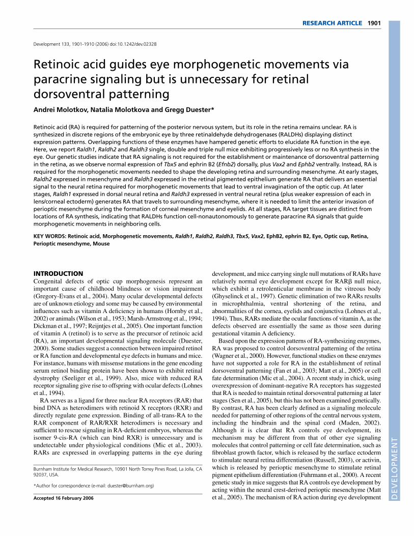
RESULTSLack of RA results in morphogenetic movementdefectsOur previous studies demonstrated that Raldh1–/– mice survive toadulthood and exhibit no noticeable defects in eye development,although they completely lose RA synthesis in the dorsal neuralretina (Fan et al., 2003). Other studies have recently shown thatRaldh3–/– mice die at birth as a result of nasal defects and exhibit amild delay in ventral retina growth (Dupé et al., 2003). We generatedan independent line of Raldh3–/– mice, as well asRaldh1–/–;Raldh3–/– double null mice, and examined the effect onocular development. Compared with wild-type and Raldh1–/–
embryos, which appeared identical at E14.5, Raldh3–/– embryosinitiated optic cup formation but exhibited completely penetrantocular defects consisting of excessive thickening of the centralneural retina, loss of the vitreous body, and a prominentretrolenticular membrane between the retina and lens (Fig. 1A-C;n=5 out of 5). Our Raldh3–/– null mutant exhibits more of a centralretina defect than that of another Raldh3–/– null mutant that wasreported to exhibit a shortening of the ventral retina (Matt et al.,2005). This could be due to strain differences, especially differencesin how efficiently Raldh2 acts in the two strains during early optic
RESEARCH ARTICLE Development 133 (10)
DEVELO
PMENT
cup formation (see below). Together, the findings from both strainssuggest that RALDH3 synthesizes the RA required to completeoptic cup and vitreous body formation.
Unexpectedly, Raldh1–/–;Raldh3–/– double null embryosexhibited not only the same ocular defects as Raldh3–/– embryos,but also excessive invasion of periocular mesenchyme anterior tothe retina, resulting in abnormal thickening of the corneal stromaand eyelid folds (Fig. 1D; n=3 out of 3). This observation revealsthat Raldh1 and Raldh3 both function to control the invasion ofperioptic mesenchyme around the retina that normally occursduring anterior eye formation (Cvekl and Tamm, 2004). We findthat loss of both Raldh1 and Raldh3 is required to observe thedefect because of functional redundancy. Another recent study alsoobserved excessive perioptic mesenchyme in Raldh1–/–;Raldh3–/–
double null embryos (Matt et al., 2005). As Raldh1 and Raldh3 areexpressed in different regions of the retina but not in the periocularmesenchyme (Fig. 2G,I), this suggests that RA synthesized in the
retinal epithelium travels as a signal to the surroundingmesenchyme to control morphogenetic movements ofmesenchymal ocular tissues.
The location of RA signaling activity in the developing eye atE10.5 was examined in whole-mount wild-type and Raldh mutantmice carrying the RARE-lacZ RA-reporter transgene (Rossant et al.,1991). Raldh3–/– embryos exhibited a large decrease in eye RAactivity compared with wild-type embryos, whereas Raldh1–/–
embryos exhibited only a modest decrease (Fig. 1E-G). Raldh3–/–
embryos completely lost RA activity in the ventral retina andolfactory pit, tissues that express Raldh3 but not Raldh1 or Raldh2at E10.5 (Fig. 2G-I). E10.5 Raldh1–/–;Raldh3–/– embryos carryingRARE-lacZ exhibited almost a total loss of RA activity in the eye,demonstrating that Raldh1 is primarily responsible for the RAactivity remaining in the eye of Raldh3–/– embryos (Fig. 1H);residual RA activity in Raldh1–/–;Raldh3–/– eyes is due to RAsynthesized by Raldh2 up to the point when its expression ends near
1903RESEARCH ARTICLEControl of eye morphogenetic movements by RA
Fig. 1. Morphogeneticmovement defects indeveloping mouse eyesdeficient for RA synthesis.(A-D) Hematoxylin and eosinstaining of frontal sectionsthrough the eye of E14.5 wild-type (WT), Raldh1–/– (R1–/–),Raldh3–/– (R3–/–) andRaldh1–/–;Raldh3–/– double null(R1;R3–/–) embryos. The double-arrow indicates thickening of theneural retina in R3–/– andR1;R3–/– embryos; notice thelack of a vitreous body and thepresence of a retrolenticularmembrane in these mutants.(E-H) Expression of RARE-lacZ (anRA-reporter transgene) in whole-mount E10.5 wild-type and mutant embryos. Note the near complete loss of RARE-lacZ expression in eyes of R1;R3–/– embryos. Olfactory pitstaining is completely dependent upon Raldh3, but forebrain staining is not dependent upon Raldh1 or Raldh3. c, cornea; D, dorsal; e, eye; el,eyelid; f, forebrain; olf, olfactory pit; pm, perioptic mesenchyme; rlm, retrolenticular membrane; V, ventral; vb, vitreous body.
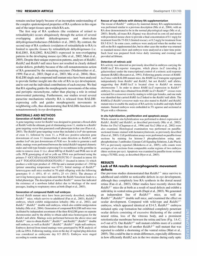
Fig. 2. Rescue of Raldh1–/–;Raldh3–/–
eye morphogenetic movements bymaternal dietary RAsupplementation. (A-F) Raldh1–/– andRaldh1–/–;Raldh3–/– littermates weresubjected to maternal dietary RAsupplementation from E8.5 until thepoint of analysis, then analyzed byhematoxylin-eosin staining of frontalsections through the eye of E14.5embryos (A,B), or RARE-lacZ expression infrontal sections of E11.5 (C,D) and E12.5(E,F) eyes. (G-K) Whole-mount in situhybridization to detect the expression ofRaldh1, Raldh2 and Raldh3 in E10.5whole-mount eyes (G-I), and Crabp1 andCrabp2 in E11.5 eyes following frontalsectioning (J,K). c, cornea; D, dorsal; el,eyelid; nr, neural retina; nr/vb, neuralretina/vitreous body junction; olf,olfactory pit; pm, perioptic mesenchyme;rpe, retinal pigment epithelium; V,ventral; vb, vitreous body.
DEVELO
PMENT
1904
E9.75 (see below). These results confirm that RARE-lacZ is aspecific marker for eye RA signaling, as its expression is totallydependent upon RA-synthesizing enzymes.
Rescue of retinal and mesenchymal ocular defectsby maternal RA treatmentAs early embryonic defects in Raldh2–/– embryos can be rescued bymaternal dietary RA supplementation (Mic et al., 2004), we usedthis method to rescue eye development in Raldh1–/–;Raldh3–/–
embryos. Following RA treatment from E8.5-E14.5, we found thatE14.5 Raldh1–/–;Raldh3–/– embryos exhibited eye development thatwas very similar to normal with a vitreous body now clearlyobservable, a neural retina of close to normal thickness, a diffuseretrolenticular membrane, and normal perioptic mesenchymegrowth (Fig. 2A,B; n=3 out of 4). Examination of E11.5 rescuedRaldh1–/–;Raldh3–/– embryos carrying RARE-lacZ revealed thatmaternally-derived RA had reached the neural retina/vitreous bodyjunction and dorsal perioptic mesenchyme (Fig. 2D; n=4 out of 4),although such RA activity was significantly less than that observedin Raldh1–/– littermates, where Raldh3 is generating RA locally(Fig. 2C). As an unrescued E11.5 Raldh1–/–;Raldh3–/– eyecompletely lacked RA activity (Fig. 7E), maternal RA wasresponsible for the RA detected in rescued mutants. Interestingly,the RA activity detected in the neural retina of rescuedRaldh1–/–;Raldh3–/– embryos was not uniformly distributed, but wasfound along the periphery of the neural retina closest to the vitreousbody (Fig. 2D). It is also apparent that such RA activity is notdetected in the dorsal-most or ventral-most neural retina whereRaldh1 and Raldh3 are expressed (Fig. 2G,I) and where RA activityis normally found at high levels (Fig. 2C), but instead is observedin the central neural retina (Fig. 2D). Analysis of E12.5 rescuedRaldh1–/–;Raldh3–/– embryos demonstrated high RA activity in thevitreous body and ventral perioptic mesenchyme, and RA was moreuniformly distributed throughout the central neural retina but wasstill not detected in the dorsal-most or ventral-most regions of theneural retina, which normally have highest levels of RA activity(Fig. 2E,F; n=2 out of 2). These results reveal that the targets of RAaction during eye development (inferred from the action of rescuinglevels of maternal RA) are distinct from the normal sites of RAsynthesis.
Our observations with the RA-reporter transgene demonstratethat the low dose of maternal dietary RA used to rescueRaldh1–/–;Raldh3–/– eye development is evidently not stimulatingRA signaling in all portions of the eye. One possible explanationfor this observation is that RA may be preferentially degraded incertain embryonic tissues, as has been observed for rhombomere4 of the hindbrain, which induces a RA-degrading enzymeencoded by Cyp26c1 to create an RA boundary (Sirbu et al., 2005).Although Cyp26a1 and Cyp26c1 function in RA degradation in thecentral retina during late eye development (E15.5), no such rolehas been found for earlier stages, and certainly no role has beenfound in the dorsal or ventral retina at any stage (Sakai et al.,2004). Another possibility for the selective appearance of RAactivity in rescued eyes is that incoming RA may be preferentiallysequestered by cellular RA-binding proteins (CRABP) thatfacilitate RA signaling (Noy, 2000). In support of this possibility,we found that Crabp1 is preferentially expressed in the vitreousbody (and weakly in perioptic mesenchyme), and that Crabp2 isexpressed in the dorsal and ventral perioptic mesenchyme (Fig.2J,K). These mesenchymal sites of CRABP expression correspondto the regions in which the highest RA activity is found during therescue.
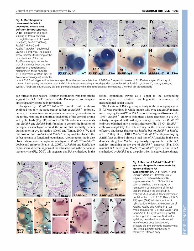
RA generated by Raldh1 controls periopticmesenchyme invasionOur experiments indicate that either Raldh1 or Raldh3 alonegenerates sufficient RA to control anterior invasion of periopticmesenchyme both dorsally and ventrally in Raldh3–/– embryos.However, at E10.5, we detected RA activity in the dorsal but notventral perioptic mesenchyme of Raldh3–/– embryos (Fig. 1G). Thisdiscrepancy was resolved by examination at a later stage (E11.5),at which point Raldh3–/– embryos did exhibit RA activity in bothdorsal and ventral mesenchyme (Fig. 3A,B). Thus, RALDH1evidently generates increasing levels of RA in the dorsal retinaduring development that are sufficient to reach the dorsal andventral perioptic mesenchyme by E11.5 to fulfill RA function inthis tissue.
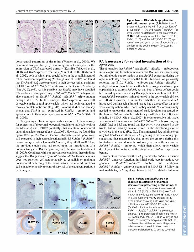
RA is required for selective cell death in periopticmesenchymeIn order to investigate RA function in the perioptic mesenchyme,we examined the effect of a loss of RA on cell proliferation andapoptosis. Ocular cell proliferation, examined in E11.5 embryos bythe detection of phosphohistone-3 (H3P), was not significantlydifferent between Raldh1–/– embryos that maintain relativelynormal RA activity and Raldh1–/–;Raldh3–/– littermates that lose RAactivity (Fig. 4A,B). The number of H3P-positive cells in Raldh1–/–
perioptic mesenchyme was 99±7.3, whereas in Raldh1–/–;Raldh3–/–
littermates the number was 95±7.6 (n=6). For retina, the number ofH3P-positive cells in Raldh1–/– embryos was 26.3±2.8, whereas inRaldh1–/–;Raldh3–/– littermates the number was 27.5±1.1 (n=6).Thus, loss of RA does not effect cell proliferation in either periopticmesenchyme or retina. However, a loss of RA does have an effecton apoptosis, as described in a recent study where dorsal and ventralregions of apoptosis detected in the perioptic mesenchyme of wild-type embryos were missing in Raldh1–/–;Raldh3–/– embryos (Mattet al., 2005). We found that these dorsal and ventral regions ofperioptic mesenchyme apoptosis were still present in E11.5Raldh1–/– embryos, but that almost no apoptotic cells weredetectable in Raldh1–/–;Raldh3–/– littermates (Fig. 4C,D; n=3 out of3). The pattern of apoptosis in the retina did not appear to besignificantly different. These findings suggest that RA derived fromeither RALDH1 or RALDH3 functions in the periopticmesenchyme to limit cell numbers by stimulating apoptosis inselective regions.
RA is unnecessary to establish or maintaindorsoventral patterning of the retinaThe unique expression patterns of Raldh1 in the dorsal retina andRaldh3 in the ventral retina led to the hypothesis that RA generateddifferentially in these two regions may somehow control
RESEARCH ARTICLE Development 133 (10)
Fig. 3. Raldh1-mediated RA signaling to perioptic mesenchyme.(A,B) RARE-lacZ expression in frontal sections of E11.5 wild-type (A)and Raldh3–/– (B) eyes. D, dorsal; pm, perioptic mesenchyme; V, ventral.
DEVELO
PMENT
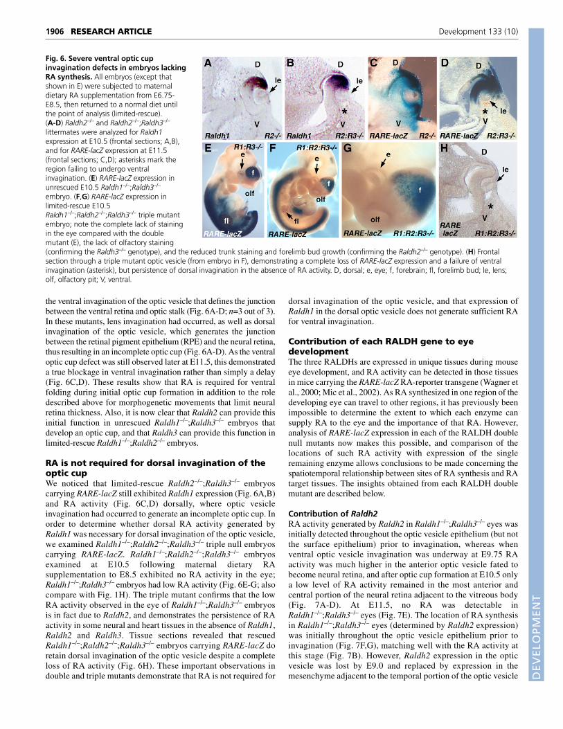
dorsoventral patterning of the retina (Wagner et al., 2000). Weexamined this possibility by examining mutant embryos for theexpression of Tbx5 expressed dorsally (Koshiba-Takeuchi et al.,2000) and Vax2 expressed ventrally (Mui et al., 2002; Barbieri etal., 2002), both of which play crucial roles in the establishment ofretinal dorsoventral patterning (McLaughlin et al., 2003). We foundthat Tbx5 and Vax2 were expressed in their correct retinal positionsin E10.5 Raldh1–/–;Raldh3–/– embryos that lack eye RA activity(Fig. 5A-C; n=3). As it is possible that Raldh2 may have suppliedRA for dorsoventral patterning in Raldh1–/–;Raldh3–/– embryos, wealso examined an Raldh1–/–;Raldh2–/–;Raldh3–/– triple mutantembryo at E10.5. In this embryo, Vax2 expression was stilldetectable in the ventral optic vesicle, which had not invaginated toform a complete optic cup (Fig. 5D). Previous studies had alreadyshown that Tbx5 is still expressed in Raldh2–/– embryos, andappears prior to the ocular expression of Raldh1 or Raldh3 (Mic etal., 2002).
RA signaling in chick embryos has been reported to be necessaryfor expression of the retinal topographic guidance molecules ephrinB2 (dorsally) and EPHB2 (ventrally) that maintain dorsoventralpatterning at later stages (Sen et al., 2005). However, we found thatephrin B2 (Efnb2 – Mouse Genome Informatics) and Ephb2 werestill expressed in their correct locations in E14.5 Raldh1–/–;Raldh3–/–
mouse embryos that lack retinal RA activity (Fig. 5E-H; n=3). Thus,the previous studies that had relied upon the introduction of adominant-negative RA receptor may have been artifactual (Sen etal., 2005). Combined with our previous observations, these findingssuggest that RA generated by Raldh1 and Raldh3 in the neural retinadoes not function cell-autonomously to establish or maintaindorsoventral patterning of the neural retina, but instead functionscell-nonautonomously to control survival of the adjacent periopticmesenchyme.
RA is necessary for ventral invagination of theoptic cupThe observation that Raldh3–/– and Raldh1–/–;Raldh3–/– embryos caninitiate optic cup formation indicates that RA is either unnecessaryfor initial optic cup formation or that Raldh2 expressed during theoptic vesicle stage can provide RA for this function. We previouslyreported that E10.5 Raldh2–/– embryos and Raldh1–/–;Raldh2–/–
embryos develop an optic vesicle that fails to invaginate into an opticcup and fails to express Raldh3, but that both of these defects couldbe rescued by maternal dietary RA supplementation limited to E8.5when Raldh2 expression is first observed in the optic vesicle (Mic etal., 2004). However, it is unclear whether the maternal RAintroduced during such a limited rescue had a direct effect on opticvesicle invagination, which does not begin until E9.5, or was simplyneeded to remove the impairment of trunk development caused bythe loss of Raldh2, which leads to a growth defect at E8.5 andlethality by E10.5 (Mic et al., 2002). In order to resolve this issue,we examined limited-rescue Raldh1–/–;Raldh2–/– embryos carryingRARE-lacZ at E8.5 and found that RA activity was detected in thetrunk, but no activity was observed in the developing eye oranywhere in the head (Fig. 7L). Thus, maternal RA administeredonly to E8.5 does not stimulate RA signaling in the developing eye,suggesting that maternal RA that enters the embryo during thislimited-rescue procedure acts posteriorly to rescue the survival ofRaldh1–/–;Raldh2–/– embryos, which then allows optic vesicledevelopment to continue to the stage when Raldh3 expressionbegins.
In order to determine whether RA generated by Raldh3 in rescuedRaldh2–/– embryos functions in initial optic cup formation, wegenerated Raldh2–/–;Raldh3–/– double null embryos.Raldh2–/–;Raldh3–/– embryos examined at E10.5-E11.5 followingmaternal dietary RA supplementation to E8.5 exhibited a failure in
1905RESEARCH ARTICLEControl of eye morphogenetic movements by RA
Fig. 4. Loss of RA curtails apoptosis inperioptic mesenchyme. (A,B) Detection ofphosphohistone 3 (H3P) in frontal sections ofE11.5 Raldh1–/– (A) and Raldh1–/–;Raldh3–/– (B)eyes reveals no difference in cell proliferation.(C,D) TUNEL assay in frontal sections of E11.5Raldh1–/– (C) and Raldh1–/–;Raldh3–/– (D) eyesreveals mesenchymal regions of apoptosis thatare lost in the double mutant (arrows). D,dorsal; V, ventral.
Fig. 5. Raldh1 and Raldh3 are notrequired to establish or maintaindorsoventral patterning of the retina. Allpanels consist of frontal sections of eyes ateither E10.5 (A-D) or E14.5 (E-H). (A) Tbx5mRNA in a wild-type embryo. (B) Vax2 mRNAin a wild-type embryo. (C) Double in situhybridization showing both Tbx5 and Vax2mRNA in a Raldh1–/–;Raldh3–/– embryo.(D) Vax2 mRNA in limited-rescueRaldh1–/–;Raldh2–/–;Raldh3–/– triple mutantembryo. (E-H) Detection of ephrin B2 mRNA(E,F) and Ephb2 mRNA (G,H) in wild-type andRaldh1–/–;Raldh3–/– embryos reveals that thedouble mutant still expresses both genes atrelatively normal levels in their correctdorsoventral positions. D, dorsal; V, ventral.
DEVELO
PMENT
1906
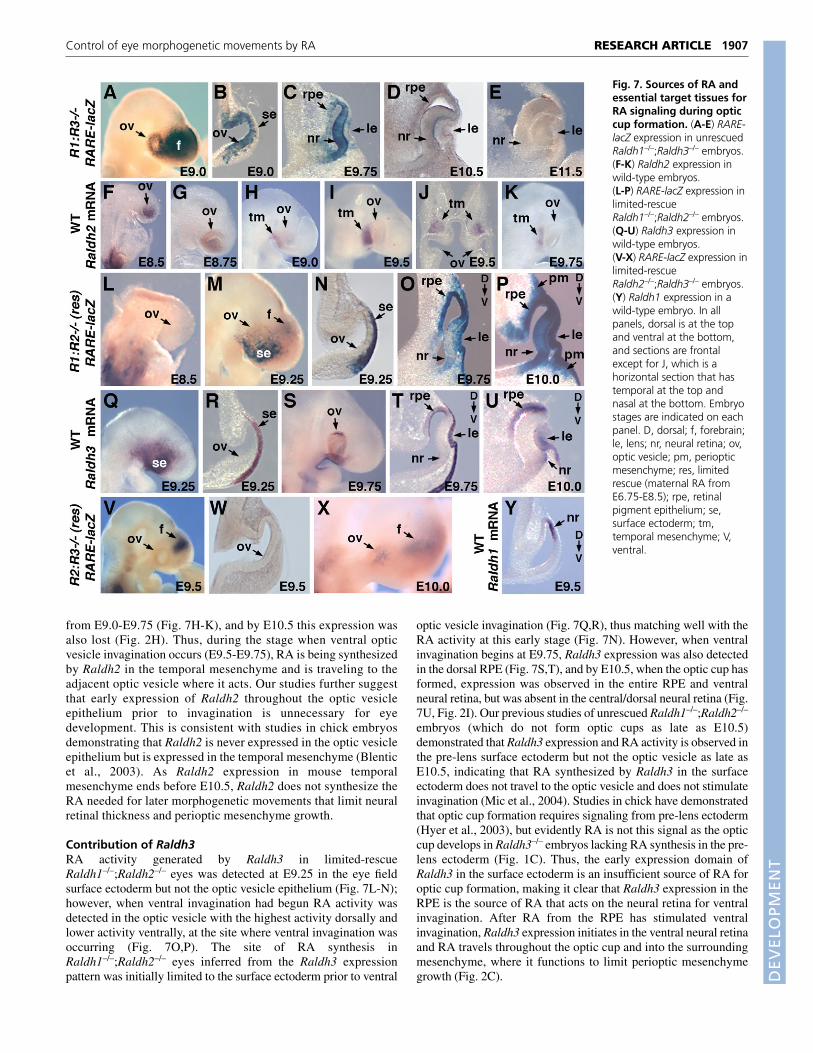
the ventral invagination of the optic vesicle that defines the junctionbetween the ventral retina and optic stalk (Fig. 6A-D; n=3 out of 3).In these mutants, lens invagination had occurred, as well as dorsalinvagination of the optic vesicle, which generates the junctionbetween the retinal pigment epithelium (RPE) and the neural retina,thus resulting in an incomplete optic cup (Fig. 6A-D). As the ventraloptic cup defect was still observed later at E11.5, this demonstrateda true blockage in ventral invagination rather than simply a delay(Fig. 6C,D). These results show that RA is required for ventralfolding during initial optic cup formation in addition to the roledescribed above for morphogenetic movements that limit neuralretina thickness. Also, it is now clear that Raldh2 can provide thisinitial function in unrescued Raldh1–/–;Raldh3–/– embryos thatdevelop an optic cup, and that Raldh3 can provide this function inlimited-rescue Raldh1–/–;Raldh2–/– embryos.
RA is not required for dorsal invagination of theoptic cupWe noticed that limited-rescue Raldh2–/–;Raldh3–/– embryoscarrying RARE-lacZ still exhibited Raldh1 expression (Fig. 6A,B)and RA activity (Fig. 6C,D) dorsally, where optic vesicleinvagination had occurred to generate an incomplete optic cup. Inorder to determine whether dorsal RA activity generated byRaldh1 was necessary for dorsal invagination of the optic vesicle,we examined Raldh1–/–;Raldh2–/–;Raldh3–/– triple null embryoscarrying RARE-lacZ. Raldh1–/–;Raldh2–/–;Raldh3–/– embryosexamined at E10.5 following maternal dietary RAsupplementation to E8.5 exhibited no RA activity in the eye;Raldh1–/–;Raldh3–/– embryos had low RA activity (Fig. 6E-G; alsocompare with Fig. 1H). The triple mutant confirms that the lowRA activity observed in the eye of Raldh1–/–;Raldh3–/– embryosis in fact due to Raldh2, and demonstrates the persistence of RAactivity in some neural and heart tissues in the absence of Raldh1,Raldh2 and Raldh3. Tissue sections revealed that rescuedRaldh1–/–;Raldh2–/–;Raldh3–/– embryos carrying RARE-lacZ doretain dorsal invagination of the optic vesicle despite a completeloss of RA activity (Fig. 6H). These important observations indouble and triple mutants demonstrate that RA is not required for
dorsal invagination of the optic vesicle, and that expression ofRaldh1 in the dorsal optic vesicle does not generate sufficient RAfor ventral invagination.
Contribution of each RALDH gene to eyedevelopmentThe three RALDHs are expressed in unique tissues during mouseeye development, and RA activity can be detected in those tissuesin mice carrying the RARE-lacZ RA-reporter transgene (Wagner etal., 2000; Mic et al., 2002). As RA synthesized in one region of thedeveloping eye can travel to other regions, it has previously beenimpossible to determine the extent to which each enzyme cansupply RA to the eye and the importance of that RA. However,analysis of RARE-lacZ expression in each of the RALDH doublenull mutants now makes this possible, and comparison of thelocations of such RA activity with expression of the singleremaining enzyme allows conclusions to be made concerning thespatiotemporal relationship between sites of RA synthesis and RAtarget tissues. The insights obtained from each RALDH doublemutant are described below.
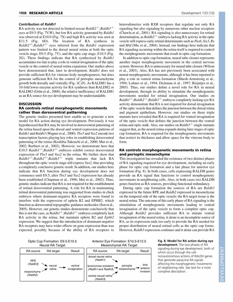
Contribution of Raldh2RA activity generated by Raldh2 in Raldh1–/–;Raldh3–/– eyes wasinitially detected throughout the optic vesicle epithelium (but notthe surface epithelium) prior to invagination, whereas whenventral optic vesicle invagination was underway at E9.75 RAactivity was much higher in the anterior optic vesicle fated tobecome neural retina, and after optic cup formation at E10.5 onlya low level of RA activity remained in the most anterior andcentral portion of the neural retina adjacent to the vitreous body(Fig. 7A-D). At E11.5, no RA was detectable inRaldh1–/–;Raldh3–/– eyes (Fig. 7E). The location of RA synthesisin Raldh1–/–;Raldh3–/– eyes (determined by Raldh2 expression)was initially throughout the optic vesicle epithelium prior toinvagination (Fig. 7F,G), matching well with the RA activity atthis stage (Fig. 7B). However, Raldh2 expression in the opticvesicle was lost by E9.0 and replaced by expression in themesenchyme adjacent to the temporal portion of the optic vesicle
RESEARCH ARTICLE Development 133 (10)
Fig. 6. Severe ventral optic cupinvagination defects in embryos lackingRA synthesis. All embryos (except thatshown in E) were subjected to maternaldietary RA supplementation from E6.75-E8.5, then returned to a normal diet untilthe point of analysis (limited-rescue).(A-D) Raldh2–/– and Raldh2–/–;Raldh3–/–
littermates were analyzed for Raldh1expression at E10.5 (frontal sections; A,B),and for RARE-lacZ expression at E11.5(frontal sections; C,D); asterisks mark theregion failing to undergo ventralinvagination. (E) RARE-lacZ expression inunrescued E10.5 Raldh1–/–;Raldh3–/–
embryo. (F,G) RARE-lacZ expression inlimited-rescue E10.5Raldh1–/–;Raldh2–/–;Raldh3–/– triple mutantembryo; note the complete lack of stainingin the eye compared with the doublemutant (E), the lack of olfactory staining(confirming the Raldh3–/– genotype), and the reduced trunk staining and forelimb bud growth (confirming the Raldh2–/– genotype). (H) Frontalsection through a triple mutant optic vesicle (from embryo in F), demonstrating a complete loss of RARE-lacZ expression and a failure of ventralinvagination (asterisk), but persistence of dorsal invagination in the absence of RA activity. D, dorsal; e, eye; f, forebrain; fl, forelimb bud; le, lens;olf, olfactory pit; V, ventral.
DEVELO
PMENT
from E9.0-E9.75 (Fig. 7H-K), and by E10.5 this expression wasalso lost (Fig. 2H). Thus, during the stage when ventral opticvesicle invagination occurs (E9.5-E9.75), RA is being synthesizedby Raldh2 in the temporal mesenchyme and is traveling to theadjacent optic vesicle where it acts. Our studies further suggestthat early expression of Raldh2 throughout the optic vesicleepithelium prior to invagination is unnecessary for eyedevelopment. This is consistent with studies in chick embryosdemonstrating that Raldh2 is never expressed in the optic vesicleepithelium but is expressed in the temporal mesenchyme (Blenticet al., 2003). As Raldh2 expression in mouse temporalmesenchyme ends before E10.5, Raldh2 does not synthesize theRA needed for later morphogenetic movements that limit neuralretinal thickness and perioptic mesenchyme growth.
Contribution of Raldh3RA activity generated by Raldh3 in limited-rescueRaldh1–/–;Raldh2–/– eyes was detected at E9.25 in the eye fieldsurface ectoderm but not the optic vesicle epithelium (Fig. 7L-N);however, when ventral invagination had begun RA activity wasdetected in the optic vesicle with the highest activity dorsally andlower activity ventrally, at the site where ventral invagination wasoccurring (Fig. 7O,P). The site of RA synthesis inRaldh1–/–;Raldh2–/– eyes inferred from the Raldh3 expressionpattern was initially limited to the surface ectoderm prior to ventral
optic vesicle invagination (Fig. 7Q,R), thus matching well with theRA activity at this early stage (Fig. 7N). However, when ventralinvagination begins at E9.75, Raldh3 expression was also detectedin the dorsal RPE (Fig. 7S,T), and by E10.5, when the optic cup hasformed, expression was observed in the entire RPE and ventralneural retina, but was absent in the central/dorsal neural retina (Fig.7U, Fig. 2I). Our previous studies of unrescued Raldh1–/–;Raldh2–/–
embryos (which do not form optic cups as late as E10.5)demonstrated that Raldh3 expression and RA activity is observed inthe pre-lens surface ectoderm but not the optic vesicle as late asE10.5, indicating that RA synthesized by Raldh3 in the surfaceectoderm does not travel to the optic vesicle and does not stimulateinvagination (Mic et al., 2004). Studies in chick have demonstratedthat optic cup formation requires signaling from pre-lens ectoderm(Hyer et al., 2003), but evidently RA is not this signal as the opticcup develops in Raldh3–/– embryos lacking RA synthesis in the pre-lens ectoderm (Fig. 1C). Thus, the early expression domain ofRaldh3 in the surface ectoderm is an insufficient source of RA foroptic cup formation, making it clear that Raldh3 expression in theRPE is the source of RA that acts on the neural retina for ventralinvagination. After RA from the RPE has stimulated ventralinvagination, Raldh3 expression initiates in the ventral neural retinaand RA travels throughout the optic cup and into the surroundingmesenchyme, where it functions to limit perioptic mesenchymegrowth (Fig. 2C).
1907RESEARCH ARTICLEControl of eye morphogenetic movements by RA
Fig. 7. Sources of RA andessential target tissues forRA signaling during opticcup formation. (A-E) RARE-lacZ expression in unrescuedRaldh1–/–;Raldh3–/– embryos.(F-K) Raldh2 expression inwild-type embryos.(L-P) RARE-lacZ expression inlimited-rescueRaldh1–/–;Raldh2–/– embryos.(Q-U) Raldh3 expression inwild-type embryos.(V-X) RARE-lacZ expression inlimited-rescueRaldh2–/–;Raldh3–/– embryos.(Y) Raldh1 expression in awild-type embryo. In allpanels, dorsal is at the topand ventral at the bottom,and sections are frontalexcept for J, which is ahorizontal section that hastemporal at the top andnasal at the bottom. Embryostages are indicated on eachpanel. D, dorsal; f, forebrain;le, lens; nr, neural retina; ov,optic vesicle; pm, periopticmesenchyme; res, limitedrescue (maternal RA fromE6.75-E8.5); rpe, retinalpigment epithelium; se,surface ectoderm; tm,temporal mesenchyme; V,ventral.
DEVELO
PMENT
1908
Contribution of Raldh1RA activity was not detected in limited-rescue Raldh2–/–;Raldh3–/–
eyes at E9.5 (Fig. 7V,W), but low RA activity generated by Raldh1was observed at E10.0 (Fig. 7X) and high RA activity was seen atE11.5 (Fig. 6D). The location of RA synthesis inRaldh2–/–;Raldh3–/– eyes inferred from the Raldh1 expressionpattern was limited to the dorsal neural retina at both the opticvesicle stage, E9.5 (Fig. 7Y), and the optic cup stage, E10.5 (Fig.2G). These findings indicate that RA synthesized by Raldh1accumulates too late to play a role in ventral invagination of the opticvesicle or the control of neural retina expansion. Our studies abovehave demonstrated that, later in development, Raldh1 does notprovide sufficient RA for vitreous body morphogenesis, but doesgenerate sufficient RA for the control of perioptic mesenchymegrowth both dorsally and ventrally (Fig. 1C,D). As RALDH1 has a10-fold lower enzyme activity for RA synthesis than RALDH2 orRALDH3 (Grün et al., 2000), the relative inefficiency of RALDH1as an RA source for eye development may be understandable.
DISCUSSIONRA controls retinal morphogenetic movementsrather than dorsoventral patterningThe genetic studies presented here enable us to generate a newmodel for RA action during eye development. Previously it washypothesized that RA may be involved in dorsoventral patterning ofthe retina based upon the dorsal and ventral expression patterns ofRaldh1 and Raldh3 (Wagner et al., 2000). Tbx5 and Vax2 encode twotranscription factors playing key roles in establishing dorsoventralpatterning of the retina (Koshiba-Takeuchi et al., 2000; Mui et al.,2002; Barbieri et al., 2002). However, we demonstrate here thatE10.5 Raldh1–/–;Raldh3–/– embryos exhibit correct dorsoventralexpression of Tbx5 and Vax2 in the retina. We further show thatRaldh1–/–;Raldh2–/–;Raldh3–/– triple mutants that lack RAthroughout the optic vesicle stage still express Vax2, thus providinga completely conclusive genetic result. In addition, our studies hereindicate that RA function during eye development does notcommence until E9.5, after Tbx5 and Vax2 expression has alreadybeen established (Chapman et al., 1996; Mui et al., 2002). Thus,genetic studies indicate that RA is not required for the establishmentof retinal dorsoventral patterning. A role for RA in maintainingretinal dorsoventral patterning was suggested from studies in chickembryos where dominant-negative RA receptors were found tointerfere with the expression of ephrin B2 and EPHB2. whichfunction as dorsoventral topographic guidance molecules (Sen et al.,2005). However, our genetic studies demonstrate conclusively thatthis is not the case, as Raldh1–/–;Raldh3–/– embryos completely lackRA activity in the retina, but maintain ephrin B2 and Ephb2expression. We suggest that the introduction of dominant-negativeRA receptors may have wider effects on gene expression than wasexpected, possibly because of the ability of RA receptors to
heterodimerize with RXR receptors that regulate not only RAsignaling but also signaling by numerous other nuclear receptors(Chawla et al., 2001). RA signaling is also unnecessary for retinaldetermination, as Raldh2–/– embryos lacking RA activity in the opticvesicle still express early retinal determinants such as Pax6, Six3, Rxand Mitf (Mic et al., 2004). Instead, our findings here indicate thatRA signaling occurring within the retina itself is required to controlthe morphogenetic movements that result in optic cup formation.
In addition to optic cup formation, neural tube closure representsanother major morphogenetic movement in the central nervoussystem. However, RA is unnecessary for neural tube closure (Wilsonet al., 2003). Also, RA has not previously been associated withneural morphogenetic movements, although it has been reported toplay a role in ventral retina formation (Marsh-Armstrong et al.,1994; Lohnes et al., 1994; Dickman et al., 1997; Reijntjes et al.,2005). Thus, our studies define a novel role for RA in neuraldevelopment, through its ability to stimulate the morphogeneticmovements needed for retinal invagination. Our studies onRaldh1–/–;Raldh2–/–;Raldh3–/– embryos completely lacking eye RAactivity demonstrate that RA is not required for dorsal invaginationof the optic vesicle that defines the junction between the dorsal retinaand pigment epithelium. However, our studies on these triplemutants have revealed that RA is required for ventral invaginationof the optic vesicle that defines the junction between the ventralretina and optic stalk. Also, our studies on Raldh3–/– single mutantssuggest that, as the neural retina expands during later stages of opticcup formation, RA is required for the morphogenetic movementsthat restrict its thickness and allow space for the vitreous body toform.
RA controls morphogenetic movements in retinaand perioptic mesenchymeThis investigation has revealed the existence of two distinct phasesof RA signaling required for eye development, including an earlyphase for optic cup formation and a late phase for anterior eyeformation (Fig. 8). In both cases, cells expressing RALDH genesprovide an RA signal that functions to control morphogeneticmovements in neighboring cells. Also, in both cases two RALDHgenes function as RA sources, providing functional redundancy.
During optic cup formation the sources of RA are Raldh3expressed in the future RPE and Raldh2 expressed in mesenchymeon the temporal side of the optic vesicle; the RA target tissue is theneural retina. The outcome of this early phase of RA signaling is thestimulation of morphogenetic movements leading to ventralinvagination of the optic vesicle to form a complete optic cup.Although Raldh2 provides sufficient RA to initiate ventralinvagination of the neural retina, it alone is an incomplete source ofRA, as its expression ends too early to provide the RA needed forproper distribution of neural retinal cells as the optic cup forms.However, Raldh3 expression continues and it alone can provide RA
RESEARCH ARTICLE Development 133 (10)
RA source
RPE(Raldh3)
mesenchyme(Raldh2)
neuralretina
ventralinvagination
RA target Result RA source
dorsal neural retina(Raldh1)
lens/cornea ectoderm(Raldh1 and Raldh3)
ventral neural retina(Raldh3)
periopticmesenchyme
limitanteriorinvasion
RA target Result
Optic Cup Formation: E9.5-E10.5Neural RA Target
Anterior Eye Formation: E10.5-E12.5Mesenchymal RA Target
Fig. 8. Model for RA action during eyedevelopment. The two phases of RAsignaling during eye development, both ofwhich occur through the cell-nonautonomous actions of RALDH genesthat generate paracrine RA signalsaffecting the morphogenetic movementsof neighboring cells. See text for a morecomplete description.
DEVELO
PMENT
that is sufficient for both the initiation of ventral invagination and theproper distribution of neural retinal cells leading to formation of anormal vitreous body cavity. Our studies suggest that RA control ofneural retina morphology does not involve the regulation of cellproliferation or apoptosis. Thus, RA may control cell movementswithin the retina.
During anterior eye formation the sources of RA are Raldh1expressed in the dorsal neural retina and Raldh3 expressed in theventral neural retina, and the RA target tissue is the periopticmesenchyme that is migrating past the optic cup. As Raldh1 andRaldh3 are also expressed at lower levels in the lens and corneaectoderm (Matt et al., 2005), this could be another source of RA thatenters the perioptic mesenchyme. The outcome of this late phase ofRA signaling is the limitation of perioptic mesenchymemorphogenetic movements that provide tissue contributing to thecorneal mesenchyme and eyelids (Cvekl and Tamm, 2004). Raldh1and Raldh3 can each alone function as sufficient sources of RA forthe control of perioptic mesenchyme invasion. Our studies, and thoseof others (Matt et al., 2005), suggest that RA may perform thisfunction by stimulating apoptosis in specific regions of the periopticmesenchyme, which would reduce cell numbers. However, arestrictive effect of RA on mesenchymal cell migration cannot beruled out. The identification of perioptic mesenchyme as a target ofRA synthesized in ocular ectodermal tissues is consistent with otherstudies demonstrating the need for perioptic mesenchyme to interactwith retina and lens for proper eye morphogenesis (Cvekl andTamm, 2004).
RALDHs function cell-nonautonomously duringeye developmentOur studies with rescued RALDH mutant mice carrying an RA-reporter transgene have revealed that the target of RA action changesduring eye morphogenesis. The initial target is the neural retina atthe optic vesicle stage, then the target switches to the periopticmesenchyme after optic cup formation. These targets are distinctfrom but adjacent to locations of RA synthesis, thus demonstratingthat RA does not need to function in the cells that synthesize RA butinstead functions in a paracrine fashion to guide the morphogeneticmovements of neighboring cells. Thus, all three RALDHs functioncell-nonautonomously. This has also been observed for posteriorneural development, where it has been determined that RAsynthesized in the somitic mesoderm by RALDH2 functions in theadjacent neuroectoderm but not in the somites themselves(Molotkova et al., 2005). Thus, in the neural tube, paracrine RAsignaling stimulated by RALDH2 occurs by mesenchymal toepithelial signaling. In the eye, we find the existence of threeparacrine RA signaling mechanisms: RALDH2 stimulatesmesenchymal to epithelial signaling for optic cup formation;RALDH3 stimulates epithelial to epithelial signaling for optic cupformation; RALDH1 and RALDH3 both stimulate epithelial tomesenchymal signaling for anterior eye formation.
Interestingly, for both neural tube and optic cup development,RALDH2 stimulates mesenchymal to epithelial signaling. This wasnot initially suspected for optic cup development, as mouse Raldh2is expressed early in the optic vesicle epithelium just after itsbudding from the forebrain, and only later exhibits expression in themesenchyme adjacent to the optic vesicle, just prior to optic cupformation. However, our studies here on RA-rescuedRaldh1–/–;Raldh2–/– embryos carrying RARE-lacZ havedemonstrated that a dose of RA sufficient to rescue overallembryonic development and optic cup formation does not stimulateRA signaling in the early optic vesicle epithelium. Thus, Raldh2
expression in the early optic vesicle is unnecessary for optic cupformation, a finding that is consistent with the observation thatRaldh2 is expressed in the temporal mesenchyme but not in the opticvesicle epithelium of chick embryos (Blentic et al., 2003). It isunclear whether Raldh2 expression in the mouse optic vesicle servesany function or whether it is an evolutionary relic.
RALDH3 provides sufficient RA to controlmorphogenetic movementsThe investigations reported here illustrate the usefulness ofcompound RALDH null mice in revealing the spatiotemporal roleof RA signaling in tissues where more than one RALDH contributesRA. Our findings make it clear that each of the three RALDH genescontributes to eye morphogenesis. Raldh1 can provide RA only forthe control of perioptic mesenchyme growth. Raldh2 is able tosupply RA only for initial optic cup formation, as its expression endstoo early to provide RA for later morphogenetic movements. Raldh3alone can supply all of the RA needed for eye morphogeneticmovements in the mouse. Knowledge of the ocular RA target tissuesgained in these studies will facilitate the identification of RA-regulated genes to further reveal the mechanisms by which RAcontrols morphogenetic movements.
We thank Ozgene Pty Ltd (Bentley, Australia) for help in generating the Raldh3null mouse, V. Papaioannou for the Tbx5 probe, G. Lemke for the Vax2 probe,N. Gale for the ephrin B2 probe, T. Pawson for Ephb2 probe, and O. Sirbu andX. Zhao for helpful discussions. This work was funded by National Institutes ofHealth grant EY013969.
ReferencesBarbieri, A. M., Broccoli, V., Bovolenta, P., Alfano, G., Marchitiello, A.,
Mocchetti, C., Crippa, L., Bulfone, A., Marigo, V., Ballabio, A. et al.(2002). Vax2 inactivation in mouse determines alteration of the eye dorsal-ventral axis, misrouting of the optic fibres and eye coloboma. Development129, 805-813.
Blentic, A., Gale, E. and Maden, M. (2003). Retinoic acid signalling centres inthe avian embryo identified by sites of expression of synthesising andcatabolising enzymes. Dev. Dyn. 227, 114-127.
Chapman, D. L., Garvey, N., Hancock, S., Alexiou, M., Agulnik, S. I., Gibson-Brown, J. J., Cebra-Thomas, J., Bollag, R. J., Silver, L. M. and Papaioannou,V. E. (1996). Expression of the T-box family genes, Tbx1-Tbx5, during earlymouse development. Dev. Dyn. 206, 379-390.
Chawla, A., Repa, J. J., Evans, R. M. and Mangelsdorf, D. J. (2001). Nuclearreceptors and lipid physiology: Opening the X-files. Science 294, 1866-1870.
Cvekl, A. and Tamm, E. R. (2004). Anterior eye development and ocularmesenchyme: new insights from mouse models and human diseases. BioEssays26, 374-386.
Dickman, E. D., Thaller, C. and Smith, S. M. (1997). Temporally-regulatedretinoic acid depletion produces specific neural crest, ocular and nervous systemdefects. Development 124, 3111-3121.
Duester, G. (2000). Families of retinoid dehydrogenases regulating vitamin Afunction: production of visual pigment and retinoic acid. Eur. J. Biochem. 267,4315-4324.
Dupé, V., Matt, N., Garnier, J.-M., Chambon, P., Mark, M. and Ghyselinck, N.B. (2003). A newborn lethal defect due to inactivation of retinaldehydedehydrogenase type 3 is prevented by maternal retinoic acid treatment. Proc.Natl. Acad. Sci. USA 100, 14036-14041.
Fan, X., Molotkov, A., Manabe, S.-I., Donmoyer, C. M., Deltour, L., Foglio, M.H., Cuenca, A. E., Blaner, W. S., Lipton, S. A. and Duester, G. (2003).Targeted disruption of Aldh1a1 (Raldh1) provides evidence for a complexmechanism of retinoic acid synthesis in the developing retina. Mol. Cell. Biol. 23,4637-4648.
Fuhrmann, S., Levine, E. M. and Reh, T. A. (2000). Extraocular mesenchymepatterns the optic vesicle during early eye development in the embryonic chick.Development 127, 4599-4609.
Ghyselinck, N. B., Dupé, V., Dierich, A., Messaddeq, N., Garnier, J.-M.,Rochette-Egly, C., Chambon, P. and Mark, M. (1997). Role of the retinoicacid receptor beta (RARb) during mouse development. Int. J. Dev. Biol. 41, 425-447.
Gregory-Evans, C. Y., Williams, M. J., Halford, S. and Gregory-Evans, K.(2004). Ocular coloboma: a reassessment in the age of molecular neuroscience.J. Med. Genet. 41, 881-891.
Grün, F., Hirose, Y., Kawauchi, S., Ogura, T. and Umesono, K. (2000).
1909RESEARCH ARTICLEControl of eye morphogenetic movements by RA
DEVELO
PMENT
1910
Aldehyde dehydrogenase 6, a cytosolic retinaldehyde dehydrogenaseprominently expressed in sensory neuroepithelia during development. J. Biol.Chem. 275, 41210-41218.
Hornby, S. J., Ward, S. J., Gilbert, C. E., Dandona, L., Foster, A. and Jones, R.B. (2002). Environmental risk factors in congenital malformations of the eye.Ann. Trop. Paediatr. 22, 67-77.
Hyer, J., Kuhlman, J., Afif, E. and Mikawa, T. (2003). Optic cup morphogenesisrequires pre-lens ectoderm but not lens differentiation. Dev. Biol. 259, 351-363.
Koshiba-Takeuchi, K., Takeuchi, J. K., Matsumoto, K., Momose, T., Uno, K.,Hoepker, V., Ogura, K., Takahashi, N., Nakamura, H., Yasuda, K. et al.(2000). Tbx5 and the retinotectum projection. Science 287, 134-137.
Lohnes, D., Mark, M., Mendelsohn, C., Dollé, P., Dierich, A., Gorry, P.,Gansmuller, A. and Chambon, P. (1994). Function of the retinoic acidreceptors (RARs) during development. (I) Craniofacial and skeletal abnormalitiesin RAR double mutants. Development 120, 2723-2748.
Maden, M. (2002). Retinoid signalling in the development of the central nervoussystem. Nat. Rev. Neurosci. 3, 843-853.
Marsh-Armstrong, N., McCaffery, P., Gilbert, W., Dowling, J. E. and Dräger,U. C. (1994). Retinoic acid is necessary for development of the ventral retina inzebrafish. Proc. Natl. Acad. Sci. USA 91, 7286-7290.
Matt, N., Dupé, V., Garnier, J.-M., Dennefeld, C., Chambon, P., Mark, M. andGhyselinck, N. B. (2005). Retinoic acid-dependent eye morphogenesis isorchestrated by neural crest cells. Development 132, 4789-4800.
McLaughlin, T., Hindges, R. and O’Leary, D. D. M. (2003). Regulation of axialpatterning of the retina and its topographic mapping in the brain. Curr. Opin.Neurobiol. 13, 57-69.
Mic, F. A., Haselbeck, R. J., Cuenca, A. E. and Duester, G. (2002). Novel retinoicacid generating activities in the neural tube and heart identified by conditionalrescue of Raldh2 null mutant mice. Development 129, 2271-2282.
Mic, F. A., Molotkov, A., Benbrook, D. M. and Duester, G. (2003). Retinoidactivation of retinoic acid receptor but not retinoid X receptor is sufficient torescue lethal defect in retinoic acid synthesis. Proc. Natl. Acad. Sci. USA 100,7135-7140.
Mic, F. A., Molotkov, A., Molotkova, N. and Duester, G. (2004). Raldh2expression in optic vesicle generates a retinoic acid signal needed forinvagination of retina during optic cup formation. Dev. Dyn. 231, 270-277.
Molotkov, A., Fan, X., Deltour, L., Foglio, M. H., Martras, S., Farrés, J., Parés,X. and Duester, G. (2002). Stimulation of retinoic acid production and growthby ubiquitously-expressed alcohol dehydrogenase Adh3. Proc. Natl. Acad. Sci.USA 99, 5337-5342.
Molotkova, N., Molotkov, A., Sirbu, I. O. and Duester, G. (2005). Requirement
of mesodermal retinoic acid generated by Raldh2 for posterior neuraltransformation. Mech. Dev. 122, 145-155.
Mui, S. H., Hindges, R., O’Leary, D. D. M., Lemke, G. and Bertuzzi, S. (2002).The homeodomain protein Vax2 patterns the dorsoventral and nasotemporalaxes of the eye. Development 129, 797-804.
Niederreither, K., Subbarayan, V., Dollé, P. and Chambon, P. (1999).Embryonic retinoic acid synthesis is essential for early mouse post-implantationdevelopment. Nat. Genet. 21, 444-448.
Noy, N. (2000). Retinoid-binding proteins: mediators of retinoid action. Biochem.J. 348, 481-495.
Reijntjes, S., Blentic, A., Gale, E. and Maden, M. (2005). The control ofmorphogen signaling: regulation of the synthesis and catabolism of retinoic acidin the developing embryo. Dev. Biol. 285, 224-237.
Rossant, J., Zirngibl, R., Cado, D., Shago, M. and Giguère, V. (1991).Expression of a retinoic acid response element-hsplacZ transgene defines specificdomains of transcriptional activity during mouse embryogenesis. Genes Dev. 5,1333-1344.
Russell, C. (2003). The roles of Hedgehogs and Fibroblast Growth Factors in eyedevelopment and retinal cell rescue. Vision Res. 43, 899-912.
Sakai, Y., Luo, T. L., McCaffery, P., Hamada, H. and Dräger, U. C. (2004).CYP26A1 and CYP26C1 cooperate in degrading retinoic acid within theequatorial retina during later eye development. Dev. Biol. 276, 143-157.
Seeliger, M. W., Biesalski, H. K., Wissinger, B., Gollnick, H., Gielen, S., Frank,J., Beck, S. and Zrenner, E. (1999). Phenotype in retinol deficiency due to ahereditary defect in retinol binding protein synthesis. Invest. Ophthalmol. Vis.Sci. 40, 3-11.
Sen, J., Harpavat, S., Peters, M. A. and Cepko, C. L. (2005). Retinoic acidregulates the expression of dorsoventral topographic guidance molecules in thechick retina. Development 132, 5147-5159.
Sirbu, I. O., Gresh, L., Barra, J. and Duester, G. (2005). Shifting boundaries ofretinoic acid activity control hindbrain segmental gene expression. Development132, 2611-2622.
Wagner, E., McCaffery, P. and Dräger, U. C. (2000). Retinoic acid in theformation of the dorsoventral retina and its central projections. Dev. Biol. 222,460-470.
Wilson, J. G., Roth, C. B. and Warkany, J. (1953). An analysis of the syndromeof malformations induced by maternal vitamin A deficiency. Effects ofrestoration of vitamin A at various times during gestation. Am. J. Anat. 92, 189-217.
Wilson, L., Gale, E. and Maden, M. (2003). The role of retinoic acid in themorphogenesis of the neural tube. J. Anat. 203, 357-368.
RESEARCH ARTICLE Development 133 (10)