development/plasticity/repair neuron ... · neuron-specificeffectsofinterleukin-1 aremediatedbya...
TRANSCRIPT

Development/Plasticity/Repair
Neuron-Specific Effects of Interleukin-1� Are Mediated by aNovel Isoform of the IL-1 Receptor Accessory Protein
Yangyang Huang,1 Dirk E. Smith,3 Osvaldo Ibanez-Sandoval,2 John E. Sims,3 and Wilma J. Friedman1
1Department of Biological Sciences, and 2Center for Molecular and Behavioral Neuroscience, Rutgers University, Newark, New Jersey 07102, and3Department of Inflammation Research, Amgen, Seattle, Washington 98119
In the CNS, interleukin-1� (IL-1�) is synthesized and released during injury, infection, and disease, mediating inflammatory responses.However, IL-1� is also present in the brain under physiological conditions, and can influence hippocampal neuronal function. Severalcell-specific IL-1-mediated signaling pathways and functions have been identified in neurons and astrocytes, but their mechanisms havenot been fully defined. In astrocytes, IL-1� induced both the p38 MAPK and NF-�B (nuclear factor �B) pathways regulating inflamma-tory responses, however in hippocampal neurons IL-1� activated p38 but not NF-�B. Additionally, IL-1� induced Src phosphorylation at0.01 ng/ml in hippocampal neurons, a dose 1000-fold lower than that used to stimulate inflammatory responses. IL-1 signaling requiresthe type 1 IL-1 receptor and the IL-1 receptor accessory protein (IL-1RAcP) as a receptor partner. We previously reported a novel isoformof the IL-1RAcP, IL-1RAcPb, found exclusively in CNS neurons. In this study, we demonstrate that AcPb specifically mediates IL-1�activation of p-Src and potentiation of NMDA-induced calcium influx in mouse hippocampal neurons in a dose-dependent manner. Micelacking the AcPb, but retaining the AcP, isoform were deficient in IL-1� regulation of p-Src in neurons. AcPb also played a modulatoryrole in the activation of p38 MAPK, but had no effect on NF-�B signaling. The restricted expression of AcPb in CNS neurons, therefore,governs specific neuronal signaling and functional responses to IL-1�.
IntroductionInterleukin-1 (IL-1) is a proinflammatory cytokine that is ex-pressed in multiple cell types in the brain (Breder et al., 1988;Hunter et al., 1992; Pitossi et al., 1997; Sims and Smith, 2010). Itsexpression is particularly high during injury and neurodegenera-tive disease (Loddick et al., 1998), as it plays a major role incoordinating the inflammatory response to these conditions.IL-1� is rapidly synthesized and released, primarily by microglia(Giulian et al., 1986; Pearson et al., 1999) and astrocytes (Davieset al., 1999; Pearson et al., 1999). Under pathophysiological con-ditions, IL-1 mediates inflammatory activity by stimulating re-lease of additional proinflammatory cytokines and growthfactors from astrocytes and microglia (Merrill and Benveniste,1996). In the hippocampus, high concentrations of IL-1 act uponneurons to inhibit synaptic strength and long-term potentiation(LTP) (Katsuki et al., 1990; Bellinger et al., 1993; Murray andLynch, 1998). In contrast, physiological levels of IL-1 promote
LTP and memory formation (Yirmiya et al., 2002). The distinctfunctions of IL-1 are mediated through the same type 1 IL-1receptor (IL-1RI), which is expressed and regulated by IL-1 inboth hippocampal neurons and astrocytes (Friedman, 2001).However, the signaling pathways for IL-1 in these two cell typesdiffer (Srinivasan et al., 2004). The precise mechanism governingthe cell type-specific IL-1 signaling is unknown and remains to beinvestigated. Two classical downstream signaling pathways areactivated following IL-1 binding to its receptors (Ninomiya-Tsujiet al., 1999). One pathway involves phosphorylation and degra-dation of the I�B subunit of NF-�B (nuclear factor �B), leadingto the release and nuclear translocation of the active NF-�B tran-scription factor and subsequent gene expression. Another path-way involves activation of the three MAPKs, ERK, JNK, and P38,and additional transcription factors such as ATF (activating tran-scription factor) and AP1, leading to nuclear gene transcription(O’Neill et al., 1990; Zhang et al., 1996; Ninomiya-Tsuji et al.,1999). Among these, NF-�B and p38 MAPK are known to beactivated by IL-1� in hippocampal astrocytes, but only p38MAPK is activated in neurons (Srinivasan et al., 2004). In addi-tion, an alternative signaling mechanism involving Src kinase hasbeen reported to be activated by IL-1� in hippocampal neurons(Viviani et al., 2003).
IL-1R accessory protein (IL-1RAcP) is a required receptorpartner in IL-1 signaling (Cullinan et al., 1998) and is expressedubiquitously. AcPb, a novel isoform of IL-1RAcP, was recentlyidentified and is mainly expressed in CNS neurons (Smith et al.,2009), but its precise function in mediating neuronal responses toIL-1 remains unclear. Here, we investigated IL-1�-mediated celltype-specific signaling pathways in hippocampal neurons and
Received Aug. 8, 2011; revised Oct. 18, 2011; accepted Oct. 30, 2011.Author contributions: W.J.F. designed research; Y.H. performed research; D.E.S. and J.E.S. contributed unpub-
lished reagents/analytic tools; O.I.-S. analyzed data; Y.H. and W.J.F. wrote the paper.This work was supported by the New Jersey Commission for Brain Injury Research and NIH/NINDS Grant
1R21NS076867. D.E.S. and J.E.S. are employees and shareholders of Amgen Corporation. We thank Barry Levin andAmbrose Dunn-Meynell for assistance with the calcium imaging, and Matthew Wilkins for excellent technicalassistance.
Correspondence should be addressed to Dr. Wilma J. Friedman, Department of Biological Sciences, RutgersUniversity, 225 University Avenue, Newark, NJ 07102. E-mail: [email protected].
Y. Huang’s present address: Department of Neuroscience and Cell Biology, UMDNJ/Robert Wood Johnson MedicalSchool, 683 Hoes Lane, Piscataway, NJ 08854.
DOI:10.1523/JNEUROSCI.4067-11.2011Copyright © 2011 the authors 0270-6474/11/3118048-12$15.00/0
18048 • The Journal of Neuroscience, December 7, 2011 • 31(49):18048 –18059
astrocytes, and the role of AcPb in each pathway. We demonstratethat AcPb governs neuron-specific functional responses to IL-1�involving Src phosphorylation and enhanced Ca 2� influx in re-sponse to NMDA receptor activation.
Materials and MethodsAnimals. Mice lacking either all AcP isoforms (total AcP �/�) or only theAcPb isoform (AcPb �/�) were generated as previously described (Smithet al., 2009) and maintained on a fully backcrossed C57BL/6 background.Briefly, the original total AcP knock-out mice were generated by deletionof exons 4 and 5, encoding crucial extracellular domains, and insertion ofa neomycin sequence; therefore, both AcP and AcPb were functionallyknocked out. AcPb-specific knock-out mice were generated by replacingthe variant AcPb-associated exon 12b with a neomycin resistance gene,while the upstream 12 exons were left intact to maintain expression ofnormal full-length AcP (Smith et al., 2009). All mice were genotypedusing primers covering either the inserted neomycin or the knocked outregion before being mated. All animal studies were conducted using theNIH guidelines for the ethical treatment of animals with approval of theRutgers Animal Care and Facilities Committee.
Astrocyte culture. Pregnant mice were killed by exposure to CO2 andsoaked in 80% ethanol for 10 min. Embryonic day 19 (E19) fetuses ofeither sex were removed under sterile conditions and kept in PBS onice. Hippocampi were dissected, dissociated by trituration, and platedon poly-D-lysine (0.1 mg/ml)-precoated flasks in NM15 medium (Ea-gle’s MEM with Earle’s salts and 2 mM L-glutamine, 15% heat-inactivated fetal bovine serum, 6 mg/ml glucose, 0.5 �g/ml penicillinand 0.5 U/ml streptomycin). Astrocytes were isolated by shakingflasks on an orbital shaker at 450 rpm for 10 min. Medium was re-placed with fresh NM15 and the flasks were returned to the incubatorfor 2 h before being shaken overnight at 225 rpm to remove neurons,microglia and oligodendrocytes. The confluent astrocytes were ex-posed to cytosine arabinoside (0.1 mM) for 3 d to eliminate any re-maining non-astrocyte cell populations. Finally, the astrocytes weretrypsinized and replated at subconfluent density. The purity of astro-cyte cultures was �99%, confirmed by immunostaining.
Neuronal culture. E16 hippocampi were dissected from mice of eithersex, dissociated, and plated in serum-free medium on poly-D-lysine (0.01ng/ml)-precoated plates. The medium consists of 1:1 mixture of Eagle’sMEM (Invitrogen) and Ham’s F12 supplemented with glucose (6 mg/ml), insulin (25 �g/ml), putrescine (60 �M), progesterone (20 nM), trans-ferrin (100 �g/ml), selenium (30 nM), penicillin (0.5U/ml) andstreptomycin (0.5 �g/ml). Neurons were cultured for 5 d in serum-freemedium and subjected to cytokine treatments. The purity of neuronalcultures was �95%, confirmed by immunostaining.
Total RNA isolation and PCR: RNA was isolated with TRIzol reagent(Invitrogen) as recommended by the manufacturer. Briefly, 1 � 10 6 cellswere lysed in 1 ml of TRIzol reagent, followed by chloroform extractionand isopropanol precipitation. The RNA was resuspended in nuclease-free water and quantified spectrophotometrically at 260/280 nm. Onemicrogram of RNA was reverse transcribed at 42°C for 2 h in a 20 �lreaction mixture using SuperScript II reverse transcriptase (Invitrogen).cDNA was amplified and quantified by regular PCR and real-timePCR. The following primer sequences were used: IL1R (forward:5�-CTACTTGGGTTCATTTGTCTCATTGTGCC-3�, reverse: 5�-TTCCACTTCCAGTAGACAAGGTCGGTGAAC-3�); mouse AcP-specificprimers (forward 5�-TGTTTCCTATGCAAGAAATGTGGAAGAAGAGG-3�, rev: 5�-TGCTTGTCATTGCTAGACCACCTGG-3�) and mouseAcPb-specific primers (forward: 5�-TGTTTCCTATGCAAGAAATGTGGAAGAAGAGG-3�, rev: 5�-ATGGGGTTGCTCAAGCGACGGTACTCCAC-3�); mouse IL-6 (forward 5�-GGAGAGGAGACTTCACAGAGG-3�, reverse: 5�-GGAAATTGGGGTAGGAAGGA-3�); actin (forward: 5�-TCATGAAGTGTGACGTTGACATCCGT-3�, reverse: 5�-CTTAGAAGCATTTGCGGTGCACGATG-3�);GAPDH(forward:5�-TTCTTGTGCAGTGCCAGCC-3�, reverse: 5�-CACCGACCTTCACCATCTTGT-3�).PCRs were performed in a 20 �l volume with goTaq Green Master Mix(Promega) and 1 �M primers according to manufacturer’s recommen-dation. PCR conditions for IL1R, AcP and AcPb were: 95°C 5 min; 95°C30 s, 50°C 30 s, 72°C 30 s for 35 cycles; and 72°C 7 min. Reaction condi-
tions for IL-6, actin, and GAPDH were identical except that the annealingtemp was 60°. PCR products were resolved on 1% agarose gel. For real-time PCR, Roche 480 SYBR Green Mastermix was used according to themanufacturer’s recommendation.
Western blots. Cells were harvested in lysis buffer containing 120mM Tris, pH 6.8, 2% SDS, and 10% glycerol supplemented with Pro-tease Inhibitor Cocktail and Phosphatase Inhibitor Cocktail (Roche).Proteins were quantified using the Bradford assay (Bio-Rad). Equalamounts of protein were run on SDS polyacrylamide gels and trans-ferred to nitrocellulose membrane. The blots were blocked in 5%nonfat milk in TBST for 1 h, and incubated with primary antibody (1:1000 in 5% milk in TBST) overnight at 4°C. The primary antibodiesare anti-p-I�B � (Cell Signaling Technology, catalog #9246), anti-I�B� (Cell Signaling Technology, catalog #9242), anti-p-p38 (Thr 180/Tyr 182) (Cell Signaling Technology, catalog #9215), anti-p38 (CellSignaling Technology, catalog #9212), anti-p-Src (Tyr 416) (Cell Sig-naling Technology, catalog #2101), anti-Src (Cell Signaling Technol-ogy, catalog #2110), anti-p-NR2B (Cell Signaling Technology catalog#4208), anti-NR2B (Cell Signaling Technology catalog #4212) anti-�actin (Sigma catalog #A5441). The blots were then washed 3 timeswith TBST, and incubated with secondary antibody (goat anti-mouse,or goat anti-rabbit labeled with horseradish peroxidase, ThermoFisher Scientific, 1:5000) for 1 h at room temperature. The mem-branes were developed using the Pierce ECL Western blot substratekit (Thermo Fisher Scientific). Blots were stripped in 62.5 mM Tris-HCl ph6.8, 2%SDS and 100 mM �-mercaptoethanol at 65°C for 5 min.Quantification of immunoblots was performed on scanned images ofthe films using Adobe Photoshop software.
Immunoprecipitation/coimmunoprecipitation-Western blot analysis.Cultured neurons or astrocytes were lysed in TNE buffer (50 mM Tris, pH7.3, 137 mM NaCl, 2 mM EDTA) containing 0.2% Triton, 60 mM octylg-lucoside, and Protease Inhibitor Cocktail. Proteins were quantified usingthe Bradford assay (Bio-Rad). Protein (500 �g) was incubated with pri-mary antibody anti-AcP/b M215 (10 ng/ml, Amgen) overnight on arocking platform at 4°C. Protein G-Sepharose was added to the lysatesand kept for an additional 3 h at 4°C. The lysates were then centrifuged at14,000 rpm for 15 min and washed with TNE buffer three times anddistilled water once. The pellets were resuspended in 1� loading bufferwith �-mercaptoethanol. The samples were then run by SDS-PAGE,transferred to nitrocellulose membrane, and probed using anti-AcP P2anti-rabbit antibody (1:1000, Amgen) which detects both AcP and AcPb.Astrocytes transfected with AcP or AcPb plasmids were used as positivecontrols.
Cell transfection. Primary mouse astrocytes were transfected using theMouse Astrocyte Nucleofector kit (Amaxa Biosystems), program T-20by Amaxa according to the manufacturer’s instructions. In brief, 2– 4million astrocytes were suspended in 100 �l of supplemented Nucleofec-tor solution in each transfection. The cell suspension was then mixedwith 3–5 �g of AcP, AcPb, empty vector DNA plasmids, 3–5 �g of �B-driven-luciferase plasmid and/or 1 �g of GFP plasmid (Amaxa Biosys-tems). Within 15 min the cell suspension was transfected with programT-20. Cells were then resuspended with warm NM-15 medium and re-plated on poly-D-lysine precoated dishes. Two hours after transfection,fresh NM-15 medium was applied to the cultures. One to 3 d after trans-fection, cells were exposed to IL-1� treatment (generously provided byDr. Ronald Hart, Department of Cell Biology and Neuroscience, RutgersUniversity, Newark, NJ).
Luciferase activity assay. Twenty-four hours after transfection, astro-cytes were treated with IL-1� (10 ng/ml) for 4 h. Protein lysates were thencollected in reporter lysis buffer (Promega, E 4030). Protein lysates werecentrifuged at 14,000 rpm for 2 min at 4°C and supernatants were storedat �80°C. Lysates were warmed at room temperature for 30 min andequal amount of protein was mixed with luciferase substrate and sub-jected to luminescence reading immediately.
Immunocytochemistry. Primary neurons and astrocytes were culturedin poly-D-lysine-precoated-Lab-tek chamber slide (Nunc). Cells weretreated with IL-1� (10 ng/ml) for 2 h, followed by fixation with 4%
Huang et al. • Neuron-Specific Il-1� Signaling and Function J. Neurosci., December 7, 2011 • 31(49):18048 –18059 • 18049
paraformaldehyde for 30 min. Fixed cells wereblocked in PBS/10% goat serum and perme-abilized with PBS/0.3%Triton X-100, then ex-posed to anti-p65 (1:500, Santa CruzBiotechnology) overnight at 4°C in PBS. Slidewells were then washed 3 in PBS, exposed for1 h at room temp to goat anti-rabbit secondaryantibodies coupled to Alexa 555 (1:500). Aftersecondary antibody, cells were washed 3 timesin PBS, followed by DAPI (10 �g/ml, Invitro-gen) application. Cells were then coverslippedwith anti-fading medium (ProLong Gold, In-vitrogen) and imaged on a Nikon TE200 In-verted Epifluorescent microscope.
Calcium imaging. Hippocampal neuronswere cultured on poly-D-lysine (0.1 mg/ml)-coated glass coverslips in SFM for 12 d beforecalcium imaging. Neurons were maintained inan extracellular medium (ECM) containing 33mM glucose in Hanks’ balanced salt solutionbuffer (in mmol/L: 135 NaCl, 5 KCl, 1 CaCl2, 1MgCl2, and 10 HEPES, pH 7.4) at 37°C duringimaging. Fluorescent imaging measurement ofintracellular Ca 2� ([Ca 2�]i) was performedusing fura-2 acetoxymethyl ester (Teflabs).Hippocampal neurons were loaded with 2.5�M fura-2 acetoxymethyl ester in ECM for 20min (37°C). Cells were washed twice with ECMto remove free fura-2 and transferred to a ther-mostatically regulated microscope chamber(37°C). Fura-2 fluorescent images were acquired every 5 s by alternatingexcitation at 340 and 380 nm, and emissions (420 – 600 nm) were col-lected using a cooled, charge-coupled device camera. For each experi-ment, baseline was established by recording for 4 min, followed with orwithout IL-1 treatment for 6 min. NMDA (10 �M) was then applied for 7min before washing for 1– 4 min. At the end of each recording, KCl (30mM) was applied to ensure calcium influx and cell viability. Only cellsthat responded to KCl were picked for quantification. Changes in[Ca 2�]I fluctuation in response to NMDA were assessed for 7 min afterNMDA application by calculating the integrated area under curve (AUC)using Origin 7.0 software (OriginLab).
Statistical analysis. All data are expressed as mean � SEM of three ormore independent experiments conducted on different culture prepara-tions. Student’s t test was used to analyze two part comparisons. One-wayANOVA followed by Tukey–Kramer post hoc tests were used for analysisof groups of three or more. For all statistical analyses, a value of p � 0.05was considered significant.
ResultsAcPb is expressed in hippocampal neurons but not astrocytesPrevious reports from our laboratory indicated that IL-1RI waspresent in both neurons and astrocytes from the rat hippocampus(Friedman, 2001), and that IL-1� stimulated different signalingpathways in these two distinct cell types (Srinivasan et al., 2004).IL-1RAcP is a known, required receptor partner in IL-1 signaling(Cullinan et al., 1998). A novel isoform of AcP, termed AcPb, thatis expressed exclusively in CNS neurons was recently identified.AcPb was able to form a complex with IL-1 and the IL-1 receptorand was found to abrogate some gene expression induced by IL-1in cortical neuron cultures (Smith et al., 2009). Thus, we investi-gated whether AcPb was responsible for the distinct IL-1� signal-ing seen in neurons and astrocytes.
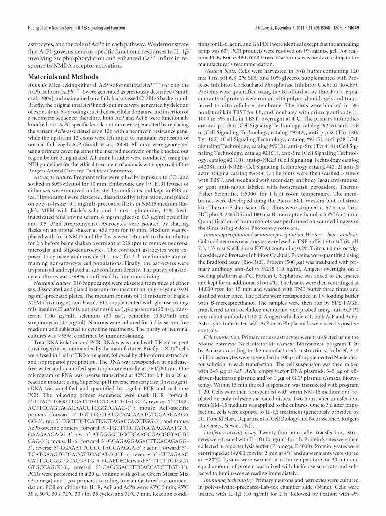
To characterize the role of AcPb in IL-1� signaling, we firstexamined the expression of IL-1RI and AcP/b in mouse neuronsand astrocytes. In accordance with previous results in the rat,IL-1RI mRNA was detected by reverse transcriptase-PCR inmouse hippocampal neurons and astrocytes (Fig. 1A). The
mRNA levels of AcP and AcPb were determined by PCR analysisusing primers designed specifically against the last two exons.AcP was detected in both neurons and astrocytes, but AcPb wasfound only in neurons (Fig. 1A). Real-time PCR quantificationshowed that hippocampal neurons express both AcP and AcPb atapproximately equal, but low, levels (Fig. 1B). In contrast, AcPbmRNA was not detected in astrocytes, and AcP levels were 7 timesabove that in neurons. To examine protein expression of AcPisoforms, immunoprecipitation (IP)-Western blot analysis wasperformed with anti-pan-AcP antibodies that recognize bothAcP and AcPb proteins at different sizes. Only AcP was detectedin astrocytes, however both AcP and AcPb proteins were presentin neurons, but very low levels (Fig. 1C), consistent with therelatively low levels of AcP and AcPb mRNA in neurons. It is notclear why the AcP/b proteins migrate slightly differently in neu-rons and astrocytes, but differential glycosylation may underliesome of the differences as reported by Smith et al. (2009).
AcPb does not influence NF-�B activation by IL-1 inhippocampal neurons and astrocytesThe NF-�B signaling pathway is known to be activated by IL-1�in hippocampal astrocytes but not in neurons (Srinivasan et al.,2004). Our previous study showed that expression of AcPb inaddition to AcP inhibited IL-1-regulated gene induction com-pared with expression of AcP alone (Smith et al., 2009), suggest-ing that the presence of AcPb might inhibit IL-1�-mediatedactivation of NF-�B. Therefore we examined whether IL-1�could activate NF-�B in the absence of AcPb.
Cultured astrocytes were prepared from wild-type, totalAcP�/�, and AcPb�/� mice. Cultures were treated with IL-1�and probed for p-I�B and total I�B by Western blot analysis. InWT astrocytes, I�B was phosphorylated within 5–10 min, accom-panied by degradation of total I�B (Fig. 2A). Total AcP deletionin astrocytes abolished I�B phosphorylation and degradation in-duced by IL-1�. However, astrocytes from AcPb�/� mice showeda similar I�B response compared with WT, consistent with the
Figure 1. Expression of IL-1R1, AcP and AcPb in primary mouse hippocampal neurons and astrocytes. A, Reverse transcriptase-PCR amplification of AcP, AcPb and IL-1RI mRNAs from hippocampal neurons and astrocytes, � actin was used as control. NTC wascontrol without cDNA for PCR. B, Relative expression of AcP and AcPb mRNA levels in hippocampal neurons and astrocytesquantified by real-time PCR. All mRNAs are expressed relative to the neuronal expression level of AcP, which was normalized to 1,using actin as an internal control (data from three different experiments with triplicate loading each). C, IP-Western blots for AcPand AcPb in hippocampal neurons and astrocytes. Protein (50 �g) from astrocytes transfected with AcP or AcPb was used aspositive control in the left two lanes. A total of 500 �g of protein from primary neurons or astrocytes was immunoprecipitated witha pan-AcP monoclonal antibody and immunoblots were analyzed with a pan-AcP antiserum.
18050 • J. Neurosci., December 7, 2011 • 31(49):18048 –18059 Huang et al. • Neuron-Specific Il-1� Signaling and Function

lack of AcPb expression in astrocytes. Since astrocytes only ex-press AcP, this suggests that only classic AcP but not AcPb wasrequired for NF-�B activation in astrocytes.
NF-�B subunits p65 and p50, have previously been shown totranslocate to the nucleus following IL-1� stimulation in astro-cytes (Friedman et al., 1996). To confirm that NF-�B was trans-
located to nuclei after I�B phosphorylation, cultured astrocyteswere immunostained for the NF-�B p65 subunit and imagedusing fluorescence microscopy. Nuclear translocation of the p65subunit was observed in WT and AcPb�/� astrocytes, but not intotal AcP�/� astrocytes (Fig. 2B), confirming the role of AcP butnot AcPb in activating NF-�B in astrocytes.
Figure 2. AcPb does not regulate NF-�B signaling. A, IL-1� induced phosphorylation of I�B in WT and AcPb �/� astrocytes but not total AcP �/� astrocytes. Cultured hippocampal astrocyteswere treated with IL-1� (10 ng/ml) for the indicated times and lysates were analyzed by Western blot for phospho-I�B, stripped and reprobed for total I�B and � actin. The blot shown isrepresentative of three independent experiments. B, p65 NF-�B subunit translocated to the nucleus in WT and AcPb �/� astrocytes but not AcP �/� astrocytes. Astrocytes were treated with IL-1�(10 ng/ml) for 2 h, fixed with 4%PFA, and immunostained for p65 (red). Cell nuclei were labeled with DAPI (blue). C, �B-luciferase activity is induced by IL1� in WT and AcPb �/�astrocytes, but notAcP �/� astrocytes. Astrocytes were transfected with �B-driven luciferase reporter plasmids. Twenty-four hours after transfection, cells were treated with IL-1� (10 ng/ml) for 4 h.Protein lysates were analyzed for luciferase activity. Data are from four different experiments with triplicate loading each. Student’s t test was used to evaluate data from cells treatedwith or without IL-1�. *p � 0.01. D, IL-6 mRNA was induced by IL1� (10 ng/ml) in WT and AcPb �/� astrocytes but not total AcP �/� astrocytes by real-time PCR analysis. Graph depicts6 h IL-1� treatment. mRNA levels were normalized to wild-type control. E, No nuclear translocation of p65 was induced by 2 h IL-1� (10 ng/ml) in hippocampal neurons. F, Nophosphorylation of I�B was detected in WT, total AcP �/� or AcPb �/� neurons in response to IL-1� (10 ng/ml). p-I�B in astrocytes was used as positive control. The blot shown isrepresentative of three independent experiments.
Huang et al. • Neuron-Specific Il-1� Signaling and Function J. Neurosci., December 7, 2011 • 31(49):18048 –18059 • 18051

To further confirm the role of AcP in mediating IL-1-induced NF-�B activity in astrocytes, WT, total AcP �/� andAcPb �/� astrocytes were transfected with a �B-driven-luciferase plasmid. Twenty-four hours after plasmid transfec-tion, astrocytes were treated with IL-1� (10 ng/ml) for 4 h, andequal amounts of protein were used for luciferase assay. Lu-ciferase activity was induced by IL-1� in WT astrocytes; thisinduction was abolished in total AcP �/� astrocytes butremained unchanged in AcPb �/� astrocytes (Fig. 2C), con-firming the essential role of classical AcP, but not AcPb, inIL-1�-induced NF-�B activity in astrocytes.
IL-1� induces expression of a number of genes via activationof NF-�B, including IL-6 (Libermann and Baltimore, 1990) andNGF (Friedman et al., 1996). To determine the roles of AcP andAcPb in IL-1�-mediated signaling in hippocampal astrocytes,real-time PCR was used to quantify IL-6 mRNA expression fromWT, total AcP�/� or AcPb�/� astrocytes treated with or withoutIL-1� (10 ng/ml) for 6 h. IL-1� elicited a 17-fold induction ofIL-6 in WT astrocytes, and the absence of total AcP completelyabolished this induction (Fig. 2D). Consistent with our previousfindings, IL-6 induction by IL-1� in AcPb�/� astrocytes was sim-ilar to WT astrocytes.
In contrast to astrocytes, no p65 nuclear translocation (Fig.2E) or I�B phosphorylation (Fig. 2F) was induced by IL-1� inhippocampal neurons of any genotype, including the AcPb�/�,indicating that removing AcPb had no effect on this pathway.
Neurons lacking AcPb did not show P-I�B and activation ofNF-�B in response to IL-1�, suggesting that the presence of AcPbwas not the determining factor for the absence of NF-�B induc-tion by IL-1� in hippocampal neurons. To confirm that the pres-ence of this protein did not suppress activation of NF-�B, weexamined whether overexpression of AcPb in astrocytes wouldinhibit I�B phosphorylation. AcPb was transfected into WT as-trocytes and no dramatic effect was observed on IL-1�-inducedI�B phosphorylation (Fig. 3), indicating that AcPb does not playa crucial role in the regulation of NF-�B by IL-1�.
Role of AcPb in IL-1�-induced p38 MAPK signaling inhippocampal neurons and astrocytesIn WT astrocytes, p38 phosphorylation was strongly induced byIL-1� (Fig. 4A). No phosphorylation of p38 was induced byIL-1� in total AcP�/� astrocytes, but astrocytes lacking onlyAcPb were similar to WT, indicating an essential role for AcP butnot AcPb in IL-1�-induced-p38 signaling in astrocytes, as withNF-�B.
Consistent with previous studies from our lab using rat neu-rons (Srinivasan et al., 2004), p38 phosphorylation was inducedby IL-1� at 5 min and was maximal by 20 min in WT mouseneurons (Fig. 4C). Eliminating total AcP abolished this induc-tion, while eliminating AcPb weakened but did not eliminate thisregulation (Fig. 4C,D). A dose-dependent induction of p-p38 byIL-1� was also observed in WT and AcPb�/� neurons, but nottotal AcP�/� neurons (Fig. 4E,F). The weakening of IL-1�-induced p38 activation in the AcPb�/� neurons suggests thatAcPb may play a modulatory role in regulating p38 activation byIL-1� in brain neurons.
Src phosphorylation by IL-1 in neurons requires AcPbAn atypical IL-1� signaling pathway that regulates Src phosphor-ylation has been reported in hippocampal neurons induced bylow doses of IL-1� (0.05 ng/ml) (Viviani et al., 2003). To inves-tigate a possible role for AcPb in regulating this pathway, culturedE16 mouse hippocampal WT neurons were exposed to IL-1� atdifferent doses for 20 min and examined for p-Src (Tyr416).IL-1� induced Src phosphorylation at a low dose (0.01 ng/ml),but not at higher doses (Fig. 5A). To test whether AcPb plays arole in IL-1-induced Src activation, WT, total AcP�/� andAcPb�/� neurons were treated with IL-1� (0.01 ng/ml/) for dif-ferent time periods. This low dose of IL-1� induced Src phos-phorylation in WT neurons within 5 min, peaked at 20 min andstayed stable for at least 40 min. Similar to the other pathwaysexamined, removal of total AcP eliminated IL-1� activation ofSrc (Fig. 5B). However, in contrast to the other pathways inves-tigated, the absence of AcPb alone was sufficient to abrogate theinduction of p-Src by IL-1� (Fig. 5B), despite the continued pres-ence of AcP, suggesting that AcPb was necessary for IL-1� toactivate p-Src.
Expression of exogenous AcPb in astrocytes induces Srcphosphorylation by IL-1To determine whether p-Src is activated by IL-1� in astrocytesthat do not express AcPb, WT astrocytes were treated with IL-1�at different doses and different time points. None of these treat-ments with IL-1� induced Src phosphorylation in WT astrocytes(Fig. 6A). However, when WT astrocytes were transfected withAcPb, IL-1� was able to activate p-Src (Fig. 6A), demonstratingthat expression of AcPb in WT astrocytes is sufficient to inducep-Src.
Since WT astrocytes express AcP, transfection of AcPb yieldsexpression of both isoforms, as in neurons. To assess whetherAcPb alone would be sufficient for IL-1� to induce p-Src, weexamined total AcP�/� astrocytes transfected with AcP alone orAcPb alone. Restoration of AcPb alone, but not AcP alone, inAcP�/� astrocytes was sufficient for IL-1� (0.01 ng/ml) to inducep-Src (Fig. 6B,C). In addition, the same experiments showed thatthis low dose of IL-1� (0.01 ng/ml) induced p-p38 in astrocytesexpressing AcP alone but not AcPb alone (Fig. 6B,D), confirmingthe role of these distinct accessory protein isoforms in activatingdistinct signaling events. We further examined whether the highdose (10 ng/ml) of IL-1� was able to induce p-p38 or p-I�B in
Figure 3. Transfection of AcPb into WT astrocytes does not inhibit I�B phosphorylationinduced by IL-1�. WT astrocytes were transfected with AcPb or an empty vector. Forty-eighthours after transfection, cells were treated with IL-1� (10 ng/ml) for 5 min. Protein lysates wereimmunoblotted for p-I�B and AcPb, then stripped and reprobed for I�B and actin as control.The blot shown is representative of four independent experiments.
18052 • J. Neurosci., December 7, 2011 • 31(49):18048 –18059 Huang et al. • Neuron-Specific Il-1� Signaling and Function

Figure 4. IL-1� induced p38 phosphorylation in hippocampal astrocytes and neurons. A, Time course of IL-1� (10 ng/ml)-induced p38 phosphorylation in WT, total AcP �/�, and AcPb �/�
astrocytes. B, Quantification of blots from four different experiments as in A. Densitometric values were expressed as p-p38/total p38, and then normalized to the treatment at 10 min (peak value).P-p38 was not detected in control samples, therefore the values were set at 0. C, Time course of IL-1� (10 ng/ml)-induced p38 phosphorylation in WT, total AcP �/�, and AcPb �/� neurons. D,Quantification of blots from four different experiments as in C. Densitometric values were expressed as p-p38/total p38, and then normalized to untreated cells (0 min) for (Figure legend continues.)
Huang et al. • Neuron-Specific Il-1� Signaling and Function J. Neurosci., December 7, 2011 • 31(49):18048 –18059 • 18053

astrocytes expressing AcPb alone. When total AcP�/� astrocyteswere reconstituted with AcPb alone, a high dose of IL-1� (10ng/ml) induced p-p38, although to a lesser degree when com-pared with astrocytes expressing AcP alone (Fig. 6E). However,expression of exogenously transfected AcPb was always lowerthan AcP, despite use of the same amount of plasmid, therefore
we normalized the relative p-P38/p38 to the level of AcP or AcPbprotein, and found no significant difference for IL-1� (10 ng/ml)activation of p-p38 after normalization (Fig. 6F). In contrast,AcPb alone was completely unable to restore activation of p-I�Bby IL-1� (Fig. 6E), while AcP restored robust activation of P-I�B.Thus, replacement of AcP with AcPb reconstituted the ability toactivate p38 MAPK but did not restore NF-�B signaling inastrocytes.
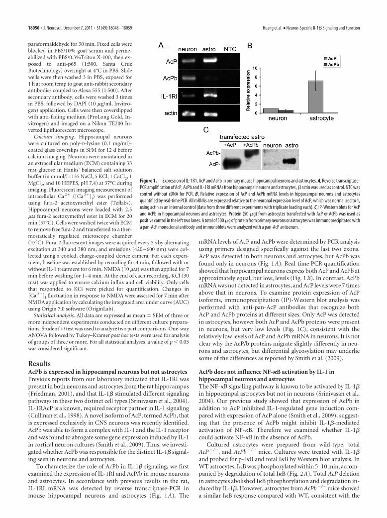
AcPb is required for IL-1-potentiated calcium influx inducedby NMDA in hippocampal neuronsA functional consequence of IL-1�-induced Src activation is tophosphorylate the NMDA receptor NR2B subunit, which colo-calizes with IL-1RI in hippocampal neurons, resulting in poten-tiation of calcium influx induced by NMDA (Viviani et al., 2003).
4
(Figure legend continued.) each genotype. E, Dose–response of IL-1�-induced p38 phos-phorylation after 20 min in WT, total AcP �/�, and AcPb �/� neurons. F, Quantification of blotsfrom three different experiments as in E. Densitometric values were expressed as p-p38/totalp38, and then normalized to untreated cells (0 ng/ml IL-1) for each genotype. The significancewas determined by one-way ANOVA with Tukey’s post hoc analysis. The p value equals to 0.0023between WT and AcPb �/� neurons treated with 100 ng/ml IL-1. Asterisks indicate valuessignificantly different from control at *p � 0.05, **p � 0.001, and ***p � 0.0001.
Figure 5. Src phosphorylation by IL-1� (0.01 ng/ml) in hippocampal neurons is dependent on AcPb. A, Dose–response treatment of IL-1� applied to cultured WT mouse hippocampal neuronsfor 20 min. Protein lysates were immunoblotted for p-Src and reprobed for total Src. Quantification of blots from three different experiments is shown below the blot. Densitometric values wereexpressed as p-Src/total Src, and then normalized to untreated cells (0 ng/ml). B, Time course of Src phosphorylation induced by IL-1� in mouse hippocampal neurons. At low concentration (0.01ng/ml), Src is phosphorylated by IL-1� in WT neurons, but not in total AcP �/� or AcPb �/� neurons. Quantification of blots from three different experiments is shown below the blot. Thesignificance was determined by one-way ANOVA with Tukey’s post hoc analysis. Asterisks indicate values significantly different from control at *p � 0.05, ***p � 0.0001.
18054 • J. Neurosci., December 7, 2011 • 31(49):18048 –18059 Huang et al. • Neuron-Specific Il-1� Signaling and Function

The same low dose of IL-1 that evoked Src phosphorylation elic-ited a sharp peak in phosphorylation of NR2B in WT hippocam-pal neurons after 5 min of treatment (Fig. 7). To assess whetherthe effects of IL-1� on Src and NR2B phosphorylation mightregulate calcium influx, preincubation of hippocampal neuronswith IL-1� (0.01 ng/ml) potentiated NMDA (10 �M)-inducedcalcium influx (Fig. 8A,D), consistent with previous reports. Inaddition, preexposing neurons to the Src kinase inhibitor, PP2(2.5 �M), blocked this effect. Consistent with our signaling resultson Src phosphorylation, higher doses of IL-1� (10 ng/ml) had noeffect on NMDA-induced calcium influx (Fig. 8A,D). To deter-mine whether AcPb was necessary for the IL-1� effect on NMDA-induced calcium influx, WT, total AcP�/� and AcPb�/� neuronswere compared. Removal of total AcP (tAcP�/�) eliminated the
IL-1� potentiation of calcium influx induced by NMDA, as ex-pected, however removal of AcPb alone (AcPb�/�) was sufficientto abrogate the IL-1 effect (Fig. 8B,C), suggesting that AcPb isrequired for IL-1�-induced Src phosphorylation and potentia-tion of NMDA-mediated calcium influx.
DiscussionAcPb in neuronal IL-1� signaling and functionIL-1 is a proinflammatory cytokine that influences many mam-malian functions. IL-1 binds the IL-1RI/IL-1RAcP receptor com-plex, however distinct signaling cascades have been described indifferent cell types with no mechanistic explanation. Within theCNS IL-1� activates different signaling pathways in hippocampalneurons and astrocytes (Viviani et al., 2003; Srinivasan et al.,
Figure 6. Effects of overexpressing AcPb in astrocytes on IL-1�-induced p-Src and p-p38 signaling. A, Time course of IL-1� (0.01 ng/ml) treatment of WT astrocytes with and withoutAcPb transfection. Expression of AcPb in WT astrocytes enabled IL-1� to induce Src phosphorylation. B, IL-1 (0.01 ng/ml)-induced p-Src in total AcP �/� astrocytes restored with AcPbbut not AcP. In contrast, restoration of AcP, but not AcPb was sufficient to restore IL-1� induction of p-p38 MAPK, even at the low dose. C and D show quantification of p-Src and p-p38MAPK, respectively, from blots from three different experiments. Densitometric values were expressed as p-Src/total Src (C) or p-p38/total p38 (D), and then normalized to untreated cells(0 min). The significance was determined by one-way ANOVA with Tukey’s post hoc analysis. Asterisk indicates values significantly different from control at p � 0.05. E, High dose of IL-1(10 ng/ml)-induced p-p38 in total AcP �/� astrocytes restored with either AcP or AcPb, but only induced p-I�B in total AcP �/� astrocytes restored with AcP. F, Quantification of p-p38induction in the tAcP �/� astrocytes transfected either with AcP or AcPb and treated with the high dose (10 ng/ml) of IL-1 for 5 min. Note in B and E that although equal amounts ofplasmid DNA for AcP or AcPb were used for transfections, expression of AcPb is always lower than AcP. Thus, when normalized to the amount of AcP or AcPb expressed, the level of p-p38activation was similar.
Huang et al. • Neuron-Specific Il-1� Signaling and Function J. Neurosci., December 7, 2011 • 31(49):18048 –18059 • 18055

2004). The discovery of AcPb, a new CNS-restricted isoform ofAcP (Smith et al., 2009), provides a novel mechanism governingdistinct responses to IL-1� in CNS neurons. Mice lacking eithertotal AcP or only AcPb were used to define neuron-specific effectson IL-1 signaling and function. Although the germ-line deletionsmay have developmental effects that influence IL-1 signaling,these studies revealed a specific role for AcPb in the regulation ofSrc activation and NMDA function.
IL-1RAcPb and SrcSrc family kinases (SFKs) can phosphorylate ion channels (Salterand Kalia, 2004) and directly modulate neuronal responses, by-passing the need for de novo gene expression. Src activity has longbeen implicated in regulating neuronal long-term potentiation(LTP) (Lu et al., 1998; Salter, 1998; Huang et al., 2001). SFKs canphosphorylate NMDA receptors and regulate their cell surfacetrafficking. Phosphorylation of the NR2B subunit has been spe-cifically associated with trafficking to synaptic sites (Goebel-Goody et al., 2009). We found that both Src and NR2B werephosphorylated in hippocampal neurons by a low dose of IL-1�(0.01 ng/ml), suggesting that IL-1 may facilitate trafficking ofNMDA receptors to synaptic sites resulting in the enhancedNMDA-induced Ca 2� influx that we observed. Consistent withthese data, IL-1 was shown to phosphorylate SFKs and NR2B invivo, eliciting hyperexcitability and potentiation of kainic acid-induced seizures (Balosso et al., 2008). Our current study dem-onstrates that AcPb specifically mediates Src activation by IL-1.
AcPb differs from AcP with a 140 aa extension in theC-terminal region. Interestingly, a related protein, IL-1-receptoraccessory protein-like 1 (IL1RAPL1), also has an extendedC-terminal region, which interacts with the neuronal calciumsensor-1 protein (NCS-1) to regulate N-type calcium channels(Gambino et al., 2007). IL1RAPL1 also associates with the majorexcitatory postsynaptic density protein, PSD-95 that plays a rolein the development and organization of dendritic spines. Muta-tions in IL1RAPL1 lead to a decrease in the number of spines andsubtle alterations in hippocampal LTP (Pavlowsky et al., 2010),and have been associated with mental retardation and autism(Piton et al., 2008). Although the C-terminal extension in AcPbhas no apparent similarity to the analogous region of IL1RAPL1,the presence of this extended domain, and the enhancement ofNMDA-induced calcium influx, suggests a similar role for AcPbin modulating excitatory neurotransmission.
Dose-dependent actions of IL-1 signaling in neuronsIL-1 has previously been shown to have a biphasic effect on hip-pocampal LTP (Goshen et al., 2007). Physiologically low levelsof IL-1 (1–10 pM; Desson and Ferguson, 2003) facilitatehippocampus-dependent memory formation (Yirmiya et al.,2002), possibly by inducing Src activity followed by rapid phos-phorylation of the NMDA receptor. Moreover, blocking IL-1 sig-naling impairs normal maintenance of LTP and memoryconsolidation (Avital et al., 2003; Spulber et al., 2009). Theseeffective doses of IL-1 actions are below the reported Kd for theIL-1R1; however, the presence of AcP and AcPb in the receptorcomplex yields a lower Kd than IL-1R1 alone. Moreover, previousstudies have demonstrated functional effects of IL-1 at doses be-low the measured Kd (Bomsztyk et al., 1989). In contrast, atpathophysiological levels, IL-1 inhibits LTP and impairs novelmemory formation (Murray and Lynch, 1998) in part via activa-tion of the p38 MAPK pathway (Kelly et al., 2003). Consistentwith this, the high dose of IL-1� (10 ng/ml) failed to elicit Srcactivation and NR2B phosphorylation, but did induce p38MAPK phosphorylation in neurons, which required both AcPand AcPb. Deletion of AcPb weakened, but did not suppress,induction of p-p38 in neurons, suggesting that AcPb partiallycontributes to activation of this pathway. Moreover, in astrocyteslacking total AcP, overexpression of either AcP alone or AcPbalone restored IL-1�-induced p38 phosphorylation.
The mechanisms and functional consequences for the distinctneuronal responses to IL-1� at physiological and pathophysio-logical doses remain unclear. One possible consequence of Srcactivation by IL-1� at low concentrations may be to constitu-tively maintain a certain level of neuronal responsiveness. Severalstudies have suggested a physiological role for IL-1� (1 pM or0.017 ng/ml) in enhancing neuronal excitability (Viviani et al.,2003; Desson and Ferguson, 2003). However, the increased excit-ability may also explain why preinjection of low doses of IL-1�prolonged bicuculline-induced seizures in mice (Vezzani et al.,2000). Thus, it is likely that at low concentrations IL-1� inducesp-Src to maintain a certain level of neuronal excitability, but mayalso increase vulnerability to insults.
In vivo, IL-1� does not directly induce neuronal death, but canexacerbate injury-induced neuronal damage (Stroemer andRothwell, 1998). In our system, treatment of hippocampal neu-rons with high concentrations of IL-1� (10 –100 ng/ml) inducedphosphorylation of p38, but not JNK or ERK, followed by activa-tion of the transcription factor CREB (Srinivasan et al., 2004).CREB regulates expression of numerous genes, including cal-cium channels, AMPA receptors, and neurotransmitters. Inter-estingly, we found that IL-1� also induced phosphorylation ofp38 at the low dose, but the role of this pathway remains unclear.It does not necessarily require AcPb, and it is not neuron specific.p38 MAPK has been implicated in mediating neuronal deathinduced by glutamate, and inhibiting p38 can attenuate neuronaldeath (Cao et al., 2004). Therefore, the activation of this pathwayeven with the low dose of IL-1� may contribute to increasedneuronal vulnerability to subsequent insults.
AcPb does not regulate NF-�BSince IL-1� activates NF-�B in many cell types, but not hip-pocampal neurons, one possibility was that AcPb suppressed ac-tivation of this pathway. However, neurons lacking AcPb did notshow NF-�B activation, and transfection of AcPb into astrocytesdid not suppress NF-�B activation, suggesting that the expressionof AcPb does not explain the inability of IL-1 to induce NF-�B
Figure 7. IL-1� (0.01 ng/ml) induced transient phosphorylation of NR2B within 5 min in WTneurons. Graph shows quantification of two independent experiments.
18056 • J. Neurosci., December 7, 2011 • 31(49):18048 –18059 Huang et al. • Neuron-Specific Il-1� Signaling and Function
signaling in neurons. Previous results from our lab showed thatTRAF6, a key adaptor protein in IL-1-mediated NF-�B activation(Cao et al., 1996b), was abundant in hippocampal astrocytes butnot detected in neurons (Srinivasan et al., 2004). TRAF6 interactswith IRAK (Cao et al., 1996a), and activates I�B kinases throughTAK1, NIK, and MEKK1 in response to IL-1 (Lee et al., 1997;Ninomiya-Tsuji et al., 1999), suggesting an essential role forTRAF6 in the NF-�B pathway. Further, dominant-negativeTRAF6 blocked NF-�B activation induced by IL-1� but not byTNF� (Cao et al., 1996b), consistent with our studies showingthat TNF� but not IL-1� activated NF-�B in hippocampal neu-rons (Choi and Friedman, 2009). Thus, it is likely the absence ofTRAF6, rather than the presence of AcPb, that explains whyIL-1� does not induce NF-�B in hippocampal neurons.
The role of AcPb versus AcPThe complexities of IL-1 signaling to elicit the broad spectrum offunctional effects are not fully understood. The association of thereceptor with a specific accessory protein is one mechanism forthe activation of distinct pathways, particularly in brain neuronsexpressing both AcP and AcPb. We show here that low concen-trations of IL-1� elicit Src phosphorylation via AcPb, whilehigher concentrations activate p38, but not Src, requiring AcP. Itis possible that the higher IL-1 concentrations elicit a more stableIL1R/AcP complex that may allow an association-dependentconformational change to occur which interferes with formationof the AcPb-dependent signaling complex. Alternatively, it is
possible that the higher concentration of IL-1� leads to the for-mation of other signaling complexes on the receptor heterodimerover time that sterically prevent association of the Src phosphor-ylating complex.
AcP can associate with other receptors of the IL-1R family,such as the IL-33 receptor ST2, and the orphan receptor IL-1receptor-related protein 2 (IL-1Rrp2). It was possible thatAcPb might associate with one of these other receptors tomodulate signaling in neurons. However, PCR analysis re-vealed ST2 expression in astrocytes but not neurons (data notshown), consistent with a previous report (Andre et al., 2005).Similarly, IL-1Rrp2 is expressed in brain glia but not neurons(Berglof et al., 2003). Thus, it is unlikely that the neuron-specific AcPb interacts with either of these other potentialcoreceptors in these cells.
The link between IL-1R1 and Src in neurons has not previ-ously been identified, but our results indicate that it requiresAcPb. Since the extended C-terminal domain of the AcPb pro-tein is unique, it may recruit distinct adapter proteins thatactivate Src instead of the classical MAPK or NF-�B pathways,which may explain why overexpression of AcPb, even in astro-cytes from total AcP �/� mice, was able to induce p-Src. In-deed, biochemical studies have shown that AcPb associateswith IL-1RI in a ligand-dependent manner (Smith et al.,2009), but fails to recruit MyD88 and IRAK4, resulting infailure to activate the classical IL-1 signaling pathways includ-
Figure 8. AcPb is required for IL-1� (0.01 ng/ml)-potentiated calcium influx induced by NMDA in hippocampal neurons. A–C, Representative changes in [Ca 2�]i measured by Fura-2 in responseto IL-1� and NMDA in WT, total AcP �/� and AcPb �/� neurons. Data are shown as the ratio of 340/380 fluorescence emission. A, WT neurons were pretreated with or without 0.01 ng/ml IL-1�,PP2 � 0.01 ng/ml IL-1�, or 10 ng/ml IL-1� before the addition of NMDA. B, C, AcPb �/� neurons (B) or total AcP �/� neurons (C) were pretreated with or without IL-1� (0.01 ng/ml) before NMDAaddition. Baseline recordings were established for 4 min followed by treatment with or without IL-1� for 6 min. NMDA (10 �M) was applied for 7 min and then washed away. At the end of eachrecording, KCl (30 mM) was applied to ensure calcium response and cell viability. D, Quantification of NMDA-induced calcium influx from 3 to 5 individual cultures. Area under the curve (AUC) fromeach group was normalized to WT neuron control, which was set at 100%. Total number of neurons analyzed for a given group is as follows: WT neuron, control (CTR), 300; WT neuron, 0.01 ng/mlIL-1�, 377; WT neuron, PP2 � 0.01 ng/ml IL-1�, 125; WT neuron, 10 ng/ml IL-1�, 73; total AcP �/� neuron, CTR, 155; total AcP �/� neuron, 0.01 ng/ml IL-1�, 158; AcPb �/� neuron, CTR, 315;AcPb �/� neuron, 0.01 ng/ml IL-1�, 271.
Huang et al. • Neuron-Specific Il-1� Signaling and Function J. Neurosci., December 7, 2011 • 31(49):18048 –18059 • 18057

ing the JNK and ERK MAPK pathways (Smith et al., 2009), aswell as NF-�B, as shown here.
IL-1 is a pleiotropic cytokine that can activate multiple signal-ing pathways in different cell types with distinct functional con-sequences. The discovery of AcPb, a novel, neuron-specific IL-1signaling protein, provides mechanistic insight into cell-specificIL-1 signaling and function in the brain.
ReferencesAndre R, Lerouet D, Kimber I, Pinteaux E, Rothwell NJ (2005) Regulation
of expression of the novel IL-1 receptor family members in the mousebrain. J Neurochem 95:324 –330.
Avital A, Goshen I, Kamsler A, Segal M, Iverfeldt K, Richter-Levin G, YirmiyaR (2003) Impaired interleukin-1 signaling is associated with deficits inhippocampal memory processes and neural plasticity. Hippocampus13:826 – 834.
Balosso S, Maroso M, Sanchez-Alavez M, Ravizza T, Frasca A, Bartfai T,Vezzani A (2008) A novel non-transcriptional pathway mediates theproconvulsive effects of interleukin-1beta. Brain 131:3256 –3265.
Bellinger FP, Madamba S, Siggins GR (1993) Interleukin 1� inhibits synap-tic strength and long-term potentiation in the rat CA1 hippocampus.Brain Res 628:227–234.
Berglof E, Andre R, Renshaw BR, Allan SM, Lawrence CB, Rothwell NJ,Pinteaux E (2003) IL-1Rrp2 expression and IL-1F9 (IL-1H1) actions inbrain cells. J Neuroimmunol 139:36 – 43.
Bomsztyk K, Stanton TH, Smith LL, Rachie NA, Dower SK (1989) Proper-ties of interleukin-1 and interferon-� receptors in B lymphoid cell line.J Biol Chem 264:6052– 6057.
Breder CD, Dinarello CA, Saper CB (1988) Interleukin-1 immunoreactiveinnervation of the human hypothalamus. Science 240:321–324.
Cao J, Semenova MM, Solovyan VT, Han J, Coffey ET, Courtney MJ (2004)Distinct requirements for p38alpha and c-Jun N-terminal kinase stress-activated protein kinases in different forms of apoptotic neuronal death.J Biol Chem 279:35903–35913.
Cao Z, Henzel WJ, Gao X (1996a) IRAK: A kinase associated with theinterleukin-1 receptor. Science 271:1128 –1131.
Cao Z, Xiong J, Takeuchi M, Kurama T, Goeddel DV (1996b) TRAF6 is asignal transducer for interleukin-1. Nature 383:443– 446.
Choi S, Friedman W (2009) Inflammatory cytokines IL-1� and TNF-� reg-ulate p75NTR expression in CNS neurons and astrocytes by distinct cell-type-specific signalling mechanisms. ASN Neuro 1:pii: e00010.
Cullinan EB, Kwee L, Nunes P, Shuster DJ, Ju G, McIntyre KW, ChizzoniteRA, Labow MA (1998) IL-1 receptor accessory protein is an essentialcomponent of the IL-1 receptor. J Immunol 161:5614 –5620.
Davies CA, Loddick SA, Toulmond S, Stroemer RP, Hunt J, Rothwell NJ(1999) The progression and topographic distribution of interleukin-1beta expression after permanent middle cerebral artery occlusion in therat. J Cereb Blood Flow Metab 19:87–98.
Desson SE, Ferguson AV (2003) Interleukin 1beta modulates rat subforni-cal organ neurons as a result of activation of a non-selective cationicconductance. J Physiol 550:113–122.
Friedman WJ (2001) Cytokines regulate expression of the type 1interleukin-1 receptor in rat hippocampal neurons and glia. Exp Neurol168:23–31.
Friedman WJ, Thakur S, Seidman L, Rabson AB (1996) Regulation of nervegrowth factor mRNA by interleukin-1 in rat hippocampal astrocytes ismediated by NFkappaB. J Biol Chem 271:31115–31120.
Gambino F, Pavlowsky A, Begle A, Dupont JL, Bahi N, Courjaret R, Gar-dette R, Hadjkacem H, Skala H, Poulain B, Chelly J, Vitale N, HumeauY (2007) IL1-receptor accessory protein-like 1 (IL1RAPL1), a pro-tein involved in cognitive functions, regulates N-type Ca 2�-channeland neurite elongation. Proc Natl Acad Sci U S A 104:9063–9068.
Giulian D, Baker TJ, Shih LC, Lachman LB (1986) Interleukin 1 of the cen-tral nervous system is produced by ameboid microglia. J Exp Med164:594 – 604.
Goebel-Goody SM, Davies KD, Alvestad Linger RM, Freund RK, BrowningMD (2009) Phospho-regulation of synaptic and extrasynaptic N-methyl-d-aspartate receptors in adult hippocampal slices. Neuroscience 158:1446 –1459.
Goshen I, Kreisel T, Ounallah-Saad H, Renbaum P, Zalzstein Y, Ben-Hur T,Levy-Lahad E, Yirmiya R (2007) A dual role for interleukin-1 inhippocampal-dependent memory processes. Psychoneuroendocrinology32:1106 –1115.
Huang Y, Lu W, Ali DW, Pelkey KA, Pitcher GM, Lu YM, Aoto H, Roder JC,Sasaki T, Salter MW, MacDonald JF (2001) CAKbeta/Pyk2 kinase is asignaling link for induction of long-term potentiation in CA1 hippocam-pus. Neuron 29:485– 496.
Hunter CA, Roberts CW, Murray M, Alexander J (1992) Detection of cyto-kine mRNA in the brains of mice with toxoplasmic encephalitis. ParasiteImmunol 14:405– 413.
Katsuki H, Nakai S, Hirai Y, Akaji K, Kiso Y, Satoh M (1990) Interleukin-1Binhibits long-term potentiation in the CA3 region of mouse hippocampalslices. Eur J Pharmacol 181:323–326.
Kelly A, Vereker E, Nolan Y, Brady M, Barry C, Loscher CE, Mills KH, LynchMA (2003) Activation of p38 plays a pivotal role in the inhibitory effectof lipopolysaccharide and interleukin-1� on long-term potentiation inrat dentate gyrus. J Biol Chem 278:19453–19462.
Lee FS, Hagler J, Chen ZJ, Maniatis T (1997) Activation of the IkappaBalpha kinase complex by MEKK1, a kinase of the JNK pathway. Cell88:213–222.
Libermann TA, Baltimore D (1990) Activation of interleukin-6 gene expres-sion through the NF-kappa B transcription factor. Mol Cell Biol10:2327–2334.
Loddick SA, Liu C, Takao T, Hashimoto K, De Souza EB (1998)Interleukin-1 receptors: cloning studies and role in central nervous sys-tem disorders. Brain Res Brain Res Rev 26:306 –319.
Lu YM, Roder JC, Davidow J, Salter MW (1998) Src activation in the induc-tion of long-term potentiation in CA1 hippocampal neurons. Science279:1363–1367.
Merrill JE, Benveniste EN (1996) Cytokines in inflammatory brain lesions:helpful and harmful. Trends Neurosci 19:331–338.
Murray CA, Lynch MA (1998) Evidence that increased hippocampal ex-pression of the cytokine interleukin-1B is a common trigger for age- andstress-induced impairments in long-term potentiation. J Neurosci18:2974 –2981.
Ninomiya-Tsuji J, Kishimoto K, Hiyama A, Inoue J, Cao Z, Matsumoto K(1999) The kinase Tak1 can activate the NIK-I�B as well as the MAPkinase cascade in the IL-1 signalling pathway. Nature 398:252–256.
O’Neill LA, Bird TA, Saklatvala J (1990) Interleukin 1 signal transduction.Immunol Today 11:392–394.
Pavlowsky A, Gianfelice A, Pallotto M, Zanchi A, Vara H, Khelfaoui M, Val-negri P, Rezai X, Bassani S, Brambilla D, Kumpost J, Blahos J, Roux MJ,Humeau Y, Chelly J, Passafaro M, Giustetto M, Billuart P, Sala C (2010)A postsynaptic signaling pathway that may account for the cognitive de-fect due to IL1RAPL1 mutation. Curr Biol 20:103–115.
Pearson VL, Rothwell NJ, Toulmond S (1999) Excitotoxic brain damage inthe rat induces interleukin-1beta protein in microglia and astrocytes: cor-relation with the progression of cell death. Glia 25:311–323.
Piton A, Michaud JL, Peng H, Aradhya S, Gauthier J, Mottron L, Cham-pagne N, Lafreniere RG, Hamdan FF, Joober R, Fombonne E, Mari-neau C, Cossette P, Dube MP, Haghighi P, Drapeau P, Barker PA,Carbonetto S, Rouleau GA (2008) Mutations in the calcium-relatedgene IL1RAPL1 are associated with autism. Hum Mol Genet17:3965–3974.
Pitossi F, del Rey A, Kabiersch A, Besedovsky H (1997) Induction of cyto-kine transcripts in the central nervous system and pituitary followingperipheral administration of endotoxin to mice. J Neurosci Res48:287–298.
Salter MW (1998) Src, N-methyl-D-aspartate (NMDA) receptors, and syn-aptic plasticity. Biochem Pharmacol 56:789 –798.
Salter MW, Kalia LV (2004) Src kinases: a hub for NMDA receptor regula-tion. Nat Rev Neurosci 5:317–328.
Sims JE, Smith DE (2010) The IL-1 family: regulators of immunity. Nat RevImmunol 10:89 –102.
Smith DE, Lipsky BP, Russell C, Ketchem RR, Kirchner J, Hensley K, HuangY, Friedman WJ, Boissonneault V, Plante MM, Rivest S, Sims JE (2009)A central nervous system-restricted isoform of the interleukin-1 receptoraccessory protein modulates neuronal responses to interleukin-1. Immu-nity 30:817– 831.
18058 • J. Neurosci., December 7, 2011 • 31(49):18048 –18059 Huang et al. • Neuron-Specific Il-1� Signaling and Function

Spulber S, Mateos L, Oprica M, Cedazo-Minguez A, Bartfai T, Winblad B,Schultzberg M (2009) Impaired long term memory consolidation intransgenic mice overexpressing the human soluble form of IL-1ra in thebrain. J Neuroimmunol 208:46 –53.
Srinivasan D, Yen JH, Joseph DJ, Friedman W (2004) Cell type specificinterleukin-1� signaling in the central nervous system. J Neurosci24:6482– 6488.
Stroemer RP, Rothwell NJ (1998) Exacerbation of ischemic brain damageby localized striatal injection of interleukin-1beta in the rat. J Cereb BloodFlow Metab 18:833– 839.
Vezzani A, Moneta D, Conti M, Richichi C, Ravizza T, De Luigi A, De SimoniMG, Sperk G, Andell-Jonsson S, Lundkvist J, Iverfeldt K, Bartfai T (2000)Powerful anticonvulsant action of IL-1 receptor antagonist on intracere-
bral injection and astrocytic overexpression in mice. Proc Natl Acad SciU S A 97:11534 –11539.
Viviani B, Bartesaghi S, Gardoni F, Vezzani A, Behrens MM, Bartfai T, Bina-glia M, Corsini E, Di Luca M, Galli CL, Marinovich M (2003)Interleukin-1beta enhances NMDA receptor-mediated intracellular cal-cium increase through activation of the Src family of kinases. J Neurosci23:8692– 8700.
Yirmiya R, Winocur G, Goshen I (2002) Brain interleukin-1 is involved inspatial memory and passive avoidance conditioning. Neurobiol LearnMem 78:379 –389.
Zhang P, Miller BS, Rosenzweig SA, Bhat NR (1996) Activation of C-junN-terminal kinase/stress-activated protein kinase in primary glial cul-tures. J Neurosci Res 46:114 –121.
Huang et al. • Neuron-Specific Il-1� Signaling and Function J. Neurosci., December 7, 2011 • 31(49):18048 –18059 • 18059