diversity in radiology starts at the top - rsna.org · a pioneer in cardiac mri, alain rahmouni,...
TRANSCRIPT

RSNA 2018 Registration Opens July 18— See Page 24
May 2018 Volume 28, Issue 5
A L S O I N S I D E :
Imaging Biomarker Promising for Liver Disease
The Role of MRI in Living Donor Liver Transplantation
Imaging Safety Movement Expands Across the Globe
Diversity in Radiology Starts at the Top

B RSNA News | May 2018

UP FRONT 2 First Impression
4 Numbers in the News
5 My Turn
RADIOLOGY’S FUTURE 16 R&E Foundation Donors
NEWS YOU CAN USE 17 Value of Membership
18 Journal Highlights
20 Radiology in Public Focus
22 Education and Funding Opportunities
24 Annual Meeting Watch
25 Member Spotlight
9
Imaging Safety Movement Expands Across the Globe
14
The Role of MRI in Living Donor Liver Transplantation
12
Imaging Biomarker Promising for Liver Disease
FEATURES
6
MAY 2018 • VOLUME 28, ISSUE 5
EDITOR
Gary J. Whitman, MD
R&E FOUNDATION CONTRIBUTING EDITOR
Theresa C. McLoud, MD
EXECUTIVE EDITOR
Shelley L. Taylor
MANAGING EDITOR
Beth Burmahl
STAFF WRITER
Jennifer Allyn
GRAPHIC DESIGNER
Eriona Baholli-Karasek
EDITORIAL ADVISORS
Mark G. Watson Executive Director
Karena Galvin Assistant Executive Director Marketing and International Affairs
Marijo Millette Director: Public Information and Communications
EDITORIAL BOARD
Gary J. Whitman, MD ChairmanVahid Yaghmai, MD Vice ChairmanEzra Bobo, MDStephen D. Brown, MDCarlo Catalano, MDDaniel A. Hamstra, MD, PhDMaureen P. Kohi, MDLaurie A. Loevner, MDTheresa C. McLoud, MDMartin P. Torriani, MDMary C. Mahoney, MDBoard Liaison
2018 RSNA BOARD OF DIRECTORS
James P. Borgstede, MD ChairmanMary C. Mahoney, MD Liaison for Publications and Communications Bruce G. Haffty, MD Liaison for ScienceMatthew A. Mauro, MD Liaison for EducationCurtis P. Langlotz, MD, PhD Liaison for Information Technology and Annual Meeting
Umar Mahmood, MD, PhDLiaison for International AffairsVijay M. Rao, MD PresidentValerie P. Jackson, MD President-Elect
Follow us for exclusive news, annual meeting offers and more!
RSNA MISSIONThe RSNA promotes excellence in patient care and healthcare delivery through education, research and technologic innovation.
Diversity in Radiology Starts at the Top

2 RSNA News | May 2018
RSNA will host a Spotlight Course in Buenos Aires, Argentina, June 8–9.
Presented entirely in Spanish, “Últimas Tendencias en Imágenes Abdominales” will focus on the latest trends in abdominal imaging. The course will include Diagno-sis Live™ sessions, giving attendees the opportunity to test their knowledge and engage with renowned global leaders in abdominal imaging.
Space is limited and early registration is encouraged to take advantage of discounted pre-booking rates. The deadline for online registration is June 1. For more information and to register, visit RSNA.org/Spotlight.
FIRST IMPRESSION
Goske Pressman Resnick
Registration DeadlineJune 1
ACR Awards 2018 Gold MedalsMarilyn J. Goske, MD, Barry D. Pressman, MD, and Donald L. Resnick, MD, will receive gold medals from the American College of Radiology (ACR) during its annual meeting this month.
Dr. Goske is a professor clinical emerita in the Department of Radiology at the University of Cincinnati. Dr. Goske was named the RSNA 2012 Outstanding Educator. She is the founding chair of the Image Gently™ campaign to promote child-sized imaging in the U.S.
Dr. Pressman is chair of the Department of Radiology at Cedar Sinai Medical Center, Los Angeles. He is a former ACR president.
Dr. Resnick is professor emeritus in the Department of Radiol-ogy at the University of California, San Diego. He was recognized in 2006 as an RSNA Outstanding Educator and serves on the RSNA Public Information Advisors Network.
Register for RSNA Spotlight Course in Argentina
New RSNA Professionalism Vignette Addresses Sexual MisconductThe RSNA Professionalism Committee has added a new professionalism vignette to the RSNA website.
“Sexual Misconduct in the Radiology Workplace” addresses a wide range of behaviors that may make a workplace or educational institution unsafe for its members. The self-guided vignette includes four scenarios that explore sexual misconduct in the radiology workplace. Radiologists read through the scenario and answer questions. Each answer raises questions about how to handle various aspects of the issue, provides responses that discuss the relevant principles of pro-fessionalism and offers references for further study.
RSNA members can access this vignette for free on the RSNA website at RSNA.org/Professionalism.
Save the Date: RSNA Spotlight Course in Paris to Focus on AI The RSNA Spotlight Course, “Practical Applications of Artificial Intelligence (AI),” will be held Sept. 23–24, 2018, in Paris, France.
This two-day course is for radiologists of all subspecialties who want to learn more about AI and its practical application to practice workflows and processes. Those completing the course will have a better understanding of how AI can help, not replace, radiologists.
Registration will open at the end of June. More information will be coming soon at RSNA.org/Spotlight2018.

May 2018 | RSNA News 3
Chew Meltzer
Marx Named SIR PresidentVictoria Marx, MD, assumed the office of president of the Society of Interventional Radiology (SIR) during the society’s annual scientific meeting in Los Angeles in March.
Dr. Marx is a professor of clinical radiology and diagnostic radiology residency program director at Keck School of Medicine of USC, Los Angeles.
Other incoming officers of SIR’s 2018–19 Executive Council include President-elect Laura Findeiss, MD, Atlanta; Secretary Michael D. Dake, MD, Stanford, CA; and Immediate Past President Suresh Vedantham, MD, St. Louis.Marx
CorrectionThe article, “Paleoradiologists Unravel the Secrets of Ancient Mummies,” in the March issue of RSNA News incorrectly stated that the Museum of Cairo is the only museum in the world with a CT scanner. RSNA News regrets the error.
The Nominating Committee for the Alexander R. Margulis Award for Scientific Excellence is accepting nominations from readers for Radiology articles published between July
2017 and June 2018. The main selection criteria are scientific quality and originality.Email your nomination, including the article citation and a brief note highlighting
the reasons for the nomination, to [email protected].
Nominate Radiology Articles for the 2018 Margulis Award
Deadline for NominationsJune 10
Chew and Meltzer Receive 2018 AUR Gold MedalsFelix S. Chew, MD, and Carolyn C. Meltzer, MD, will receive gold medals from the Association of University Radiologists (AUR) during its annual meeting this month.
Dr. Chew is professor and section chief of musculoskeletal radiology at the University of Washington School of Medicine, Seattle. A former AUR president, Dr. Chew previously served as chair of the RSNA Education Exhibits Committee Musculoskeletal Subcommittee.
Dr. Meltzer is the William Patterson Timmie Professor and Chair of the Department of Radiology and Imaging Sciences and the associate dean for research at Emory University School of Medicine, Atlanta. She is an RSNA third vice president and serves on the RSNA Research and Education (R&E) Board of Trustees and the Public Information Advisors Network.
2017 RSNA President Richard L. Ehman, MD, (left) presented the 2017 Margulis Award to
Walter H. Backes, PhD, at the RSNA annual meeting.
4 RSNA News | May 2018
FIRST IMPRESSION
May 2018 • Volume 28, Issue 5 Published monthly by the Radiological Society of North America, Inc. 820 Jorie Blvd., Oak Brook, IL 60523-2251. Printed in the USA.
Postmaster: Send address corrections or changes to: RSNA News, 820 Jorie Blvd., Oak Brook, IL 60523-2251Non-member subscription rate is $20 per year; $10 of active members’ dues is allocated to a subscription of RSNA News.
LETTERS TO THE [email protected] 1-630-571-7837 fax
[email protected] 1-888-600-0064 1-630-590-7770
Contents of RSNA News copyrighted ©2018, RSNA. RSNA is a registered trademark of the Radiological Society of North America, Inc.
REPRINTS AND [email protected] 1-630-571-7829 1-630-590-7724 fax
[email protected] Lisa Lazzaretto Assistant Director: Corporate Relations 1-630-571-7818
Numbers in the News
29Percentage of academic radiologists who are women, according to the Association of American Medical Colleges. Read more about the role of leadership in diversity and inclusion in radiology on Page 6.
90Percentage of liver cancer cases related to chronic liver disease. We report on recent Radiology research on a promising biomarker in managing chronic liver disease on Page 12.
1,015Number of imaging proce-dures performed during the 2016 Rio de Janeiro Sum-mer Olympics. Read about new Radiology research on the role of imaging in eval-uating athletes’ injuries on Page 20.
FDA Approves U.S.-Based Source of Tc-99mThe most widely used radioisotope in medical imaging, Technetium-99m (Tc-99m), may once again be extensively available in the United States due to a new approval from the U.S. Food and Drug Administration (FDA).
The FDA, with guidance and licensing support from the Nuclear Regulatory Commission, has approved the RadioGenix System, a unique system for producing Tc-99m.
The NRC will license NorthStar Medical Radioisotopes, LLC, the company that created the RadioGenix System, to enable the Tc-99m to be made into sodium pertechnetate Tc-99m injec-tions. These can be injected intravenously, instilled into the bladder or eye or used with other FDA-approved imaging drugs to examine specific tissues and organs.
For more information about the potential domestic shortage of TC-99m, visit www.FDA.gov.
A pioneer in cardiac MRI, Alain Rahmouni, MD, died Jan. 26 in France. He was 61. Dr. Rahmouni was noted throughout France and Europe for his work to promote radiology and champion innovations that benefited patients. He was a dedicated educator eager to share the latest scientific innovations with young radiologists. Dr. Rahmouni was head of radiology and medical imaging at Henri-
Mondor University Hospitals, Créteil, France. He earned his medical degree at Sorbonne Universite, Paris, and completed his residency at Hospital Dijon, France. Dr. Rahmouni completed his fellowship at Henri-Mondor University Hospital, where he co-founded the radiology school in 1989 with Elias Zerhouni, MD. Dr. Rahmouni was a former vice secretary general for the Société
Française de Radiologie (SFR). Within RSNA, he served as a manuscript reviewer and author for Radiology and also published in RadioGraphics. Dr. Rahmouni presented at several RSNA annual meetings.
In MemoriamAlain Rahmouni, MD
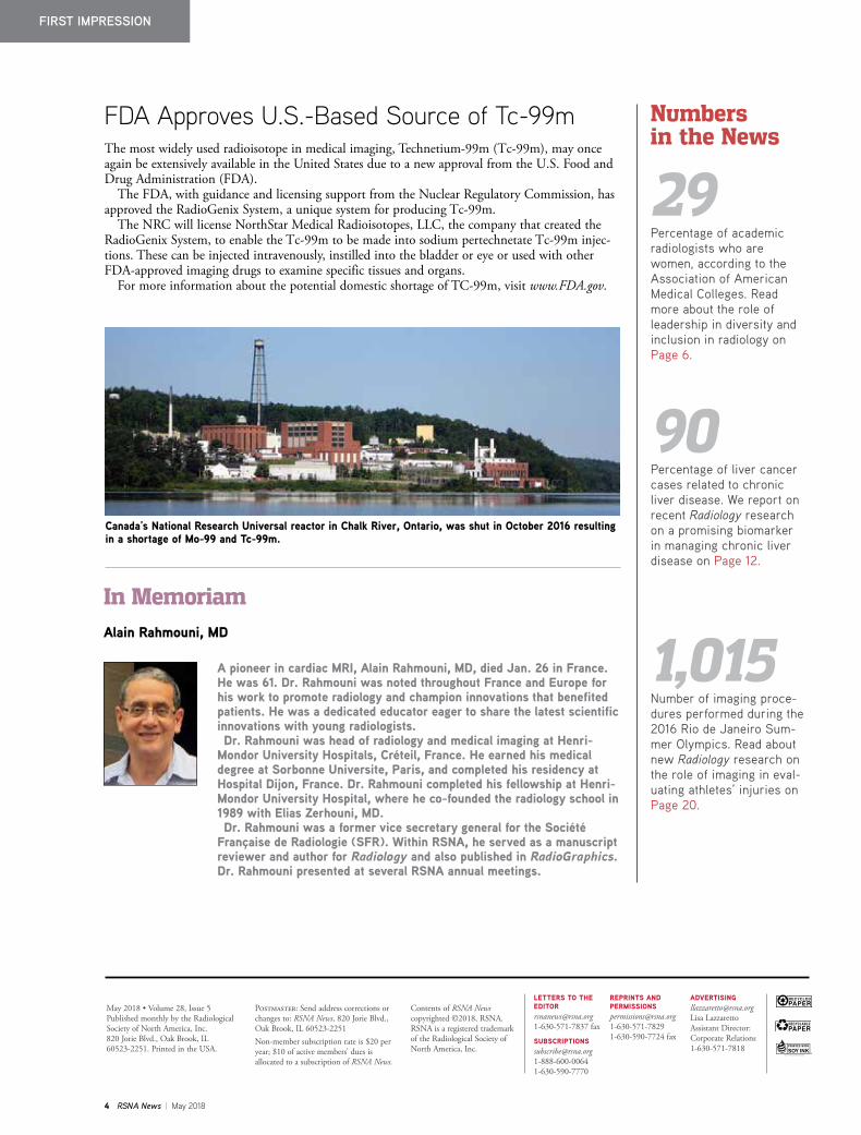
Canada’s National Research Universal reactor in Chalk River, Ontario, was shut in October 2016 resulting in a shortage of Mo-99 and Tc-99m.

May 2018 | RSNA News 5
My Turn:
RSNA’s International Advisory Committee Promotes Radiology Across the GlobeBY RENATO ADAM MENDONÇA, MD, PHD
The RSNA International Advisory Committee (IAC) oversees four regional committees: Asia/Oceania, Europe, Latin America and Middle East/Africa. The chair for each regional committee is also a member of the IAC. The chairs provide updates on its work throughout the year to the IAC at the in-person meeting held during the RSNA annual meeting in Chicago. The Regional Committees are responsible for representing radiologists in their regions and their interactions with RSNA. Members of these commit-tees provide direction on how RSNA can most effectively engage members in their respective geographical areas. By providing local insights on how radiology oper-ates and country specific feedback, the regional committees have been instrumen-tal in the Society’s international growth.
Since 2016, RSNA has offered two Spotlight Courses and will conduct its third course this year. The Spotlight Courses were developed as a way to increase accessibility of RSNA education to the international community and to fill any educational gaps in the region. The RSNA Board Committee on Inter-national Affairs (BCIA) works with the corresponding regional committee to select the topic of focus that is most relevant to radiologists in the region and identify course directors. The first Spotlight Course, “Emergency Radiology: Interactive Course with Cases”, took place in June 2016 in Cancun, Mexico. The second course, “MSK: Interactive Course with Cases,” took place in May 2017 in Bogotá, Colombia. Both courses had approximately 200 attendees from 18 different countries. The upcoming course, “Latest Trends in Abdominal Imaging,” will be held in Buenos Aires, Argentina June 8–9, 2018.
RSNA recently introduced the Interna-tional Travel Stipend Program which was developed through the IAC for eligible RSNA members to partially defray travel expenses related to attending the RSNA annual meeting in Chicago. Beginning in 2018, up to 50 awards of $500 and complimentary registration to the RSNA meeting will be available for eligible candidates.
In 2007, RSNA introduced Country Presents to the RSNA annual meeting. Country Presents features programming that highlights a country’s contributions to RSNA and the radiology community. With input from the IAC, two coun-tries are invited to participate each year. Colombia and Israel were highlighted at RSNA 2017.
The IAC is also responsible for plan-ning the highly anticipated International Trends Session that takes place at the RSNA annual meeting. This invita-tion-only session brings together leaders from around the world to share ideas and best practices on a topic of global impor-tance to the profession. The 2017 Trends session, “Impact of Artificial Intelligence and Machine Learning on International Radiology,” was a topic of great interest. As machine learning and artificial intel-ligence will increasingly affect the global radiology community, RSNA will con-tinue to offer opportunities surrounding this topic, including a course developed from this discussion which will be offered at RSNA 2018. The International Trends session at RSNA 2018 will also continue discussion on this topic.
The International Advisory Committee is dedicated to its charge to effectively represent RSNA members and greatly appreciates the importance that the Soci-ety places on our recommendations. I am honored to work with and lead such an essential constituency of RSNA.
Renato Adam Mendonça, MD, PhD, is the chair of the RSNA International Advisory Committee (IAC) and chief of the Department of Neuroradiology at Delboni Auriemo in São Paulo, Brazil. Dr. Mendonça also serves as Scientific Director for the Radiological and Diagnostic Imaging Society of São Paulo (SPR).

6 RSNA News | May 2018
FEATURES
Championing diversity and seeking ways to institute inclusion can be encouraged from within any level of an organization. However, according to advocates who are working to improve diversity in radiology, the best way to change the culture and ethics of an entire organization is for the directives to come from top leadership, backed by the necessary resources and investments needed to foster change.
Many departments of radiology are doing both: embracing over- arching university or health system initiatives on diversity and inclu-sion as well as identifying specific needs within its own division.
Closing the Gender GapIn 2005, faculty at Memorial Sloan Kettering (MSK) Cancer Center in New York City identified several issues impacting the growth and development of the female faculty. Laura Liberman, MD, a radiologist specializing in breast imaging at MSK, was asked to lead the new Pro-gram for Women Faculty Affairs. She and her team were charged with creating a baseline of where the institution was regarding diversity — particularly gender diversity within faculty levels.
“Early in the course of leading the Program for Women Faculty Affairs, we found that there was a higher representation of women in the junior faculty compared to in the senior faculty in all specialties, not just radiology,” Dr. Liberman said. “We worked to create programs for junior faculty that would benefit female faculty and their male colleagues as well.”
These initiatives were so successful that they served as a model for future growth. In 2012, the Office of Faculty Development (OFD) was created within the Office of the President. Dr. Liberman is now the director of the OFD, and the Program for Women Faculty Affairs exists under the OFD umbrella.
The OFD is responsible for developing initiatives for faculty at all levels, as well as expanding the pipeline for future physicians and scientists through student programs. The OFD has seen tremendous gains in representation of women on the faculty of MSK. Women now comprise 46 percent of all MSK hospital faculty, an increase from 37 percent in 2005 and higher than the 40 percent representation of women in the clinical sciences reported in the most recent Association of American Medical Colleges (AAMC) data, showing that women comprise 29 percent of academic radiology faculty.
“We also collaborate with the MSK Office of Diversity Programs to enhance career development of faculty from backgrounds under-repre-sented in medicine," Dr. Liberman said, noting that diversity among physicians fosters different perspectives, enhancing research and clin-ical care. "The fact that our initiative came from the Office of the President at MSK helps us gain traction. This is not just talk for us. It is how we see the future of our workforce.”
Diversity in Radiology Starts at the TopBY JENNIFER ALLYN
“All leaders must be innovative, have a clear vision and the courage to take risks. For the specific task of creating a diverse and inclusive environment, leaders must make an intentional commitment to the goal.”
STEPHANIE SPOTTSWOOD, MD

May 2018 | RSNA News 7
Identifying and Eliminating BiasVanderbilt University Medical Center in Nashville, TN, also has support from top leadership for its diversity initiatives. The School of Medicine’s Office for Diversity Affairs supports the radiology department’s Office of Diversity, Equity and Inclusion.
The Office of Diversity, Equity and Inclusion has made significant inroads in improving diversity among radiology house staff (residents) with its multi-fac-eted approach that includes taking advan-tage of many of Vanderbilt’s larger diver-sity initiatives.
“The primary aims of the radiology department’s Office of Diversity, Equity and Inclusion are to recruit talented, underrepresented minorities to our
residency program, by diversifying our applicant pool. This initiative was designed to develop and maintain a cul-turally and demographically diverse group of residents. This effort is supported by faculty and resident training on cultural proficiency and unconscious bias,” said Stephanie Spottswood, MD, professor of radiology and associate vice chair for Diversity, Equity and Inclusion at Van-derbilt.
To that effect, Vanderbilt University Medical Center has undertaken an ini-tiative to acknowledge and help mitigate bias in recruitment decisions and during patient care. A staff-led group trained in the principles of unconscious bias has begun to provide training for more than
20,000 Vanderbilt employees. In addition, radiology faculty enrich-
ment programs educate and support both new and current faculty: An annual Diver-sity Grand Rounds lectureship recognizes the research and clinical achievements of nationally renowned, underrepresented minority physicians within radiology. The radiology department’s Leadership Intervention to Further the Training of Female Faculty (LIFT OFF) program was designed to improve access to opportuni-ties for women’s faculty development and advancement and improve clarification of expectations about the role and path of advancement.
Eleby R. Washington IV, MD, a radiol-ogy resident at Vanderbilt who serves as
Editor’s Note: This is the second in a series of articles on diversity and inclusion in radiology.
Liberman Spottswood Washington Norbash
Continued on next page

8 RSNA News | May 2018
FEATURE
the resident liaison to the Office of Diver-sity, Equity and Inclusion, has a unique vision for his career path in radiology.
“I was drawn to radiology because it is a technologically driven field with the possibility of profound and immediate impact on patient care,” Dr. Washington said. “I recognized the lack of diversity, but that did not negatively affect my deci-sion to select radiology. To the contrary, I personally saw it as an opportunity to contribute to and positively help effect change in my desired field.”
Vanderbilt’s efforts are having an impact. The radiology program has increased its applicant pool of minority students from 7.5 percent to 13.5 per-cent over the four years since the Office of Diversity, Equity and Inclusion was founded. Currently, 19 percent of its diagnostic radiology residents are women and 13 percent are from groups consid-ered to be underrepresented in medicine.
“As an academic medical center, it’s important that we recruit and retain a diverse group of faculty and trainees,” Dr. Spottswood said. “Diversity and inclusion improve problem-solving, enrich the learning environment and ensure that we have adequate represen-tation in medicine, which in turn helps alleviate the barriers to healthcare facing underserved populations.”
Campus-Wide Diversity GoalsAt the University of California, San Diego (UCSD), Alexander Norbash, MD, professor and chair of the Department of Radiology who also serves as the associate vice chancellor of equity, diversity and inclusion, agrees with the ultimate end-goals of diversity initiatives.
“At UCSD, our charge is to prepare students to succeed in a diverse and com-plex society,” Dr. Norbash said. “Newer generations of medical students expect diversity and inclusion in their depart-ments and expect to be mindful of diversity issues. They expect to work in an environment of civility and respect where they also will treat patients with civility and respect. We can’t draw students to medicine without showing strong and directed efforts in ensuring professional-ism and managing diversity.”
Throughout his career, Dr. Norbash has seen how the potential richness of learning from those who are different can be stymied by common misconceptions.
To that end, the department of radiol-ogy at UCSD has launched a diversity committee that is actively engaged in this space. It supports the initiatives from the UCSD’s Office of the Vice Chancellor for Diversity, which is developing a campus wide strategic plan for inclusive excellence.
The department Residency Selection Committee actively looks at gender equity to ensure that the short list is rep-resentative of all applicants. The depart-ment also initiated a wellness program that is charged with addressing issues like bullying and burnout.
“Any department of radiology diversity programs are intended to be complemen-tary with the larger university initiatives,” Dr. Norbash said. “This ensures that we foster a teaching, research and learning environment that strengthens our ties as radiologists by being inclusive and equi-table to faculty and students, while lever-aging what we are learning in all areas of the university."
Strong Leaders NeededWhile the approaches to help engage women and minorities in radiology are different across institutions, there are similarities when it comes to the type of leadership traits needed to drive this change, either at the university or department level.
Leaders need to remember that every-one has a role to play in this space, so paying attention to the potential of the people already working with the initiative and those that still need to be recruited, is important, according to Dr. Norbash.
“Establishing the need and the credibility of diversity initiatives is mission-critical,” he said. “The world is changing in an accelerated manner. We must address the broadest range of talent in order to identify and mobilize the strongest and most creative possible future workforce.”
Dr. Washington adds that leaders must have empathy.
“Even if you have had significantly different life experiences than others, leaders need to be able to build a sense of community without stagnation,” Dr. Washington said. “Championing new ideas, building on past successes and continually moving forward, while bringing the entire team along, are paramount to success in building diversity and inclusion.”
Dr. Spottswood agrees that the responsibility rests with leadership.
“All leaders must be innovative, have a clear vision and the courage to take risks,” she said. “For the specific task of creating a diverse and inclusive environment, leaders must make an intentional commitment to the goal.”
Continued from previous page

May 2018 | RSNA News 9
Imaging Safety Movement Expands Across the GlobeBY NICK KLENSKE
In 2010, leading North American radiology organizations including RSNA introduced Image Wisely®, a program dedicated to raising awareness about optimizing the radiation used in medically necessary imaging studies and eliminating unnecessary procedures in adult patients.
Image Wisely was launched by RSNA, the American College of Radiology (ACR), the American Association of Physicists in Medicine (AAPM) and the American Soci-ety of Radiological Technologists (ASRT) with the goal of championing the safe and effective use of medical imaging for adult patients and disseminating these concepts to the radiology profession and other med-ical professionals. RSNA is also a member of the Image Gently Alliance, a coalition of healthcare organizations dedicated to pro-viding safe, high quality pediatric imaging worldwide. The Image Gently® campaign was launched in 2007.
In the years that followed, other medi-cal imaging safety initiatives have formed across the globe, including in Europe, Africa, Japan, Latin America and Canada.
For example, EuroSafe Imaging, launched in 2014, is the European Society of Radiology’s flagship initiative for pro-moting quality and safety in medical imag-ing. EuroSafe Imaging was designed to support the International Atomic Energy Agency Call for Action issued in Bonn, Germany, in 2012.
While there is general agreement in the radiology community that certain imaging procedures carry risk, there is also con-sensus that with appropriate utilization, low-dose protocols and implementation of
programs that evaluate patient doses the potential benefits far outweigh that risk.
“What is unique about Image Wisely is that it is not about promoting the use of lower doses, but about finding the optimal amount of radiation needed to produce high-quality images,” said Richard L. Morin, PhD, Emeritus Department of Radiology and Brooks Hollern Professor Emeritus at the Mayo Clinic and co-chair of the Image Wisely Executive Committee and ACR representative. Elliot K. Fish-man, MD, serves as co-chairman of the Image Wisely Executive Committee and as the RSNA representative.
The launch of ImageWisely.org offered individuals and practices the opportunity to pledge to optimize the use of radiation in adult patients undergoing imaging. While the pledge began as a one-time commitment, participants are now asked to make the pledge annually. (See Web Extras.)
Joining for a Common GoalAs Image Wisely and EuroSafe Imaging began to grow and gain traction, radiol-ogy organizations around the world were inspired to develop their own safe imaging programs.
“Image Wisely is widely respected among the international community, with other organizations often turning to us for advice,” Dr. Morin said.
However, Dr. Morin is quick to note that this does not mean all organizations are identical. Instead, each organization reflects the region’s unique imaging culture and needs.
Launched in 2015, Canada Safe Imaging (CSI) brings together all organizations and participants involved in medical radiation safety and invites them to join an initiative designed to promote radiation safety in all institutions.
Leão FilhoMansouri Kumamaru Kawooya
Continued on next page

10 RSNA News | May 2018
FEATURE
“As healthcare is a provincial responsi-bility, we want to achieve coordination and standardization across the country, along with creating tools to help regulators and accreditors in their work,” said David A. Koff, MD, FRCPC, professor and chair, Department of Radiology, McMaster Uni-versity, Ontario, Canada.
Rather than reinventing the wheel, Dr. Koff said CSI is leveraging the wealth of information collected by other campaign websites, including Image Wisely.
“The definition of what it means to ‘image wisely’ is no different in Canada than in the United States or other devel-oped countries,” he said. “But limited access to equipment and long wait times make excess utilization less likely in Can-ada than in the U.S.”
LATINSAFE, which was also launched in 2015, is an educational initiative pri-marily designed to produce material for a better understanding of imaging techniques that use ionizing radiation for diagnosis and treatment. “LATINSAFE was strongly influenced by the Image Wisely initiative, especially our website and the taking of a pledge,” said Hilton Muniz Leão Filho, MD, director of LATINSAFE and an
abdominal radiologist at Hospital das Clínicas da Faculdade de Medicina, São Paulo, Brazil.
In Africa, AFROSAFERAD — founded in 2015 — is focused on applying the 3As: awareness, appropriateness and audit, said Michael G. Kawooya, MBChB, MMed (Rad), PhD, at the Ernest Cook Ultrasound Research and Education Insti-tute at Mengo Hospital, Kampala, Uganda.
“A major achievement under this action has been the launch of AFROSAFERAD, which is Africa’s radiation protection cam-paign platform,” Dr. Kawooya said. “This platform is instrumental in facilitating the application of the 3As, especially awareness.”
Japan Safe Radiology, founded in 2016, is working to improve the safety and
efficiency of medical imaging by imple-menting structural reforms. Although the organization is primarily focused on setting standards for dose usage, members recently began adding radiation awareness activities to its agenda as well.
“Image Wisely inspired us to start including activities that raise awareness about opportunities to eliminate unneces-sary imaging examinations and to lower the amount of radiation used to what is needed to acquire quality medical images,” said Kanako Kunishmima Kumamaru, MD, PhD, associate professor, Department of Radiology at Juntendo University School of Medicine, Japan.
Established safety campaigns also served as models for ArabSafe launched in 2017.
Continued from previous page
The ISR Quality and Safety Alliance (ISRQSA), co-chaired by the EuroSafe Imaging and Image Gently chairs and including representatives of the regional safe imaging campaigns (above), met at the International Atomic Energy Agency conference, held in Vienna in December 2017. Image courtesy of ISRQSA.
Fishman Koff FrijaMorin

May 2018 | RSNA News 11
ArabSafe is dedicated to establishing a culture of radiation safety among Arabic speaking countries and ensuring the appropriate use of radiation in medicine.
“We saw the Image Wisely campaign as something that we could adapt to the needs of Arab countries,” said Boudjema Mansouri, MD, professor of medical imaging and radiology at the University Hospital of Bab El Oued, Algiers. “Being able to collaborate with our counter-parts in the U.S. and learn from their resources and expertise has allowed us to implement the structural reforms needed to improve the safety and efficiency of medical imaging.”
Healthcare System Drives Focus in EuropeWhile EuroSafe Imaging is in line with other imaging safety organizations ded-icated to supporting and strengthening medical radiation protection, EuroSafe Imaging follows a holistic, inclusive and multi-stakeholder approach to realizing that goal.
EuroSafe Imaging was launched under the general umbrella of the European regulations for medical applications. Updated in 2018, the regulations address the full scope of medical radiation pro-tection, including occupational issues.
“Our experience is that having a comprehensive regulatory body in a given country makes it easier to take a professional approach to implementing radiation protection policies into daily practice,” said Guy Frija, MD, chair of EuroSafe Imaging and co-chair of the International Society of Radiology Quality and Safety Alliance. “This in part helps explain why EuroSafe Imaging tends to take a more holistic
approach to radiation protection.” Dr. Morin notes that this difference
is largely tied to healthcare in Europe, which mainly operates under a single payer system, negating the need for a campaign pledge model.
“In places with single payer systems, such as Europe, you do not need pledges as you can advocate for and regulate the maximum level of radiation allowed for a particular type of study,” Dr. Morin said. “Such a system simply doesn’t work in the U.S., which is why the focus of Image Wisely is on raising awareness and using the pledge to self-regulate.”
Raising Awareness Across the GlobeAlthough each of these organizations is taking a unique approach, all are mak-ing impressive gains in their common commitment to strengthening radiation protection across the world. According to Dr. Morin, the Image Wisely campaign, along with its international counterparts, are succeeding in their goal to optimize radiation dose.
“There is no question that since the launch of these initiatives, the dose index has gone down,” Dr. Morin said. “Image Wisely has played a substantial role in accomplishing this by raising awareness – both here at home and abroad.”
And that is the role Image Wisely will continue to play, Dr. Fishman said.
“The future of radiation safety initia-tives like Image Wisely remains relevant to improve patient care,” Dr. Fishman said. “We will continue to engage our professional colleagues to optimize the use of radiation in medically necessary imaging. And will continue to update and expand our educational materials to spread awareness on this issue.”
“Being able to collaborate with our counterparts in the U.S. and learn from their resources and expertise has allowed us to implement the structural reforms needed to improve the safety and efficiency of medical imaging.”
BOUDJEMA MANSOURI, MD
Radiation Safety Cases Offered on ImageWisely.org.In 2013, Image Wisely®
launched its first Image Wisely Radiation Safety Case — a series of free, online and mobile-compatible educational offerings that allow radiologists, imaging technologists and med-ical physicists to assess their own understanding of important radiation safety concepts such as radiation dose monitoring and optimization.
To access the full list of cases, including the new-est case, “Child-sizing CT Dose: Optimizing Patient Care Through Quality Improvement– Pediatric and Adult Imaging (developed by Image Gently®),” go to ImageWisely.org.
WEB EXTRAS Image Wisely® offers resources
and information to radiologists, medical physicists, other imaging practitioners and patients. For more information and to take the Image Wisely pledge, go to ImageWisely.org.

12 RSNA News | May 2018
FEATURE
Imaging Biomarker Shows Promise for Management of Chronic Liver DiseaseBY LYNN ANTONOPOULOS
A recent Radiology study shows the potential use of 18F fluorocholine PET/CT as a non-invasive imaging biomarker for chronic liver disease.
While the modality is already increasingly recognized as useful in detecting some cancers, the study demonstrated a signif-icant association between 18F fluorocho-line uptake and necro-inflammatory and fibrotic changes typically found in patients with liver disease.
“Chronic liver disease is a major cause of mortality worldwide, and over 90 percent of liver cancers are associated with it, so we felt it was worthwhile to pursue the topic of molecular imaging in the disease,” said the study’s lead author, Sandi Alexander Kwee, MD, PhD, an associate professor at the University of Hawaii Cancer Center and John A. Burns School of Medicine and program director for PET research at Hamamatsu/Queen’s PET Imaging Center in Honolulu, HI.
Dr. Kwee and his team were conducting a clinical trial evaluating 18F fluorocholine PET/CT for liver cancer when they recog-nized a chance to look beyond its utility in cancer assessment.
“We collected tumor samples from patients after surgery, and these samples contained the liver tumor and adjacent liver tissues, giving us the opportunity for radiologic-pathologic correspondence to study chronic liver disease along with liver cancer,” he said.
The team performed preoperative 18 F fluorocholine PET/CT on 48 patients with Child-Pugh class A or B disease who had resectable liver tumors. The relatively small population of the island created certain challenges with the study, Dr. Kwee said.
“The accrual of subjects for this trial took approximately five years to complete,” Dr. Kwee said. “A fair amount of effort was required to maintain reasonable consis-tency in imaging, biospecimen collection, etc. over the study period to minimize potential biases.”
The researchers obtained mean liver standardized uptake value (SUVmean) measurements from the PET images to assess liver choline metabolism. They also applied several histologic indexes to non-tumor liver tissue from the specimens to evaluate the presence of inflammation and fibrosis as well as other features com-mon to chronic liver disease.
The researchers found significant cor-relation between liver SUVmean and Knodell histologic activity index (HAI), aspartate aminotransferase level and ala-nine aminotransferase level. They did not find significant correlation between albumin-bilirubin score, fibrosis-4 index, Model for End Stage Liver Disease score, albumin level, total bilirubin level or age.
“As a somewhat skeptical researcher, I was surprised that the measurements obtained with PET showed reasonably good accuracy for discriminating early liver fibrosis, since detecting mild liver disease is often problematic for existing methods.”
SANDI ALEXANDER KWEE, MD, PHD
Kwee

May 2018 | RSNA News 13
WEB EXTRAS Access the study, “PET/
CT with 18F Fluorocholine as an Imaging Biomarker for Chronic Liver Disease: A Preliminary Radiopathologic Correspondence Study in Patients with Liver Cancer,” at RSNA.org/Radiology.
Multi-Institutional Study NeededThe researchers analyzed the differences between individually scored components of the HAI as ordi-nal classifiers of necro-inflammation and fibrosis and identified significant variance in liver SUVmean among groups stratified by portal inflammation, piecemeal necrosis and fibrosis. Significant differ-ences in liver SUVmean values were also noted between Metavir fibrosis stage F0 and higher, including F1, which indicates early fibrosis.
“As a somewhat skeptical researcher, I was sur-prised that the measurements obtained with PET showed reasonably good accuracy for discriminating early liver fibrosis, since detecting mild liver disease is often problematic for existing methods,” Dr. Kwee said. “But because our study comprised a very narrow patient group, these results need to be vali-dated across a broader cohort of patients.”
He suggested a rigorous, multi-institutional study be conducted to assess the generalizability of their findings. “Emerging treatments and approaches that could potentially reverse liver fibrosis and optimize liver transplant selection may justify the develop-ment of imaging techniques to anatomically map and quantitatively track liver dysfunction,” Dr. Kwee said.
However, he acknowledged that cost-effectivenss needs to be proven with any expansion of clinical use of PET. For non-oncological applications such as measuring organ function, he noted that a conven-tional PET/CT or PET/MRI design may not be necessary. Instead, more cost-efficient implementa-tion of PET could be pursued without the CT or MRI component.
“With modern, sensitive PET detectors and a PET tracer with rapid biodistribution kinetics such as 18F fluorocholine, radiation exposure is further reduced while speeding up the exam,” Dr. Kwee said. “I hope to see novel scanner development being informed by the potential for broader clinical applications of PET.”
Images in a 59-year-old man with HCV. Substantial portal inflammation on hematoxylin-eosin–stained slice (g) and bridging fibrosis (Metavir stage, F2) on Masson trichrome–stained liver slice (h), resulting in a Knodell score of 7. Corresponding maximum intensity projection image (i) shows moderately diminished liver SUVmean of 7.6. Liver tumor indicated by arrow.© RSNA 2018. All rights reserved. Printed with permission.
g
h
i

14 RSNA News | May 2018
FEATURE
MRI Plays Increasingly Vital Role in Living Donor Liver TransplantationBY RICHARD DARGAN
Advances in MRI have made the modality a powerful tool for imaging potential living liver donors, providing a “one-stop shop” that can, in many cases, significantly reduce or completely eliminate the need for pre-procedural CT, according to experts in the field.
Liver transplantation is the best hope for patients with end-stage liver disease and/or hapatocel-lular carcinoma. However, the number of livers available every year from deceased organ donors continues to fall well short of demand.
Lack of available organs pushed the development of liv-ing-donor liver transplantation, a procedure in which donors give portion of their liver to recipients. The procedure was first developed for pediatric patients in the 1980s and expanded into the adult popula-tion the following decade.
“The median wait time for a liver can be very long, and some patients may even die during the waiting time,” said Kartik S. Jhaveri, MD, director of abdominal MRI at the University Health Network, University of Toronto, Canada, which has one of the largest living-donor liver transplant pro-grams in North America. “When a family member or someone close to the patient is willing to donate part of a liver, that certainly helps relieve the time pressure of waiting for a transplant.”
Living-donor liver transplantation takes
advantage of regenerative powers that allow the liver to grow back to its original size in only a few weeks. In adults, surgeons replace the diseased liver with the right half of the donor’s liver, while pediatric patients receive a portion or the whole donor’s left lobe.
Thorough imaging evaluation of the donor liver is vital to ensure that a liver is suitable (in addition to clinical, biological and psychso-cial evaluations). Imaging assess-ment focuses on three key areas in the donor liver: fat content, vascular system and biliary tree anatomy, which consists of the bile duct and the associated net-work of smaller ducts that carry fat-digesting bile from the liver to the digestive tract.
“Liver anatomy, volume, morphology, the hepatic artery, the portal and hepatic veins and biliary anatomy are all very important when considering donors, in addition to any important incidental finding (e.g., liver or renal tumor),” said Bachir Taouli, MD, director of body MRI at the Icahn School of Medicine at Mount Sinai Hospital in New York City. “The anatomy can be complex; you have to be very careful so that you minimize risk to
the donor and avoid complications that may affect both the recipient and donor.”
MRI Offers Advantages Over CTCT was the favored option for imaging donor livers through the first decade of the 21st century, while MRI was limited primarily to bile duct assessment due to its spatial resolution limitations. But advances in contrast agents and pulse sequences has expanded the role of MRI.
“MRI has progressed enormously in terms of image quality, acquisition speed and reporting,” said Dr. Taouli, who reported that his institution performs approximatley 20 living-donor transplants every year. “MRI has become almost the only way to examine living donors for liver transplantation.”
Using MRI, the potential donor is examined for bile duct anatomy, liver fat/iron and blood vessels in one scan. In the bile duct, where most of the post-trans-plant complications occur, it can help prevent or minimize biliary complications such as post-transplant leakages, infections and abnormal biliary drainage in donors.
“The anatomy of the bile duct greatly affects surgery,” Dr. Taouli said. “There are two different containers that drain bile: one on the left, one on the right. If the surgeon does not know where to cut
“MRI has progressed enormously in terms of image quality, acquisition and reporting. It has become an essential way to examine suitability of potential living donors for liver transplantation.”
KARTIK S. JHAVERI , MD
Jhaveri
Taouli

May 2018 | RSNA News 15
and stitch the bile ducts, a patient could be left with a lobe that has no drainage, and if the liver cannot drain bile it will not function.”
The amount of fat in the donor liver is also critical to a successful transplant, said Dr. Jhaveri, who published a 2017 review in the American Journal of Roentgenology demonstrating how MRI has rapidly evolved as a noninvasive alternative to invasive approaches to liver transplantation.
Research has shown that the presence of fat in a donated liver has a negative effect on the recipient after transplantation. By evaluating the liver for fat content, MRI techniques help reduce the need for a liver biopsy in donor candidates.
“The distribution of fat in the donor liver can be heterogeneous,” said Dr. Jhaveri, who has also contributed
MRI-liver related research to Radiology and RadioGraphics. “MRI allows us to assess the entire liver for fat, as opposed to a biopsy that samples only a small area.”
MRI is Becoming “One-Stop Shop” for Liver Donor ImagingCT still has advantages over contrast- enhanced MR angiography for assessing vascular anatomy, particularly in the smaller blood vessels, Dr. Jhaveri said. But MR angiography is closing the gap as the technology evolves.
All these advances point to a role for multi-sequence MRI as a “one-stop shop” for liver donors that could miti-gate or even eliminate the need for CT, Dr. Jhaveri said. Potential donors could undergo MRI first and move on to CT only if more information is needed.
“We are already performing MRI to assess the biliary tract,” Dr. Jhaveri said. “Now, we can use it to assess arterial anat-omy and liver fat, enabling patients to be comprehensively evaluated in one scan. This represents significant cost savings for healthcare and eliminates radiation exposure to the donors who are typically healthy individuals.”
Using this protocol, Dr. Jhaveri esti-mates that up to 30 to 40 percent of the living-donor patients at his hospital would not need a CT scan.
“MRI is already a standard approach,” he said. “We have been doing it for many years now. It is a constant process of eval-uation, and with continued improvements in the technology, we may one day be able to switch completely to MRI.”
Hepatic arterial (left), portal venous (middle) and hepatic venous (right) anatomy displayed in liver donor as part of a comprehensive liver donor protocol. All images courtesy of Kartik S. Jhaveri, MD.
Biliary anatomy displayed with MR cholangiographic (MRCP) (left) and gadoxetic acid enhanced MRCP in a liver donor.
Liver fat quantification by MRI in a potential liver donor. Image at left shows a liver fat fraction map; at right, a color display reveals the percentage of liver fat (lipid).

16 RSNA News | May 2018
RADIOLOGY’S FUTURE
The RSNA Research & Education Foundation thanks the following donors for gifts made January 24, 2018 through February 21, 2018.
Individual DonorsDonors who give $1,500 or more per year qualify for the RSNA Presidents Circle. Their names are shown in bold face.
$5,000 – $9,999Mary & Allen F. Turcke, MD
$2,500 – $4,999Shirley Baron, PhD In memory of Richard L. Baron, MDJoanne & Gerald A. Mandell, MD
$1,500 – $2,499Judith & G. Donald Frey, PhD
$500 – $1,499Cynthia & Paolo P. Lim, MDPaul D. Radecki, MDSherry & Michael M. Raskin, MD, JD, MBA
Dean A. Genth & Gary W. Swenson, MDRichard D. White, MD
$300 – $499Yessenia Davila & Alvin A. Almodovar, MDRamesh Avva, MDDon Barrie, MDKaren M. Bickett, MDLisa C. Blacklock, MDJudith & Giles W. Boland, MDWilliam T. Bottoms Jr., MDDean A. Bruschwein, MDKatia P. Miranda & Jose F. Castro Santos, MD
Nagaramesh Chinapuvvula, MBBSAngel R. Colon, MDGina M. Constantine, MDHelen L. Corcoran, MDAdela & Freddy Drews, MDGale Tiemtcheu Mimon Fride & Ambroise Mathurin Dzogang Temdemno, MD
Janice L. Elder, MDJill Evans, MBBSSean C. Fell, MDWarren B. Freitag, MDColleen & Michael W. Gabriele, MDPablo A. Gamboa, MDRichard F. Grzybowski, DOPuneet Gupta, MDDean Gute, MD, PhDAmos Q. Habib, MDWilliam C. Harrison, MD
Jessica H. Hayward, MDLloyd E. Hendrix, MDThu-Anh Hoang, MDPatricia A. Hudgins, MDGeraldine M. Jacobson, MD, MPH & Monroe Reed
John E. Jordan, MDHoward Kahen, MDGeorg A. Katz, MDLawrence J. Keating, MDJane J. Kim, MDOliver Kress, MDAmy Kiyo Hara, MD & Mark D. Kuo, MDLaurie M. Lomasney, MDMichael W. Mitchell, MDNancy Mohsen, MDNivine & James W. Moseley, MDKambiz Motamedi, MDJamil S. Muasher, MDFaysal Mudarris, MDLeverett Neville, MDJeffrey W. Peeke, MDJoao M. Pisco, MDDenise & Matthew S. Pollack, MDThomas Pope, MD & Jennifer Cranny, MD
Cori & Robert A. Posniak, MDMari E. Schenk, MDVineeta Sethi, MBBSSandy W. Shultz, MDMichele & Joseph R. Steele Jr., MDJason W. Stephenson, MDRichard G. Waggener, MDMartha C. Wasserman, MDWinston S. Whitney, MDCorinne B. Winston, MDMichael J. Wolf, MDChristopher P. Wood, MDSteve L. Yang, MDSebastian Yevenes Aravena, MD
$299 or LessEbtehaj D. Al Shehri, MBBSLiva Andrejeva-Wright, MD & David WrightEamonn C. Bannan, MDPaul A. Bilow, MDKathleen R. Brandt, MDDuy Q. Bui, MDCarrie L. Carlin, MD & Anthony Carlin
Mary Foster-Carre & Joseph N. Carre, MDSumblina A. Chaudhary, MDBrian H. Ching, DOEllen M. Chung, MDGlenn C. Cook, MDMichael R. Cooley, MDClaudia Cotes, MDAndrew J. Degnan, MDLiten & Eric DeNaut In memory of William G. Bradley Jr., MD, PhD
In memory of Grace SteiningerPeter F. Duggan, MDKevin S. Dunham, MDFloyd D. Dunnavant, MDPatricia & Maksym S. Dymek, MDJeanie & Don Egan In memory of William G. Bradley Jr., MD, PhD
Carmeno G. Filho Sr., MBBSGregory B. Foremny, MDAkira Fujikawa, MDMerav Galper, BA, MDAngelica Ramirez, MD & Victor M. Garcia-Gallegos, MD, PhD
Joann M. Gierbolini, MDMarc J. Gollub, MDMina F. Hanna, MBBCh, MScJacquelyn & Roger K. Harned, MDJulie Harreld, MDMary R. & Donald P. Harrington, MDDaniel Hayward, BAKathleen Golueke-Heredia & Sergio L. Heredia, MD
Kumiko & Masaaki Hori, MDJoshua JadwinDelia M. Keating, MDLeena M. Ketonen, MD, PhDJulie & Steven P. Knight, MDTroy Koch, MDLisa & Marc D. Kohli, MDSambasiva R. Kottamasu, MDJean L. Kraft, MD Loren E. Laybourn, MD, MSEERobert F. Leonardo, MDElizabeth Lopez Rivera, MDWilliam G. Maaiah, MDVasantha & Mahadevappa Mahesh, MS, PhD
Michael Mair, MDKathleen Byington, DO
Daniele Marin, MDAntonella & Angelo Marrone, MDLaszlo Miskolczi, MDWilliam M. Molpus, MDStephanie L. Eschenbach, MDMargie & John R. Muhm, MDRaymond A. Murphy, MD, PhD Hiroshi Nakahara, MDAmy M. Neville, MD & Steven M. Townsend
Lien & Dan T. Nguyen, MDChristoph A. Paselk, MDPenny Kereiakes Pomeranz & Stephen Jory Pomeranz, MD
In memory of William G. Bradley Jr., MD, PhD
Mark Pugach, MDPamela Quinn In memory of Carol LazaroffNestor A. Rangel Ovalle, MDE. Russell & Julia R. RitenourKay A. Rodgers, MBBSPablo Rodriguez Covili, MDRoberto R. Rojas, MDBenjamin M. Romney, MDNoah D. Sabin, MD, JDNiloufar Khoobehi & Hamid Salamipour, MD
Robin Samit, JD In memory of William G. Bradley Jr., MD, PhD
Samir P. Shah, MDJulian T. Simmons, MDJustin W. Skweres, MDKatherine & Alex L. Sleeker, MDDeborah H. Goldstein & David E. Sosnovik, MD
Rodney D. Taft, MDShelley Taylor In memory of William G. Bradley Jr., MD, PhD
Nina L. Terry, MD, JDMark Toatley, MSSabah S. Tumeh, MDDaniel R. Vargas, MDFrancisco C. Vargas, MDTeodor Vasile, MDJavier Villanueva-Meyer, MDIsak Vorster, MBChBMichelle L. Walker, MSEllen B. Wetter, MD
Visionary DonorsThe following individuals are recognized for cumulative lifetime donations.
RESEARCH & EDUCATION FOUNDATION DONORS
SILVER VISIONARY ($10,000)Vasantha & Mahadevappa Mahesh, MS, PhD
BRONZE VISIONARY ($5,000)Paul A. Bilow, MDJoanne & Gerald A. Mandell, MD

May 2018 | RSNA News 17
YOUR DONATIONS IN ACTION
Role of the Angiotensin Pathway and Redox State in Carotid Plaque Permeability
McNally
Joseph Scott McNally, MD, PhD, used his 2014 Bayer HealthCare/RSNA Research Scholar Grant to investigate carotid intraplaque hemorrhage to determine if it was a better indicator of stroke risk than traditional stenosis. While the study found that angiotensin levels did not correlate with carotid intraplaque hemorrhage, it did lead to surprising results that have prompted Dr. McNally to seek future grant funding for additional research. “As with much scientific research, the original focus of my RSNA-funded work led to another discovery,” Dr. McNally said. “Surprisingly, we found one of the main predictors of carotid intraplaque hemorrhage volume was low vitamin D status, which may regulate local plaque inflammation. Future clinical studies are needed to determine if vitamin D can decrease carotid plaque vulnerability and prevent future strokes.”
RSNA Offers Affordable Membership as Residents Transition into PracticeThere are numerous things that can be challenging about transitioning to full-time practice. Maintaining your RSNA membership should not be one of them.
Residents and fellows transitioning from training receive reduced RSNA membership rates during their first and second years of practice — just $100 in year one and $200 in year two.
This RSNA benefit gives transitioning members time to settle into the profession before paying the full membership fee in year three. Under the program, transitioning mem-bers receive all the benefits of full membership, including subscriptions to Radiology, RadioGraphics and RSNA News, free standard admission (with advanced registration) to the RSNA annual meeting and free access to hundreds of online CME opportunities.
For more information, contact [email protected] or 1-877-RSNA-MEM (1-877-776-2636) or 1-630-571-7873 outside the U.S. and Canada.
Value of Membership

18 RSNA News | May 2018
NEWS YOU CAN USE
The following are highlights from the current issues of RSNA’s two peer-reviewed journals.
Journal Highlights
Progressive Reduction in Gray Matter in Patients with Schizophrenia Assessed with MR Imaging by Using Causal Network Analysis.
Listen to Radiology Editor David A. Bluemke, MD, PhD, discuss this month’s research you need to know. Podcasts summarize the importance and context of selected recent articles. Subscribe today at RSNA.org/Radiology-Podcasts and never miss a single episode.
Highlights include:
"High-Risk Breast Lesions: A Machine Learning Model to Predict Pathologic Upgrade and Reduce Unnecessary Surgical Excision," Manisha Bahl, MD, MPH, and colleagues.
"Generalist versus Subspecialist Characteristics of the U.S. Radiologist Workforce," Andrew B. Rosenkrantz, MD, MPA, and colleagues.
"Zero Echo Time Imaging of the Shoulder: Enhanced Osseous Detail by Using MR Imaging," Ryan E. Breighner, PhD, and colleagues.
Morphometric studies have revealed gray matter (GM) volume atrophy in subcortical regions of the thalamus and basal ganglia and cortical GM including the frontal, temporal and occipital lobes as well as the cerebellum, indicating the importance of multiregional abnormalities in the pathologic mechanism of schizophrenia.
In an article published online in Radiology (RSNA.org/Radiology), Yuchao Jiang, PhD, from the Clinical Hospital of Chengdu Brain Science Institute, China, and colleagues inves-
tigated the interregional relationship of progressive brain damage to understand the pathologic deviations of neurodevel-
opment in patients with schizophrenia. T1-weighted MRI images of 97 patients with schizophrenia
were evaluated after being broken into subgroups based on the progressive stage of schizophrenia duration. GM volume was compared with that of healthy control subjects.
With greater disease duration, reduction in GM volume began in the thalamus and progressed to the frontal lobe fol-lowed by progression to the temporal and occipital cortices and the cerebellum. The thalamus was shown to be the primary hub of the directional network and exhibited positive causal effects on the frontal, temporal and occipital regions as well as on the cerebellum. The frontal regions, which were identified to be transitional points, projected causal effects to the occipital lobe, temporal regions and the cerebellum and received causal effects from the thalamus.
“These results imply a hierarchy of structural brain damage and the important role of the thalamus and frontal lobe in dis-ease progression. Our work provides further evidence to suggest that schizophrenia is associated with progressive gray matter abnormalities,” the authors conclude.
Causal networks show causal effects of gray matter atrophy pattern in patients with schizophrenia. Causal networks were constructed by applying Granger causality (GC) analysis to sequenced morphometric data according to the ranks of schizophrenia duration from low to high. The thalamus (Montreal Neurologic Institute coordinates: 1, -13, 10) was used as the seed region on the basis of voxel-based morphometric anal-ysis. Considering the reduction in the gray matter volume of thalamus in patients with schizophrenia regions with positive GC values indicate the same gray matter volume alteration (reduction) lagged behind thalamus atrophy, which may suggest it is driven by the thalamus. GC values were transformed to z values.(Radiology;2018;InPress) © RSNA 2018. All rights reserved. Printed with permission.

May 2018 | RSNA News 19
MR Imaging of the Fetal Face: Comprehensive Review
“Imaging and Management of Blunt Cerebrovascular Injury,” Aaron M. Rutman, MD, and colleagues. “Approach to Pulmonary Hypertension: From CT to Clinical Diagnosis,” Felipe Aluja Jaramillo, MD, and colleagues. “Multiparametric MR Imaging of the Prostate after Treatment of Prostate Cancer,” Pritesh Patel, MD, and colleagues.
Listen to RadioGraphics Editor Jeffrey S. Klein, MD, and authors discuss the following articles from recent issues of RadioGraphics at RSNA.org/RG-Podcasts.
This article meets the criteria for AMA PRA Category 1 Credit™. SA-CME is available online only.
Fetal facial deformities seen at MRI can serve as diagnostic clues in various syn-dromes. Proper evaluation of fetal facial deformities can help in prognostication, family counseling and prenatal or early postnatal intervention.
In an article published in the May-June issue of RadioGraphics (RSNA.org/ RadioGraphics), Murali Nagarajan, MD, from Rush University Medical Center, Chicago, and colleagues discuss imaging used to identify various structural anom-alies in the fetal face, including cosmetic anomalies and potentially life-threatening conditions.
While ultrasonography (US) is the recommended imaging method for the fetal face, the modality can sometimes be lim-ited owing to suboptimal fetal positioning and shadowing artifacts from developing bone structures. MRI can be used in such cases and can help confirm or rule out equivocal findings seen at US. It can also help in the evaluation of various associa-tions and the treatment approach.
MRI features of the fetal face are classi-fied into groups depending on the location of the abnormality: orofacial clefts, orbital anomalies, nasal anomalies, facial masses,
external ear anomalies and abnormal face shape or profile. These anomalies can
be isolated, associated with intracranial, spinal or dental anomalies or can be part of various syndromes, thus serving as diagnostic clues.
“Knowledge of the spectrum of com-mon and uncommon facial malformations and their appearances at fetal MR imaging is helpful to confirm and supplement US findings and also is essential for family counseling and prognostication and for planning postnatal management,” the authors write.
Hypertelorism and hypotelorism in two different
fetuses. (a) Hypertelorism in a fetus at 22 weeks of gestation: Axial MR image of the bilateral
orbits shows an interocular distance (double-headed arrow) above the 95th percentile for the age. (b) Hypotelorism in a fetus at 21 weeks of gestation: Axial MR image of the bilateral orbits
shows abnormally medially located orbits with an intero-
cular distance (double-headed arrow) below the fifth percentile
for the age.RadioGraphics 2018;38;3;InPress
© RSNA 2018. All rights reserved. Printed with permission.
a b

20 RSNA News | May 2018
NEWS YOU CAN USE
Radiology in Public Focus
A press release was sent to the medical news media for the following article appearing in a recent issue of Radiology.
Imaging Plays Key Role in Evaluating Injuries at OlympicsThe Olympic Games give elite athletes a chance at athletic triumph, but also carry a risk of injury. When injuries occur, it is critical that they be evaluated quickly. Onsite imaging services play an import-ant role in the management of Olympic athletes with sports-related injuries and disorders, according to a new study.
Ali Guermazi, MD, PhD, professor and vice chair in the Department of Radiology at Boston University School of Medicine and musculoskeletal radiol-ogist at Boston Medical Center, and col-leagues reviewed the use of radiography, ultrasound (US) and MRI during the 2016 Rio de Janeiro Summer Olympics. The 2016 games drew more than 11,000 athletes from 206 different countries. During the games, a total of 1,015 radiologic examinations were performed on participating athletes.
The researchers collected and analyzed data from the imaging exams. These data were categorized according to gender, age, participating country, type of sport and body part.
The results showed that 1,101 injuries occurred in 718 of the 11,274 athletes. Of the 1,015 imaging exams performed, 304 (30 percent) were x-ray, 104 (10.2
percent) were US and 607 (59.8 percent) were MRI. Overall, imaging was used to help diagnose sports-related injuries in 6.4 percent of athletes competing in the Olympic Games.
Among the sports, gymnastics (artistic) had the highest percentage of athletes who utilized imaging (15.5 percent), followed by Taekwondo (14.2 percent) and beach volleyball (13.5 per-cent). Athletics (track and field) had the most examinations (293, including 53 x-rays, 50 US and 190 MRIs).
Eighty-four percent of stress injuries were seen in the lower extremities. Stress injuries were most commonly seen in athletics, volleyball, artistic gymnastics and fencing. Fractures were most com-monly found in athletics, hockey and cycling. Nearly half were upper extremity fractures.
“Imaging continues to be crucial for establishing fast and relevant diagnoses that help in medical decision making during these events. Anticipated absence from competition or training is often based on imaging findings, and in cases of severe injury, imaging can help further determine the best therapeutic approach,” the authors write.
Images in a sprinter with acute anterior thigh pain sustained while training. (a) Ultrasound image of anterior thigh shows complete rupture of proximal rectus femoris muscle (arrowheads) with major distal retraction (arrows). Origin of proximal tendon (arrowheads) is located at anterior inferior iliac spine (*). (b) Fat-suppressed T2-weighted MR imaging demonstrates distal retraction of proximal rectus femoris muscle (arrows).© RSNA 2018. All rights reserved. Printed with permission.

May 2018 | RSNA News 21
Media Coverage of RSNAIn January, 1,889 RSNA-related news stories were tracked in the media. These stories reached an estimated 968 million people.
Coverage included U.S. News & World Report, The Washington Post, Fox News Channel, Bloomberg News, KABC-TV (Los Angeles), KTLA-TV (Los Angeles), WGN-AM (Chicago), WFXT-TV (Boston), WPXI-TV (Pittsburgh), KDKA-TV (Pittsburgh), KOMO-TV (Seattle), WJBK-TV (Detroit), WebMD, Yahoo! Finance, Daily Mail (UK), Milwaukee Journal Sentinel, Pittsburgh Post-Gazette, Medscape, The Arizona Republic, ScienceDaily, Drugs.com, and Auntminnie.com.
Become a RadiologyInfo.org AffiliateIs your institution looking for trusted radiology information to share with your patients? Become a RadiologyInfo.org Affiliate and link to this patient-directed website that receives more than 1.3 million visits per month.
RadiologyInfo.org accepts no advertising, is free to patients and features easy-to-understand descriptions of over 230 procedures, exams and disease topics covering diagnostic and interventional radiology, nuclear medicine, radiation therapy and radiation safety.
For more information, visit the affiliates page or email [email protected].
RadiologyInfo.org Spotlights Professions in Radiology
May Public Information Outreach Focuses on Stroke Awareness In recognition of American Stroke Month in May, RSNA is distribut-ing public service announcements (PSAs) focusing on stroke imaging, interventional treatments for stroke and the importance of immediate emergency help when the signs of stroke occur.
Do you know someone who is interested in becoming a radiolo-gist? Encourage them to visit the “ What Does a Radiologist Do?” page at RadiologyInfo.org to learn more about radiology specialties, the role of radiologists in patient healthcare and what it takes to become a radiologist. Check out the “Professions in Radiology” page for more topics and information on radiology careers in diagnostic radiology, interventional radiology, nuclear medicine and radiation therapy.
RadiologyInfo.org Scores Record Number of VisitsThere were 1,709,786 visits to RadiologyInfo.org in March – a new, all-time record for visits in one month for the RSNA/ACR public information website. March marks the third month in a row in 2018 that RadiologyInfo.org set new monthly records for visits.
22 RSNA News | May 2018
NEWS YOU CAN USE
Education and Funding Opportunities
RSNA Online Learning Center Adds New CoursesRSNA has added 43 new courses to its portfolio. New courses captured at previous RSNA Annual Meetings include:
Education Innovation Grant Amount: Up to $175,000 per year for three years ($525,000 maximum)Topic: Point-of-care education
Education Development Grant Amount: $30,000 to $100,000 per year for up to three years ($300,000 maximum)
Topics:• Imaging cancer• Patient education• Physician burnout• Women and minorities in radiology leadership
New R&E Education Grants AvailableThe RSNA Research & Education (R&E) Foundation is offering new education grants, the first of which will be awarded in 2019. The request for application and instructions for the pre-application process will be available in June. Now is the time to start developing ideas and gathering your team. The topics are:
Clinical Trials Methodology Workshop
Over the course of the six-day workshop, participants will learn how to develop
protocols for the clinical evaluation of imaging modalities. Each trainee will be expected to develop a protocol for a clinical study, ready to include in an application for external funding.
The workshop will be held at the Marriott Resort in Coronado, CA, Jan. 5–11, 2019.
There are no fees associated with the workshop. For online applications and additional information, visit RSNA.org/CTMW.
Application DeadlineJune 15
Essentials Series: These courses are geared toward radiologists who are new to a specific specialty. New courses added to the Essentials catalog include Essentials of Neuro Imaging, Chest Imaging and Genitourinary Imaging.
Hot Topics: These courses cover content deemed to be of particular importance by the RSNA Board of Directors. Hot Topic courses added to the catalog include Novel Concepts in Hepatobiliary Tumor Imaging: Current Concerns and Cybersecurity for Medical Imaging.
Refresher Courses: Most refresher courses include an audio-visual presentation as well as an English language transcript. New Refresher Courses added to the catalog include Morbidity and Mortality, Imaging for Personalized Medicine: Thorax, and Evidence-Based Neuroradiology.Visit RSNA.org/Education to access any of these new courses.
For more information go to RSNA.org/Foundation

May 2018 | RSNA News 23
RSNA Recognizes Physics Module ReviewersThe RSNA Education Center would like to thank the following universities for their staff assistance in updating the RSNA/AAPM Physics Modules in 2016 and 2017:
Advanced Course in Grant Writing Applications are now being accepted for this course designed to assist participants prepare and submit a National Institutes of Health, National Science Foundation, or equivalent, grant application. This
course is beneficial for junior faculty members in radiology, radiation oncology or nuclear medicine programs. The course, held at RSNA Headquarters in Oak Brook, IL, will consist of four 1/2-day sessions:
Introduction to Academic Radiology (ITAR)
This program exposes second-year residents to academic radiology and demonstrates the importance of research
in radiologic sciences. Successful applicants will be assigned to either a seminar held during the 2018 RSNA annual meeting in Chicago, Nov. 25–Nov. 29, or the ARRS annual meeting in Honolulu, HI, May 5–10, 2019.
A $1,000 award will be made to the departments of accepted applicants to be used to help advance the applicants’ academic careers. There are no fees associated with this workshop. For more information and an application, visit RSNA.org/ITAR.
Introduction to Academic Radiology for Scientists (ITARSc)
Postdoctoral fellows and early-stage researchers in biomedical engineering and the imaging sciences, who received their degrees within the past six years, are
invited to apply for this opportunity held during RSNA 2018. The program consists of a combination of dedicated
programming for ITARSc participants and shared sessions with participants of the ITAR program. Selected participants will receive a $1,000 stipend to offset travel and hotel costs as well as free registration for the RSNA annual meeting. Applications are available at RSNA.org/ITARSc.
Application DeadlineJuly 1
2016Emory UniversityImage Perception and Performance Evaluation
Medical University of South CarolinaMRI: Siting and Environmental Protection
Thomas Jefferson UniversityImage Quality, Artifacts and Safety
University of California, Los AngelesBasic Radiation Biology
University of ChicagoBasic Ultrasound Imaging and Display
University of ColoradoMRI: Quality, Bioeffects and Safety
University of KentuckyRadionuclide Dosimetry and Nuclear Regulations
University of MichiganHenry Ford Health SystemsFluroscopy Systems
Application DeadlineJuly 15
2017Augusta UniversityDigital X-ray Imaging
Brigham and Women’s Hospital, Harvard Medical SchoolImage Processing and Reconstruction
Florida HospitalFundamentals of Radiation Protection
Hofstra Northwell School of MedicineAtoms, Radiation and Radioactivity
Massachusetts General HospitalCT Systems
Medical University of South CarolinaRadiation Measurements and Units
University of ChicagoMammographic Image Quality and Dose
University of ManitobaRadiation Effects
Yale University School of MedicineRadiographic and Mammographic Systems
• Session I: Sept. 14–15, 2018 • Session II: Oct. 26–27, 2018
• Session III: Jan. 25–26, 2019 • Session IV: May 3–4, 2019
Accepted participants are responsible for travel expenses for each session. Hotel accommodations will be provided by RSNA. There are no fees associated with this course. For more information and an application, visit RSNA.org/AGW.
Application DeadlineJuly 1

24 RSNA News | May 2018
NEWS YOU CAN USE
July 18 Registration opens for all attendees
July 18 Program available online in Meeting Central
Oct. 26 Advance Registration Deadline; after this date rates increase $160 for most categories
Reserve Your Hotel Room through RSNA
RSNA 2018 HICT Session Call for Abstracts Opens June 1The process for submitting abstracts for the High Impact Clinical Trial (HICT) session at RSNA 2018 opens June 1. The session features the latest cutting-edge clinical science and research. It will provide a forum for practice-changing clinical research across radiology with the goal to present the most significant work in the field.
Submissions qualifying for consideration include:• First presentations of the primary endpoints of a trial• Presentations of new data or secondary analyses of a trial where the primary data has been presented previously• A new registry or new data/analyses from a registry• The latest and “hottest” findings in translational imaging sciences that have immediate clinical implications
Submission deadline is Aug. 1 at noon Central Time. Authors of accepted submissions will be notified Aug. 15. For more information, visit RSNA.org/Annual-Meeting.
RSNA 2018 Registration Opens July 18 Mark your calendar for July 18 and complete your registration for this year’s RSNA annual meeting. Meeting Central (Meeting.RSNA.org) is the comprehensive tool for planning your visit to RSNA 2018 including the meeting program, exhibitor list, and more. Visit RSNA.org/Annual-Meeting to register and for up-to-date information about RSNA 2018.
Begin planning for RSNA 2018 by reserving your hotel room in Chicago. RSNA has negotiated special room rates for meeting attendees. Additional savings can be earned by reserving your hotel room before Sept. 12. Hotel information is available at RSNA.org/hotel-reservations.
Why reserve with RSNA?• We offer exclusive rates for RSNA 2018 attendees.• Our hotel partners offer a wide range of options from
economy to 5-star accommodations.• Dedicated customer service professionals work on your
behalf to make sure your reservation is right and we assist with any hotel issues, questions or concerns.
Important Dates for RSNA 2018
OBSERVEBEFORE
YOURESERVE!
Experient is the only certified partner to reserve your 2018 RSNA hotel rooms!
Be aware of fraudulent and counterfeit websites—only reserve your RSNA hotel reservations through
Experient, our trusted partner since 1980.
Oct. 27 Canceling a hotel reservation as of this date will result in the forfeiture of the hotel deposit equal to one night’s room and tax
Nov. 25 – 30 104th Scientific Assembly & Annual Meeting
Annual Meeting Watch
May 2018 | RSNA News 25
Next month, RSNA News reports on the shortage of radiologists in the U.K. and Scotland, including solutions for closing the gap.
COMING NEXT
MONTH
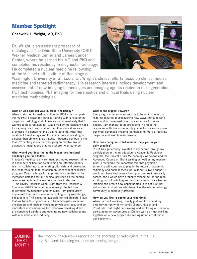
Dr. Wright is an assistant professor of radiology at The Ohio State University (OSU) Wexner Medical Center and James Cancer Center, where he earned his MD and PhD and completed his residency in diagnostic radiology. He completed a nuclear medicine fellowship at the Mallinckrodt Institute of Radiology at Washington University in St. Louis. Dr. Wright’s clinical efforts focus on clinical nuclear medicine and targeted radiotherapy. His research interests include development and assessment of new imaging technologies and imaging agents related to next-generation PET technologies, PET imaging for theranostics and clinical trials using nuclear medicine methodologies.
Member SpotlightChadwick L. Wright, MD, PhD
What or who sparked your interest in radiology?When I returned to medical school in 2004 after complet-ing my PhD, I began my clinical training with a rotation in diagnostic radiology and I knew almost immediately that I wanted to be a radiologist. I was awed by the constant need for radiologists to assist all of the other clinical service providers in diagnosing and treating patients. After that rotation, I found x-rays and CT scans more interesting to discuss than abnormal lab values. It became obvious to me that 21st century medicine was going to revolve around diagnostic imaging and that was where I wanted to be.
What would you describe as the biggest professional challenge you face today?In today’s healthcare environment, protected research time is absolutely critical for establishing an interdisciplinary team of collaborators, generating pilot data and developing the leadership skills to establish an independent research program. One challenge for all physician-scientists is the increased demand for our clinical services as the clinical reimbursements and revenues continue to decline. An RSNA Research Seed Grant from the Research & Education (R&E) Foundation gave me protected time to advance my research and innovate. I am particularly impressed that the Foundation is willing to try new things, because it is THE resource available for radiologists. I love that we have this opportunity to be radiologists, radiation oncologists and nuclear medicine physicians today and be innovators and visionaries for tomorrow, breaking down pre-conceived barriers and opening up new collaborations within academia and industry.
What is the biggest reward?Every day, my personal mission is to be an innovator, to redefine failures as discovering new ways that just don’t work and to make medicine more effective for more people. I am thankful to be practicing in a field that resonates with this mission. My goal is to use and improve our most advanced imaging technology to more effectively diagnose and treat human disease.
How does being an RSNA member help you in your daily practice?RSNA has generously invested in my career through my participation in the Introduction to Academic Radiology program, the Clinical Trials Methodology Workshop and the Advanced Course on Grant Writing as well as my research grant. I recognize the important role that physician- scientists will continue to play in the future of academic radiology and nuclear medicine. Without RSNA’s support I would not have had several key opportunities in my early career, and I would have probably missed out on the most exciting part of radiology — the chance to innovate beyond imaging and create new opportunities. It is not just indi-viduals and institutions who benefit — the whole radiology community is positively affected.
How do you like to spend your free time?When I am not working, I really just want to spend my time having fun with my family (Sarah, Huxley and Rosalind). That might be traveling and exploring our national parks, going on adventures at Disney World or just working together on a new project like setting up an art studio in our basement.

Plan Now. Save BIG!RESERVE YOUR RSNA 2018 HOTEL NOW!New! Begin preparing for RSNA 2018 and reserve your hotel in advance. Get a jump on planning and enjoy the savings when you secure your reservation through RSNA.
Online Reservations Open Plan now to take advantage of the best room selection in downtown Chicago plus RSNA early-discount rates. The earlier you reserve, the more you save! Visit RSNA.org/Hotel-Reservations to secure your discounted RSNA 2018 hotel reservation now!
MTG994.1 Hotel Carrier Sheet for RSNA News Color FIN.indd 1 12/18/17 11:53 AM