ci doi: 10.1102/1470-7330.2005 - … · alain rahmouni∗, alain luciani∗ and emmanuel itti†...
TRANSCRIPT

Cancer Imaging(2005)5, S102–S106DOI: 10.1102/1470-7330.2005.0037 CI
MULTIDISCIPLINARY SYMPOSIUM: HAEMATOLOGICALMALIGNANCIES
Wednesday 5 October 2005, 08:30–10:00
Quantitative CT analysis for assessing response inlymphoma (Cheson’s criteria)
Alain Rahmouni ∗, Alain Luciani ∗ and Emmanuel Itti †
Departments of∗Radiology and†Nuclear Medicine, Centre Hospitalo-Universitaire Henri Mondor, 51 Avenue duMarechal de Lattre de Tassigny, 94010 Creteil, France
Corresponding address: Alain Rahmouni, MD, Imagerie Medicale, CHU Henri Mondor, 51, avenue du Marechal deLattre de Tassigny, 94010 Creteil cedex, France. E-mail:[email protected]
Abstract
Standardized CT-based criteria used for lymphoma staging and follow-up and the current role of FDG-PET arereviewed. The current CT-based International Workshop Criteria (IWC) still have the main advantage of representingstandardized criteria allowing comparability of clinical trials in patients with lymphoma. However, functional imagingwith integrated IWC and FDG-PET provide more accurate response assessment, and challenge the current paradigm.Although integration of FDG-PET in IWC requires validation in a prospective trial with a large number of patients,new long-term clinical and therapeutic trials probably need to be designed using these new and hopefully standardizedfunctional criteria. This potentially could allow a more risk-adapted approach to the treatment of aggressivelymphoma: intensive (reinforced) therapies for non-responders vs. less intensive therapies for good responders withthe main goal of improved clinical outcome.
Keywords: Lymphoma; CT analysis; Cheson’s criteria.
Introduction
Since 1999, the International Workshop Criteria (IWC)have been widely accepted for response assessmentin patients with non-Hodgkin’s lymphoma (NHL).IWC also ensure comparability among clinical trials[1] .Multi-agent chemotherapy regimen have transformedaggressive lymphoma from a fatal disease to a potentiallycurable one, but no more than half of all patients arecured[2] . More aggressive, but also potentially moretoxic treatments are now available. At diagnosis, thetherapeutic regimen will be chosen in part accordingto the International Prognostic Index, a well establishedpredictor of outcome, including the performance statusaccording to the Eastern Cooperative Oncology Groupscale, the patient’s age, serum lactate dehydrogenaselevel, Ann Arbor stage (Table 1) (reflecting the anatomicextent and the tumoral mass primarily obtained byCT and bone marrow biopsy), and finally the numberof extranodal sites[3] . In order to assess response to
treatment, the IWC are primarily based on computedtomography (CT) although bone marrow biopsy (BMB)and clinical and biochemical information are also takeninto account; IWC should also discriminate rapid respon-ders to standard induction likely to show better and moredurable response, from non-responders who could benefitfrom an early change of therapeutic orientation[4] . Theinability of CT to differentiate between viable tumour,necrosis, or fibrosis in residual mass(es) in patients withotherwise complete clinical response has led to only CT-based size changes after treatment being considered toassign a response designation in the IWC[1] . Functionalimaging, particularly using fluorodeoxyglucose positronemission tomography (FDG-PET) has been shown toprovide additional metabolic information such as tumourviability in residual masses[5] . Gallium citrate andmagnetic resonance (MR) imaging have also been usedfor this purpose in patients with lymphoma[6–8].
1470-7330/05/010102 + 05 c© 2005 International Cancer Imaging Society

Multidisciplinary symposium: haematological malignancies S103
Table 1 Ann Arbor staging classification of thoracic lymphoma
Stage Area of involvement
I A single lymph node region or a single localised involvement of an extralymphatic siteII Two or more lymph node regions on the same side of the diaphragmIII Lymph node regions on both sides of the diaphragmIV Diffuse involvement of one or more extranodal organs with or without lymph node involvement
Additional qualifiersA Absence of systemic symptomsB Presence of systemic symptomsS Involvement of spleenE Localised extralymphatic site involvementH Involvement of liverM Diffuse involvement of bone marrow
International Workshop Criteria (IWC)
The standardized criteria for response assessment,proposed by Cheson and colleagues, include five desig-nations[1,9]:
(1) Complete response (CR) is the complete disap-pearance of all detectable evidence of diseaseon CT, and all disease-related symptoms, andnormalization of biochemical abnormalities, andnormal bone marrow biopsy (BMB). Previouslyinvolved nodes on CT more than 1.5 cm in theirgreatest axial diameter must regress to less than1.5 cm, and previously measured nodes of 1.1–1.5 cm must decrease to less than 1 cm.
(2) CRu (uncertain) corresponds to CR criteria butwith a residual mass more than 1.5 cm in greatestaxial diameter that has regressed by more than75%.
(3) Partial response (PR) is at least 50% reduction inthe sum of the product of the greatest diameters(SPD) of the six largest nodes with no increasein the size of other nodes and no new sites ofdisease. Splenic and hepatic nodules must regressby at least 50% in the SPD. BMB is irrelevant fordetermination of PR.
(4) Stable disease (SD) is less than a PR but isnot progressive disease. Progressive disease (PD)is more than 50% increase in the sum of theproduct of the greatest diameters of any previouslyabnormal node, or appearance of any new lesionsduring or at the end of therapy.
(5) Relapsed disease (RD) is the appearance of anynew lesion or increase in size of more than 50% ofpreviously involved sites or nodes in patients whoachieved CR or CRu.
Main limitations of CT-based IWC
At staging, assessment of nodal involvement by CTis limited by its low specificity in the case of small
nodes, especially less than 1.5 cm in diameter[9] . Spleeninvolvement by CT is based on an enlarged spleenand/or hypodense nodules. However, spleen size can varywith age and other associated diseases. Detection ofspleen nodules will depend upon CT technical parametersincluding contrast injection rate and timing. Whenisolated, a spleen nodule can be difficult to characterizeby CT alone. In young patients, when upper rangemeasurements of the normal thymus according to age areobtained, involvement by lymphoma can be difficult toassess with certainty.
Extranodal sites of involvement can be difficult todetect by CT. Bone marrow involvement is usually notidentified unless a lytic bone lesion is present. CTappearance of a small pulmonary nodule can be non-specific. Subtle changes in gastrointestinal wall thicknesscan be difficult to assess on CT[10,11].
At CT follow-up of nodal masses, a residual mass ispresent in approximately 40% of NHL patients treated bychemotherapy and/or radiation. This led to the conceptof ‘complete remission uncertain’ (CRu) which reflectsthe unknown significance of persistent CT abnormalitiesin patients who otherwise seem to be in CR. Previousstudies showing that only 10%–20% of such patients haveevidence of disease in these residual masses, considerablylimits the value of CT for prediction of clinical outcomeof NHL [11–13]. Due to its good spatial resolution, CT isalso able to detect nodes less than 1 cm but these nodesare considered normal although residual disease cannottheoretically be ruled out.
In the case of extranodal involvement, residual massescan also be seen in cases of gastrointestinal involvement;size changes of wall thickness are also difficult toestimate[11]. In the case of a lytic bone involvement,bone remodelling is usually delayed with no ad integrumrecovery. Pulmonary involvement can be difficult toassess during treatment due to drug-related and/orinfectious complications.
Although IWC have been useful to standardizeresponse to treatment, functional imaging has been shownto improve detection of sites of involvement, and toprovide metabolic tissue characterization[14].

S104 Multidisciplinary symposium: haematological malignancies
(a)
(b)
(c)
Figure 1 A 32-year-old woman with aggressive non-Hodgkin’s lymphoma. (a) Unenhanced CT scan ofthe thorax performed during percutaneous biopsyshowing a 7× 4 cm large anterior mediastinal mass.(b) Based on sole CT follow-up findings, the patientwas classified as complete response (uncertain) (CRu)according to IWC after 3 months of treatment witha 3× 2 cm large residual mass consistent with≥75%decrease in size (arrows). (c) FDG-PET performed 3days after CT did not show any significant residualuptake within the mass allowing the reclassification ofthe response to treatment as complete response (CR).
The emergence of fluoro-2-deoxy-D-glucose positronemission tomography (FDG-PET) in the clinical arma-mentarium and its increasing availability have recentlyprovided an alternative to gallium-citrate scan and MRimaging, which were previously employed to detectactive disease.
Integrated IWC and FDG-PET
Compared to CT staging, Mooget al. have reportedmore accurate staging by FDG-PET in NHL[15]; 8%of patients were up-staged at diagnosis because of theidentification of additional disease sites. After treatment,several studies have demonstrated that persistence of anincreased glycolytic activity on FDG-PET was associatedwith a high relapse rate, while the latter was low in thecase of a negative scan[5,16]. Only one recent report hasshown that a response classification based on integrationof FDG-PET results into IWC should provide a moreaccurate response assessment than IWC alone in patientswith lymphoma. In a retrospective study of 54 patientswith aggressive NHL, Juweidet al.showed that only 61%had concordant response designations between integratedIWC including FDG-PET results vs. IWC alone[9] . Themost pronounced discordance was observed in the CRuby IWC designation, in which all CRu patients werereclassified as CR in case of no FDG uptake or PRin case of FDG uptake. All patients reclassified as CRremained progression free at a median of more than32 months. An example of a residual mass detectedby CT but with no FDG uptake on PET is shown inFig. 1. The other major discordance was found in thePR by IWC designation in which half the patients werereclassified as CR by integrated IWC and FDG-PET.All but one of these reclassified CR patients remainedwithout evidence of disease progression at a median ofmore than 32 months. In contrast, only two of ninepatients with concordant PR designation by IWC andintegrated FDG-PET, and IWC alone were progressionfree at follow-up. Based on the Kaplan–Meier method,this study demonstrated that integrated IWC and FDG-PET was a statistically significant independent predictorfor progression-free survival.
Using FDG-PET results at follow-up will substantiallyincrease the proportion of CR designation by IWC, andprobably cancel the CRu designation. It will also showtwo distinct subgroups within the PR by IWC: a subgroupof PET-positive patients with poor outcome and anotherof PET-negative patients with excellent outcome. Thebiological explanation is likely related to the fact thatthe residual CT abnormalities represent necrosis and/orfibrosis in the majority of the PET-negative patients,whereas the abnormalities represent active tumour in themajority of the PET-positive patients.
One must, however, take into account that owing toits high spatial resolution, CT can identify extranodal

Multidisciplinary symposium: haematological malignancies S105
involvement sometimes not apparent on PET, thustriggering the need for combined PET/CT, and forcombined radiological and nuclear medicine workout ofimage interpretation, as illustrated by Fig. 2.
(b)
(c)
(a)
Figure 2 A 25-year-old male patient with aggressivenon-Hodgkin’s lymphoma. (a) Enhanced CT scan ofthe thorax showed a small 1 cm large left hilar node(arrow), and multiple subcareneal enlarged lymphnodes. (b) FDG-PET showed increased uptake ofall nodes detected by CT including the centimetrichilar node (arrow). However, FDG-PET failed toidentify multiple disseminated lung nodules consistentwith pulmonary involvement as confirmed by thedisappearance of these CT findings after completionof the first line of treatment.
Conclusion
Current CT-based IWC still has the main advantage ofrepresenting standardized criteria allowing comparability
of clinical trials in patients with lymphoma. However,functional imaging with integrated IWC and FDG-PETprovide more accurate response assessment, and chal-lenge the current paradigm. As FDG-PET combined withvisual correlation with a contrast-enhanced CT accuratelypredicts progression-free survival, fused FDG-PET andCT images acquired during the same examination willyield similar or superior results. Although integration ofFDG-PET in IWC requires validation in a prospectivetrial with a large number of patients, new long-term clin-ical and therapeutic trials probably need to be designedusing these new and hopefully standardized functionalcriteria. This potentially could allow a more risk-adapted approach to treatment of aggressive lymphoma:intensive (reinforced) therapies for non-responders vs.less intensive therapies for good responders with the maingoal of improved clinical outcome.
References
[1] Cheson BD, Horning SJ, Coiffier Bet al. Report of aninternational workshop to standardize response criteriafor non-Hodgkin’s lymphomas. NCI Sponsored Interna-tional Working Group. J Clin Oncol 1999; 17: 1244.
[2] Vose JM. Current approaches to the management of non-Hodgkin’s lymphoma. Semin Oncol 1998; 25: 483–91.
[3] Shipp M, Harrington D, Anderson Jet al. A predictivemodel for aggressive non-Hodgkin’s lymphoma. TheInternational Non-Hodgkin’s Lymphoma Prognostic Fac-tors Project. N Engl J Med 1993; 329: 987–94.
[4] Haioun C, Itti E, Rahmouni Aet al. [18F]Fluoro-2-deoxy-D-glucose positron emission tomography (FDG-PET) inaggressive lymphoma: an early prognostic tool for pre-dicting patient outcome. Blood 2005; prepublished onlineApril 28, 2005 (DOI 10.1182/blood-2005-01-0272).
[5] Jerusalem G, Beguin Y, Fassotte MFet al. Whole-body positron emission tomography using 18F-fluorodeoxyglucose for posttreatment evaluation inHodgkin’s disease and non-Hodgkin’s lymphoma hashigher diagnostic and prognostic value than classicalcomputed tomography scan imaging. Blood 1999; 94:429–33.
[6] Israel O, Mor M, Epelbaum Ret al. Clinical pretreatmentrisk factors and Ga-67 scintigraphy early during treatmentfor prediction of outcome of patients with aggressive non-Hodgkin lymphoma. Cancer 2002; 94: 873–8.
[7] Rahmouni A, Divine M, Lepage Eet al. Mediastinal lym-phoma: quantitative changes in gadolinium enhancementat MR imaging after treatment. Radiology 2001; 219:621–8.
[8] Janicek M, Kaplan W, Neuberg D, Canellos GP, Shul-man LN, Shipp MA. Early restaging gallium scans predictoutcome in poor-prognosis patients with aggressive non-Hodgkin’s lymphoma treated with high-dose CHOPchemotherapy. J Clin Oncol 1997; 15: 1631–7.
[9] Juweid ME, Wiseman GA, Vose JMet al. Responseassessment of aggressive non-Hodgkin’s lymphoma byintegrated International Workshop Criteria and fluorine-18-fluorodeoxyglucose positron emission tomography.J Clin Oncol 2005; 23: 4652–61.

S106 Multidisciplinary symposium: haematological malignancies
[10] Kumar R, Xiu Y, Potenta Set al. 18F-FDG PET forevaluation of the treatment response in patients withgastrointestinal tract lymphomas. J Nucl Med 2004; 45:1796–1803.
[11] Surbone A, Longo DL, DeVita Jr VTet al. Residualabdominal masses in aggressive non-Hodgkin’s lym-phoma after combination chemotherapy: significanceand management. J Clin Oncol 1988; 6: 1832–7.
[12] Fuks JZ, Aisner J, Wiernik PH. Restaging laparotomy inthe management of the non-Hodgkin lymphomas. MedPediatr Oncol 1982; 10: 429–38.
[13] Coiffier B, Lepage E. Prognosis of aggressive lym-phomas: a study of five prognostic models with patients
included in the LNH-84 regimen. Blood 1989; 74:558–64.
[14] Juweid ME, Cheson BD. Role of positron emissiontomography in lymphoma. J Clin Oncol 2005; 23:4577–80.
[15] Moog F, Bangerter M, Diederichs CGet al. Extranodalmalignant lymphoma: detection with FDG PET versusCT. Radiology 1998; 206: 475–81.
[16] Mikhaeel NG, Timothy AR, O’Doherty MJ, Hain S,Maisey MN. 18-FDG-PET as a prognostic indicator inthe treatment of aggressive non-Hodgkin’s lymphoma—comparison with CT. Leuk Lymphoma 2000; 39:543–53.
Cancer Imaging(2005)5, S106–S112 DOI: 10.1102/1470-7330.2005.0038
MRI and PET in monitoring response in lymphoma
Alain Rahmouni ∗, Alain Luciani ∗ and Emmanuel Itti †
Departments of∗Radiology and†Nuclear Medicine, Centre Hospitalo-Universitaire Henri Mondor, 51 Avenue duMarechal de Lattre de Tassigny, 94010 Creteil, France
Corresponding address: Alain Rahmouni, MD, Imagerie Medicale, CHU Henri Mondor, 51, avenue du Marechalde Lattre de Tassigny, 94010 Creteil cedex, France. E-mail:[email protected]
Abstract
The potential of FDG-PET and MRI in monitoring response to treatment in lymphoma is reviewed. Both FDG-PETand MRI can provide whole body imaging. Both also share the advantage of combining functional and anatomicalinformation. At present, hybrid FDG-PET and MDCT is the best technique for monitoring response to treatment,especially early response to treatment. Early assessment of response to treatment has the potential to tailor therapy.MR imaging is useful especially in assessing bone marrow and central nervous system involvement.
Keywords: MRI; FDG-PET; monitoring response; lymphoma.
Introduction
So-called ‘functional imaging’ has been used in theevaluation of lymphoma to supplement the informationobtained from computed tomography (CT). The mainlimitations of CT-based International Workshop Criteria(ICW) are: (1) the limited accuracy of CT at initialstaging for assessing lymphoma in small nodes (<1 to1.5 cm), bone marrow, or various extranodal sites; (2) theinability of CT to differentiate active disease within aresidual mass; and (3) the limited ability of CT to assessearly response to treatment although more aggressive,but also potentially more toxic treatments are nowavailable[1] . CT, however, remains the method of choicefor initial measurements of involved sites and detectionof complications such as adjacent organ compression.During follow-up, CT monitors size changes, and isuseful for diagnosing treatment complications. Moreover,
CT can also be seen as a potential ‘functional’ imaging.Dugdaleet al. showed that perfusion values measured atCT decrease when lymphoma masses become inactive(Fig. 1)[2] . To date, however, no study has confirmedthese preliminary data. Other functional imaging toolsare used for the evaluation of residual masses. Galliumimaging is not an accurate technique for detectingsites of involvement at diagnosis, with frequent false-negative results compared to CT, and also false-positivepara-hilar uptake[3] . However, when an initial nodalsite is gallium avid at diagnosis, follow-up galliumscans assess tumour activity during treatment. Janiceket al. demonstrated that early restaging gallium scansdelineate patients who are likely to have prolongeddisease-free survival from those who fail to respondto intensive therapy[4] . Patients whose tumours remainGa-positive midway through chemotherapy have a pooroutcome[4] . However, gallium imaging is a complex

Multidisciplinary symposium: haematological malignancies S107
technique with several disadvantages when compared to[18F]fluorodeoxyglucose positron emission tomography(FDG-PET) including: lower contrast and resolution, theneed to perform acquisition 48 h after gallium citrateinjection, higher dosimetry, and longer scanning time.Furthermore, gallium binds to plasma transferrin afterwhich it binds to receptors on the surface of lymphomacells leading to potentially false negative results. FDGuptake reflects a general metabolic process commonto most malignant tissues with therefore probably lessfalse negative results. The objective of this paper is toreview the potential of FDG-PET and magnetic resonanceimaging (MRI) in monitoring response to treatment inlymphoma.
FDG-PET
Briefly, FDG is transported into viable cells by glucosetransporter molecules, where it is phosphorylated byhexokinase into FDG-6-phosphate, just as glucoseis normally phosphorylated into glucose-6-phosphate.Unlike glucose-6-phosphate, however, FDG-6-phosphateundergoes no further metabolism within the cell. More-over, its dephosphorylation by glucose-6-phosphataseis a relatively slow process in comparison to that ofglucose-6-phosphatase. This, combined with the fact thatFDG-6-phosphate cannot easily cross the cell membrane,results in entrapment of FDG-6-phosphate within viablecells. Malignant cells have an increased rate of aerobicglycolysis, compared to normal tissue. They also have agreater number of glucose transport molecules at the cellsurface and a lower level of glucose-6-phosphatase.
Fluorine-18 is a positron emitter. The emitted positronpenetrates only a few millimetres into tissues beforecombining with an electron. The particle pair thenannihilates and its mass is entirely converted intoenergy. This energy takes the form of two 511 keVannihilation photons, emitted at approximately 180◦
from each other. Detection of both photons is termedcoincidence detection, and this is the principle by whichPET operates. Fluorine-18 has a half-life of 110 min,allowing acquisition of images over 30–120 min.
The biodistribution of FDG can be affected byvarious physiologic factors[5] . Blood glucose levels havean impact on FDG uptake through (a) competitivedisplacement of FDG by plasma glucose, and (b) patientsbeing asked to fast for 6 h prior to imaging. Goodcontrol of blood glucose is essential; a level of lessthan 150 mg/dl is desirable. Because the primary routeof FDG excretion is renal, good hydration is required.Muscle relaxants may be used to reduce muscle uptake.Patients are asked to remain silent after injection. Theusual dose of FDG is 10–15 mCi. To our knowledge,there is no contra-indication for FDG administration. PETimaging is initiated approximately 60 min following theinjection of FDG.
Hybrid FDG-PET/CT
Hybrid PET/CT scanners combine a PET and a CTmachine housed back-to-back. This enables imageacquisition in the same position with PET and CT,thus enabling the precise combination of the anatomicinformation provided by CT and the functional dataprovided by FDG-PET. Although most centres acquireunenhanced CT images, other centres perform singlephase enhanced CT with orally administered water-soluble iodinated contrast media[6] . This could allowoptimal CT images as multidetector CT (MDCT) is nowstandard in new hybrid PET/CT devices.
Monitoring response to treatment withFDG-PET
Staging of lymphoma
The accuracy of FDG-PET as an imaging tool forprimary staging of lymphoma suffers from the absenceof a systematic pathological correlation (Fig. 2). In ourexperience, PET alone is concordant with conventionalimaging and bone marrow biopsy (BMB) in only 80%of cases[7] , better than conventional imaging and BMBin 8% and worse in 12%. Among these latter cases, one-third account for bone marrow involvement undetectedby PET. Other studies have reported similar results[8] .Moog et al. showed that FDG-PET was more accuratefor detecting nodal lymphoma than CT[8] . Seven lymphnode regions unremarkable on conventional CT showedincreased uptake of FDG. Staging was changed in the4/60 patients with these seven confirmed additional PETfindings: from stage I to II in one patient, and from stageII to III in three patients. The clinically relevant questionof how PET impacts on the staging of lymphoma andabove all, whether or not up- or down-staging leads tochanges in therapeutic strategies, has been addressed insome studies with variable results. For Shoderet al.PET-FDG could contribute to changes in clinical stage in 44%and changes in treatment in more than 60% of cases[9] .
FDG-PET: significance of positive findings
Several limitations of FDG-PET have been reported:physiologic muscles may take up the radiotracer andshow increased activity on the PET images (Fig. 2)[6] ;This muscle uptake is easily identified when compared toCT images. Similarly, physiologic uptake by the kidneys,bowel and liver can be distinguished on combined CTimages. Physiologic FDG uptake has also been reportedin brown fat[10]. Other false-positive findings of FDG-PET, more difficult to recognize, include inflammatorychanges caused by infectious or inflammatory processessuch as viral infections, bronchitis, aspergilloma, sar-coidosis, etc.[10,11]

S108 Multidisciplinary symposium: haematological malignancies
(a) (b)
Figure 1 A 78-year-old patient with mediastinal NHL. (a) Enhanced baseline CT scan showed an enhancing7 × 4 cm large mediastinal mass. (b) After three cycles of chemotherapy, enhanced CT scan of the mediastinumdemonstrated partial response, but with decreased enhancement of the residual mass. After completion oftreatment, the patient was considered in complete response (uncertain).
Response to treatment
PET is able to distinguish between active tumourand inactive residual masses often present followingtreatment (Fig. 3)[12–14]. This use is probably themost important especially in aggressive NHL andHodgkin’s lymphoma. False-positive findings at the siteof residual masses may be seen, however, due to reboundthymic hyperplasia or post-therapy inflammatory changesespecially following radiotherapy as well as infectiousor inflammatory processes outside the site of residualmasses[14]. The diffusely increased bone marrow uptakeoften observed during treatment and related to theadministration of growth factor is usually linked to bonemarrow hyperplasia and should not be misinterpretedas specific involvement (Fig. 3). FDG-PET results aftertreatment can predict therapy outcomes[15]. However,a negative PET scan cannot rule out the presence ofminimal residual disease[5] .
Early response to first-line treatment
The clinical parameters incorporated in the InternationalPrognostic Index (IPI) grossly reflect the biologicalheterogeneity of lymphoma. In this respect, the durationof a complete remission might be significantly moreinfluenced by the chemosensitivity than by the initialIPI factors[7] . Consequently, an early evaluation duringtreatment leading to an alternative treatment mightimprove outcome. Several studies have established thatinterim FDG-PET scans after 1–3 cycles of chemotherapyprovide valuable information regarding early assessmentof response and survival. Conventional chemotherapycan induce a rapid decrease of FDG uptake as soon as
7 days after treatment[16]. Spaepenet al.have shown theimportant prognostic value of mid-treatment FDG-PETin monitoring 70 cases of aggressive NHL[17]. Thirty-three patients showed persistence of abnormal FDGuptake and none of them achieved a durable completeremission, whereas 37 showed a negative scan. Out ofthe 37 patients, 31 remained in compete remission. Morerecently, we have confirmed the early (after two cycles)prognostic impact of FDG-PET in terms of responseand survival[16]. At mid-induction, ‘early PET’ wasconsidered negative in 54 patients and positive in 36.The outcome differed significantly between PET-negativeand PET-positive groups; the predictive value of ‘earlyPET’ was observed in both the lower-risk and higher-risk groups, indicating prognostic independence from IPI.Therefore, FDG-PET should guide first-line strategies inlymphoma. The role of PET scanning for post-therapysurveillance without clinical or biochemical or CTevidence of disease (complete remission status) remainscontroversial primarily because of the potential for adisproportionate fraction of false-positive findings poten-tially resulting in increased cost without proven benefitfrom early PET detection of disease compared withstandard conventional methods[18]. Large prospectivestudies are therefore needed to determine whether routinesurveillance by PET results in meaningful changes inpatient management[14].
MRI
T2 signal
The signal intensity of lymphoma at MR imaging changesduring the course of the disease[19]. Active untreated

Multidisciplinary symposium: haematological malignancies S109
(a)
(b) (d)
(c)
Figure 2 A 41-year-old patient presenting with cervical enlarged lymph nodes with NHL involvement. (a) CTscan of the upper mediastinum showed an anterior mediastinal mass (arrows), without FDG uptake on PET.(b) This mass could correspond to an enlarged thymus. (c) Post-treatment CT scan performed 3 monthslater showed that the anterior mass was no longer present. (d) FDG-PET was normal; the linear cervicaluptake (arrow) corresponded to physiologic muscle uptake. This case demonstrates the potential discordancesbetween CT and FDG-PET due to the absence of pathological examination of the anterior mediastinalmass.
tumour tissue contains an excess of free water whichincreases the signal intensity on T2 WI. With successfultreatment, cellular elements and the water content ofthe tumour are reduced while the collagen and fibroticstroma of the original tumour account for the maincomponent of the signal[20]. These factors reduce thesignal intensity of the residual mass on T2 WI andhave been used for predicting relapse in a residual mass.However, the sensitivity of MR imaging in the predictionof relapse in a residual mass ranges from 45% to 90%,with a specificity ranging from 80% to 90%[21,22]. Low
sensitivity is mainly due to necrosis, immature fibrotictissue, oedema and inflammation that can simulate thehigh T2 SI of a viable tumour.
Gadolinium injection
Gadolinum (Gd) enhancement of lymphoma of themediastinum changes during the course of the disease.The mean Gd enhancement of residual masses aftertreatment is substantially weaker than that observed

S110 Multidisciplinary symposium: haematological malignancies
(a)
(b)
(c)
(d)
Figure 3 A 25-year-old patient with NHL presenting with a soft tissue sternal mass associated with lyticsternal bone on CT (a) and increased isolated uptake on FDG-PET (b) concordant with CT findings. (c) Aftertreatment, CT showed decrease of the soft tissue mass, but the lytic lesion of the sternum remained unchanged.The designation of response to treatment was difficult on CT. (d) FDG-PET showed no residual uptake in thepreviously involved sternum, but diffuse uptake of the whole bone marrow consistent with marrow regenerationdue to growth factor treatment.
before treatment in patients in complete remission.Enhancement of these inactive residual masses decreasesmarkedly to the same level as that of muscle[23]. This maybe explained by a higher degree of vascularization anda larger extracellular compartment in the active cellulartumour compared with dense immature fibrotic tissue.Due to different enhancement of lymphomatous massesat diagnosis, MR evaluation of residual masses requires apre-treatment baseline MR study for comparison. Furtherstudies with more recent MR techniques of perfusionanalysis are required for comparison with FDG-PET.MR imaging suffers from limited field of view analysiscompared to MDCT and PET. Furthermore, various
impairments such as motion artefacts alter the overallimage analysis especially in the mediastinum.
Potential of MRI
Preliminary studies have suggested a potential roleof diffusion MR imaging in oncologic patients byallowing the detection of water motion over smalldistances[24]. The development of body MR usingmulti-channel phased array surface coils combined withparallel imaging techniques could enable whole body MRdiffusion imaging in cancer patients. No study has yet

Multidisciplinary symposium: haematological malignancies S111
(a) (b) (c) (d)
Figure 4 A 28-year-old patient with NHL with spine and epidural involvement. (a) T1 WI showed twoepidural masses (arrows). The bone marrow signal is consistent with the young age of the patient or withdiffuse involvement. (b) Fat suppressed T2 WI demonstrated multiple focal bone marrow lesions. (c) T1 GEWI before injection and (d) 35 s after Gd-chelates injection showed early enhancement of focal lesions withoutenhancement of the remaining marrow. Bone marrow biopsy in the iliac crest was normal.
been published assessing the impact of such techniquesin staging and monitoring lymphoma.
New contrast agents taken up by the reticulo-endothelial system are now available, such as super-paramagnetic iron oxides (USPIO). USPIO are takenup by normal and hypercellular bone marrow, but notby neoplastic lesions, thereby providing significantlydifferent enhancement patterns on T2-weighted MRimages[25]. USPIO could therefore help in differentiatingnormal from lymphomatous bone marrow.
MRI in bone marrow involvement
In patients with lymphoma, MR identification of focallesions within the bone marrow is important for patientstaging according to the Ann Arbor classification. BMBremains the gold standard in this setting, but its sensitivityis sub-optimal as the biopsy site may not reflect theentire bone marrow compartment. Bilateral iliac crestBMB usually increases the sensitivity of unilateral BMB.Early during bone marrow infiltration, tumour cells donot displace bone marrow fat cells; the amount offat cells remains normal. Subsequently, replacement ofnormal marrow by tumour cells leads to a reductionin T1 SI and an increase in T2 SI. Focal lesions areeasily detected when using appropriate MR techniquessuch as fat suppressed T2 WI before treatment. Several
studies have shown that NHL induces angiogenesis[26].Dynamic contrast-enhanced MR images can demonstrateincreased bone marrow enhancement in patients withlymphoproliferative diseases and marrow involvement(Fig. 4)[27,28]. Contrast enhancement decreases aftertreatment in good responders.
Conclusion
Both FDG-PET and MRI can provide whole bodyimaging. Both also share the advantage of combiningfunctional and anatomical information. Hybrid FDG-PETand MDCT is the best technique for monitoring responseto treatment, especially early response to treatment. Earlyassessment of response to treatment has the potentialto tailor therapy. MR imaging is especially useful inassessing bone marrow and central nervous systeminvolvement.
References[1] Haioun C, Lepage E, Gisselbrecht Cet al. High-dose
therapy followed by stem cell transplantation in partialresponse after first-line induction therapy for aggressivenon-Hodgkin’s lymphoma. Ann Oncol 1998; 9 (Suppl 1):S5–8.
[2] Dugdale PE, Miles KA, Bunce I, Kelley BB, Leggett DA.CT measurement of perfusion and permeabilitywithin lymphoma masses and its ability to assess grade,

S112 Multidisciplinary symposium: haematological malignancies
activity, and chemotherapeutic response. J Comput AssistTomogr 1999; 23: 540–7.
[3] Bar-Shalom R, Yefremov N, Haim Net al. Camera-basedFDG PET and 67Ga SPECT in evaluation of lymphoma:comparative study. Radiology 2003; 227: 353–60.
[4] Janicek M, Kaplan W, Neuberg D, Canellos GP, Shul-man LN, Shipp MA. Early restaging gallium scans predictoutcome in poor-prognosis patients with aggressive non-Hodgkin’s lymphoma treated with high-dose CHOPchemotherapy. J Clin Oncol 1997; 15: 1631–7.
[5] Kazama T, Faria SC, Varavithya V, Phongkitkarun S,Ito H, Macapinlac HA. FDG PET in the evaluation oftreatment for lymphoma: clinical usefulness and pitfalls.Radiographics 2005; 25: 191–207.
[6] Kapoor V, McCook BM, Torok FS. An introduction toPET-CT imaging. Radiographics 2004; 24: 523–43.
[7] Haioun C, Itti E, Rahmouni A, Meignan M, Reyes F. PETscan in the therapeutic strategy. Hematol J 2004; 5 (Suppl3): S149–53.
[8] Moog F, Bangerter M, Diederichs CGet al. Lymphoma:role of whole-body 2-deoxy-2-[F-18]fluoro-D-glucose(FDG) PET in nodal staging. Radiology 1997; 203:795–800.
[9] Schoder H, Meta J, Yap Cet al. Effect of whole-body (18)F-FDG PET imaging on clinical staging andmanagement of patients with malignant lymphoma.J Nucl Med 2001; 42: 1139–43.
[10] Castellucci P, Nanni C, Farsad Met al. Potential pitfallsof 18F-FDG PET in a large series of patients treated formalignant lymphoma: prevalence and scan interpretation.Nucl Med Commun 2005; 26: 689–94.
[11] Castellucci P, Zinzani P, Pourdehnad Met al. (18)F-FDGPET in malignant lymphoma: significance of positivefindings. Eur J Nucl Med Mol Imaging 2005 (Epubahead of print).
[12] Jerusalem G, Beguin Y, Fassotte MFet al. Whole-body positron emission tomography using18F-fluorodeoxyglucose for posttreatment evaluation inHodgkin’s disease and non-Hodgkin’s lymphoma hashigher diagnostic and prognostic value than classicalcomputed tomography scan imaging. Blood 1999; 94:429–33.
[13] Spaepen K, Stroobants S, Dupont Pet al. Prognosticvalue of positron emission tomography (PET) withfluorine-18 fluorodeoxyglucose ([18F]FDG) after first-line chemotherapy in non-Hodgkin’s lymphoma: is[18F]FDG-PET a valid alternative to conventional diag-nostic methods? J Clin Oncol 2001; 19: 414–19.
[14] Juweid ME, Cheson BD. Role of positron emissiontomography in lymphoma. J Clin Oncol 2005; 23:4577–80.
[15] Juweid ME, Wiseman GA, Vose JMet al. Responseassessment of aggressive non-Hodgkin’s lymphoma byintegrated International Workshop Criteria and fluorine-18-fluorodeoxyglucose positron emission tomography.J Clin Oncol 2005; 23: 4652–61.
[16] Haioun C, Itti E, Rahmouni Aet al. [18F]Fluoro-2-deoxy-D-glucose positron emission tomography (FDG-PET) in aggressive lymphoma: an early prognostic toolfor predicting patient outcome. Blood 2005 (Epub aheadof print).
[17] Spaepen K, Stroobants S, Dupont Pet al. Earlyrestaging positron emission tomography with (18)F-fluorodeoxyglucose predicts outcome in patientswith aggressive non-Hodgkin’s lymphoma. Ann Oncol2002; 13: 1356–63.
[18] Jerusalem G, Beguin Y, Fassotte MFet al. Early detectionof relapse by whole-body positron emission tomographyin the follow-up of patients with Hodgkin’s disease. AnnOncol 2003; 14: 123–30.
[19] Rahmouni A, Tempany C, Jones R, Mann R, Yang A,Zerhouni E. Lymphoma: monitoring tumor size andsignal intensity with MR imaging. Radiology 1993; 188:445–51.
[20] Nyman RS, Rehn SM, Glimelius BL, Hagberg HE,Hemmingsson AL, Sundstrom CJ. Residual mediastinalmasses in Hodgkin disease: prediction of size with MRimaging. Radiology 1989; 170: 435–40.
[21] Gasparini MD, Balzarini L, Castellani MRet al. Currentrole of gallium scan and magnetic resonance imagingin the management of mediastinal Hodgkin lymphoma.Cancer 1993; 72: 577–82.
[22] Devizzi L, Maffioli L, Bonfante V et al. Comparisonof gallium scan, computed tomography, and magneticresonance in patients with mediastinal Hodgkin’s disease.Ann Oncol 1997; 8 (Suppl 1): 53–6.
[23] Forsgren G, Nyman R, Glimelius B, Hagberg H, Rehn S,Hemmingsson A. Gd-DTPA-enhanced MR imaging inmediastinal Hodgkin’s disease. Acta Radiol 1994; 35:564–9.
[24] Sato C, Naganawa S, Nakamura Tet al. Differentiationof noncancerous tissue and cancer lesions by apparentdiffusion coefficient values in transition and peripheralzones of the prostate. J Magn Reson Imaging 2005; 21:258–62.
[25] Daldrup-Link HE, Rummeny EJ, Ihssen B, Kienast J,Link TM. Iron-oxide-enhanced MR imaging of bonemarrow in patients with non-Hodgkin’s lymphoma: dif-ferentiation between tumor infiltration and hypercellularbone marrow. Eur Radiol 2002; 12: 1557–66.
[26] Ribatti D, Vacca A, Nico B, Fanelli M, Roncali L,Dammacco F. Angiogenesis spectrum in the stroma of B-cell non-Hodgkin’s lymphomas. An immunohistochemi-cal and ultrastructural study. Eur J Haematol 1996; 56:45–53.
[27] Montazel JL, Divine M, Lepage E, Kobeiter H, Breil S,Rahmouni A. Normal spinal bone marrow in adults:dynamic gadolinium-enhanced MR imaging. Radiology2003; 229: 703–9.
[28] Rahmouni A, Montazel JL, Divine Met al. Bone marrowwith diffuse tumor infiltration in patients with lympho-proliferative diseases: dynamic gadolinium-enhanced MRimaging. Radiology 2003; 229: 710–17.

Multidisciplinary symposium: haematological malignancies S113
Cancer Imaging(2005)5, S113–S119 DOI: 10.1102/1470-7330.2005.0032
Role of imaging to choose treatment
Christophe Ferm e∗, Daniel Vanel †, Vincent Ribrag ∗ and Th eo Girinski ‡
Departments of∗Medicine,†Radiology and‡Radiation Therapy, Institut Gustave Roussy, 39 rue CamilleDesmoulins, 94805 Villejuif, France
Corresponding address: Daniel Vanel, Department of Radiology, Institut Gustave Roussy, 39 rue CamilleDesmoulins, 94805 Villejuif, France. E-mail:[email protected]
Abstract
Radiologists perform various examinations at every step of lymphomas. The role of imaging is atypical for a ‘classical’oncologic radiologist, as multiple non-radiological criteria are combined to decide on treatments. A good knowledgeof the practical use of the results helps the radiologist to seek the useful pieces of information. In treatment evaluation,uncertain complete response is only used in lymphomas. Imaging is changing, with the emergence of PET and wholebody MRI but CT remains the key examination today. The WHO criteria are the only ones used to evaluate treatmentresults on CT, even though the use of PET is increasingly used, with better and better results.
Keywords: Lymphoma; CT; staging; treatment evaluation.
Introduction
The management of lymphomas is quite different fromthe rest of oncology. A good understanding of thepractical consequences of imaging findings helps theradiologist to be efficient. Histology is by far themain prognostic factor, and has the highest impacton treatment choices. A follicular lymphoma, evenwith multiple lesions, may not require any treatment,whereas a limited high grade lymphoma will requirevery aggressive treatment. Then multiple other criteria,including imaging, are involved in the choice of treat-ment. Computed tomography (CT) is the main imagingmodality, as it is reproducible and widely available.Magnetic resonance (MR) imaging is especially efficientto evaluate bone marrow. Positron emission tomography(PET) has increasing value in the initial staging andevaluation of early effectiveness of treatment.
Definition of criteria for radiologicevaluation
Radiologic pre-treatment evaluation procedures includechest radiographs (posteroanterior and lateral) and CTof the thorax, abdomen, and pelvis. These investigationsare needed to define a number of parameters that are ofimportance in terms of prognosis, and should be takeninto consideration when planning treatment.
Extent of disease
The extent of disease is evaluated according to theAnn Arbor staging classification and Cotswolds’ revision
(Table 1)[1] or St Jude staging system used forchildhood non-Hodgkin’s lymphoma and adult patientswith Burkitt’s lymphoma (Table 2).
Among the criteria are:
• The number of peripheral nodal sites involved.Peripheral nodal sites are defined as cervical (oneor several anatomic groups), axillary, inguinal orfemoral right and left.
• Tumour volume. A mediastinal mass is defined asbulky on a posteroanterior chest radiograph whenthe maximum width is equal to or greater thanone-third of the internal transverse diameter of thethorax at the level of the T5–T6 interspace[2] .A large mediastinal mass is defined as MT ratio≥0.35. This criterion is mainly used for Hodgkin’slymphoma. According to Cotswolds’ modificationsof the Ann Arbor classification, a palpable lymphnode or conglomerate node mass must be 10 cmor greater in largest diameter to be recorded asbulky. The nodal and extranodal targets defined forassessment of response are measured bidimensionallyaccording to the WHO criteria[3] . RECIST criteriacannot be used.
Prognostic index and score
A number of prognostic scores are used to defineprognosis and to make medical decisions for differententities according to the WHO classification of malignantlymphoma[4] .
S114 Multidisciplinary symposium: haematological malignancies
(a)
(b)
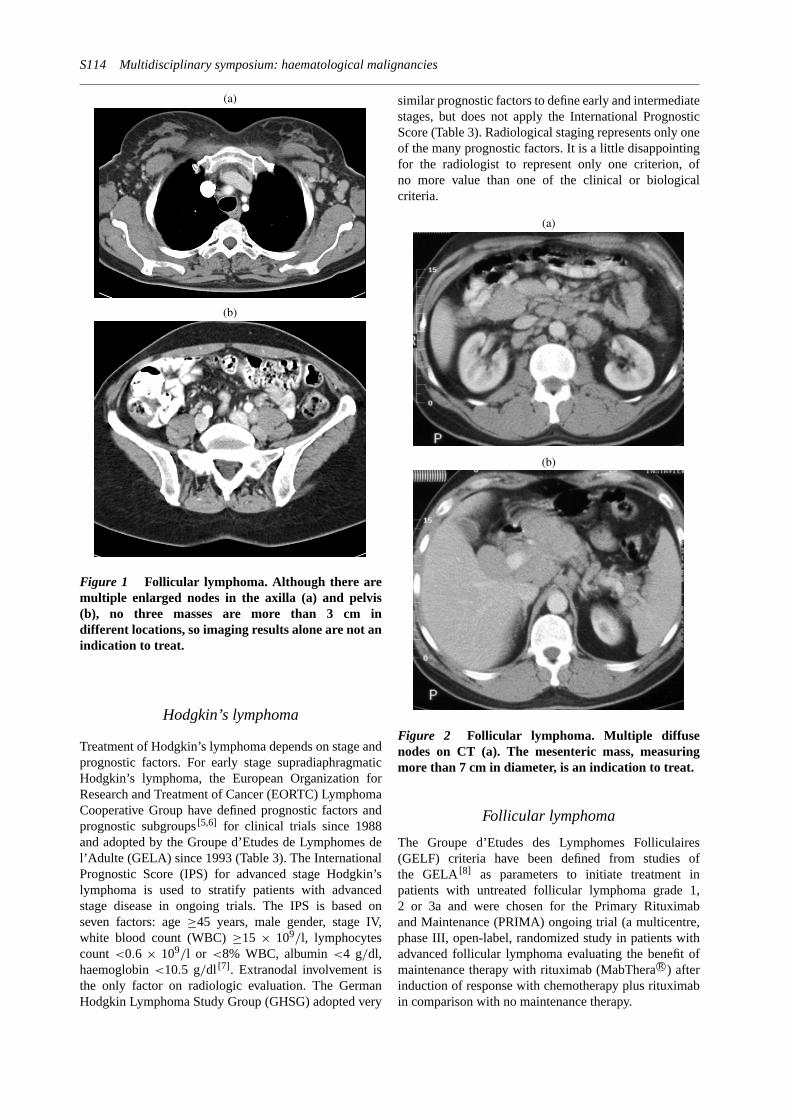
Figure 1 Follicular lymphoma. Although there aremultiple enlarged nodes in the axilla (a) and pelvis(b), no three masses are more than 3 cm indifferent locations, so imaging results alone are not anindication to treat.
Hodgkin’s lymphoma
Treatment of Hodgkin’s lymphoma depends on stage andprognostic factors. For early stage supradiaphragmaticHodgkin’s lymphoma, the European Organization forResearch and Treatment of Cancer (EORTC) LymphomaCooperative Group have defined prognostic factors andprognostic subgroups[5,6] for clinical trials since 1988and adopted by the Groupe d’Etudes de Lymphomes del’Adulte (GELA) since 1993 (Table 3). The InternationalPrognostic Score (IPS) for advanced stage Hodgkin’slymphoma is used to stratify patients with advancedstage disease in ongoing trials. The IPS is based onseven factors: age≥45 years, male gender, stage IV,white blood count (WBC)≥15 × 109/l, lymphocytescount <0.6 × 10⁹/l or <8% WBC, albumin<4 g/dl,haemoglobin<10.5 g/dl [7] . Extranodal involvement isthe only factor on radiologic evaluation. The GermanHodgkin Lymphoma Study Group (GHSG) adopted very
similar prognostic factors to define early and intermediatestages, but does not apply the International PrognosticScore (Table 3). Radiological staging represents only oneof the many prognostic factors. It is a little disappointingfor the radiologist to represent only one criterion, ofno more value than one of the clinical or biologicalcriteria.
(a)
(b)
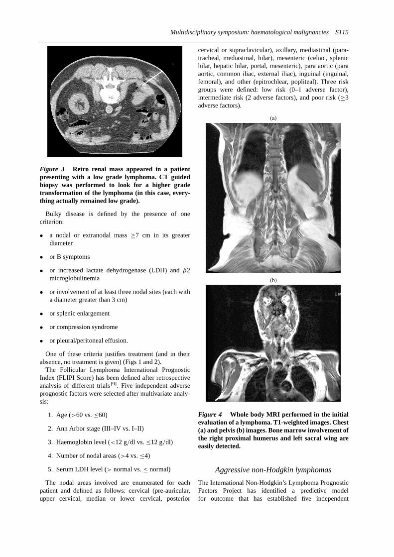
Figure 2 Follicular lymphoma. Multiple diffusenodes on CT (a). The mesenteric mass, measuringmore than 7 cm in diameter, is an indication to treat.
Follicular lymphoma
The Groupe d’Etudes des Lymphomes Folliculaires(GELF) criteria have been defined from studies ofthe GELA[8] as parameters to initiate treatment inpatients with untreated follicular lymphoma grade 1,2 or 3a and were chosen for the Primary Rituximaband Maintenance (PRIMA) ongoing trial (a multicentre,phase III, open-label, randomized study in patients withadvanced follicular lymphoma evaluating the benefit ofmaintenance therapy with rituximab (MabTheraR©) afterinduction of response with chemotherapy plus rituximabin comparison with no maintenance therapy.
Multidisciplinary symposium: haematological malignancies S115
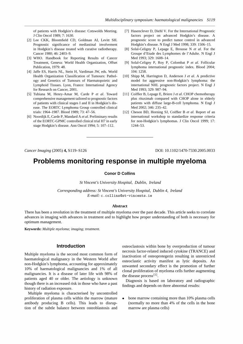
Figure 3 Retro renal mass appeared in a patientpresenting with a low grade lymphoma. CT guidedbiopsy was performed to look for a higher gradetransformation of the lymphoma (in this case, every-thing actually remained low grade).
Bulky disease is defined by the presence of onecriterion:
• a nodal or extranodal mass≥7 cm in its greaterdiameter
• or B symptoms
• or increased lactate dehydrogenase (LDH) andβ2microglobulinemia
• or involvement of at least three nodal sites (each witha diameter greater than 3 cm)
• or splenic enlargement
• or compression syndrome
• or pleural/peritoneal effusion.
One of these criteria justifies treatment (and in theirabsence, no treatment is given) (Figs 1 and 2).
The Follicular Lymphoma International PrognosticIndex (FLIPI Score) has been defined after retrospectiveanalysis of different trials[9] . Five independent adverseprognostic factors were selected after multivariate analy-sis:
1. Age (>60 vs.≤60)
2. Ann Arbor stage (III–IV vs. I–II)
3. Haemoglobin level (<12 g/dl vs.≤12 g/dl)
4. Number of nodal areas (>4 vs.≤4)
5. Serum LDH level (> normal vs.≤ normal)
The nodal areas involved are enumerated for eachpatient and defined as follows: cervical (pre-auricular,upper cervical, median or lower cervical, posterior
cervical or supraclavicular), axillary, mediastinal (para-tracheal, mediastinal, hilar), mesenteric (celiac, splenichilar, hepatic hilar, portal, mesenteric), para aortic (paraaortic, common iliac, external iliac), inguinal (inguinal,femoral), and other (epitrochlear, popliteal). Three riskgroups were defined: low risk (0–1 adverse factor),intermediate risk (2 adverse factors), and poor risk (≥3adverse factors).
(a)
(b)
Figure 4 Whole body MRI performed in the initialevaluation of a lymphoma. T1-weighted images. Chest(a) and pelvis (b) images. Bone marrow involvement ofthe right proximal humerus and left sacral wing areeasily detected.
Aggressive non-Hodgkin lymphomas
The International Non-Hodgkin’s Lymphoma PrognosticFactors Project has identified a predictive modelfor outcome that has established five independent

S116 Multidisciplinary symposium: haematological malignancies
prognostic factors: age (>60 years adverse), stage I–II vs. III–IV (III–IV adverse), number of extranodalsites (>1 adverse), performance status (low statusEastern Cooperative Oncology Group (ECOG)>2adverse), serum LDH (elevated level adverse)[10]. Theage-adjusted International Prognostic Index (stage,performance status, serum LDH) is applied worldwideto stratify patients into prognostic subgroups.
Burkitt’s lymphoma
The Ann Arbor staging system is not well suited topatients with Burkitt’s lymphoma, which is predomi-nantly extranodal (bone marrow and/or central nervoussystem involvement not always accompanied by nodalinvolvement). The most widely used classification is theSt Jude staging system used for childhood non-Hodgkin’slymphoma (Table 2). Staging procedures should bedone within 24–48 h because of the rapidity of tumourgrowth. Peritoneal or pleural effusions are particularlyhelpful for the diagnosis and should be documentedwith cytogenetics, immunophenotyping and genotyping.Tumour burden is a prognostic factor and evaluation oftumour size is necessary for restaging under treatment.Magnetic resonance imaging of the head and neck, chest,abdomen, skeleton, and central nervous system (CNS)may be indicated in some circumstances but is not usedroutinely.
Impact of radiologic evaluation ontreatment decision
Initial staging at diagnosis
Hodgkin’s lymphoma
In early stage supradiaphragmatic Hodgkin’s lymphoma,the standard treatment is a combination of chemotherapyfollowed by radiotherapy. The number of cycles ofchemotherapy (ABVD regimen (adriamycin, bleomycin,vinblastine, dacarbazine) as standard) depends on prog-nostic subgroups, three cycles for favourable groupsand four cycles for unfavourable groups. Radiother-apy is delivered to responding patients and limitedto initially involved areas: ‘involved fields’ radiationtherapy.
As the treatment subgroups (unfavourable andfavourable) are defined by adverse prognostic factors,a careful evaluation of the extent of disease and tumoursize is a condition for a risk adapted strategy: numberof cycles of chemotherapy, radiotherapy delivered to allinitially involved areas (evaluation of axillary areas onchest CT), and planned doses of radiotherapy deliveredaccording to the response to chemotherapy.
In advanced stage Hodgkin’s lymphoma, the standardtreatment is chemotherapy alone with ABVD regimen,
eight cycles if a complete response or complete responseuncertain is achieved after 6 cycles.
Tumour volume as a large mediastinal mass has noimpact on the choice of first line chemotherapy, however,the tumour size is measured for the evaluation of responseto initial chemotherapy (first evaluation recommendedafter four cycles).
Follicular lymphoma
The initial evaluation is of importance to identifypatients with a small tumour burden and a slowprogression of disease for whom a watch and waitpolicy is recommended by the majority of centres andthose with adverse risk factors as previously describedwho need to be treated. Chemotherapy (cyclophos-phamide, vincristine and prednisolone (CVP), CHOPregimen or Fludarabine-based regimens) plus rituximab(MabTheraR©), a chimeric/human monoclonal antibodythat is directed specifically against the B-cell antigenCD20, is considered in many countries as the standard forfirst-line treatment of patients with follicular lymphoma.
Diffuse large-B-cell lymphoma
The CHOP regimen (cyclophosphamide, doxorubicin,vincristine, prednisone) combined with rituximab givenin eight cycles is the standard of care for elderly patients(≥60 years old) with diffuse large-B-cell lymphoma[11].
Evaluation of the Ann Arbor stage is the mainparameter evaluated on imaging with an impact on thedefinition of initial treatment based on the age-adjustedInternational Prognostic Index (Fig. 4).
Burkitt’s lymphoma
Treatment protocols based on combination chemotherapyregimens designed for children, consisting of intensivedoses of alkylating agents given in combination withmethotrexate, vincristine, cytarabine, have been shown tobe highly effective for patients with Burkitt’s lymphoma,whether adults or children. CNS prophylaxis includingintrathecal methotrexate and cytarabine is an essentialcomponent of therapy. Treatment subgroups are definedon the basis of bone marrow and/or CNS involvementat diagnosis: patients without bone marrow and/or CNSinvolvement (stages 1–3 of the St Jude staging system)and patients with bone marrow and/or CNS involvementwho are stratified according to age and CNS involvementbecause of the toxicity of methotrexate and cytarabine.Appropriate measures for the prevention of tumour lysissyndrome are highly recommended. The role of rituximabis being evaluated in prospective trials.

Multidisciplinary symposium: haematological malignancies S117
Table 1 Ann Arbor staging classification and Cotswolds revision
Stage I I Involvement of a single lymph node region.IE Localized involvement of a single extralymphatic organ or site.
Stage II II Involvement of two or more lymph node regions on the same side of the diaphragm.IIE Localized involvement of a single associated extralymphatic organ or site and of one or more lymph node regions on the
same side of the diaphragm.Right and left hilum: one area each, independent of mediastinum; number of anatomic nodal areas to be indicated by a
subscript (II4).
Stage III III Involvement of lymph node regions on both sides of the diaphragm.III 1 Upper abdomen (splenic, hilar, celiac, or portal nodes).III 2 Lower abdomen (paraaortic, iliac, mesenteric nodes).IIIE Involvement of lymph node regions on both sides of the diaphragm accompanied by localized involvement of an
extralymphatic organ or sitea.
Stage IV IV Disseminated (multifocal) involvement of one or more extralymphatic sites with or without associated lymph nodeinvolvement or isolated extralymphatic organ involvement with distant (non-regional) nodal involvement.
The absence or presence of fever>38◦C, drenching sweats during the last month, and/or weight loss of 10% or more ofbody weight in 6 months are to be noted in all cases by the suffix letters A or B, respectively.
X Bulky disease,>1/3 widening of mediastinum at T5–T6 level, or>10 cm maximum dimension of nodal mass.CR(u) Unconfirmed/uncertain complete remission (residual imaging abnormality).
aIn FLIPPI, spleen involvement is categorized as stage IV.
Table 2 St Jude staging system used for childhood non-Hodgkin’s lymphomaa
Stage Definition
I Single tumour (extranodal)Single anatomic area (nodal)Excluding mediastinum or abdomen
II Single tumour (extranodal) with regional node involvementPrimary gastrointestinal tumour with or without involvement of associated mesenteric nodes only, grossly completely resectedOn same side of diaphragm:Two or more nodal areasTwo single (extranodal) tumours with or without regional node involvement
III On both sides of the diaphragm:Two single tumours (extranodal)Two or more nodal areasAll primary intrathoracic tumours (mediastinal, pleural, thymic)All extensive primary intra-abdominal diseaseAll primary paraspinal or epidural tumours regardless of other sites
IV Any of the above with initial central nervous system or bone marrow involvement (<25%)
aPatients with more than 25% of blast cells in the bone marrow are considered to have acute-B-cell leukaemia.
Response assessments during and aftertreatment
Response is classified as complete response (CR),complete response unconfirmed (CRu), partial response(PR), stable disease or progressive disease, according tostandardized response criteria[12] (Table 4).
The impact of response assessment depends on theplanned treatment for the different types of lympho-mas.
Hodgkin’s lymphoma
In early stage supradiaphragmatic Hodgkin’s lymphoma,response is evaluated after initial chemotherapy andbefore radiation therapy, to confirm the planned radio-therapy for responding patients and the doses of radiation
therapy to initially involved areas. Patients with stabledisease or progression are treated with salvage therapy.
In advanced stage Hodgkin’s lymphoma, an inter-mediate response evaluation is recommended at theend of the 4th and 6th cycles. Patients who achieveat least a PR at the end of the 4th cycle continuethe chemotherapy and are restaged after six cycles.Patients who achieve a CR/Cru after six cycles usuallyreceive consolidation chemotherapy. Patients with apartial response after initial chemotherapy, are submittedto additional investigations (PET scan, MRI, biopsy ofmediastinal or infradiaphragmatic node or mass) in orderto define if a salvage treatment is indicated. Responseis evaluated after eight cycles, patients with a partialresponse and documented active disease are candidatesfor additional treatment (radiation therapy in the case oflocalized nodal disease).

S118 Multidisciplinary symposium: haematological malignancies
Table 3 Hodgkin’s lymphoma, risk factors according to cooperative treatment groupsa
EORTC GHSG Canada
Risk factors (RF) (A) Mediastinal mass MT≥0.35 (A) Mediastinal mass MT≥0.35(B) Age≥50 years (B) Extra nodal site E (B) Age>40 years(C) (A) and ESR≥50 or (B) and ESR≥30 (C) ESR≥50 mm without or≥30 mm (C) ESR>50
with (B) symptoms(D) ≥4 nodal areas (D)>3 nodal areas (D)≥3 sites
StageFavourable (F) I–II without RF I–II without RF I–II without RFUnfavourable (UF) I–II with 1 or+RF I–IIA with 1 or +RF I–II with RFOr intermediate advanced III–IV IIB with A/B; III–IV
aGHSG, German Hodgkin’s Lymphoma Study Group; EORTC, European Organisation for Research and Treatment of Cancer; GELA, Grouped’Etudes des Lymphomes de l’Adulte; ESR, erythrocyte sedimentation rate; MT ratio, ratio of the largest transverse diameter of the mass to thetransverse diameter of the thorax at the level of T5–T6.
Table 4 Response criteria for non-Hodgkin’s lymphoma: International Working Group recommendations[12]
Response category Physical examination Lymph nodes Lymph node masses Bone marrow
CR Normal Normal Normal NormalCRu Normal Normal Normal Indeterminate
Normal Normal ≥75% decrease Normal or indeterminatePR Normal Normal Normal Positive
Normal ≥50% decrease ≥50% decrease IrrelevantDecrease in liver/spleen ≥50% decrease ≥50% decrease Irrelevant
Relapse/progression Enlarging liver/spleen; new sites New or increased New or increased Reappearance
Follicular lymphomas
The PRIMA study has been designed to compare themaintenance schedule of one infusion of rituximab every2 months for 24 months vs. observation until progression,relapse, death or institution of new treatment for follicularlymphoma in patients responding to an inductionstandard regimen of rituximab plus chemotherapy (CVP,CHOP, fludarabine, cyclophosphamide, mitoxantrone(FCM)). After induction treatment (six or eight cyclesof chemotherapy combined with eight infusions ofrituximab), responding patients (complete response CRor partial response PR will be randomized to maintenancetherapy vs. no further treatment (observation). Patientswith stable or progressive disease will be discontinuedfrom study treatment.
Diffuse large B cell lymphomas
The treatment is delivered according to two phases, aninduction phase and a consolidation phase. Evaluationof response after the induction phase is performed toidentify the responding patients (CR or PR) for whomthe consolidation phase is confirmed and patients withstable disease or progression for whom a new treatment isnecessary. After the completion of the planned treatment,a final restaging is performed, usually within 4 weeksof the last treatment, to define patients with CR orCRu who are submitted to the follow-up evaluation,and patients with partial response who are submitted todisease documentation (PET scan and/or histological),
in order to decide a new anti-lymphoma therapy (Fig.3).
Burkitt’s lymphoma
Response to the initial cyclophosphamide, vincristine,prednisone (COP) regimen is a prognostic factor andneeds to be evaluated early at day 7 by clinicalexamination and ultrasonography of an abdominal mass.Response must be evaluated further after the firstcycle of consolidative chemotherapy. Surgical resectionis indicated for patients with residual mass afterconsolidative chemotherapy, in order to define salvagetreatment with intensive chemotherapy and autologousstem cell transplant for patients with documented partialresponse.
Conclusion
Treating lymphoma combines multiple criteria based onyears of trials. Although they may look very strange, theyare probably efficient. Radiology plays a well-definedrole among those criteria. Their extensive knowledge willallow the radiologist to be an active and useful part of theteam in charge of the patient.
References
[1] Lister TA, Crowther D, Sutcliffe SBet al. Report of acommittee convened to discuss the evaluation and staging
Multidisciplinary symposium: haematological malignancies S119
of patients with Hodgkin’s disease: Cotswolds Meeting.J Clin Oncol 1989; 7: 1630.
[2] Lee CKK, Bloomfield CD, Goldman AI, Levitt SH.Prognostic significance of mediastinal involvementin Hodgkin’s disease treated with curative radiotherapy.Cancer 1980; 46: 2403–9.
[3] WHO. Handbook for Reporting Results of CancerTreatment, Geneva: World Health Organization, OffsetPublication, 1979: 48.
[4] Jaffe ES, Harris NL, Stein H, Vardiman JW, eds. WorldHealth Organization Classification of Tumours: Pathol-ogy and Genetics of Tumours of Haematopoietic andLymphoid Tissues. Lyon, France: International Agencyfor Research on Cancer, 2001.
[5] Tubiana M, Henry-Amar M, Carde Pet al. Towardcomprehensive management tailored to prognostic factorsof patients with clinical stages I and II in Hodgkin’s dis-ease. The EORTC Lymphoma Group controlled clinicaltrials: 1964–1987. Blood 1989; 73: 47–56.
[6] Noordijk E, Carde P, Mandard Aet al. Preliminary resultsof the EORTC-GPMC controlled clinical trial H7 in earlystage Hodgkin’s disease. Ann Oncol 1994; 5: 107–112.
[7] Hasenclever D, Diehl V. For the International Prognosticfactors project on advanced Hodgkin’s disease. Aprognostic score to predict tumor control in advancedHodgkin’s disease. N Engl J Med 1998; 339: 1506–15.
[8] Solal-Celigny P, Lepage E, Brousse Net al. For theGroupe d’Etude des Lymphomes de l’Adulte. N Engl JMed 1993; 329: 1608–14.
[9] Solal-Celigny P, Roy P, Colombat Pet al. Follicularlymphoma international prognostic index. Blood 2004;104: 1258.
[10] Shipp M, Harrington D, Anderson Jet al. A predictivemodel for aggressive non-Hodgkin’s lymphoma: theinternational NHL prognostic factors project. N Engl JMed 1993; 329: 987–94.
[11] Coiffier B, Lepage E, Briere Jet al. CHOP chemotherapyplus rituximab compared with CHOP alone in elderlypatients with diffuse large-B-cell lymphoma. N Engl JMed 2002; 346: 235–42.
[12] Cheson BD, Horning SJ, Coiffier Bet al. Report of aninternational workshop to standardize response criteriafor non-Hodgkin’s lymphomas. J Clin Oncol 1999; 17:1244–53.
Cancer Imaging(2005)4, S119–S126 DOI: 10.1102/1470-7330.2005.0033
Problems monitoring response in multiple myeloma
Conor D Collins
St Vincent’s University Hospital, Dublin, Ireland
Corresponding address: St Vincent’s University Hospital, Dublin 4, IrelandE-mail: [email protected]
Abstract
There has been a revolution in the treatment of multiple myeloma over the past decade. This article seeks to correlateadvances in imaging with advances in treatment and to highlight how proper understanding of both is necessary foroptimum management.
Keywords: Multiple myeloma; imaging; treatment.
Introduction
Multiple myeloma is the second most common form ofhaematological malignancy in the Western World afternon-Hodgkin’s lymphoma, accounting for approximately10% of haematological malignancies and 1% of allmalignancies. It is a disease of later life with 98% ofpatients aged 40 or older. The aetiology is unknownthough there is an increased risk in those who have a pasthistory of radiation exposure.
Multiple myeloma is characterised by uncontrolledproliferation of plasma cells within the marrow (matureantibody producing B cells). This leads to disrup-tion of the subtle balance between osteoblastosis and
osteoclastosis within bone by overproduction of tumournecrosis factor-related induced cytokine (TRANCE) andinactivation of osteoprotegerin resulting in unrestrictedosteoclastic activity manifest as lytic deposits. Anunwanted secondary effect is the promotion of furtherclonal proliferation of myeloma cells further augmentingthe disease process[1] .
Diagnosis is based on laboratory and radiographicfindings and depends on three abnormal results:
• bone marrow containing more than 10% plasma cells(normally no more than 4% of the cells in the bonemarrow are plasma cells)

S120 Multidisciplinary symposium: haematological malignancies
• generalised osteopaenia and/or lytic bone deposits onplain film radiography
• blood serum and/or urine containing an abnormalprotein.
In about 75% of all cases of multiple myeloma the para-protein present (M protein) will correspond with one typeof immunoglobulin. In about 60% of cases an abnormalprotein, known as Bence–Jones protein may also be foundin the urine. Measuring the amount of paraprotein in theblood or urine is of value in the diagnosis of myelomaand in monitoring the response to treatment.
Staging
The clinical staging system devised by Durie and Salmondistinguishes different patient subgroups in terms oftumour mass and disease aggression and still oftendetermines management[2] . Patients with at least twolytic foci are classified in advanced disease subgroupsand aggressive systemic treatment is usually indicated.However, this staging system has recently been replacedby one based entirely on serumβ2 microglobulin andserum albumin levels[3] (Table 1). Although patientoutcome is affected by abnormalities of chromosome13 it does not add to the prognostic power of the newinternational staging system[3,4].
Table 1 New international staging system[3]
Stage I Serumβ2 microglobulin<3.5 mg/l
Serum albumin≥3.5 g/dlStage II Not I or IIIa
Stage III Serumβ2 microglobulin≥5.5 mg/l
aThere are two categories for stage II: serumβ2 microglobulin<3.5 mg/l but serum albumin<3.5 g/dl or serumβ2 microglobulin3.5–5.5 mg/l irrespective of the serum albumin level.
Therapy
The International Myeloma Foundation and UKMyeloma Forum (with the support of the BritishCommittee for Standards in Haematology) shouldbe regarded as the preferred source of detailedguidance on treatment[5,6]. Treatment strategy is directedtowards adequate analgesia, rehydration, managementof hypercalcaemia and renal impairment, and treatmentof infection. The response categories (complete, nearcomplete, partial, minimal, stable and progressive) aredetermined primarily by the level of M protein present.M protein is the level of monoclonal protein measured byprotein electrophoresis in serum or 24 h urine. Changesin M protein should be supported with other evidence oftreatment benefit to confirm response[5] .
Chemotherapy is indicated for management of symp-tomatic myeloma. High dose therapy using melphalan
and prednisolone can produce complete remission inup to 75% of patients[7,8]. In recent years thalidomide(and its more potent immunomodulatory analoguelenalidomide) has been recognised as a valuable drug forthe treatment of myeloma[9,10]. Other new agents enter-ing clinical trials include conventional drugs (Doxil),cytokines (Avastin), biological agents (Betathine) andagents such as arsenic trioxide[10–12].
The most serious morbidity in these patients arisesfrom destructive bone deposits which cause severeintractable pain and pathological fractures often resultingin deformity and disability. Vertebroplasty and kypho-plasty have been performed to alleviate bone painfrom collapsed vertebrae and restore vertebral bodyheight[13–15]. The introduction of the bisphosphonategroup of drugs has transformed this aspect of the disease.They bind to bone at sites of active bone remodelling andcan therefore inhibit myelomatous bone damage arrestingthe destructive cycle described above[16,17]. These agents(used in conjunction with cytotoxic chemotherapy) havebeen found to be superior to chemotherapy alone indecreasing the incidence of pathological fractures andbone pain and may lead to prolonged survival[18–21].
Autologous transplantation has an established place inthe treatment of myeloma. It is the treatment of choicefor patients aged under 65 and can be considered inolder age groups (with good performance status) carryinga procedure related mortality of less than 5%[10,22,23].At present the added benefit of double or tandemtransplantation versus a single autologous transplant isnot known.
Radiation therapy is reserved for patients with spinalcord compression secondary to vertebral body collapseassociated with a soft tissue mass or pathologicalfractures elsewhere associated with a soft tissue mass.It can be very effective but permanently destroys normalbone marrow stem cells in the treatment field.
Myeloma is generally considered incurable. It is aslowly progressing disease with long periods of relativeinactivity. Relapse occurs in virtually all cases. Oncurrent treatment regimens patients younger than 70years can expect a median survival of 5 years (dependingon stage)[7,12]. Death results from bacterial infection,renal insufficiency and thromboembolism.
Side effects of therapy andcomplications: the role of radiology
Drug therapy
Infection is the single most dangerous complication formyeloma patients with the patient most at risk in the first3 months of front-line therapy[5] . Myeloma is associatedwith a higher incidence of infective discitis and cerebritisin part due to cytotoxic therapy induced immunosup-pression associated with corticosteroid therapy[24–26].Central venous catheters represent a potential source

Multidisciplinary symposium: haematological malignancies S121
of bacteraemia[27]. Magnetic resonance imaging (MRI)enables early identification followed by percutaneousneedle aspirate using computed tomography (CT) toconfirm the diagnosis and provide information regardingchoice of antibiotic[28]. Melphalan is associated withincreased pancytopaenia, mucositis and pulmonary com-plications[29–32]. Plain film radiography and CT scanningare the appropriate imaging investigations. High doses ofcorticosteroids may cause spinal fractures and avascularnecrosis of the femoral heads (amongst other bones).MRI is useful for assessing both these conditions. Whenthalidomide is used in combination with dexamethasoneit carries a 16% incidence of deep vein thrombosis(DVT) [33,34]. Abdominal discomfort resulting fromconstipation is also a well-recognised side effect ofthalidomide and can be readily assessed radiologicallyusing a supine plain radiograph of the abdomen. Arecently reported side-effect is interstitial pneumonitiswhich can be identified on high resolution CT[35]. Thenewer class of drug Bortezomib (a proteasome inhibitorreserved for relapsed disease) is associated with cytopae-nia and a decrease in platelet count to<50 000 mm3
occurs in almost 30% of patients increasing the risk ofhaemorrhage[36]. This drug has not been associated withan increased incidence of DVT in trials conducted todate[12]. Chronic bisphosphonate use is associated withrenal damage (monitored with regular serum creatininelevels) and osteonecrosis of the mandible[37,38]. Regulardental check-ups in association with an orthopantomo-gram enable early diagnosis of the latter.
Marrow transplantation
Allogeneic transplant is a high risk procedure withreported mortality of 15%–20% in the best centres dueprimarily to infection and graft vs. host disease[39,40].Encephalopathic changes (which are reversible) maydevelop as a result of cyclosporin therapy[41]. In patientsundergoing non-myeloablative or ‘mini’ allogeneic trans-plants there is a high risk of acute (32%–39%) andchronic (32%–46%) graft versus host disease in reportedseries[10]. Imaging depends on symptomatology andconsists of plain film radiography, CT and MRI asrequired.
Complications
Spinal cord compression resulting from vertebral bodycollapse may occur in up to 25% of patients andhas been described as the presenting feature in 12%of patients[42–44]. Early recognition of back pain andneurological symptoms is essential. Magnetic resonanceis the imaging investigation of choice. Pathologicalfractures are common, occurring in 50% of patients[45].Fractures of the tubular bones heal readily with normalamounts of callus but extensive fractures may require
insertion of intramedullary nails. Myelofibrosis manifestby diffuse low signal on both T1 weighted (T1W)and short tau inversion recovery (STIR) sequences andamyloidosis manifest by focal areas of decreased signalon T1W and STIR sequences are other recognisedcomplications[46].
Renal impairment is common in myeloma and affectsup to half of all patients at some stage in their illness.This is usually a consequence of amyloidosis rather thanplasma cell infiltration[47]. Other possible causes includehypercalcaemia, dehydration, hyperuricaemia, infectionor the action of nephrotoxic drugs. Unfortunately severalof the drugs that are used to treat myeloma have anadverse effect on kidney function. Secondary amyloidoccurs in approximately 10% of cases and in theearly stages ultrasound demonstrates enlarged kidneyswith increased cortical reflectivity. Amyloid protein isdeposited mainly in the cortex so that corticomedullarydifferentiation is preserved and the pyramids are normalin size[48]. Radiolabelled serum amyloid P componentscintigraphy is a non-invasive and quantitative methodfor imaging amyloid deposits though it is less effectivein myeloma associated amyloid than other forms ofamyloid[49].
Radiology of responding/relapsingdisease
The role of radiology in the assessment of treatmentresponse is limited and sequential quantification ofbiological markers of disease (monoclonal protein levelsand bone marrow plasmacytosis) are usually sufficient toassess response to chemotherapy.
Anatomical imaging
Plain film radiography
Almost 80% of patients with multiple myeloma willhave radiological evidence of skeletal involvement atdiagnosis manifest in four different appearances: solitarydeposit (plasmacytoma); diffuse skeletal involvement(myelomatosis); generalised osteopaenia; and sclerosingmyeloma[46]. The most common sites include thevertebrae, ribs, skull and pelvis, whereas involvement ofthe distal bones is unusual. In early stage disease therole of the plain radiograph is limited with myelomadeposits often not visualised[50,51]. Myeloma lesions aresharply defined, small lytic areas (average size 20 mm)of bone destruction with no reactive bone formation. Atpost mortem these lesions are due to nodular replacementof marrow and bone by plasma cells. Although myelomaarises within the medulla, disease progression mayproduce infiltration of the cortex, invasion of theperiosteum and large extraosseous soft tissue masses.The pattern of destruction may be geographic, moth

S122 Multidisciplinary symposium: haematological malignancies
eaten or permeated. Generalised osteopaenia may bethe only bone manifestation of myeloma in up to15% of patients. Vertebral body collapse is the usualmanifestation of this subtype which should not beconfused with non-myelomatous osteoporosis whichoccurs in many older patients. On plain film radiographyshrinking or sclerosing deposits indicate a response tochemotherapy and/or radiation therapy. The additionof bisphosphonate compounds as antiosteoclast agentsleads to bone strengthening which further accentuatesthese features. Persistence of radiological abnormalitiesshould not be considered evidence of active disease, sincethey may represent residual osteolysis in the absence ofplasma cell proliferation.
Computed tomography
A wide range of findings have been described in CTof myeloma. These include sharp, lytic foci of smalland relatively homogenous size with no sclerotic rim,diffuse faint osteolysis, an angioma-like appearance dueto the presence of thickened vertical trabeculae andexpansile deposits[52]. Myelomatous marrow often showsan abnormally high attenuation value compared withnormal marrow. Discrete interruption of the corticalcontour may be seen. CT can accurately depict the extentof associated soft tissue masses and can direct needlebiopsy for histological diagnosis. In treated lytic depositsdisappearance of soft tissue masses and reappearanceof a continuous cortical contour and of a fatty marrowcontent may be observed. The advent of multidetectorCT (MDCT) provides more detailed information on therisk of vertebral fractures compared with plain filmradiography and MRI[53]. In patients who are severelydisabled or who are unable to undergo MRI examinationthis is a useful alternative imaging technique[54].
Magnetic resonance imaging
MRI is used routinely due to its high sensitivityand its ability to directly visualise bone marrow. Theimaging patterns in multiple myeloma can be classifiedas normal, focal, diffuse and variegated[55,56]. A moredetailed discussion on the MRI patterns is presentedelsewhere[57]. The lack of specificity of the MRI patternsshould be noted. The focal and diffuse patterns maybe observed in both metastatic disease from primarysolid tumours and in other haematological malignancies,especially lymphoma and leukaemia. Differentiationbetween red marrow hyperplasia secondary to anaemia,infection, malignant or treated marrow infiltration canbe extremely difficult. Normal marrow heterogeneitiesmay mimic the variegated pattern although in mostcases high signal intensity on T2 weighted images andcontrast enhancement help distinguish relevant smallmarrow abnormalities from normal haematopoietic focithat generally show intermediate signal intensity on T2
weighted images and no contrast enhancement on T1weighted images.
Several criteria exist for differentiating benign frommalignant vertebral body compression fractures[58,59].However, these should be applied with caution to patientswith multiple myeloma as normal signal intensity withina compressed vertebral body on spinal MR imagesdoes not preclude the diagnosis of multiple myeloma.In a study of 224 vertebral fractures in patients withknown multiple myeloma Lecouvetet al. found that67% appeared benign on MRI and 38% of their 37patients had benign fractures only at diagnosis[60]. Inpatients with osteoporotic or post traumatic vertebralcompression of recent onset MRI will usually show signalalteration that parallels one of the end plates, involvesless than half of the vertebral body, does not extendto the pedicles and enhances homogenously followingintravenous contrast. Diffusion weighted MRI may alsoprove to be a useful method to apply to the differentialdiagnosis of compression fractures[61].
Patients being treated for multiple myeloma may sufferacute back pain secondary to vertebral body collapse evenafter effective chemotherapy. This is due to resolution ofthe tumour mass that was supporting the bony cortex.Thirty-five new vertebral compression fractures werediscovered on post-treatment MR images of 29 patientswith multiple myeloma in remission[62]. In anotherstudy, 131 vertebral compression fractures appeared in37 patients with multiple myeloma after the onset oftherapy[61]. Conversely, progression of disease may alsobe responsible for a new compression fracture and MRImay be useful in differentiating between these twoclinical settings. It has been shown that patients witheither normal marrow appearance or less than ten focallesions on pre-treatment MR images had significantlylonger fracture free survival than patients with more than10 focal lesions or with diffuse patterns on pre-treatmentMR images[63].
Interpretation of post treatment MRI changes can bedifficult as there is a wide spectrum of possible treatmentinduced changes on MRI depending on the pattern ofbone marrow infiltration. There has also been little long-term follow-up of these patients. Although MRI is moresensitive than the skeletal survey it is often difficult todifferentiate inactive from active disease. Changes incontrast enhancement between the pre and post treatmentMR examinations have been studied. The lack of lesionenhancement or only a peripheral rim enhancementseen after treatment can be indicative of responsivedeposits. Focal marrow lesions may remain identicalor decrease in size[62,64,65]. Local radiation therapy offocal complex deposits induces a rapid decrease inthe soft tissue extension and appearance of presumablynecrotic, avascular central areas within the deposit on T1weighted images with a later decrease in lesion size[66].In diffuse marrow abnormalities, increased marrow signalis usually observed on post treatment T1 weighted

Multidisciplinary symposium: haematological malignancies S123
images due to reappearance of fat cells within morehydrated cellular components. Conversion of a diffuseto a focal or variegated pattern is also frequent[62].Post treatment MRI of the bone marrow may provideimportant information for patients with equivocal clinicaland laboratory results as well as for patients with non-secretory myeloma.
In patients with advanced disease stages treated withconventional chemotherapy, patients with normal MRfindings at diagnosis have better response to treatmentand a longer survival than those with focal or diffusemarrow abnormalities at MR imaging[67]. This featurehas not yet been assessed in patients treated withmarrow transplantation. Patients undergoing therapy withthalidomide have more favourable outcomes (betteroverall survival rate and prolonged event free survival)with a normal post-treatment MRI than those withpersistent focal deposits[9] .
After bone marrow transplantation, bone marrowgenerally has a high signal on T1 weighted images butfocal residual deposits are frequent[68]. The prognosticsignificance of these abnormalities is uncertain as patientswith these residual abnormalities did not have a pooreroutcome than those with normal post transplantation MRIscans[69]. Increased marrow cellularity due to marrowstimulating factors and decreased signal due to marrowhaemosiderosis resulting from repeated transfusions mayalso be present on post-transplantation MR images.Despite the superiority of MRI over radiographs forspinal and pelvic lesion detection, an MRI survey limitedto these areas may be less sensitive than the conventionalskeletal survey which may detect deposits in the skull andrib [70].
In patients with clinical relapse new focal depositsor an increase in size of deposits previously presentcan be identified with MRI. Conversion of a normal orvariegated pattern to a diffuse pattern indicates severerelapse on follow-up MRI scans. MRI is also useful inassessing status of leptomeninges as abnormal enhance-ment representing tumour spread has been reported in 18out of 1856 treated patients in one series[71]. In patientswith a solitary bone plasmacytoma MR screening ofthe spine and pelvis will usually reveal radiographicallyunsuspected deposits in up to 80% of patients thussuggesting true myeloma from the outset. This finding isassociated with a poor response to localised radiotherapyand an earlier development of systemic disease than inpatients with a negative MRI survey[72].
High levels of serum beta 2 microglobulin correlatewith a poor prognosis and remain the single mostpowerful determinant of outcome[73]. No correlationbetween this finding and appearances on MRI has yetbeen demonstrated. Long term prospective studies arerequired to establish the significance and prognostic valueof the different MRI patterns of marrow involvementand their correlation with various laboratory valuesparticularly in patients undergoing transplantation.
Functional imaging
Conventional scintigraphy
Although abnormal tracer uptake has been shownto indicate residual activity on conventional skeletalscintigraphy, osteoblastic activity due to healing vertebralbody fractures, fractures elsewhere in bony skeletonand drug therapy (particularly bisphosphonates) willalso give rise to increased isotope uptake[16,17,74].99mTechnetium methoxyisobutylisonitrile (99mTc-MIBI)has been shown to be superior to plain film radiographyand skeletal scintigraphy in detecting bone and bonemarrow involvement[75–78]. Different patterns of99mTc-MIBI uptake have been described with multiple myeloma(negative, diffuse, focal, combined focal and diffuse) andsemiquantitative evaluation of these patterns showed asignificant correlation with clinical status and stage ofthe disease[79]. A negative scan in a patient with multiplemyeloma indicates early stage disease or post treatmentremission while the presence of focal uptake and/orintense diffuse bone marrow uptake suggests an advancedstage of active disease. A subsequent follow-up studyinvolving 22 patients showed a significant correlationbetween the scintigraphic findings and clinical status postchemotherapy[80]. However, evaluation of the treatedpatient using this tracer may be compromised if drugresistance is present. If this is manifest as Pgp expression,dual phase imaging (at 10 min and 4 h followinginjection) helps differentiate but if Bcl-2 is expressedcorrelation with other imaging is necessary[81].
FDG-PET
Positron emission tomography (PET) using the glucoseanalogue fluorine-18 fluorodeoxyglucose has also proveduseful. In one series comprising 28 patients FDG-PETwas true positive in almost 93% of the radiographicallydocumented osteolytic deposits and demonstrated agreater extent of disease than plain film radiography in61% of patients[82]. Other studies have demonstrated itsreliability in detecting active myeloma both within boneand at extramedullary sites and its ability to differentiatebetween new active disease and inactive (treated)sites[83–85]. In a recent study involving 13 patients usingFDG-PET, nine of whom had undergone therapy, PETproved superior to anatomical imaging in identifyingsites of active residual disease[86]. Patients showingno abnormal or decreased FDG uptake demonstratedclinical improvement. False negative results may occurdue to limitations with spatial resolution resulting indeposits less than 0.5 cm not being detected. Falsepositive results may arise from inflammatory changesassociated with radiation or chemotherapy so at least2 months should elapse post therapy before this studyis performed. FDG-PET is also accurate in assessing

S124 Multidisciplinary symposium: haematological malignancies
disease status in patients with extraosseous myeloma (5%of all myeloma patients)[84,87,88]. Although no study hasbeen published to date using integrated PET/CT imagingthis technique is likely to be complementary to plainfilm radiography and/or MRI as anatomical assessmentof myeloma deposits will still be necessary. Interestingly,a recent study comparing MIBI with PET indicated thatMIBI identified more disease sites[89].
Conclusion
The wide variety of options now available for treatingmultiple myeloma means that the radiologist has tobe ever more alert in differentiating imaging featuresdue to side-effects and complications of therapy fromthose related to responding/relapsing disease. Thereis still an important role for the skeletal survey withMRI/MDCT being reserved for bones that require closerevaluation. The benefits of bisphosphonate therapy can beassessed using dual energy X-ray absortiometry (DEXA)scanning. The increasing availability of metabolicimaging means that FDG-PET/CT is likely to playan increasing role particularly in assessing status ofextramedullary disease.
References
[1] Tricot G. New insights into role of microenvironment inmultiple myeloma. Lancet 2000; 355: 248–50.
[2] Durie BG, Salmon SE. A clinical staging system formultiple myeloma. Correlation of measured myelomacell mass with presenting clinical features, response totreatment, and survival. Cancer 1975; 36: 842–54.
[3] Greipp PR, San Miguel J, Durie BGet al. Internationalstaging system for myeloma. J Clin Oncol 2005; 23:3412–20.
[4] Facon T, Avel-Loiseau H, Guillarin Get al. IntergroupeFrancophone du Myeloma. Chromosome 13 abnormali-ties identified by FISH analysis and serum B2 microglob-ulin produce a powerful myeloma staging systemfor patients receiving high dose therapy. Blood 2001; 97:1566–71.
[5] Durie BGM, Kyle RA, Belch Aet al. Myeloma manage-ment guidelines, a consensus report from the ScientificAdvisors of the International Myeloma Foundation.Haematol J 2003; 4: 379–98.
[6] UK Myeloma Forum. The diagnosis and management ofmultiple myeloma. Br J Haematol 2001; 115: 522–40.
[7] Sirohi B, Powles R. Multiple myeloma. Lancet 2004; 363:875–87.
[8] Barlogie B, Shaughnessy J, Tricot Get al. Treatment ofmultiple myeloma. Blood 2004; 103: 20–32.
[9] Barlogie B, Zangari M, Spencer Tet al. Thalidomide inthe management of multiple myeloma. Semin Hematol2001; 38: 250–9.
[10] Harousseau JL, Shaughnessy Jr J, Richardson P. Multiplemyeloma. Hematology (Am Soc Hematol Educ Program)2004; 237–56.
[11] Bruno B, Rotta M, Giaccone L. New drugs for treatmentof multiple myeloma. Lancet Oncol 2004; 5: 1–16.
[12] Morgan GJ, Davies FE. Evolving treatment strategies formyeloma. Br J Cancer 2005; 92: 217–21.
[13] Cotten A, Dewatre F, Cortet Bet al. Percutaneousvertebroplasty for osteolytic metastases and myeloma:effects of the percentage of lesion filling and the leakageof methyl methacrylate at clinical follow-up. Radiology1996; 200: 525–30.
[14] Theodorou DJ, Theodorou SJ, Sartoris DJ. Treatment ofosteoporosis: current status and recent advances. ComprTher 2002; 28: 109–22.
[15] Dudeney S, Lieberman IH, Reinhardt MK, Hussein M.Kyphoplasty in the treatment of osteolytic vertebral com-pression fractures as a result of multiple myeloma. J ClinOncol 2002; 20: 2382–7.
[16] Conte P, Coleman R. Bisphosphonates in the treatment ofskeletal metastases. Semin Oncol 2004; 31: 59–63.
[17] Lipton A. Pathophysiology of bone metastases: how thisknowledge may lead to therapeutic intervention. J SupportOncol 2004; 2: 205–13 (discussion 213–4, 216–7, 219–20).
[18] Kanis JA, McCloskey EV. Bisphosphonates in multiplemyeloma. Cancer 2000; 88: 3022–32.
[19] Theriault RL, Hortobagyi GN. The evolving role ofbisphosphonates. Semin Oncol 2001; 28: 284–90.
[20] Berenson JR, Hillner BE, Kyle RAet al. AmericanSociety of Clinical Oncology clinical practice guidelines:the role of bisphosphonates in multiple myeloma. J ClinOncol 2002; 20: 3719–36.
[21] Durie BGM, Jacobson J, Barlogie B, Crowley J.Magnitude of response with myeloma frontline ther-apy does not predict outcome: importance of time to pro-gression in Southwest Oncology Group ChemotherapyTrials. J Clin Oncol 2004; 22: 1857–63.
[22] Barlogie B, Jagannath S, Vesole DHet al. Superiority oftandem autologous transplantation over standard therapyfor previously untreated multiple myeloma. Blood 1997;89: 789–93.
[23] Zaidi AA, Vesole DH. Multiple myeloma: an old diseasewith new hope for the future. CA: Cancer J Clin 2001; 51:273–85.
[24] Burton CH, Fairham SA, Millet B, DasGupta R,Sivakumaran M. Unusual aetiology of persistent backpain in a patient with multiple myeloma: infectiousdiscitis. J Clin Pathol 1998; 51: 633–4.
[25] Tung GA, Rogg JM. Diffusion-weighted imaging ofcerebritis. AJNR Am J Neuroradiol 2003; 24: 1110–3.
[26] Desikan R, Barlogie B, Sawyer Jet al. Results of high-dose therapy for 1000 patients with multiple myeloma:durable complete remissions and superior survival in theabsence of chromosome 13 abnormalities. Blood 2000;95: 4008–10.
[27] Bucher E, Trampuz A, Donati L, Zimmerli W.Spondylodiscitis associated with bacteraemia dueto coagulase-negative staphylococci. Eur J ClinMicrobiol Infect Dis 2000; 19: 118–20.
[28] Chew FS, Kline MJ. Diagnostic yield of CT-guided per-cutaneous aspiration procedures in suspected spontaneousinfectious diskitis. Radiology 2001; 218: 211–4.
[29] Williams L, Beveridge RA, Rifkin RMet al. Increasedpulmonary toxicity results from a 1-day versus 2-day
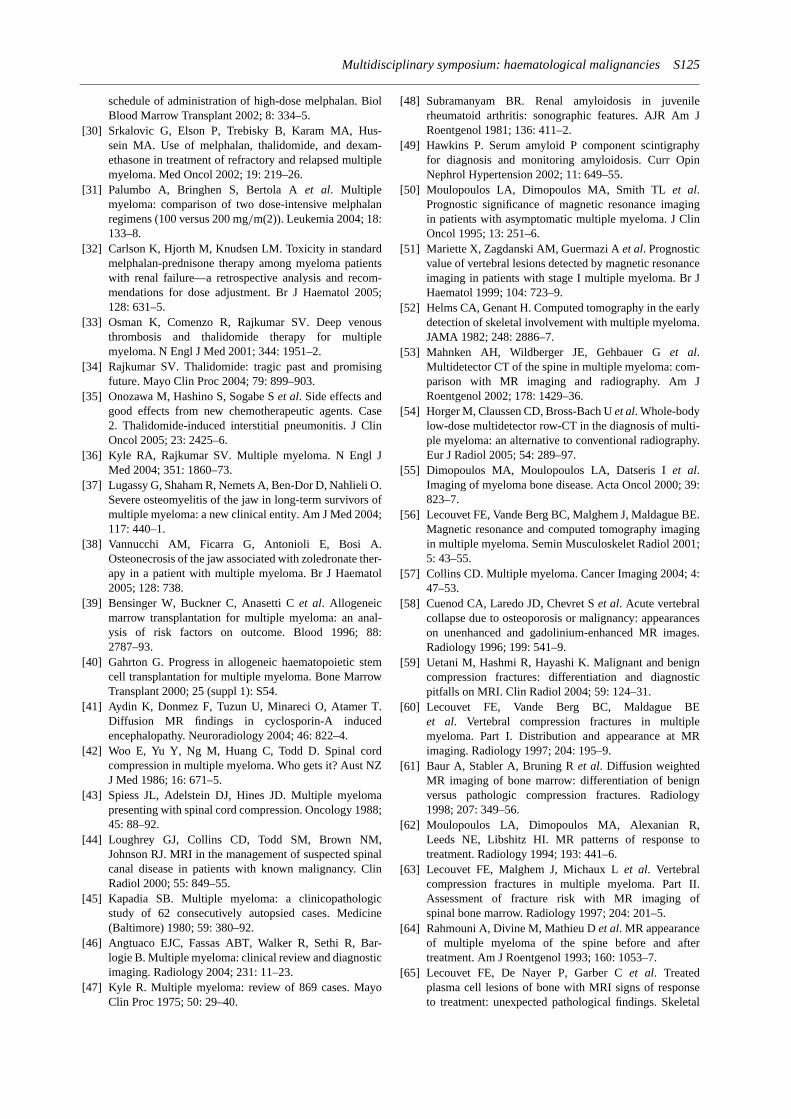
Multidisciplinary symposium: haematological malignancies S125
schedule of administration of high-dose melphalan. BiolBlood Marrow Transplant 2002; 8: 334–5.
[30] Srkalovic G, Elson P, Trebisky B, Karam MA, Hus-sein MA. Use of melphalan, thalidomide, and dexam-ethasone in treatment of refractory and relapsed multiplemyeloma. Med Oncol 2002; 19: 219–26.
[31] Palumbo A, Bringhen S, Bertola Aet al. Multiplemyeloma: comparison of two dose-intensive melphalanregimens (100 versus 200 mg/m(2)). Leukemia 2004; 18:133–8.
[32] Carlson K, Hjorth M, Knudsen LM. Toxicity in standardmelphalan-prednisone therapy among myeloma patientswith renal failure—a retrospective analysis and recom-mendations for dose adjustment. Br J Haematol 2005;128: 631–5.
[33] Osman K, Comenzo R, Rajkumar SV. Deep venousthrombosis and thalidomide therapy for multiplemyeloma. N Engl J Med 2001; 344: 1951–2.
[34] Rajkumar SV. Thalidomide: tragic past and promisingfuture. Mayo Clin Proc 2004; 79: 899–903.
[35] Onozawa M, Hashino S, Sogabe Set al. Side effects andgood effects from new chemotherapeutic agents. Case2. Thalidomide-induced interstitial pneumonitis. J ClinOncol 2005; 23: 2425–6.
[36] Kyle RA, Rajkumar SV. Multiple myeloma. N Engl JMed 2004; 351: 1860–73.
[37] Lugassy G, Shaham R, Nemets A, Ben-Dor D, Nahlieli O.Severe osteomyelitis of the jaw in long-term survivors ofmultiple myeloma: a new clinical entity. Am J Med 2004;117: 440–1.
[38] Vannucchi AM, Ficarra G, Antonioli E, Bosi A.Osteonecrosis of the jaw associated with zoledronate ther-apy in a patient with multiple myeloma. Br J Haematol2005; 128: 738.
[39] Bensinger W, Buckner C, Anasetti Cet al. Allogeneicmarrow transplantation for multiple myeloma: an anal-ysis of risk factors on outcome. Blood 1996; 88:2787–93.
[40] Gahrton G. Progress in allogeneic haematopoietic stemcell transplantation for multiple myeloma. Bone MarrowTransplant 2000; 25 (suppl 1): S54.
[41] Aydin K, Donmez F, Tuzun U, Minareci O, Atamer T.Diffusion MR findings in cyclosporin-A inducedencephalopathy. Neuroradiology 2004; 46: 822–4.
[42] Woo E, Yu Y, Ng M, Huang C, Todd D. Spinal cordcompression in multiple myeloma. Who gets it? Aust NZJ Med 1986; 16: 671–5.
[43] Spiess JL, Adelstein DJ, Hines JD. Multiple myelomapresenting with spinal cord compression. Oncology 1988;45: 88–92.
[44] Loughrey GJ, Collins CD, Todd SM, Brown NM,Johnson RJ. MRI in the management of suspected spinalcanal disease in patients with known malignancy. ClinRadiol 2000; 55: 849–55.
[45] Kapadia SB. Multiple myeloma: a clinicopathologicstudy of 62 consecutively autopsied cases. Medicine(Baltimore) 1980; 59: 380–92.
[46] Angtuaco EJC, Fassas ABT, Walker R, Sethi R, Bar-logie B. Multiple myeloma: clinical review and diagnosticimaging. Radiology 2004; 231: 11–23.
[47] Kyle R. Multiple myeloma: review of 869 cases. MayoClin Proc 1975; 50: 29–40.
[48] Subramanyam BR. Renal amyloidosis in juvenilerheumatoid arthritis: sonographic features. AJR Am JRoentgenol 1981; 136: 411–2.
[49] Hawkins P. Serum amyloid P component scintigraphyfor diagnosis and monitoring amyloidosis. Curr OpinNephrol Hypertension 2002; 11: 649–55.
[50] Moulopoulos LA, Dimopoulos MA, Smith TLet al.Prognostic significance of magnetic resonance imagingin patients with asymptomatic multiple myeloma. J ClinOncol 1995; 13: 251–6.
[51] Mariette X, Zagdanski AM, Guermazi Aet al. Prognosticvalue of vertebral lesions detected by magnetic resonanceimaging in patients with stage I multiple myeloma. Br JHaematol 1999; 104: 723–9.
[52] Helms CA, Genant H. Computed tomography in the earlydetection of skeletal involvement with multiple myeloma.JAMA 1982; 248: 2886–7.
[53] Mahnken AH, Wildberger JE, Gehbauer Get al.Multidetector CT of the spine in multiple myeloma: com-parison with MR imaging and radiography. Am JRoentgenol 2002; 178: 1429–36.
[54] Horger M, Claussen CD, Bross-Bach Uet al. Whole-bodylow-dose multidetector row-CT in the diagnosis of multi-ple myeloma: an alternative to conventional radiography.Eur J Radiol 2005; 54: 289–97.
[55] Dimopoulos MA, Moulopoulos LA, Datseris Iet al.Imaging of myeloma bone disease. Acta Oncol 2000; 39:823–7.
[56] Lecouvet FE, Vande Berg BC, Malghem J, Maldague BE.Magnetic resonance and computed tomography imagingin multiple myeloma. Semin Musculoskelet Radiol 2001;5: 43–55.
[57] Collins CD. Multiple myeloma. Cancer Imaging 2004; 4:47–53.
[58] Cuenod CA, Laredo JD, Chevret Set al. Acute vertebralcollapse due to osteoporosis or malignancy: appearanceson unenhanced and gadolinium-enhanced MR images.Radiology 1996; 199: 541–9.
[59] Uetani M, Hashmi R, Hayashi K. Malignant and benigncompression fractures: differentiation and diagnosticpitfalls on MRI. Clin Radiol 2004; 59: 124–31.
[60] Lecouvet FE, Vande Berg BC, Maldague BEet al. Vertebral compression fractures in multiplemyeloma. Part I. Distribution and appearance at MRimaging. Radiology 1997; 204: 195–9.
[61] Baur A, Stabler A, Bruning Ret al. Diffusion weightedMR imaging of bone marrow: differentiation of benignversus pathologic compression fractures. Radiology1998; 207: 349–56.
[62] Moulopoulos LA, Dimopoulos MA, Alexanian R,Leeds NE, Libshitz HI. MR patterns of response totreatment. Radiology 1994; 193: 441–6.
[63] Lecouvet FE, Malghem J, Michaux Let al. Vertebralcompression fractures in multiple myeloma. Part II.Assessment of fracture risk with MR imaging ofspinal bone marrow. Radiology 1997; 204: 201–5.
[64] Rahmouni A, Divine M, Mathieu Det al. MR appearanceof multiple myeloma of the spine before and aftertreatment. Am J Roentgenol 1993; 160: 1053–7.
[65] Lecouvet FE, De Nayer P, Garber Cet al. Treatedplasma cell lesions of bone with MRI signs of responseto treatment: unexpected pathological findings. Skeletal

S126 Multidisciplinary symposium: haematological malignancies
Radiol 1998; 27.[66] Lecouvet FE, Richard F, Vande Berg BCet al. Long term
effects of localised spinal radiation therapy on vertebralfractures and focal lesions: appearance in patients withmultiple myeloma. Br J Haematol 1997; 96: 743–5.
[67] Lecouvet FE, Vande Berg BC, Michaux Let al. StageIII multiple myeloma: clinical and prognostic value ofspinal bone marrow MR imaging. Radiology 1998; 209:653–60.
[68] Agren B, Reidberg U, Isberg B, Svensson L, Aspelin P.MR imaging of multiple myeloma patients with bonemarrow transplants. Acta Radiol 1998; 39: 36–42.
[69] Lecouvet FE, Dechambre S, Malghem J, Ferrant A, VandeBerg BC, Maldague B. Bone marrow transplantationin patients with multiple myeloma: prognostic signif-icance of MR imaging. Am J Roentgenol 2001; 176:91–6.
[70] Lecouvet FE, Malghem J, Michaux Let al. Skeletal sur-vey in multiple myeloma: radiographic versus MR imag-ing survey. Br J Haematol 1999; 106: 35–9.
[71] Fassas AB, Muwalla F, Berryman Tet al. Myeloma of thecentral nervous system: association with high-risk chro-mosomal abnormalities, plasmablastic morphology andextramedullary manifestations. Br J Haematol 2002; 117:103–8.
[72] Moulopoulos LA, Dimopoulos MA, Weber D, Fuller L,Libshitz HI, Alexanian R. Magnetic resonance imagingin the staging of solitary plasmacytoma of bone. J ClinOncol 1993; 11: 1311–5.
[73] Sezer O, Niemoller K, Jakob Cet al. Relationshipbetween bone marrow angiogenesis and plasma cellinfiltration and serum beta 2 microglobulin levelsin patients with multiple myeloma. Ann Haematol 2001;80: 598–601.
[74] Bataille R, Chevalier J, Rossi M, Sany J. Bonescintigraphy in plasma-cell myeloma. A prospective studyof 70 patients. Radiology 1982; 145: 801–4.
[75] Catalano L, Pace L, Califano Cet al. Detection of focalmyeloma lesions by Tc-99m sestaMIBI scintigraphy.Haematologica 1999; 84: 119–24.
[76] Fonti R, Del Vecchio S, Zannetti Aet al. Bone marrowuptake of Tc-99m MIBI in patients with multiplemyeloma. Eur J Nucl Med 2001; 25: 714–20.
[77] Alexandrakis MG, Kyriakou DS, Passam F, Kouk-ouraki S, Karkavitsas N. Value of Tc-99m sestamibiscintigraphy in the detection of bone lesions in multiple
myeloma. Ann Haematol 2001; 80: 349–63.[78] Alper E, Gurel M, Evrensel T, Ozkocaman V, Akbunar T,
Demiray M. 99mTc-MIBI scintigraphy in untreated stageIII multiple myeloma: comparison with X-ray skeletalsurvey and bone scintigraphy. Nucl Med Commun 2003;24: 537–42.
[79] Pace L, Catalano L, Pinto AMet al. Different patterns oftechnetium 99m-sestamibi uptake in multiple myeloma.Eur J Nucl Med 1998; 25: 714–20.
[80] Pace L, Catalano L, Del Vecchio Set al. Predictive valueof technetium-99m sestamibi in patients with multiplemyeloma and potential role in follow-up. Eur J Nucl Med2001; 28: 304–12.
[81] Pace L, Del Vecchio S, Salvatore M. Technetium 99msestamibi in multiple myeloma. Radiology 2005; 234:312–3.
[82] Schirrmeister H, Bommer L, Buck AKet al. Initial resultsin the assessment of multiple myeloma using 18F-FDGPET. Eur J Nucl Med Mol Imaging 2002; 29: 361–6.
[83] Jadvar H, Conti PS. Diagnostic utility of FDG PET inmultiple myeloma. Skeletal Radiol 2002; 31: 690–4.
[84] Durie BGM, Waxman AD, D’Agnolo A, Williams CM.Whole body 18F-FDG PET identifies high risk myeloma.J Nucl Med 2002; 43: 1457–63.
[85] Orchard K, Barrington S, Buscombe J, Hilson A,Prentice HG, Mehta A. Fluorodeoxyglucose positronemission tomography imaging for the detection of occultdisease in multiple myeloma. Br J Haematol 2002; 117:133–5.
[86] Bredella MA, Steinbach L, Caputo G, Segall G,Hawkins R. Value of FDG PET in the assessment ofpatients with multiple myeloma. Am J Roentgenol 2005;184: 1199–204.
[87] Kato T, Tsukamoto E, Nishioka Tet al. Early detectionof bone marrow involvement in extramedullary plas-macytoma by whole-body F-18 FDG positron emissiontomography. Clin Nucl Med 2000; 25: 870–3.
[88] Patlas M, Hadas-Halpern I, Libson E. Imaging findings ofextraosseous multiple myeloma. Cancer Imaging 2002; 2:120–2.
[89] Mileshkin L, Blum R, Seymour JF, Patrikeos A,Hicks RJ, Prince HM. A comparison of fluorine-18fluoro-deoxyglucose PET and technetium-99m sestamibiin assessing patients with multiple myeloma. Eur JHaematol 2004; 72: 32–7.