efr3s are palmitoylated plasma membrane proteins that ... · naveen bojjireddy1, maria luisa...
TRANSCRIPT
Jour
nal o
f Cel
l Sci
ence
RESEARCH ARTICLE
EFR3s are palmitoylated plasma membrane proteins that controlresponsiveness to G-protein-coupled receptors
Naveen Bojjireddy1, Maria Luisa Guzman-Hernandez1, Nathalie Renee Reinhard2, Marko Jovic1 andTamas Balla1,*
ABSTRACT
The yeast Efr3p protein is a main regulator of the Stt4p
phosphatidylinositol 4-kinase at contact sites between the
endoplasmic reticulum and the plasma membrane. A mutation in its
fly homologue Rbo, leads to diminished light responses in the eye
attributed to progressively impaired PLC signaling. Here, we find that
Efr3s plays a role in maintaining responsiveness to the type-I
angiotensin II (AngII) receptors. siRNA-mediated depletion of
EFR3A and EFR3B impaired the sustained phase of cytosolic Ca2+
response to high concentration of AngII in HEK293 cells that express
wild type but not truncated AGTR1 (AT1a receptor), missing the
phosphorylation sites. Efr3 depletion had minimal effect on the
recovery of plasma membrane phosphoinositides during stimulation,
and AT1 receptors still underwent ligand-induced internalization. A
higher level of basal receptor phosphorylation and a larger response
was observed after stimulation. Moreover, Gq activation more rapidly
desensitized after AngII stimulation in Efr3 downregulated cells. A
similar but less pronounced effect of EFR3 depletion was observed on
the desensitization of the cAMP response after stimulation with
isoproterenol. These data suggest that mammalian Efr3s contribute to
the control of the phosphorylation state and, hence, desensitization of
AT1a receptors, and could affect responsiveness of G-protein-
coupled receptors in higher eukaryotes.
KEY WORDS: EFR3, GPCR, Receptor desensitization, Angiotensin
II, PI 4-kinase, Phosphoinositide
INTRODUCTIONA selected group of G-protein-coupled receptors (GPCRs) activate
phospholipase C (PLC) by coupling to heterotrimeric Gq/11 proteins
to induce an increase of cytosolic Ca2+ and, depending on the cell
type, trigger a variety of downstream responses. PLC enzymes
hydrolyze the plasma membrane lipid, phosphatidylinositol 4,5-
bisphosphate [PtdIns(4,5)P2] to generate the two messenger
molecules, Ins(1,4,5)P3 and diacylglycerol (Berridge, 1984).
Owing to the limited size of the PtdIns(4,5)P2 pools at the plasma
membrane, continuous signal generation requires a steady
resynthesis of this lipid by PI 4-kinase and PIP 5-kinase enzymes.
The PI 4-kinase implicated in this process is PI4KA (Balla et al.,
2008; Balla et al., 2005), whose yeast orthologue Stt4 is also
responsible for the maintenance of the PtdIns(4,5)P2 pools at the
plasma membrane (Audhya and Emr, 2002).
Cells possess several protective mechanisms to prevent
excessive stimulation of their GPCRs. High concentrations of
GPCR ligand often lead to rapid receptor desensitization that
diminishes the ability of the receptor to couple to G proteins. The
most common cause of desensitization is due to the rapid
phosphorylation of receptors by one of several GPCR kinases
(GRKs) that lead to uncoupling from the G proteins and
association with arrestins (Moore et al., 2007). Binding to
arrestin triggers receptor endocytosis, thereby limiting the
number of the receptors on the cell surface. However, arrestin
association also lends a new signaling profile to endocytosed
receptors that trigger specific cellular responses from endocytic
compartments (Luttrell and Lefkowitz, 2002; Reiter et al., 2012;
Wei et al., 2003). Although these molecular events have been
studied and characterized in great detail, little is known about the
processes that restore the sensitivity of receptors at the plasma
membrane (Vasudevan et al., 2011). Early studies on b-
adrenergic receptors suggested that dephosphorylation requires
the receptors to be endocytosed (Yu et al., 1993) but this
hypothesis has never been thoroughly investigated.
This present study was intended to investigate the possible role
of the two mammalian EFR3 proteins, EFR3A and EFR3B, in the
regulation of the PI4KA enzyme in mammalian cells. The yeast
orthologue of these proteins, Efr3p, was found to be essential for
the localization of the yeast PI4K Stt4 into discrete signaling
domains and, together with another protein, Ypp1, to control Stt4
function (Baird et al., 2008). A similar role has been described in
the mammalian system, where EFR3 and the TTC7 proteins were
shown to keep an active pool of PI4KA at the plasma membrane
(Nakatsu et al., 2012). While studying the role of EFR3 in the
maintenance of the signaling phosphoinositide pools, we
discovered that EFR3 proteins are important for AT1 receptors
to maintain their signaling competence, an effect that was not
caused directly by depletion of the phosphoinositide pool but,
rather, the regulation of the phosphorylation state of the receptor.
Therefore, our studies identified EFR3 as a component of the
AT1 receptor signaling cascade that controls receptor re-
sensitization and, thereby, could serve as a hitherto
unrecognized player in determining AT1 receptor and, possibly,
other GPCR responsiveness.
RESULTSEFR3s are widely expressed but show distinct enrichment invarious tissuesMammalian cells contain two EFR3 proteins, EFR3A and EFR3B
with several putative splice variants annotated in GenBank. To
determine the expression pattern of the two EFR3 isoforms, we
1Section on Molecular Signal Transduction, Program for DevelopmentalNeuroscience, Eunice Kennedy Shriver NICHD, National Institutes of Health,Bethesda, MD 20892, USA. 2Swammerdam Institute for Life Sciences, Universityof Amsterdam, 1012 WX Amsterdam, The Netherlands.
*Author for correspondence ([email protected])
Received 9 June 2014; Accepted 29 October 2014
� 2015. Published by The Company of Biologists Ltd | Journal of Cell Science (2015) 128, 118–128 doi:10.1242/jcs.157495
118

Jour
nal o
f Cel
l Sci
ence
performed quantitative PCR (qPCR) using various mouse tissues.Fig. 1 shows that the two EFR3 isoforms are widely expressed,
although they show notable differences: while EFR3A showedhighest expression in the testis, EFR3B had highest expression inthe brain – followed by small intestine and the eye – but showedvery low expression in testis and kidney. These results did not
suggest a narrow tissue-specific function for the two EFR3proteins.
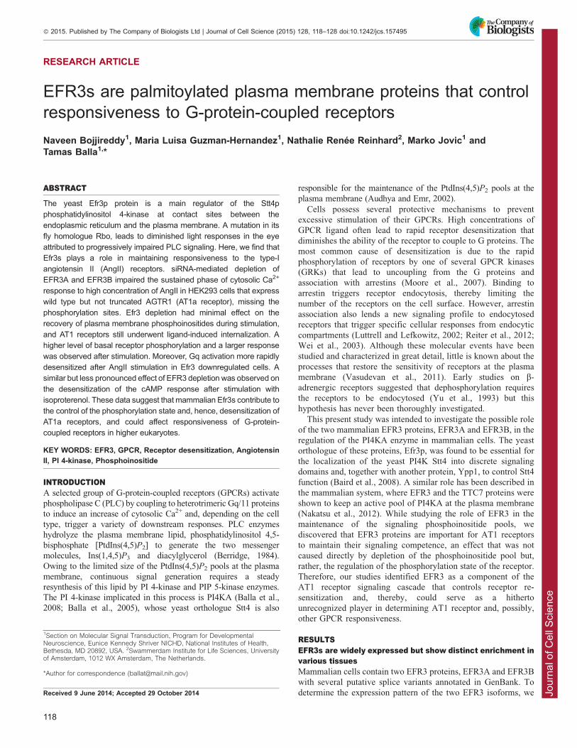
EFR3s are localized to the plasma membranevia palmitoylationTo determine the subcellular distribution of the two EFR3
proteins, we obtained cDNAs of both of them and epitope-taggedthem at their C-termini with either a hemagglutinin (HA)-tag,green fluorescent protein (GFP) or monomeric red fluorescent
protein (mRFP). When these proteins were expressed in HEK293cells or COS-7 cells, they localized to the plasma membranealthough a small amount was also detected in the cytosol(Fig. 2A). Similar plasma membrane localization was found
with the HA-tagged versions in fixed and immunostained cells(not shown). A shorter form of EFR3B truncated at the N-terminus (missing 148 residues) was found to be cytosolic
only (Fig. 2A). The presence of cysteine residues (four in EFR3Aand three in EFR3B) within this N-terminal segment – whichare conserved in the EFRs of higher eukaryotes but are missing
in yeast – raised the possibility that the full-length proteins arekept in the membrane by palmitoylation. Therefore, we mutatedthe four cysteine to serine residues in EFR3A and found this
mutant protein to be located in the cytosol (Fig. 2B). By usingmetabolic labeling of cells that overexpress GFP-tagged EFR3Aor GFP-tagged EFR3B with [3H]-palmitate, we were able toconfirm that, indeed, EFR3A and EFR3B are palmitoylated
proteins (Fig. 2C). Last, we determined whether the isolated N-terminus (residues 1–37 of EFR3A) is sufficient to target GFP tothe plasma membrane. We found that this construct is mostly
localized to the Golgi complex and that it requires the cysteineresidues for the Golgi localization (Fig. 2B). This suggested thatother determinants of the full-length protein are needed to
localize the protein to the plasma membrane. Our resultscollectively suggested that, contrary to previous suggestions,
EFR3 proteins are not transmembrane proteins but peripheralmembrane proteins associated with the plasma membranethrough palmitoylation. The same conclusion about EFR3localization and palmitoylation was reached in a recently
published study (Nakatsu et al., 2012).
EFR3s are important for sustained Ca2+ signalingIf EFR3 proteins are important for the correct functioning ofPI4KA, one expects that the phosphoinositide pools at the plasmamembrane are depleted upon strong stimulation of Gq/11-coupled
receptors, because the PI4KA enzyme was found to be primarilyresponsible for the synthesis of PtdIns4P in the plasma membraneto serve as precursor for PtdIns(4,5)P2 (Balla et al., 2008; Balla
et al., 2005). Such PtdIns(4,5)P2 depletion happens when theenzyme is pharmacologically inhibited, as shown initially byusing high concentrations of wortmannin or phenylarsine oxide ata concentration that primarily inhibits the PI4KA enzyme (Balla
et al., 2008) or, recently, by specific PI4KA inhibitors (Bojjireddyet al., 2014). The easiest way to test for such an effect was tofollow the cytosolic Ca2+ increase after stimulation because the
sustained phase of this response requires continuousPtdIns(4,5)P2 resynthesis (Nakanishi et al., 1995). For thisexperiment, we used HEK293 cells that stably express the rat
type-1A angiotensin II receptor (AT1a, also known as AGTR1)and to which we refer to hereafter as HEK-AT1. The AngIIresponses of these cells have been thoroughly characterized in our
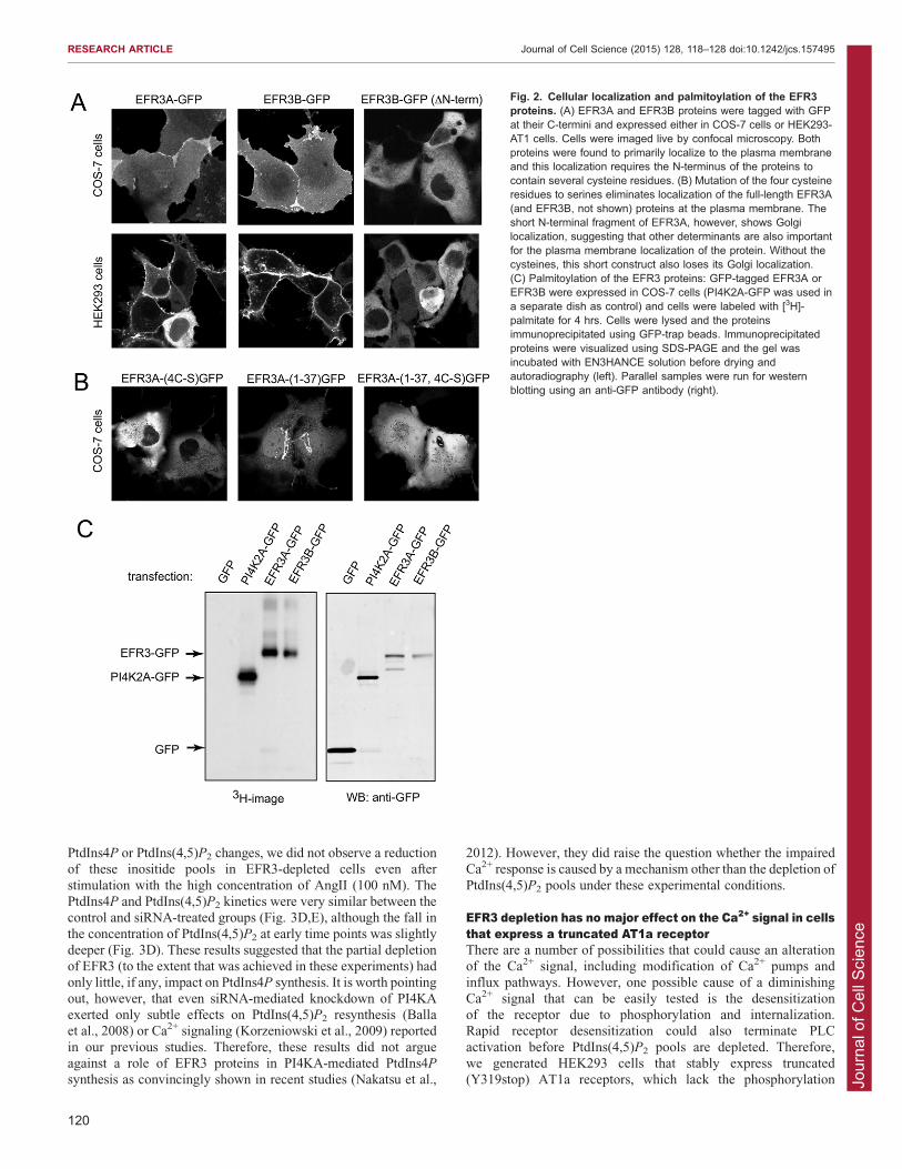
laboratory. We used small interfering RNA (siRNA)-mediatedsilencing (for 3 days) to knockdown EFR3 isoforms (either aloneor in combination) and studied the Ca2+ responses of Fura-2-loaded cells following the application of AngII (100 nM). The
efficiency of the EFR3 knockdown was tested using western blotanalysis of either the endogenous EFR3A or transfected EFR3Aand EFR3B (Fig. 3A,B). We found that, following stimulation
with 100 nM AngII, EFR3A- and EFR3B-depleted cells showed aCa2+ response with a diminished plateau phase, whereas cellstreated with control siRNA maintained the sustained Ca2+ plateau
that is characteristic of these cells (Fig. 3C, left panel). Notably,the Ca2+ response following stimulation with 1 nM of AngII wasvery similar between EFR3A and EFR3B-depleted cells(EFR3AB si) and control si cells (Fig. 3C, right panel).
Knockdown of the individual EFR3s have indicated thatknockdown of EFR3B had a stronger effect than that of EFR3A(not shown). Therefore, some of the later experiments were
performed only by knocking down EFR3B. These data wereconsistent with a depletion of the PtdIns(4,5)P2 pools at theplasma membrane in the EFR3A/B-depleted cells which would
be more severe at a higher level of stimulation.
EFR3-depleted cells do not show prominent defects inPtdIns4P and PtdIns(4,5)P2 resynthesis during stimulationNext we tested the effects of EFR3 downregulation by using RNAinterference (RNAi) on the PtdIns4P and PtdIns(4,5)P2 responsesto stimulation with AngII using metabolic labeling of these lipids.
Cells were labeled with 32P-phosphate or myo-[3H]inositol for3 hrs or 24 hrs, respectively, before stimulation with AngII(100 nM) for the indicated times. Lipids were extracted and
separated by thin layer chromatography (TLC) and quantifiedusing PhosphorImaging (for 32P) or densitometry after exposure ofplates that had been treated with EN3HANCE2 solution to X-ray
film ([3H]inositol). As shown in Fig. 3D for changes in 32P-labeled
Fig. 1. Relative expression of EFR3A and EFR3B mRNAs in variousmouse tissues. Total RNA from various mouse tissues was isolated andcDNA synthesized as described in Materials and Methods. Real-time qPCRwas performed using SYBR Green and primers specific for the respectivemRNAs (see Materials and Methods). The signal was then normalized to thatof 18S rRNA. Means+s.e.m. (n56) are shown from duplicate determinationsof three independent mouse and tissue isolations. See the predominantexpression of EFR3B in the brain and that of EFR3A in the testis.
RESEARCH ARTICLE Journal of Cell Science (2015) 128, 118–128 doi:10.1242/jcs.157495
119
Jour
nal o
f Cel
l Sci
ence
PtdIns4P or PtdIns(4,5)P2 changes, we did not observe a reductionof these inositide pools in EFR3-depleted cells even after
stimulation with the high concentration of AngII (100 nM). ThePtdIns4P and PtdIns(4,5)P2 kinetics were very similar between thecontrol and siRNA-treated groups (Fig. 3D,E), although the fall in
the concentration of PtdIns(4,5)P2 at early time points was slightlydeeper (Fig. 3D). These results suggested that the partial depletionof EFR3 (to the extent that was achieved in these experiments) had
only little, if any, impact on PtdIns4P synthesis. It is worth pointingout, however, that even siRNA-mediated knockdown of PI4KAexerted only subtle effects on PtdIns(4,5)P2 resynthesis (Ballaet al., 2008) or Ca2+ signaling (Korzeniowski et al., 2009) reported
in our previous studies. Therefore, these results did not argueagainst a role of EFR3 proteins in PI4KA-mediated PtdIns4P
synthesis as convincingly shown in recent studies (Nakatsu et al.,
2012). However, they did raise the question whether the impairedCa2+ response is caused by a mechanism other than the depletion of
PtdIns(4,5)P2 pools under these experimental conditions.
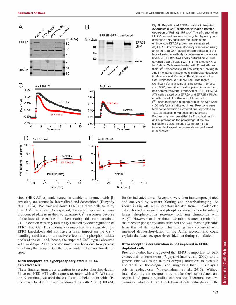
EFR3 depletion has no major effect on the Ca2+ signal in cellsthat express a truncated AT1a receptorThere are a number of possibilities that could cause an alterationof the Ca2+ signal, including modification of Ca2+ pumps and
influx pathways. However, one possible cause of a diminishingCa2+ signal that can be easily tested is the desensitizationof the receptor due to phosphorylation and internalization.Rapid receptor desensitization could also terminate PLC
activation before PtdIns(4,5)P2 pools are depleted. Therefore,we generated HEK293 cells that stably express truncated(Y319stop) AT1a receptors, which lack the phosphorylation
Fig. 2. Cellular localization and palmitoylation of the EFR3proteins. (A) EFR3A and EFR3B proteins were tagged with GFPat their C-termini and expressed either in COS-7 cells or HEK293-AT1 cells. Cells were imaged live by confocal microscopy. Bothproteins were found to primarily localize to the plasma membraneand this localization requires the N-terminus of the proteins tocontain several cysteine residues. (B) Mutation of the four cysteineresidues to serines eliminates localization of the full-length EFR3A(and EFR3B, not shown) proteins at the plasma membrane. Theshort N-terminal fragment of EFR3A, however, shows Golgilocalization, suggesting that other determinants are also importantfor the plasma membrane localization of the protein. Without thecysteines, this short construct also loses its Golgi localization.(C) Palmitoylation of the EFR3 proteins: GFP-tagged EFR3A orEFR3B were expressed in COS-7 cells (PI4K2A-GFP was used ina separate dish as control) and cells were labeled with [3H]-palmitate for 4 hrs. Cells were lysed and the proteinsimmunoprecipitated using GFP-trap beads. Immunoprecipitatedproteins were visualized using SDS-PAGE and the gel wasincubated with EN3HANCE solution before drying andautoradiography (left). Parallel samples were run for westernblotting using an anti-GFP antibody (right).
RESEARCH ARTICLE Journal of Cell Science (2015) 128, 118–128 doi:10.1242/jcs.157495
120
Jour
nal o
f Cel
l Sci
ence
sites (HEK-AT1D) and, hence, is unable to interact with b-
arrestins, and cannot be internalized and desensitized (Hunyadyet al., 1994). We knocked down EFR3s in these cells to studytheir Ca2+ responses. As expected, the cells displayed a more-pronounced plateau in their cytoplasmic Ca2+ responses because
of the lack of desensitization. Remarkably, this more-sustainedCa2+ elevation was only minimally affected by downregulation ofEFR3 (Fig. 4A). This finding was important as it suggested that
EFR3 knockdown did not have a main impact on the Ca2+-handling machinery or a massive effect on the phosphoinositidepools of the cell and, hence, the impaired Ca2+ signal observed
with wild-type AT1a receptor must have been due to a processinvolving the receptor tail that does contain the phosphorylationsites.
AT1a receptors are hyperphosphorylated in EFR3-depleted cellsThese findings turned our attention to receptor phosphorylation.
Since our HEK-AT1 cells express receptors with a FLAG-tag atthe N-terminus, we used these cells and labeled them with 32P-phosphate for 4 h followed by stimulation with AngII (100 nM)
for the indicated times. Receptors were then immunoprecipitated
and analyzed by western blotting and phosphorimaging. Asshown in Fig. 4B, AT1a receptors isolated from EFR3-depletedcells, showed increased basal phosphorylation and a substantiallylarger phosphorylation response following stimulation with
AngII. However, at later times (20 minutes after stimulation),the receptor phosphorylation subsided and was indistinguishablefrom that of the controls. This finding was consistent with
impaired dephosphorylation of the AT1a receptor and couldexplain the faster receptor desensitization during stimulation.
AT1a receptor internalization is not impaired in EFR3-depleted cellsPrevious studies have suggested that EFR3 is important for bulk
endocytosis of membranes (Vijayakrishnan et al., 2009), and agenetic link was found in flies carrying mutations in dynaminand the EFR3 homologue, Rbo, suggesting that EFR3 plays arole in endocytosis (Vijayakrishnan et al., 2010). Without
internalization, the receptor may not be dephosphorylated andregain its G protein signaling competence. Therefore, next weexamined whether EFR3 knockdown affects endocytosis of the
Fig. 3. Depletion of EFR3s results in impairedcytoplasmic Ca2+ response without a notabledepletion of PtdIns(4,5)P2. (A) The efficiency of anEFR3A knockdown was investigated by using twodifferent siRNA duplexes; the levels of theendogenous EFR3A protein were measured.(B) EFR3B knockdown efficiency was tested usingan expressed GFP-tagged protein because of thelack of suitable antibody to determine endogenouslevels. (C) HEK293-AT1 cells cultured on 25 mmcoverslips were treated with the indicated siRNAsfor 3 days. Cells were loaded with Fura-2/AM andtheir Ca2+ responses to 100 nM (left) or 1 nM (right)AngII monitored in ratiometric imaging as describedin Materials and Methods. The difference of theCa2+ responses to 100 nM AngII was highlysignificant (for analyzing all time points .60 sec,P,0.0001); we either used unpaired t-test or thenon-parametric Mann–Whitney test. (D,E) HEK293-AT1 cells treated with EFR3A and EFR3B siRNAsor with a control siRNA were labeled with[32P]phosphate for 3 h before stimulation with AngII(100 nM) for the indicated times. Reactions wereterminated and lipids extracted and separated byTLC as detailed in Materials and Methods.Radioactivity was quantified by Phosphorimagingand expressed as the percentage of the pre-stimulatory value. Means6s.e.m. from threeindependent experiments are shown performedin duplicates.
RESEARCH ARTICLE Journal of Cell Science (2015) 128, 118–128 doi:10.1242/jcs.157495
121
Jour
nal o
f Cel
l Sci
ence
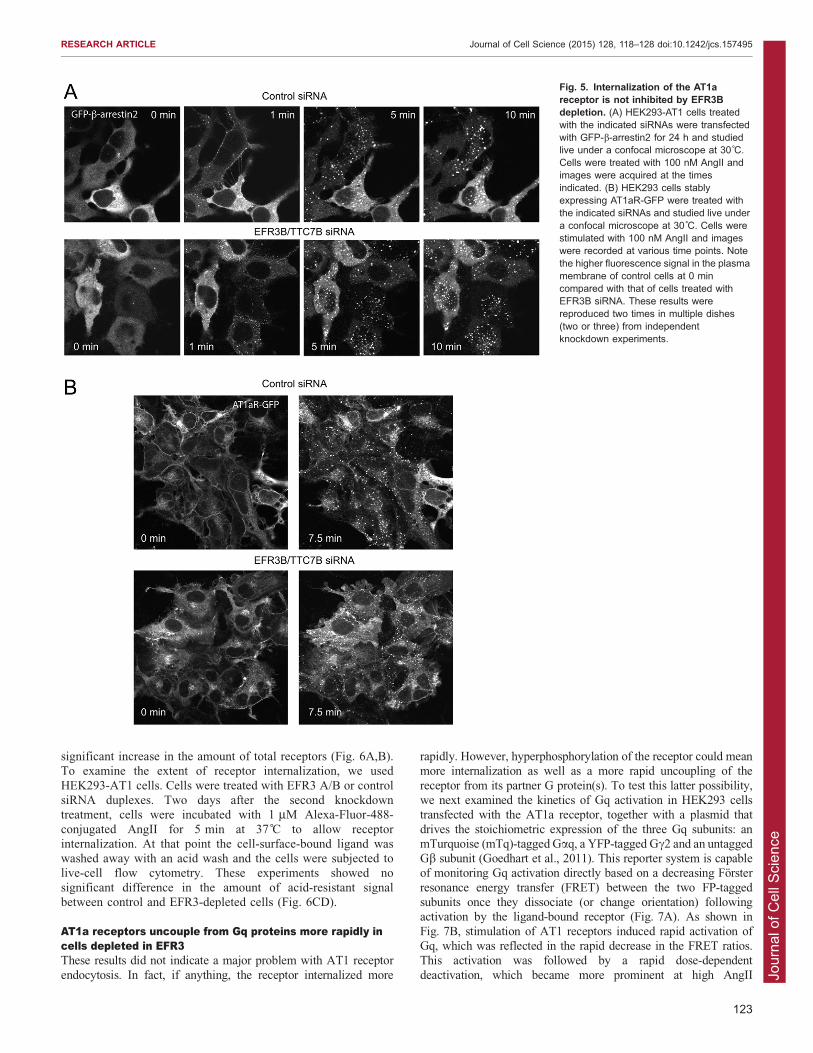
AT1a receptor. For this, we used several different approaches.First, we examined the translocation of GFP-tagged b-arrestin2(GFP-b-arrestin2) in the HEK293-AT1 cell line after stimulation
with AngII. In these experiments we used siRNA against EFR3Bonly. These experiments showed that EFR3 knockdown had noappreciable effect on the ability of the AT1 receptor to recruit
GFP-b-arrestin2 to the membrane or on subsequent intracellulartrafficking of the receptor–b-arrestin2 complex (Fig. 5A). Similarresults were found with GFP-tagged b-arrestin1, although the
association of this construct with the receptor was lesspronounced both in the control and EFR3-depleted cells (notshown).
Second, we followed the distribution of the AT1 receptors in astable cell line expressing the AT1a receptor tagged with GFP atthe C-terminus (AT1aR-GFP) (Hunyady et al., 2002). In previousstudies we have thoroughly characterized the trafficking of
AT1aR-GFP (Hunyady et al., 2002). There was a notabledecrease in the GFP signal at the plasma membrane of EFR3B-depleted cells before AngII stimulation relative to the internal
signal (Fig. 5B). However, AT1aR-GFP showed similarclustering and endocytosis regardless of whether cells weretreated with control or EFR3 siRNAs (Fig. 5B).
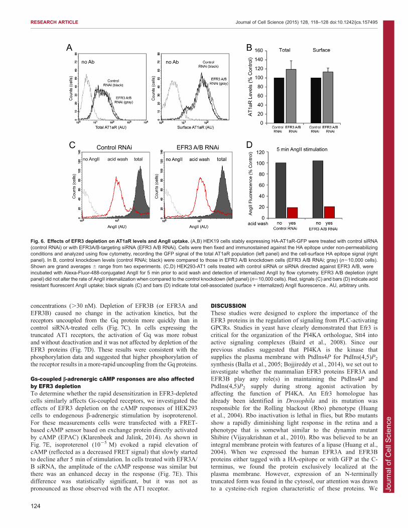
Third, we performed a more-quantitative assessment ofreceptor distribution by using flow cytometry. We determinedthe total number of receptors and that of cell-surface receptors by
using HEK293 cells expressing AT1a receptors that were HA-tagged at the N-terminus and GFP-tagged at the C-terminus (HA-AT1aR-GFP). Cells were treated with siRNAi targeting EFR3A
and EFR3B or with control siRNA prior to trypsinization,pelleting and fixation in 4% PFA. Cells were next immunostainedusing a mouse antibody against the extracellular HA epitope
under non-permeabilizing conditions. Secondary antibodystaining was performed using PerCP-conjugated goat anti-mouse antibody, and the total receptor population (GFP;Fig. 6A, left panel) and the surface receptor population (Ab
signal; Fig. 6A, right panel) were analyzed using flow cytometry.These studies showed no decrease in the amount of cell-surfacereceptors in EFR3-depleted cells and a slight but statistically not
Fig. 4. Depletion of EFR3s failed to affect the cytoplasmic Ca2+
response of cells that express a C-terminally truncated AT1areceptor and augmented phosphorylation of the wild-type AT1areceptor. (A) HEK293 cells stably transfected with an AT1a receptortruncated at its C-terminus (to remove all phosphorylation sites) weretreated with siRNAs targeting EFR3A and EFR3B or with a controlsiRNA. Measurements of cytoplasmic Ca2+ responses to AngII were asdescribed in the legend to Fig. 3 (B) Phosphorylation of the wild-typeAT1a receptor immunoprecipitated from HEK293-AT1 cells labeled with[32P]phosphate and stimulated with AngII for the indicated times. Notethe already increased phosphorylation of the receptor before and thelarger response after AngII stimulation. Representative phosphorimageof three similar observations. (C) Quantification and summary of threeindependent experiments. All radioactivity values in each experimentwere expressed as the percentage of the value measured at the5 minute time-point in EFR3AB-depleted cells. Means6s.e.m. (n53).
RESEARCH ARTICLE Journal of Cell Science (2015) 128, 118–128 doi:10.1242/jcs.157495
122
Jour
nal o
f Cel
l Sci
ence
significant increase in the amount of total receptors (Fig. 6A,B).To examine the extent of receptor internalization, we used
HEK293-AT1 cells. Cells were treated with EFR3 A/B or controlsiRNA duplexes. Two days after the second knockdowntreatment, cells were incubated with 1 mM Alexa-Fluor-488-
conjugated AngII for 5 min at 37 C to allow receptorinternalization. At that point the cell-surface-bound ligand waswashed away with an acid wash and the cells were subjected tolive-cell flow cytometry. These experiments showed no
significant difference in the amount of acid-resistant signalbetween control and EFR3-depleted cells (Fig. 6CD).
AT1a receptors uncouple from Gq proteins more rapidly incells depleted in EFR3These results did not indicate a major problem with AT1 receptor
endocytosis. In fact, if anything, the receptor internalized more
rapidly. However, hyperphosphorylation of the receptor could meanmore internalization as well as a more rapid uncoupling of the
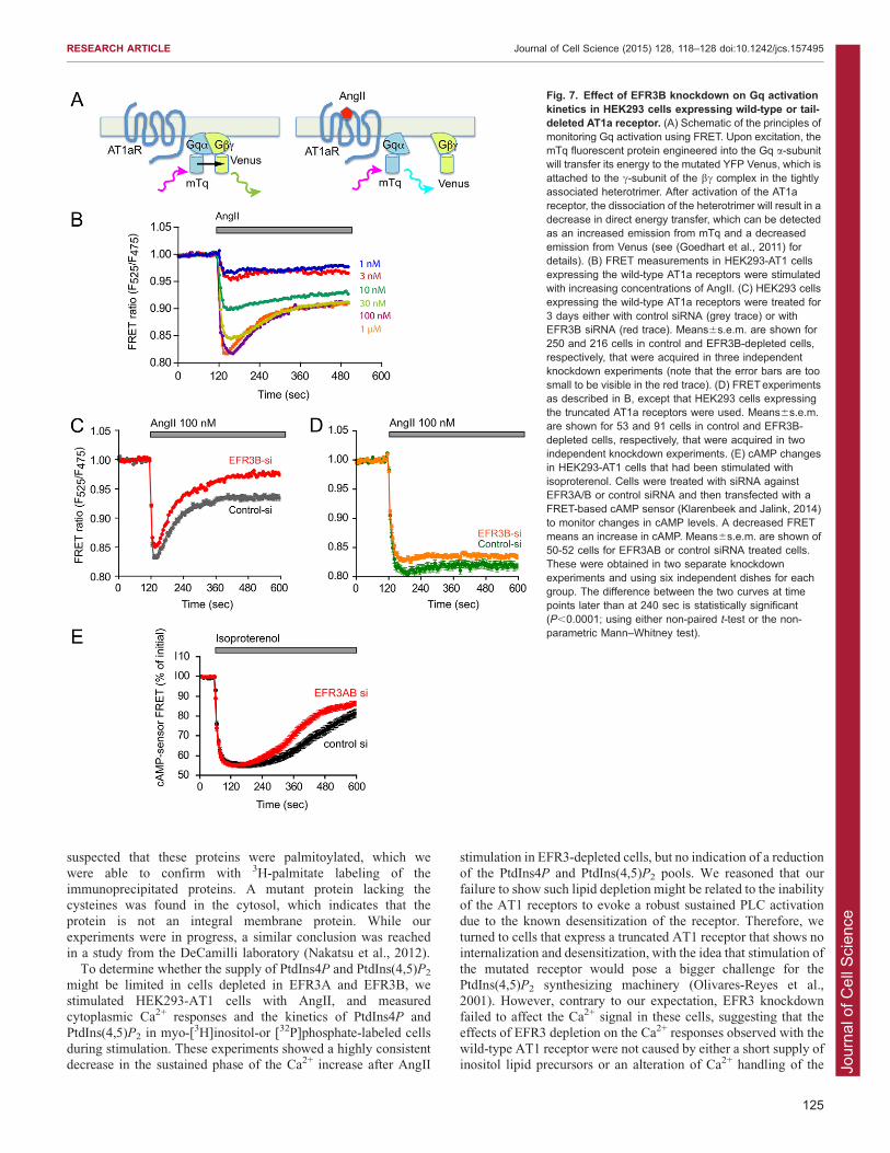
receptor from its partner G protein(s). To test this latter possibility,we next examined the kinetics of Gq activation in HEK293 cellstransfected with the AT1a receptor, together with a plasmid that
drives the stoichiometric expression of the three Gq subunits: anmTurquoise (mTq)-tagged Gaq, a YFP-tagged Gc2 and an untaggedGb subunit (Goedhart et al., 2011). This reporter system is capableof monitoring Gq activation directly based on a decreasing Forster
resonance energy transfer (FRET) between the two FP-taggedsubunits once they dissociate (or change orientation) followingactivation by the ligand-bound receptor (Fig. 7A). As shown in
Fig. 7B, stimulation of AT1 receptors induced rapid activation ofGq, which was reflected in the rapid decrease in the FRET ratios.This activation was followed by a rapid dose-dependent
deactivation, which became more prominent at high AngII
Fig. 5. Internalization of the AT1areceptor is not inhibited by EFR3Bdepletion. (A) HEK293-AT1 cells treatedwith the indicated siRNAs were transfectedwith GFP-b-arrestin2 for 24 h and studiedlive under a confocal microscope at 30˚C.Cells were treated with 100 nM AngII andimages were acquired at the timesindicated. (B) HEK293 cells stablyexpressing AT1aR-GFP were treated withthe indicated siRNAs and studied live undera confocal microscope at 30˚C. Cells werestimulated with 100 nM AngII and imageswere recorded at various time points. Notethe higher fluorescence signal in the plasmamembrane of control cells at 0 mincompared with that of cells treated withEFR3B siRNA. These results werereproduced two times in multiple dishes(two or three) from independentknockdown experiments.
RESEARCH ARTICLE Journal of Cell Science (2015) 128, 118–128 doi:10.1242/jcs.157495
123
Jour
nal o
f Cel
l Sci
ence
concentrations (.30 nM). Depletion of EFR3B (or EFR3A andEFR3B) caused no change in the activation kinetics, but thereceptors uncoupled from the Gq protein more quickly than in
control siRNA-treated cells (Fig. 7C). In cells expressing thetruncated AT1 receptors, the activation of Gq was more robustand without deactivation and it was not affected by depletion of the
EFR3 proteins (Fig. 7D). These results were consistent with thephosphorylation data and suggested that higher phosphorylation ofthe receptor results in a more-rapid uncoupling from the Gq proteins.
Gs-coupled b-adrenergic cAMP responses are also affectedby EFR3 depletionTo determine whether the rapid desensitization in EFR3-depleted
cells similarly affects Gs-coupled receptors, we investigated theeffects of EFR3 depletion on the cAMP responses of HEK293cells to endogenous b-adrenergic stimulation by isoproterenol.
For these measurements cells were transfected with a FRET-based cAMP sensor based on exchange protein directly activatedby cAMP (EPAC) (Klarenbeek and Jalink, 2014). As shown in
Fig. 7E, isoproterenol (1025 M) evoked a rapid elevation ofcAMP (reflected as a decreased FRET signal) that slowly startedto decline after 5 min of stimulation. In cells treated with EFR3A/
B siRNA, the amplitude of the cAMP response was similar butthere was an enhanced decay in the response (Fig. 7E). Thisdifference was statistically significant, but it was not aspronounced as those observed with the AT1 receptor.
DISCUSSIONThese studies were designed to explore the importance of theEFR3 proteins in the regulation of signaling from PLC-activating
GPCRs. Studies in yeast have clearly demonstrated that Efr3 iscritical for the organization of the PI4KA orthologue, Stt4 intoactive signaling complexes (Baird et al., 2008). Since our
previous studies suggested that PI4KA is the kinase thatsupplies the plasma membrane with PtdIns4P for PtdIns(4,5)P2
synthesis (Balla et al., 2005; Bojjireddy et al., 2014), we set out to
investigate whether the mammalian EFR3 proteins EFR3A andEFR3B play any role(s) in maintaining the PtdIns4P andPtdIns(4,5)P2 supply during strong agonist activation byaffecting the function of PI4KA. An Efr3 homologue has
already been identified in Drosophila and its mutation wasresponsible for the Rolling blackout (Rbo) phenotype (Huanget al., 2004). Rbo inactivation is lethal in flies, but Rbo mutants
show a rapidly diminishing light response in the retina and aphenotype that is somewhat similar to the dynamin mutantShibire (Vijayakrishnan et al., 2010). Rbo was believed to be an
integral membrane protein with features of a lipase (Huang et al.,2004). When we expressed the human EFR3A and EFR3Bproteins either tagged with a HA-epitope or with GFP at the C-
terminus, we found the protein exclusively localized at theplasma membrane. However, expression of an N-terminallytruncated form was found in the cytosol, our attention was drawnto a cysteine-rich region characteristic of these proteins. We
Fig. 6. Effects of EFR3 depletion on AT1aR levels and AngII uptake. (A,B) HEK19 cells stably expressing HA-AT1aR-GFP were treated with control siRNA(control RNAi) or with EFR3A/B-targeting siRNA (EFR3 A/B RNAi). Cells were then fixed and immunostained against the HA epitope under non-permeabilizingconditions and analyzed using flow cytometry, recording the GFP signal of the total AT1aR population (left panel) and the cell-surface HA epitope signal (rightpanel). In B, control knockdown levels (control RNAi; black) were compared to those in EFR3 A/B knockdown cells (EFR3 A/B RNAi; gray) (n510,000 cells).Shown are grand averages 6 range from two experiments. (C,D) HEK293-AT1 cells treated with control siRNA or siRNA directed against EFR3 A/B, wereincubated with Alexa-Fluor-488-conjugated AngII for 5 min prior to acid wash and detection of internalized AngII by flow cytometry. EFR3 A/B depletion (rightpanel) did not alter the rate of AngII internalization when compared to the control knockdown (left panel) (n510,000 cells). Red, signals (C) and bars (D) indicate acidresistant fluorescent AngII uptake; black signals (C) and bars (D) indicate total cell-associated (surface + internalized) AngII fluorescence.. AU, arbitrary units.
RESEARCH ARTICLE Journal of Cell Science (2015) 128, 118–128 doi:10.1242/jcs.157495
124
Jour
nal o
f Cel
l Sci
ence
suspected that these proteins were palmitoylated, which we
were able to confirm with 3H-palmitate labeling of theimmunoprecipitated proteins. A mutant protein lacking thecysteines was found in the cytosol, which indicates that the
protein is not an integral membrane protein. While ourexperiments were in progress, a similar conclusion was reachedin a study from the DeCamilli laboratory (Nakatsu et al., 2012).
To determine whether the supply of PtdIns4P and PtdIns(4,5)P2
might be limited in cells depleted in EFR3A and EFR3B, westimulated HEK293-AT1 cells with AngII, and measuredcytoplasmic Ca2+ responses and the kinetics of PtdIns4P and
PtdIns(4,5)P2 in myo-[3H]inositol-or [32P]phosphate-labeled cellsduring stimulation. These experiments showed a highly consistentdecrease in the sustained phase of the Ca2+ increase after AngII
stimulation in EFR3-depleted cells, but no indication of a reduction
of the PtdIns4P and PtdIns(4,5)P2 pools. We reasoned that ourfailure to show such lipid depletion might be related to the inabilityof the AT1 receptors to evoke a robust sustained PLC activation
due to the known desensitization of the receptor. Therefore, weturned to cells that express a truncated AT1 receptor that shows nointernalization and desensitization, with the idea that stimulation of
the mutated receptor would pose a bigger challenge for thePtdIns(4,5)P2 synthesizing machinery (Olivares-Reyes et al.,2001). However, contrary to our expectation, EFR3 knockdownfailed to affect the Ca2+ signal in these cells, suggesting that the
effects of EFR3 depletion on the Ca2+ responses observed with thewild-type AT1 receptor were not caused by either a short supply ofinositol lipid precursors or an alteration of Ca2+ handling of the
Fig. 7. Effect of EFR3B knockdown on Gq activationkinetics in HEK293 cells expressing wild-type or tail-deleted AT1a receptor. (A) Schematic of the principles ofmonitoring Gq activation using FRET. Upon excitation, themTq fluorescent protein engineered into the Gq a-subunitwill transfer its energy to the mutated YFP Venus, which isattached to the c-subunit of the bc complex in the tightlyassociated heterotrimer. After activation of the AT1areceptor, the dissociation of the heterotrimer will result in adecrease in direct energy transfer, which can be detectedas an increased emission from mTq and a decreasedemission from Venus (see (Goedhart et al., 2011) fordetails). (B) FRET measurements in HEK293-AT1 cellsexpressing the wild-type AT1a receptors were stimulatedwith increasing concentrations of AngII. (C) HEK293 cellsexpressing the wild-type AT1a receptors were treated for3 days either with control siRNA (grey trace) or withEFR3B siRNA (red trace). Means6s.e.m. are shown for250 and 216 cells in control and EFR3B-depleted cells,respectively, that were acquired in three independentknockdown experiments (note that the error bars are toosmall to be visible in the red trace). (D) FRETexperimentsas described in B, except that HEK293 cells expressingthe truncated AT1a receptors were used. Means6s.e.m.are shown for 53 and 91 cells in control and EFR3B-depleted cells, respectively, that were acquired in twoindependent knockdown experiments. (E) cAMP changesin HEK293-AT1 cells that had been stimulated withisoproterenol. Cells were treated with siRNA againstEFR3A/B or control siRNA and then transfected with aFRET-based cAMP sensor (Klarenbeek and Jalink, 2014)to monitor changes in cAMP levels. A decreased FRETmeans an increase in cAMP. Means6s.e.m. are shown of50-52 cells for EFR3AB or control siRNA treated cells.These were obtained in two separate knockdownexperiments and using six independent dishes for eachgroup. The difference between the two curves at timepoints later than at 240 sec is statistically significant(P,0.0001; using either non-paired t-test or the non-parametric Mann–Whitney test).
RESEARCH ARTICLE Journal of Cell Science (2015) 128, 118–128 doi:10.1242/jcs.157495
125
Jour
nal o
f Cel
l Sci
ence
cells. Instead, these results indicated that EFR3s controls thereceptor itself through a process that is linked to the tail region of
the receptor that is lacking in the deletion mutant. Since this regioncontains the phosphorylation sites, next we examined thephosphorylation status of the receptor and found that EFR3depletion caused an increased basal phosphorylation and an
enhanced phosphorylation response following stimulation. Inparallel experiments we also determined that AT1 receptorinternalization and the subsequent trafficking were not inhibited
in EFR3-depleted cells. These findings together pointed to apossible defect in the ability of the receptor to maintain Gqactivation in EFR3-depleted cells caused by hyperphosphorylation
and uncoupling from the G-proteins, in other words, a more rapiddesensitization. This was studied directly by using a FRET-basedGq activation sensor (Adjobo-Hermans et al., 2011) and the results
showed that EFR3-depletion, indeed, yielded faster inactivation ofthe wild-type receptor but not the tail-deleted mutant.
Taken all these findings together, we assume that there is adefect in the resensitization of the receptors from their
desensitized state in EFR3-depleted cells. This would alsoexplain the rapidly diminishing light responses of the Rbo flyand their increased pool of PtdIns(4,5)P2 due to the inability of
the photoreceptor to couple transducin and activate PLC (Huanget al., 2004). There is limited knowledge on how phosphorylatedGPCRs are dephosphorylated and return to their active state. In
Drosophila, a Ca2+-activated protein phosphatase, a product ofthe rdgC gene, was found to be responsible for thedephosphorylation of the photoreceptor (Steele et al., 1992;
Vinos et al., 1997). Mammalian homologues of rdgC have beenidentified as PPEF1 and PPEF2 (Montini et al., 1997; Shermanet al., 1997), but mice deficient in these proteins had no problemswith GPCR desensitization or reactivation, either in the retina or
other tissues (Ramulu et al., 2001). In mammalian cells severalphosphatases can dephosphorylate GPCRs, but it is generallyagreed that PP2A dephosphorylates b2-receptors and, perhaps,
other GPCRs as well (Vasudevan et al., 2011). A special ‘latent’pool of PP2A residing in internal membranes was postulated to besufficient to act on b2-receptors (Yang et al., 1988) and it was
suggested that b-adrenergic receptors have to be internalized andrecycled to regain their coupling competence (Yu et al., 1993).However, other studies showed that GRK-mediated GPCRphosphorylations could be reversed at the plasma membrane
(Iyer et al., 2006). Since EFR3s themselves do not showinternalization we assume that their function is linked to theplasma membrane. It should also be noted that the higher level of
receptor phosphorylation still subsides after a 20-minutestimulation. This suggests either a delayed dephosphorylationresponse or the existence of more than one mechanism
responsible for receptor dephosphorylation at different stages ofreceptor trafficking. No signs of a defect in AT1 receptorinternalization or subsequent trafficking were observed in the
present study. The notable decrease in the AT1aR-GFP signal atthe plasma membrane relative to the cytoplasm in EFR3-depletedcells may suggest a problem with the return of the phosphorylatedand internalized receptors to the plasma membrane. However,
flow cytometry did not indicate decreased levels of receptors onthe cell surface after EFR3A/B knockdown, and did not findaccumulation of receptor-containing vesicles beneath the plasma
membrane at light-microscopy level.Recent reports have shown that EFR3 and TTC7 proteins are
responsible for the recruitment of PI4KA to the plasma
membrane in yeast (Baird et al., 2008) and also in mammalian
cells (Nakatsu et al., 2012), a finding we were able to confirm(N.B. and T.B., unpublished observation). The question then
naturally arises whether the rapid desensitization of the receptorin EFR3-depleted cells is related to a defect in the functionof PI4KA. We found no indication of a problem regardingmaintenance of the PtdIns(4,5)P2 pool in EFR3-depleted cells,
even when stimulated through a non-desensitizing AT1R.However, it is important to emphasize that these results do notargue against the role of EFR3s in the control of PI4KA
function. They merely suggest that, at the level of EFR3knockdown we achieved, PI4KA can still supply the necessaryphosphoinositides. It should be also noted that it has proven to be
extremely difficult to achieve PI4KA knockdowns that wouldlimit PtdIns4P production at the plasma membrane (Balla et al.,2008; Balla et al., 2005). This, however, also suggests that the
process that controls the resensitization of the AT1R is moresensitive to EFR3 depletion than the PI4KA-mediatedmaintenance of the PtdIns(4,5)P2 pool. Furthermore, PI4KA-depleted cells had shown no signs of rapid desensitization of the
AT1a receptor in our previous studies (Korzeniowski et al.,2009).
We also examined, whether the same phenomenon is observed
when the cells are stimulated through a Gs-coupled receptor andfound that the cAMP response to isoproterenol in HEK293 cellsthat express endogenous b–receptors is also affected by a
knockdown of EFR3A/B, although the effect was not aspronounced as the one observed with the AT1a receptor. Thismay be caused by the fact that we did not directly measure the
coupling of Gs but only cAMP levels, and the latter might notreflect the extent of desensitization so directly. More studies willbe needed to extend the generality of this phenomenon to otherGPCRs and to address the exact mechanism by which receptor
resensitization occurs. Nevertheless, the cAMP studies alsosuggest that the observed phenomenon is not related to changesof phosphoinositide, as evoked by Gq-coupled receptors.
In summary, the present results identify a hithertounrecognized and unexpected function of EFR3 proteins inmammalian cells, namely their importance in the control of
GPCR responsiveness by affecting receptor phosphorylation.AT1Rs in EFR3-depleted cells are hyperphosphorylated anduncouple from Gq proteins more rapidly after stimulation, butthey maintain their ability to internalize. These findings highlight
the caveats in our knowledge of the mechanism(s) that controlGPCR dephosphorylation and resensitization, and shouldfacilitate further studies to uncover the molecular details of
these processes, as well as the role of the EFR3 proteins within.
MATERIALS AND METHODSMaterialsAll chemicals were of the highest analytical grade. Angiotensin II
(human octapeptide; AngII) was from Bachem (Torrance, CA).
[c32P]ATP (6000 Ci/mmol) was purchased from Perkin-Elmer. Myo-
[3H]inositol (30–80 Ci/mmol) was from Amersham and American
Radiolabeled Chemicals (St Louis, MO). The monoclonal anti-
hemagglutinin (HA) antibody (Ab) (HA.11) was from Covance, the
anti-FLAG and the EFR3A antibodies were from Sigma (St Louis, MO).
Antibodies against tubulin and actin were from Sigma Aldrich and Cell
Signaling, respectively.
Plasmids and cellsThe GFP-tagged b-arrestin2 (Barak et al., 1997) was kindly provided by
Marc Caron (Duke University, Durham, NC). The Gq FRET construct
(Goedhart et al., 2011) was a kind gift from Joachim Goedhart and
RESEARCH ARTICLE Journal of Cell Science (2015) 128, 118–128 doi:10.1242/jcs.157495
126
Jour
nal o
f Cel
l Sci
ence
Theodorus W. Gadella Jr. (University of Amsterdam). The HEK293-AT1
cells stably express the rat AT1a angiotensin receptor (Balla et al., 2005).
HEK293-AT1-GFP cells stably express the rat AT1a angiotensin receptor
fused to GFP (AT1aR-GFP) (Hunyady et al., 2002), and the HEK-AT1Dcell line stably expresses the rat AT1a angiotensin receptor with a stop at
Y319 (Hunyady et al., 1994). EST clones for EFR3A (EHS1001-
213247615) and EFR3B (MHS6278-202802246) (IHS1382-8680872,
containing the N-terminal EFR3B piece missing from clone MHS6278-
202802246) were from Open Biosystems. The coding sequence of
EFR3A, EFR3B short form and long form were subcloned into the
pCDNA3.1(+) vector with an HA-tag at C-termini. For localization
studies EFR3A and 3B were also subcloned into pEGFP-N1 and mRFP-
N1 plasmids. Palmitoylation sites EFR3A-GFP (amino acids 5-CCCC-
11) and EFR3B-GFP (amino acids 4-CGCC-9) were mutated to EFR3A
(5-SSSS-11) and EFR3B (4-SGSS-9) by site directed mutagenesis using
the Quikchange mutagenesis kit from Promega.
Transfection of cellsCells (50,000 cells/well) were plated onto 25-mm-diameter circular glass
coverslips treated with Poly-lysine in 6-well plates and plasmid DNAs
(0.5–1 mg/well) were transfected with the indicated constructs
(Szentpetery et al., 2009) using Lipofectamine2000 reagent (Invitrogen)
and OPTI-MEM (Invitrogen) following the manufacturer’s instructions.
For siRNA treatment, cells were either cultured on coverslips in 6-well
dishes for imaging or 12-well dishes for metabolic labeling. Cells were
treated with 100 nM siRNA using oligofectamine. Experiments were
carried out with the knockdown cells 3 days post siRNA treatment.
Live-cell imagingAfter 20–24 hrs of transfection, cells were washed on the glass coverslips
with a modified Krebs-Ringer solution, containing 120 mM NaCl,
4.7 mM KCl, 1.2 mM CaCl2, 0.7 mM MgSO4, 10 mM glucose, 10 mM
Na-HEPES pH 7.4 and the coverslip was placed into a metal chamber
(Atto, Invitrogen) that was mounted on a heated stage (35 C). Cells were
incubated in 1 ml of the Krebs-Ringer buffer and the stimulating agents
were dissolved and added in 200 ml warm buffer removed from the cells.
Cells were examined by using inverted microscopes. Confocal images
were obtained with a Zeiss LSM 510-META laser confocal microscope
(Carl Zeiss MicroImaging, Inc.) using a 636oil-immersion objective
equipped with an objective heater (Bioptech).
Cytoplasmic Ca2+ measurementsHEK-AT1 cells (46105 cells) cultured on coverslips were treated with
siRNA as described above. Cells were loaded with 3 mM Fura 2/AM at
room temperature for 45 min in HEPES-buffered M199-Hanks’ salt
solution containing 200 mM sulfinpyrazone and 0.06% pluronic acid.
Ca2+ measurements were performed in modified Krebs-Ringer solution
(see above) containing 200 mM sulfinpyrazone. Single-cell Ca2+
measurements were carried out at room temperature using an Olympus
IX70 inverted microscope equipped with Lamda-DG4 illuminator and a
MicroMAX-1024BFT digital camera with appropriate filters. Data were
acquired using MetaFluor software (Molecular Devices).
Analysis of myo-[3H]inositol- or [32P]phosphate-labeled lipidsHEK293-AT1 cells plated on 12-well plates (at a density of 30,000 cells/
ml) were labeled with myo-[3H]inositol (20 mCi/ml) in 1 ml of inositol-
free DMEM supplemented with 2% dialyzed FBS for 24 h or with 2 mCi/
ml o-[32P]phosphate for 3 h in phosphate-free DMEM supplemented with
2% dialyzed FBS. Cells were stimulated with AngII (100 nM) for the
indicated times or left unstimulated. Reactions were terminated by the
addition of ice-cold perchloric acid (5% final concentration), and cells
were kept on ice for 30 min. Cells were scraped off the plates and frozen
and thawed once; they were then centrifuged and the cell pellet was
processed to extract the phosphoinositides by an acidic chloroform–
methanol extraction followed by thin-layer chromatography (TLC)
essentially as described previously (Balla et al., 2008; Nakanishi et al.,
1995). TLC plates were sprayed with EN3HANCE2 solution (Perkin-
Elmer) and subjected to autoradiography (in the case of 3H-labeling) or
analyzed without spraying using a PhosphorImager (for the 32P-labeled
samples). Autographic films were exposed several times to find the
correct exposure time for densitometric analysis.
Analysis of incorporation of [3H]-palmitateThe procedure described by Linder et al. was followed (Linder et al.,
1995). Briefly, COS-7 cells were transiently transfected with pGFP-N1-
EFR3A or pEGFP-N1-EFR3B. After 24 hrs, cells were metabolically
labeled with [3H]-palmitate (0.3 mCi/ml) for 4 hrs. Prior to lysis,
cells were washed once with ice-cold PBS pH 7.4, lysates were
immunoprecipitated with GFP-trap (ChromoTek GmbH, Germany)
overnight and washed three times with lysis buffer. Proteins were
resolved using SDS-PAGE, and the gel was incubated with EN3HANCE
solution for 2 hrs, dried and subjected to autoradiography. pEGFP-N1-
PI4K2A was used as a positive control in this experiment.
Analysis of receptor phosphorylationHEK293-AT1 cells (ctrl siRNA- or EFR3AB siRNA-treated) in 6-well
plates were metabolically labeled with 100 mCi/mL ortho-32PO4 for 4 hrs
at 37 C in phosphate-free Dulbecco’s modified Eagle’s medium (DMEM)
containing 2% dialyzed FBS. Cells were stimulated with 100 nM AngII for
the indicated time points and lysed in ice-cold lysis buffer [50 mM Tris-
HCl pH 7.4, 150 mM NaCl, 1 mM EDTA, 0.25% sodium deoxycholate,
1% NP-40, 1 mM DTT, 1 mM 4-(2-aminoethyl)benzenesulfonyl fluoride,
10 mg/ml aprotinin and 10 mg/ml leupeptin]. Cell lysates were incubated
on ice for 15 min and centrifuged at 16,000 g. Supernatants were further
incubated with a mixture of anti-HA and anti-FLAG tag antibodies and
protein-G–agarose beads overnight at 4 C. Immunoprecipitates were
further washed three times with lysis buffer and incubated in sample
buffer for 1 h at 48 C followed by SDS-PAGE; the gel was dried and
subjected to analysis using a PhosphorImager.
FRET analysis of Gq activation and cAMP changesTo explore the kinetics of the Gaq or cAMP Forster resonance energy
transfer (FRET) reporter, the appropriate HEK293 cells were cultured on
glass coverslips and transfected with the pGb-2A-YFP-Gc2-IRES-Gaq-
mTq construct (kind gift from Joachim Goedhart and Theodorus Gadella,
University of Amsterdam) (Goedhart et al., 2011) or the EPAC-based
cAMP FRET sensor (Klarenbeek and Jalink, 2014) (kindly provided by
Dr Kees Jalink, The Netherlands Cancer Institute, Amsterdam) for 24 h.
Coverslips were placed into a metal Attofluor cell chamber (Invitrogen)
and the cells were incubated in 1 ml modified Krebs-Ringer buffer (see
above) at room temperature. Cells were stimulated with AngII (0.1–
100 nM) (for Gq activation) or isoproterenol (1025 M) (for cAMP)
(stimuli, dissolved in 200 ml buffer removed from the cells, were added
with proper mixing). FRET measurements were performed on an
Olympus IX70 inverted microscope equipped with a Lamda-DG4
illuminator (Sutter Instruments, Novato, CA) and a MicroMAX-
1024BFT digital camera (Roper Scientific, Trenton, NJ). A beam-
splitter (DualView, Photometrics) with the appropriate filter sets was
used to obtain a time-lapse of cyan and yellow fluorescent protein (CFP
and YFP, respectively) images. Data acquisition and processing was
performed by using MetaFluor software (Molecular Devices), where
YFP:CFP ratios were calculated after background-subtraction and
normalized to the initial pre-stimulatory ratio values. For the EFR3A
and EFR3B siRNA experiments, HEK293-AT1 cells were treated with
the respective siRNAs (100 nM) for 3 days before transfection with the
pGb-2A-YFP-Gc2-IRES-GaqmTq or the EPAC-FRET construct.
Flow cytometryMeasurement of AngII internalization rates by flow cytometry was
performed on HEK293-AT1 cells at sub-confluent density that had been
treated with EFR3 A/B or control siRNA duplexes. Two days following
the second knockdown treatment cells were incubated with 1 mM Alexa-
Fluor-488-conjugated AngII for 5 min, washed with the acid wash
solution (150 mM NaCl and 50 mM acetic acid, pH 4.0) in order to
remove the surface-bound ligand, and assayed by live-cell flow
cytometry.
RESEARCH ARTICLE Journal of Cell Science (2015) 128, 118–128 doi:10.1242/jcs.157495
127
Jour
nal o
f Cel
l Sci
ence
Analysis of total and surface AT1aR levels was performed in HEK19
cells stably expressing HA-AT1aR-GFP that had been treated for EFR3
A/B or control knockdown prior to trypsinization, pelleting and fixation
in 4% PFA. Cells were next immunostained with a mouse antibody
against the extracellular HA epitope under the non-permeabilizing
conditions. Secondary antibody staining was performed using the PerCP-
conjugated goat anti-mouse secondary antibody. Data acquisition was
performed on Becton Dickinson FACScan cytometer (Franklin Lakes,
NJ) using CELLQuest software, processing 10,000 cells per sample.
Quantitative RT-PCRTotal RNA from various mouse tissues (replicate tissues from three mice)
were isolated using RNeasy Kit (Qiagen), and cDNA was synthesized
using Omniscript reverse transcriptase and random hexamers according
manufacturers instructions (Qiagen). Real-time quantitative PCR (RT-
qPCR) was performed in duplicates using SYBR green (Bio-rad). Primers
used for RT-qPCR were EFR3A-F: 59-CAGCTGACAAAGAAGAG-
AAC-39, EFR3B-R: 59-ACTGAGCCTGGATAGAATAC-39 and EFR3B-
F: 59-GAGCATGAGGAGTGCATGTTCCAG-39, EFR3B-R: 59- GAAA-
CCTGTGGATACCTGAAGGAGGG-39. RT-qPCR results for the mRNA
levels of each gene were normalized to level of 18S rRNA.
AcknowledgementsThe plasmid for the simultaneous expression of the three Gq subunits was kindlyprovided by Joachim Goedhart and Theodorus W. Gadella Jr. (SwammerdamInstitute for Life Sciences, University of Amsterdam, The Netherlands) and theEPAC-based cAMP sensor by Kees Jalink (The Netherlands Cancer Institute,Amsterdam). Confocal imaging was performed at the Microscopy and ImagingCore of the National Institute of Child Health and Human Development (NICHD),NIH, with the kind assistance of V. Schram and J. T. Russell.
Competing interestsThe authors declare no competing interests.
Author contributionsN.B. performed Ca2+, lipid labeling, palmitoylation, receptor phosphorylation andcAMP experiments and contributed to planning experiments and writing themanuscript. M.L.G.-H. performed Ca2+ measurements and the internalizationexperiments. N.R.R. performed the Gq FRET measurements. M.J. performed theflow cytometric analysis of AT1 receptor quantifications. T.B. designed theexperiments, prepared figures and wrote the manuscript.
FundingThis research was supported by the Intramural Research Program of the NICHD,National Institutes of Health (NIH). N.R.R. was supported by a grant from the JoeKolk Foundation sponsoring her visit to the NIH as part of her undergraduateprogram from the Swammerdam Institute for Life Sciences, University ofAmsterdam, The Netherlands. Deposited in PMC for release after 12 months.
ReferencesAdjobo-Hermans, M. J., Goedhart, J., van Weeren, L., Nijmeijer, S., Manders,E. M., Offermanns, S. and Gadella, T. W., Jr (2011). Real-time visualization ofheterotrimeric G protein Gq activation in living cells. BMC Biol. 9, 32.
Audhya, A. and Emr, S. D. (2002). Stt4 PI 4-kinase localizes to the plasmamembraneand functions in the Pkc1-mediated MAP kinase cascade. Dev. Cell 2, 593-605.
Baird, D., Stefan, C., Audhya, A., Weys, S. and Emr, S. D. (2008). Assembly ofthe PtdIns 4-kinase Stt4 complex at the plasma membrane requires Ypp1 andEfr3. J. Cell Biol. 183, 1061-1074.
Balla, A., Tuymetova, G., Tsiomenko, A., Varnai, P. and Balla, T. (2005). Aplasma membrane pool of phosphatidylinositol 4-phosphate is generated byphosphatidylinositol 4-kinase type-III alpha: studies with the PH domains of theoxysterol binding protein and FAPP1. Mol. Biol. Cell 16, 1282-1295.
Balla, A., Kim, Y. J., Varnai, P., Szentpetery, Z., Knight, Z., Shokat, K. M. andBalla, T. (2008). Maintenance of hormone-sensitive phosphoinositide pools inthe plasma membrane requires phosphatidylinositol 4-kinase IIIalpha. Mol. Biol.Cell 19, 711-721.
Barak, L. S., Ferguson, S. S., Zhang, J. and Caron, M. G. (1997). A beta-arrestin/green fluorescent protein biosensor for detecting G protein-coupledreceptor activation. J. Biol. Chem. 272, 27497-27500.
Berridge, M. J. (1984). Inositol trisphosphate and diacylglycerol as secondmessengers. Biochem. J. 220, 345-360.
Bojjireddy, N., Botyanszki, J., Hammond, G., Creech, D., Peterson, R., Kemp,D. C., Snead, M., Brown, R., Morrison, A., Wilson, S. et al. (2014).Pharmacological and genetic targeting of pPI4KA reveals its important role inmaintaining plasma membrane PtdIns4p and PtdIns(4,5)p2 levels. J. Biol.Chem. 289, 6120-6132.
Goedhart, J., van Weeren, L., Adjobo-Hermans, M. J., Elzenaar, I., Hink, M. A.and Gadella, T. W., Jr (2011). Quantitative co-expression of proteins at thesingle cell level – application to a multimeric FRET sensor. PLoS ONE 6,e27321.
Huang, F. D., Matthies, H. J., Speese, S. D., Smith, M. A. and Broadie, K.(2004). Rolling blackout, a newly identified PIP2-DAG pathway lipase requiredfor Drosophila phototransduction. Nat. Neurosci. 7, 1070-1078.
Hunyady, L., Bor, M., Balla, T. and Catt, K. J. (1994). Identification of acytoplasmic Ser-Thr-Leu motif that determines agonist-induced internalization ofthe AT1 angiotensin receptor. J. Biol. Chem. 269, 31378-31382.
Hunyady, L., Baukal, A. J., Gaborik, Z., Olivares-Reyes, J. A., Bor, M.,Szaszak, M., Lodge, R., Catt, K. J. and Balla, T. (2002). Differential PI 3-kinasedependence of early and late phases of recycling of the internalized AT1angiotensin receptor. J. Cell Biol. 157, 1211-1222.
Iyer, V., Tran, T. M., Foster, E., Dai, W., Clark, R. B. and Knoll, B. J. (2006).Differential phosphorylation and dephosphorylation of beta2-adrenoceptor sitesSer262 and Ser355,356. Br. J. Pharmacol. 147, 249-259.
Klarenbeek, J. and Jalink, K. (2014). Detecting cAMP with an EPAC-basedFRET sensor in single living cells. Methods Mol. Biol. 1071, 49-58.
Korzeniowski, M. K., Popovic, M. A., Szentpetery, Z., Varnai, P., Stojilkovic,S. S. and Balla, T. (2009). Dependence of STIM1/Orai1-mediated calcium entryon plasma membrane phosphoinositides. J. Biol. Chem. 284, 21027-21035.
Linder, M. E., Kleuss, C. and Mumby, S. M. (1995). Palmitoylation of G-proteinalpha subunits. Methods Enzymol. 250, 314-330.
Luttrell, L. M. and Lefkowitz, R. J. (2002). The role of beta-arrestins in thetermination and transduction of G-protein-coupled receptor signals. J. Cell Sci.115, 455-465.
Montini, E., Rugarli, E. I., Van de Vosse, E., Andolfi, G., Mariani, M., Puca,A. A., Consalez, G. G., den Dunnen, J. T., Ballabio, A. and Franco, B. (1997).A novel human serine-threonine phosphatase related to the Drosophila retinaldegeneration C (rdgC) gene is selectively expressed in sensory neurons ofneural crest origin. Hum. Mol. Genet. 6, 1137-1145.
Moore, C. A., Milano, S. K. and Benovic, J. L. (2007). Regulation of receptortrafficking by GRKs and arrestins. Annu. Rev. Physiol. 69, 451-482.
Nakanishi, S., Catt, K. J. and Balla, T. (1995). A wortmannin-sensitivephosphatidylinositol 4-kinase that regulates hormone-sensitive pools ofinositolphospholipids. Proc. Natl. Acad. Sci. USA 92, 5317-5321.
Nakatsu, F., Baskin, J. M., Chung, J., Tanner, L. B., Shui, G., Lee, S. Y.,Pirruccello, M., Hao, M., Ingolia, N. T., Wenk, M. R. et al. (2012). PtdIns4Psynthesis by PI4KIIIa at the plasma membrane and its impact on plasmamembrane identity. J. Cell Biol. 199, 1003-1016.
Olivares-Reyes, J. A., Smith, R. D., Hunyady, L., Shah, B. H. and Catt, K. J.(2001). Agonist-induced signaling, desensitization, and internalization of aphosphorylation-deficient AT1A angiotensin receptor. J. Biol. Chem. 276,37761-37768.
Ramulu, P., Kennedy, M., Xiong, W. H., Williams, J., Cowan, M., Blesh, D., Yau,K. W., Hurley, J. B. and Nathans, J. (2001). Normal light response,photoreceptor integrity, and rhodopsin dephosphorylation in mice lacking bothprotein phosphatases with EF hands (PPEF-1 and PPEF-2). Mol. Cell. Biol. 21,8605-8614.
Reiter, E., Ahn, S., Shukla, A. K. and Lefkowitz, R. J. (2012). Molecularmechanism of b-arrestin-biased agonism at seven-transmembrane receptors.Annu. Rev. Pharmacol. Toxicol. 52, 179-197.
Sherman, P. M., Sun, H., Macke, J. P., Williams, J., Smallwood, P. M. andNathans, J. (1997). Identification and characterization of a conserved family ofprotein serine/threonine phosphatases homologous to Drosophila retinaldegeneration C. Proc. Natl. Acad. Sci. USA 94, 11639-11644.
Steele, F. R., Washburn, T., Rieger, R. and O’Tousa, J. E. (1992). Drosophilaretinal degeneration C (rdgC) encodes a novel serine/threonine proteinphosphatase. Cell 69, 669-676.
Szentpetery, Z., Balla, A., Kim, Y. J., Lemmon, M. A. and Balla, T. (2009). Livecell imaging with protein domains capable of recognizing phosphatidylinositol4,5-bisphosphate; a comparative study. BMC Cell Biol. 10, 67.
Vasudevan, N. T., Mohan, M. L., Goswami, S. K. and Naga Prasad, S. V. (2011).Regulation of b-adrenergic receptor function: an emphasis on receptorresensitization. Cell Cycle 10, 3684-3691.
Vijayakrishnan, N., Woodruff, E. A., III and Broadie, K. (2009). Rolling blackoutis required for bulk endocytosis in non-neuronal cells and neuronal synapses.J. Cell Sci. 122, 114-125.
Vijayakrishnan, N., Phillips, S. E. and Broadie, K. (2010). Drosophila rollingblackout displays lipase domain-dependent and -independent endocyticfunctions downstream of dynamin. Traffic 11, 1567-1578.
Vinos, J., Jalink, K., Hardy, R. W., Britt, S. G. and Zuker, C. S. (1997). A Gprotein-coupled receptor phosphatase required for rhodopsin function. Science277, 687-690.
Wei, H., Ahn, S., Shenoy, S. K., Karnik, S. S., Hunyady, L., Luttrell, L. M. andLefkowitz, R. J. (2003). Independent beta-arrestin 2 and G protein-mediatedpathways for angiotensin II activation of extracellular signal-regulated kinases 1and 2. Proc. Natl. Acad. Sci. USA 100, 10782-10787.
Yang, S. D., Fong, Y. L., Benovic, J. L., Sibley, D. R., Caron, M. G. andLefkowitz, R. J. (1988). Dephosphorylation of the beta 2-adrenergic receptorand rhodopsin by latent phosphatase 2. J. Biol. Chem. 263, 8856-8858.
Yu, S. S., Lefkowitz, R. J. and Hausdorff, W. P. (1993). Beta-adrenergic receptorsequestration. A potential mechanism of receptor resensitization. J. Biol. Chem.268, 337-341.
RESEARCH ARTICLE Journal of Cell Science (2015) 128, 118–128 doi:10.1242/jcs.157495
128