elastase-induced parenchymal disruption and airway hyper ... · likely to affect airway narrowing...
TRANSCRIPT

ORIGINAL RESEARCHpublished: 04 January 2017
doi: 10.3389/fphys.2016.00657
Frontiers in Physiology | www.frontiersin.org 1 January 2017 | Volume 7 | Article 657
Edited by:
Walter Araujo Zin,
Federal University of Rio de Janeiro,
Brazil
Reviewed by:
Bela Suki,
Boston University, USA
Jane Elizabeth Bourke,
Monash University, Australia
*Correspondence:
Reinoud Gosens
Specialty section:
This article was submitted to
Respiratory Physiology,
a section of the journal
Frontiers in Physiology
Received: 27 September 2016
Accepted: 14 December 2016
Published: 04 January 2017
Citation:
Van Dijk EM, Culha S, Menzen MH,
Bidan CM and Gosens R (2017)
Elastase-Induced Parenchymal
Disruption and Airway Hyper
Responsiveness in Mouse Precision
Cut Lung Slices: Toward an Ex vivo
COPD Model. Front. Physiol. 7:657.
doi: 10.3389/fphys.2016.00657
Elastase-Induced ParenchymalDisruption and Airway HyperResponsiveness in Mouse PrecisionCut Lung Slices: Toward an Ex vivoCOPD ModelEline M. Van Dijk 1, 2, Sule Culha 1, 2, Mark H. Menzen 1, 2, Cécile M. Bidan 3 and
Reinoud Gosens 1, 2*
1Department of Molecular Pharmacology, University of Groningen, Groningen, Netherlands, 2Groningen Research Institute
for Asthma and COPD, University Medical Center Groningen, University of Groningen, Groningen, Netherlands, 3Université
Grenoble Alpes, Centre National de la Recherche Scientifique, LIPhy, Grenoble, France
Background: COPD is a progressive lung disease characterized by emphysema
and enhanced bronchoconstriction. Current treatments focused on bronchodilation
can delay disease progression to some extent, but recovery or normalization of loss
of lung function is impossible. Therefore, novel therapeutic targets are needed. The
importance of the parenchyma in airway narrowing is increasingly recognized. In COPD,
the parenchyma and extracellular matrix are altered, possibly affecting airway mechanics
and enhancing bronchoconstriction. Our aim was to set up a comprehensive ex vivo
Precision Cut Lung Slice (PCLS) model with a pathophysiology resembling that of COPD
and integrate multiple readouts in order to study the relationship between parenchyma,
airway functionality, and lung repair processes.
Methods: Lungs of C57Bl/6J mice were sliced and treated ex vivo with elastase
(2.5 µg/ml) or H2O2 (200 µM) for 16 h. Following treatment, parenchymal structure,
airway narrowing, and gene expression levels of alveolar Type I and II cell repair were
assessed.
Results: Following elastase, but not H2O2 treatment, slices showed a significant
increase in mean linear intercept (Lmi), reflective of emphysema. Only elastase-treated
slices showed disorganization of elastin and collagen fibers. In addition, elastase
treatment lowered both alveolar Type I and II marker expression, whereas H2O2
stimulation lowered alveolar Type I marker expression only. Furthermore, elastase-treated
slices showed enhanced methacholine-induced airway narrowing as reflected by
increased pEC50 (5.87 at basal vs. 6.50 after elastase treatment) and Emax values (47.96
vs. 67.30%), and impaired chloroquine-induced airway opening. The increase in pEC50
correlated with an increase in mean Lmi.
Conclusion: Using this model, we show that structural disruption of elastin fibers leads
to impaired alveolar repair, disruption of the parenchymal compartment, and altered
airway biomechanics, enhancing airway contraction. This finding may have implications
for COPD, as the amount of elastin fiber and parenchymal tissue disruption is associated

Van Dijk et al. Parenchymal Disruption Enhances Airway Reactivity
with disease severity. Therefore, we suggest that PCLS can be used to model certain
aspects of COPD pathophysiology and that the parenchymal tissue damage observed in
COPD contributes to lung function decline by disrupting airway biomechanics. Targeting
the parenchymal compartment may therefore be a promising therapeutic target in the
treatment of COPD.
Keywords: airway mechanics, extracellular matrix, chronic obstructive pulmonary disease
INTRODUCTION
Chronic obstructive pulmonary disease (COPD) is a disablingdisease and life-threatening. COPD is characterized by anabnormal inflammatory response of the lungs to noxiousparticles or gases, and this abnormal response is associated withprogressive airflow limitation. Long-term exposure to cigarettesmoke is a major risk factor for the development of COPD(Pauwels et al., 2001; Rabe et al., 2007). Major pathophysiologicalcharacteristics that enhance the decline of lung function andcontribute to airway obstruction are inflammation, airway wallremodeling, bronchoconstriction, mucus hypersecretion, highlevels of oxidative stress, and an abnormal increase in airspaces (emphysema; Barnes et al., 2003). Worldwide, COPDis the fourth leading cause of death. By 2020 it expected tobe the third leading cause of death (COPD, 2016). Availabletreatments can delay disease progression to some extent, butrecovery or normalization of loss of lung function is notpossible. Bronchodilators are currently the golden standardfor COPD treatment as they reduce bronchoconstriction andairflow obstruction by inducing airway smooth muscle (ASM)relaxation. However, even the most recent combination therapiesof long-acting β2 agonists and long-acting anticholinergics havea limited effect on improving lung function since they improvetrough FEV1 by a range of ∼150–200 mL only (Buhl et al., 2015;Vogelmeier et al., 2013). Although this improvement is clinicallysignificant, clearly other factors than the ASM contribute to thedecline of lung function in COPD.
The parenchyma around the airways is known to have animportant role in airway narrowing (Bidan et al., 2015). Thealveolar tissue is defined by the parenchyma and it consists of alarge proportion of the extracellularmatrix (ECM) and interstitialcells. The parenchymal compartment is connected to the airwaysvia parenchymal tethers. These tethers transmit forces to theairways, linking chest movements during inspiration to airwayopening (Suki and Bates, 2008, 2011; Lauzon et al., 2012). Theparenchymal mechanical and structural properties therefore havea major impact on airway mechanics. In healthy lung tissue,the parenchyma maintains the elastic recoil which counteractsairway narrowing. Parenchymal tethers on the outside of anairway transmit trans-pulmonary pressure to the airway wall,thereby opposing the shortening of the ASM (Ma and Bates,2014). As a result, parenchymal tethering supports the relaxanteffect of deep inspiration and reduces bronchoconstriction (Anet al., 2007). Changes of parenchymal properties are thereforelikely to affect airway narrowing (Bidan et al., 2015). In COPD,the parenchyma and ECM are altered, leading to a loss of elasticrecoil (Eurlings et al., 2014; Bidan et al., 2015). These alterations,
caused by the presence of elastolytic enzymes and oxidativestress, lead to damage of the parenchyma, and ECM (Chungand Adcock, 2008; Kirkham and Barnes, 2013). Therefore, theincreased levels elastolytic enzymes and oxidative likely affectairway mechanics in COPD and enhance bronchoconstriction.In order to have a better understanding of enhanced airwaynarrowing in COPD, it is therefore important to investigate therole of the parenchymal compartment. This role is especiallyinteresting in mild or moderate COPD, as changes in theparenchymal compartment are not very profound yet in thisstage, and possibly still reversible. In order to develop atherapeutic approach to restore parenchymal properties andpossibly reduce airway narrowing in COPD, a model in whichthe underlying pathological processes can be investigated isneeded. A previous study by Khan et al. demonstrated thatex vivo elastase treatment of mouse lung slices enhanced thevelocity of acetylcholine-induced contraction, while the velocityof relaxation was significantly suppressed (Khan et al., 2007).These results indicate that parenchymal damage may indeedenhance airway narrowing.
In the present work, we aimed to expand this modelby studying the impact of low level elastase and oxidativestress exposure on airway narrowing and relaxation in relationto parenchymal and ECM structure, and (in)activation ofalveolar epithelial repair. We used an ex vivo Precision CutLung Slice (PCLS) model to mimic these aspects of COPDpathophysiology. PCLS, unlike isolated airway rings, have theadvantage that the attachments between the small airways andthe surrounding parenchyma remain intact during preparation.Therefore, PCLS are suitable to study the relationship betweenparenchymal structure and airway function. In addition, thebenefit of such an ex vivo model is that it enables all theexperimental conditions to be performed within the sameanimal and thus helps limit the number of animals neededand their discomfort as compared to an in vivo approach. Wehypothesized that ex vivo treatment of lung slices with elastaseor H2O2 would damage the parenchyma and therefore enhancebronchoconstriction.
MATERIALS AND METHODS
Antibodies and ReagentsMethacholine (MCh) was obtained from ICN Biomedicals(Zoetermeer, the Netherlands). Alexa Fluor R© 488 Phalloidinwas purchased from Life technologies. Mouse anti-E-cadherinwas obtained from BD Biosciences (Bedford, MA, USA), andCy3-conjugated secondary antibody was purchased from JacksonImmunoResearch (West Grove PA, USA). Elastase from porcine
Frontiers in Physiology | www.frontiersin.org 2 January 2017 | Volume 7 | Article 657
Van Dijk et al. Parenchymal Disruption Enhances Airway Reactivity
pancreas Type IV and chloroquine were received from Sigma-Aldrich (Zwijndrecht, The Netherlands).
AnimalsC57bl/6 male and female mice (weight 23–41 g; age 12–40weeks) were obtained from Innoser (Lelystad, The Netherlands).Animals were maintained on mouse chow and tap water adlibitum in a humidity- and temperature-controlled room at 24◦Cwith a 12 h light/dark cycle. All experiments were performedaccording to the national guidelines and upon approval of theexperimental procedures by the local Animal Care and Usecommittee of Groningen University, DEC number 6815A.
Precision-Cut Lung SlicesPrecision-cut lung slices were prepared as described previously(Oenema et al., 2013). Animals were euthanized by subcutaneousinjection with ketamine (40 mg/kg, Alfasan, Woerden, TheNetherlands) and dexdomitor (0.5 mg/kg, Orion Pharma,Mechelen, Belgium). Following euthanization, the trachea wascannulated, and the animal was ex-sanguinated via the aortaabdominalis. Subsequently, the lungs were inflated through thecannula with a low melting-point agarose solution (1.5% finalconcentration (Gerbu Biotechnik GmbH,Wieblingen, Germany)in CaCl2 (0.9 mM), MgSO4 (0.4 mM), KCl (2.7 mM), NaCl(58.2 mM), NaH2PO4 (0.6 mM), glucose (8.4 mM), NaHCO3 (13mM), HEPES (12.6 mM), sodium pyruvate (0.5 mM), glutamine(1 mM), MEM-amino acids mixture (1:50), and MEM-vitaminsmixture (1:100), pH = 7.2). Following inflation, lungs wereplaced on ice for 15 min, so that the agarose could solidify forslicing. Next, the lungs were separated into individual lobes.These lobes were used to prepare tissue cores, after which thelobes were sliced at a thickness of 250 µm, which was the samefor all further experimental procedures. Slicing was performedin medium composed of CaCl2 (1.8 mM), MgSO4 (0.8 mM),KCl (5.4 mM), NaCl (116.4 mM), NaH2PO4 (1.2 mM), glucose(16.7 mM), NaHCO3 (26.1 mM), HEPES (25.2 mM), pH = 7.2,using a tissue slicer (CompresstomeTM VF- 300 microtome,Precisionary Instruments, San Jose CA, USA). Lung slices wereincubated in a humid atmosphere under 5% CO2/95% air at37◦C. Every 30min slices were washed (four times in total). PCLSwere incubated in DMEM supplemented with sodium pyruvate(1 mM), MEM non-essential amino acid mixture (1:100; Gibco R©
by Life Technologies), gentamycin (45 µg/ml; Gibco R© by LifeTechnologies), penicillin (100 U/ml), streptomycin (100 µg/ml),and amphotericin B (1.5 µg/ml; Gibco R© by Life Technologies).Slices were cultured at 37◦C in a humidified atmosphere under5% CO2/95% air in 12-well tissue culture plates, using threeto four slices per well. Matched slices from the same mousewere treated with elastase (0–2.5 µg/ml, Sigma Aldrich) or H2O2
(0–800 µM, Merck) for 16 h. Following treatment, slices werewashed twice with medium and incubated for another 24 h afterwhich the slices were collected. Previous work from our lab(unpublished) demonstrated that mouse lung slice viability ispreserved after 72 h of culturing, as mitochondrial activity didnot change. This indicates that the lung slice is viable for at least3 days. Our experiments were all performed within 56 h aftersacrifice.
mRNA Isolation and Real-Time PCRTotal RNA was extracted from PCLS by using the Maxwell16 instrument and corresponding Maxwell 16 LEV simplyRNA tissue kit (Promega, Madison, USA) for automatedpurification according to manufacturer’s instructions. TheReverse Transcription System (Promega, Madison, USA) wasused to reverse transcribe equal amounts of total RNA (1 µg).cDNA was diluted four times after which 1 µl was subjected toreal-time PCR This was done with the Illumina Eco PersonalQPCR System (Westburg, Leusden, the Netherlands) usingFastStart Universal SYBR Green Master (Rox) from RocheApplied Science (Mannheim, Germany). Real-time PCR wasperformed with denaturation at 95◦C for 30 s, annealing at 59◦Cfor 30 s and extension at 72◦C for 30 s for 40 cycles followed by5 min at 72◦C. The amount of target gene was normalized tothe endogenous reference genes β-2 microglobulin (B2M) andribosomal protein L13A (RPL13). Genetic markers in treated oruntreated slices from the same mouse were incubated in parallelfor a similar time interval and were compared and expressedas percent basal. Analysis of RT-PCR data was performed usingLinRegPCR analysis software (Ruijter et al., 2009, 2013). Primersets used to analyze gene expression are shown in Table 1.
Tissue Staining and Confocal LaserScanning Microscopy to VisualizeParenchymal CellsTo visualize the parenchyma of the PCLS, slices were stained forF-actin and E-cadherin. PCLS were fixed for 15 min at 4◦C incytoskeletal buffer (CB) (10 mM Tris base, 150 mM NaCl, 5 mMEGTA, 5 mM MgCl2, and 5 mM glucose at pH 6.1) containing3% paraformaldehyde (PFA). PCLS were then permeabilized byincubation for 5 min at 4◦C in CB containing 3% PFA and0.3% Triton X-100. Subsequently, PCLS were washed twice with4◦C CB. For immunofluorescence microscopy, fixed PCLS werefirst blocked for 1 h at room temperature in Cyto-TBS buffer(200 mM Tris base, 154 mM NaCl, 20 mM EGTA and 20 mMMgCl2at pH 7.2) containing 1% bovine serum albumin and
TABLE 1 | Primers used for RT-PCR analysis.
mRNA Primer
Mouse B2m Fwd 5′-ACCGTCTACTGGGATCGAGA-3′
Rev 5′-TGCTATTTCTTTCTGCGTGCAT-3′
Mouse Rpl13a Fwd 5′-AGAAGCAGATCTTGAGGTTACGG-3′
Rev 5′-GTTCACACCAGGAGTCCGTT-3′
Mouse Aqp5 Fwd 5′-CTTGTGGGGATCTACTTCACCG-3′
Rev 5′-AAGTAGAGGATTGCAGCCAGG-3′
Mouse T1α Fwd 5′-TCACCCCAATAGAGATGGCTTG-3′
Rev 5′-GGGCAAGTTGGAAGCTCTCTT-3′
Mouse Rage Fwd 5′-CACAGGCTCTGTGGGTGAG-3′
Rev 5′-TTCAGCTCTGCACGTTCCTC-3′
Mouse Con43 Fwd 5′-TCCTTTTCCTTTGACTTCAGCCTC-3′
Rev 5′-TCTGAAAATGAAGAGCACCGACA-3′
Mouse Sftpc Fwd 5′-GGAGCACCGGAAACTCAGAA-3′
Rev 5′-GGAGCCGCTGGTAGTCATAC-3′
Frontiers in Physiology | www.frontiersin.org 3 January 2017 | Volume 7 | Article 657
Van Dijk et al. Parenchymal Disruption Enhances Airway Reactivity
2% normal donkey serum. PCLS were incubated with primaryantibody (E-Cadherin, 1:100, BD biosciences) overnight at 4◦C inCyto-TBS containing 0.1% Tween 20 (Cyto-TBST). The next day,PCLS were incubated with Alexa Fluor R© 488 Phalloidin (1:100,Life technologies) and Cy3-conjugated secondary antibody (1:50,Jackson ImmunoResearch) for 2 h at room temperature in Cyto-TBST containing 1% BSA. Between incubation steps slices werewashed with Cyto-TBST. Following staining, coverslips weremounted using ProLong Gold antifade reagent (Invitrogen).Fluorescence was determined with a confocal laser scanningmicroscope (CLSM) equipped with true confocal scanner (TCS;SP8 Leica, Heidelberg, Germany), using a 200x lens. To avoidbleed through, sequential scans were performed. AlexaFluor488was excited using the 488 nm blue laser line, and CyTM3 wasexcited using the 552 nm green laser line. All images wererecorded in the linear range, at an image resolution of 1024 ×
1024 pixels and with a pinhole size of 1 Airy unit, while avoidinglocal saturation. The images presented here show a single z-scan.ImageJ 1.48d was used to further process images (Schindelinet al., 2012).
2-Photon Imaging and Autofluorescence toVisualize the ECM2-Photon and multiphoton excitation fluorescence (MPEF)imaging were used to visualize collagen and elastin polymers,respectively, as described previously (Abraham and Hogg,2010). Following stimulation with either elastase or H2O2,PCLS were washed twice with PBS and directly mounted oncoverslips. Under excitation at 820 nm, the collagen bundlesnaturally emitted a second harmonic generation signal collectedaround 410 nm. Elastin was visualized by using its endogenousfluorescence. Images from elastin were generated by usinginfrared laser (excitation wavelength 880 nm). The measuredbroadband emission spectrum ranged from 455 to 650 nm with apeak at∼500 nm.
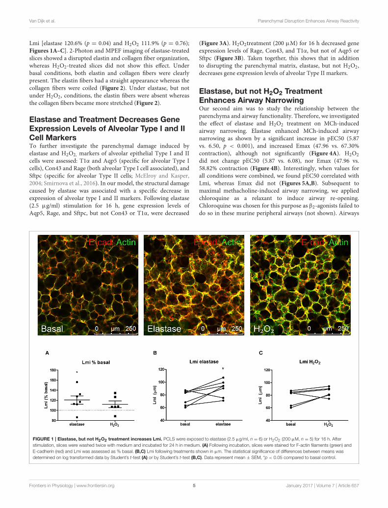
Mean Linear Intercept (Lmi)To assess emphysema in the PCLS, the mean linear intercept(Lmi) was determined as a measure of mean distance of freeairspace, as described previously (van der Strate et al., 2006).Following staining with Alexa Fluor R© 488 Phalloidin (1:100, Lifetechnologies) the alveolar structure was visualized by confocalmicroscopy (magnification 200x). Two fields per animal wereused to determine Lmi. As the lungs were filled with agaroseunder a varying pressure, Lmi differs between animals due tothe experimental protocol. However, in this PCLS model animalsserved as their own control as all experimental conditions wereperformed within the same animal. Hence, the treatment effecton Lmi was normalized (percent basal) within the animal,and these normalized values were compared between animals.Therefore, the variance in Lmi caused by the experimentalprotocol was excluded.
Airway Narrowing StudiesAirway narrowing studies were performed within the samemouse on untreated slices and on slices treated with elastase(2.5 µg/ml) or H2O2 (200 µM). Dose response curves for
MCh (10−9–10−3M) were recorded, after which the airwayswere dilated using the bitter taste receptor agonist chloroquine(10−3M, Sigma-Aldrich). A nylon mesh and metal washer wereused to fixate the lung slice, as described previously (Rosner et al.,2014). Lung slice images were captured in time-lapse (1 frameper 2 s) using a microscope (Eclipse, TS100; Nikon). To quantifyairway luminal area, image acquisition software (NIS-elements;Nikon) was used. Luminal area is expressed as percent basal.
Data Analysis (Statistics)Values reported for all data are represented as mean ± SEM.The statistical significance of differences between means wasdetermined on log transformed data by Student’s t-test orone-way ANOVA, followed by a Bonferroni correction whereappropriate. Log-transformation was performed prior to thestatistic calculations when data were normalized to percentage ofbaseline. While such normalization can be useful to demonstratean effect of treatment relative to control, it distorts Gaussiandistribution of data. Log transformation of is then necessaryto re-obtain Gaussian distribution and parametric testing is insuch cases allowed. Differences were considered to be statisticallysignificant when p < 0.05.
RESULTS
Elastase, but Not H2O2 Treatment Disruptsthe ParenchymaOur first aim was to create tissue damage in the PCLS resemblingthe tissue damage seen in COPD. Therefore, we first investigatedwhether elastase and H2O2 were able to induce parenchymaltissue damage. PCLS were treated with increasing concentrationsof elastase (0, 0.16, 0.31, 0.63, 1.25, and 2.5 µg/ml) and H2O2
(0, 50, 100, 200, 400, 800 µM) and ECM and alveolar epithelialmarkers were assessed. Based on these initial results (data notshown due to a low n), 2.5 µg/ml elastase and 200 µM of H2O2
were chosen for all the following experiments. Following elastasetreatment gene expression levels of Sftpc decreased (moderately),with little difference between the dosage used. The effect ofelastase treatment on ECM marker gene expression levels wasrather variable. Confocal images of the parenchymal structuredemonstrated that only the highest dose of 2.5 µg/ml elastaseenhanced Lmi. Based on these observations, we chose to workwith 2.5 µg/ml elastase in further experiments. H2O2 treatmentinduced small increases or decreases of Sftpc and ECM markers,depending on the concentration. The highest decrease in Sftpcand ECM markers was observed at the highest dose of 800 µM.However, at this dose we observed visually that the PCLS werevery fragile and too disintegrated to use. Therefore, the effectof H2O2 800 µM treatment on Sftpc and ECM gene expressionlevels is more likely to reflect general cell death than a specificeffect of H2O2. Based on these findings we chose to work with 200µM H2O2, as these slices showed only small decreases in Sftpcand ECM gene expression levels and were still intact. To assessemphysema in the PCLS, the Lmi was determined as a measure ofmean distance of free airspace. Following elastase, but not H2O2
stimulation, the mean free distance in air spaces was increasedas compared to basal (set at 100%) as reflected by an enhanced
Frontiers in Physiology | www.frontiersin.org 4 January 2017 | Volume 7 | Article 657
Van Dijk et al. Parenchymal Disruption Enhances Airway Reactivity
Lmi [elastase 120.6% (p = 0.04) and H2O2 111.9% (p = 0.76);Figures 1A–C]. 2-Photon and MPEF imaging of elastase-treatedslices showed a disrupted elastin and collagen fiber organization,whereas H2O2-treated slices did not show this effect. Underbasal conditions, both elastin and collagen fibers were clearlypresent. The elastin fibers had a straight appearance whereas thecollagen fibers were coiled (Figure 2). Under elastase, but notunder H2O2, conditions, the elastin fibers were absent whereasthe collagen fibers became more stretched (Figure 2).
Elastase and Treatment Decreases GeneExpression Levels of Alveolar Type I and IICell MarkersTo further investigate the parenchymal damage induced byelastase and H2O2, markers of alveolar epithelial Type I and IIcells were assessed: T1α and Aqp5 (specific for alveolar Type Icells), Con43 and Rage (both alveolar Type I cell associated), andSftpc (specific for alveolar Type II cells; McElroy and Kasper,2004; Smirnova et al., 2016). In our model, the structural damagecaused by elastase was associated with a specific decrease inexpression of alveolar type I and II markers. Following elastase(2.5 µg/ml) stimulation for 16 h, gene expression levels ofAqp5, Rage, and Sftpc, but not Con43 or T1α, were decreased
(Figure 3A). H2O2treatment (200 µM) for 16 h decreased geneexpression levels of Rage, Con43, and T1α, but not of Aqp5 orSftpc (Figure 3B). Taken together, this shows that in additionto disrupting the parenchymal matrix, elastase, but not H2O2,decreases gene expression levels of alveolar Type II markers.
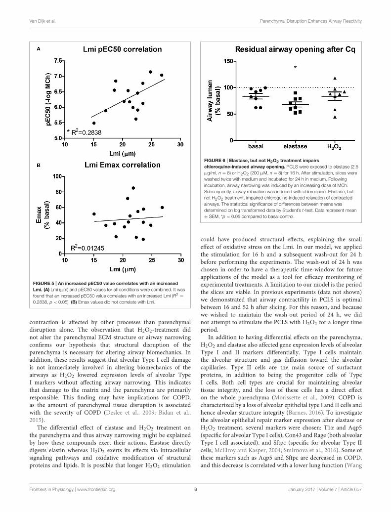
Elastase, but not H2O2 TreatmentEnhances Airway NarrowingOur second aim was to study the relationship between theparenchyma and airway functionality. Therefore, we investigatedthe effect of elastase and H2O2 treatment on MCh-inducedairway narrowing. Elastase enhanced MCh-induced airwaynarrowing as shown by a significant increase in pEC50 (5.87vs. 6.50, p < 0.001), and increased Emax (47.96 vs. 67.30%contraction), although not significantly (Figure 4A). H2O2
did not change pEC50 (5.87 vs. 6.08), nor Emax (47.96 vs.58.82% contraction (Figure 4B). Interestingly, when values forall conditions were combined, we found pEC50 correlated withLmi, whereas Emax did not (Figures 5A,B). Subsequent tomaximal methacholine-induced airway narrowing, we appliedchloroquine as a relaxant to induce airway re-opening.Chloroquine was chosen for this purpose as β2-agonists failed todo so in these murine peripheral airways (not shown). Airways
FIGURE 1 | Elastase, but not H2O2 treatment increases Lmi. PCLS were exposed to elastase (2.5 µg/ml, n = 6) or H2O2 (200 µM, n = 5) for 16 h. After
stimulation, slices were washed twice with medium and incubated for 24 h in medium. (A) Following incubation, slices were stained for F-actin filaments (green) and
E-cadherin (red) and Lmi was assessed as % basal. (B,C) Lmi following treatments shown in µm. The statistical significance of differences between means was
determined on log transformed data by Student’s t-test (A) or by Student’s t-test (B,C). Data represent mean ± SEM, *p < 0.05 compared to basal control.
Frontiers in Physiology | www.frontiersin.org 5 January 2017 | Volume 7 | Article 657

Van Dijk et al. Parenchymal Disruption Enhances Airway Reactivity
FIGURE 2 | Elastase, but not H2O2 treatment disrupts the structural organization of elastin and collagen. PCLS were exposed to elastase (2.5 µg/ml) or
H2O2 (200 µM) for 16 h. After stimulation, slices were washed twice with medium and incubated for 24 h in medium. 2-Photon and multiphoton excitation
fluorescence (MPEF) imaging were used to visualize collagen and elastin polymers. Following elastase, but not H2O2, elastin and collagen showed a disrupted fiber
organization.
in slices treated with elastase did not fully return to their originalsize after chloroquine-induced relaxation, in contrast to slicestreated with H2O2 (Figure 6). Taken together, these findingsindicate that elastase, but not H2O2 treatment enhances airwaynarrowing in PCLS and limits airway re-opening to its originalsize.
DISCUSSION
COPD is characterized by progressive airflow limitation, and it isincreasingly recognized that the parenchymal compartment mayplay an important role in airway narrowing. Our aim was to setup an ex vivo PCLS model mimicking structural abnormalitiesthat resemble COPD and to integrate multiple readouts in orderto study their inter-relationship. Tissue damage was establishedin the PCLS with either elastase or H2O2. We show that exvivo elastase, but not H2O2, treatment leads to disruption ofthe parenchymal compartment and to enhanced MCh-inducedairway narrowing. Elastase-induced damage was reflected by anincreased Lmi and a disorganization of elastin and collagen fibers.The structural damage caused by elastase was associated with aspecific decrease in both alveolar Type I and II markers (Aqp5,Rage, and Sftpc) mRNA expression. H2O2 treatment decreasedgene expression levels of alveolar Type I cells (Rage, Con43,and T1α) only. As COPD is characterized by both alveolar Type
I and II injury, this result indicates that in the PCLS, elastasetreatment better reproduces tissue damage seen in COPD thanH2O2 treatment.
We observed a clear relationship between lung structure andairway function. This was demonstrated by the finding thatelastase treatment enhanced MCh-induced airway narrowing asreflected by an increased pEC50 and Emax values as comparedto control slices. However, a limitation of the PCLS model whenstudying lung structure and airway biomechanics is the lack ofpre-strain or cyclic stretching due to tidal breathing. The relativedistribution of forces within the lung has been proposed as amain player in tissue destruction, and hence is a key contributorto disease development (Kononov et al., 2001; Suki and Bates,2011; Yi et al., 2016). In vivo pre-strain and cyclic stretchingare likely able to enhance tissue degradation and are thereforelikely to influence lung tissue biomechanics. This should betaken into consideration when interpreting the findings of thepresented ex vivo PCLS model. The enhanced airway narrowingobserved in our model is unlikely due to a direct effect of elastaseon smooth muscle α-actin or the M3 receptor, as elastase didnot increase their gene expression levels (data not shown). Inaddition, an increased pEC50 value correlated with an increasedLmi. Furthermore, elastase treatment impaired chloroquine-induced airway re-opening. Interestingly, it has been shownthat in untreated mouse PCLS, chloroquine, but not the
Frontiers in Physiology | www.frontiersin.org 6 January 2017 | Volume 7 | Article 657

Van Dijk et al. Parenchymal Disruption Enhances Airway Reactivity
FIGURE 3 | Elastase and H2O2 treatment alter mRNA expression levels
of alveolar makers. PCLS were exposed to (A) elastase (2.5 µg/ml, n = 9) or
(B) H2O2 (200 µM, n = 14) for 16 h. After stimulation, slices were washed
twice with medium and incubated for 24 h in medium. The statistical
significance of differences between means was determined on log
transformed data by Student’s t-test followed by a Bonferroni correction. Data
represent mean ± SEM, *p < 0.05 compared to basal control.
β2-agonist salbutamol, is able tomaintain efficacy with increasingcontraction (Donovan et al., 2014). Therefore, whereas β2-agonists are sensitive to functional antagonism (Lemoine et al.,1992), this is not the case for chloroquine. This indicates thatthe impaired chloroquine-induced relaxation following elastasetreatment is more likely explained by as loss of elastic recoildue to the absence of functional elastin fibers than by functionalantagonism. The complete airway re-opening with choloroquinein H2O2 treated slices supports this. A previous study byKhan et al. demonstrated that ex vivo elastase treatment ofmouse lung slices enhanced the velocity of acetylcholine-inducedcontraction while the velocity of relaxation was significantlysuppressed (Khan et al., 2007). In their study, airway narrowingstudies using acetylcholine (ACh) were performed directly after16 h elastase incubation period. The finding by Khan et al.that elastase enhances contraction velocity is similar to ourobservation that elastase enhances airway narrowing after 16h incubation and additional 24 h washout period. Althoughunquantified, Khan et al. observed rupture of the parenchymal
FIGURE 4 | Elastase, but not H2O2 treatment enhances MCh-induced
airway narrowing. PCLS were exposed to elastase (2.5 µg/ml, n = 10) or
H2O2 (200 µM, n = 10) for 16 h. After stimulation, slices were washed twice
with medium and incubated for 24 h in medium. Following incubation,
MCh-induced airway narrowing was assessed. Lung slice images were
captured in time-lapse (1 frame per 2 s) using an inverted phase contrast
microscope (Eclipse, TS100; Nikon). Airway luminal area was quantified using
image acquisition software (NIS-elements; Nikon), and expressed as percent
basal. (A) MCh-induced airway narrowing following elastase treatment.
Elastase treatment increased pEC50 values significantly (p < 0.05, compared
to basal control). (B) MCh-induced airway narrowing following H2O2 treament.
The statistical significance of differences between means was determined on
log transformed data by one-way ANOVA followed by Bonferonni testing. Data
represent mean ± SEM.
tethering to the airway at the peak of contraction followingelastase treatment. These results fit with our data showingthat increased Lmi correlates with increased pEC50 values, andimplies that structural damage is the cause of enhanced airwaynarrowing at both the 16 and 40 h time point. Furthermore, inanother study Khan et al. demonstrated that 16 h treatment ofPCLS with collagenase increased velocity of ACh-induced airwaycontraction to a similar extent as elastase, whereas relaxationvelocities were affected to a lesser extent (Khan et al., 2010).Together, the findings in the studies by Khan et al. and the presentstudy indicate that parenchymal damage indeed enhances airwaynarrowing, and that PCLS are a suitable model to study thisinteraction.
In contrast to elastase, H2O2 did not alter Lmi, the pEC50value or chloroquine-induced airway opening. Collectively, theabove-mentioned results show that elastase, and to a lesserextent H2O2, treatment of PCLS induces tissue damage similarto hallmarks of COPD. In addition, these results indicate thatparenchymal disruption is linked to an increased sensitivity ofthe airways to MCh-induced contraction, whereas the maximal
Frontiers in Physiology | www.frontiersin.org 7 January 2017 | Volume 7 | Article 657
Van Dijk et al. Parenchymal Disruption Enhances Airway Reactivity
FIGURE 5 | An increased pEC50 value correlates with an increased
Lmi. (A) Lmi (µm) and pEC50 values for all conditions were combined. It was
found that an increased pEC50 value correlates with an increased Lmi (R2 =
0.2838, p < 0.05). (B) Emax values did not correlate with Lmi.
contraction is affected by other processes than parenchymaldisruption alone. The observation that H2O2-treatment didnot alter the parenchymal ECM structure or airway narrowingconfirms our hypothesis that structural disruption of theparenchyma is necessary for altering airway biomechanics. Inaddition, these results suggest that alveolar Type I cell damageis not immediately involved in altering biomechanics of theairways as H2O2 lowered expression levels of alveolar TypeI markers without affecting airway narrowing. This indicatesthat damage to the matrix and the parenchyma are primarilyresponsible. This finding may have implications for COPD,as the amount of parenchymal tissue disruption is associatedwith the severity of COPD (Deslee et al., 2009; Bidan et al.,2015).
The differential effect of elastase and H2O2 treatment onthe parenchyma and thus airway narrowing might be explainedby how these compounds exert their actions. Elastase directlydigests elastin whereas H2O2 exerts its effects via intracellularsignaling pathways and oxidative modification of structuralproteins and lipids. It is possible that longer H2O2 stimulation
FIGURE 6 | Elastase, but not H2O2 treatment impairs
chloroquine-induced airway opening. PCLS were exposed to elastase (2.5
µg/ml, n = 8) or H2O2 (200 µM, n = 8) for 16 h. After stimulation, slices were
washed twice with medium and incubated for 24 h in medium. Following
incubation, airway narrowing was induced by an increasing dose of MCh.
Subsequently, airway relaxation was induced with chloroquine. Elastase, but
not H2O2 treatment, impaired chloroquine-induced relaxation of contracted
airways. The statistical significance of differences between means was
determined on log transformed data by Student’s t-test. Data represent mean
± SEM, *p < 0.05 compared to basal control.
could have produced structural effects, explaining the smalleffect of oxidative stress on the Lmi. In our model, we appliedthe stimulation for 16 h and a subsequent wash-out for 24 hbefore performing the experiments. The wash-out of 24 h waschosen in order to have a therapeutic time-window for futureapplications of the model as a tool for efficacy monitoring ofexperimental treatments. A limitation to our model is the periodthe slices are viable. In previous experiments (data not shown)we demonstrated that airway contractility in PCLS is optimalbetween 16 and 52 h after slicing. For this reason, and becausewe wished to maintain the wash-out period of 24 h, we didnot attempt to stimulate the PCLS with H2O2 for a longer timeperiod.
In addition to having differential effects on the parenchyma,H2O2 and elastase also affected gene expression levels of alveolarType I and II markers differentially. Type I cells maintainthe alveolar structure and gas diffusion toward the alveolarcapillaries. Type II cells are the main source of surfactantproteins, in addition to being the progenitor cells of TypeI cells. Both cell types are crucial for maintaining alveolartissue integrity, and the loss of these cells has a direct effecton the whole parenchyma (Morissette et al., 2009). COPD ischaracterized by a loss of alveolar epithelial type I and II cells andhence alveolar structure integrity (Barnes, 2016). To investigatethe alveolar epithelial repair marker expression after elastase orH2O2 treatment, several markers were chosen: T1α and Aqp5(specific for alveolar Type I cells), Con43 and Rage (both alveolarType I cell associated), and Sftpc (specific for alveolar Type IIcells; McElroy and Kasper, 2004; Smirnova et al., 2016). Some ofthese markers such as Aqp5 and Sftpc are decreased in COPD,and this decrease is correlated with a lower lung function (Wang
Frontiers in Physiology | www.frontiersin.org 8 January 2017 | Volume 7 | Article 657

Van Dijk et al. Parenchymal Disruption Enhances Airway Reactivity
et al., 2007; Zhao et al., 2014). Following injury of alveolarepithelial cells, the level of expression of alveolar markers maychange because of an altered regulation or because of the death ofcells expressing the marker. Although both treatments caused adecrease in themRNA expression levels of alveolar type I markers(Rage, Con43, T1α), only elastase treatment reduced the alveolarType II marker Sftpc. Alveolar Type II cells are secretory cells,which release components of surfactant and ECM (Fehrenbach,2001). In addition, they also release the extracellular antioxidantsGpx3 and Sod3 (Folz et al., 1997; Burk et al., 2011; Yamadaet al., 2012). H2O2 may not alter Sftpc gene expression levelsbecause of the antioxidant properties of the alveolar Type IIcells, thereby explaining the different effect of elastase and H2O2
on alveolar maker expression levels. A limitation of this studyis that we did not assess PCLS reactivity directly following 16h of treatment. Hence, we cannot exclude the possibility thatH2O2 had an effect on gene expression levels at this earlier timepoint that was lost following 24 h incubation. However, as weaim to use this model for monitoring experimental treatmentsin the future we are interested in creating a model with stableparenchymal disruption. The effect of elastase on expressionlevels of alveolar Type I and II markers may be explained viaseveral mechanisms. First, elastase-induced elastin fragments areknown chemokines for macrophages in an adult murine modelof emphysema, and elastin fragments can impair alveologenesis(Houghton et al., 2006; Masood et al., 2015). Second, mechanicalstimuli are well-known to affect proliferation and differentiationof stem cells (Shah et al., 2014). As elastase disrupts theparenchyma, it reorganizes the distribution of mechanical forceswithin the tissue and may therefore affect the differentiationof alveolar Type II cells. Together, these mechanisms couldexplain how elastase is capable of affecting alveolar markerexpression levels. Collectively, the above-mentioned resultsindicate that elastase-treated PCLS serve as a better modelto study the specific relationship between parenchymaldisruption and airway narrowing than H2O2-treatedPCLS.
COPD is a progressive disease, and currently no treatmentis capable of stopping or reversing the progression of lungdecline. Novel therapeutic targets are clearly needed, and theparenchyma is a good candidate. Recently, there has beenincreasing recognition of the potential importance of theparenchymal compartment and its role in lung tissue mechanicsin many lung diseases, including COPD (Suki and Bates, 2011;Bidan et al., 2015). In COPD, the parenchyma and ECM arealtered (Eurlings et al., 2014; Bidan et al., 2015). The mostabundant changes in the ECM are reductions in the expressionor functional organization of elastin fiber, leading to a loss ofelastic recoil. Alterations in elastin expression are already presentin mild to moderate COPD, and seen in both airways andalveoli of COPD patients (Black et al., 2008). Furthermore, thisreduction in elastin expression seems to be similar in mild tomoderate and severe COPD (Eurlings et al., 2014). Interestingly,alveolar wall elastin fiber structure is altered in patients withsevere COPD. Compared to healthy subjects, elastin fibers fromCOPD patients are significantly less densely packed, unraveledand loose (Deslee et al., 2009). This indicates that, even though
elastin expression is similar in both mild to moderate andsevere COPD, the disruption of the structural organization ofelastin might contribute to the continuous decline of elasticrecoil observed in small airways and parenchyma of patientswith COPD (Bidan et al., 2015). The same appears to be thecase for collagen in COPD. Studies about the total expressionlevels of collagen in COPD are inconsistent, but it has beenobserved that collagen fibers are more disorganized in severeCOPD as compared tomild tomoderate COPD (Tjin et al., 2014).Furthermore, recent studies demonstrate that expression of genesassociated with elastogenesis is altered in COPD (Brandsmaet al., 2015, 2016). Among the most upregulated genes werefibulin-5 (FBLN5), elastin (ELN), latent transforming growthfactor β binding protein 2 (LTBP2), and microfibrillar associatedprotein 4 (MFAP4), which are all implicated in elastogenesis.In addition to elevated gene expression levels of FBLN5this study demonstrated that cleaved, possibly non-functionalFBLN5 protein was present in COPD lung tissue, indicatingan impaired repair response. Targeting these elastogenesispathways in COPD may therefore represent a novel therapeutictarget.
Disorganization of elastin and a changed organization ofcollagen fibers were also observed in our PCLS model followingtreatment with elastase, demonstrating that elastase treatmentmimics the damage seen in COPD lung tissue. Under basalconditions, both elastin and collagen fibers were present.Following elastase treatment, the elastin fibers were lost andthe collagen fibers became more stretched. The importance ofECM fiber structure is demonstrated by the finding that tissuestrips from elastase-treated mice failed at ∼50% less stress thancontrol animals, even though the treated animals had a 50%increase in total collagen content of the lung (Ito et al., 2005).These results agree with our finding that collagen fibers becomemore stretched following elastase treatment; if collagen isalready stretched maximally, it will break at a lower deformationcompared to control. Moreover, due to structural remodeling,the yield stress (breaking point) of collagen is lower in theemphysematous lung (Suki and Bates, 2008). A lower yield stressof collagen also means a lower resistance to airway contraction,explaining why structural remodeling of collagen can lead toan enhanced airway narrowing, even though total collagenexpression levels are increased and Lmi is unchanged. Takentogether, these reported findings show that the ECM in the airwayand parenchymal compartments of COPD patients is alteredas compared to non-COPD controls. One of the most evidentalterations is the changed expression level and disorganizationof elastin and collagen fibers. In our PCLS model, elastasetreatment increased the Lmi, disrupted the fiber organizationof elastin and collagen, and enhanced methacholine-inducedairway narrowing as reflected by an increased pEC50 value.Importantly, increased Lmi values correlated with increasedpEC50 values. Furthermore, elastase treatment impairedchloroquine-induced airway re-opening. Taken together,the above-mentioned findings stress the importance of thecontribution of the parenchymal ECM structure to airwaynarrowing. It is likely that the enhanced bronchoconstrictionin COPD is at least partly caused by the parenchymal tissue
Frontiers in Physiology | www.frontiersin.org 9 January 2017 | Volume 7 | Article 657

Van Dijk et al. Parenchymal Disruption Enhances Airway Reactivity
damage. This role of the parenchymal compartment in airwaynarrowing is especially interesting in mild or moderate COPD,as changes in the parenchymal compartment are not veryprofound yet in stage, and possibly still reversible. Therefore, theparenchyma represents a promising target in the treatment ofCOPD.
In summary, these results demonstrate that PCLS can beused to model structural defects in COPD in a comprehensivemanner. To our knowledge, this is the first study integratingmultiple read-out parameters such as the parenchyma, epithelialrepair marker expression and airway function in one model.Using this model, we show that structural disruption of theparenchymal compartment leads to altered biomechanics ofthe airways, enhancing airway contraction. This finding mayhave implications for COPD, as the amount of parenchymaltissue disruption is associated with the severity of the disease.Therefore, we suggest that the parenchymal tissue damageobserved in COPD contributes to lung function decline bydisrupting airway biomechanics. Targeting the parenchymalcompartment may therefore be a promising therapeutic target inthe treatment of COPD.
AUTHOR CONTRIBUTIONS
EV, CB, and RG contributed to the conception or design ofthe work. EM, SC, and MM contributed to the acquisition andanalysis of data. All authors contributed to the interpretationof data. All authors drafted the work or revised it critically forimportant intellectual content. All authors approved the finalversion of the manuscript.
FUNDING
We would like to thank the Netherlands Organization forScientific Research and the Netherlands Lung Foundationfor financial support (Vidi grant: 016.116.309 and 6.1.14.009,respectively).
ACKNOWLEDGMENTS
We would like to thank Klaas Sjollema (University MedicalCenter Groningen) for providing help with the confocal, 2-photon, and multiphoton imaging.
REFERENCES
Abraham, T., and Hogg, J. (2010). Extracellular matrix remodeling of lung
alveolar walls in three dimensional space identified using second harmonic
generation and multiphoton excitation fluorescence. J. Struct. Biol. 171,
189–196. doi: 10.1016/j.jsb.2010.04.006
An, S. S., Bai, T. R., Bates, J. H., Black, J. L., Brown, R. H., Brusasco, V., et al. (2007).
Airway smooth muscle dynamics: a common pathway of airway obstruction in
asthma. Eur. Respir. J. 29, 834–860. doi: 10.1183/09031936.00112606
Barnes, P. J. (2016). Inflammatory mechanisms in patients with chronic
obstructive pulmonary disease. J. Allergy Clin. Immunol. 138, 16–27.
doi: 10.1016/j.jaci.2016.05.011
Barnes, P. J., Shapiro, S. D., and Pauwels, R. A. (2003). Chronic obstructive
pulmonary disease: molecular and cellular mechanisms. Eur. Respir. J. 22,
672–688. doi: 10.1183/09031936.03.00040703
Bidan, C. M., Veldsink, A. C., Meurs, H., and Gosens, R. (2015). Airway
and extracellular matrix mechanics in COPD. Front. Physiol. 6:346.
doi: 10.3389/fphys.2015.00346
Black, P. N., Ching, P. S., Beaumont, B., Ranasinghe, S., Taylor, G., and Merrilees,
M. J. (2008). Changes in elastic fibres in the small airways and alveoli in COPD.
Eur. Respir. J. 31, 998–1004. doi: 10.1183/09031936.00017207
Brandsma, C. A., van den Berge, M., Postma, D. S., Jonker, M. R., Brouwer,
S., Paré, P. D., et al. (2015). A large lung gene expression study identifying
fibulin-5 as a novel player in tissue repair in COPD. Thorax 70, 21–32.
doi: 10.1136/thoraxjnl-2014-205091
Brandsma, C. A., van den Berge, M., Postma, D., and Timens, W. (2016). Fibulin-
5 as a potential therapeutic target in COPD. Expert Opin. Ther. Targets 20,
1031–1033. doi: 10.1517/14728222.2016.1164696
Buhl, R., Maltais, F., Abrahams, R., Bjermer, L., Derom, E., Ferguson, G., et al.
(2015). Author correction. Tiotropium and olodaterol fixed-dose combination
versus mono-components in COPD (GOLD 2-4). Eur. Respir. J. 45, 1763.
doi: 10.1183/09031936.50136014
Burk, R. F., Olson, G. E., Winfrey, V. P., Hill, K. E., and Yin, D. (2011).
Glutathione peroxidase-3 produced by the kidney binds to a population
of basement membranes in the gastrointestinal tract and in other tissues.
Am. J. Physiol. Gastrointest. Liver Physiol. 301, G32–G38. doi: 10.1152/ajpgi.
00064.2011
Chung, K. F., and Adcock, I. M. (2008). Multifaceted mechanisms in COPD:
inflammation, immunity, and tissue repair and destruction. Eur. Respir. J. 31,
1334–1356. doi: 10.1183/09031936.00018908
COPD (2016).Anonymous From the Global Strategy for the Diagnosis, Management
and Prevention of COPD, Global Initiative for Chronic Obstructive Lung Disease
(GOLD) 2016. Available online at: http://goldcopd.org/
Deslee, G., Woods, J. C., Moore, C. M., Liu, L., Conradi, S. H., Milne, M., et al.
(2009). Elastin expression in very severe human COPD. Eur. Respir. J. 34,
324–331. doi: 10.1183/09031936.00123008
Donovan, C., Simoons, M., Esposito, J., Ni Cheong, J., Fitzpatrick, M., and
Bourke, J. E. (2014). Rosiglitazone is a superior bronchodilator compared to
chloroquine and beta-adrenoceptor agonists in mouse lung slices. Respir. Res.
15:29. doi: 10.1186/1465-9921-15-29
Eurlings, I. M., Dentener,M. A., Cleutjens, J. P., Peutz, C. J., Rohde, G. G.,Wouters,
E. F., et al. (2014). Similar matrix alterations in alveolar and small airway walls
of COPD patients. BMC Pulm. Med. 14:90. doi: 10.1186/1471-2466-14-90
Fehrenbach, H. (2001). Alveolar epithelial type II cell: defender of the alveolus
revisited. Respir. Res. 2, 33–46. doi: 10.1186/rr36
Folz, R. J., Guan, J., Seldin, M. F., Oury, T. D., Enghild, J. J., and Crapo, J. D. (1997).
Mouse extracellular superoxide dismutase: primary structure, tissue-specific
gene expression, chromosomal localization, and lung in situ hybridization. Am.
J. Respir. Cell Mol. Biol. 17, 393–403. doi: 10.1165/ajrcmb.17.4.2826
Houghton, A. M., Quintero, P. A., Perkins, D. L., Kobayashi, D. K., Kelley,
D. G., Marconcini, L. A., et al. (2006). Elastin fragments drive disease
progression in a murine model of emphysema. J. Clin. Invest. 116, 753–759.
doi: 10.1172/JCI25617
Ito, S., Ingenito, E. P., Brewer, K. K., Black, L. D., Parameswaran, H., Lutchen, K.
R., et al. (2005). Mechanics, nonlinearity, and failure strength of lung tissue in
a mouse model of emphysema: possible role of collagen remodeling. J. Appl.
Physiol. 98, 503–511. doi: 10.1152/japplphysiol.00590.2004
Khan, M. A., Ellis, R., Inman, M. D., Bates, J. H., Sanderson, M. J., and Janssen, L.
J. (2010). Influence of airway wall stiffness and parenchymal tethering on the
dynamics of bronchoconstriction. Am. J. Physiol. Lung Cell. Mol. Physiol. 299,
L98–L108. doi: 10.1152/ajplung.00011.2010
Khan, M. A., Kianpour, S., Stämpfli, M. R., and Janssen, L. J. (2007). Kinetics of in
vitro bronchoconstriction in an elastolytic mouse model of emphysema. Eur.
Respir. J. 30, 691–700. doi: 10.1183/09031936.00025907
Kirkham, P. A., and Barnes, P. J. (2013). Oxidative stress in COPD. Chest 144,
266–273. doi: 10.1378/chest.12-2664
Kononov, S., Brewer, K., Sakai, H., Cavalcante, F. S., Sabayanagam, C. R., Ingenito,
E. P., et al. (2001). Roles of mechanical forces and collagen failure in the
development of elastase-induced emphysema. Am. J. Respir. Crit. Care Med.
164 (10 Pt 1), 1920–1926. doi: 10.1164/ajrccm.164.10.2101083
Frontiers in Physiology | www.frontiersin.org 10 January 2017 | Volume 7 | Article 657

Van Dijk et al. Parenchymal Disruption Enhances Airway Reactivity
Lauzon, A. M., Bates, J. H., Donovan, G., Tawhai, M., Sneyd, J., and
Sanderson,M. J. (2012). Amulti-scale approach to airway hyperresponsiveness:
from molecule to organ. Front. Physiol. 3:191. doi: 10.3389/fphys.2012.
00191
Lemoine, H., Overlack, C., Köhl, A., Worth, H., and Reinhardt, D.
(1992). Formoterol, fenoterol, and salbutamol as partial agonists for
relaxation of maximally contracted guinea pig tracheae: comparison of
relaxation with receptor binding. Lung 170, 163–180. doi: 10.1007/BF001
74319
Ma, B., and Bates, J. H. (2014). Mechanical interactions between adjacent airways
in the lung. J. Appl. Physiol. 116, 628–634. doi: 10.1152/japplphysiol.011
80.2013
Masood, A., Yi, M., Belcastro, R., Li, J., Lopez, L., Kantores, C., et al. (2015).
Neutrophil elastase-induced elastin degradation mediates macrophage influx
and lung injury in 60% O2-exposed neonatal rats. Am. J. Physiol. Lung Cell.
Mol. Physiol. 309, L53–L62. doi: 10.1152/ajplung.00298.2014
McElroy, M. C., and Kasper, M. (2004). The use of alveolar epithelial type I
cell-selective markers to investigate lung injury and repair. Eur. Respir. J. 24,
664–673. doi: 10.1183/09031936.04.00096003
Morissette, M. C., Parent, J., and Milot, J. (2009). Alveolar epithelial and
endothelial cell apoptosis in emphysema: what we know and what we need to
know. Int. J. Chron. Obstruct. Pulmon. Dis. 4, 19–31. doi: 10.2147/COPD.S4432
Oenema, T. A., Maarsingh, H., Smit, M., Groothuis, G. M., Meurs, H.,
and Gosens, R. (2013). Bronchoconstriction induces TGF-beta release
and airway remodelling in guinea pig lung slices. PLoS ONE 8:e65580.
doi: 10.1371/journal.pone.0065580
Pauwels, R. A., Buist, A. S., Ma, P., Jenkins, C. R., Hurd, S. S., and GOLD
Scientific Committee (2001). Global strategy for the diagnosis, management,
and prevention of chronic obstructive pulmonary disease: National Heart,
Lung, and Blood Institute andWorld Health Organization Global Initiative for
Chronic Obstructive Lung Disease (GOLD): executive summary. Respir. Care
46, 798–825. doi: 10.1164/ajrccm.163.5.2101039
Rabe, K. F., Hurd, S., Anzueto, A., Barnes, P. J., Buist, S. A., Calverley, P.,
et al. (2007). Global strategy for the diagnosis, management, and prevention
of chronic obstructive pulmonary disease: GOLD executive summary. Am. J.
Respir. Crit. Care Med. 176, 532–555. doi: 10.1164/rccm.200703-456SO
Rosner, S. R., Ram-Mohan, S., Paez-Cortez, J. R., Lavoie, T. L., Dowell, M.
L., Yuan, L., et al. (2014). Airway contractility in the precision-cut lung
slice after cryopreservation. Am. J. Respir. Cell Mol. Biol. 50, 876–881.
doi: 10.1165/rcmb.2013-0166MA
Ruijter, J. M., Pfaffl, M. W., Zhao, S., Spiess, A. N., Boggy, G., Blom, J., et al.
(2013). Evaluation of qPCR curve analysis methods for reliable biomarker
discovery: bias, resolution, precision, and implications. Methods 59, 32–46.
doi: 10.1016/j.ymeth.2012.08.011
Ruijter, J. M., Ramakers, C., Hoogaars, W. M., Karlen, Y., Bakker, O., van
den Hoff, M. J., et al. (2009). Amplification efficiency: linking baseline and
bias in the analysis of quantitative PCR data. Nucleic Acids Res. 37:e45.
doi: 10.1093/nar/gkp045
Schindelin, J., Arganda-Carreras, I., Frise, E., Kaynig, V., Longair, M., Pietzsch, T.,
et al. (2012). Fiji: an open-source platform for biological-image analysis. Nat.
Methods 9, 676–682. doi: 10.1038/nmeth.2019
Shah, N., Morsi, Y., and Manasseh, R. (2014). From mechanical stimulation to
biological pathways in the regulation of stem cell fate. Cell Biochem. Funct. 32,
309–325. doi: 10.1002/cbf.3027
Smirnova, N. F., Schamberger, A. C., Nayakanti, S., Hatz, R., Behr, J., and
Eickelberg, O. (2016). Detection and quantification of epithelial progenitor
cell populations in human healthy and IPF lungs. Respir. Res. 17, 83.
doi: 10.1186/s12931-016-0404-x
Suki, B., and Bates, J. H. (2008). Extracellular matrix mechanics in
lung parenchymal diseases. Respir. Physiol. Neurobiol. 163, 33–43.
doi: 10.1016/j.resp.2008.03.015
Suki, B., and Bates, J. H. (2011). Lung tissue mechanics as
an emergent phenomenon. J. Appl. Physiol. 110, 1111–1118.
doi: 10.1152/japplphysiol.01244.2010
Tjin, G., Xu, P., Kable, S. H., Kable, E. P., and Burgess, J. K. (2014). Quantification
of collagen I in airway tissues using second harmonic generation. J. Biomed.
Opt. 195:36005. doi: 10.1117/1.JBO.19.3.036005
van der Strate, B. W., Postma, D. S., Brandsma, C. A., Melgert, B. N., Luinge,
M. A., Geerlings, M., et al. (2006). Cigarette smoke-induced emphysema:
a role for the B cell? Am. J. Respir. Crit. Care Med. 173, 751–758.
doi: 10.1164/rccm.200504-594OC
Vogelmeier, C. F., Bateman, E. D., Pallante, J., Alagappan, V. K., D’Andrea, P.,
Chen, H., et al. (2013). Efficacy and safety of once-daily QVA149 compared
with twice-daily salmeterol-fluticasone in patients with chronic obstructive
pulmonary disease (ILLUMINATE): a randomised, double-blind, parallel
group study. Lancet Respir. Med. 1, 51–60. doi: 10.1016/S2213-2600(12)70052-8
Wang, K., Feng, Y. L., Wen, F. Q., Chen, X. R., Ou, X. M., Xu, D., et al.
(2007). Decreased expression of human aquaporin-5 correlated with mucus
overproduction in airways of chronic obstructive pulmonary disease. Acta
Pharmacol. Sin. 28, 1166–1174. doi: 10.1111/j.1745-7254.2007.00608.x
Yamada, Y., Limmon, G. V., Zheng, D., Li, N., Li, L., Yin, L., et al. (2012). Major
shifts in the spatio-temporal distribution of lung antioxidant enzymes during
influenza pneumonia. PLoS ONE 7:e31494. doi: 10.1371/journal.pone.0031494
Yi, E., Sato, S., Takahashi, A., Parameswaran, H., Blute, T. A., Bartolák-
Suki, E., et al. (2016). Mechanical forces accelerate collagen digestion
by bacterial collagenase in lung tissue strips. Front. Physiol. 7:287.
doi: 10.3389/fphys.2016.00287
Zhao, R., Liang, X., Zhao, M., Liu, S. L., Huang, Y., Idell, S., et al. (2014).
Correlation of apical fluid-regulating channel proteins with lung function in
human COPD lungs. PLoS ONE 9:e109725. doi: 10.1371/journal.pone.0109725
Conflict of Interest Statement: The authors declare that the research was
conducted in the absence of any commercial or financial relationships that could
be construed as a potential conflict of interest.
Copyright © 2017 Van Dijk, Culha, Menzen, Bidan and Gosens. This is an open-
access article distributed under the terms of the Creative Commons Attribution
License (CC BY). The use, distribution or reproduction in other forums is permitted,
provided the original author(s) or licensor are credited and that the original
publication in this journal is cited, in accordance with accepted academic practice.
No use, distribution or reproduction is permitted which does not comply with these
terms.
Frontiers in Physiology | www.frontiersin.org 11 January 2017 | Volume 7 | Article 657