filtering for indels variant calling and · variant calling and filtering for indels erik garrison...
TRANSCRIPT

Variant calling and filtering for INDELs
Erik GarrisonSeqShop @ University of Michigan

Overview
1. Genesis of insertion/deletion (indel) polymorphism
2. Standard approaches to detecting indels3. Assembly-based indel detection4. Haplotype-based indel detection5. Primary filtering: Bayesian variant calling6. Post-call filtering: SVM7. Graph-based resequencing approaches

An INDEL
A mutation that results from the gain or loss of sequence.
AATTAGCCATTA
AATTA--CATTA

INDEL genesis
A number of processes are known to generate insertions and deletions in the process of DNA replication:
● Replication slippage● Double-stranded break repair● Structural variation (e.g. mobile element
insertions, CNVs)

DNA replication
http://www.stanford.edu/group/hopes/cgi-bin/wordpress/2011/02/all-about-mutations/

Polymerase slippage
http://www.stanford.edu/group/hopes/cgi-bin/wordpress/2011/02/all-about-mutations/

Insertions and deletions via slippage
Energetic signatures of single base bulges: thermodynamic consequences and biological implications. Minetti CA, Remeta DP, Dickstein R, Breslauer KJ - Nucleic Acids Res. (2009)

Double-stranded break repair
Possible anti-recombinogenic role of Bloom’s syndrome helicase in double-strand break processing. doi: 10.1093/nar/gkg834

NHEJ-derived indels
DNA Slippage Occurs at Microsatellite Loci without Minimal Threshold Length in Humans: A Comparative Genomic Approach. Leclercq S, Rivals E, Jarne P - Genome Biol Evol (2010)

Structural variation (SV)
Transposable elements (in this case, an Alu) are sequences that can copy and paste themselves into genomic DNA, causing insertions.
Deletions can also be mediated by these sequences via other processes.
http://www.nature.com/nrg/journal/v3/n5/full/nrg798.html

Overview
1. Genesis of insertion/deletion (indel) polymorphism
2. Standard approaches to detecting indels3. Assembly-based indel detection4. Haplotype-based indel detection5. Primary filtering: Bayesian variant calling6. Post-call filtering: SVM7. Graph-based resequencing approaches

Calling INDEL variation
Can we quickly design a process to detect indels from alignment data?
What are the steps you’d do to find the indel between these two sequences?

Indel finder
We could start by finding the long matches in both sequences at the start and end:

Indel finder
We can see this more easily like this:
CAAATAAGGTTTGGAGTTCTATTATACAAATAAGGTTTGGAAATTTTCTGGAGTTCTATTATA

Indel finder
The match structure implies that the sequence that doesn’t match was inserted in one sequence, or lost from the other.
CAAATAAGGTTT-----------GGAGTTCTATTATACAAATAAGGTTTGGAAATTTTCTGGAGTTCTATTATA
So that’s easy enough….

Something more complicated
These sequences are similar to the previous ones, but with different mutations between them.
They are still (kinda) homologous but it’s not easy to see.

Pairwise alignment
One solution, assuming a particular set of alignment parameters, has 3 indels and a SNP:
But if we use a higher gap-open penalty, things look different:

Alignment as interpretation
Different parameterizations can yield different results.
Different results suggest “different” variation.
What kind of problems can this cause? (And how can we mitigate these issues?)
First, let’s review standard calling approaches.

Standard variant calling approach
Genome (FASTA) Variation (VCF)
alignment andvariant calling
Reads (FASTQ)

Alignments to candidatesReference
Reads
Variant observations

The data exposed to the callerReference
Haplotype information is lost.

INDELs have multiple representations and require normalization for standard calling
Left alignment allows us to ensure that our representation is consistent across alignments and also variant calls.
https://www.biostars.org/p/66843/ user sa9

example: 1000G PhaseI low coveragechr15:81551110, ref:CTCTC alt:ATATA
ref: TGTCACTCGCTCTCTCTCTCTCTCTCTATATATATATATTTGTGCATalt: TGTCACTCGCTCTCTCTCTCTATATATATATATATATATTTGTGCAT
ref: TGTCACTCGCTCTCTCTCTCTCTCTCT------ATATATATATATTTGTGCATalt: TGTCACTCGCTCTCTCTCTCT------ATATATATATATATATATTTGTGCAT
Interpreted as 3 SNPs
Interpreted as microsatellite expansion/contraction

example: 1000G PhaseI low coveragechr20:708257, ref:AGC alt:CGA
ref: TATAGAGAGAGAGAGAGAGCGAGAGAGAGAGAGAGAGGGAGAGACGGAGTTalt: TATAGAGAGAGAGAGAGCGAGAGAGAGAGAGAGAGAGGGAGAGACGGAGTT
ref: TATAGAGAGAGAGAGAGAGC--GAGAGAGAGAGAGAGAGGGAGAGACGGAGTTalt: TATAGAGAGAGAGAGAG--CGAGAGAGAGAGAGAGAGAGGGAGAGACGGAGTT

Overview
1. Genesis of insertion/deletion (indel) polymorphism
2. Standard approaches to detecting indels3. Assembly-based indel detection4. Haplotype-based indel detection5. Primary filtering: Bayesian variant calling6. Post-call filtering: SVM7. Graph-based resequencing approaches

Problem: inconsistent indel representation makes alignment-based variant calling difficult
If alleles are represented in multiple ways, then to detect them correctly with a single-position based approach we need:1. An awesome normalization method2. Perfectly consistent filtering (so we represent
our entire context correctly in the calls)3. Highly-accurate reads

Solution: assembly and haplotype-driven detection
We can shift our focus from the specific interpretation in the alignments:- this is a SNP- whereas this is a series of indels… and instead focus on the underlying sequences.
Basically, we use the alignments to localize reads, then process them again with assembly approaches to determine candidate alleles.

Variant detection by assembly
Multiple methods have been developed by members of the 1000G analysis group:● Global joint assembly
○ cortex○ SGA (localized to 5 megabase chunks)
● Local assembly○ Platypus (+cortex)○ GATK HaplotypeCaller
● k-mer based detection○ FreeBayes (anchored reference-free windows)

Assembly
http://www.nature.com/nrmicro/journal/v7/n4/full/nrmicro2088.html

Using colored graphs (Cortex)
Variants can be called using bubbles in deBruijn graphs.
Method is completely reference-free, except for reporting of variants. The reference is threaded through the colored graph.
Many samples can be called at the same time.
from Iqbal et. al., "De novo assembly and genotyping of variants using colored de Bruijn graphs." (2012)

String graphs (SGA)
http://www.homolog.us/blogs/2012/02/13/string-graph-of-a-genome/
A string graph has the advantage of using less memory to represent an assembly than a de Bruijn graph. In the 1000G, SGA is run on alignments localized to ~5mb chunks.

Discovering alleles using graphs (GATK HaplotypeCaller)

Why don't we just assemble?
Assembly-based calls tend to have high specificity, but sensitivity suffers.
from Iqbal et. al., "De novo assembly and genotyping of variants using colored de Bruijn graphs." (2012)
The requirement of exact kmer matches means that errors disrupt coverage of alleles.
Existing assembly methods don't just detect point mutations--- they detect haplotypes.

Indel validation, 191 AFR samples
High-depth miSeq sequencing-based validation on 4 samples.Local assembly methods (BI2, BC, SI1)* have higher specificity than baseline mapping-based calls (BI1), but lower sensitivity. Global assembly (OX2) yielded very low error, but also low sensitivity.*The local assembly-based method Platypus (OX1) had a genotyping bug which caused poor performance.

Site-frequency spectrum, SNPs
phase3-like setchr20
*RepeatSeq is a microsatellite caller

Site-frequency spectrum, indels
phase3-like set chr20

Overview
1. Genesis of insertion/deletion (indel) polymorphism
2. Standard approaches to detecting indels3. Assembly-based indel detection4. Haplotype-based indel detection5. Primary filtering: Bayesian variant calling6. Post-call filtering: SVM7. Graph-based resequencing approaches

Finding haplotype polymorphisms
AGAACCCAGTGCTCTTTCTGCT
AGAACCCAGTGGTCTTTCTGCT
AGAACCCAGTG TCTTTCTGCTCG
AGAACCCAGTGCTCTATCTGCT
AGAACCCAGTG TCTGCTCTCTAGTCTT
Two reads
Their alignment
a SNP
Another readshowing a SNP on the same haplotype as the first
A variant locus implied by alignments
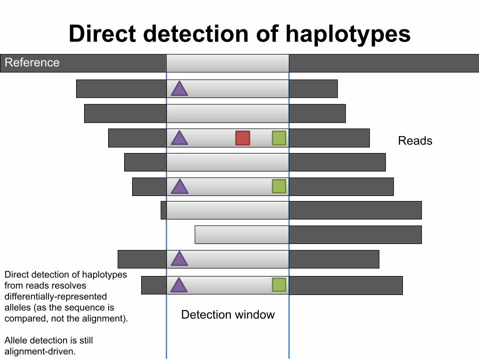
Direct detection of haplotypes
Detection window
Reference
Reads
Direct detection of haplotypes from reads resolves differentially-represented alleles (as the sequence is compared, not the alignment).
Allele detection is still alignment-driven.


Why haplotypes?
- Variants cluster.- This has functional significance.- Observing haplotypes lets us be more
certain of the local structure of the genome.- We can improve the detection process itself
by using haplotypes rather than point mutations.
- We get the sensitivity of alignment-based approaches with the specificity of assembly-based ones.

Sequence variants cluster
In ~1000 individuals, ½ of variants are within ~22bp of another variant.
Variance to mean ratio (VMR) = 1.4.

The functional effect of variants depends on other nearby variants on the same haplotype
AGG GAG CTGArg Glu Leu
reference:
AGG TAG CTGArg Ter ---
apparent:
AGG TTG CTGArg Leu Leu
actual:
OTOF gene – mutations cause profound recessive deafness
Apparent nonsense variant, one YRI homozygote
Actually a block substitution that results in a missense substitution
(Daniel MacArthur)

Importance of haplotype effects: frame-restoring indels
● Two apparent frameshift deletions in the CASP8AP2 gene (one 17 bp, one 1 bp) on the same haplotype
● Overall effect is in-frame deletion of six amino acids
(Daniel MacArthur)

Frame-restoring indels in1000 Genomes Phase I exomes
chr6:117113761, GPRC6A (~10% AF in 1000G)
chr6:32551935, HLA-DRB1 (~11% AF in 1000G)
ref: ATTGTAATTCTCA--TA--TT--TGCCTTTGAAAGCalt: ATTGTAATTCTCAGGTAATTTCCTGCCTTTGAAAGC
ref: CCACCGCGGCCCGCGCCTG-C-TCCAGGATGTCCAlt: CCACCGCGG--CGCGCCTGTCTTCCAGGAGGTCC

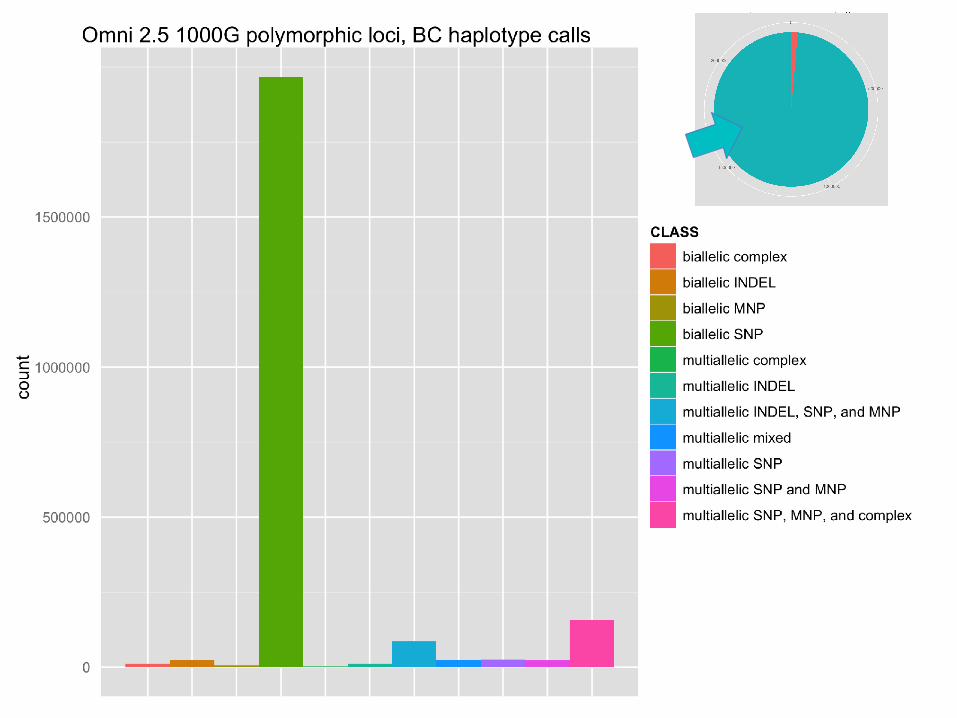
Impact on genotyping chip design
monomorphic loci
● Biallelic SNPs detected during the 1000 Genomes Pilot project were used to design a genotyping microarray (Omni 2.5).
● When the 1000 Genomes samples were genotyped using the chip, 100k of the 2.5 million loci showed no polymorphism (monomorphs).


Measuring haplotypes improves specificity
Indels from AFR191 sample set, 1000G phase2 testing.
Excess of 1bp insertions is driven by bubble artifacts in sequencing.

2bp MNPs and dinucleotide intermediates
reference
alte
rnat
e

Direct detection of haplotypes can remove directional bias associated with
alignment-based detection
CAGT
CGGC
TGAC
TGAC
CGGC
CAGT
CA/TG TG/CA
A→G transition to CpG intermediate
Deamination of methyl-C to T
Same process on opposite strand

Overview
1. Genesis of insertion/deletion (indel) polymorphism
2. Standard approaches to detecting indels3. Assembly-based indel detection4. Haplotype-based indel detection5. Primary filtering: Bayesian variant calling6. Post-call filtering: SVM7. Graph-based resequencing approaches

Filtering INDELs
As with SNPs, sequencing error rates are high.
So, we need to filter.
The standard filter of NGS is the Bayesian variant caller.
Combines population-based priors and data from many samples to make high-quality calls.

Bayesian (visual) intuition
Figures from http://oscarbonilla.com/2009/05/visualizing-bayes-theorem/
A = samples with a variant at some locus
We have a universe of individuals.
B = putative observations of variant at some locus

probability(A|B)
We want to estimate the probability that we have a real polymorphism "A" given "|" that we observed variants in our alignments "B".

In our case it's a bit more like this...
Observations (B) provide pretty good sensitivity, but poor specificity.

The model
● Bayesian model estimates the probability of polymorphism at a locus given input data and the population mutation rate (~pairwise heterozygosity) and assumption of “neutrality” (random mating).
● Following Bayes theorem, the probability of a specific set of genotypes over some number of samples is:○ P(G|R) = ( P(R|G) P(G) ) / P(R)
● Which in FreeBayes we extend to:○ P(G,S|R) = ( P(R|G,S) P(G)P(S) ) / P(R)○ G = genotypes, R = reads, S = locus is well-
characterized/mapped○ P(R|G,S) is our data likelihood, P(G) is our prior estimate
of the genotypes, P(S) is our prior estimate of the mappability of the locus, P(R) is a normalizer.

Handling non-biallelic/diploid casesWe compose our data likelihoods, P(Reads|Genotype) using a discrete multinomial sampling probability:
X
X
Our priors, P(Genoypes), follow the Ewens Sampling Formula and the discrete sampling probability for genotypes.

Are our locus and alleles sequenceable?In WGS, biases in the way we observe an allele (placement, position, strand, cycle, or balance in heterozygotes) are often correlated with error. We include this in our posterior P(G,S|R), and to do so we need an estimator of P(S).
neutral strand bias cycle bias placement bias
allele imbalance

The detection process
alignments
candidates
genotype likelihoods
haplotypes
sample posterior space
find maximuma posteriori genotyping
position
position
position
position
position
position
bayesian model
output record

Overview
1. Genesis of insertion/deletion (indel) polymorphism
2. Standard approaches to detecting indels3. Assembly-based indel detection4. Haplotype-based indel detection5. Primary filtering: Bayesian variant calling6. Post-call filtering: SVM7. Graph-based resequencing approaches
SVM filtering
INDEL detection is hard.
A priori models can’t capture all types of error.
It’s especially difficult when we try to make a consensus set from lots of input variant callers.
We can use classifiers like Support Vector Machines (SVM) to further improve results.

SVM classifier
Find a hyperplane (here a line in 2D) which separates observations.

SVM classifier
The best separating hyperplane is determined by maximum margin between groups we want to classify.

SVM filtering in the 1000 Genomes
25 human populations X ~100 samples each.

1000G variant integration process
* The filtering process I’ll discuss.
*

SVM approach for INDEL filtering
Extract features that tend to vary with respect to call quality:● call QUALity● read depth● sum of base qualities● inbreeding coefficient (heterozygosity)● entropy of sequence at locus● mapping quality● allele frequency in population● read pairing rate● etc.

SVM approach for INDEL filtering
Now, use overlaps in validation samples or sites to determine likely errors and true calls.
Use this list + annotations of the calls to train an SVM model.
Apply the model to all the calls, filter, and measure validation rate of the whole set.

Application of SVM to 1000G INDELs
Raw validation rates of indels in 1000G phase 3, “MVNCall” set.
Tony Marcketta and Adam Auton

Application of SVM to 1000G INDELs
Filtering results, using SVM-based method.
Anthony Marcketta and Adam Auton
Passing SVM Failing SVM

Application of SVM to 1000G INDELs
Correlation between allele frequency and observation counts.
Anthony Marcketta and Adam Auton
Passing SVM Failing SVM

Indel results from 1000G
Comparing the phase3 results to the genotypes for indels in the subset of samples for which we also had high-quality, high-coverage genomes from Complete Genomics.


Overview
1. Genesis of insertion/deletion (indel) polymorphism
2. Standard approaches to detecting indels3. Assembly-based indel detection4. Haplotype-based indel detection5. Primary filtering: Bayesian variant calling6. Post-call filtering: SVM7. Graph-based resequencing approaches

We know the variants,so why not use them in our analysis?
We resequence new genomes and compare them to a single reference haplotype.
To determine anything more than short variants, we must do everything de novo.
If we could merge sequence and variation, we could detect known alleles of arbitrary scale and divergence with minimal cost.

Pan-genomes as graphs
We can combine sequence and variation using a variant graphs, or graph reference.
*This representation is directed (5’ to 3’), and acyclic.

Building the variant graph
Deniz Kural, Boston College

Local alignment against the graph
Christopher Lee, Catherine Grasso, Mark F. Sharlow. Multiple sequence alignment using partial order graphs. Bioinformatics, 2002.

Local alignment against the graph
Deniz Kural, Boston College

“Striped” string/DAG alignment
We improved performance of our aligner >10-fold by generalizing Farrar’s striped Smith-Waterman algorithm to DAGs. GSSW
Farrar, Bioinformatics (2006); Rognes, BMC Bioinformatics (2011); Zhao, PLoS One (2014)
max of H, E vectors
copy H, E vectors
Data dependencies across DAG are limited to H and E vectors.
*Implemented using SSE2 instruction set.

Seeding graph-based alignments
linear reference x x x
? ?
graph reference
x x x
Test imperfectly-mapped reads against graph.

Detecting variation on the variant graph
G
A
AGCCTA
AGTACGTAGCT CCTATG GGCCAG
read supporting variant allele
agtacg ggccag
read supporting reference alleleagtacg cctatg ggtcag
Detecting variation on the graph

Graph-based alignments with glia
gliaBAM BAM freebayes
VCF VCFunion alleles with exact
breakpoints, used to build local graph
genotyped input alleles
1000G released
alignments (bwa)
**“flattened” into reference space,
with pseudo-reads of large insertions.

Application to 1000G variant integration
Brian D'Astous

Unifying calls from many methods
*Tests from 1000G “phase3-like” chr20.

realigned to variant graph
raw alignments

glia reduces reference bias
deletions insertions SNPs

glia reduces reference bias
Standard alignment is frustrated even by small variants!
Ratio between observations before and after realignment to graph of union variants
Improvement in observation support
dens
ity

Improving genotype likelihoods
Imputation of variant calls on chr20 via SHAPEIT 2. Imputed results are tested against Complete Genomics samples in 1000 Genomes.
We do as well for high-quality indels as SNPs!
SET GRP N RR RA AA ALT ALLSVM indels UM 6743 0.285 1.008 2.947 1.698 0.561SVM indels BC* 6743 0.034 0.673 0.245 0.521 0.129
SNPs BCM 404270 0.029 1.373 0.445 1.093 0.111
Olivier Delaneau, Androniki Menelaou, Jonathan Marchini
Genotype Likelihood = P(data|genotype)
* includes glia realignment
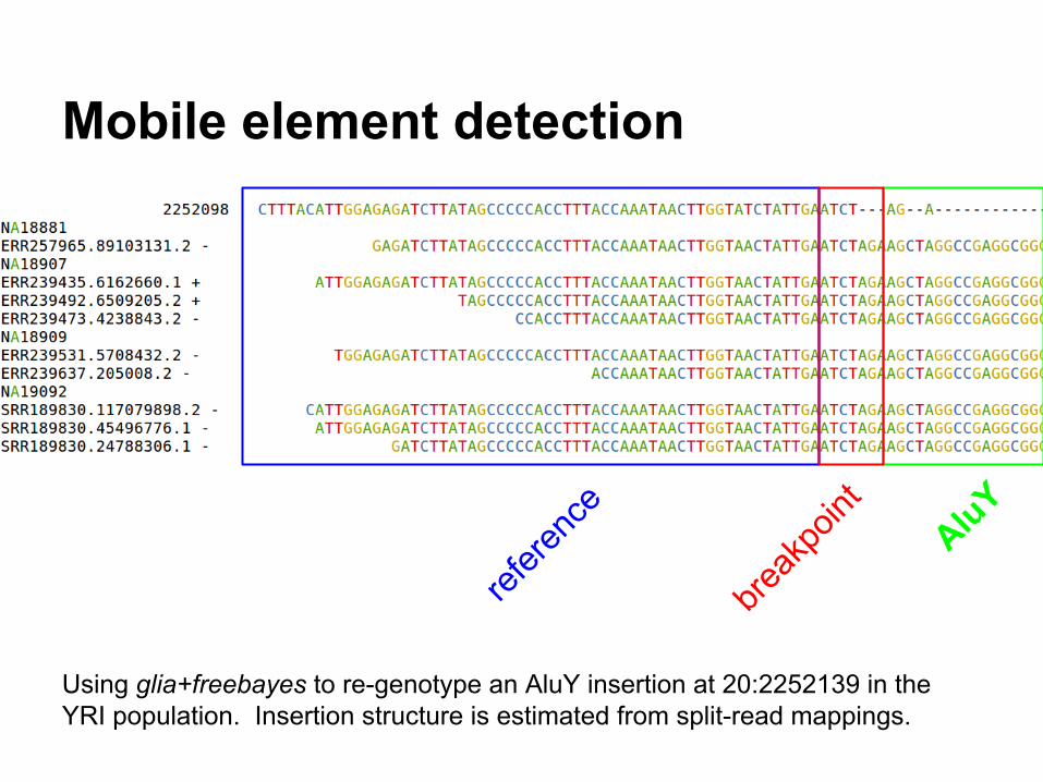
Mobile element detection
Using glia+freebayes to re-genotype an AluY insertion at 20:2252139 in the YRI population. Insertion structure is estimated from split-read mappings.
refere
nce
AluY
break
point

Alu genotyping efficiency
We re-call a substantial fraction of known (validated) Alus in 1000G low-coverage bwa alignments.
set re-genotyped Alus %
Pilot 2 data (source) 282 99.6
PCR-free NA12878 281 99.3
5x NA12878 (low-coverage) 173 61.1
Stewart et. al 2011. A Comprehensive Map of Mobile Element Insertion Polymorphisms in Humans. PLoS Genetics.

Genotyping large deletions
Input SVs were generated by DELLY on deep, PCR-free samples used for validation in the 1000 Genomes Project.
When using this set as our reference, we can regenotype around 70% of such events in low-coverage samples.

Performance using 1000G phase 3 SNPs and indels >1% frequency
depth snp AUC diff indel AUC diff
5 6.02% 1.87%
10 1.07% 0.78%
20 0.26% 0.37%
30 0.08% 0.40%
50 0.02% 1.2%
Deep-coverage 100bp Illumina data on NA12878 was downsampled to 5, 10, 20, 30, and 50-fold. Calling by both freebayes and freebayes+glia (realigning to 1000G variants >1% MAF), and comparing the results to the Genome In a Bottle truth set demonstrates marked improvement in sensitivity, particularly at low-coverage.

Questions?
…