general introduction to ecg - ecg.utah.edu · compute the 5 basic measurements: hr, pr interval,...
TRANSCRIPT
General Introduction to ECG
Reading Assignment (p2-16 in PDF ‘Outline’)
Objectives
1. Practice the 5-step ‘Method’
2. Differential Diagnosis: R & L axis deviation
3. Differential Diagnosis: Poor R-wave progression
4. Differential Diagnosis: Prominent Anterior Forces

Welcome to the “5-Step Method”
Mearurements: Rhythm (s): Conduction: Waveform: Interpretation:
A= V=
PR=
QRS=
QT=
Axis=
1. Compute the 5 basic measurements: HR, PR interval, QRS duration, QT interval, Axis
2. What’s the basic rhythm and other rhythm statements (e.g., PACs and PVC’s)
3. Any conduction abnormalities (SA blocks, AV blocks (Types I or II), and IV blocks
4. Waveform abnormalities beginning with P waves, QRS complexes, ST-T, and U waves
5. Final interpretations: Normal ECG or Borderline or Abnormal ECG (list final conclusions)
ECG #:

30 year old woman (explain the sequence of activation from sinus node to ventricular
muscle)
What are ‘septal’ q-waves?
1-1

Mearurements: Rhythm (s): Conduction: Waveform: Interpretation:
A=55 V=55 Normal Sinus rhythm Normal SA, AV, IV conduction
Sequence of conduction:• SA node → (RA→LA) →AV
node →His Bundle →RBB & LBB →LAF & LPF & LSF →Purkinje network →left septal surface (onset of QRS)
• Normal P, QRS, ST, T; notenormal U waves in precordial leads (*)
• Septal q‘s in II, III, aVF
(onset of ventricular activation
begins on the left ventricular
septal surface resulting in
small septal q-waves)
Normal ECG (septal q-waves normally seen in II, III, aVF in ECG‘s when the QRS axis is > +60; see arrows)PR=140
QRS=100
QT=430
Axis= +801-1
*
*
*
*

I
II
III
1-2
65 Year old womanWhere are the ‘septal’ q-waves?

I
II
III
Mearurements: Rhythm (s): Conduction: Waveform: Interpretation:
A=65 V=65 Sinus Rhythm Normal SA, AV, IV • Normal P, QRS, ST-T• Septal q-waves I, aVL (arrows)
(onset of ventricular activation
begins in the left ventricular
septal surface)
Normal ECG (septal q-waves are normally are seen in leads I, and aVL when the QRS axis is < +60)
PR=169
QRS=70
QT=380
Axis= +301-2
I
II
III
Age 22
1-3
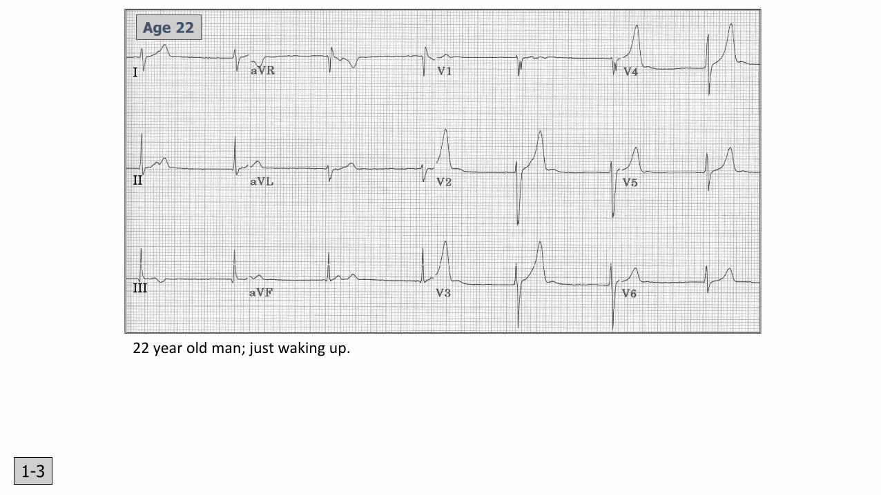
22 year old man; just waking up.

Mearurements: Rhythm (s): Conduction: Waveform: Interpretation:
A=48 V=48 Junctional escape rhythm
(Escape rhythms serve as backup pacemakers when the primary pacemaker gets too slow or when heart block prevents primary pacemaker from reaching the ventricles)
• Normal IV • Normal QRS, ST, T, U (*)• Retrograde P waves after the QRS in
the ST segment, best seen in II, III, aVF (arrows); it‘s like someone took a bite out of the T wave!
Note: normal U waves are best seen in leads V2-5 (*); these are the best leads to see U waves especially at slow heart rates.
Abnormal ECG (likely a normal variant in an athlete)1. Slight right axis deviation (can be
normal in 22 year old man)2. Junctional escape rhythm (probably
due to vagal slowing of the sinus rate in a healthy athlete; sinus rhythm would reappear after light exercise)
PR= none
QRS=90
QT=400
Axis= +100
I
II
III
Age 22
1-3
*
*
*

1-4
F, Age 27
27 year old woman; feeling anxious.

1-4
Mearurements: Rhythm (s): Conduction: Waveform: Interpretation:
A=110 V=110 Ectopic atrial tachycardia Normal AV, IV • Inverted P waves II, III, aVF;upright P waves in lead avR; (low atrial ectopic pacemaker)
• Normal QRS, ST, T waves
Note: In the horizontal plane (V1-6)
ectopic atrial P waves may look
normal in morphology; i.e., upright in
direction.
Abnormal ECG:1. Rhythm (this rhythm abnormality can be the result of various internal or external stress perturbations; e.g., hypoxia, stimulants, sepsis, et al.
Brief ectopic atrial rhythms (usually 3-6
beat runs) are common in otherwise
healthy people (may also occur in sick
individuals)
PR=120
QRS=80
QT=300
Axis= +10

Just an ordinary guy getting an insurance physical.
1-5

1-5
Mearurements: Rhythm (s): Conduction: Waveform: Interpretation:
A= 80 V=80 Sinus rhythm Normal SA, AV, slight IV conduction delay
• Normal P• rS in II, III, aVF (SIII >SII)
• Small q in I, aVL
• Delayed QRS transition in
horizontal plane (V5); note
persistent S waves in V5-6.
Abnormal ECG:1) Left anterior fascicular block (LAFB is
the most common IV conduction disorder)
The left bundle branches into two (sometimes three) fascicles: anterior, (septal), and posterior. (see pp 55-58 in the Outline)
PR= 120
QRS=110
QT=360
Axis= -60

I
II
III
1-6
F, Age 87 (sick and dehydrated)

I
II
III
1-6
Mearurements: Rhythm (s): Conduction: Waveform: Interpretation:
A=80 V=80 Sinus rhythm with 2 PACs (*)
Note: The PAC’s are early
beats with different P wave
morphology; the first PAC is
followed by a pause (longer RR
cycle)
Normal SA, AV, IV • Normal P, QRS• Slight ST depression V5-6• T inversion in III, V2-4
Abnormal ECG:1) Prolonged QT (upper limit @ 80 bpm
is ~380 ms); many etiologies to
consider!
2) Nonspecific ST-T abnormalities
(consider abnormal electrolytes,
drugs, various heart diseases, etc)
3) Rhythm: 2 PACs
PR=160
QRS=80
QT=480
Axis= -20
* *

V1-6: Differential Diagnoses
• Poor R-wave progression (small or no r-waves V1-3, + R:SV4 <1)
• Normal variant (esp. in women)
• Misplaced precordial leads
• Left ventricular hypertrophy
• Anterior and anteroseptal MI
• LBBB and incomplete LBBB
• Left anterior fascicular block
• Emphysema and COPD
• Some cases of WPW
• Diffuse infiltrative diseases
• Dextrocardia
• Prominent anterior forces (PAF: R:S V1-2 ≥1)
• Normal variant
• Misplaced precordial leads
• Right ventricular hypertrophy
• RBBB and incomplete RBBB
• ‘True Posterior’ (now called Lateral) MI
• Some cases of WPW
• Left septal fascicular block
• Muscular dystrophy

68 y.o. woman (History of hypertension on Rx)
1-7

1-7
Mearurements: Rhythm (s): Conduction: Waveform: Interpretation:
A=85 V=85 Sinus rhythm Normal SA, AV, IV • Increase P terminal force V1 (arrow)• Multiple voltage criteria for LVH• Poor R wave progression V1-4• ST depression, T inversion in I, aVL,
V5-6
Abnormal ECG1. LAE2. LVH with strain pattern (seen in LV
pressure overload conditions like aortic stenosis, hypertensive heart disease, IHSS)
(See p61 in the 2018 pdf Outline for various LVH criteria; ECG criteria for LVH has very poor sensitivity but high specificity)
PR=140
QRS=90
QT=360
Axis= +15

18 year old woman who is pretty sick!
1-8
I
II
III

I
II
III
1-8
Mearurements: Rhythm (s): Conduction: Waveform: Interpretation:
A= 100 V=100 Sinus tachycardia Normal SA, AV, IV • PII. V2 ≥ 2.5 mm (arrows)
• Prominent anterior forces
(PAF) with qR pattern in V1
• ST depression, T wave
inversion multiple leads
Abnormal ECG:1. Right atrial enlargement (RAE)2. RVH with strain pattern3. Heart rate (tachycardia)
(This is a patient with primary pulmonary hypertension; severe right heart disease)
(See p64 in the 2018 pdf Outline for
various RVH criteria)
PR=180
QRS=80
QT=330
Axis= +130

1-9
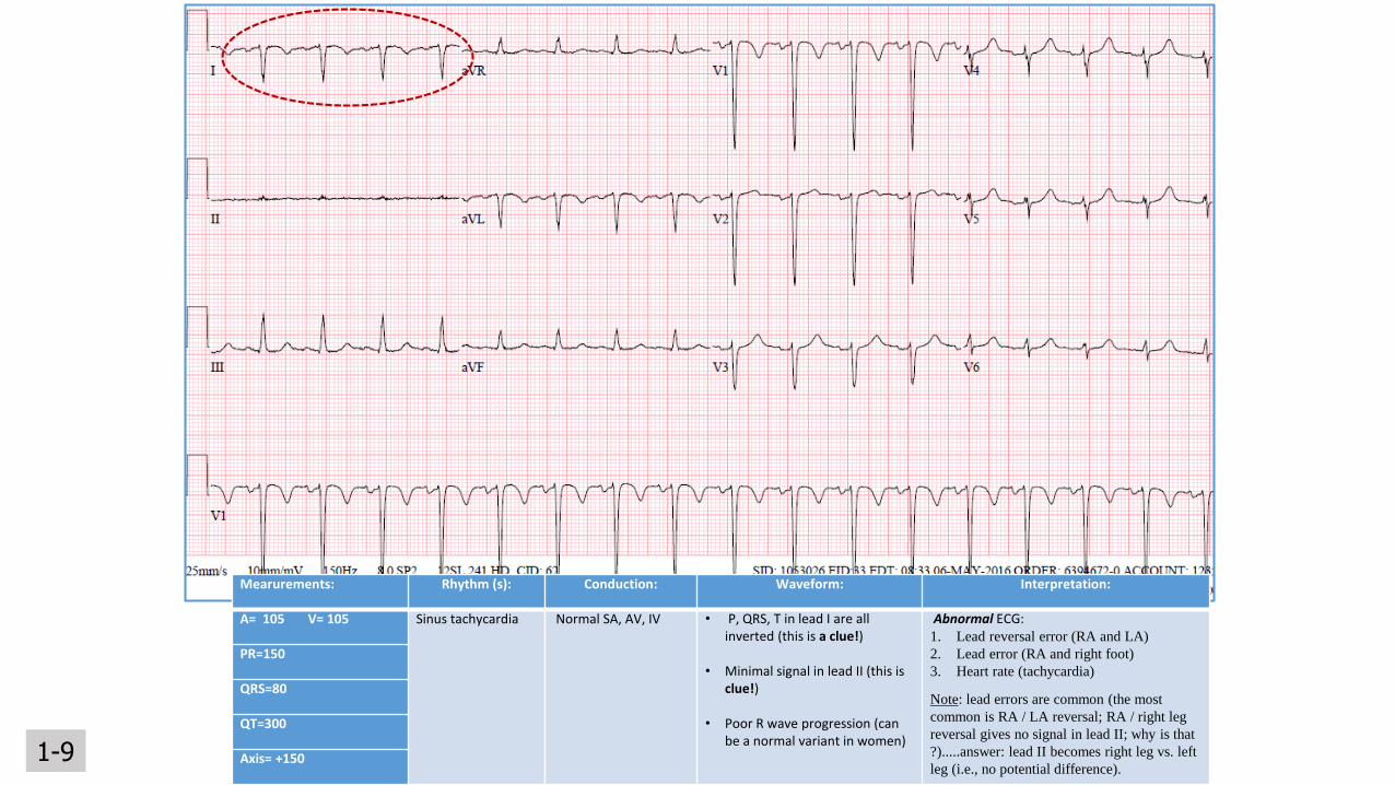
35 year old woman admitted for acute alcohol intoxication
What else went wrong?
1-9
Mearurements: Rhythm (s): Conduction: Waveform: Interpretation:
A= 105 V= 105 Sinus tachycardia Normal SA, AV, IV • P, QRS, T in lead I are all inverted (this is a clue!)
• Minimal signal in lead II (this is clue!)
• Poor R wave progression (can be a normal variant in women)
Abnormal ECG:1. Lead reversal error (RA and LA)
2. Lead error (RA and right foot)
3. Heart rate (tachycardia)
Note: lead errors are common (the most
common is RA / LA reversal; RA / right leg
reversal gives no signal in lead II; why is that
?).....answer: lead II becomes right leg vs. left
leg (i.e., no potential difference).
PR=150
QRS=80
QT=300
Axis= +150

1-10
Oh, oh….. What to do ?
I
II
III

1-10
Mearurements: Rhythm (s): Conduction: Waveform: Interpretation:
A=110 V=110 Sinus tachycardia Normal SA, AV, IV Much artifact (but you can still recognize aspects of the ECG waveform (see lead III)
Artifact precludes accurate ECG interpretation; sinus tachycardia is present.
Artifact in this case is from a patient with Parkinson‘s disease (skeletal muscle).
PR=140
QRS=70
QT=300
Axis=?
I
II
III