guidelines and standards -...
TRANSCRIPT

GUIDELINES AND STANDARDS
FromCentroM
Washington H
Hospital, Belo
Belo Horizont
Francisco, Ca
(J.L.C.); Funda
Johns Hopkin
Unit, Medical
(J.A.M.-N.); Ho
de Investigacio
University, Pop
the Federal Un
Institute, S~ao P
Paulo, Brazil (M
The following
Caryn Bern, M
rea,MD, has re
support fromT
Carlos A. Mor
Research, TDR
Bayer, and BM
is amember of
Recommendations for Multimodality CardiacImaging in Patients with Chagas Disease: A Reportfrom the American Society of Echocardiography inCollaboration With the InterAmerican Association
of Echocardiography (ECOSIAC) and theCardiovascular Imaging Department of theBrazilian Society of Cardiology (DIC-SBC)
Harry Acquatella, MD, FASE (Chair), Federico M. Asch, MD, FASE (Co-Chair),Marcia M. Barbosa, MD, PhD, FASE, Marcio Barros, MD, PhD, Caryn Bern, MD, MPH,
Joao L. Cavalcante, MD, FASE, Luis Eduardo Echeverria Correa, MD, Joao Lima, MD, Rachel Marcus, MD,Jose Antonio Marin-Neto, MD, PhD, RicardoMigliore, MD, PhD, Jose Milei, MD, PhD, Carlos A. Morillo, MD,Maria Carmo Pereira Nunes, MD, PhD,Marcelo Luiz Campos Vieira,MD, PhD, and Rodolfo Viotti, MD*,Caracas,Venezuela;Washington,District ofColumbia;BeloHorizonte and S~aoPaulo, Brazil; SanFrancisco,California; Pittsburgh,Pennsylvania; Floridablanca, Colombia; Baltimore, Maryland; SanMartin and Buenos Aires, Argentina; andHamilton,
Ontario, Canada
Keywords: Chagas, Cardiomyopathy, Left ventricular aneurysm, Heart failure, Echocardiography, Cardiacmagnetic resonance
In addition to the collaborating societies listed in the title, this document is endorsed by the following AmericanSociety of Echocardiography International Alliance Partners: the Argentinian Federation of Cardiology, theArgentinian Society of Cardiology, the British Society of Echocardiography, the Chinese Society ofEchocardiography, the Echocardiography Section of the Cuban Society of Cardiology, the EchocardiographySection of the Venezuelan Society of Cardiology, the Indian Academy of Echocardiography, the IndianAssociation of Cardiovascular Thoracic Anaesthesiologists, the Iranian Society of Echocardiography, theJapanese Society of Echocardiography, the Mexican Society of Echocardiography and Cardiovascular Imaging,and the Saudi Arabian Society of Echocardiography.
edico deCaracas, SanBernardino, Caracas, Venezuela (H.A.);Medstar
ospital Center, Washington, District of Columbia (F.M.A., R.M.); Socor
Horizonte, Brazil (M.M.B.); Faculdade de Sa�ude e Ecologia Humana,
e, Brazil (M.B.); the University of California, San Francisco, San
lifornia (C.B.); the University of Pittsburgh, Pittsburgh, Pennsylvania
cion Cardiovascular de Colombia, Floridablanca, Colombia (L.E.E.C.);
s University, Baltimore, Maryland (J.L.); the Interventional Cardiology
School of Ribeir~ao Preto, University of S~ao Paulo, S~ao Paulo, Brazil
spital Eva Per�on, San Martin, Argentina (R.M., R.V.); Consejo Nacional
nes Cientificas y Tecnicas, Buenos Aires, Argentina (J.M.); McMaster
ulation Health Research Institute, Hamilton, Ontario, Canada (C.A.M.);
iversity of Minas Gerais, Belo Horizonte, Brazil (M.C.P.N.); and Heart
aulo University Medical School, Hospital Israelita Albert Einstein, S~ao
.L.C.V.).
authors reported relationships with one or more commercial interests:
D, MPH, has consulted for Chemogroup. Luis Eduardo Echeverria Cor-
ceived a researchgrant fromRoche. JoaoLima,MD,has receivedgrant
oshibaMedical Systems. RachelMarcus,MD, is a consultant for Bayer.
illo, MD, has received grants from the Canadian Institute for Health
-WHO (BENEFIT trial), Merck Shape & Dohme (STOP-CHAGAS trial),
S/Pfizer; has been a speaker for Bayer and Boehringer Ingelheim; and
the advisory board for BostonScientific. The following authors reported
no actual or potential conflicts of interest in relation to this document: Harry Acquatella,
MD, FASE, FedericoM. Asch,MD, FASE,MarciaM. Barbosa,MD, PhD, FASE,Marcio
Barros,MD, PhD, Jo~ao L. Cavalcante,MD, FASE, Jose AntonioMarin-Neto,MD, PhD,
Ricardo Migliore, MD, PhD, Jose Milei, MD, PhD, Maria Carmo Pereira Nunes, MD,
PhD, Marcelo Luiz Campos Vieira, MD, PhD, and Rodolfo Viotti, MD.
Reprint requests: American Society of Echocardiography, 2530Meridian Parkway,
Suite 450 Durham, NC 27713 USA (Email: [email protected]).
* The American Society of Echocardiography and the writing group sadly note the
passing of Dr. Rodolfo Viotti inMarch 2017, while this document was beingwritten.
It was our honor to work with Dr. Viotti on a topic that was very dear to him
throughout his long career.
Attention ASE Members:
Visit www.aseuniversity.org to earn free continuing medical education credit
through an online activity related to this article. Certificates are available for
immediate access upon successful completion of the activity. Nonmembers
will need to join the ASE to access this great member benefit!
0894-7317/$36.00
Copyright 2017 by the American Society of Echocardiography.
https://doi.org/10.1016/j.echo.2017.10.019
3

TABLE OF CONTENTS
I. Introduction 4II. Epidemiology of ChD: Geographic Distribution Worldwide and in theUnited States 4III. Pathophysiology Related to Imaging and Clinical Presentation 5IV. Special Features of Electrocardiography and Each Imaging Modality inRelation to ChD 6
IV.a. Electrocardiography and Continuous Rhythm Monitoring 6
IV.b. Echocardiography 7
IV.b.i. M-mode and 2D echocardiography 7
IV.b.ii. Three-dimensional echocardiography 9
IV.b.iii. Strain and speckle tracking echocardiography 11
IV.c. CMR and CT 12
IV.d. Nuclear Medicine 16V. Ventricular Function 17
V.a. LV Systolic Function 17
V.b. LV Diastolic Function 18
V.c. RV Function 18VI. Recommendations for the Use of Imaging Modalities According to theStage of theDisease:Diagnostic,Monitoring, andPrognostic Implications 19
VI.a. Acute ChD 19
VI.b. Chronic ChD 19
VI.b.i. Silent or asymptomatic Chagas disease (Stages A, B1 and B2):Monitoring of LV function and myocardial damage 19
VI.b.ii. Symptomatic chronicChagas heart disease (StagesC andD) 20
VI.b.ii.1. LV function and heart failure (HF) 20
VI.b.ii.2. Thromboembolism 20
VI.b.ii.3. Cardiac arrhythmias 21VII. Summary and Conclusions 21
Abbreviations
2D = Two-dimensional
3D = Three-dimensional
ASE = American Society of Echocardiography
ChD = Chagas disease
ChHD = Chagas heart disease
CMR = Cardiac magnetic resonance
CT = Computed tomography
ECG = Electrocardiographic
HF = Heart failure
LA = Left atrial
LGE = Late gadolinium enhancement
LV = Left ventricular
LVEF = Left ventricular ejection fraction
PW Doppler = Pulsed wave Doppler
RV = Right ventricular
RVEF = Right ventricular ejection fraction
TEE = Transesophageal echocardiography
TTE = Transthoracic echocardiography
4 Acquatella et al Journal of the American Society of EchocardiographyJanuary 2018
I. INTRODUCTION
Chagas disease (ChD) is a significant public health problem in mostLatin American countries. Observed mainly in rural areas, in recentdecades it has spread to cities and to nonendemic countries, mostlyas a result of migration of infected people. Increasing numbers ofcases are now being identified in the United States, Spain, and othercountries, which makes its diagnosis and management of increasinginterest worldwide.
During an antimalarial campaign in Lassance (Minas Gerais,Brazil) in 1909, Carlos Chagas identified the parasite Trypanosomacruzi, its vector for transmission (triatomine bugs, called differentlyin each country: kissing bug, barbeiro, vinchuca, chinche, etc.), anddescribed the initial cases of the disease.1 Transmission occursmainly through the bite of these vectors but may also occur byblood transfusion, from mother to fetus, oral ingestion of contami-nated foods, organ transplantation, and laboratory accidents.Vector control programs have substantially diminished T. cruzi andChD incidence. However, about 70 million people remain at riskfor acquiring the infection.2
The diagnosis of ChD is made by epidemiologic history and by twoor more positive serologic tests. There are two clinical phases of T.cruzi infection: acute ChD, seen early after acquiring the infection,and chronic ChD, lasting for decades. About 70% to 80% of individ-uals with chronic T. cruzi infection remain asymptomatic (indetermi-nate form), while 20% to 30% develop cardiac and/orgastrointestinal disease.3 Patients with chronic Chagas heart disease(ChHD) are staged according to the severity of myocardial damageand symptoms of congestive heart failure (HF; Table 1).4,5
Assessment by electrocardiography is mandatory because theearliest signs of ChHD are generally conduction system defectsand/or ventricular arrhythmias. The introduction of various cardiacimaging modalities, such as echocardiography, nuclear medicine,computed tomography (CT), cardiac magnetic resonance (CMR),and chest radiography, provides valuable information on cardiacstructure and function.
The purpose of this document is to provide recommendations forthe use of cardiac ultrasound and other imagingmodalities in the diag-nosis, classification, and risk assessment of myocardial damage fromearly to advanced forms of ChHD.
II. EPIDEMIOLOGY OF ChD: GEOGRAPHIC DISTRIBUTION
WORLDWIDE AND IN THE UNITED STATES
ChD is caused by the protozoan parasite T. cruzi, transmittedwhen feces of an infected triatomine vector enters the mamma-lian host through the bite wound or mucous membranes.6
Infection is lifelong in the absence of treatment. Vector-bornetransmission occurs in parts of North America, Central America,and South America, with geographic distribution determinedboth by the ecology of the triatomine vectors and factors suchas housing conditions that govern contact between vectors andthe human population.7 Transmission can also occur throughtransfusion of infected blood components, organ and bonemarrow transplantation, and from mother to fetus. Outbreaksattributed to contaminated food or drink have been reported innorthern South America.8 In addition, many T. cruzi–infected in-dividuals have moved from endemic rural villages to LatinAmerican cities, and hundreds of thousands now live in the

Table 1 Stages of ChHD
Acute Phase
Chronic Phase
Indeterminate Form, A
Chagas Cardiomyopathy
B1 B2 C D
Infected by
T. cruzi and
findingsof acute ChD
Positive serology
Normal ECG findings
No heart disease or HF
Structural
cardiomyopathy
(abnormal ECG orechocardiographic
findings) but normal LV function
No HF
LV dysfunction
No HF
LV dysfunction
HF (current or prior)
Refractory HF
despite optimal
medical therapy
Modified from Andrade et al.4 and Bern et al.5
Journal of the American Society of EchocardiographyVolume 31 Number 1 Acquatella et al 5
United States, Spain, and other nonendemic countries outsideLatin America (Figures 12 and 29,10).
T. cruzi infects many mammalian species. The vector speciesresponsible for the majority of human infections are considered do-mestic because they are adapted to living in cracks in mud wallsand thatch roofs of rustic rural houses.11 Inhabitants of infested housesare repeatedly exposed to vectors and parasites over many years. Inhighly endemic villages, a high percentage of the adult population isinfected, with the prevalence rising with increasing age.12 The preva-lence of cardiac morbidity also increases with age.13 An estimated20% to 30% of T. cruzi–infected individuals eventually developChagas cardiomyopathy, but in endemic or previously endemic set-tings, a much higher percentage of the elderly may have cardiacsigns.3
Latin America has made substantial progress in decreasing T. cruzitransmission, largely through residual insecticide application to con-trol domestic infestation.14 The estimated global ChD prevalencedeclined from 18million in 1991, when the first regional control initia-tive began, to approximately 6 million in 2010.2,6,14 The PanAmerican Health Organization has certified interruption oftransmission by domestic vectors in several countries of SouthAmerica and Central America.15,16 T. cruzi serologic screening isconducted in most blood banks in Latin America and the UnitedStates, and some countries have systematic screening for congenitalChD. Nevertheless, ChD remains the most important parasiticdisease in the Western Hemisphere.
The southern half of the continental United States has estab-lished enzootic transmission cycles, with infected vectors andmammalian hosts such as raccoons, opossums, wood rats, and do-mestic dogs.7,17 Nevertheless, the majority of infected residents areLatin American immigrants infected in their home countries, andinfected individuals are found in nearly every state. On the basisof the Latin American immigrant population and estimates ofprevalence in their home countries, an estimated 300,000 T.cruzi–infected immigrants reside in the United States.9 Locally ac-quired vector-borne infection has been documented in a handfulof cases over the past 60 years and has been inferred in blood do-nors for whom acquisition of the infection in Latin America hasbeen ruled out or judged unlikely.7,18,19 Direct assessments ofprevalence in the United States are sparse and have beenrestricted to small-scale surveys or case series in populations cho-sen because of anticipated high risk (e.g., Latin American immi-grants with nonischemic heart disease).20-22 Because of lowprovider awareness, cases of Chagas cardiomyopathy likely gounrecognized, and women at risk for vertical transmission totheir infants are not screened.23,24 Thus, more work is needed
to raise awareness and improve the knowledge base of providersin the United States. In addition, more extensive epidemiologicstudies and better diagnostic and treatment availability arerequired.
Key Points� ChD is a vector-borne zoonotic disease endemic to the Americas, which could be
underdiagnosed if aggressive screening campaigns are not pursued.
� Substantial progress has been made in decreasing vector- and blood-borne T. cruzi
transmission through residual insecticide application and housing improvement,
but there remain 6 million infected people and 70 million at risk for infection in
the Americas.
� Zoonotic transmission occurs in the southern half of the United States, but T. cruzi–in-
fectedmigrants from Latin America greatly outnumber those infected locally. Patients
with ChD are found in nearly every state of the United States and in other countries
outside of the Americas.
III. PATHOPHYSIOLOGY RELATED TO IMAGING AND
CLINICAL PRESENTATION
The pathophysiology of myocardial damage in chronic ChHD is com-plex andmultifactorial. ChHD is an acquired inflammatory cardiomy-opathy caused by three key pathologic processes: inflammation, celldeath, and fibrosis. A consensus is now emerging that parasite persis-tence and parasite-driven adverse immune response play a pivotalrole in the pathogenesis of ChHD.25
Because of these underlying pathogenic mechanisms, a variety ofstructural and functional cardiovascular abnormalities have beenshown in patients with ChHD.26 Cardiac structural cells affected bythe inflammatory process include the myocytes, leading to myocytol-ysis and contraction band necrosis and irreversible lesions of thespecialized conduction system and cardiac neural cells. The progres-sive destruction of normal cardiac constituents leads to a markedreparative and reactive fibrosis, characterized by a dense interstitialaccumulation of collagen that encloses fibers or groups of myocardialfibers.27 This explains the frequent occurrence of atrioventricular andintraventricular blocks, sinus node dysfunction, malignant ventriculararrhythmias, and sudden death in patients with ChHD.28 Recentstudies in the experimental model of chronic T. cruzi infection haveshown that at early stages, the coalescence of focal areas of myocar-dial inflammation, necrosis, and fibrosis typically results in the appear-ance of left ventricular (LV) segmental wall motion abnormalities, ahallmark of ChHD.29 At later stages, these regional derangementsgradually cause progressive impairment of global myocardial contrac-tile function. The ultimate consequence of is dilated cardiomyopathywith biventricular dysfunction and congestive HF.28

6 Acquatella et al Journal of the American Society of EchocardiographyJanuary 2018
Derangements of the coronarymicrocirculation, including increasedplatelet activity, microthrombi, microvascular spasm, and endothelialdysfunction, have been reported in animal models of T. cruzi infectionand in studies of humans with ChD.30 These phenomena precedeand may be causally related to the development of segmental wall mo-tion abnormalities. Abnormal reactivity to vasodilator and vasocon-strictor stimuli has also been reported in the epicardial coronaryarteries of patients with ChD. Overall, these derangements in the coro-nary microcirculation are likely to cause ischemic myocytolic necrosisand reparative fibrosis that are clinically expressed as ischemic-likesymptoms, electrocardiographic changes, and perfusion defectsdescribed in ChHD patients with angiographically normal coronary ar-teries. In the advanced stages of ChHD, ventricular aneurysms are de-tected mostly in watershed zones between principal coronary arterybranches, such as regions between the left anterior descending and pos-terior descending coronary arteries and the right coronary and leftcircumflex coronary arteries, which supply the apex and the basal-posterior wall of the left ventricle, respectively.31
Virtually all pathophysiologic aspects of chronic ChHD can be de-tected using various imaging modalities, which are discussed in thefollowing sections.
Figure 1 Estimated prevalence of T. cruzi infection per 100 ha-bitants per country. (A) South America. (B) Mexico and CentralAmerica.2
IV. SPECIAL FEATURES OF ELECTROCARDIOGRAPHY AND
EACH IMAGING MODALITY IN RELATION TO ChD
IV.a. Electrocardiography and Continuous RhythmMonitoring
Electrocardiographic (ECG) abnormalities are often the first indicatorof cardiac involvement in ChD. Electrocardiography remains a cost-effective diagnostic test that should be performed routinely once sero-logic confirmation is obtained. Recent regional guidelines provide aclass I recommendation with level of evidence C for the indicationof 12-lead electrocardiography in the diagnosis and risk stratificationof patients with ChD.4
By and large, the acute presentation of ChD is usually mildly symp-tomatic and manifests as a flu-like event. In approximately 5% ofcases, acute myocarditis may clinically manifest with a wide rangeof ECG abnormalities. The most frequent findings are nonspecificand common to any myocarditis and include sinus tachycardia,diffuse abnormal repolarization, low QRS voltage, and atrioventric-ular block. With severe myocarditis, higher degrees of atrioventricularblock and intraventricular conduction disturbances (bundle branchand fascicular blocks) may occur.32 In a series of acute Chagasmyocarditis cases, ECG alterations were documented in 66% of thepatients. Abnormal repolarization was the most common finding(37%), and the most frequent arrhythmia was inappropriate sinustachycardia (9%), followed by atrial premature complexes (8%). Inthis series, only 2% developed right bundle branch block.33
Abnormal ECG findings during the acute phase may have prognosticimplications, as reported by Porto34 in a classic series of patients pre-senting with acute Chagas myocarditis.
Electrocardiography is instrumental in classifying the stage of dis-ease in patients in the chronic phase of ChD. The lack of ECG abnor-malities stages T. cruzi carriers in the indeterminate phase of thedisease (stage A; Table 1). Normal ECG findings are rare in the pres-ence of moderate or severe LV dysfunction, while a greater number ofECG alterations correlates with worse LV function, especially if leftbundle branch block is present. Of note, ECG abnormalities werestrongly associated with ChHD in the Retrovirus EpidemiologyDonor Study II, with a high negative predictive value (95%) for
Chagas cardiomyopathy. This finding suggests that in rural areaswith very limited resources, normal ECG findings without other imag-ing modalities (such as echocardiography) could be sufficientscreening in asymptomatic T. cruzi–infected individuals.35
The earliest ECG abnormalities of ChHD usually involve the conduc-tion system, manifesting most frequently as right bundle branch blockand/or left anterior fascicular block. Second- and third-degree atrioventric-ular block have also been strongly relatedwithChagas cardiomyopathy inendemic populations. Sinus node dysfunctionmay present as episodes ofsinoatrial block with bradycardia or ectopic atrial tachycardia (Figure 3).

Figure 2 Estimated number of ChD cases per country and their status of vector transmission (2009). Obtained with permission fromthe World Health Organization (http://gamapserver.who.int/mapLibrary/Files/Maps/Global_Chagas_2009.png).
Journal of the American Society of EchocardiographyVolume 31 Number 1 Acquatella et al 7
Complex ventricular arrhythmias such as ventricular tachycardia occureven in patients without overt HF but tend to be associated with moreadvanced stages of ChHD and worse prognosis.36,37
Electrocardiography is also a useful tool for risk stratification. Thepresence of premature ventricular contractions, increased QT-intervaldispersion, low-voltage QRS, QRS fragmentation, and prolongedQRS duration have all been associated with a worse prognosis.38-40
Once ECG abnormalities arise, they imply disease progression (stageB; Table 1), preceding the appearance of HF symptoms (stages C andD). ECG abnormalities are frequent and related primarily with nonspe-cific repolarization alterations (30%–40%), right bundle branch blockin conjunction with left anterior fascicular block (20%–35%), prema-ture ventricular ectopic beats (5%–10%), and atrial fibrillation (5%–10%).4,41 These findings have been recently supported by Echeverr�ıaet al.,42 who by using electrocardiography for Chagas cardiomyopathystaging predicted disease progression as determined by elevation in N-terminal pro–brain natriuretic peptide and high-sensitivity troponin.
Twenty-four-hour ambulatory ECG monitoring (Holter) is recom-mended in patients with symptoms suggestive of cardiac arrhythmias(palpitations, presyncope, or syncope) or the presence of certain ECGfindings, such as sinus bradyarrhythmias (heart rate < 40 beats/minand/or prolonged sinus pauses), second-degree atrioventricular block,or frequent and/or repetitive (bursts of) ventricular extrasystoles.4,43
Holter monitoring may identify patients at risk for sudden deathand unmask early signs of cardiac autonomic dysfunction withreduced heart rate variability.44 In asymptomatic patients or thosewith rare symptoms, Holter monitoring will have a low diagnostic
yield and is usually not routinely indicated. An implantable cardiacmonitor may be considered in patients with other markers of risksuch as depressed LV or right ventricular (RV) function, regionalwall motion abnormalities, or syncope with frequent premature ven-tricular complexes or palpitations.
In summary, electrocardiography should be performed in all patientsidentified with positive T. cruzi serology. The rationale for this strategy isbased on the cost-effectiveness and wide availability of this diagnostictest, in addition to high negative predictive value to rule out cardiomy-opathy in T. cruzi–infected individuals. Electrocardiography serves twopurposes: staging and prediction of disease progression. The appro-priate timing and frequency for performing electrocardiography duringfollow-up remains debatable and is not evidence based. It seemsreasonable that in individuals with a normal baseline ECG findings,follow-up ECG testing may be performed at least every 5 to 10 years.Further screening with biomarkers and cardiac imaging may be selec-tively performed, depending on baseline ECG findings. Patients withfrequent premature ventricular complexes, nonsustained ventriculartachycardia, and other bradytachyarrhythmias identified by Holtermonitoring should undergo further assessment of LV systolic function.
IV.b. Echocardiography
IV.b.i. M-Mode and Two-Dimensional Echocardiography.ChHD is an inflammatory cardiomyopathy that may affect themyocardium in a segmental or regional manner. On imaging, theheart may appear normal, have localized segmental ventricular

Figure 3 ECG progression of a single patient with ChHD over 23 years of follow-up. (A) At 31 years of age, sinus bradycardia, ventricularbigeminy, narrow QRS (0.10 sec), and ST-T convex upward with inverted T wave (suggestive of apical aneurysm, subsequently demon-stratedby two-dimensional echo). (B)At 44 years of age, sinus rhythm, noextrasystoles, and anew right bundle branchblockwith left ante-rior fascicularblock (QRSduration increased to0.134sec), andprecordialR-wavesizedecreased. (C)At54yearsof age, sinusbradycardia,no extrasystoles, left anterior fascicular block is presentwithout right bundle branchblock (whichappears on exercise), andQRSduration is0.126 sec. These changesmay explain different rates of ECGfindings. Corresponding 2Dechocardiographic images at 54 years of age areshown in Figure 5.
8 Acquatella et al Journal of the American Society of EchocardiographyJanuary 2018

Figure 4 Dilated ChHD cardiomyopathy (stage D). Four-chamber view echocardiograms of two patients with ChHD in congestive HFwith severe global hypokinesis and low ejection fraction, similar to other dilated cardiomyopathies. A pacemaker wire is seen in theright heart chambers (arrow). (Left) The left ventricle (LV) and left atrium (LA) are severely dilated with normal size right ventricle (RV).(Right) Biventricular and biatrial severe dilatation. RA, Right atrium.
Journal of the American Society of EchocardiographyVolume 31 Number 1 Acquatella et al 9
abnormalities (wall motion, thinning, aneurysms, etc.), or manifest asa globally dilated cardiomyopathy with associated valvular heart dis-ease (mainly functional mitral and tricuspid regurgitation). In general,echocardiographic evaluation should be performed as suggested bythe American Society of Echocardiography (ASE) guidelines onchamber quantification,45 with a special emphasis on LV functionand morphology, RV function, and valvular disease. As previouslymentioned, the absence of ECG abnormalities mostly rules out signif-icant cardiomyopathy. However, it is reasonable to perform at least asingle echocardiographic examination (baseline evaluation) on everypatient with positive serology for ChD and repeat during follow-up ifthe ECG findings become abnormal to document disease progres-sion (Table 1). Patients with symptomatic ChHD (HF) may presentwith predominantly hypokinetic, dilated left ventricles with dimin-ished LV ejection fraction (LVEF), or biventricular dilatation(Figure 4). Although infrequent, even asymptomatic subjects maydisplay subtle abnormalities by two-dimensional (2D) and three-dimensional (3D) echocardiography, such as small aneurysms orwall motion abnormalities.
Global LV Function. Initial rural surveys using basic M-mode and2D echocardiography were useful for clinical staging by estimatinganatomic and functional damage and assessing LV function.46,47
Current ASE guidelines recommend routine clinical evaluationusing 2D and 3D echocardiography to estimate LV, RV, left atrial(LA) and right atrial volumes and dimensions and for assessment ofLV and RV function.45 Global LV systolic function should be ad-dressed on 2D echocardiography by calculation of LVEF throughthe biplane method of disks (the Simpson rule). The endocardialborder should be traced at the interface of the compacted myocar-dium and the LV cavity at end-systole and end-diastole in the apicalfour- and two-chamber views. Although the accuracy of LV volumesand LVEF is higher when using 3D echocardiography (which is there-fore preferred whenever available), 2D echocardiography has theadvantage of easiness and wider availability. However, the presenceof apical LV aneurysms present a challenge to the use of the methodof disks, as apical aneurysms frequently cannot be included within theultrasound field (Figure 5).
Regional Wall Motion Abnormalities. LV segmental abnormalitiesare common in ChHD at any stage of the disease. These are locatedmainly at the LV apex (Figure 6, Videos 1 and 2, available at www.
onlinejase.com) and inferior and inferolateral walls (Figures 7 and 8)butmay also affect other LVorRV segments.48 Technically, it is importantto perform a comprehensive examination from multiple windows, aswall motion abnormalities should be demonstrated in at least twodifferent views to avoid false-positive results. The use of ultrasoundcontrast agents for LVopacification (Figure 9) andwallmotion evaluationis recommendedwhen images are suboptimal in at least two contiguoussegments.49,50 In addition, contrast could be particularly useful for thedetection of small aneurysms and thrombus, typical of ChHD.51 In a re-viewof 2D echocardiographic series of patients withChHD, among 920asymptomatic patients with mild cardiac damage, the prevalence of LVaneurysm was 8.5%, while it increased to 55% in patients with moreadvanced cardiac disease.47 Similarly, LV apical abnormalities had alow prevalence in those with normal ECG findings but increased to24% in those with abnormal ECG findings. Other common contractileabnormalities involve the inferolateral or inferior walls, with prevalenceof up to 23% in symptomatic patients.
Valvular Disease. A comprehensive evaluation in patients with ChDshould include careful examination of the cardiac valves.Two-dimensional echocardiography is used to evaluate valvular andsubvalvular structure and, together with a thorough Doppler exami-nation, provide a good understanding of the severity and etiologyof different valvular diseases and dysfunction.52,53 Functionalincompetence of the mitral and tricuspid valves is common asChHD advances (Figure 10). Ventricular remodeling with progressivedysfunction, dyssynchrony, valvular annular dilation, tethering of thesubvalvular apparatus, fibrosis, and atrial enlargement may inducevarious degrees of valve dysfunction. An understanding of these alter-ations will help in determining the need and proper strategy for ther-apeutic interventions.
IV.b.ii. Three-Dimensional Echocardiography. Cardiac cham-bers are 3D structures with complex anatomy and variable shape.Therefore, the accuracyof2Dechocardiography for evaluationof cardiacstructure, shape, and dimensions is limited, as it requires some degree ofreconstruction and geometric assumptions. Three-dimensional echocar-diography, on the other hand, allows visualization of cardiac chambersin their entirety without geometric assumptions.45,54-56 Creation of 3Dechocardiographic images requires specialized transducers foracquisition of a volume (pyramid) of data rather than a slice (2Dechocardiography). The 3D volume could be acquired through a single

Figure 5 Challenges in 2D echocardiographic LVEF and volume evaluation by the method of disks (Simpson rule) in patients withChHD with apical aneurysm. Four-chamber and two-chamber apical views (biplane method) in a 54-year-old patient with ChHD.LV volumes are increased, and LVEF is mildly decreased. ECG follow-up is shown in Figure 3C. Notice the LV apical aneurysm, espe-cially in mid-systole (right). As in this case, large apical aneurysms are frequently difficult to contain within the image, and thereforeadequate tracing of the cavity/endocardial interface may not be feasible.
10 Acquatella et al Journal of the American Society of EchocardiographyJanuary 2018
heartbeat or by stitching together smaller volumes in consecutive beats.Although 3D echocardiography has significant advantages over 2Dechocardiography, as mentioned above, there are limitations, relatedmostly to lower temporal and spatial resolution or stitching artifact withmultibeat acquisition. Nevertheless, by direct visualization of the entireleft ventricle, 3D echocardiography avoids foreshortening of the leftventricle from the apical windows and facilitates measurement of LVvolumes and LVEF by direct endocardial contour tracing, rather thanassumptions of LV shape from 2D apical views with the single orbiplane method of disks (Figure 11). Three-dimensional echocardiogra-phy is currently well validated compared with other 3D imaging tech-niques such as CMR and cardiac CT.45,54,56 This concept also appliesto visualization and volume measurements of other cardiac chambers,such as the left atrium and right ventricle (Figure 12).57,58 As with
patients with other forms of cardiomyopathy, 3D echocardiographyshould be used in patients with ChD to evaluate cardiac chamber sizeand ventricular function.45 Currently, LV volumes and LVEF can bemeasured using a variety of semiautomated software that are less time-consuming and improve reproducibility.55 In patients with ChHD pre-senting with HF, it is important to have information concerning RV sizeand function,which isparticularly challengingwith2Dechocardiography.The use of 3D echocardiography allows an accurate analysis of RV vol-umes and RV ejection fraction (RVEF; Figure 12). In addition, the useof 3D or 3D-derived 2D images (biplane, Xplane, etc.) could help in de-tecting small LV aneurysms that would otherwise be overlooked by 2Dechocardiography because of foreshortening.
The analysis of mitral regurgitation severity in patients with chronicChagas cardiomyopathy should include 3D echocardiography as part

Figure 6 Left ventricular apical aneurysm and thrombus (stage B2). Apical four- and two-chamber views (left and right) of a 48-year-old patient with chronic ChHD who presented with a right arm embolic event. A left ventricular apical aneurysm (ANEU) shows bloodstasis with small thrombus (STA-THRB). Themid and basal segments of the left ventricle (LV) had normal contractility. LVEFwas 45%.Right ventricle (RV), left atrium (LA), and right atrium (RA) were roughly spared from disease.
Figure 7 Inferolateral wall fibrosis and akinesis, M-mode, andautopsy specimen. Long-axis slow sweepM-mode echocardio-gram (A) and cardiac specimen (B) from a 52-year-old male pa-tient with ChHD, HF, and arrhythmias, showing extensivescarring and akinetic inferolateral wall (posterior wall [PW]),extensive to apex, contrasting with relatively preserved septal(S) systolic motion and thickening. The coronary arteries werenormal at autopsy. (Reproduced with permission from Acqua-tella et al.46).
Journal of the American Society of EchocardiographyVolume 31 Number 1 Acquatella et al 11
of the comprehensive mitral valve evaluation to assess the structureand morphology of the leaflets and subvalvular apparatus. Althougha dilated annulus with tethering of the leaflets from a dilated leftventricle (functional or secondary mitral regurgitation) is the mostcommon finding in ChHD, the examination should evaluate othercoexisting valvular abnormalities.52-54 Therefore, careful attentionshould be paid to valvular and subvalvular function and structure toevaluate for presence, location, and extent of prolapse, redundanttissue, clefts, rheumatic changes, annular dimensions, cuspseparation, and other abnormalities that are not specific for ChHD.Color 3D echocardiography may be used to identify multiple ornoncircumferential jets, measure the vena contracta area, or, incombination with continuous-wave Doppler echocardiography,calculate regurgitant orifice area or regurgitant volume and fractionusing the proximal isovelocity surface area method. Comprehensiveanalysis with information derived from 2D, 3D, and Doppler echo-cardiography may be useful for the prediction of success in future at-tempts at mitral valve repair (surgical or percutaneous), similar toother etiologies of functional mitral regurgitation.
IV.b.iii. Strain and Speckle-Tracking Echocardiography.Myocardial deformation imaging is a relatively new technique forquantitative assessment of myocardial contractility.59 Strain is a mea-sure ofmyocardial deformation, defined as the change in length of themyocardium relative to the original length. Strain rate is the rate ofchange in strain.60 Myocardial deformation imaging with echocardi-ography can be measured using Doppler tissue imaging and 2Dand 3D speckle-tracking echocardiography (Figures 13 and 14).Tissue Doppler–derived strain has several limitations, particularly inrelation to angle dependency and noise interference. Therefore,strain measurement based on speckle-tracking, which is not angledependent, has become a method of choice to assess myocardialdeformation.
Speckle-tracking echocardiography has allowed the increased recog-nition of subclinicalmyocardial dysfunction, particularly in patientswith
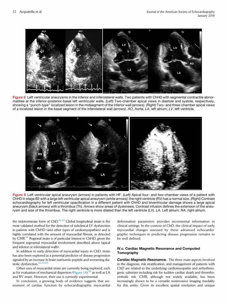
Figure 8 Left ventricular aneurysms in the inferior and inferolateral walls. Two patients with ChHD with segmental contractile abnor-malities at the inferior-posterior-basal left ventricular walls. (Left) Two-chamber apical views in diastole and systole, respectively,showing a ‘‘punch-type’’ localized lesion in the midsegment of the inferior wall (arrows). (Right) Two- and three-chamber apical viewsof a localized lesion in the basal segment of the inferolateral wall (arrows). AO, Aorta; LA, left atrium; LV, left ventricle.
Figure 9 Left ventricular apical aneurysm (arrows) in patients with HF. (Left) Apical four- and two-chamber views of a patient withChHD in stage B2with a large left ventricular apical aneurysm (white arrows); the right ventricle (RV) has a normal size. (Right)Contrastechocardiography for left ventricular opacification in a different patient with ChHD and biventricular damage shows a large apicalaneurysm (black arrows)with a thrombus (Th). Arrows show areas of dyskinesis. Contrast infusion defines the extension of the aneu-rysm and size of the thrombus. The right ventricle is more dilated than the left ventricle (LV). LA, Left atrium; RA, right atrium.
12 Acquatella et al Journal of the American Society of EchocardiographyJanuary 2018
the indeterminate form of ChD.61-63 Global longitudinal strain is themost validated method for the detection of subclinical LV dysfunctionin patients with ChHD (and other types of cardiomyopathies) and ishighly correlated with the amount of myocardial fibrosis, as detectedby CMR.64 Regional strain is of particular interest in ChHD, given thefrequent segmental myocardial involvement described above (apicaland inferior or inferolateral walls).
In addition to early detection of myocardial injury in ChD, strainhas also been explored as a potential predictor of disease progressionsignaled by an increase in brain natriuretic peptide and worsening dia-stolic dysfunction.62,63,65
Other uses of myocardial strain are currently being explored, suchas for evaluation of mechanical dispersion (Figure 14),66 as well as LAand RV strain. However, their use is currently experimental.
In conclusion, a growing body of evidence suggests that ass-essment of cardiac function by echocardiographic myocardial
deformation parameters provides incremental information inclinical settings. In the context of ChD, the clinical impact of earlymyocardial changes assessed by these advanced echocardio-graphic techniques in predicting disease progression remains tobe well defined.
IV.c. Cardiac Magnetic Resonance and ComputedTomography
Cardiac Magnetic Resonance. The three main aspects involvedin the diagnosis, risk stratification, and management of patients withChD are related to the underlying cardiomyopathy and arrhythmo-genic substrate including risk for sudden cardiac death and thrombo-embolic risk. CMR, although not widely available, has beenincreasingly shown to be a versatile noninvasive imaging modalityfor this entity. Given its excellent spatial resolution and unique

Figure 10 Mitral regurgitation (MR). A 47-year-old woman withChHD in HF (stage D) and severe MR. The color Doppler imageacquired from the apical four-chamber view shows a largeeccentric regurgitant color area (red arrow) with ‘‘wall-hugging’’appearance, directed to the left pulmonary veins and reachingthe roof of the left atrium (LA), allowing a qualitative visual esti-mation as severe MR. Regurgitant volume was >50 mL/beat.The left ventricle (LV) is severely hypokinetic and dilated; LVEFwas 25%. It is recommended to perform a comprehensive MRevaluation with multiple qualitative and quantitative parameters.RA, Right atrium; RV, right ventricle.
Journal of the American Society of EchocardiographyVolume 31 Number 1 Acquatella et al 13
capability of tissue characterization of myocardial edema and fibrosis,CMR can provide good insight into the pathophysiology of thedisease.67
CMR protocols begin with cine sequences using steady-state freeprecession, which allow accurate assessment of the severity andextent of biventricular involvement by accurate calculation of ejectionfraction and evaluation of wall motion abnormalities. As mentionedpreviously, LV systolic dysfunction is the strongest predictor ofmorbidity and mortality in ChD. Asymptomatic LV systolic dysfunc-tion is even more prevalent than symptomatic HF. Importantly, diag-nosis at a subclinical stage may help in preventing or delayingprogression of the disease by appropriate therapeutic interventions.Regional wall motion abnormalities, including the typical apical aneu-rysm, can be readily recognized by this technique without the poten-tial limitations of inadequate echocardiographic windows.
The use of noncontrast T2-weighted sequences allows the evalua-tion of myocardial edema, which can occur through all phases of thedisease and with good correlation with the traditional late gadoliniumenhancement (LGE).68 LGE is assessed using T1-weighted sequences,approximately 10 to 15 min after gadolinium administration. As aparamagnetic extracellular contrast agent, gadolinium will distributeinto the areas where there is expansion of the interstitium related
to myocardial fibrosis or necrosis. For adequate LGE imaging, thenormal or unaffected myocardium signal is nulled (black), whereasthe involved myocardial segments will have a prolonged washoutfrom decreased capillary density, causing shortening of T1 and there-fore a higher or brighter signal.69
Several patterns of LGE have been described in patients withChHD, including subendocardial and transmural (both are difficultto distinguish from prior myocardial infarction), midwall, or subepi-cardial. LGE tends to more commonly involve the basal inferolateralsegments and LV apex (Figure 15, Video 3; available at www.onlinejase.com). Myocardial fibrosis can be found in up to 8% of pa-tients with positive serology for ChD despite normal ECG and echo-cardiographic findings.70 Importantly, the presence and extent ofmyocardial fibrosis correlate well with the New York HeartAssociation functional class and likelihood of ventricular arrhythmias,particularly when a transmural pattern is found extending beyondtwo or more contiguous segments.71,72 In addition, myocardialfibrosis inversely correlates with LV systolic function.5 Therefore,assessment of fibrosis by LGE imaging has been shown to be an excel-lent marker of disease severity.
CMR with LGE imaging can also evaluate thromboembolic risk.Specifically, the high spatial resolution of CMRmakes it the best imag-ing modality to detect intracardiac thrombi related to LV aneurysms.LV thrombus, which carries a high risk for stroke and peripheral embo-lism, could bemissed by echocardiography in patients with limited im-age quality, even when using ultrasound contrast agents.73,74
Although CMR imaging is an excellent tool for the risk stratifica-tion and prognosis of patients with ChD, it is not readily availableto most patients living in rural endemic areas. In addition, CMRstudies in patients who have received prior implantable cardiac de-vices such as defibrillators, conventional pacemakers, and cardiac re-synchronization devices, are at this time relatively contraindicated.
Cardiac CT. The literature on cardiac CT in ChD is limited to iso-lated case reports. Data acquisition is performed following intrave-nous iodine contrast injection with ECG synchronization. Differentscanning protocols are available that can be restricted to imagingonly the diastolic phase (prospective acquisition, less radiation, andcommonly used for coronary computed tomographic angiography)or imaging the entire cardiac cycle (retrospective acquisition, more ra-diation), which allows quantification of cardiac function. The 3D dataset is postprocessed offline using multiplanar reconstruction for visu-alization of the entire cardiovascular anatomy in any given plane, withexcellent spatial resolution.
Similar to CMR image acquisition, cardiac arrhythmias such asatrial fibrillation or premature atrial or ventricular contractions canproduce image artifacts, making it difficult for study analysis andinterpretation. Technological advances with availability of a largernumber of detector rows (i.e., $256) allows volumetric coverageof the entire heart with one beat, therefore minimizing some ofthese problems.
Cardiac CT could be considered in three particular clinical sce-narios for patients with ChD: (1) To exclude significant coronary ar-tery disease in patients with low to intermediate pretest probability.This differential diagnosis could be particularly challenging in patientswith ChHD presenting with cardiomyopathy and/or wall motion ab-normalities. Coronary computed tomographic angiography has anexcellent negative predictive value to exclude coronary artery diseasein patients with low and intermediate pretest probability.75 (2) In theplanning of complex electrophysiologic procedures. Patients withChHD have larger epicardial compared with endocardial substrate

Figure 11 LVEF by 3D echocardiography. Three-dimensional four-chamber apical view of a 56-year-old man with HF in New YorkHeart Association functional class III and depressed left ventricular systolic function. Evaluation of left ventricular volumes andLVEF was done by 3D echocardiography using an automated adaptive analytics algorithm for quantification. LV end-diastolic (ED)volume was 67 mL; LV end-systolic (ES) volume was 42 mL; LVEF was 37%; left atrial end-systolic volume was 25 mL. HR, Heartrate; LA, left atrium; LV, left ventricle; SV, stroke volume.
Figure 12 RV function by 3D echocardiography. RV 3D echocardiography in a 49-year-old patient with ChHD and HF presenting inNewYork Heart Association functional class III. In addition, the results for RV longitudinal strain (RVLS) by speckle-tracking are shown(top left). RV end-diastolic volume (EDV) was 149.5ml; RV end-systolic volume (ESV) was 104.6ml; RVEFwas 30%; RV stroke volume(SV) was 44.9 mL; septal RVLS was �9.34%; free wall RVLS was �9.55%.
14 Acquatella et al Journal of the American Society of EchocardiographyJanuary 2018

Figure 13 Global longitudinal strain of the left and right ventricles. (A) Abnormal LV longitudinal strain findings in a patient with ChHDwithright bundle branch block, reduced LVEF, and prior symptoms of heart failure (stage C). (Top left) LV longitudinal strain in apical four-chamber view; note thedelayedpeak strain of the septal segments (yellowandblue tracings) typical of right bundle branchblock. (Top right)Apical two-chamber view. (Bottom left) Apical three-chamber view. (Bottom right) ‘‘Bull’s-eye’’ plot of strain values for each myocardialsegment. (B) Abnormal findings of RV longitudinal strain (global strain [GS] was �6.5%, free wall strain was �12.6%) in a patient withChHD (asymptomatic with abnormal ECG findings and decreased LVEF, stage B2). (Top left) Apical four-chamber view. (Bottom left)Regional strain values. (Top right) Time-strain curves. (Bottom right) M-mode parametric colorization. 4CH, Four-chamber; A2C, apicaltwo-chamber; A4C, apical four-chamber; ANT, anterior; ANT_SEPT, anteroseptal; AVC, aortic valve closure; Avg, average; FR, framerate; GLPS, global longitudinal peak strain; HR, heart rate; INF, inferior; LAT, lateral; LAX, long-axis; POST, posterior; SEPT, septal.
Journal of the American Society of EchocardiographyVolume 31 Number 1 Acquatella et al 15

Figure 14 Mechanical dispersion by myocardial strain imaging. Longitudinal strain curves from apical four-chamber view displayingsix of the 18 LV segments used to calculate mechanical dispersion. (Left)Mechanical dispersion in a patient with ChHD without ven-tricular arrhythmias. (Right) A patient with ChHD who presented with LV dysfunction and a previous episode of sustained ventriculartachycardia. The time to maximal myocardial shortening of longitudinal strain was markedly dispersed compared to the patientwithout arrhythmias. AVC, Aortic valve closure.
Figure 15 CMR with LGE. Small, focal LV apical aneurysm in a25-year-old patient with ChHD presenting with a stroke. De-layed gadolinium enhancement shows an LV aneurysm withmyocardial fibrosis (white within the apical myocardium).Video 3 with cine noncontrast CMR shows the apical dyskinesisand small aneurysm.
16 Acquatella et al Journal of the American Society of EchocardiographyJanuary 2018
areas for ventricular arrhythmias.7 This puts patients at risk for coro-nary artery injury during ablation, a complication that can be avoidedby properly planning the procedure with advanced imaging.Coregistration of cardiac computed tomographic 3D data sets withelectroanatomic mapping data allows better understanding of theanatomic relationship of the arrhythmia-substrate areas and the coro-nary arteries, therefore increasing ablation safety.73 (3) To evaluate LVfunction and morphology in patients with difficult echocardiographicwindows who are unable to undergo CMR because of device incom-patibility. Retrospective gated acquisition following intravenouscontrast injection can provide quantification of cardiac function aswell as detection of regional wall motion abnormalities, apical aneu-rysms, and intracardiac thrombi.
Key Points� CMR is a versatile imaging modality for ChHD, allowing exquisite visualization of
heart function, anatomy, and tissue characterization.
� Presence of myocardial fibrosis by CMR with LGE imaging has been associated with
increased cardiac arrhythmias and risk for sudden cardiac death.
� Cardiac CT can provide ancillary noninvasive evaluation of coronary artery anatomy
and atherosclerosis. Coregistration of 3D data sets with electrophysiologic electrical
mapping during ablations might improve procedural safety and outcomes.
IV.d. Nuclear Medicine
Various nuclearmedicine imagingmodalities have been used to assessbiventricular function, myocardial perfusion, innervation, and inflam-mation in patients with ChD. Those with proven clinical value aredescribed here.
Radionuclide Planar Gated Angiography. This method can beused for the evaluation of global biventricular function in patients forwhom echocardiography is hindered by technical problems, prevent-ing optimal imaging or adequate quantitative evaluation.76-78 Thismethod was once considered the gold standard for themeasurement of LVEF because it allows the averaging of hundredsof cardiac cycles without resorting to any geometric assumptions inthe usually distorted LV shape exhibited by patients with ChHDwith aneurysms or other wall motion abnormalities.
Single-Photon Emission Computed Tomographic Myocar-
dial Perfusion Scintigraphy. In patients with ChHD, perfusiondefects may be related to concomitant epicardial coronary diseaseor, more frequently, to microvascular disease in the setting of normalresults on coronary angiography. More specifically related to ChHD,fixed perfusion defects seen during both stress and rest conditionspredominate in LV regions exhibiting advanced wall motion impair-ment (such as akinesis or dyskinesis) and are interpreted as represent-ing areas of fibrosis. Reversible perfusion defects, on the other hand,denote the occurrence of myocardial ischemia due to microvascularflow disturbances. These ischemic defects have been describedeven in Chagas patients with the indeterminate form of the diseaseand are topographically correlated with myocardial areas exhibitingmore significant wall motion abnormalities at later stages of the car-diomyopathy.79,80 Moreover, the increase over time in the extent

Journal of the American Society of EchocardiographyVolume 31 Number 1 Acquatella et al 17
of myocardial fibrosis, as denoted by the transformation of previouslyreversible into fixed perfusion defects, correlates with a decrease inLVEF.81
Myocardial Sympathetic Innervation Imaging. Myocardialscintigraphy with 123I-metaiodobenzylguanidine can be used for thein vivo assessment of cardiac sympathetic innervation integrity in pa-tients with ChHD. Regional sympathetic denervation is an earlyderangement in the pathophysiology of ChHD, preceding the devel-opment of regional LV contraction disturbances. In fact, abnormalmetaiodobenzylguanidine uptake has been shown in most Chagaspatients with no apparent cardiac involvement.31,82 Impairedregional myocardial sympathetic innervation has also beenassociated with the occurrence of sustained ventricular tachycardiain patients with ChHD.83,84
V. VENTRICULAR FUNCTION
V.a. LV Systolic Function
Echocardiography is the imaging modality of choice to determine LVstructure and function in ChD, complementing information providedby clinical status and electrocardiography.4,47 In selectedasymptomatic patients with normal ECG findings, furtherevaluation with echocardiography may be recommended to classifythe presence and severity of myocardial damage, based mainly ondetection of subtle regional wall motion defects.4
T. cruzi–infected patients in the indeterminate formmay have subtlechanges inLV segmental contractility detected eitherby conventional orspeckle-tracking echocardiography.61,85 Once the disease progresses toheart damage with development of ECG changes consistent withChHD, increased LV diameters associated with segmental wallmotion abnormalities are usually seen. In more advanced diseasewith HF, diffuse hypokinesis with enlargement of all cardiacchambers is the predominant characteristic (Figures 4 and 5).47,86
Regional LV contractility abnormalities represent a characteristicaspect of ChD and may occur in any stage of the disease. The fre-quency of these abnormalities varies according to the stage of the dis-ease. In asymptomatic patients with preserved LVEF, wall motionabnormalities can be detected in up to 10% of patients, particularlyin inferolateral basal segments (Figures 6–9).48 As the disease pro-gresses to a dilated cardiomyopathy, the prevalence of regional abnor-malities increases to approximately 50% of patients.47,87 Althoughsegmental wall motion abnormalities are one of the most commonfindings of cardiac involvement in ChD, the mechanism has notbeen defined. It has been hypothesized that microvascularinvolvement leads to ischemia in distal areas of the coronaryterritories.88 The diagnosis of LV segmental lesions is essential,because it permits the identification of individuals at risk for wors-ening of LV function and ventricular arrhythmias.66,89,90
Apical aneurysm is a hallmark of ChHD, so the presence ofapical aneurysms may help differentiate this disease from idio-pathic cardiomyopathy.37,48 The prevalence of aneurysmsvaries widely, because of the heterogeneity of the populationanalyzed and the accuracy of the imaging method used for itsdiagnosis. Importantly, apical aneurysms may be missed if adetailed echocardiographic examination is not performed. Tobetter identify apical aneurysms, multiple views, oftenunconventional or foreshortened with the transducer slightlyangled to visualize the apex, may be required. An apicalaneurysm is usually defined as a well-delineated dyskinetic area
with marked wall thinning at the apex involving opposing wallsand limited by an area of normal contraction. Aneurysm size isvariable (Figures 6, 8, and 9) and is frequently difficult todistinguish from a healed transmural myocardial infarction.48
LV apical aneurysms are found in 8.5% of asymptomatic patients,but this increases to 55% (ranging from 47% to 64%) in patientswith moderate or severe LV systolic dysfunction. RV aneurysmsare unusual, but some patients have apical aneurysms affectingboth ventricles. An autopsy study showed that 82% of aneurysmswere found at the LV apex, 9% in the right ventricle, and 9% inboth ventricles.91
Mural thrombi can be associated with aneurysms (Figure 6) and areimportant risk factors for systemic embolisms in patients withChHD.91-94 The prevalence of LV thrombus in clinical studies isapproximately 20%, but it may be higher in more advanced diseasestages, as suggested by autopsy studies that showed LV thrombosisin 35% to 44% of patients with ChHD who died of congestive HFor sudden cardiac death.95,96
AssessmentofLVSystolicFunctionbyEchocardiography. Thequantification of LV size and function constitutes one of the earliest indi-cations for echocardiography. The accuracy of the determination ofglobal and regional LV function with echocardiography has improveddramatically over the past decade with several technological develop-ments.97
A pioneering study using M-mode echocardiography demon-strated hypokinesis of the posterior wall with relative preservationof septal motion in patients with ChHD (Figure 7).46 In advancedChHD, a diffuse hypokinesis was detected with a nonspecific pattern,indistinguishable from idiopathic cardiomyopathy.98
Previous studies have used the change in the M-mode LV dimen-sion from diastole to systole to calculate the percentage of fractionalshortening and LVEF. However, this method is no longer recommen-ded in ASE guidelines45 and is particularly unreliable in patients withChD with pronounced LV dilation or in those with either markedsegmental wall motion abnormalities or aneurysms. Therefore, mostrecent studies have used 2D echocardiography for the assessmentof LV function and to quantify LVEF.
Three-dimensional echocardiography has been shown to quantifyLV volumes and function more accurately, as no geometric assump-tions are required (Figure 11). Given the frequency of segmental ab-normalities and aneurysms, it seems most appropriate to include fullvolume 3D echocardiography of the left and right ventricles in anyechocardiographic examination of patients with suspected ChHD.However, there are limited data to date on the additional value of3D echocardiography for the evaluation of the left ventricle and leftatrium in ChD.65,99
Regardless of the parameter used to estimate ventricular systolicdysfunction, themost consistent independent risk predictor of mortal-ity in ChD is impaired LV function.39 In a systematic review, echocar-diographic or cine ventriculographic evidence of decreasedcontractility, either qualitatively or quantitatively expressed, wasfound to be strongly associated with an increased risk for mortalityin the majority of the studies reviewed.100 Although LVEF has beenthe most widely used prognostic variable, other echocardiographicparameters have been reported to be useful in the risk stratificationof patients with impaired LV systolic function.101,102 Additionalinformation about myocardial perfusion and fibrosis from CMR orsingle-photon emission computed tomographic imaging can helpassess prognosis (see specific sections in this document for moredetails).

18 Acquatella et al Journal of the American Society of EchocardiographyJanuary 2018
Key Points� Echocardiography is the most common imaging modality used to detect
cardiac involvement in patients with ChD, even in those with normal ECG
findings.
� Segmental LV wall motion abnormalities may be found in early stages of the disease,
especially at the apex and in the inferior and inferolateral walls.
� Apical aneurysms are the hallmark lesion in ChD, which is helpful in distinguishing
ChHD from other cardiomyopathies.
� Late manifestation of ChD is characterized by diffuse LV hypokinesis with enlarge-
ment of all cardiac chambers.
� LV systolic dysfunction is a powerful predictor of mortality in ChD.
V.b. LV Diastolic Function
Assessment of diastolic function includes evaluation of myocardialrelaxation, ventricular stiffness, and estimation of LV filling pres-sures. As in other cardiomyopathies, diastolic dysfunction usuallyprecedes systolic dysfunction.103,104 Therefore, it is reasonable toassume that most patients with myocardial disease also havediastolic dysfunction. As recommended by the updated ASE andEuropean Association of Cardiovascular Imaging guidelines,evaluation of diastolic function and LV filling pressures is basedmostly on four parameters: LA volume index, peak tricuspidregurgitation velocity, mitral annular tissue Doppler velocity (e0),and E/e0 ratio.105 Diastolic dysfunction is initially manifested byimpaired relaxation, an energy-dependent process that leads to areduction in early passive filling of the left ventricle and increasein active filling (low E and high A waves in mitral inflow, grade Idiastolic dysfunction).106 With the development of systolicdysfunction, the increase in diastolic volume produces a rise in dia-stolic pressures that results in a decrease in atrial contribution toventricular filling, an increase in LA pressure, and LA dilatation(grade II). Patients with HF typically show grade II or III diastolicdysfunction with a decrease in atrial function (high E and low Awaves).
Doppler Tissue Imaging. Peak e0 velocity obtained by pulsedDoppler tissue imaging is decreased in patients with ChD withECG abnormalities, systolic dysfunction, and HF but is frequentlynormal in the indeterminate form. The combined use of peak ve-locity of the E wave of mitral flow and e0 (E/e0 ratio) is a surrogatefor LV end-diastolic filling pressures (E/e0 < 8 reflects normalpressures, and E/e0 $ 15 reflects elevated pressures).105 E/e0 isgradually increased from indeterminate to more advanced formsof ChHD.107-109 An elevated E/e0 ratio > 15 is a strong predictorof poor outcomes in patients with ChHD with mild to moderateLV dysfunction,102 and it correlates with functional class, brainnatriuretic peptide level, and detection of fibrosis by gadoliniumdelayed enhancement with CMR.110 However, patients withChHD may present with some confounding factors, includingatrial fibrillation or RV pacing, that limit the accuracy of mitralannular velocities and E/e0 ratio in the assessment of diastolicfunction.
Color M-Mode Flow Velocity Propagation. Flow velocitypropagation is decreased in patients with ChD with systolicdysfunction and HF but may be normal in patients without ECGand regional contraction abnormalities.47 The apical contractilityabnormality may contribute to a diminished diastolic suction in pa-tients with ChHD, showing a decreased slope of flow propagationvelocity.
� LV diastolic dysfunction is a common finding in early stages of Chagas cardiomyopa-
thy because of extensive fibrotic changes in the myocardium.
� Evaluation of diastolic function bymeans of E/e0 and LA volume index has additional,
Key Points
independent prognostic value in patients with systolic dysfunction and HF.
� The accuracy of some variables used to assess diastolic function (such as E and e0) isreduced in the presence of arrhythmias and RV pacing, which is common in patients
with ChHD.
V.c. RV Function
The right ventricle is composed of three parts, inlet, main cavity, andoutlet (infundibulum), giving it a complex geometry that is difficult toanalyze in a single biplane view and making its volume calculationimpossible by the use of 2D geometric assumptions. In addition,the thin walls, prominent trabeculations, and presence of the moder-ator band further contribute to the difficult task of defining the RVendocardial borders. Thus, as opposed to the left ventricle, RVEF by2D echocardiography is not recommended. Although 3D echocardi-ography seems to be a promising technique, it is still difficult to obtainadequate RV images to allow a routinely useful RVEF calculation inclinical practice. Instead, other easier to obtain parameters havebeen shown to be clinically useful and are recommended by recentASE guidelines.45,111,112 These parameters are tricuspid annularplane systolic excursion (obtained by M-mode echocardiography ofthe tricuspid valve annulus), fractional area change (obtained by thedifference between the end-diastolic and end-systolic RV areas,divided by the diastolic area times 100), tissue Doppler systolic veloc-ity of the tricuspid annulus (RV s0), and RV myocardial performanceindex (Tei index, which expresses both RV systolic and diastolic func-tion). Although indices such as tricuspid annular plane systolic excur-sion and RV s0 express basal, longitudinal contractility, they have goodcorrelation with RV global systolic function and may be useful for as-sessing RV systolic function in a variety of clinical scenarios. Morerecently, strain has emerged as an echocardiographic tool for the me-chanical evaluation of the ventricles, allowing the detection of subclin-ical dysfunction, while traditional indices of systolic function, such asejection fraction, are still preserved. RV free wall strain (Figure 13B) isa novel technique for evaluating RV systolic function in ChD.113
The Right Ventricle in ChD. RV involvement is common inChHD and has been described even in the indeterminate form by nu-clear and biopsy studies.98,114,115 However, in clinical practice, RVdysfunction assessed by traditional methods of echocardiography isusually not seen in the absence of LV dysfunction.
RV dysfunction in ChHD seems to be multifactorial and could begenerated by the burden of chronic pulmonary hypertension second-ary to LV systolic dysfunction or, most importantly, by direct damageto the RV myocardium with chronic myocarditis with progressivefibrosis that affects the myocardium of both ventricles. Chagas pa-tients with RV dysfunction may have low cardiac output without clin-ical evidence of elevated LV filling pressures or pulmonarycongestion, so they can be surprisingly stable clinically without acutesymptoms of dyspnea. However, RV dysfunction is still related to anominous prognosis.116
RV Evaluation by Echocardiography in ChD. Few studies havesystematically analyzed the RV in the context of ChD. However, earlyabnormalities have been described even in the indeterminate form,such as shortening of the RV isovolumetric contraction time or lowRV s0 on Doppler tissue imaging.117,118 Moreover, RV Tei index

Journal of the American Society of EchocardiographyVolume 31 Number 1 Acquatella et al 19
provides incremental prognostic information to that of moretraditional risk factors, such as New York Heart Associationfunctional class and LV function.116 The value of RV strain is still un-clear in ChD.61,113 Occasionally, RV apical aneurysm is the onlydetectable abnormality.91
RV Evaluation by CMR. CMR in ChD has been used almostexclusively for LV evaluation. However, the incremental value ofCMR to other traditional imaging modalities in structural and func-tional evaluation of the right ventricle has been recently high-lighted.119 Although less commonly present, similar features tothose described for the left ventricle can be explored and describedin the right ventricle (RVEF, aneurysms, thrombus, fibrosis, andinflammation patterns).113,119 Advanced RV dysfunction isfrequently present in ChHD once severe LV dysfunction hasdeveloped.
� RV dysfunction is a typical feature of chronic Chagas cardiomyopathy.
� Although more frequently found in the presence of LV dysfunction, RV dysfunction
Key Points
has been described in patients with the indeterminate form of ChHD.
� Because of inherent anatomic and functional peculiarities linked to the morphology
of the right ventricle, diagnosis of early RV dysfunction in ChHD is sometimes chal-
lenging using most echocardiographic approaches and is more amenable to methods
such as CMR or radionuclide angiography.
VI. RECOMMENDATIONS FOR THE USE OF IMAGING
MODALITIES ACCORDING TO THE STAGE OF THE DISEASE:
DIAGNOSTIC, MONITORING, AND PROGNOSTIC
IMPLICATIONS
VI.a. Acute ChD
As stated previously, acute ChD is frequently unrecognized, andmost patients with chronic ChD do not remember having beenacutely ill. Therefore, reports of acute disease are very limitedand reflect the findings of patients who were particularly symptom-atic and more likely to seek medical attention. Electrocardiographyhas been used extensively for the detection of cardiac abnormal-ities in acute ChD, partly because of its low cost and portability.In contrast, echocardiography has been incorporated only morerecently. The largest published echocardiographic series on acuteChD included 58 subjects, with a prevalence of abnormal echocar-diographic findings slightly higher than 50%.120 Pericardial effusionwas the most common abnormal finding on echocardiography(42% of cases), while decreased LVEF was present in 37%.Apical or anterior dyskinesis was found in 21%, and only 6%had LV dilation. Abnormal ECG findings were present in 41%.Acute myocarditis was documented by myocardial biopsy or nec-ropsy in half of the patients.
There have been a few reports of groups with orally trans-mitted Chagas infection.121,122 In these patients, beverages orfood were contaminated either by infected triatomines or theirfeces. The absence of contact with the vector and of traditionalcutaneous and Roma~na signs (eye swelling) may confuse thediagnosis of orally transmitted ChD with other more commoninfections. The clinical presentation may be severe because ofthe massive load of parasites. The majority of patients weresymptomatic and 59% showed ECG abnormalities.Echocardiographic findings were similar to those described inthe previous paragraph. Importantly, as its presentation isfrequently vague in symptoms and ECG and echocardiographic
findings, the likelihood of suspecting acute ChD in nonendemicareas is actually very low.
ChD reactivation could happen in the setting of immunosup-pression and may present similarly to acute ChD. Sporadic casereports exist in patients with human immunodeficiency virus orwith drug-induced immunosuppression such as after transplanta-tion.123
In summary, among the few series reported of acute ChD withechocardiographic examination, symptoms were present in 98% ofpatients.120,121,124-126 The most common echocardiographic findingwas mild to moderate pericardial effusion, with some cases oftamponade requiring pericardial drainage. Signs of congestion dueto HF or cardiac tamponade were observed in 24%, diminishedLVEF in 35%, and regional wall motion abnormalities in 28%,while ECG abnormalities were the most frequent finding.
Recommendations� Echocardiography should be performed whenever acute ChD is suspected.
� A febrile illness accompanied by myocarditic findings on echocardiography and/or
pericardial fluid should raise suspicion for acute ChD in endemic countries and in
Latin American immigrants with immunosuppressed states living in nonendemic
countries.
� As acute myocarditis is a common cardiac presentation, assessment of ejection frac-
tion and wall motion abnormalities is of critical importance.
� Hypotension may be a sign of hemodynamically significant pericardial effusion lead-
ing to cardiac tamponade.
VI.b. Chronic ChD
The spectrum of chronic ChD spans from the indeterminate form toovert HF (Figure 1). Stages A and B are silent or asymptomatic andcan last for many years or decades. Indeed, in most patients the dis-ease never progresses to the more advanced symptomatic stages CandD. The role of cardiac imaging and the frequency of examinationsis different for each of these stages.
VI.b.i. Silent or Asymptomatic ChD (Stages A, B1, and B2):
Monitoring of LV Function and Myocardial Damage.Chronically infected individuals become an intermediate-phase reser-voir of T. cruzi infection known as the indeterminate phase. This isdefined by the presence of two general criteria: (1) at least two posi-tive serologic tests based on the detection of a specific immunoglob-ulin G antibody or a positive direct demonstration of the parasite inblood or tissue and (2) the absence of signs and symptoms ofChD.127,128 More than two thirds of T. cruzi–infected individualsremain in the clinically indeterminate phase throughout theirlives.129 However, some of these patients evolve to a chronic formwith clinically evident heart involvement. Conversion to ChHDwith new ECG abnormalities or evidence of definite cardiomyopathyhas been reported at rates of 1.8% to 5% per year.35,129 This rate ofconversion may be higher if cardiac examination is performed withmore sensitive imaging techniques for detection of early myocardialdamage, such as advanced echocardiography (strain) or CMR (forfibrosis and inflammation).4,61,70 Indeed, anatomopathologic studieswith endomyocardial biopsy have demonstrated fibrosis orinflammatory cardiac damage in patients with ChD with nootherwise apparent cardiomyopathy.130
Electrocardiography has been the traditional modality of choice forChD, both because of the specificity of its findings in endemic areasand because of the excellent prognosis associated with normalECG findings. Echocardiography, although more difficult to providein a resource-limited environment, is the most useful imaging tool

20 Acquatella et al Journal of the American Society of EchocardiographyJanuary 2018
for the evaluation, classification, and follow-up of patients withChHD, including those with the indeterminate form.4 In early stagesof cardiac involvement, echocardiography may demonstratesegmental LV wall motion abnormalities (anywhere in the spectrumfrom hypokinesis to dyskinesis or aneurysms) and diastolic dysfunc-tion, even when ECG findings are normal.47,131,132 The LV areasmost commonly involved are inferior, inferolateral, and apex andfrequently do not respect the distribution of coronary arteryterritories.48 These segmental abnormalities could identify individualsat risk for ventricular function deterioration or ventricular arrhythmiaon Holter89,90 and should, in fact, reclassify the patient to the chronicstage of ChHD (stage B1).
ChD also leads to impairment of LV diastolic function, which canoccur early in the process, including in patients without LV systolicdysfunction. The prevalence and severity of diastolic dysfunctiongradually increase as the disease progress from the indeterminateform (present in 10% of cases) to more advanced stages, in which itis found in nearly all patients.65,108,110
The use of advanced imaging modalities such as CMR in the early,subclinical stages of ChD is limited by their cost and availability.Although there is additive value compared with electrocardiographyand echocardiography, the clinical significance of CMR-specific find-ings during these early stages is still unclear. In patients with the inde-terminate form, the presence of myocardial fibrosis (by LGE), edema(hyperintensity in T2-weighted sequences), and hyperemia(T1-weighted myocardial early gadolinium enhancement sequences)has been demonstrated in 12%, 31%, and 25% of cases, respec-tively.68 Whether early therapeutic interventions would be justifiedon the basis of these findings seems to be unlikely, as patients withthe undetermined form of the disease have an excellent prognosis,and their life expectancy is similar to individuals without ChD.129
Evaluation of LV function is of great value in determining ChDprognosis because LV dysfunction is one of the most consistent inde-pendent predictors of death identified in most series.37 Given thewide availability, low cost, and high accuracy in cardiac function eval-uation, it is recommended to consider performing echocardiographyas part of the initial evaluation of patients with positive serology andwhenever there are changes in clinical or electrocardiographic status.
Recommendations� Electrocardiography and echocardiography should be performed as part of the initial
evaluation of all patients with newly diagnosed ChD, to exclude LV dysfunction and an-
eurysms as well as conduction abnormalities (right bundle branch, left anterior fascic-
ular, and atrioventricular block) or arrhythmias.
� ECG follow-up is reasonable at least every 2 to 5 years in patients with ChD with the
indeterminate form.
� Echocardiography should be performed if any changes in ECGfindings or clinical con-
dition suggesting possible HF are noted.
� The use of advanced imaging modalities to detect silent myocardial damage (such as
strain imaging or CMR) in the indeterminate stage is currently not recommended, as it
has limited clinical value.
VI.b.ii. SymptomaticChronicChHD(StagesCandD). Morbidityand prognosis of patients with advanced ChHD is almost exclusivelyrelated to three conditions: HF, thromboembolism, and cardiac arrhyth-mias. The goals of imaging are to identify the substrate for these condi-tions to occur, that is, the presence of LV dysfunction and mitralregurgitation, LVaneurysms or mural thrombus, and myocardial fibrosisand/or inflammation. To achieve these goals, a comprehensive echocar-diographic examination should be done according to the ASE chamberquantification and valvular regurgitation guidelines, whichwould include2D, Doppler, and preferably more advanced novel techniques such as
3D echocardiography and strain.45,53 CMR can also achieve all thesegoals and should be considered an alternative to echocardiography orin some cases as a complementary technique.
VI.b.ii.1. LV Function and HF. As previously stated, a prominentfeature of HF due to ChHD involves the inflammation with subse-quent necrosis and reparative fibrosis of the myocardium in both atriaand ventricles and the specialized intracardiac conduction system. HFin chronic Chagas myocarditis can present with regional wall motionabnormalities or LV global systolic dysfunction (typically in moreadvanced stages of the disease).
Although in essence the clinical presentation has similar character-istics to other dilated cardiomyopathies, a worse prognosis for patientshaving HF of Chagas etiology has been suggested.133
Basic Imaging Goals for Patients at Risk or Presenting with
HF:. Although echocardiography is the preferred method, CMR orother imaging modalities could achieve most or some of these goals:
� Determine the LVEF by 3D echocardiography or the method of disks andgrade as normal, mild, moderate, or severe global LV systolic dysfunction.
� Evaluate the presence, location, and grade of regional wall motion abnor-malities using a 16-segment model of the left ventricle, including descriptionof aneurysms.
� Estimate LV remodeling by measuring LV dimensions and volumes.� Evaluate RV dimensions and function by fractional area change, tricuspidannular plane systolic excursion, and RV s0.
� Evaluate the presence and grading of LV diastolic dysfunction by using E/Amitral inflow ratio, mitral annular e0, tricuspid regurgitation velocity, and LAvol-ume index. In addition, estimate LV filling pressures by using the E/e0 ratio.105
� Estimate systolic pulmonary artery pressure by peak tricuspid regurgitationvelocity (if tricuspid regurgitation is present).
� Estimate right atrial pressure by measuring the inferior vena cava diameterduring expiration and the percentage of reduction in the inspiratory phase.
� Evaluate for the presence, severity, mechanism, and etiology of mitral andtricuspid regurgitation.
� Evaluate for the presence of thrombi in the atria and ventricles.
VI.b.ii.2. Thromboembolism. Thromboembolic events are rela-tively frequent in ChD and represent the third most common causeof death in this entity.92,134,135 Even early stages of the disease maypresent with dyskinetic segments or ventricular aneurysms,predisposing to thromboembolism.93 Although aneurysms are themost significant source of thrombus, advanced cardiomyopathy alsopredisposes to other embolic sources such as atrial fibrillation orvenous stasis. Therefore, careful examination of the LA appendage(in patients with atrial fibrillation or flutter), pacemaker or implantablecardioverter-defibrillator leads, and so on, by transesophageal echo-cardiography may be warranted. Importantly, embolic events couldpresent both as systemic and as pulmonary embolism. Stroke is themost common clinically recognized form of embolism, followed bylimb ischemia and pulmonary embolism. Ventricular dysfunction, api-cal aneurysms (Figure 6), mural thrombus, LA enlargement withblood stasis, and cardiac arrhythmias are important risk factors forthrombosis in ChD that should be evaluated in patients withChHD presenting with thromboembolism.93
Basic Imaging Goals for Patients at Risk or Presenting with
Thromboembolism:.� Determine the presence, location, and size of potential sources of embo-lism (left ventricle, right ventricle, LA appendage, cardiac devices, etc.).This evaluation may require advanced imaging modalities such ascontrast echocardiography, transesophageal echocardiography, CMR,or cardiac CT.

Journal of the American Society of EchocardiographyVolume 31 Number 1 Acquatella et al 21
VI.b.ii.3. Cardiac Arrhythmias. Cardiac arrhythmias play a signif-icant role in the natural history and prognosis of patients with Chagascardiomyopathy.136 Sudden cardiac death remains one the leadingcauses of death in ChHD and is usually triggered by malignant ven-tricular arrhythmias that are almost invariably related to regional scar-ring and fibrosis, resulting in regional wall motion abnormalities. Aswith other cardiomyopathies, 2D echocardiography is the mostreadily available and cost-effective test for assessment of LV and RVfunction and detection of wall motion abnormalities and LV aneu-rysms, all of which are important variables to determine prognosisand disease progression.4,89,133,137 Wall motion abnormalities,mechanical dispersion, and abnormal global longitudinal strain(Figures 13 and 14) have been associated with a higher frequency ofventricular arrhythmias even in the presence of preserved global LVsystolic function.47,66,89
CMRprovides additional information that may improve predictionof malignant ventricular arrhythmias and sudden cardiac death.ChHD is characterized by diffuse myocardial fibrosis that can be de-tected by delayed enhanced gadolinium techniques, defining the sub-strate for ventricular tachycardia.72 These malignant rhythmdisturbances may also be detected in association with irreversibleperfusion defects seen with myocardial scintigraphy or with regionsof ventricular sympathetic denervation as shown by metaiodobenzyl-guanidine myocardial scintigraphy.
Basic Imaging Goals for Patients at Risk for or Presenting with
Malignant Arrhythmias:.� Two-dimensional echocardiography is recommended as the preferred imagingtool in patients with Chagas cardiomyopathy, as it provides information onprognostic variables such as LVEF and segmental wall motion abnormalities.
� CMR can provide detailed insight into the myocardial substrate for malig-nant arrhythmias by accurate assessment of LVEF, regional wall motion ab-normalities, and detection of myocardial fibrosis and edema.
� Nuclear medicine methods constitute alternative ways to stratify risk for ma-lignant arrhythmia in patients with ChHD by detection of perfusion defects,myocardial fibrosis, and regional sympathetic denervation.
Recommendations for Imaging in SymptomaticStages of ChHD
� The overall goals of imaging are to identify the substrate for HF, thromboembolism,
and malignant arrhythmias such as the presence of LV dysfunction, LV aneurysms
or thrombus,myocardial fibrosis or inflammation, and regional sympathetic denerva-
tion.
� Although echocardiography is in general the preferred method, CMR is a valuable
alternative and could achieve most of these goals. Nuclear angiography is valuable
for evaluation of LV and RV function, while nuclear scintigraphy is helpful in detect-
ing myocardial perfusion defects, fibrosis, or denervation.
� Monitoring of cardiac structure and function by echocardiography (and/or CMR)
should be performed at least yearly in patients at stage B or higher, as it provides
important prognostic information and could assist in making therapeutic decisions.
� In patients at stage B or higher, the imaging report should always include
^ LVEF,
^ Regional wall motion abnormalities and aneurysms,
^ LV diastolic function,
^ RV function,
^Mitral and tricuspid regurgitation,
^ Pulmonary artery systolic pressure,
^ Presence of intracardiac thrombus, and
^Myocardial edema or fibrosis.
VII. SUMMARY AND CONCLUSIONS
Although prevalent mostly in Latin America, ChD is nowadays pre-sent in traditionally nonendemic areas such as the United States
and Europe. Therefore, physicians around the globe should be awareof the presence of this disease and have a basic understanding of howto diagnose and treat it. This document seeks to provide a deep under-standing of the potential role that each cardiac imaging modality playsin the management of patients at risk and those already diagnosedwith ChD.
The diagnosis of ChHD is based on epidemiology, positiveserology, and clinical and imaging findings. Staging is based on thepresence of cardiac involvement and symptoms of HF. As an inflam-matory cardiomyopathy, ChHD may affect the myocardium in aglobal or focal manner. The typical findings are focal hypokinesis, an-eurysms, fibrosis or mural thrombus, most commonly affecting the LVapex. More advanced stages could be difficult to distinguish fromother dilated cardiomyopathies.
Screening for heart disease with electrocardiography (conductionabnormalities such as right bundle branch block and arrhythmias) iswidely accepted because of its low cost and wide availability. Moreadvanced cardiac evaluation with echocardiography, CMR, CT, ornuclear modalities has incremental value and is critical in determiningthe best treatment options and in predicting prognosis. Among all cur-rent imaging options, echocardiography should be the initial imagingmodality given its low cost, safety, and availability.
SUPPLEMENTARY DATA
Supplementary data related to this article can be found at https://doi.org/10.1016/j.echo.2017.10.019.
REFERENCES
1. Lewinsohn R. Carlos Chagas and the discovery of Chagas’ disease(American trypanosomiasis). J R Soc Med 1981;74:451-5.
2. Chagas disease in Latin America: an epidemiological update based on2010 estimates. Wkly Epidemiol Rec 2015;90:33-43.
3. Ribeiro AL, Marcolino MS, Prineas RJ, Lima-Costa MF. Electrocardio-graphic abnormalities in elderly Chagas disease patients: 10-yearfollow-up of the Bambui Cohort Study of Aging. J Am Heart Assoc2014;3:e000632.
4. Andrade JP,Marin-Neto JA, Paola AA, Vilas-Boas F, Oliveira GM, Bacal F,et al. I Latin American guidelines for the diagnosis and treatment of Cha-gas cardiomyopathy. Arq Bras Cardiol 2011;97:1-48.
5. Bern C, Montgomery SP, Herwaldt BL, Rassi A, Marin-Neto JA,Dantas RO, et al. Evaluation and treatment of Chagas disease in theUnited States: a systematic review. JAMA 2007;298:2171-81.
6. Rassi A, Marin-Neto JA. Chagas disease. Lancet 2010;375:1388-402.7. Bern C, Kjos S, Yabsley MJ, Montgomery SP. Trypanosoma cruzi and
Chagas’ disease in the United States. Clin Microbiol Rev 2011;24:655-81.8. Shikanai-Yasuda MA, Carvalho NB. Oral transmission of Chagas disease.
Clin Infect Dis 2012;54:845-52.9. Bern C, Montgomery SP. An estimate of the burden of Chagas disease in
the United States. Clin Infect Dis 2009;49:e52-4.10. Gascon J, Bern C, Pinazo MJ. Chagas disease in Spain, the United States
and other non-endemic countries. Acta Trop 2010;115:22-7.11. Schofield CJ, Galv~ao C. Classification, evolution, and species groups
within the Triatominae. Acta Trop 2009;110:88-100.12. Samuels AM, Clark EH, Galdos-Cardenas G, Wiegand RE, Ferrufino L,
Menacho S, et al. Epidemiology of and impact of insecticide sprayingon Chagas disease in communities in the Bolivian Chaco. PLoS NeglTrop Dis 2013;7:e2358.
13. Fernandez AB, Nunes MC, Clark EH, Samuels A, Menacho S, Gomez J,et al. Electrocardiographic and echocardiographic abnormalities in Cha-gas disease: findings in residents of rural Bolivian communities hyperen-demic for Chagas disease. Glob Heart 2015;10:159-66.

22 Acquatella et al Journal of the American Society of EchocardiographyJanuary 2018
14. Rassi A, Marcondes de Rezende J. American trypanosomiasis (Chagasdisease). Infect Dis Clin North Am 2012;26:275-91.
15. Hashimoto K, Schofield CJ. Elimination of Rhodnius prolixus in CentralAmerica. Parasit Vectors 2012;5:45.
16. Schofield CJ, Jannin J, Salvatella R. The future of Chagas disease control.Trends Parasitol 2006;22:583-8.
17. Tenney TD, Curtis-Robles R, Snowden KF, Hamer SA. Shelter dogs assentinels for Trypanosoma cruzi transmission across Texas. Emerg InfectDis 2014;20:1323-6.
18. Cantey PT, Stramer SL, Townsend RL, Kamel H, Ofafa K, Todd CW,et al. The United States Trypanosoma cruzi Infection Study: evidencefor vector-borne transmission of the parasite that causes Chagas dis-ease among United States blood donors. Transfusion 2012;52:1922-30.
19. Garcia MN, Aguilar D, Gorchakov R, Rossmann SN, Montgomery SP,Rivera H, et al. Evidence of autochthonous Chagas disease in south-eastern Texas. Am J Trop Med Hyg 2015;92:325-30.
20. Edwards MS, Rench MA, Todd CW, Czaicki N, Steurer FJ, Bern C, et al.Perinatal screening for Chagas disease in southern Texas. J Pediatric InfectDis Soc 2015;4:67-70.
21. Garcia MN, Murray KO, Hotez PJ, Rossmann SN, Gorchakov R,Ontiveros A, et al. Development of Chagas cardiacmanifestations amongTexas blood donors. Am J Cardiol 2015;115:113-7.
22. Kapelusznik L, Varela D, Montgomery SP, Shah AN, Steurer FJ,Rubinstein D, et al. Chagas disease in Latin American immigrantswith dilated cardiomyopathy in New York City. Clin Infect Dis2013;57:e7.
23. Stimpert KK, Montgomery SP. Physician awareness of Chagas disease,USA. Emerg Infect Dis 2010;16:871-2.
24. Verani JR, Montgomery SP, Schulkin J, Anderson B, Jones JL. Survey ofobstetrician-gynecologists in the United States about Chagas disease.Am J Trop Med Hyg 2010;83:891-5.
25. Tarleton RL, Zhang L. Chagas disease etiology: autoimmunity or parasitepersistence. Parasitol Today 1999;15:94-9.
26. Rassi A Jr., Rassi A, Marin-Neto JA. Chagas heart disease: pathophysio-logic mechanisms, prognostic factors and risk stratification. Mem Inst Os-waldo Cruz 2009;104(suppl 1):152-8.
27. Rossi MA. The pattern of myocardial fibrosis in chronic Chagas’ heart dis-ease. Int J Cardiol 1991;30:335-40.
28. Marin-Neto JA, Cunha-Neto E, Maciel BC, Sim~oes MV. Pathogenesis ofchronic Chagas heart disease. Circulation 2007;115:1109-23.
29. de Oliveira LF, Romano MM, de Carvalho EE, Cabeza JM, Salgado HC,Fazan J�unior R, et al. Histopathological correlates of global and segmentalleft ventricular systolic dysfunction in experimental chronic Chagas car-diomyopathy. J Am Heart Assoc 2016;5:e002786.
30. Rossi MA. Microvascular changes as a cause of chronic cardiomyopathyin Chagas’ disease. Am Heart J 1990;120:233-6.
31. Sim~oesMV, Pintya AO, Bromberg-Marin G, Sarabanda AV, Antloga CM,Pazin-Filho A, et al. Relation of regional sympathetic denervation andmyocardial perfusion disturbance to wall motion impairment in Chagas’cardiomyopathy. Am J Cardiol 2000;86:975-81.
32. Garzon SA, Lorga AM, Nicolau JC. Electrocardiography in Chagas’ heartdisease. Sao Paulo Med J 1995;113:802-13.
33. Marques J, Mendoza I, Noya B, Acquatella H, Palacios I, Marques-Mejias M. ECGmanifestations of the biggest outbreak of Chagas diseasedue to oral infection in Latin-America. Arq Bras Cardiol 2013;101:249-54.
34. Porto CC. The electrocardiogram in the prognosis and evolution of Cha-gas’ disease. Arq Bras Cardiol 1964;17:313-46.
35. Sabino EC, Ribeiro AL, Salemi VM, Di Lorenzo Oliveira C, Antunes AP,Menezes MM, et al. Ten-year incidence of Chagas cardiomyopathyamong asymptomatic Trypanosoma cruzi-seropositive former blood do-nors. Circulation 2013;127:1105-15.
36. Cardoso R, Garcia D, Fernandes G, He LI, Lichtenberger P, Viles-Gonzalez J, et al. The prevalence of atrial fibrillation and conductionabnormalities in Chagas’ disease: a meta-analysis. J Cardiovasc Electro-physiol 2016;27:161-9.
37. Nunes MC, Dones W, Morillo CA, Encina JJ, Ribeiro AL, Council OC-DOTISOC. Chagas disease: an overview of clinical and epidemiologicalaspects. J Am Coll Cardiol 2013;62:767-76.
38. Baranchuk A, Femenia F, L�opez-Diez JC, Muratore C, Valentino M,Retyk E, et al. Fragmented surface ECG was a poor predictor of appro-priate therapies in patients with Chagas’ cardiomyopathy and ICD im-plantation (Fragmented ECG in Chagas’ Cardiomyopathy Study). AnnNoninvasive Electrocardiol 2014;19:43-9.
39. Rassi A, Little WC, Xavier SS, Rassi SG, Rassi AG, Rassi GG, et al. Devel-opment and validation of a risk score for predicting death in Chagas’heart disease. N Engl J Med 2006;355:799-808.
40. Salles G, Xavier S, Sousa A, Hasslocher-Moreno A, Cardoso C. Prog-nostic value of QT interval parameters for mortality risk stratification inChagas’ disease: results of a long-term follow-up study. Circulation2003;108:305-12.
41. Marcolino MS, Palhares DM, Ferreira LR, Ribeiro AL. Electrocardiogramand Chagas disease: a large population database of primary care patients.Glob Heart 2015;10:167-72.
42. Echeverr�ıa LE, Rojas LZ, Calvo LS, Roa ZM, Rueda-Ochoa OL,Morillo CA, et al. Profiles of cardiovascular biomarkers according toseverity stages of Chagas cardiomyopathy. Int J Cardiol 2017;227:577-82.
43. Gasc�on J, Albajar P, Ca~nas E, Flores M, G�omez i Prat J, Herrera RN, et al.Diagnosis, management and treatment of chronic Chagas’ heart diseasein areas where Trypanosoma cruzi infection is not endemic. Rev Esp Car-diol 2007;60:285-93.
44. Ribeiro AL, Lombardi F, Sousa MR, Lins Barros MV, Porta A, Costa ValBarros V, et al. Power-law behavior of heart rate variability in Chagas’ dis-ease. Am J Cardiol 2002;89:414-8.
45. Lang RM, Badano LP,Mor-Avi V, Afilalo J, ArmstrongA, Ernande L, et al.Recommendations for cardiac chamber quantification by echocardiogra-phy in adults: an update from the American Society of Echocardiographyand the European Association of Cardiovascular Imaging. J Am SocEchocardiogr 2015;28:1-39.e14.
46. Acquatella H, Schiller NB, Puigb�o JJ, Giordano H, Su�arez JA, Casal H,et al. M-mode and two-dimensional echocardiography in chronic Cha-gas’ heart disease. A clinical and pathologic study. Circulation 1980;62:787-99.
47. Acquatella H. Echocardiography in Chagas heart disease. Circulation2007;115:1124-31.
48. Viotti RJ, Vigliano C, Laucella S, Lococo B, Petti M, Bertocchi G, et al.Value of echocardiography for diagnosis and prognosis of chronic Cha-gas disease cardiomyopathy without heart failure. Heart 2004;90:655-60.
49. Hundley WG, Kizilbash AM, Afridi I, Franco F, Peshock RM,Grayburn PA. Effect of contrast enhancement on transthoracic echocar-diographic assessment of left ventricular regional wall motion. Am J Car-diol 1999;84:1365-8, A8.
50. Mulvagh SL, Rakowski H, Vannan MA, Abdelmoneim SS, Becher H,Bierig SM, et al. American Society of Echocardiography consensus state-ment on the clinical applications of ultrasonic contrast agents in echocar-diography. J Am Soc Echocardiogr 2008;21:1179-201.
51. Thanigaraj S, Schechtman KB, P�erez JE. Improved echocardiographicdelineation of left ventricular thrombus with the use of intravenoussecond-generation contrast image enhancement. J Am Soc Echocardiogr1999;12:1022-6.
52. Lancellotti P, Tribouilloy C, Hagendorff A, Popescu BA, Edvardsen T,Pierard LA, et al. Recommendations for the echocardiographic assess-ment of native valvular regurgitation: an executive summary from theEuropean Association of Cardiovascular Imaging. Eur Heart J CardiovascImaging 2013;14:611-44.
53. Zoghbi WA, Adams D, Bonow RO, Enriquez-Sarano M, Foster E,Grayburn PA, et al. Recommendations for noninvasive evaluation ofnative valvular regurgitation: a report from the American Society ofEchocardiography developed in collaboration with the Society for Car-diovascular Magnetic Resonance. J Am Soc Echocardiogr 2017;30:303-71.

Journal of the American Society of EchocardiographyVolume 31 Number 1 Acquatella et al 23
54. Lang RM, Badano LP, Tsang W, Adams DH, Agricola E, Buck T, et al.EAE/ASE recommendations for image acquisition and display usingthree-dimensional echocardiography. J Am Soc Echocardiogr 2012;25:3-46.
55. Tsang W, Salgo IS, Medvedofsky D, Takeuchi M, Prater D, Weinert L,et al. Transthoracic 3D echocardiographic left heart chamber quantifica-tion using an automated adaptive analytics algorithm. JACC CardiovascImaging 2016;9:769-82.
56. Vieira ML, Nomura CH, Tranchesi B, de Oliveira WA, Naccarato G,Serpa BS, et al. Real-time three-dimensional echocardiographic left ven-tricular systolic assessment: side-by-side comparison with 64-slice multi-detector cardiac computed tomography. Eur J Echocardiogr 2010;11:257-63.
57. Endo Y, Maddukuri PV, Vieira ML, Pandian NG, Patel AR. Quantifica-tion of right ventricular volumes and function by real timethree-dimensional echocardiographic longitudinal axial plane method:validation in the clinical setting. Echocardiography 2006;23:853-9.
58. Maddukuri PV, Vieira ML, DeCastro S, Maron MS, Kuvin JT, Patel AR,et al. What is the best approach for the assessment of left atrial size? Com-parison of various unidimensional and two-dimensional parameters withthree-dimensional echocardiographically determined left atrial volume. JAm Soc Echocardiogr 2006;19:1026-32.
59. Geyer H, Caracciolo G, Abe H, Wilansky S, Carerj S, Gentile F, et al.Assessment of myocardial mechanics using speckle tracking echocardi-ography: fundamentals and clinical applications. J Am Soc Echocardiogr2010;23:351-69.
60. Voigt JU, Pedrizzetti G, Lysyansky P, Marwick TH, Houle H, Baumann R,et al. Definitions for a common standard for 2D speckle tracking echo-cardiography: consensus document of the EACVI/ASE/Industry TaskForce to standardize deformation imaging. Eur Heart J Cardiovasc Imag-ing 2015;16:1-11.
61. Barbosa MM, Costa Rocha MO, Vidigal DF, Bicalho Carneiro RC,Ara�ujo RD, Palma MC, et al. Early detection of left ventricular contrac-tility abnormalities by two-dimensional speckle tracking strain in Chagas’disease. Echocardiography 2014;31:623-30.
62. Garc�ıa-�Alvarez A, Sitges M, Regueiro A, Poyatos S, Jesus Pinazo M,Posada E, et al. Myocardial deformation analysis in Chagas heart diseasewith the use of speckle tracking echocardiography. J Card Fail 2011;17:1028-34.
63. Silva CE, Ferreira LD, Peixoto LB, Monaco CG, Gil MA, Ortiz J, et al.Evaluation of segmentary contractility in Chagas’ disease by using the in-tegral of the myocardial velocity gradient (myocardial strain) obtainedthrough tissue Doppler echocardiography. Arq Bras Cardiol 2005;84:285-91.
64. Gomes VA, Alves GF, Hadlich M, Azevedo CF, Pereira IM, Santos CR,et al. Analysis of regional left ventricular strain in patients with Chagasdisease and normal left ventricular systolic function. J Am Soc Echocar-diogr 2016;29:679-88.
65. Nascimento CA, Gomes VA, Silva SK, Santos CR, Chambela MC,Madeira FS, et al. Left atrial and left ventricular diastolic function inchronic Chagas disease. J Am Soc Echocardiogr 2013;26:1424-33.
66. Barros MV, Leren IS, Edvardsen T, Haugaa KH, Carmo AA, Lage TA,et al. Mechanical dispersion assessed by strain echocardiography is asso-ciated with malignant arrhythmias in Chagas cardiomyopathy. J Am SocEchocardiogr 2016;29:368-74.
67. Rochitte CE, Nacif MS, de Oliveira J�unior AC, Siqueira-Batista R,Marchiori E, Uellendahl M, et al. Cardiac magnetic resonance in Chagas’disease. Artif Organs 2007;31:259-67.
68. Torre~ao JA, Ianni BM, Mady C, Naia E, Rassi CH, Nomura C, et al.Myocardial tissue characterization in Chagas’ heart disease by cardiovas-cular magnetic resonance. J Cardiovasc Magn Reson 2015;17:97.
69. Mewton N, Liu CY, Croisille P, Bluemke D, Lima JA. Assessment ofmyocardial fibrosis with cardiovascular magnetic resonance. J Am CollCardiol 2011;57:891-903.
70. Lee-Felker SA, Thomas M, Felker ER, Traina M, Salih M, Hernandez S,et al. Value of cardiac MRI for evaluation of chronic Chagas disease car-diomyopathy. Clin Radiol 2016;71:618.e1-7.
71. Mello RP, Szarf G, Schvartzman PR, Nakano EM, Espinosa MM,Szejnfeld D, et al. Delayed enhancement cardiac magnetic resonance im-aging can identify the risk for ventricular tachycardia in chronic Chagas’heart disease. Arq Bras Cardiol 2012;98:421-30.
72. Rochitte CE, Oliveira PF, Andrade JM, Ianni BM, Parga JR, Avila LF, et al.Myocardial delayed enhancement by magnetic resonance imaging in pa-tients with Chagas’ disease: a marker of disease severity. J Am Coll Car-diol 2005;46:1553-8.
73. Weinsaft JW, Kim HW, Crowley AL, Klem I, Shenoy C, Van Assche L,et al. LV thrombus detection by routine echocardiography: insightsinto performance characteristics using delayed enhancement CMR.JACC Cardiovasc Imaging 2011;4:702-12.
74. Weinsaft JW, Kim J, Medicherla CB, Ma CL, Codella NC, Kukar N, et al.Echocardiographic algorithm for post-myocardial infarction LVthrombus: a gatekeeper for thrombus evaluation by delayed enhance-ment CMR. JACC Cardiovasc Imaging 2016;9:505-15.
75. Arbab-Zadeh A, Miller JM, Rochitte CE, Dewey M, Niinuma H,Gottlieb I, et al. Diagnostic accuracy of computed tomography coronaryangiography according to pre-test probability of coronary artery diseaseand severity of coronary arterial calcification. The CORE-64 (CoronaryArtery Evaluation Using 64-Row Multidetector Computed TomographyAngiography) international multicenter study. J Am Coll Cardiol 2012;59:379-87.
76. Arreaza N, Puigb�o JJ, Acquatella H, Casal H, Giordano H, Valecillos R,et al. Radionuclide evaluation of left-ventricular function in chronic Cha-gas’ cardiomyopathy. J Nucl Med 1983;24:563-7.
77. Bellido CA, Guibourg HC, Chwojnik A, Marrero HG, Argento R,Arce LC. Radioisotopic evaluation of left ventricular function in patientswith Chagas disease. Rev Clin Esp 1984;172:279-81.
78. Kuschnir E, Sgammini H, Castro R, Evequoz C, Ledesma R, Brunetto J.Evaluation of cardiac function by radioisotopic angiography, in patientswith chronic Chagas cardiopathy. Arq Bras Cardiol 1985;45:249-56.
79. Abuhid IM, Pedroso ER, Rezende NA. Scintigraphy for the detection ofmyocardial damage in the indeterminate form of Chagas disease. ArqBras Cardiol 2010;95:30-4.
80. Peix A, Garc�ıa R, S�anchez J, Cabrera LO, Padr�on K, Vedia O, et al.Myocardial perfusion imaging and cardiac involvement in the indetermi-nate phase of Chagas disease. Arq Bras Cardiol 2013;100:114-7.
81. Hiss FC, Lascala TF, Maciel BC, Marin-Neto JA, Sim~oes MV. Changes inmyocardial perfusion correlate with deterioration of left ventricular sys-tolic function in chronic Chagas’ cardiomyopathy. JACC Cardiovasc Im-aging 2009;2:164-72.
82. Landesmann MC, da Fonseca LM, de B Pereira B, do Nascimento EM,Rosado-de-Castro PH, de Souza SA, et al. Iodine-123 metaiodobenzyl-guanidine cardiac imaging as a method to detect early sympatheticneuronal dysfunction in Chagasic patients with normal or borderlineelectrocardiogram and preserved ventricular function. Clin Nucl Med2011;36:757-61.
83. Gadioli LP, Miranda CH, Pintya AO, de Figueiredo AB, Schmidt A,Maciel BC, et al. The severity of ventricular arrhythmia correlates withthe extent of myocardial sympathetic denervation, but not with myocar-dial fibrosis extent in chronic Chagas cardiomyopathy: Chagas disease,denervation and arrhythmia. J Nucl Cardiol 2017. In press.
84. Miranda CH, Figueiredo AB, Maciel BC, Marin-Neto JA, Sim~oes MV.Sustained ventricular tachycardia is associated with regional myocardialsympathetic denervation assessed with 123I-metaiodobenzylguanidine inchronic Chagas cardiomyopathy. J Nucl Med 2011;52:504-10.
85. Pazin-Filho A, Romano MM, Gomes Furtado R, de Almeida Filho OC,Schmidt A, Marin-Neto JA, et al. Left ventricular global performanceand diastolic function in indeterminate and cardiac forms of Chagas’ dis-ease. J Am Soc Echocardiogr 2007;20:1338-43.
86. Ribeiro AL, Nunes MP, Teixeira MM, Rocha MO. Diagnosis and man-agement of Chagas disease and cardiomyopathy. Nat Rev Cardiol2012;9:576-89.
87. Nunes MC, Barbosa MM, Ribeiro AL, Colosimo EA, Rocha MO. Leftatrial volume provides independent prognostic value in patients withChagas cardiomyopathy. J Am Soc Echocardiogr 2009;22:82-8.

24 Acquatella et al Journal of the American Society of EchocardiographyJanuary 2018
88. Milei J, Fern�andez Alonso G, Vanzulli S, Storino R, Matturri L, Rossi L.Myocardial inflammatory infiltrate in human chronic Chagasic cardiomyop-athy: Immunohistochemical findings. Cardiovasc Pathol 1996;5:209-19.
89. Barros ML, Ribeiro A, Nunes MC, Rocha MO. Association between leftventricular wall motion abnormalities and ventricular arrhythmia in theindeterminate form of Chagas disease. Rev Soc Bras Med Trop 2011;44:213-6.
90. Pazin-Filho A, Romano MM, Almeida-Filho OC, Furuta MS, Viviani LF,Schmidt A, et al. Minor segmental wall motion abnormalities detected inpatients with Chagas’ disease have adverse prognostic implications. Braz JMed Biol Res 2006;39:483-7.
91. Oliveira JS, Mello De Oliveira JA, Frederigue U, Lima Filho EC. Apicalaneurysm of Chagas’s heart disease. Br Heart J 1981;46:432-7.
92. Carod-Artal FJ, Gascon J. Chagas disease and stroke. Lancet Neurol2010;9:533-42.
93. NunesMC, Kreuser LJ, Ribeiro AL, Sousa GR, Costa HS, Botoni FA, et al.Prevalence and risk factors of embolic cerebrovascular events associatedwith Chagas heart disease. Glob Heart 2015;10:151-7.
94. Nunes MC, Barbosa MM, Rocha MO. Peculiar aspects of cardiogenicembolism in patients with Chagas’ cardiomyopathy: a transthoracicand transesophageal echocardiographic study. J Am Soc Echocardiogr2005;18:761-7.
95. Arteaga-Fern�andez E, Barretto AC, Ianni BM, Mady C, Lopes EA,Vianna CB, et al. Cardiac thrombosis and embolism in patients havingdied of chronic Chagas cardiopathy. Arq Bras Cardiol 1989;52:189-92.
96. Samuel J, Oliveira M, Correa De Araujo RR, Navarro MA, Muccillo G.Cardiac thrombosis and thromboembolism in chronic Chagas’ heart dis-ease. Am J Cardiol 1983;52:147-51.
97. Picard MH, Popp RL, Weyman AE. Assessment of left ventricular func-tion by echocardiography: a technique in evolution. J Am Soc Echocar-diogr 2008;21:14-21.
98. Pereira Nunes MC, Barbosa MM, Ribeiro AL, Amorim Fenelon LM,Rocha MO. Predictors of mortality in patients with dilated cardiomyop-athy: relevance of Chagas disease as an etiological factor. Rev EspCardiol2010;63:788-97.
99. Mancuso FJ, Almeida DR, Mois�es VA, Oliveira WA, Mello ES, Poyares D,et al. Left atrial dysfunction in Chagas cardiomyopathy is more severethan in idiopathic dilated cardiomyopathy: a study with real-timethree-dimensional echocardiography. J Am Soc Echocardiogr 2011;24:526-32.
100. Rassi A, Rassi SG. Predictors of mortality in chronic Chagas disease:a systematic review of observational studies. Circulation 2007;115:1101-8.
101. Nunes MC, Carmo AA, Rocha MO, Ribeiro AL. Mortality prediction inChagas heart disease. Expert Rev Cardiovasc Ther 2012;10:1173-84.
102. Nunes MP, Colosimo EA, Reis RC, Barbosa MM, da Silva JL, Barbosa F,et al. Different prognostic impact of the tissue Doppler-derived E/e0 ratioon mortality in Chagas cardiomyopathy patients with heart failure. JHeart Lung Transplant 2012;31:634-41.
103. Caeiro T, Amuchastegui LM,Moreyra E, Gibson DG. Abnormal left ven-tricular diastolic function in chronic Chagas’ disease: an echocardio-graphic study. Int J Cardiol 1985;9:417-24.
104. Combellas I, Puigbo JJ, Acquatella H, Tortoledo F, Gomez JR. Echocardio-graphic features of impaired left ventricular diastolic function in Chagas’sheart disease. Br Heart J 1985;53:298-309.
105. Nagueh SF, Smiseth OA, Appleton CP, Byrd BF, Dokainish H,Edvardsen T, et al. Recommendations for the evaluation of left ventricu-lar diastolic function by echocardiography: an update from the AmericanSociety of Echocardiography and the European Association of Cardio-vascular Imaging. J Am Soc Echocardiogr 2016;29:277-314.
106. Migliore RA, Guerrero FT, Armenti A, Fern�andez C, Adaniya ME,Iannariello J, et al. Diastolic function in Chagas disease. Medicina (BAires) 1990;50:537-42.
107. Barros MV, Rocha MO, Ribeiro AL, Machado FS. Doppler tissue imagingto evaluate early myocardium damage in patients with undeterminedform of Chagas’ disease and normal echocardiogram. Echocardiography2001;18:131-6.
108. Barros MV, Machado FS, Ribeiro AL, Rocha MO. Diastolic function inChagas’ disease: an echo and tissue Doppler imaging study. Eur J Echo-cardiogr 2004;5:182-8.
109. Migliore RA, Adaniya ME, Tamagusuku H, Lapuente A. Assessment ofdiastolic function in Chagas disease with pulsed Doppler tissue imaging.Medicina (B Aires) 2003;63:692-6.
110. Garcia-Alvarez A, Sitges M, Pinazo MJ, Regueiro-Cueva A, Posada E,Poyatos S, et al. Chagas cardiomyopathy: the potential of diastolicdysfunction and brain natriuretic peptide in the early identification of car-diac damage. PLoS Negl Trop Dis 2010;4.
111. Leong DP, Grover S, Molaee P, Chakrabarty A, Shirazi M, Cheng YH,et al. Nonvolumetric echocardiographic indices of right ventricular sys-tolic function: validation with cardiovascular magnetic resonance andrelationshipwith functional capacity. Echocardiography 2012;29:455-63.
112. Rudski LG, Lai WW, Afilalo J, Hua L, Handschumacher MD,Chandrasekaran K, et al. Guidelines for the echocardiographic assess-ment of the right heart in adults: a report from the American Societyof Echocardiography endorsed by the European Association of Echocar-diography, a registered branch of the European Society of Cardiology,and the Canadian Society of Echocardiography. J Am Soc Echocardiogr2010;23:685-713.
113. Moreira HT, Volpe GJ, Marin-Neto JA, Nwabuo CC, Ambale-Venkatesh B, Gali LG, et al. Right ventricular systolic dysfunction in Cha-gas disease defined by speckle-tracking echocardiography: a comparativestudy with cardiac magnetic resonance imaging. J Am Soc Echocardiogr2017;30:493-502.
114. Mady C, de Moraes AV, Galiano N, D�ecourt LV. Hemodynamic study ofthe indeterminate form of Chagas’ disease. Arq Bras Cardiol 1982;38:271-5.
115. Marin-Neto JA, Bromberg-Marin G, Pazin-Filho A, Sim~oes MV,Maciel BC. Cardiac autonomic impairment and earlymyocardial damageinvolving the right ventricle are independent phenomena in Chagas’ dis-ease. Int J Cardiol 1998;65:261-9.
116. Nunes MC, Rocha MO, Ribeiro AL, Colosimo EA, Rezende RA,Carmo GA, et al. Right ventricular dysfunction is an independent predic-tor of survival in patients with dilated chronic Chagas’ cardiomyopathy.Int J Cardiol 2008;127:372-9.
117. Barros MV, Machado FS, Ribeiro AL. Da Costa Rocha MO. Detection ofearly right ventricular dysfunction in Chagas’ disease using Doppler tissueimaging. J Am Soc Echocardiogr 2002;15:1197-201.
118. Furtado RG, Frota DC, Silva JB, Romano MM, Almeida Filho OC,Schmidt A, et al. Right ventricular Doppler echocardiographic study ofindeterminate form of Chagas disease. Arq Bras Cardiol 2015;104:209-17.
119. Moreira HT, Volpe GJ, Marin-Neto JA, Ambale-Venkatesh B,Nwabuo CC, Trad HS, et al. Evaluation of right ventricular systolic func-tion in Chagas disease using cardiac magnetic resonance imaging. CircCardiovasc Imaging 2017;10.
120. Parada H, Carrasco HA, A~nez N, Fuenmayor C, Inglessis I. Cardiacinvolvement is a constant finding in acute Chagas’ disease: a clinical,parasitological and histopathological study. Int J Cardiol 1997;60:49-54.
121. Alarc�on de Noya B, D�ıaz-Bello Z, Colmenares C, Ruiz-Guevara R,Mauriello L, Zavala-Jaspe R, et al. Large urban outbreak of orally acquiredacute Chagas disease at a school in Caracas, Venezuela. J Infect Dis 2010;201:1308-15.
122. Guimar~aes FN, da Silva NN, Clausell DT, de Mello AL, Rapone T,Snell T, et al. Epidemic out-brake of Chaga’s disease in Teutonia (Es-trela-Rio Grande do Sul) probably due to gastrointestinal infection. Hos-pital (Rio J) 1968;73:1767-804.
123. Sartori AM, Shikanai-Yasuda MA, Amato Neto V, Lopes MH. Follow-upof 18 patients with human immunodeficiency virus infection and chronicChagas’ disease, with reactivation of Chagas’ disease causing cardiac dis-ease in three patients. Clin Infect Dis 1998;26:177-9.
124. Bastos CJ, Aras R, Mota G, Reis F, Dias JP, de Jesus RS, et al. Clinical out-comes of thirteen patients with acute Chagas disease acquired throughoral transmission from two urban outbreaks in northeastern Brazil.PLoS Negl Trop Dis 2010;4:e711.

Journal of the American Society of EchocardiographyVolume 31 Number 1 Acquatella et al 25
125. Carme B, Aune I, Nguyen G, Aznar C, Beaudet B. Four cases of acuteChagasic myocarditis in French Guiana. Am J Trop Med Hyg 2001;64:162-3.
126. Pinto AY, Valente SA, Valente VC. Emerging acute Chagas disease inAmazonian Brazil: case reports with serious cardiac involvement. Braz JInfect Dis 2004;8:454-60.
127. Ribeiro AL, Rocha MO. Indeterminate form of Chagas disease: consider-ations about diagnosis and prognosis. Rev Soc Bras Med Trop 1998;31:301-14.
128. Teixeira AR, Hecht MM, Guimaro MC, Sousa AO, Nitz N. Pathogenesisof Chagas’ disease: parasite persistence and autoimmunity. Clin Micro-biol Rev 2011;24:592-630.
129. Dias JC. The indeterminate form of human chronic Chagas’ disease: aclinical epidemiological review. Rev Soc BrasMed Trop 1989;22:147-56.
130. Carrasco Guerra HA, Palacios-Pr€u E, Dagert de Scorza C, Molina C,Inglessis G,Mendoza RV. Clinical, histochemical, and ultrastructural corre-lation in septal endomyocardial biopsies from chronic Chagasic patients:detection of early myocardial damage. Am Heart J 1987;113:716-24.
131. Maciel BC, de Almeida Filho OC, Schmidt A, Marin-Neto JA. Ventricularfunction in Chagas’ heart disease. Sao Paulo Med J 1995;113:814-20.
132. Ortiz J, Barretto AC, Matsumoto AY, Monaco CA, Ianni B,Marotta RH, et al. Segmental contractility changes in the indetermi-nate form of Chagas’ disease. Echocardiographic study. Arq Bras Car-diol 1987;49:217-20.
133. Martinelli Filho M, De Siqueira SF, Moreira H, Fagundes A, Pedrosa A,Nishioka SD, et al. Probability of occurrence of life-threatening ventricu-lar arrhythmias in Chagas’ disease versus non-Chagas’ disease. PacingClin Electrophysiol 2000;23:1944-6.
134. Oliveira-Filho J, Viana LC, Vieira-de-Melo RM, Faical F, Torre~ao JA,Villar FA, et al. Chagas disease is an independent risk factor for stroke:baseline characteristics of a Chagas Disease cohort. Stroke 2005;36:2015-7.
135. Rassi A, Rassi SG, Rassi A. Sudden death in Chagas’ disease. Arq BrasCardiol 2001;76:75-96.
136. Bestetti RB, Dalbo CM, Arruda CA, Correia Filho D, Freitas OC. Predic-tors of sudden cardiac death for patients with Chagas’ disease: a hospital-derived cohort study. Cardiology 1996;87:481-7.
137. Carrasco HA, Guerrero L, Parada H, Molina C, Vegas E, Chuecos R. Ven-tricular arrhythmias and left ventricular myocardial function in chronicChagasic patients. Int J Cardiol 1990;28:35-41.