guillain-barre syndromeskreanimation.fr/wp-content/uploads/2015/12/guillain...guillain-barre...
TRANSCRIPT
GUILLAIN-BARRE SYNDROME
University of Versailles Saint-Quentin en Yvelines
Raymond Poincaré Teaching Hospital
Garches - France

STATEMENTS
1. Guillain-Barré Syndrome (GBS) is the most frequent cause of acute peripheral paralysis.
2. GBS is secondary to an acute immune-mediated polyneuropathy
3. GBS can be differentiated in various clinical and electrophysiological sub-types
4. Its gravity includes respiratory failure, cardio-vascular autonomic dysfunction, and long-term disability

INCIDENCE
1. INCIDENCE: 0.5–2.0 cases/100 000/year
2. SEX RATIO (F/M): < 1.0
3. AGE RATIO (O/Y): > 1.0
4. OUTBREAK: NO SEASONAL VARIATION
5. PRECEDING SYMPTOMS: 70-90%
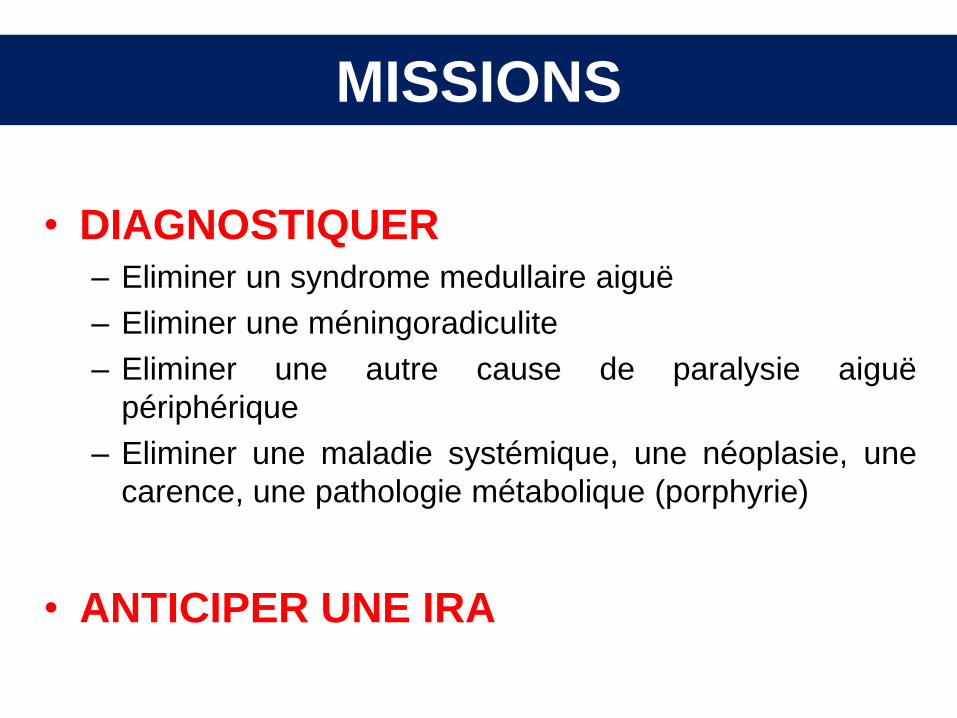
MISSIONS
• DIAGNOSTIQUER – Eliminer un syndrome medullaire aiguë
– Eliminer une méningoradiculite
– Eliminer une autre cause de paralysie aiguë
périphérique
– Eliminer une maladie systémique, une néoplasie, une
carence, une pathologie métabolique (porphyrie)
• ANTICIPER UNE IRA

ACUTE FLACCID WEAKNESS
SENSORIMOTOR
PARALYSIS
PURE MOTOR
PARALYSIS
1. ± MRI
2. CSF ANALYSIS
3. BLOOD TESTS (ESR)
4. EMG (Axonal PN,
Demyelinating PN)
1. BLOOD TESTS (K+)
2. CSF ANALYSIS
3. EMG (Myopathy, NMJ,
Neuropathy)
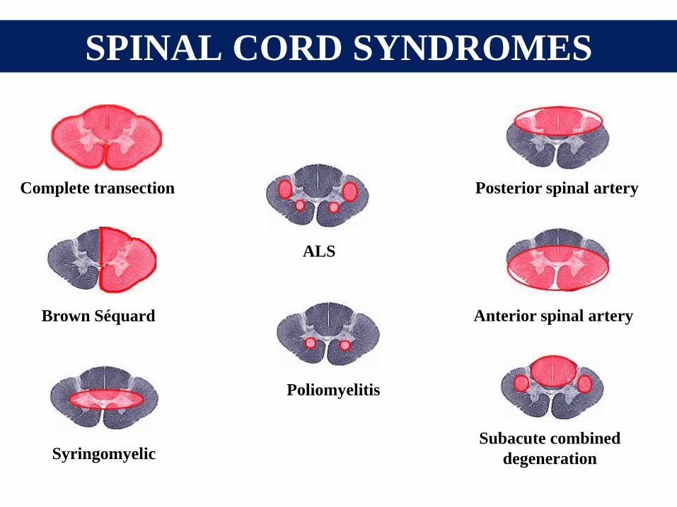
SPINAL CORD SYNDROMES
Syringomyelic
Complete transection
Brown Séquard Anterior spinal artery
Posterior spinal artery
Subacute combined
degeneration
ALS
Poliomyelitis

Pyramidal signs
Sensory level
Cauda-equina syndrome
SPINAL CORD MRI
ACUTE SENSORIMOTOR HYPOREFLEXIC
PARALYSIS

ACUTE SENSORIMOTOR
HYPOREFLEXIC PARALYSIS
ESR NORMAL ESR INCREASED
CSF
NORMAL
TOXIC (Thallium, arsenic…)
METABOLIC (Gly, Vit …)
VASCULITIS (SLE…)
PRIMARY GBS
VASCULITIS (SLE…)
INCREASED
CSF CELLS MENINGORADICULITIS MENINGORADICULITIS
INCREASED
CSF
PROTEIN
PRIMARY GBS
CANCER, LYMPHOMA,
VASCULITIS, DIPHTERIA,
HIV.
CANCER, LYMPHOMA,
VASCULITIS, DIPHTERIA,
HIV.

PURE MOTOR HYPOREFLEXIC DEFICIT
SIGNS K+ CSF EMG
PERIODIC
PARALYSIS EXERCISE MYOPATHY
MYASTHENIA
GRAVIS
VARIATION
EYE MVT NM JUNCTION
BOTULISM
FOOD
POISONNING
PUPILL
NM JUNCTION
POLIO
MYELITIS
TRAVEL
DIARRHEA
CELLS
ANTERIOR
HORN CELLS
PORPHYRIC
NEUROPATHY
CONFUSION
PAIN
POLY
NEUROPATHY
PRIMARY
GBS
INFECTION
ASCENDANT
PROTEIN
POLY
NEUROPATHY
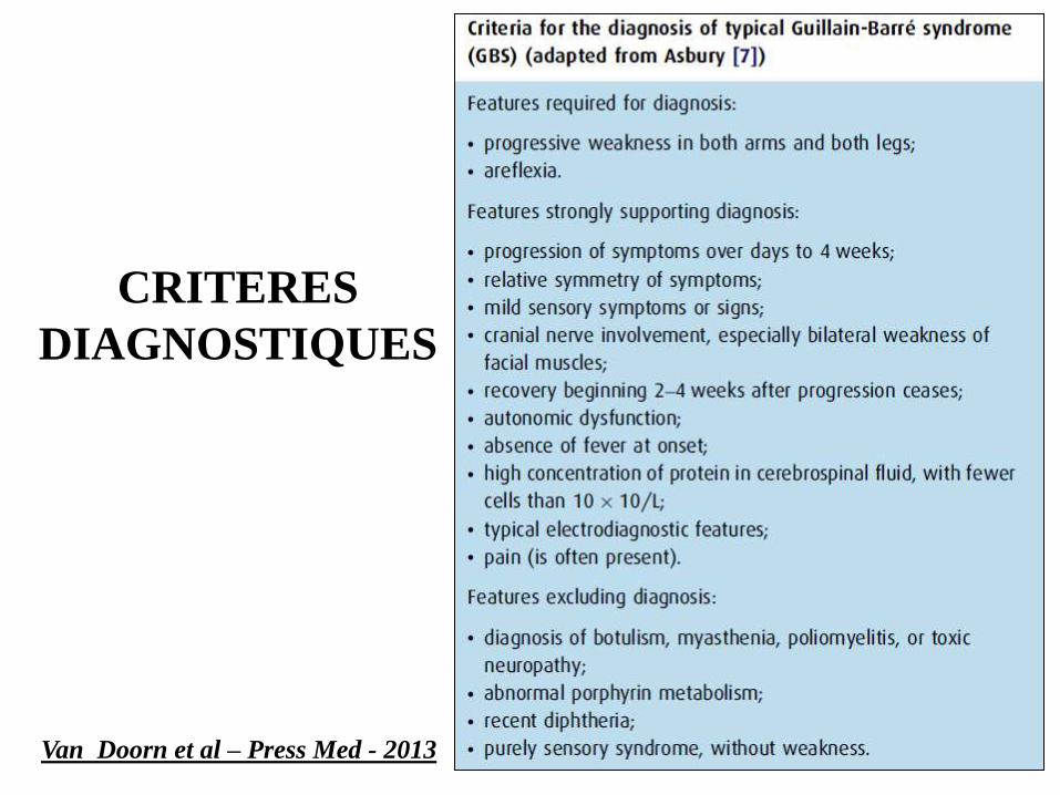
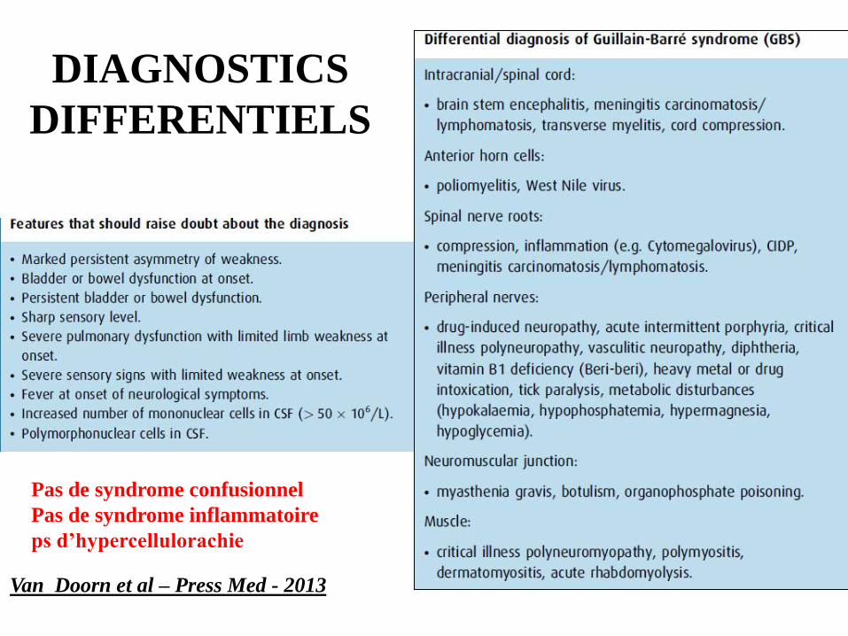
Van Doorn et al – Press Med - 2013
CRITERES
DIAGNOSTIQUES
Van Doorn et al – Press Med - 2013
DIAGNOSTICS
DIFFERENTIELS
Pas de syndrome confusionnel
Pas de syndrome inflammatoire
ps d’hypercellulorachie

GUILLAIN-BARRE SYNDROME
COURSE
Day 0 Time
Motor deficit
Extension Recovery
Plateau
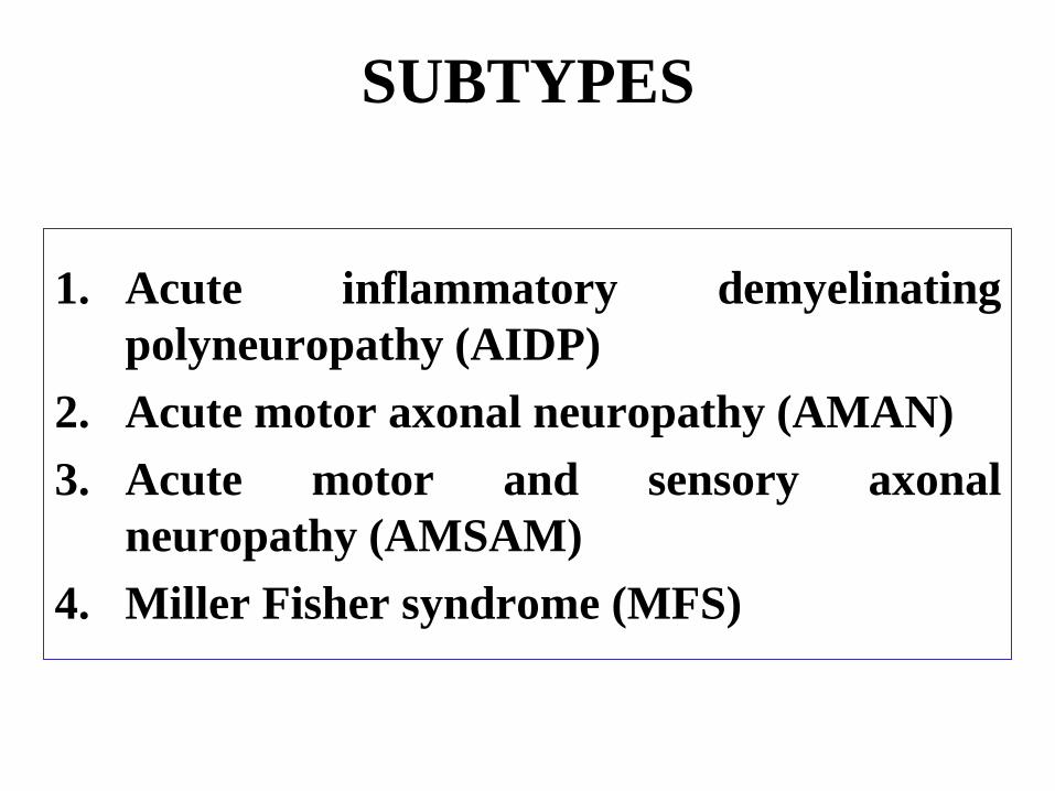
SUBTYPES
1. Acute inflammatory demyelinating
polyneuropathy (AIDP)
2. Acute motor axonal neuropathy (AMAN)
3. Acute motor and sensory axonal
neuropathy (AMSAM)
4. Miller Fisher syndrome (MFS)
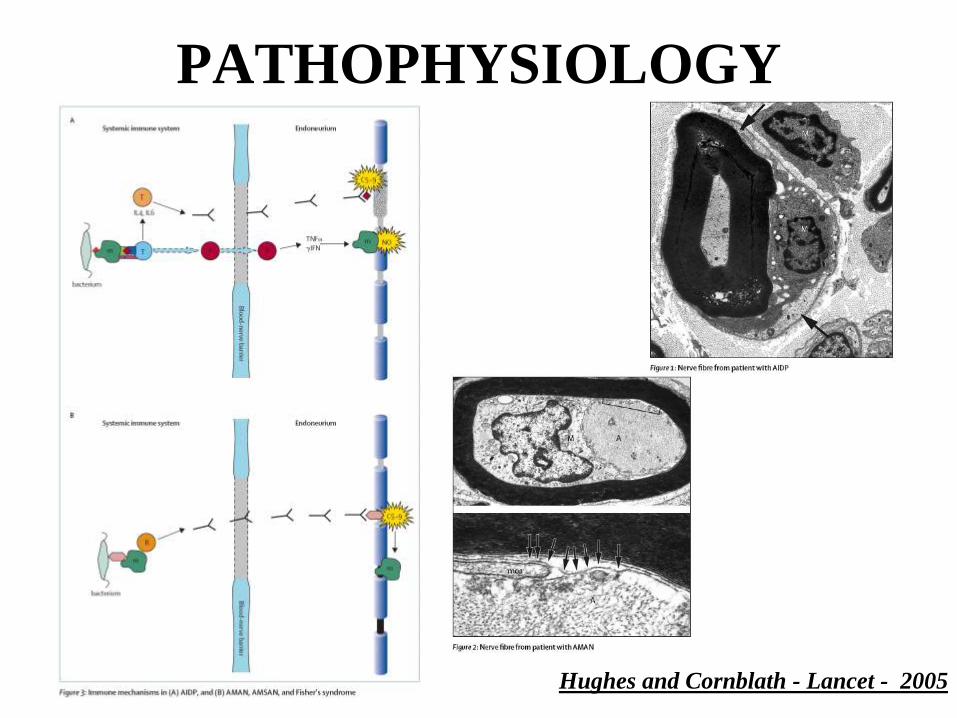
PATHOPHYSIOLOGY
Hughes and Cornblath - Lancet - 2005

ANTIGANGLIOSIDE
ANTIBODIES
Hughes and Cornblath - Lancet - 2005
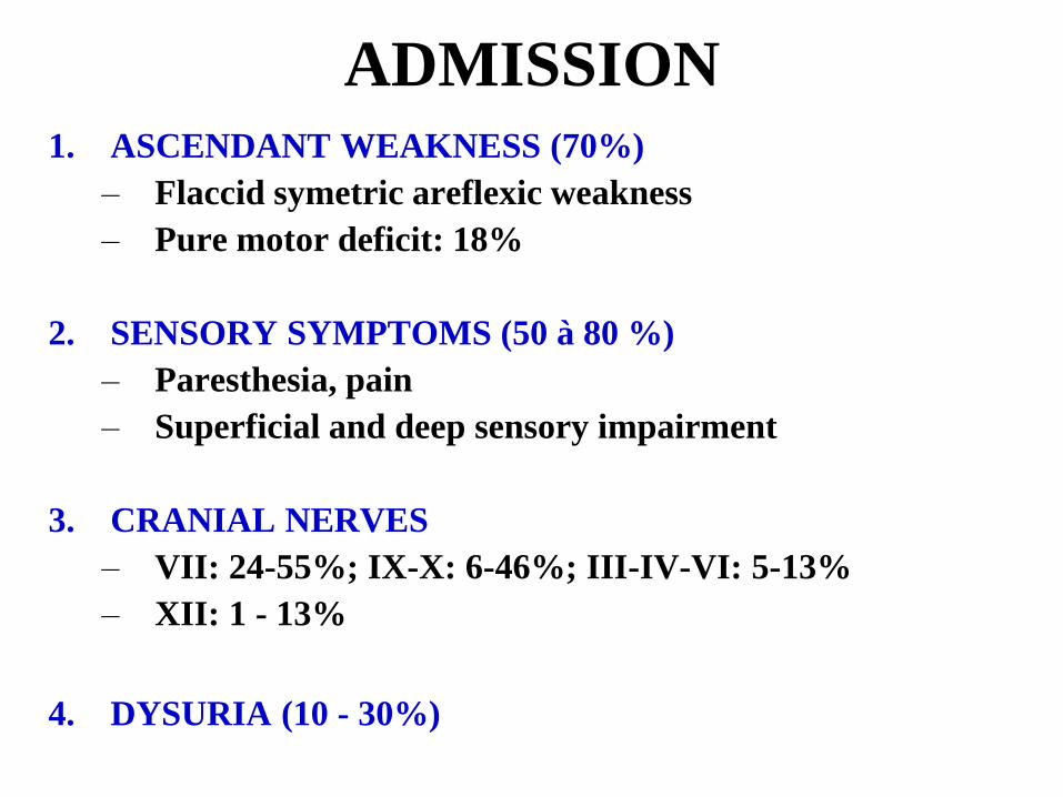
ADMISSION 1. ASCENDANT WEAKNESS (70%)
– Flaccid symetric areflexic weakness
– Pure motor deficit: 18%
2. SENSORY SYMPTOMS (50 à 80 %)
– Paresthesia, pain
– Superficial and deep sensory impairment
3. CRANIAL NERVES
– VII: 24-55%; IX-X: 6-46%; III-IV-VI: 5-13%
– XII: 1 - 13%
4. DYSURIA (10 - 30%)

SCALES
MRC sum score
Kleyweg et al. - Muscle Nerve - 1991
Disability grade

SEVERITY AT ADMISSION
1. SWALLOWING IMPAIRMENT
- In 6 to 46%
2. RESPIRATORY DYSFUNCTION
- Respiratory symptoms in 40 to 60%
- Vital capacity < 1 L in 16%
3. CV AUTONOMIC DYSFUNCTION
- in about 15%
- Correlated with weakness

ANTICIPATING ACUTE
RESPIRATORY FAILURE
IN GUILLAIN-BARRE
SYNDROME

MECHANICAL VENTILATION
1. Required in 20 to 30%
2. Median time from admission to MV: 2 days
3. Median MV duration: 21 days
4. Up to 1/3 weaned from MV within 3 weeks

MECHANISMS
Diaphragm
Accessory
Abdominal
CV
Pimax
Pemax
cough
Atelectasia
Bulbar
dysfunction Aspiration
Phrenic
IX-X
Hypoventilation
Hypoxaemia
T8-T12
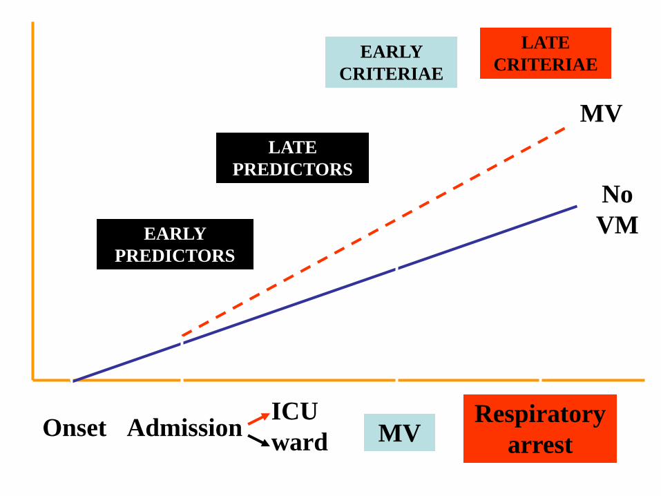
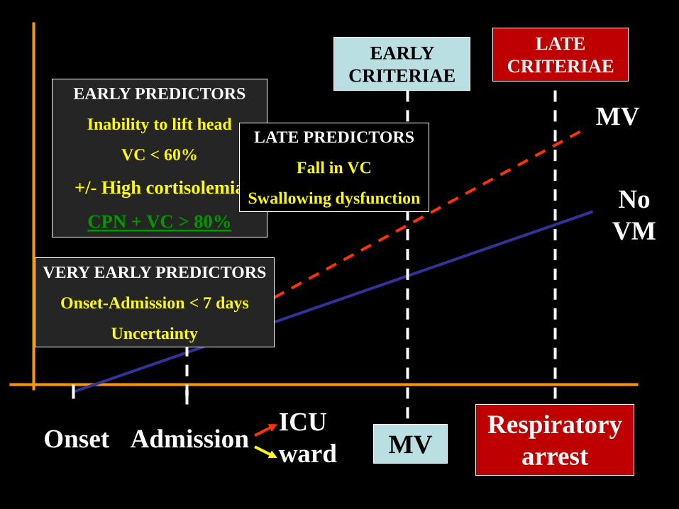
Admission Onset MV
EARLY
PREDICTORS
EARLY
CRITERIAE
LATE
CRITERIAE
LATE
PREDICTORS
Respiratory
arrest
ICU
ward
MV
No
VM

CRITERIA FOR INTUBATION
MAJOR CRITERIA
1. Signs of respiratory distress
2. VC< 15 ml/kg, Pimax ou Pemax < 25 cm H2O
3. PaCO2 > 6,4 kPa
4. PaO2 < 7.5 kPa (FiO2= 0,21)
MINOR CRITERIA
1. Expectoration inefficient
2. Severe bulbar dysfunction
3. Atelectasis
Ropper Neurology 1985 - Wijdicks Neurology 1998

THE RISKS
• RESPIRATORY AND CARDIAC ARREST
• ASPIRATION
• ARDS
Orlikowski et al - ICM - 2007
N= 87
VAP = 67 (75%)
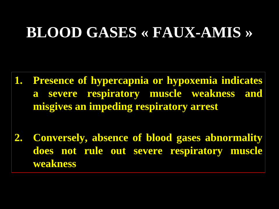
BLOOD GASES « FAUX-AMIS »
1. Presence of hypercapnia or hypoxemia indicates
a severe respiratory muscle weakness and
misgives an impeding respiratory arrest
2. Conversely, absence of blood gases abnormality
does not rule out severe respiratory muscle
weakness

HYPOXEMIA
• Can be induced by increased PaCO2
– physiological law
– Check that decrease in PAO2 is explained by increase in PaO2
• Pneumoniae, atelectasia…
– Check the chest x Ray
• Pulmonary embolism
– To be suspected if chest x ray is normal
– Frequent in this patient

EARLY PREDICTORS
INABILITY OR 95% CI p
PROGRESSION < 7 days 2.51 (1.7 – 3.8) < 0.0001
STAND 2.53 (1.4 – 3.3) < 0.0005
RISE ELBOW 2.99 (1.8 – 4.9) < 0.0001
RISE HEAD 4.34 (2.7 – 6.7) < 0.0001
COUGH 9.09 (4.0 – 20.00) < 0.0001
CYTOLYSIS 2.09 (1.4 – 3.2) < 0.0005
n = 722
Sharshar et al-Crit Care Med-2003

EARLY PREDICTORS
n= 196
Inability OR 95% CI p Value
PROGRESSION
< 7 days 5.0 (1.4 – 5.7) < 0.003
HEAD 5.0 (1.9 – 12.5) < 0.0011
VC < 60% 2.86 (2.4 – 10.0) < 0.0001
Sharshar et al-Crit Care Med-2003
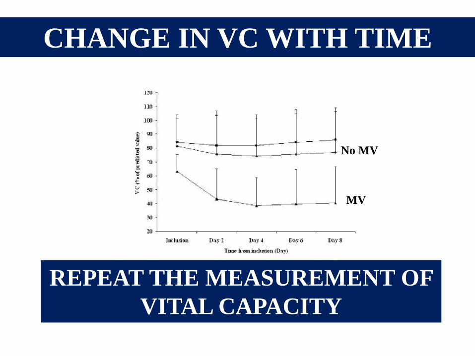
CHANGE IN VC WITH TIME
Durand et al – Neurology - 2005
MV
No MV
REPEAT THE MEASUREMENT OF
VITAL CAPACITY

OTHER PREDICTORS
1. 30 to 50% fall in VC
or VC < 20 ml/kg
2. 30% fall in Pimax or Pemax
or Pimax ou Pemax< 30 to 40 cmH2O
3. Bulbar dysfunction
Hahn et al Arch Neurol 2001
Chevrolet et al Am Rev Respir Dis 1991
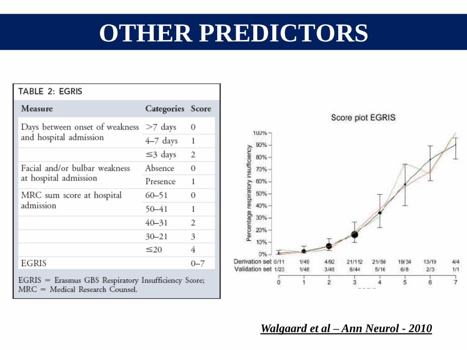
OTHER PREDICTORS
Walgaard et al – Ann Neurol - 2010

ELECTROPHYSIOLOGY
• Phrenic ENMG
– Not helpful (Durand – Neurology – 2005)
• Standard ENMG
– Demyeliting form at higher risk (Durand–
Eur J Neurol 2003)
– No risk if no conduction block on common
peroneal nerve + VC >80% (Durand –
Lancet Neurology – 2006)
Durand et al – Lancet Neurology - 2006

CORTISOLEMIA
Variable no MV
(Group 2)
MV > 24h
(Group 3) P*
n 60 17
[Cortisol]T0 (ng/ml) 20.4 ± 9.6 28.5 ± 12.1 0.01
[Cortisol]T60 (ng/ml) 42.4 ± 14.8 53.1 ± 16.8 0.05
Strauss et al – CCM - 2009
[Cortisol]T0 correlated with occurrence of dysautonomia

FEAR
N (%)
Marcadet et al - Submitted
0
20
40
60
80
100
Dyspnea Pain Weakness Uncertainty
No MV
MV
Admission Onset MV
EARLY PREDICTORS
Inability to lift head
VC < 60%
+/- High cortisolemia
CPN + VC > 80%
EARLY
CRITERIAE
LATE
CRITERIAE
LATE PREDICTORS
Fall in VC
Swallowing dysfunction
Respiratory
arrest
ICU
ward
MV
No
VM
VERY EARLY PREDICTORS
Onset-Admission < 7 days
Uncertainty

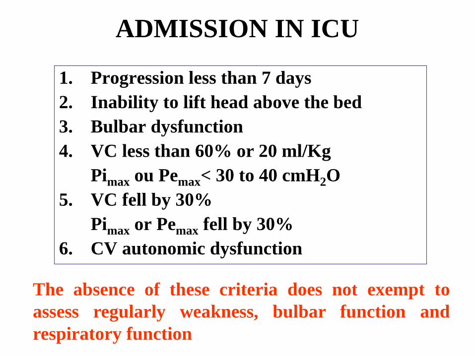
ADMISSION IN ICU
1. Progression less than 7 days
2. Inability to lift head above the bed
3. Bulbar dysfunction
4. VC less than 60% or 20 ml/Kg
Pimax ou Pemax< 30 to 40 cmH2O
5. VC fell by 30%
Pimax or Pemax fell by 30%
6. CV autonomic dysfunction
The absence of these criteria does not exempt to assess
regularly weakness, bulbar function and respiratory
function

OTHER ABNORMALITIES
HYPONATREMIAE
1. Hyponatremia < 133 mmol/L : 31%
2. Pseudohyponatremia due to IvIg: 46%
3. Hyponatremia: worst outcome ?
LIVER DYSFUNCTION
1. Cytolysis: 25%
2. Secondary to CMV: 25%
3. Predictors of MV
Colls Int Med J 2003; Oomes et al Neurology 1996;
Sharshar et al Crit Care Med 2003
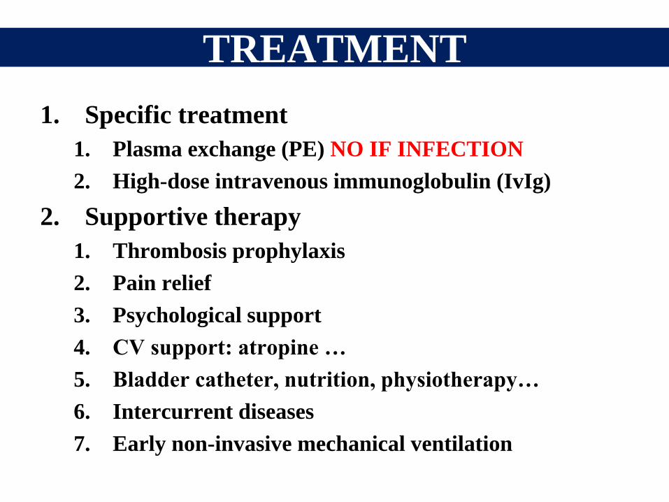
TREATMENT
1. Specific treatment
1. Plasma exchange (PE) NO IF INFECTION
2. High-dose intravenous immunoglobulin (IvIg)
2. Supportive therapy
1. Thrombosis prophylaxis
2. Pain relief
3. Psychological support
4. CV support: atropine …
5. Bladder catheter, nutrition, physiotherapy…
6. Intercurrent diseases
7. Early non-invasive mechanical ventilation
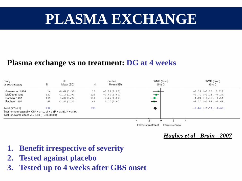
PLASMA EXCHANGE
Plasma exchange vs no treatment: DG at 4 weeks
Hughes et al - Brain - 2007
1. Benefit irrespective of severity
2. Tested against placebo
3. Tested up to 4 weeks after GBS onset

IMMUNOGLOBULIN
Hughes et al - Brain - 2007
Plasma exchange vs IvIg: DG at 4 weeks
1. Tested against PE
2. Tested in mild or severe GBS
3. Tested within 2 weeks after GBS onset
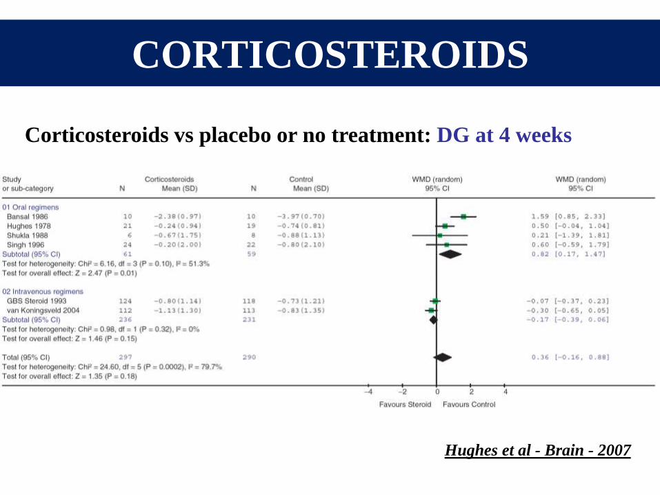
CORTICOSTEROIDS
Hughes et al - Brain - 2007
Corticosteroids vs placebo or no treatment: DG at 4 weeks
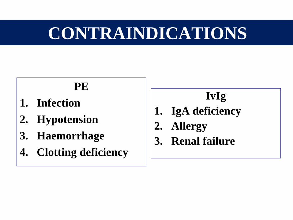
CONTRAINDICATIONS
PE
1. Infection
2. Hypotension
3. Haemorrhage
4. Clotting deficiency
IvIg
1. IgA deficiency
2. Allergy
3. Renal failure
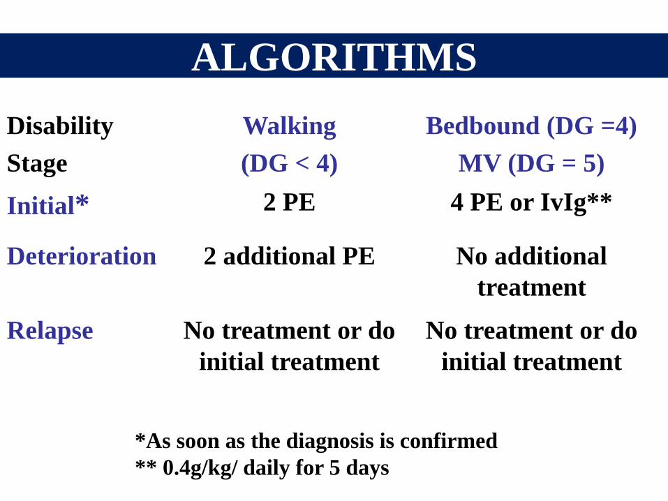
ALGORITHMS
Disability
Stage
Walking
(DG < 4)
Bedbound (DG =4)
MV (DG = 5)
Initial* 2 PE 4 PE or IvIg**
Deterioration 2 additional PE No additional
treatment
Relapse No treatment or do
initial treatment
No treatment or do
initial treatment
*As soon as the diagnosis is confirmed
** 0.4g/kg/ daily for 5 days
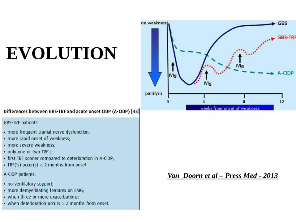
EVOLUTION
Van Doorn et al – Press Med - 2013

Raymond Poincaré
THANK YOU
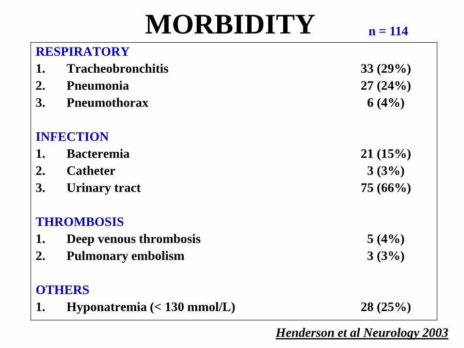
MORBIDITY RESPIRATORY
1. Tracheobronchitis 33 (29%)
2. Pneumonia 27 (24%)
3. Pneumothorax 6 (4%)
INFECTION
1. Bacteremia 21 (15%)
2. Catheter 3 (3%)
3. Urinary tract 75 (66%)
THROMBOSIS
1. Deep venous thrombosis 5 (4%)
2. Pulmonary embolism 3 (3%)
OTHERS
1. Hyponatremia (< 130 mmol/L) 28 (25%)
Henderson et al Neurology 2003
n = 114
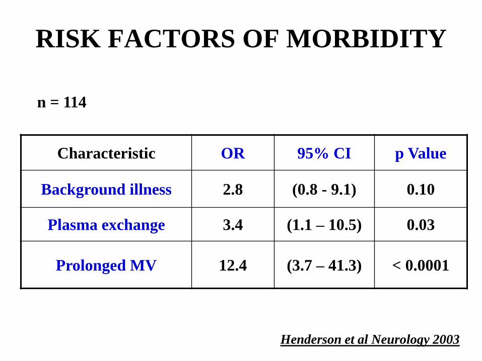
RISK FACTORS OF MORBIDITY
Characteristic OR 95% CI p Value
Background illness 2.8 (0.8 - 9.1) 0.10
Plasma exchange 3.4 (1.1 – 10.5) 0.03
Prolonged MV 12.4 (3.7 – 41.3) < 0.0001
Henderson et al Neurology 2003
n = 114
ADMISSION IN ICU
1. Progression less than 7 days
2. Inability to lift head above the bed
3. Bulbar dysfunction
4. VC less than 60% or 20 ml/Kg
Pimax ou Pemax< 30 to 40 cmH2O
5. VC fell by 30%
Pimax or Pemax fell by 30%
6. CV autonomic dysfunction
The absence of these criteria does not exempt to
assess regularly weakness, bulbar function and
respiratory function

PUZZLE
Pathogen agent
1. C. jejuni
2. CMV
3. Others
Anti-Ganliosides
1. Ant-GM1
2. Anti-GM2
3. Anti-G1QB
Subtypes
1. AIDP
2. AMAN
3. AMSAM
4. MFS
Clinic
1. Sensorimotor
2. Pure motor
3. Cranial nerves
Severity
1. Aspiration
2. Intubation
3. Dysautonomia
Outcome
1. Recovery
2. Sensory sequellae
3. Motor sequellae

WHY NORMOCAPNIC IS NOT
« SAFE »
Tid
al
Volu
me
Normocapnic
but no reserve
because low
VC
Respiratory
arrest if
anything
happpen
Healthy subject
Can increase his VT
Patients with NMD
cannot
increase his VT

ACUTE FLACCID WEAKNESS
SENSORIMOTOR
PARALYSIS
PURE MOTOR
PARALYSIS
1. ± MRI
2. CSF ANALYSIS
3. BLOOD TESTS (ESR)
4. EMG (Axonal PN,
Demyelinating PN)
1. BLOOD TESTS (K+)
2. CSF ANALYSIS
3. EMG (Myopathy, NMJ,
Neuropathy)

Pyramidal signs
Sensory level
Cauda-equina syndrome
SPINAL CORD MRI
ACUTE SENSORIMOTOR HYPOREFLEXIC PARALYSIS

TRACHEOSTOMY
Prolonged MV > 21 days
1. Elder patients
2. Preexisting pulmonary disease
3. Pulmonary function dint/d12 ratio < 1
1. PF score = CV + Pimax + Pemax
2. Sensitivity = 70%, specificity: 100%
Lawn and Wijdicks Muscle Nerve 1999, 2000

ACUTE SENSORIMOTOR
HYPOREFLEXIC PARALYSIS
ESR NORMAL ESR INCREASED
CSF
NORMAL
TOXIC (Thallium, arsenic…)
METABOLIC (Gly, Vit …)
VASCULITIS (SLE…)
PRIMARY GBS
VASCULITIS (SLE…)
INCREASED
CSF CELLS MENINGORADICULITIS MENINGORADICULITIS
INCREASED
CSF
PROTEIN
PRIMARY GBS
CANCER, LYMPHOMA,
VASCULITIS, DIPHTERIA,
HIV.
CANCER, LYMPHOMA,
VASCULITIS, DIPHTERIA,
HIV.

PURE MOTOR
HYPOREFLEXIC DEFICIT
SIGNS K+ CSF EMG
PERIODIC
PARALYSIS EXERCISE MYOPATHY
MYASTHENIA
GRAVIS
VARIATION
EYE MVT NM JUNCTION
BOTULISM
FOOD
POISONNING
PUPILL
NM JUNCTION
POLIO
MYELITIS
TRAVEL
DIARRHEA
CELLS
ANTERIOR
HORN CELLS
PORPHYRIC
NEUROPATHY
CONFUSION
PAIN
POLY
NEUROPATHY
PRIMARY
GBS
INFECTION
ASCENDANT
PROTEIN
POLY
NEUROPATHY
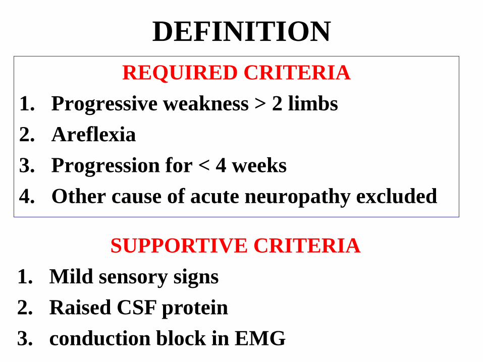
DEFINITION
REQUIRED CRITERIA
1. Progressive weakness > 2 limbs
2. Areflexia
3. Progression for < 4 weeks
4. Other cause of acute neuropathy excluded
SUPPORTIVE CRITERIA
1. Mild sensory signs
2. Raised CSF protein
3. conduction block in EMG
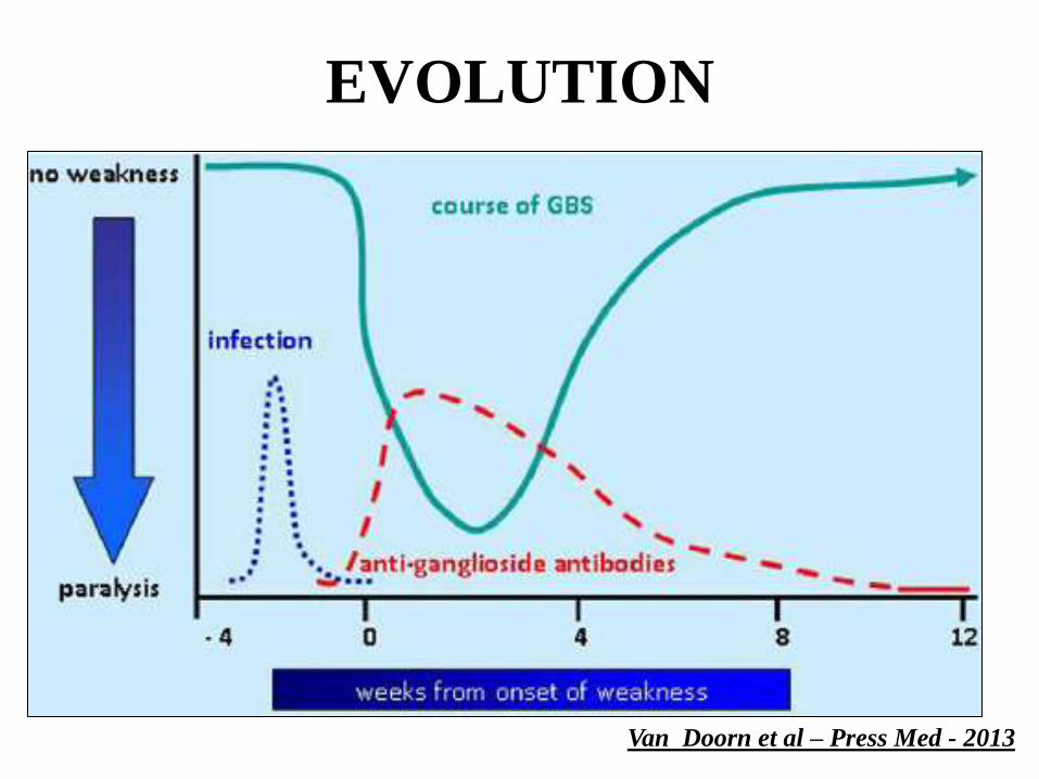
EVOLUTION
Van Doorn et al – Press Med - 2013