healthmed - volume 7 / number 8 / 2013 histological...
TRANSCRIPT

HealthMED - Volume 7 / Number 8 / 2013
Journal of Society for development in new net environment in B&H 2445
Abstract
Considering that ethanol caused number of health problems in the world , the present study was initiated to investigate the histological haz-ardous effects of ethanol on the liver and spleen. Animals were divided into 4 groups, the first group served as a control group, the second, third and fourth received 1,2 and 6 ml/kg/bw of 70% ethanol respectively. At the day 5 post treatment, the liver and spleen sections were prepared for the histological study. Ethanol intake induced marked histological alterations in the liver and spleen that correlated with the dose taken, low dose induced liver and spleen injury mainly as cytoplasmic degeneration in liver and abnormal structure of spleen, whereas, high doses of ethanol resulted in fibrosis in liver and splenomegaly.
Key words: Ethanol, mice, liver, spleen.
Introduction
Ethanol has been a part of the human diet for centuries. However, its consumption in excess can result in a number of health problems, most nota-bly liver damage , Ethanol cannot be excreted and must be metabolized, primarily by the liver (Berg et al., 2002). Alcoholic Liver Disease (ALD) is a blanket term in which conditions related specifi-cally to the liver and alcohol use fall under. The most prevalent types of Alcoholic Liver Disease, or ALD, are fatty liver, alcoholic hepatitis, and cir-rhosis. Often, as people continue to drink heavily, they progress from fatty liver to hepatitis to cirrho-sis. All three of the disorders can occur together ( Robert et al., 2004). Fatty Liver, which occurs af-ter acute alcohol ingestion, is generally reversible with abstinence and is not thought to predispose to any chronic form of liver disease if abstinence and/or moderation are maintained (Kyrsten, 2004). It
is estimated by the National Institutes of Health that about 20 percent of alcoholics and heavy drinkers develop fatty liver, or steatosis (Robert et al., 2004). Other sources estimate that 90-100 per-cent of patients with heavy drinking will develop this disease (Ismail and Riely, 2006). The condi-tion can lead to death if alcohol consumption is not reduced or stopped, Alcohol can be a factor in several other forms of liver disease not specifical-ly attributed to it, and may interact with risk fac-tors for other forms of liver disease. An example of this is people with alcohol-related cirrhosis are at a higher risk of developing liver cancer. Those with Hepatitis B or C accompanied with heavy drinking are at a much higher risk of cirrhosis than with heavy drinking alone (Robert et al., 2004). Clinical evidence supports a correlation between excessive alcohol consumption and certain bacte-rial infections. For example, alcoholics who have developed cirrhosis of the liver are predisposed to spontaneous bacterial peritonitis. Phagocytes are an important defense against infection in this part of the body and defects in phagocytic cell func-tion, observed in many alcoholics, may predispose these individual to peritoneal infection (Roselle, 1992). There is considerable evidence indicating that ethanol consumption alters immune system function and leads to increased susceptibility to infections and neoplastic diseases (Nath and Sza-bo, 2009; Lau et al., 2009; Nava‐Aguilera et al., 2009; Szabo and Mandrekar, 2009).
Materials and Methods
Animals and experimental designMale swiss mice weighed 25 to 30 g were ob-
tained from King Saoud university animal house. Mice were provided with water and balanced diet ad libitum. 20 adult swiss mice were randomly di-vided into 4 groups of 5 mice in each as following:
Histological study on the hazardous effects of ethanol on liver and spleen in Swiss albino mice Badr Abdullah Aldahmash1, Doaa Mohamed El-Nagar2
1 Medical Laboratory Department, College of Health Sciences, King Saud University, Riyadh, Kingdom of Saudi Arabia,2 Faculty of Sciences and Humanities, University of Salman bin Abdul Aziz, Kingdom of Saudi Arabia.

2446
HealthMED - Volume 7 / Number 8 / 2013
Journal of Society for development in new net environment in B&H
- Group 1: control group ( normal mice without treatment).
- Group 2 : mice treated with 1ml /kg/bw of 70% ethanol orally by stomach gavage for consecutive 5 days.
- Group 3 : mice treated with 2ml/kg/bw of 70% ethanol orally by stomach gavage for consecutive 5 days.
- Group 4: mice treated with 6ml/kg/bw of 70% ethanol orally by stomach gavage for consecutive 5 days.
Histological examinationMice were sacrificed in the sixth day of the
beginning of the experiment, livers and spleens were collected and cut into small pieces and fixed 10% neutral buffered formalin, then embedded in paraffin and sectioned. The sections were stained with Hx&E stain for histopathological examinati-on and photographing.
Results
1. LiverThe liver of normal mice (untreated) consists of
a vast interanastomosing network of hepatocytes arranged in single-cell thick plates separated by vas-cular sinusoids. The hepatocytes along with vascu-lar channels form organized micro structures which serve as structural and functional units. The liver is composed of innumerable lobules, each of which is a hexagonal structure consisting of a central vein surrounded by radiating hepatocyte plates. Howe-ver, another concept of a functional unit defines an acinus as the functional unit in relation to terminal portal branches and terminal hepatic venule. Portal tracts surround the classical lobules. An interlobular portal vein is also shown (Figure 1).
Histopathological examination of liver of mice received 1ml/kg/bw of 70% ethanol orally for 5 days showed hazardous effects on liver repre-sented by appearance of many necrotic foci (N), vacuolar degeneration (vc) in the cytoplasm of hepatocytes, some cells showed abundant nuclei, others showed pyknotic (P) nuclei where others appeared without nuclei. Dilatation in blood sinu-soids with kuppfer (K)cells besides to existence of lymphocytic infiltration foci were abundant (L) (Figures 2a&2b). Sections stained by masson’s
trichrome technique showed wide dilated and branched central vien (CV) (Figure 2c),moreover, Figure 2d revealed congested and dilated central vein with destructed wall and surrounded by ne-crotic areas.
Figure 1. Uuntreated liver section showed nor-mal central vein (CV) surrounded by normal he-patocytes (mag.x200 H&E)
Examination of liver sections of mice received oral administration of 2ml/kg/bw of 70% ethanol for 5 days showed more complicated alterations remarked by destruction in the vein wall surroun-ded by aggregations of lymphocytes and necrotic areas (N), in addition, pyknotic nuclei were abun-dant in some cells, and other cells showed comple-te degeneration in its nuclei, moreover, sections revealed fusion of cells due to degeneration of cell walls (Figures 3a&3b). Whereas, trichrome stai-ned sections (Figures 3c&3d) displayed wide ne-crotic areas were filled with erythrocytic exudates, in addition to, wide dilated blood sinusoids with kupfer cells, dilated portal vein appeared surro-unded by concentric layers of collagenous fibers with lymphocytes in between.
Livers of mice received oral administration of 6ml/kg/bw of 70% ethanol revealed the worst hazardous features in liver tissues compared with previous doses mainfested by cytoplasmic and nuclear degeneration in hepatocytes, some cells showed pyknotic nuclei (p), dilatation in blood si-nusoids in addition to appearance of kupfer cells in the sinusoids, presence of necrotic areas besides to blood exudates among liver cells and appearance of blood congestion in dilated central vein (Figure 4a), moreover, Figure 4b showed some aggregati-ons of lymphocytes in liver tissue. Concentrated

HealthMED - Volume 7 / Number 8 / 2013
Journal of Society for development in new net environment in B&H 2447
layers of collagenous fibers were deposited in the tissue accompanied by lymphocytic infiltration and blood exudates, moreover, dilated central vein surrounded by thick concentric layers of collage-nous fibers besides to presence of necrotic areas were seen (Figures 4c&4d).
2. SpleenSpleen is a large lymphoid organ that contains
two main compartments known as “the white pulp and the red pulp“. Spleen is covered with a fibro-us capsule from which trabeculae enters into the parenchyma. The fibrous trabeculae ramify thro-ughout the spleen and form supportive sheathing around blood vessels. The white pulp consists of lymphoid follicles, Each B-lymphoid follicle contains a distinct marginal zone of lymphocytes
(outer rim of lymphocytes) around the mantle zone of the follicle (inner rim of lymphocytes). The marginal zone consists of loosely arranged lymphocytes. This ‘perifollicular zone’ is the bo-undary between white and red pulp and serves as the area where macrophages are abundant, The macrophages serve as the ‘custom officers’ for the newly entered red and white blood cells and parti-culate material. The red pulp is the area of spleen in between white pulp and consists of open sinu-ses and cellular cords, Splenic sinuses are open vascular spaces lined by a discontinuous layer of endothelial cells and supported by a fenestrated basal lamina and reticular fibers. The surrounding cellular splenic cords provide a tissue framework maintaining the network of sinuses.
Figure 2. Liver sections treated with 1ml/kg/bw of ethanol showed marked changes (2a mag.x400 H&E) necrotic areas (N), vacuolar degeneration (VC), kupffer cells (K)and blood sinusoids (S) (2b mag.x400 H&E) lymphocytic infiltration (L) and pyknotic nuclei (P). (2c mag.x400 M.Tr.) dilated central vein, (2d mag.x400 M.Tr.) dilated central vein with erythrocytic congestion surrounded by necrotic foci (N).

2448
HealthMED - Volume 7 / Number 8 / 2013
Journal of Society for development in new net environment in B&H
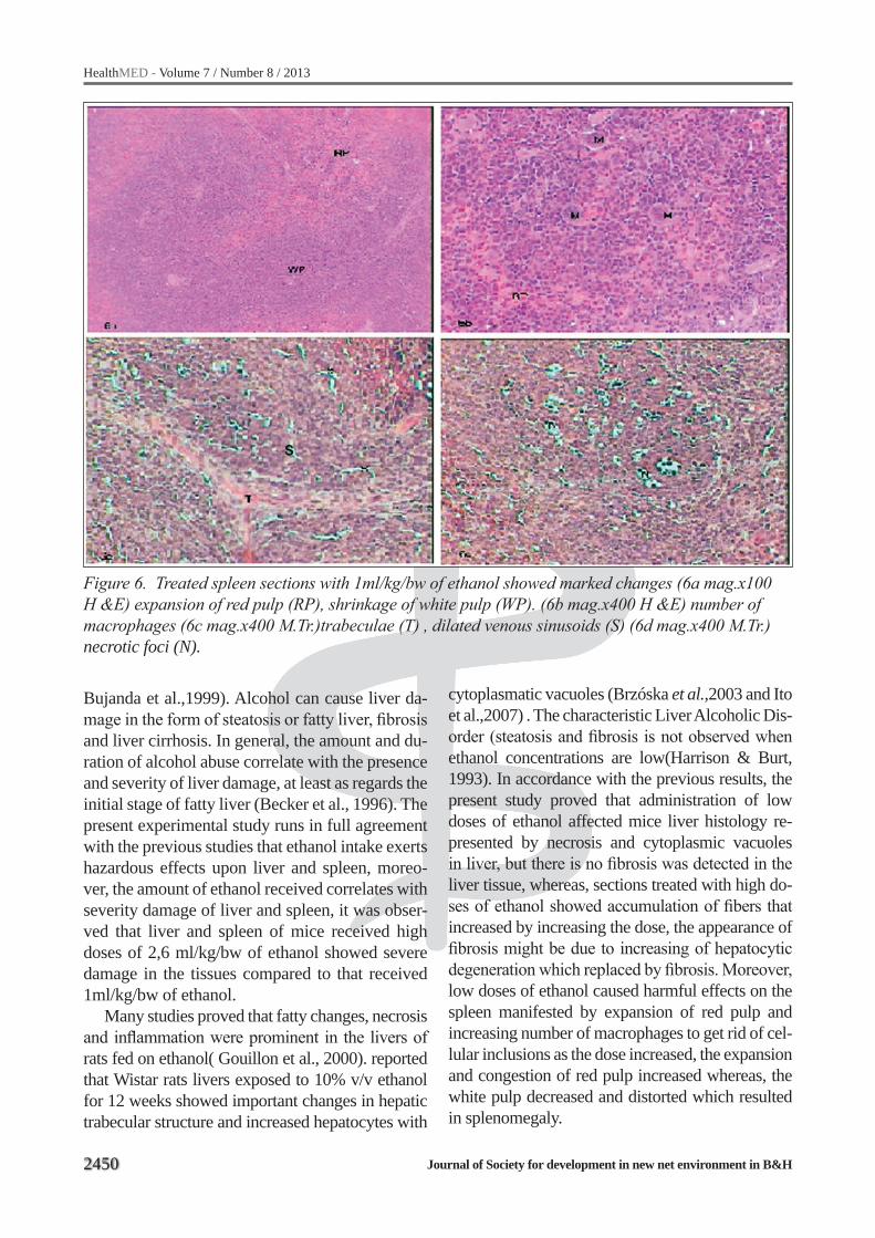
Examination of mice spleen sections received 1 ml/kg/bw oral administration of 70% ethanol for 5 days showed marked changes in the spleen tissue represented by expansion of red pulp due to venous congestion, presence of number of resi-dent macrophages to get rid of abnormal red blo-od cells and cellular inclusions due to the venous congestion (Figures 6a &6b). Dilatation in venous sinuse that lined with endothelial cells besides to necrotic foci appeared in Figures 6c and 6d.
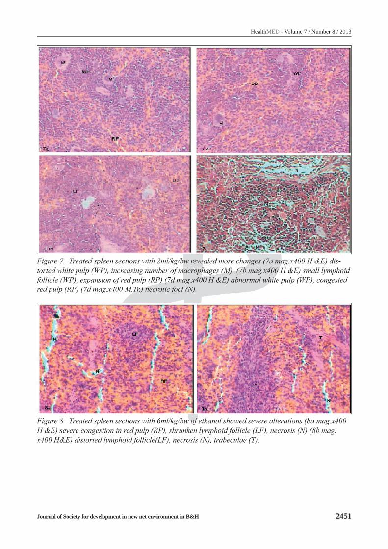
Spleen sections of mice received oral admini-stration of 2 ml/kg/bw of 70% ethanol revealed abnormal architecture of spleen tissue remarked by distorted lymphoid follicles (white pulp) with resident macrophages in the marginal zone of the follicles, increasing the expansion of red pulp due to the increasing of venous congestion in addition to the dilatation in the venous sinuses ( Figures 7a, 7b & 7c), whereas, Figure 7d showed wide necro-
tic areas in the lymphoid follicle due to the dege-neration of B and T lymphocytes.
By increasing the dose of 70% ethanol to 6ml/kg/bw spleen sections showed very hazardous effects on the spleen represented by splenomegaly due to severe congestion that venous sinuses com-pletely disappeared because of filling of red blood cells, in other side, distorted white pulp appeared with small sized lymph follicles surrounded by ne-crotic areas (Figures 8a &8b).
Discussion
Ethanol, which is a weak anesthetic, is toxic for both humans and animals, and exerts important negative effects upon the liver, brain, heart, skele-tal muscle, pancreas, hematological and immune systems, gastrointestinal apparatus and endocrine system (Rottenberg, 1992, Rodés et al.,1990 and
Figure 3. Liver sections treated with 2ml/kg/bw of ethanol showed more complicated alterations (3a mag.x400 H&E) central vein (CV) with destructed wall surrounded by necrotic areas (N) and lymphocytic infiltration (L). (3b mag.x400 H&E) dilated blood sinusoids (bs), pyknotic nuclei of he-patocytes (P), necrotic foci (N). (3c mag.x400 M.Tr.) wide necrotic area(N) filled with erythrocytic exudates (Ex). (3d mag.x400 M.Tr.) dilated vein (V) surrounded by fibrotic layer (F).

HealthMED - Volume 7 / Number 8 / 2013
Journal of Society for development in new net environment in B&H 2449
Figure 4. Liver sections treated with 6ml/kg/bw of ethanol showed hazardous feathures (4a mag.x400 H&E) necrotic area (N) , pyknotic nuclei (P) , hepatocytic degeneration (d) (4b mag. X400 H&E) aggregations of lymphocytes (L) (4c mag.x400 M.Tr.) collagenous fibers (F) stained blue in the tissue, lymphocytic infiltration (L) and blood exudates (EX). (4d mag.x400 M.Tr.) dilated central vein (CV) surrounded by collagenous fibers (F) and wide necrotic areas (N).
Figure 5. Untreated spleen sections showed normal architecture (5a mag.x100 H&E) white pulp (WP) and red pulp (RP) (5b magx400 H&E)lymphoid follicle consists of lymphocytes (L), macrophages (M)
2450
HealthMED - Volume 7 / Number 8 / 2013
Journal of Society for development in new net environment in B&H
Bujanda et al.,1999). Alcohol can cause liver da-mage in the form of steatosis or fatty liver, fibrosis and liver cirrhosis. In general, the amount and du-ration of alcohol abuse correlate with the presence and severity of liver damage, at least as regards the initial stage of fatty liver (Becker et al., 1996). The present experimental study runs in full agreement with the previous studies that ethanol intake exerts hazardous effects upon liver and spleen, moreo-ver, the amount of ethanol received correlates with severity damage of liver and spleen, it was obser-ved that liver and spleen of mice received high doses of 2,6 ml/kg/bw of ethanol showed severe damage in the tissues compared to that received 1ml/kg/bw of ethanol.
Many studies proved that fatty changes, necrosis and inflammation were prominent in the livers of rats fed on ethanol( Gouillon et al., 2000). reported that Wistar rats livers exposed to 10% v/v ethanol for 12 weeks showed important changes in hepatic trabecular structure and increased hepatocytes with
Figure 6. Treated spleen sections with 1ml/kg/bw of ethanol showed marked changes (6a mag.x100 H &E) expansion of red pulp (RP), shrinkage of white pulp (WP). (6b mag.x400 H &E) number of macrophages (6c mag.x400 M.Tr.)trabeculae (T) , dilated venous sinusoids (S) (6d mag.x400 M.Tr.) necrotic foci (N).
cytoplasmatic vacuoles (Brzóska et al.,2003 and Ito et al.,2007) . The characteristic Liver Alcoholic Dis-order (steatosis and fibrosis is not observed when ethanol concentrations are low(Harrison & Burt, 1993). In accordance with the previous results, the present study proved that administration of low doses of ethanol affected mice liver histology re-presented by necrosis and cytoplasmic vacuoles in liver, but there is no fibrosis was detected in the liver tissue, whereas, sections treated with high do-ses of ethanol showed accumulation of fibers that increased by increasing the dose, the appearance of fibrosis might be due to increasing of hepatocytic degeneration which replaced by fibrosis. Moreover, low doses of ethanol caused harmful effects on the spleen manifested by expansion of red pulp and increasing number of macrophages to get rid of cel-lular inclusions as the dose increased, the expansion and congestion of red pulp increased whereas, the white pulp decreased and distorted which resulted in splenomegaly.
HealthMED - Volume 7 / Number 8 / 2013
Journal of Society for development in new net environment in B&H 2451
Figure 7. Treated spleen sections with 2ml/kg/bw revealed more changes (7a mag.x400 H &E) dis-torted white pulp (WP), increasing number of macrophages (M), (7b mag.x400 H &E) small lymphoid follicle (WP), expansion of red pulp (RP) (7d mag.x400 H &E) abnormal white pulp (WP), congested red pulp (RP) (7d mag.x400 M.Tr.) necrotic foci (N).
Figure 8. Treated spleen sections with 6ml/kg/bw of ethanol showed severe alterations (8a mag.x400 H &E) severe congestion in red pulp (RP), shrunken lymphoid follicle (LF), necrosis (N) (8b mag. x400 H&E) distorted lymphoid follicle(LF), necrosis (N), trabeculae (T).
2452
HealthMED - Volume 7 / Number 8 / 2013
Journal of Society for development in new net environment in B&H
References
1. Becker U, Deis A, Sorensen TI, Gronbaek M, Borch-Johnsen K, et al. Prediction of risk of liver disease by alcohol intake, sex and age: A prospective population study. Hepatology, 1996; 23: 1025-1029.
2. Berg JM, Tymoczko JLeof, Stryer L. Ethanol Alters Energy Metabolism in the Liver. Biochemistry. 5th edition. 2002.
3. Brozoska M, Moniuszko J, Marcinkiewicz B, Sawic-ki B. Liver and kidney function and histology in rats exposed to cadmium and ethanol. Alcohol Alcohol 2003; 38: 2-10.
4. Bujanda L, Gutiérrez-Stampa MA, Marimón JM. El vino a dosis moderadas; salud o enfermedad. Med Clin (Barc) 1999; 112: 29-35.
5. Gouillon Z, Lucas D, Li J, Hagbjork A, French B, et al. Inhibition of Ethanol-Induced Liver Disease in the Intragastric Feeding Rat Model by Chlormethiazole. Exp Biol Med. 2000; 224(4), 302-308.
6. Harrisond J, Burt D. Pathology of alcoholic liver dise-ase. Bailliére’s Clin Gastroenterol 1993; 7: 641 -662.
7. Ismail M, Riely C. Alcoholic Fatty Liver. emedicine from WebMD. October 31, 2006.
8. Ito T, Molina M, Andriolo A, Borges R. The combi-nation of atorvastatin and ethanol is not more hepa-totoxic to rats than the administration of each drug alone. Br J Med Biol Res 2007; 40: 343-348.
9. Fairbanks K, Alcoholic Liver Disease. Disease Ma-nagement FProject. The Cleveland Clinic, December 13, 2004.
10. Lau A, von D. V, Sander M, MacGuill M, Lanzke N, Spies C. Alcohol use disorder and perioperative immu-ne dysfunction. Anesth. Analg. 2009; 108: 916-920
11. Nath B, Szabo G. Alcohol‐induced modulation of si-gnaling pathways in liver parenchymal and nonpa-renchymal cells: implications for immunity. Semin. Liver Dis. 2009; 29: 166-177.
12. Nava-Aguilera E, Andersson N, Harris E, Mitchell S, Hamel C, et al. Risk factors associated with re-cent transmissionof tuberculosis: systematic review and meta-analysis. Int. J Tuberc. Lung Dis. 2009; 13: 17-26.
13. Mann RE, Smart RG, Govoni R. The Epidemiology of Alcoholic Liver Disease. National Institute on Alcohol Abuse and Alcoholism Publication. Septem-ber 29, 2004.
14. Rodés J, Urbano-Márquez A, Bach L. Alcohol y en-fermedad. Barcelona, Prous JR 1990.
15. Roselle G. Alcohol and the immune system. Alcohol Health & Research World. 1992.
16. Rottenberg H. Liver Cell Membrane Adaptation to Chronic Alcohol Consumption . Liver pathology and Alcohol. Drug and Alcohol Reviews. 1992; Volume 2: 91-115, DOI: 10.1007/978-1-4612-0421-3_3
17. Szabo G, Mandrekar P. A recent perspective on alco-hol, immunity, and host defense. Alcohol Clin Exp. 2009; Res 33: 220-232.
Corresponding AuthorBadr Abdullah Aldahmash,Medical Laboratory Department,College of Health Sciences,King Saud University,Riyadh,Kingdom of Saudi Arabia,E-mail: [email protected]