high resolution photoemission study of the formation and thermal stability of mg silicide on silicon
TRANSCRIPT
Thin Solid Films 519 (2011) 1861–1865
Contents lists available at ScienceDirect
Thin Solid Films
j ourna l homepage: www.e lsev ie r.com/ locate / ts f
High resolution photoemission study of the formation and thermal stability ofMg silicide on silicon
Patrick Casey ⁎, Greg HughesSchool of Physical Sciences, Dublin City University, Glasnevin, Dublin 9, Ireland
⁎ Corresponding author.E-mail address: [email protected] (P. Casey
0040-6090/$ – see front matter © 2010 Elsevier B.V. Aldoi:10.1016/j.tsf.2010.10.028
a b s t r a c t
a r t i c l e i n f oArticle history:Received 21 April 2010Received in revised form 18 August 2010Accepted 13 October 2010Available online 21 October 2010
Keywords:SilicideHigh resolution photoemissionAnnealingThermal stabilityGrowth mechanismCoating processes
The focus of this study is to use high resolution synchrotron based photoemission to investigate the initialgrowth mode of magnesium silicide which has been formed by both stepwise and continuous deposition ofmetallic Mg onto a thermally grown ultra-thin Si oxide surface. The findings suggest that stepwise depositionofMg initially results in the growth ofMg silicide islands on the surface. Furthermagnesiumdeposition leads tothe growth of metallic Mg on the surface of these silicide islands, along with the continued growth of silicidespecies on the uncovered oxide surface. However, it has been shown that continuous deposition of Mg resultsin considerably less silicide growth. The thermal stability of Mg silicide and amechanism for high temperaturesilicide growth have also been studied using conventional X-ray photoelectron spectroscopy. The resultssuggest that the presence of oxidised Si acts as a barrier to Si diffusion during vacuum annealing, hencepreventing the growth of further Mg silicide. It has also been shown that metallic Mg desorbs from the surfacebelow 300 °C, while Mg silicide is not stable at temperatures above 500 °C in contrast to other metal silicides.
).
l rights reserved.
© 2010 Elsevier B.V. All rights reserved.
1. Introduction
The growth of Mg silicide by the deposition of Mg onto Sisubstrates has been the focus of significant investigation, due to itsthermal instability and reported semiconducting properties [1–3]. In arecent study we have shown that the growth of Mg silicide onoxidised Si surfaces acts as a vital intermediate step in the formationof Mg silicate dielectric layers [4]. This present study utilizes highresolution synchrotron radiation based photoemission to investigatethe interface formation between a thermally grown ultra-thin oxideand the deposited magnesium film. The work of Lee et al. [5] hasshown that the rate of Mg deposition onto Si (111) surfaces stronglyinfluences the growth characteristics of both Mg silicide and metallicMg. They report that depositingMg in a stepwise fashion, with at least1 h between deposition stages, results in the creation of round shapedMg islands on the surface of an amorphousMg silicide layer. However,deposition of Mg in a continuous fashion results in the formation ofMg islands with a hexagonal structure on a flat Mg silicide overlayer.Lee et al. [5] have also suggested that the formation of such hexagonalstructures may act as a barrier to further Mg atoms reaching the Mgsilicide surface. While photoemission techniques have previouslybeen used to analyse the growth of Mg silicide [1,6,7], the focus of thisstudy is to use synchrotron radiation based high resolution core levelphotoemission to characterise the growth mode of Mg silicide on an
ultra-thin silicon oxide using both stepwise and continuous deposi-tion, and determine if there is any difference in the amount of silicideformed using the different deposition approaches.
The technique of vacuum annealing has previously been used topromote the growth of metal-silicide species following metaldeposition on silicon surfaces [8–10]. In this study the thermalstability of Mg silicide and the mechanism of silicide growth have alsobeen investigated using standard X-ray photoelectron spectroscopy(XPS) after a series of high temperature vacuum anneals. Resultssuggest that factors such as the presence of Si oxide and the thermalstability of both themetal and themetal-silicide species on the surfaceare crucial in determining the effectiveness of promoting Mg silicidegrowth using vacuum annealing.
2. Experimental details
The soft X-ray photoemission experiments were carried out on theSGM1 beamline at the Astrid synchrotron in the University of Aarhusin an ultra high vacuum (UHV) system consisting of a preparationchamber (1×10⁻⁸ mbar) and an analysis chamber (1×10⁻⁹ mbar)allowing sample transfer under UHV conditions. The SCIENTA SES-200electron energy analyser collects photoelectrons over a solid angle of8° centred at 40° from the direction of the incoming photons. The SGMmonochromator and the SCIENTA analyser were set up such that thecombined instrumental resolution was 70 meV for the Si 2p acquiredwith 130 eV photons andMg 2p acquired with 79 eV photons. Prior tomagnesium deposition, an ultra-thin SiO2 film was grown onatomically clean boron doped p-type Si(111) of resistivity 1–3 mΩ
1862 P. Casey, G. Hughes / Thin Solid Films 519 (2011) 1861–1865
cm (2–5×1019 cm−3) which had been cleaned by flash annealing to1050 °C several times in ultra high vacuum. The oxide was grown inan oxygen partial pressure of 5×10⁻⁷ mbar at 500 °C for 30 minwhich resulted in a self-limiting oxide similar to that reported byMorgen et al. [11]. Standard X-ray photoelectron spectroscopy (XPS)analysis was carried out in a VG Microtech electron spectrometer at abase pressure of 1×10⁻⁹ mbar. The photoelectrons were excitedwitha Mg Kα (hν=1253.6 eV) X-ray source and an electron energyanalyser operating at a 20 eV pass energy yielded an overall resolutionof 1.2 eV. Magnesium metal was evaporated from a resistively heatedtantalum pouch which contained high purity (99.9%) magnesiumchips onto Si substrates at room temperature. The deposition rate ofmagnesium metal was approximately 0.1 /s and was carried out at achamber pressure of 5×10⁻⁹ mbar. High temperature vacuumannealing was performed at a pressure of 5×10⁻⁹ mbar, withsamples being held at the target temperature for 30 min. XPS corelevel spectra were curve fitted using the A-Analyser peak fittingsoftware, using Voigt profiles composed of Gaussian and Lorentzianline shapes in a 3:1 ratio and using a Shirley-type background. The fullwidth at half maximum (FWHM) of the Si 2p substrate peak takenusing synchrotron radiation based XPS (SRXPS) was 0.5 eV and theoxides, silicides and silicate component peaks were in the range0.6 eV to 1.1 eV. The metallic Mg component of the Mg 2p profile isfitted with an asymmetric doublet, with a FWHM of 0.6 eV. All othercomponents within the Mg 2p (Mg silicide, oxidised Mg) fitted usingstandard, symmetric, doublet peaks with FWHM in the range of 0.7 to1.0 eV.
3. Results and discussions
3.1. Influence of the Mg deposition method on Mg Silicide growth
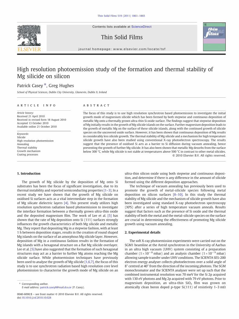
The effect of stepwise Mg deposition onto an ultra-thin thermallygrown Si oxide layer is shown using curve fitted Si 2p spectra in Fig. 1,taken at 130 eV photon energy using synchrotron radiation basedphotoemission. The acquisition of photoemission spectra resulted in agap of at least 1 h between successive depositions. The initially grownthermal oxide shows evidence for the presence of the four separate Sioxidation states, including the sub-stoichiometric Si oxide states ( Si1+,Si2+, and Si3+ ) and the fully oxidised Si4+. These oxidation states areresolvedbycurvefitting inagreementwithprevious studies [12,13]. Thethickness of the self-limiting oxide was calculated to be approximately0.3 nm, effectivelymeaning that it is onemonolayer thick, as previouslyreported by Miyata et al. [14]. The spectra show that Mg deposition
106 104 102 100 98 96
Si 1+Si 2+
Si 3+Si 4+ Si o Mg Silicide
Si 2p @130 eV
1 nm Mg
0.5 nm Mg
0.2 nm Mg
Si Oxide
Binding Energy / eV
Fig. 1. Curve fitted Si 2p spectra, acquired using SRXPS at 130 eV photon energyfollowing the stepwise deposition of 0.2, 0.5 and 1 nmMg onto an ultra-thin (~0.3 nm)thermally grown Si oxide surface.
results in the growth of a peak at 1.1 eV lower biding energy (LBE) withrespect to the Si bulk (Si°) peak. This peak has been attributed to thepresence of Mg silicide, in agreementwith the work of Brause et al. [15]and van Burren et al. [16]. While numerous silicide phases exist formetals suchasNi and Ti [17], the only reportedphaseofMg silicide is themetal richMg2Si [15,17], and as such a single silicide peak is used duringcurve fitting.
Curve fitting data taken from the Si 2p spectra in Fig. 1 indicatea~10% reduction in the ratio of the Si oxide and bulk signals as afunction of Mg deposition (i.e. (Σ Oxide peaks)/Bulk peak). Anincrease in the area of the Si3+ oxidation state with respect to the Si4+
state, from 0.6:1 to 1:1, is also observed. Both results indicate thatoxygen has been lost from the Si oxide, due to the formation of Mgsilicide involving Si atoms which were initially within the Si oxidelayer. This is in agreement with previous results which have shownthat Mg can react with Si atoms in a fully oxidised environment [4]and is consistent with the accepted behavior of reactive metals onSiO2 [18,19]. However, given that the Si oxide layer had an initialthickness of only 1 monolayer, silicide growth may not have occurredexclusively within the oxide layer and may have also incorporatedatoms from the Si bulk. While the formation of Mg silicide andremoval of oxygen from the oxide layer are examples of chemicalinteraction between the substrate and the deposited Mg, the spectrain Fig. 1 also show evidence for physical interactions in the form of anincreased binding energy separation between the Si 2p bulk and oxidepeaks. This differential core level shift between the substrate Si and Sioxide is attributed to anMg induced dipole field across the oxide layerand/or changes in the band bending at the SiOx/Si interface [20].
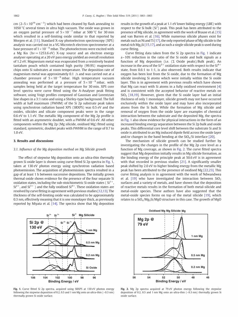
The mechanism of silicide growth can be studied further byinvestigating the changes in the profile of the Mg 2p core level as afunction of Mg coverage, as shown in Fig. 2. The curve fitted spectrasuggest that Mg deposition initially results in Mg silicide formation, asthe binding energy of the principle peak at 50.6 eV is in agreementwith that recorded in previous studies [21]. A significantly smallerpeak shifted by 2.0 eV to higher binding energy from the metallic Mgpeak has been attributed to the presence of oxidised Mg [22,23]. Thiscurve fitting analysis is in agreement with the work of Ndwandweaet al. [19] who have investigated the interaction between SiO2
surfaces and a variety of metals, and have shown that the depositionof reactive metals results in the formation of both metal-silicide andmetal-oxide species. These authors have also suggested that themetal-oxide species forms on top of the metal silicide [19], whichrelates to a SiOx/Mg2Si/MgO structure in this case. The growth of MgO
56 54 52 50 48 46
1 nm Mg
0.5 nm Mg
0.2 nm Mg
Oxidised Mg Mg Silicide Mg
Mg 2p @79 eV
Binding Energy / eV
Fig. 2. Mg 2p spectra acquired at 79 eV photon energy following the stepwisedeposition of 0.2, 0.5 and 1 nm Mg onto an ultra-thin (~0.3 nm) thermally grown Sioxide surface.
1863P. Casey, G. Hughes / Thin Solid Films 519 (2011) 1861–1865
observed in Fig. 2 is also in agreement with the removal of oxygenfrom the SiOx layer seen in Fig. 1, and is further evidenced by thegrowth of a MgO component peak within the O 1s core level spectrum(not shown). It can be seen from Fig. 2 that continued deposition leadsto the emergence of a metallic Mg component, at 49.6 eV bindingenergy, which acts to attenuate the other component peakswithin theMg 2p peak profile.
The work of Pretorius [17] suggests that the growth of metal-silicide thin films can be predicted based both on bulk thermodynamicdata and on the relative concentration of the Si and metal atoms.Therefore, the high heat of formation of Mg2Si (−1661 kJ/mol)compared to either MgO (−602 kJ/mol) or SiO2 (−859 kJ/mol) wouldsuggest that metallic Mgwould only be present on the surface if it wasin contact with previously formed Mg silicide or MgO structuresrather than on the Si oxide surface. If this is the case, thesimultaneously observed growth of both metallic Mg (Fig. 2) andMg silicide (Fig. 1) seen after the 2nd and 3rd Mg depositions isconsistent with the formation of silicide island structures on theoxidised silicon surface. The room temperature formation of metallicMg on the surface of silicide island structures has previously beenreported by Galkin et al. [2]. The effects of stepwiseMg deposition on aSi oxide surface can therefore be explained by the initial formation ofMg silicide/MgO islands, and the growth of metallic Mg on the surfaceof these islands.
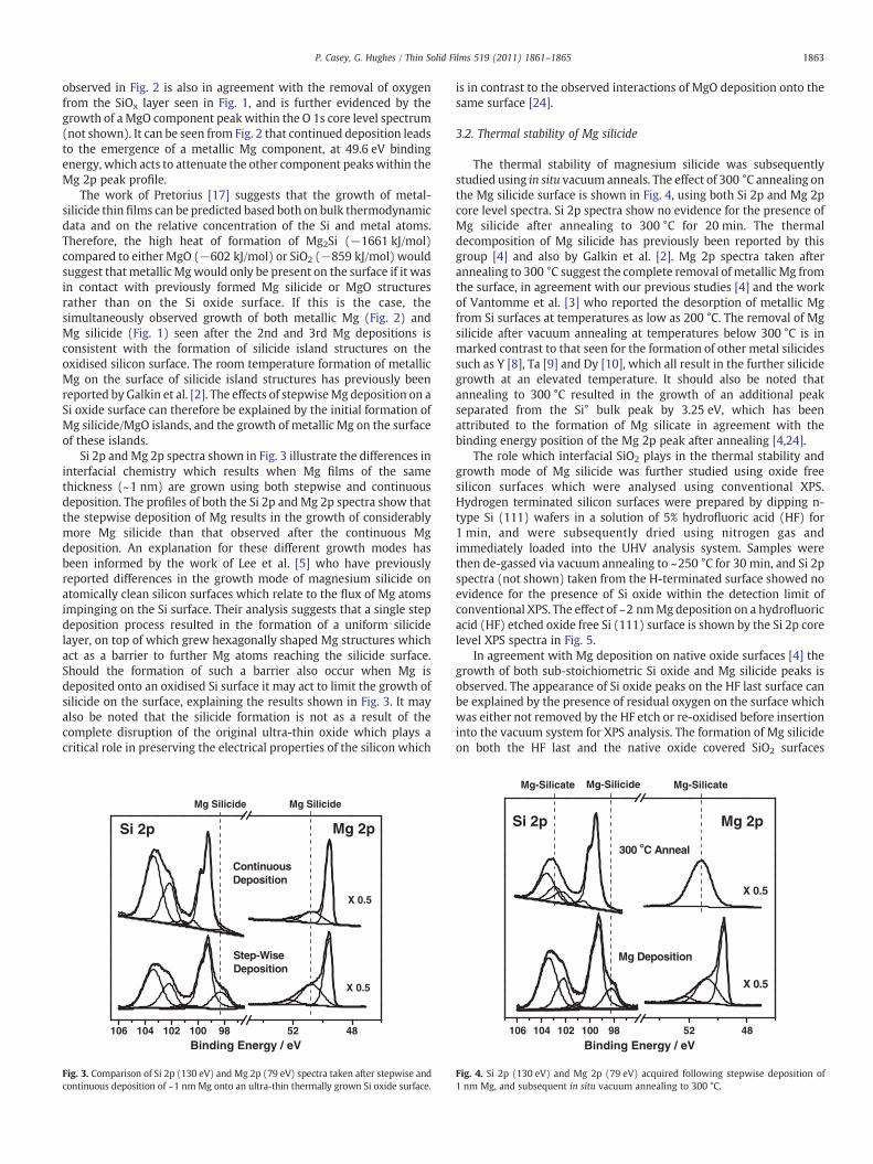
Si 2p andMg 2p spectra shown in Fig. 3 illustrate the differences ininterfacial chemistry which results when Mg films of the samethickness (~1 nm) are grown using both stepwise and continuousdeposition. The profiles of both the Si 2p and Mg 2p spectra show thatthe stepwise deposition of Mg results in the growth of considerablymore Mg silicide than that observed after the continuous Mgdeposition. An explanation for these different growth modes hasbeen informed by the work of Lee et al. [5] who have previouslyreported differences in the growth mode of magnesium silicide onatomically clean silicon surfaces which relate to the flux of Mg atomsimpinging on the Si surface. Their analysis suggests that a single stepdeposition process resulted in the formation of a uniform silicidelayer, on top of which grew hexagonally shaped Mg structures whichact as a barrier to further Mg atoms reaching the silicide surface.Should the formation of such a barrier also occur when Mg isdeposited onto an oxidised Si surface it may act to limit the growth ofsilicide on the surface, explaining the results shown in Fig. 3. It mayalso be noted that the silicide formation is not as a result of thecomplete disruption of the original ultra-thin oxide which plays acritical role in preserving the electrical properties of the silicon which
106 104 102 100 98 52 48
Mg Silicide Mg Silicide
X 0.5
X 0.5
Mg 2p
Step-WiseDeposition
ContinuousDeposition
Si 2p
Binding Energy / eV
Fig. 3. Comparison of Si 2p (130 eV) and Mg 2p (79 eV) spectra taken after stepwise andcontinuous deposition of ~1 nmMg onto an ultra-thin thermally grown Si oxide surface.
is in contrast to the observed interactions of MgO deposition onto thesame surface [24].
3.2. Thermal stability of Mg silicide
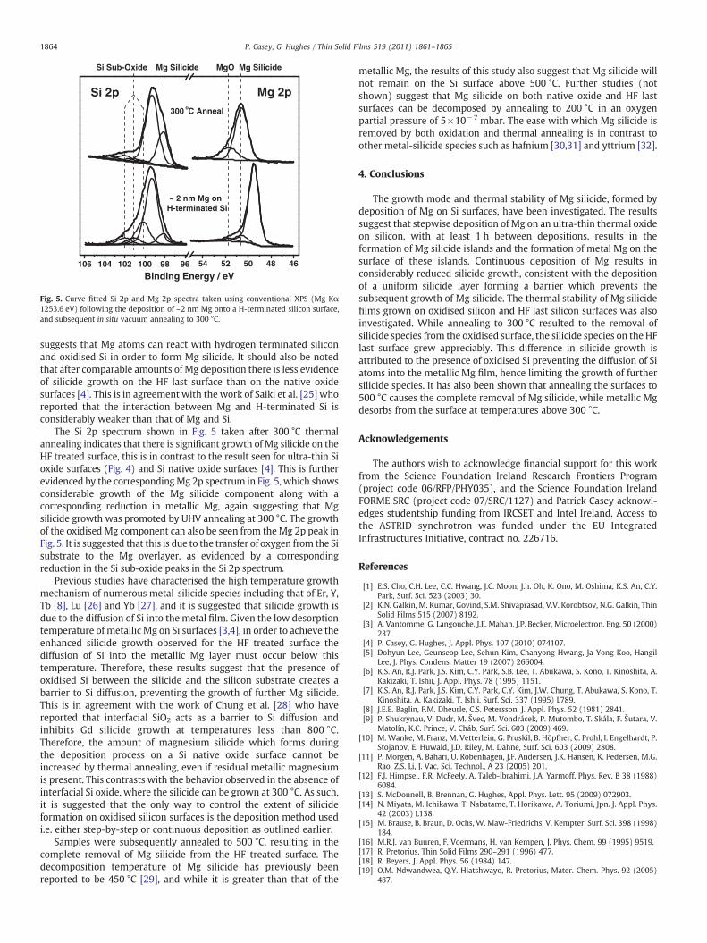
The thermal stability of magnesium silicide was subsequentlystudied using in situ vacuum anneals. The effect of 300 °C annealing onthe Mg silicide surface is shown in Fig. 4, using both Si 2p and Mg 2pcore level spectra. Si 2p spectra show no evidence for the presence ofMg silicide after annealing to 300 °C for 20 min. The thermaldecomposition of Mg silicide has previously been reported by thisgroup [4] and also by Galkin et al. [2]. Mg 2p spectra taken afterannealing to 300 °C suggest the complete removal of metallic Mg fromthe surface, in agreement with our previous studies [4] and the workof Vantomme et al. [3] who reported the desorption of metallic Mgfrom Si surfaces at temperatures as low as 200 °C. The removal of Mgsilicide after vacuum annealing at temperatures below 300 °C is inmarked contrast to that seen for the formation of other metal silicidessuch as Y [8], Ta [9] and Dy [10], which all result in the further silicidegrowth at an elevated temperature. It should also be noted thatannealing to 300 °C resulted in the growth of an additional peakseparated from the Si° bulk peak by 3.25 eV, which has beenattributed to the formation of Mg silicate in agreement with thebinding energy position of the Mg 2p peak after annealing [4,24].
The role which interfacial SiO2 plays in the thermal stability andgrowth mode of Mg silicide was further studied using oxide freesilicon surfaces which were analysed using conventional XPS.Hydrogen terminated silicon surfaces were prepared by dipping n-type Si (111) wafers in a solution of 5% hydrofluoric acid (HF) for1 min, and were subsequently dried using nitrogen gas andimmediately loaded into the UHV analysis system. Samples werethen de-gassed via vacuum annealing to ~250 °C for 30 min, and Si 2pspectra (not shown) taken from the H-terminated surface showed noevidence for the presence of Si oxide within the detection limit ofconventional XPS. The effect of ~2 nmMg deposition on a hydrofluoricacid (HF) etched oxide free Si (111) surface is shown by the Si 2p corelevel XPS spectra in Fig. 5.
In agreement with Mg deposition on native oxide surfaces [4] thegrowth of both sub-stoichiometric Si oxide and Mg silicide peaks isobserved. The appearance of Si oxide peaks on the HF last surface canbe explained by the presence of residual oxygen on the surface whichwas either not removed by the HF etch or re-oxidised before insertioninto the vacuum system for XPS analysis. The formation of Mg silicideon both the HF last and the native oxide covered SiO2 surfaces
106 104 102 100 98 52 48
X 0.5
X 0.5
Mg-Silicide Mg-SilicateMg-Silicate
Mg 2pSi 2p
300 oC Anneal
Mg Deposition
Binding Energy / eV
Fig. 4. Si 2p (130 eV) and Mg 2p (79 eV) acquired following stepwise deposition of1 nm Mg, and subsequent in situ vacuum annealing to 300 °C.
106 104 102 100 98 96 54 52 50 48 46
Si Sub-Oxide MgO
300 oC Anneal
~ 2 nm Mg onH-terminated Si
Si 2p Mg 2p
Mg Silicide Mg Silicide
Binding Energy / eV
Fig. 5. Curve fitted Si 2p and Mg 2p spectra taken using conventional XPS (Mg Kα1253.6 eV) following the deposition of ~2 nm Mg onto a H-terminated silicon surface,and subsequent in situ vacuum annealing to 300 °C.
1864 P. Casey, G. Hughes / Thin Solid Films 519 (2011) 1861–1865
suggests that Mg atoms can react with hydrogen terminated siliconand oxidised Si in order to form Mg silicide. It should also be notedthat after comparable amounts of Mg deposition there is less evidenceof silicide growth on the HF last surface than on the native oxidesurfaces [4]. This is in agreement with the work of Saiki et al. [25] whoreported that the interaction between Mg and H-terminated Si isconsiderably weaker than that of Mg and Si.
The Si 2p spectrum shown in Fig. 5 taken after 300 °C thermalannealing indicates that there is significant growth of Mg silicide on theHF treated surface, this is in contrast to the result seen for ultra-thin Sioxide surfaces (Fig. 4) and Si native oxide surfaces [4]. This is furtherevidenced by the correspondingMg 2p spectrum in Fig. 5, which showsconsiderable growth of the Mg silicide component along with acorresponding reduction in metallic Mg, again suggesting that Mgsilicide growth was promoted by UHV annealing at 300 °C. The growthof the oxidised Mg component can also be seen from theMg 2p peak inFig. 5. It is suggested that this is due to the transfer of oxygen from the Sisubstrate to the Mg overlayer, as evidenced by a correspondingreduction in the Si sub-oxide peaks in the Si 2p spectrum.
Previous studies have characterised the high temperature growthmechanism of numerous metal-silicide species including that of Er, Y,Tb [8], Lu [26] and Yb [27], and it is suggested that silicide growth isdue to the diffusion of Si into the metal film. Given the low desorptiontemperature of metallic Mg on Si surfaces [3,4], in order to achieve theenhanced silicide growth observed for the HF treated surface thediffusion of Si into the metallic Mg layer must occur below thistemperature. Therefore, these results suggest that the presence ofoxidised Si between the silicide and the silicon substrate creates abarrier to Si diffusion, preventing the growth of further Mg silicide.This is in agreement with the work of Chung et al. [28] who havereported that interfacial SiO2 acts as a barrier to Si diffusion andinhibits Gd silicide growth at temperatures less than 800 °C.Therefore, the amount of magnesium silicide which forms duringthe deposition process on a Si native oxide surface cannot beincreased by thermal annealing, even if residual metallic magnesiumis present. This contrasts with the behavior observed in the absence ofinterfacial Si oxide, where the silicide can be grown at 300 °C. As such,it is suggested that the only way to control the extent of silicideformation on oxidised silicon surfaces is the deposition method usedi.e. either step-by-step or continuous deposition as outlined earlier.
Samples were subsequently annealed to 500 °C, resulting in thecomplete removal of Mg silicide from the HF treated surface. Thedecomposition temperature of Mg silicide has previously beenreported to be 450 °C [29], and while it is greater than that of the
metallic Mg, the results of this study also suggest that Mg silicide willnot remain on the Si surface above 500 °C. Further studies (notshown) suggest that Mg silicide on both native oxide and HF lastsurfaces can be decomposed by annealing to 200 °C in an oxygenpartial pressure of 5×10⁻⁷ mbar. The ease with which Mg silicide isremoved by both oxidation and thermal annealing is in contrast toother metal-silicide species such as hafnium [30,31] and yttrium [32].
4. Conclusions
The growth mode and thermal stability of Mg silicide, formed bydeposition of Mg on Si surfaces, have been investigated. The resultssuggest that stepwise deposition of Mg on an ultra-thin thermal oxideon silicon, with at least 1 h between depositions, results in theformation of Mg silicide islands and the formation of metal Mg on thesurface of these islands. Continuous deposition of Mg results inconsiderably reduced silicide growth, consistent with the depositionof a uniform silicide layer forming a barrier which prevents thesubsequent growth of Mg silicide. The thermal stability of Mg silicidefilms grown on oxidised silicon and HF last silicon surfaces was alsoinvestigated. While annealing to 300 °C resulted to the removal ofsilicide species from the oxidised surface, the silicide species on the HFlast surface grew appreciably. This difference in silicide growth isattributed to the presence of oxidised Si preventing the diffusion of Siatoms into the metallic Mg film, hence limiting the growth of furthersilicide species. It has also been shown that annealing the surfaces to500 °C causes the complete removal of Mg silicide, while metallic Mgdesorbs from the surface at temperatures above 300 °C.
Acknowledgements
The authors wish to acknowledge financial support for this workfrom the Science Foundation Ireland Research Frontiers Program(project code 06/RFP/PHY035), and the Science Foundation IrelandFORME SRC (project code 07/SRC/1127) and Patrick Casey acknowl-edges studentship funding from IRCSET and Intel Ireland. Access tothe ASTRID synchrotron was funded under the EU IntegratedInfrastructures Initiative, contract no. 226716.
References
[1] E.S. Cho, C.H. Lee, C.C. Hwang, J.C. Moon, J.h. Oh, K. Ono, M. Oshima, K.S. An, C.Y.Park, Surf. Sci. 523 (2003) 30.
[2] K.N. Galkin, M. Kumar, Govind, S.M. Shivaprasad, V.V. Korobtsov, N.G. Galkin, ThinSolid Films 515 (2007) 8192.
[3] A. Vantomme, G. Langouche, J.E. Mahan, J.P. Becker, Microelectron. Eng. 50 (2000)237.
[4] P. Casey, G. Hughes, J. Appl. Phys. 107 (2010) 074107.[5] Dohyun Lee, Geunseop Lee, Sehun Kim, Chanyong Hwang, Ja-Yong Koo, Hangil
Lee, J. Phys. Condens. Matter 19 (2007) 266004.[6] K.S. An, R.J. Park, J.S. Kim, C.Y. Park, S.B. Lee, T. Abukawa, S. Kono, T. Kinoshita, A.
Kakizaki, T. lshii, J. Appl. Phys. 78 (1995) 1151.[7] K.S. An, R.J. Park, J.S. Kim, C.Y. Park, C.Y. Kim, J.W. Chung, T. Abukawa, S. Kono, T.
Kinoshita, A. Kakizaki, T. Ishii, Surf. Sci. 337 (1995) L789.[8] J.E.E. Baglin, F.M. Dheurle, C.S. Petersson, J. Appl. Phys. 52 (1981) 2841.[9] P. Shukrynau, V. Dudr, M. Švec, M. Vondrácek, P. Mutombo, T. Skála, F. Šutara, V.
Matolín, K.C. Prince, V. Cháb, Surf. Sci. 603 (2009) 469.[10] M. Wanke, M. Franz, M. Vetterlein, G. Pruskil, B. Höpfner, C. Prohl, I. Engelhardt, P.
Stojanov, E. Huwald, J.D. Riley, M. Dähne, Surf. Sci. 603 (2009) 2808.[11] P. Morgen, A. Bahari, U. Robenhagen, J.F. Andersen, J.K. Hansen, K. Pedersen, M.G.
Rao, Z.S. Li, J. Vac. Sci. Technol., A 23 (2005) 201.[12] F.J. Himpsel, F.R. McFeely, A. Taleb-Ibrahimi, J.A. Yarmoff, Phys. Rev. B 38 (1988)
6084.[13] S. McDonnell, B. Brennan, G. Hughes, Appl. Phys. Lett. 95 (2009) 072903.[14] N. Miyata, M. Ichikawa, T. Nabatame, T. Horikawa, A. Toriumi, Jpn. J. Appl. Phys.
42 (2003) L138.[15] M. Brause, B. Braun, D. Ochs, W. Maw-Friedrichs, V. Kempter, Surf. Sci. 398 (1998)
184.[16] M.R.J. van Buuren, F. Voermans, H. van Kempen, J. Phys. Chem. 99 (1995) 9519.[17] R. Pretorius, Thin Solid Films 290–291 (1996) 477.[18] R. Beyers, J. Appl. Phys. 56 (1984) 147.[19] O.M. Ndwandwea, Q.Y. Hlatshwayo, R. Pretorius, Mater. Chem. Phys. 92 (2005)
487.

1865P. Casey, G. Hughes / Thin Solid Films 519 (2011) 1861–1865
[20] H. Ofner, R. Hofmann, J. Kraft, F.P. Netzer, J.J. Paggel, K. Horn, Phys. Rev. B 50 (1994)15120.
[21] K.S. An, R.J. Park, J.S. Kim, C.Y. Park, C.Y. Kim, J.W. Chung, T. Kinoshita, A. Kakizakff,J. Electron. Spectrosc. Relat. Phenom. 80 (1996) 165.
[22] C. Chen, S.J. Splinter, T. Do, N.S. Mclntyre, Surf. Sci. 382 (1997) L652.[23] M. Kurth, P.C.J. Graat, E.J. Mittemeijer, Appl. Surf. Sci. 220 (2003) 60.[24] B. Brennan, S. McDonnell, G. Hughes, Thin Solid Films 518 (2010) 1980.[25] K. Saiki, K. Nishita, Y. Ariga, A. Koma, J. Vac. Sci. Technol., A 17.5 (1999) 2911.
[26] A. Travlos, P. Aloupogiannis, G. Weber, G. Robaye, J. Appl. Phys. 70 (1991) 7620.[27] K.S. Chi, L.J. Chen, J. Appl. Phys. 92 (2002) 927.[28] K.B. Chung, Y.K. Choi, M.H. Jang, M. Noh, C.N. Whang, J. Appl. Phys. 94 (2003) 212.[29] W. Xi-Na, W. Yong, Z. Jin, Z. Tian-Chong, M. Zeng-Xia, G. Yang, X. Qi-Kun, D.
Xiao-Long, Z. Xiao-Na, H. Xiao-Dong, Z. Ze, Chin. Phys. B 18 (2009) 3079.[30] J.H. Lee, M. Ichikawa, J. Appl. Phys. 92 (2002) 1929.[31] J.H. Lee, N. Miyata, M. Kundu, M. Ichikawa, Phys. Rev. B 66 (2002) 233309.[32] J.J. Chambers, G.N. Parsons, J. Appl. Phys. 90 (2001) 918.